Localization of relaxin receptors in arteries and veins, and region-specific increases in compliance...
13
The FASEB Journal • Research Communication Localization of relaxin receptors in arteries and veins, and region-specific increases in compliance and bradykinin-mediated relaxation after in vivo serelaxin treatment Maria Jelinic,* ,† Chen-Huei Leo,* ,1 Emiel D. Post Uiterweer, ‡,,1 Shaun L. Sandow, § Jonathan H. Gooi,* Mary E. Wlodek, † Kirk P. Conrad, Helena Parkington, ¶ Marianne Tare, ¶ and Laura J. Parry* ,2 *Department of Zoology and † Department of Physiology, The University of Melbourne, Parkville, Victoria, Australia; ‡ Department of Obstetrics, University Medical Center Utrecht, Utrecht, The Netherlands; § Department of Physiology, University of New South Wales, Sydney, New South Wales, Australia; Department of Physiology and Functional Genomics, University of Florida, Gainesville, Florida, USA; and ¶ Department of Physiology, Monash University, Clayton, Victoria, Australia ABSTRACT Relaxin is a potent vasodilator of small resistance arteries and modifies arterial compliance in some systemic vascular beds, yet receptors for relaxin, such as RXFP1, have only been localized to vascular smooth muscle. This study first aimed to localize RXFP1 in rat arteries and veins from different organ beds and determine whether receptors are present in endothelial cells. We then tested the hypothesis that region-specific vascular effects of relaxin may be influ- enced by the cellular localization of RXFP1 within different blood vessels. The aorta, vena cava, mesen- teric artery, and vein had significantly higher (P<0.05) RXFP1 immunostaining in endothelial cells compared with vascular smooth muscle, whereas the femoral artery and vein and small pulmonary arteries had higher (P<0.01) RXFP1 immunostaining in the vascular smooth muscle. Male rats were treated subcutaneously with recombinant human relaxin-2 (serelaxin; 4 g/h) for 5 d; vasodilation and compliance in mesenteric and femoral arteries and veins were compared with placebo controls. Serelaxin significantly (P0.04) reduced wall stiffness and increased volume compliance in mesen- teric arteries but not in the other vessels examined. This was associated with changes in geometrical prop- erties, and not compositional changes in the extracel- lular matrix. Serelaxin treatment had no effect on acetylcholine-mediated relaxation but significantly (P< 0.001) enhanced bradykinin (BK)-mediated relaxation in mesenteric arteries, involving enhanced nitric oxide but not endothelium-derived hyperpolarization or vaso- dilatory prostanoids. In conclusion, there is differential distribution of RXFP1 on endothelial and smooth mus- cle across the vasculature. In rats, mesenteric arteries exhibit the greatest functional response to chronic serelaxin treatment.—Jelinic, M., Leo, C-H., Post Uiter- weer, E. P., Sandow, S. L., Gooi, J. H., Wlodek, M. E., Conrad, K. P., Parkington, H., Tare, M., Parry, L. J. Local- ization of relaxin receptors in arteries and veins, and region- specific increases in compliance and bradykinin-mediated relaxation after in vivo serelaxin treatment. FASEB J. 28, 000 – 000 (2014). www.fasebj.org Key Words: RXFP1 vasodilation It is well established that the peptide hormone relaxin plays an important role in the profound mater- nal vascular adaptations that occur in pregnancy, spe- cifically targeting the renal and uterine vasculature (1–3). Beneficial cardiovascular effects of relaxin, which are attributed to alterations in the renal and 1 These authors contributed equally to this work. 2 Correspondence: Department of Zoology, The University of Melbourne, Parkville, VIC, 3010, Australia. E-mail: ljparry@ unimelb.edu.au doi: 10.1096/fj.13-233429 This article includes supplemental data. Please visit http:// www.fasebj.org to obtain this information. Abbreviations: -SMA, -smooth muscle actin; ACh, acetyl- choline; Akt, protein kinase B; AUC, area under curve; BK, bradykinin; COX, cyclooxygenase; DAB, 3,3=-diaminobenzi- dine; ECM, extracellular matrix; EDH, endothelium-derived hyperpolarization; EC 50 , half-maximal effective concentra- tion; eNOS, endothelial nitric oxide synthase; E max , maxi- mum contraction; FAM, 6-carboxy fluorescein; ID, inner diameter; Indo, indomethacin; L-NAME, N -nitro-l-arginine synthase; methyl ester; NBF, neutral buffered formalin; NO, nitric oxide; NOS, nitric oxide Nos3, endothelial nitric oxide synthase III; NBF, neutral buffered formalin; OD, outer diameter; PBS, phosphate-buffered saline; pD 2 , log EC 50 ; PE, phenylephrine; PGI 2 , prostacyclin; PI3K, phosphatidylinositol-3- kinase; PLP, paraformaldehyde-lysine-periodate; PSS, physiolog- ical saline solution; R max , maximum relaxation; Rn18S, ribo- somal 18S; RXFP1, relaxin/insulin-like family peptide receptor 1; RXFP2, relaxin/insulin-like family peptide receptor 2; WT, wall thickness 1 0892-6638/14/0028-0001 © FASEB The FASEB Journal article fj.13-233429. Published online September 13, 2013.
Transcript of Localization of relaxin receptors in arteries and veins, and region-specific increases in compliance...

The FASEB Journal • Research Communication
Localization of relaxin receptors in arteries and veins,and region-specific increases in compliance andbradykinin-mediated relaxation after in vivoserelaxin treatment
Maria Jelinic,*,† Chen-Huei Leo,*,1 Emiel D. Post Uiterweer,‡,�,1 Shaun L. Sandow,§
Jonathan H. Gooi,* Mary E. Wlodek,† Kirk P. Conrad,� Helena Parkington,¶
Marianne Tare,¶ and Laura J. Parry*,2
*Department of Zoology and †Department of Physiology, The University of Melbourne, Parkville,Victoria, Australia; ‡Department of Obstetrics, University Medical Center Utrecht, Utrecht, TheNetherlands; §Department of Physiology, University of New South Wales, Sydney, New South Wales,Australia; �Department of Physiology and Functional Genomics, University of Florida, Gainesville,Florida, USA; and ¶Department of Physiology, Monash University, Clayton, Victoria, Australia
ABSTRACT Relaxin is a potent vasodilator of smallresistance arteries and modifies arterial compliance insome systemic vascular beds, yet receptors for relaxin,such as RXFP1, have only been localized to vascularsmooth muscle. This study first aimed to localizeRXFP1 in rat arteries and veins from different organbeds and determine whether receptors are present inendothelial cells. We then tested the hypothesis thatregion-specific vascular effects of relaxin may be influ-enced by the cellular localization of RXFP1 withindifferent blood vessels. The aorta, vena cava, mesen-teric artery, and vein had significantly higher (P<0.05)RXFP1 immunostaining in endothelial cells comparedwith vascular smooth muscle, whereas the femoralartery and vein and small pulmonary arteries hadhigher (P<0.01) RXFP1 immunostaining in the vascularsmooth muscle. Male rats were treated subcutaneouslywith recombinant human relaxin-2 (serelaxin; 4 �g/h)for 5 d; vasodilation and compliance in mesenteric andfemoral arteries and veins were compared with placebo
controls. Serelaxin significantly (P�0.04) reduced wallstiffness and increased volume compliance in mesen-teric arteries but not in the other vessels examined.This was associated with changes in geometrical prop-erties, and not compositional changes in the extracel-lular matrix. Serelaxin treatment had no effect onacetylcholine-mediated relaxation but significantly (P<0.001) enhanced bradykinin (BK)-mediated relaxationin mesenteric arteries, involving enhanced nitric oxidebut not endothelium-derived hyperpolarization or vaso-dilatory prostanoids. In conclusion, there is differentialdistribution of RXFP1 on endothelial and smooth mus-cle across the vasculature. In rats, mesenteric arteriesexhibit the greatest functional response to chronicserelaxin treatment.—Jelinic, M., Leo, C-H., Post Uiter-weer, E. P., Sandow, S. L., Gooi, J. H., Wlodek, M. E.,Conrad, K. P., Parkington, H., Tare, M., Parry, L. J. Local-ization of relaxin receptors in arteries and veins, and region-specific increases in compliance and bradykinin-mediatedrelaxation after in vivo serelaxin treatment. FASEB J. 28,000–000 (2014). www.fasebj.org
Key Words: RXFP1 � vasodilation
It is well established that the peptide hormonerelaxin plays an important role in the profound mater-nal vascular adaptations that occur in pregnancy, spe-cifically targeting the renal and uterine vasculature(1–3). Beneficial cardiovascular effects of relaxin,which are attributed to alterations in the renal and
1 These authors contributed equally to this work.2 Correspondence: Department of Zoology, The University
of Melbourne, Parkville, VIC, 3010, Australia. E-mail: [email protected]
doi: 10.1096/fj.13-233429This article includes supplemental data. Please visit http://
www.fasebj.org to obtain this information.
Abbreviations: �-SMA, �-smooth muscle actin; ACh, acetyl-choline; Akt, protein kinase B; AUC, area under curve; BK,bradykinin; COX, cyclooxygenase; DAB, 3,3=-diaminobenzi-dine; ECM, extracellular matrix; EDH, endothelium-derivedhyperpolarization; EC50, half-maximal effective concentra-tion; eNOS, endothelial nitric oxide synthase; Emax, maxi-mum contraction; FAM, 6-carboxy fluorescein; ID, innerdiameter; Indo, indomethacin; L-NAME, N�-nitro-l-argininesynthase; methyl ester; NBF, neutral buffered formalin; NO,nitric oxide; NOS, nitric oxide Nos3, endothelial nitric oxidesynthase III; NBF, neutral buffered formalin; OD, outerdiameter; PBS, phosphate-buffered saline; pD2, �log EC50; PE,phenylephrine; PGI2, prostacyclin; PI3K, phosphatidylinositol-3-kinase; PLP, paraformaldehyde-lysine-periodate; PSS, physiolog-ical saline solution; Rmax, maximum relaxation; Rn18S, ribo-somal 18S; RXFP1, relaxin/insulin-like family peptide receptor1; RXFP2, relaxin/insulin-like family peptide receptor 2; WT,wall thickness
10892-6638/14/0028-0001 © FASEB
The FASEB Journal article fj.13-233429. Published online September 13, 2013.

systemic vasculature, have also been demonstrated incongestive and acute heart failure patients in preclini-cal studies and phase II/III clinical trials (4–6). Inconscious normotensive and hypertensive male andfemale rats, acute intravenous and chronic subcutane-ous administration of relaxin increases cardiac outputand global arterial compliance and reduces systemicvascular resistance, without affecting mean arterialpressure (7, 8). Relaxin has also been shown to reducemean arterial pressure in rat models of hypertension(9, 10) and increase coronary flow in rat and guinea pighearts (11).
These beneficial cardiovascular effects of relaxin innormotensive rats occur in parallel with increases inrenal plasma flow and glomerular filtration rate (12),and are underpinned by a reduction in myogenicconstriction of the small renal arteries (13). Vasodila-tory responses to in vivo relaxin treatment have alsobeen demonstrated in rat mesenteric arteries (13–15),whereas ex vivo relaxin treatment induces vasodilationin isolated rodent small renal and mesenteric arteries(16–18) and human small gluteal and subcutaneousarteries but not pulmonary resistance arteries (18, 19).Only one study to date has assessed veins; incubation ofmesenteric veins with relaxin causes relaxation but doesnot affect myogenic reactivity (17). In small resistancearteries, vascular tone is modulated by endothelium-derived factors, including nitric oxide (NO), prostacy-clin (PGI2), and non-NO/PGI2 endothelium-derivedhyperpolarization (EDH) (20). Relaxin appears to actthrough an endothelium-dependent NO-mediated va-sodilator pathway (13–15). In small renal arteries, rapidrelaxin-induced vasodilation involves phosphatidylino-sitol-3-kinase (PI3K; ref. 18). Relaxin also directly stim-ulates NO production from cultured human coronaryartery and aortic endothelial cells, with endothelial NOsynthase (eNOS) phosphorylation via PI3K activationand phosphorylation of protein kinase B (Akt; Ser473)(18). Currently, it is not known whether PGI2 and EDHcontribute to the vasodilator effects of relaxin.
The vascular actions of relaxin extend to modifica-tion of passive wall compliance. Chronic subcutaneousrelaxin infusion in rats and mice increases arterialcompliance in small renal (7, 21), mesenteric (17),uterine (22), brain parenchymal (23, 24) and carotidarteries (25). In mice administered relaxin, the relativeincrease in small renal artery compliance is mediatedby both geometric and compositional (decreased colla-gen) remodeling (21). Specifically, there is an increasein unpressurized wall area and wall-thickness-to-lumenarea ratio, increased smooth muscle cell density and adecrease in collagen-to-total-protein. Relaxin infusionin nonpregnant rats also increases wall thickness andinner diameter of brain parenchymal arterioles (23).The effects of relaxin on arterial compliance appear tobe region specific, as relaxin treatment has no effect onthe external iliac (21) and middle cerebral arteries(23), or mesenteric veins (17).
One explanation for the region-specific vascular ef-fects of relaxin might be differential expression of
relaxin/insulin-like family peptide receptor 1 (RXFP1)in arteries and veins and/or between endothelial andsmooth muscle cells. RXFP1 gene and protein areexpressed in the thoracic aorta and small renal andmesenteric arteries, but not in the cerebral vasculature,of male and female rodents (14, 24, 26, 27) but thesestudies did not localize RXFP1 within the artery wall.We recently colocalized RXFP1 with �-smooth muscleactin (�-SMA) muscle-positive cells in the media ofuterine arteries (3), demonstrating expression of re-laxin receptors in the vascular smooth muscle. Local-ization of RXFP1 in endothelial cells is less convincing,despite evidence of a direct action of relaxin on aorticand coronary artery endothelial cells to evoke NOrelease (18, 28). In summary, the specific localization ofRXFP1 in vascular smooth muscle cells is consistentwith the hypothesis that relaxin acts directly on bloodvessels to influence arterial remodeling and compli-ance, but RXFP1 is yet to be localized to endothelialcells in intact arteries or veins.
Therefore, in this study we tested the hypothesis thatregion-specific vascular effects of relaxin may be influ-enced by the distribution of RXFP1 within differentblood vessels in males. The main objectives of this studywere to localize RXFP1 in a variety of arteries and veinsfrom different organ beds and determine whetherreceptors are present in smooth muscle or endothelialcells; and compare the effects of chronic subcutaneousin vivo relaxin treatment on specific vasodilator path-ways and passive mechanical wall properties in mesen-teric and femoral arteries and veins.
MATERIALS AND METHODS
Animal preparation
All experimental protocols were approved by The Universityof Melbourne Faculty of Science Animal ExperimentationEthics Committee. Male Wistar rats aged 12–14 wk (�300 gbody weight) were purchased from Monash Animal Services(Clayton, VIC, Australia). Male rats were used in all experi-ments because nonpregnant females produce estrogen fromthe ovaries, which could confound the experimental out-comes. Rats were housed in the Animal Facility (Departmentof Zoology, The University of Melbourne, VIC, Australia) ona 12/12-h light-dark cycle and had access to standard rat chow(Specialty Feed, Glen Forrest, WA, Australia) and water adlibitum.
Experimental protocols
Localization of RXFP1 in arteries and veins
Rats were anesthetized with isofluorane and euthanized bycervical dislocation. Thoracic and abdominal aortae, pulmo-nary, mesenteric, femoral arteries, and mesenteric and fem-oral veins were dissected from each animal (n�3–4). Bloodvessels were then placed in ice-cold 0.1 M phosphate bufferedsaline (PBS), and excess fat and loose connective tissue wereremoved before fixing in 10% neutral buffered formalin(NBF). The kidneys were dissected, sectioned bilaterally, andfixed in PLP fixative (8% paraformaldehyde, 0.2 M lysine,
2 Vol. 28 January 2014 JELINIC ET AL.The FASEB Journal � www.fasebj.org
0.01 M periodate in 0.1 M PBS) for 24 h, and then transferredto 0.1 M PBS.
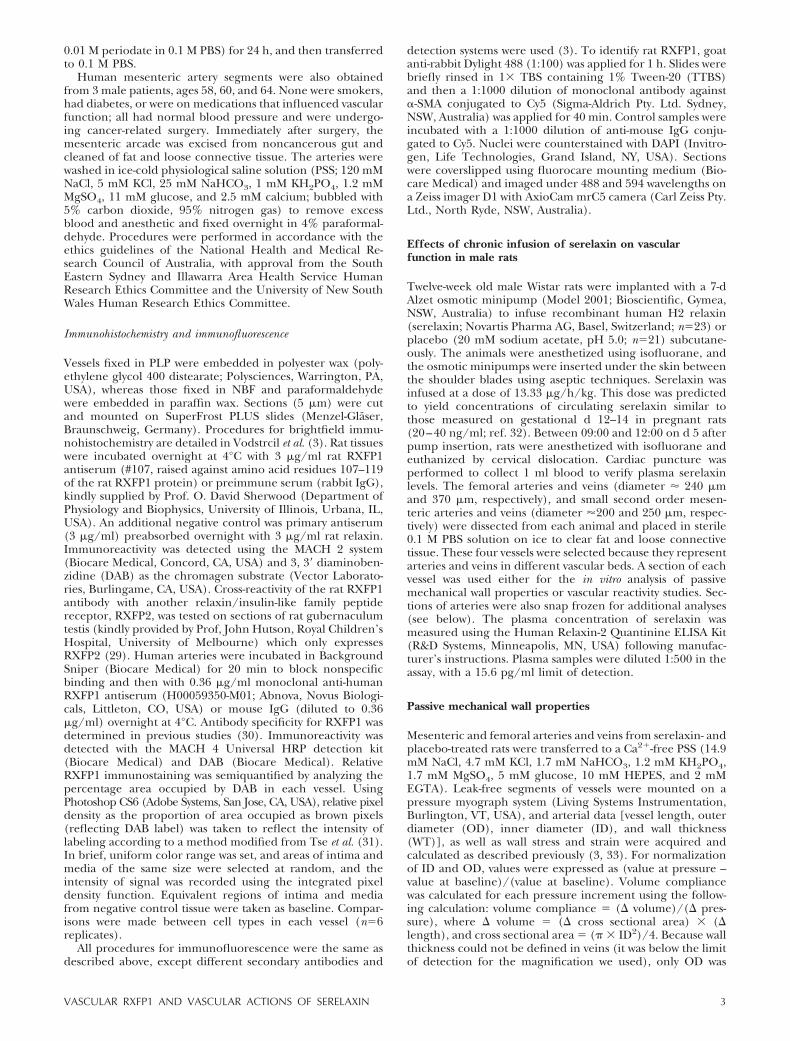
Human mesenteric artery segments were also obtainedfrom 3 male patients, ages 58, 60, and 64. None were smokers,had diabetes, or were on medications that influenced vascularfunction; all had normal blood pressure and were undergo-ing cancer-related surgery. Immediately after surgery, themesenteric arcade was excised from noncancerous gut andcleaned of fat and loose connective tissue. The arteries werewashed in ice-cold physiological saline solution (PSS; 120 mMNaCl, 5 mM KCl, 25 mM NaHCO3, 1 mM KH2PO4, 1.2 mMMgSO4, 11 mM glucose, and 2.5 mM calcium; bubbled with5% carbon dioxide, 95% nitrogen gas) to remove excessblood and anesthetic and fixed overnight in 4% paraformal-dehyde. Procedures were performed in accordance with theethics guidelines of the National Health and Medical Re-search Council of Australia, with approval from the SouthEastern Sydney and Illawarra Area Health Service HumanResearch Ethics Committee and the University of New SouthWales Human Research Ethics Committee.
Immunohistochemistry and immunofluorescence
Vessels fixed in PLP were embedded in polyester wax (poly-ethylene glycol 400 distearate; Polysciences, Warrington, PA,USA), whereas those fixed in NBF and paraformaldehydewere embedded in paraffin wax. Sections (5 �m) were cutand mounted on SuperFrost PLUS slides (Menzel-Gläser,Braunschweig, Germany). Procedures for brightfield immu-nohistochemistry are detailed in Vodstrcil et al. (3). Rat tissueswere incubated overnight at 4°C with 3 �g/ml rat RXFP1antiserum (#107, raised against amino acid residues 107–119of the rat RXFP1 protein) or preimmune serum (rabbit IgG),kindly supplied by Prof. O. David Sherwood (Department ofPhysiology and Biophysics, University of Illinois, Urbana, IL,USA). An additional negative control was primary antiserum(3 �g/ml) preabsorbed overnight with 3 �g/ml rat relaxin.Immunoreactivity was detected using the MACH 2 system(Biocare Medical, Concord, CA, USA) and 3, 3= diaminoben-zidine (DAB) as the chromagen substrate (Vector Laborato-ries, Burlingame, CA, USA). Cross-reactivity of the rat RXFP1antibody with another relaxin/insulin-like family peptidereceptor, RXFP2, was tested on sections of rat gubernaculumtestis (kindly provided by Prof, John Hutson, Royal Children’sHospital, University of Melbourne) which only expressesRXFP2 (29). Human arteries were incubated in BackgroundSniper (Biocare Medical) for 20 min to block nonspecificbinding and then with 0.36 �g/ml monoclonal anti-humanRXFP1 antiserum (H00059350-M01; Abnova, Novus Biologi-cals, Littleton, CO, USA) or mouse IgG (diluted to 0.36�g/ml) overnight at 4°C. Antibody specificity for RXFP1 wasdetermined in previous studies (30). Immunoreactivity wasdetected with the MACH 4 Universal HRP detection kit(Biocare Medical) and DAB (Biocare Medical). RelativeRXFP1 immunostaining was semiquantified by analyzing thepercentage area occupied by DAB in each vessel. UsingPhotoshop CS6 (Adobe Systems, San Jose, CA, USA), relative pixeldensity as the proportion of area occupied as brown pixels(reflecting DAB label) was taken to reflect the intensity oflabeling according to a method modified from Tse et al. (31).In brief, uniform color range was set, and areas of intima andmedia of the same size were selected at random, and theintensity of signal was recorded using the integrated pixeldensity function. Equivalent regions of intima and mediafrom negative control tissue were taken as baseline. Compar-isons were made between cell types in each vessel (n�6replicates).
All procedures for immunofluorescence were the same asdescribed above, except different secondary antibodies and
detection systems were used (3). To identify rat RXFP1, goatanti-rabbit Dylight 488 (1:100) was applied for 1 h. Slides werebriefly rinsed in 1� TBS containing 1% Tween-20 (TTBS)and then a 1:1000 dilution of monoclonal antibody against�-SMA conjugated to Cy5 (Sigma-Aldrich Pty. Ltd. Sydney,NSW, Australia) was applied for 40 min. Control samples wereincubated with a 1:1000 dilution of anti-mouse IgG conju-gated to Cy5. Nuclei were counterstained with DAPI (Invitro-gen, Life Technologies, Grand Island, NY, USA). Sectionswere coverslipped using fluorocare mounting medium (Bio-care Medical) and imaged under 488 and 594 wavelengths ona Zeiss imager D1 with AxioCam mrC5 camera (Carl Zeiss Pty.Ltd., North Ryde, NSW, Australia).
Effects of chronic infusion of serelaxin on vascularfunction in male rats
Twelve-week old male Wistar rats were implanted with a 7-dAlzet osmotic minipump (Model 2001; Bioscientific, Gymea,NSW, Australia) to infuse recombinant human H2 relaxin(serelaxin; Novartis Pharma AG, Basel, Switzerland; n�23) orplacebo (20 mM sodium acetate, pH 5.0; n�21) subcutane-ously. The animals were anesthetized using isofluorane, andthe osmotic minipumps were inserted under the skin betweenthe shoulder blades using aseptic techniques. Serelaxin wasinfused at a dose of 13.33 �g/h/kg. This dose was predictedto yield concentrations of circulating serelaxin similar tothose measured on gestational d 12–14 in pregnant rats(20–40 ng/ml; ref. 32). Between 09:00 and 12:00 on d 5 afterpump insertion, rats were anesthetized with isofluorane andeuthanized by cervical dislocation. Cardiac puncture wasperformed to collect 1 ml blood to verify plasma serelaxinlevels. The femoral arteries and veins (diameter � 240 �mand 370 �m, respectively), and small second order mesen-teric arteries and veins (diameter �200 and 250 �m, respec-tively) were dissected from each animal and placed in sterile0.1 M PBS solution on ice to clear fat and loose connectivetissue. These four vessels were selected because they representarteries and veins in different vascular beds. A section of eachvessel was used either for the in vitro analysis of passivemechanical wall properties or vascular reactivity studies. Sec-tions of arteries were also snap frozen for additional analyses(see below). The plasma concentration of serelaxin wasmeasured using the Human Relaxin-2 Quantinine ELISA Kit(R&D Systems, Minneapolis, MN, USA) following manufac-turer’s instructions. Plasma samples were diluted 1:500 in theassay, with a 15.6 pg/ml limit of detection.
Passive mechanical wall properties
Mesenteric and femoral arteries and veins from serelaxin- andplacebo-treated rats were transferred to a Ca2-free PSS (14.9mM NaCl, 4.7 mM KCl, 1.7 mM NaHCO3, 1.2 mM KH2PO4,1.7 mM MgSO4, 5 mM glucose, 10 mM HEPES, and 2 mMEGTA). Leak-free segments of vessels were mounted on apressure myograph system (Living Systems Instrumentation,Burlington, VT, USA), and arterial data [vessel length, outerdiameter (OD), inner diameter (ID), and wall thickness(WT)], as well as wall stress and strain were acquired andcalculated as described previously (3, 33). For normalizationof ID and OD, values were expressed as (value at pressure –value at baseline)/(value at baseline). Volume compliancewas calculated for each pressure increment using the follow-ing calculation: volume compliance � ( volume)/( pres-sure), where volume � ( cross sectional area) � (length), and cross sectional area � (� � ID2)/4. Because wallthickness could not be defined in veins (it was below the limitof detection for the magnification we used), only OD was
3VASCULAR RXFP1 AND VASCULAR ACTIONS OF SERELAXIN
measured, and vascular stiffness was evaluated by calculatingchange in OD/pressure.
Collagen and elastin assay
Collagen content was measured in abdominal aorta, mesen-teric, and femoral arteries using the Sircol collagen dyebinding assay (Biocolor, Carrickfergus, UK) as describedpreviously (21). Elastin content was only determined inmesenteric and femoral arteries (Fastin elastin assay kit;Biocolor). Arteries were lyophilized, and elastin was solubi-lized using the hot oxalic acid digestion method. Samples andstandards (ranging from 0–50 �g) were then incubated inelastin precipitating reagent at 4°C overnight. Final pelletswere resuspended in 250 �l of dye dissociation reagent.Absorbance was determined at 540 nm. and relative elastincontent was quantified as the ratio of elastin concentration(�g/ml) to dry weight (mg).
Vascular reactivity
Rings (1–2 mm in length) of mesenteric and femoral arteriesand veins from serelaxin (n�15) and placebo (n�14) treatedrats were mounted on a four-channel Mulvany-Halpern myo-graph (model 610M; Danish Myo Technology, Aarhus, Den-mark). The vessels were then stretched in increments to atension equivalent to �70 mmHg for arteries and �20 mmHgfor veins. Vascular smooth muscle and endothelial cell func-tion were tested as described previously (33, 34). Bloodvessels were submaximally (50–60% maximal constriction)preconstricted with phenylephrine (PE; �10�9 to 10�6 M),and endothelium-dependent relaxation was assessed withincreasing concentrations of acetylcholine (ACh; 10�10 to10�5 M; all arteries and veins) or bradykinin (BK; 10�10 to10�6 M; mesenteric arteries and veins because BK causedcontraction in the other vessels) applied cumulatively. Re-sponses to ACh and BK were recorded before and after NOSand cyclooxygenase blockade with N�-nitro-l-arginine methylester (L-NAME; 2�10�4 M) and/or indomethacin (Indo;10�6 M), respectively. The relaxation remaining in the pres-ence of both inhibitors was attributed to EDH (35). Smoothmuscle contraction was examined in vessels exposed to in-creasing concentrations of PE (10�9 to 10�4 M) appliedcumulatively. The relative contribution of NO, PGI2, andEDH to relaxation evoked by BK was determined by analyzingthe area under curve (AUC) of the BK-response curves. Theresponses attributed to each component were calculated usingthe following equations: PGI2 contribution � AUC (L-NAMEalone) � AUC (L-NAMEIndo); NO contribution � AUC (noinhibitors) – AUC (L-NAME alone); EDH contribution � AUC(L-NAMEIndo).
Quantitative PCR
RNA was extracted from individual arteries, and cDNA wassynthesized from 0.5 �g of RNA in a 20 �l reaction containingrandom hexamers (50 ng/�l) and 200 U of Superscript III(Invitrogen, Mulgrave, VIC, Australia; ref. 3). The compara-tive cycle threshold (2�Ct) method of quantitative real-timepolymerase chain reaction (qPCR) was used to quantify eNOSIII (Nos3) gene expression in the mesenteric arteries andveins of serelaxin and placebo treated male rats. Rat-specificforward/reverse primers and 6-carboxyl fluorescein (FAM)-labeled TaqMan probes were designed and purchased fromBiosearch Technologies (Novato, CA, USA). qPCR was per-formed on the Opticon 3 PCR machine (Bio-Rad, West Ryde,NSW, Australia) using 96-well reaction plates with 20-�lreactions in triplicate containing SensiMix (Bioline, Alexan-
dria, NSW, Australia) and 10 �M of primers and FAM-labeledprobe. Ribosomal 18S (Rn18s) was the reference gene. Neg-ative template controls substituting cDNA with water or RTnegative controls substituting the reverse transcriptase in thecDNA synthesis, were included on each plate. For eachsample, the mean Rn18s CT triplicate value was subtractedfrom the mean gene of interest triplicate CT value to normal-ize gene of interest expression to the reference gene. Thesenormalized data were then presented as a relative value(means�se).
Statistical analysis
Data are expressed as means � se with n representing thenumber of rats. The stress-strain and volume compliance datawere analyzed with repeated measures 2-way ANOVA (treat-ment vs. strain) with Bonferroni post hoc analysis using Graph-Pad Prism 5 (GraphPad Software, San Diego, CA, USA).Independent t tests assessed statistical differences in collagenand elastin concentrations between placebo- and serelaxin-treated rats. Concentration-response curves were constructedusing GraphPad Prism and sigmoidal curves were fitted usingthe least squares method and analyzed using repeated mea-sures 2-way ANOVA (treatment vs. concentration). Contrac-tions to PE were expressed as percentage contraction to highKPSS. ACh- and BK-evoked relaxation was expressed as per-centage of precontraction evoked by PE. The concentrationof drug required to evoke a half-maximal response (EC50) wasdetermined, and the pD2 (�log EC50), maximum contraction(Emax), and maximum relaxation (Rmax) were comparedusing independent t tests.
RESULTS
Localization of RXFP1 in arteries and veins
Immunoreactive RXFP1 was localized in small renalarteries, abdominal aorta, vena cava, large and smallpulmonary arteries, mesenteric and femoral arteries,and veins of rats, but the cellular distribution of RXFP1differed between vessel types (Figs. 1 and 2 and Table 1). Inthe kidney, RXFP1 was predominantly localized to thesmall renal artery and renal tubules (Fig. 1A, D). Therewas no RXFP1 staining in the preimmune serum neg-ative controls (Fig. 1B) or in sections that were incu-bated with antiserum preabsorbed with rat relaxin(data not shown). It was important to demonstratespecificity to RXFP1, as the Rxfp2 gene is expressed inblood vessels (26). The absence of RXFP1 immuno-staining in rat gubernaculum testis (Fig. 1C), whichonly expresses RXFP2 (29), is evidence that the ratRXFP1 antibody used in this study did not cross-reactwith RXFP2. Double-labeling immunofluorescenceconfirmed colocalization of RXFP1 with �-SMA invascular smooth muscle cells within the media of smallrenal arteries and not veins (Fig. 1D, G). When one ofthe primary antisera was replaced with preimmuneserum, there was only positive staining for RXFP1 (Fig.1E, H) or �-SMA (Fig. 1F). The negative controls withpreimmune rabbit serum revealed little backgroundfluorescence (Fig. 1E, F; insets). Endothelial cells weredifficult to identify in these small renal artery cross-
4 Vol. 28 January 2014 JELINIC ET AL.The FASEB Journal � www.fasebj.org

sections but at higher magnification, there appeared tobe RXFP1 immunostaining in cells lining the lumenthat did not stain positive for �-SMA (Fig. 1I).
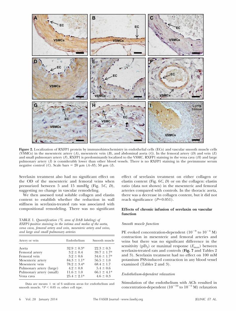
Comparisons between endothelial and vascularsmooth muscle cells revealed differences in relativeRXFP1 immunostaining in rat arteries and veins (Fig. 2and Table 1). In the mesenteric artery and vein, RXFP1was localized to both endothelial cells and the smoothmuscle (Fig. 2A, B) but with significantly (P 0.05)higher immunostaining in endothelial cells comparedwith smooth muscle cells (Table 1). There was noRXFP1 staining in the preimmune serum negativecontrols (Fig. 2C). RXFP1 immunostaining in endothe-lial cells was also significantly (P 0.05) higher in theaorta and vena cava (Table 1, Fig. 2G, H). In thefemoral artery and vein and small pulmonary artery,RXFP1 was predominantly localized to smooth muscle(Fig. 2D–F) and significantly (P 0.05) higher com-pared with endothelial cells (Table 1). RXFP1 immu-nostaining was relatively low in the large pulmonaryartery (Fig. 2I).
In human mesenteric arteries, immunoreactiveRXFP1 was clearly localized in endothelial cells, withless intense immunostaining in the vascular smoothmuscle and adventitia (Fig. 3). There was no RXFP1staining in the negative control with mouse IgG (Fig.3, insets).
Effects of chronic infusion of serelaxin on passivemechanical wall properties
Rats treated with serelaxin had significantly increasedplasma recombinant human serelaxin concentrations(39.93�4.06 ng/ml) compared with placebo-treatedrats, in which no human serelaxin was detected. Therewas no significant difference in WT, OD, and ID in anyvessel analyzed at baseline (5 mmHg; Tables 2 and 3).Over the pressurization range, ID of mesenteric arteriesincreased to a significantly greater extent (P�0.03) inthe serelaxin-treated animals compared with controls(Fig. 4A). There was no significant difference in ODand WT responses to increasing pressure between thetwo groups (Fig. 4C, E). There was a significant shift tothe right of the stress-strain curve of mesenteric arteriesfrom serelaxin-treated rats (F1,10�5.55, P�0.04) com-pared with controls (Fig. 5A), indicative of an overallreduction in passive wall stiffness. Furthermore, overallvolume compliance was significantly (F1,10�5.04,P�0.03) increased in the mesenteric arteries of sere-laxin-treated rats compared with controls (Fig. 6A).
In femoral arteries, treatment with serelaxin had nosignificant effect on normalized OD, ID, and WT (Fig.4B, D, F) or wall stiffness compared with controls (Fig.5B). Furthermore, there was no significant differencein volume compliance across the pressurization range.
CA B
-ve -ve
FD E
IHG
EC
VSMCs
EC EC
GB
-ve
IEL
Figure 1. Localization of RXFP1protein in rat small renal arteriesby immunohistochemistry (A–C)and immunofluorescence (D–I). Immunoreactive RXFP1 ispredominantly localized to thevascular smooth muscle of themedia (A). ImmunofluorescentRXFP1 (E, H) and �-SMA (F),with the overlay (D, G, I) dem-onstrate colocalization. Highermagnification illustrates RXFP1in endothelial cells that did notstain positive for �-SMA (I, ar-row). There is no RXFP1 stain-ing in the preimmune serumnegative controls (B and C, E, F,insets) and rat gubernaculumtestis (GB; C). VSMC, vascularsmooth muscle cell, EC, endo-thelial cell. Scale bars �50 �m(A); 500 �m (C). Note the highlevel of autofluorescence in theinternal elastic lamina (IEL).
5VASCULAR RXFP1 AND VASCULAR ACTIONS OF SERELAXIN
Serelaxin treatment also had no significant effect onthe OD of the mesenteric and femoral veins whenpressurized between 5 and 15 mmHg (Fig. 5C, D),suggesting no change in vascular remodeling.
We then assessed total soluble collagen and elastincontent to establish whether the reduction in wallstiffness in serelaxin-treated rats was associated withcompositional remodeling. There was no significant
effect of serelaxin treatment on either collagen orelastin content (Fig. 6C, D) or on the collagen: elastinratio (data not shown) in the mesenteric and femoralarteries compared with controls. In the thoracic aorta,there was a decrease in collagen content, but it did notreach significance (P�0.051).
Effects of chronic infusion of serelaxin on vascularfunction
Smooth muscle function
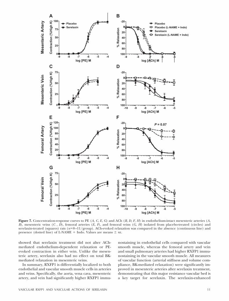
PE evoked concentration-dependent (10�9 to 10�4 M)contraction in mesenteric and femoral arteries andveins but there was no significant difference in thesensitivity (pD2) or maximal response (Emax) betweenserelaxin-treated rats and controls (Fig. 7 and Tables 2and 3). Serelaxin treatment had no effect on 100 mMpotassium PSS-induced contraction in any blood vesselexamined (Tables 2 and 3).
Endothelium-dependent relaxation
Stimulation of the endothelium with ACh resulted inconcentration-dependent (10�10 to 10�5 M) relaxation
VSMCs
EC
G
VSMCs
EC
EE
VSMC
H
EC
I
VSMCs
EC
C
VSMCs
AEC
VSMCs
EC
B
VSMCs
VSMCs
F
EC
DEC
VSMCs
E F
LungLungtissuetissueLungtissue
Figure 2. Localization of RXFP1 protein by immunohistochemistry in endothelial cells (ECs) and vascular smooth muscle cells(VSMCs) in the mesenteric artery (A), mesenteric vein (B), and abdominal aorta (G). In the femoral artery (D) and vein (E)and small pulmonary artery (F), RXFP1 is predominantly localized to the VSMC. RXFP1 staining in the vena cava (H) and largepulmonary artery (I) is considerably lower than other blood vessels. There is no RXFP1 staining in the preimmune serumnegative control (C). Scale bars � 20 �m (A–H); 50 �m (I).
TABLE 1. Quantification (% area of DAB labeling) ofRXFP1-positive staining in the intima and media of the aorta,vena cava, femoral artery and vein, mesenteric artery and veins,and large and small pulmonary arteries
Artery or vein Endothelium Smooth muscle
Aorta 32.9 � 0.3* 22.3 � 0.3Femoral artery 3.2 � 0.4 39.7 � 1.7*Femoral vein 3.2 � 0.6 34.6 � 1.7*Mesenteric artery 84.3 � 1.1* 56.5 � 1.0Mesenteric vein 78.2 � 3.4* 68.4 � 1.7Pulmonary artery (large) 4.2 � 0.8 5.4 � 0.6Pulmonary artery (small) 11.6 � 1.0 66.1 � 4.1*Vena cava 25.4 � 2.1* 4.6 � 0.3
Data are means � se of 6 uniform areas for endothelium andsmooth muscle. *P 0.05 vs. other cell type.
6 Vol. 28 January 2014 JELINIC ET AL.The FASEB Journal � www.fasebj.org

in mesenteric and femoral arteries and veins but sere-laxin treatment had no significant effect on either thesensitivity (pD2) or Rmax compared with controls (Fig. 7and Tables 2 and 3). The effects of serelaxin treatmenton EDH-mediated relaxation evoked by ACh were alsoevaluated; 75% of femoral and mesenteric veins, butnot arteries, developed spontaneous tone after L-NAME Indo incubation, but this did not differ between sere-laxin and placebo-treated rats. L-NAME Indo shiftedthe concentration-relaxation curves to ACh to the rightin all blood vessels, indicating the involvement of bothNO and/or vasodilator prostanoids (Fig. 7). In thefemoral artery, blockade with L-NAME Indo reducedmaximum relaxation to ACh (to 10.91�5.08%) inserelaxin-treated rats compared with controls (to 33.14�10.09%), but this was not significant (P�0.07; Fig. 7F).In all other vessels examined, the EDH-mediated relax-ation evoked by ACh was unaffected by serelaxin treat-ment (Fig. 7).
We expanded our analysis of endothelium-depen-dent relaxation in the mesenteric arteries and veinsusing BK. In mesenteric arteries of serelaxin-treated
rats the concentration-dependent relaxation curve wassignificantly augmented compared with controls (P 0.001;Fig. 8A). Post hoc analysis revealed that 10�9 and 10�8 MBK caused significantly (P 0.01) greater relaxation inserelaxin-treated animals compared with controls. Sen-sitivity (pD2) to BK was also significantly (P�0.03)greater in serelaxin-treated animals (9.24�0.6) com-pared with controls (7.73�0.10) (Fig. 8A, Table 2).Analysis of the area under the curve indicated that thiswas due to a significant (P�0.02) increase in thecomponent attributed to NO. Conversely, in the pres-ence of L-NAME and L-NAME Indo there was nosignificant difference in BK-mediated relaxation be-tween serelaxin and placebo-treated rats (Fig. 8A),indicating the absence of a PGI2 component. There wasno significant difference in BK-evoked relaxation inmesenteric veins of serelaxin and placebo-treated rats(Fig. 8B, Table 2).
To investigate whether the serelaxin-enhanced BK-mediated vasodilation involves an increase in totaleNOS, we analyzed Nos3 gene expression in mesen-teric arteries and veins. There was no significantdifference in Nos3 expression between placebo andserelaxin-treated animals in these two vessels (Sup-plemental Fig. 1).
DISCUSSION
The major findings of this study were that receptors forrelaxin, RXFP1, were differentially localized in endo-thelial and smooth muscle cells of arteries and veins.RXFP1 was predominantly expressed in endothelialcells in the mesenteric artery and vein, and also in theaorta and vena cava. In contrast, the femoral artery andvein and small pulmonary arteries had higher RXFP1immunostaining in the vascular smooth muscle. Chronicinfusion with serelaxin for 5 d reduced vessel wallstiffness in the mesenteric arteries but not in the othervessels examined. This was primarily associated withvascular remodeling and changes in geometrical prop-erties, and not compositional changes in the extracel-lular matrix (ECM). Serelaxin treatment had no effecton ACh-mediated relaxation. However, BK-mediatedrelaxation in mesenteric arteries, but not veins, wasenhanced following serelaxin treatment. This was dueto an increase in the NO component of BK-mediatedrelaxation and not to changes in EDH or vasodilatorprostanoids. Overall, our data demonstrate increases incompliance and BK-mediated relaxation but only in themesenteric artery of healthy serelaxin-treated rats.
Previous studies have shown Rxfp1 gene expression insmall renal and mesenteric arteries, as well as thoracicaortae isolated from mice and rats (26, 27) and inhuman subcutaneous arteries (18). In the rat uterineartery, RXFP1 protein is predominantly localized to thevascular smooth muscle. Our data now demonstrateRXFP1 in endothelial cells of intact mesenteric arteriesof rats and humans, and veins, aorta and the vena cavaof rats. Importantly, the large pulmonary and femoral
A
-ve
EC
VSMCs
-ve
B
Figure 3. A) Localization of RXFP1 protein by immunohisto-chemistry in a human mesenteric artery (representative ofarteries from all 3 patients). B) RXFP1 immunostaining isdetected in endothelial cells (ECs) and vascular smoothmuscle cells (VSMCs) but not in the mouse IgG negativecontrols (insets). Scale bars � 100 �m (A), 20 �m (B).
7VASCULAR RXFP1 AND VASCULAR ACTIONS OF SERELAXIN
artery and femoral vein had few RXFP1-positive endo-thelial cells illustrating differences between vessel types.These data represent a significant advance in ourunderstanding of serelaxin’s vascular actions becausethey illustrate the diversity in RXFP1 expression be-tween arteries and veins, and suggest that the effects ofserelaxin vary across the vasculature.
To test our hypothesis that region-specific vascularresponses to serelaxin are due, in part, to differentiallocalization of RXFP1 in endothelial and vascularsmooth muscle cells, we first analyzed passive mechan-ical wall properties in mesenteric and femoral arteriesand veins because the cellular distribution of RXFP1differed between these two vascular beds. In the mes-enteric artery, serelaxin treatment reduced passive ar-terial wall stiffness, which is consistent with previous
studies (7, 21). This was associated with a significantincrease in ID, without a change in WT. Serelaxintreatment also increased volume compliance in the lowpressure range, which reflects the ability of the artery tolengthen. Conversely, there were no changes in circum-ferential wall stiffness or volume compliance in thefemoral artery. We suggest that this is not related toRXFP1 localization because both vessel types haveRXFP1 in the vascular smooth muscle. An alternativeexplanation for the lack of a response in the femoralartery is that the dose of serelaxin and duration ofinfusion may not have been sufficient to reach thresh-old levels to induce substantial vascular remodeling.The media of the femoral artery is thicker and consistsof more ECM than a resistance artery, so it is alsopossible that a greater extent of arterial remodeling is
TABLE 2. Mesenteric artery and vein dimensions at baseline (5 and 2 mmHg, respectively)
Parameter
Mesenteric artery Mesenteric vein
Placebo n Serelaxin n Placebo n Serelaxin n
Stress-strain (K) 7.09 � 1.67 6 6.08 � 0.53 8 N/M – N/M –Baseline OD (�m) 150.4 � 6.8 6 149.4 � 9.5 8 251.3 � 37.2 7 251.3 � 34.59 6Baseline ID (�m) 123.5 � 5.3 6 116.6 � 9.2 8 N/M – N/M –Baseline WT (�m) 13.44 � 1.37 6 16.39 � 0.81 8 N/M – N/M –Hi K Emax (mN/mm) 9.59 � 0.37 13 9.95 � 0.55 13 3.79 � 0.35 14 3.88 � 0.48 13PE pD2 5.93 � 0.13 8 5.81 � 0.14 8 N/D 10 N/D 8PE Emax (% Hi K) 99.74 � 4.01 8 97.85 � 4.37 8 60.38 � 7.39 10 54.83 � 7.22 8ACh pD2 7.97 � 0.09 8 8.00 � 0.06 8 7.15 � 0.52 8 6.71 � 0.22 8ACh Rmax (%) 101.57 � 0.78 8 99.85 � 0.82 8 84.06 � 12.71 8 84.06 � 10.07 8ACh (L-NAMEIndo) pD2 7.25 � 0.09 6 7.49 � 0.13 6 N/D 5 N/D 7ACh (L-NAMEIndo) Rmax (%) 99.17 � 2.40 6 99.92 � 2.50 6 17.67 � 10.82 5 16.98 � 8.52 7BK pD2 7.73 � 0.10 9 9.24 � 0.61* 9 8.88 � 0.14 8 8.65 � 0.12 9BK Rmax (%) 92.87 � 2.28 9 98.45 � 1.51 9 109.1 � 1.66 8 105.5 � 1.62 9BK (L-NAME) pD2 7.45 � 0.25 5 7.22 � 0.44 7 8.15 � 0.21 5 8.15 � 0.24 7BK (L-NAME) Rmax (%) 85.11 � 7.62 7 92.94 � 6.15 8 55.99 � 4.21 7 59.08 � 5.48 9BK (L-NAMEIndo) pD2 7.46 � 0.17 4 7.20 � 0.15 6 8.23 � 0.26 6 7.69 � 0.24 7BK (L-NAMEIndo) Rmax (%) 85.84 � 7.32 7 92.94 � 9.49 6 58.87 � 9.21 7 59.06 � 5.21 7
OD, outer diameter; ID, inner diameter; WT, wall thickness. Vascular reactivity data: PE, phenylephrine; ACh, acetylcholine; BK,bradykinin; Hi K, 100 mM potassium PSS; L-NAME, nitric oxide synthase inhibitor; Indo, indomethacin; Emax, maximum response; Rmax,maximum relaxation; pD2, sensitivity (�log EC50); N/M, not measurable, N/D, not defined. All values are expressed as mean � se; n � samplesize. *P 0.05 vs. placebo controls.
TABLE 3. Femoral artery and vein dimensions at baseline (5 mmHg)
Parameter
Femoral artery Femoral vein
Placebo n Serelaxin n Placebo n Serelaxin n
Stress-strain (K) 3.81 � 0.52 6 3.34 � 0.45 8 N/M – N/M –Baseline OD (�m) 243.3 � 16.3 6 234.2 � 13.5 8 373.3 � 26.9 7 375.3 � 31.5 6Baseline ID (�m) 169.2 � 16.4 6 157.2 � 16.8 8 N/M – N/M –Baseline WT (�m) 37.08 � 1.23 6 44.83 � 5.80 8 N/M – N/M –Hi K Emax (mN/mm) 15.07 � 0.97 13 15.01 � 0.87 12 3.56 � 0.11 14 3.30 � 0.23 13PE pD2 5.72 � 0.12 8 5.63 � 0.12 8 6.03 � 0.15 11 6.05 � 0.20 10PE Emax (% Hi K) 95.57 � 2.96 8 98.79 � 4.54 8 54.07 � 3.31 11 50.39 � 5.75 10ACh pD2 6.98 � 0.09 10 6.85 � 0.13 9 7.26 � 0.15 11 7.14 � 0.21 8ACh Rmax (%) 91.91 � 3.40 10 91.15 � 5.94 9 96.95 � 2.24 11 95.16 � 3.75 8ACh (L-NAMEIndo) pD2 N/D 9 N/D 8 6.34 � 0.32 9 5.72 � 0.31 7ACh (L-NAMEIndo) Rmax (%) 66.86 � 10.09 9 89.09 � 5.08 8 45.31 � 10.70 9 48.93 � 11.15 7
OD, outer diameter; ID, inner diameter; WT, wall thickness. Vascular reactivity data: PE, phenylephrine; ACh, acetylcholine; Hi K, 100 mMpotassium PSS; L-NAME, nitric oxide synthase inhibitor; Indo, indomethacin; Emax, maximum response; pD2, sensitivity (�log EC50); N/M , notmeasurable; N/D, not defined. All values are expressed as mean � se; n � sample size.
8 Vol. 28 January 2014 JELINIC ET AL.The FASEB Journal � www.fasebj.org

required to influence wall stiffness and compliance.Another hypothesis is related to the known effects ofshear stress on the vascular wall, which activates vascu-lar remodeling pathways (36). Relaxin decreases vascu-lar resistance (37) and increases blood flow velocity (3).It is therefore possible that the differential effects ofserelaxin on vascular remodeling between resistanceand capacitance arteries are related to differences inblood flow (or shear stress). Only one study has re-ported improvements in elasticity in a large capacitanceartery (carotid artery) after relaxin treatment but this
was in senescent, spontaneously hypertensive rats (25).The duration of relaxin treatment in that study was alsoconsiderably longer (2 wk) and the dose higher (�20�g/kg/h). Consistent with previous studies (17), sere-laxin treatment had no effect on wall stiffness inmesenteric and femoral veins. However, this was notexplained by the absence of RXFP1 in the vascularsmooth muscle, and is more likely related to differencesin venous wall structure or the relationship betweenshear stress and vascular remodeling.
The consensus view is that relaxin promotes vascular
0 20 40 60 80 100 1200.0
0.2
0.4
0.6
0.8
1.0
Mesenteric Artery
0 20 40 60 80 100 1200.0
0.2
0.4
0.6
0.8
Femoral Artery
0 20 40 60 80 100 1200.0
0.5
1.0
1.5
2.0PlaceboSerelaxin
0 20 40 60 80 100 120-0.6
-0.4
-0.2
0.0
0 20 40 60 80 100 120-0.8
-0.6
-0.4
-0.2
0.0D
C
B
A
F
E
*
0 20 40 60 80 100 1200.0
0.1
0.2
0.3
0.4
0.5P = 0.06
Nom
aliz
ed ID
(µm
)N
omal
ized
ID (µ
m)
Nom
aliz
ed O
D (µ
m)
Nom
aliz
ed W
T (µ
m)
Nom
aliz
ed O
D (µ
m)
Nom
aliz
ed W
T (µ
m)
Figure 4. Normalized passive ID (A, B), (OD; C, D), and WT (E, F) against intraluminal pressures in the mesenteric (A, C, E)and femoral arteries (B, D F) from placebo-treated (solid circles, n�6) and serelaxin-treated (open circles, n�8) rats. Values aremeans � se. *P 0.05 vs. placebo controls.
Mesenteric Artery
0.0 0.2 0.4 0.6 0.8 1.00
50
100
150
200
250
Strain
Stre
ss (k
Pa)
Femoral Artery
0.0 0.5 1.0 1.5 2.00
50
100
150
200
250
SerelaxinPlacebo
Strain
Stre
ss (k
Pa)
Mesenteric Vein
0 2 4 6 8 10 12 14 160
10
20
30
40
50
60
Pressure (mmHg)
Cha
nge
in O
D(%
bas
elin
e)
Femoral Vein
0 2 4 6 8 10 12 14 160
10
20
30
40
Pressure (mmHg)
Cha
nge
in O
D(%
bas
elin
e)
*
A
C
B
D
Figure 5. Stress-strain relationships in mesen-teric (A) and femoral (B) arteries, and pres-sure-OD relationships in mesenteric (C) andfemoral (D) veins from from placebo-treated(solid circles, n�6–7) and serelaxin-treated(open circles, n�7–8) male rats. Values aremeans � se. *P 0.05 vs. placebo controls.
9VASCULAR RXFP1 AND VASCULAR ACTIONS OF SERELAXIN
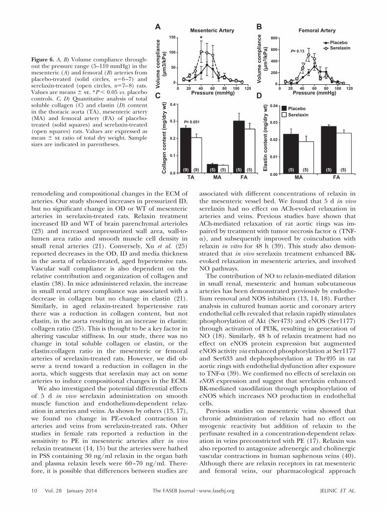
remodeling and compositional changes in the ECM ofarteries. Our study showed increases in pressurized ID,but no significant change in OD or WT of mesentericarteries in serelaxin-treated rats. Relaxin treatmentincreased ID and WT of brain parenchymal arterioles(23) and increased unpressurized wall area, wall-to-lumen area ratio and smooth muscle cell density insmall renal arteries (21). Conversely, Xu et al. (25)reported decreases in the OD, ID and media thicknessin the aorta of relaxin-treated, aged hypertensive rats.Vascular wall compliance is also dependent on therelative contribution and organization of collagen andelastin (38). In mice administered relaxin, the increasein small renal artery compliance was associated with adecrease in collagen but no change in elastin (21).Similarly, in aged relaxin-treated hypertensive ratsthere was a reduction in collagen content, but notelastin, in the aorta resulting in an increase in elastin:collagen ratio (25). This is thought to be a key factor inaltering vascular stiffness. In our study, there was nochange in total soluble collagen or elastin, or theelastin:collagen ratio in the mesenteric or femoralarteries of serelaxin-treated rats. However, we did ob-serve a trend toward a reduction in collagen in theaorta, which suggests that serelaxin may act on somearteries to induce compositional changes in the ECM.
We also investigated the potential differential effectsof 5 d in vivo serelaxin administration on smoothmuscle function and endothelium-dependent relax-ation in arteries and veins. As shown by others (13, 17),we found no change in PE-evoked contraction inarteries and veins from serelaxin-treated rats. Otherstudies in female rats reported a reduction in thesensitivity to PE in mesenteric arteries after in vivorelaxin treatment (14, 15) but the arteries were bathedin PSS containing 30 ng/ml relaxin in the organ bathand plasma relaxin levels were 60–70 ng/ml. There-fore, it is possible that differences between studies are
associated with different concentrations of relaxin inthe mesenteric vessel bed. We found that 5 d in vivoserelaxin had no effect on ACh-evoked relaxation inarteries and veins. Previous studies have shown thatACh-mediated relaxation of rat aortic rings was im-paired by treatment with tumor necrosis factor � (TNF-�), and subsequently improved by coincubation withrelaxin in vitro for 48 h (39). This study also demon-strated that in vivo serelaxin treatment enhanced BK-evoked relaxation in mesenteric arteries, and involvedNO pathways.
The contribution of NO to relaxin-mediated dilationin small renal, mesenteric and human subcutaneousarteries has been demonstrated previously by endothe-lium removal and NOS inhibitors (13, 14, 18). Furtheranalysis in cultured human aortic and coronary arteryendothelial cells revealed that relaxin rapidly stimulatesphosphorylation of Akt (Ser473) and eNOS (Ser1177)through activation of PI3K, resulting in generation ofNO (18). Similarly, 48 h of relaxin treatment had noeffect on eNOS protein expression but augmentedeNOS activity via enhanced phosphorylation at Ser1177and Ser633 and dephosphorylation at Thr495 in rataortic rings with endothelial dysfunction after exposureto TNF-� (39). We confirmed no effects of serelaxin oneNOS expression and suggest that serelaxin enhancedBK-mediated vasodilation through phosphorylation ofeNOS which increases NO production in endothelialcells.
Previous studies on mesenteric veins showed thatchronic administration of relaxin had no effect onmyogenic reactivity but addition of relaxin to theperfusate resulted in a concentration-dependent relax-ation in veins preconstricted with PE (17). Relaxin wasalso reported to antagonize adrenergic and cholinergicvascular contractions in human saphenous veins (40).Although there are relaxin receptors in rat mesentericand femoral veins, our pharmacological approach
Col
lage
n co
nten
t (m
g/dr
y w
t)
Elas
tin c
onte
nt (m
g/dr
y w
t)
(9) (9)
DC
Mesenteric Artery
0 20 40 60 80 100 1200
50
100
150
Pressure (mmHg)
Femoral Artery
0 20 40 60 80 100 120
200
400
600
800PlaceboSerelaxin
Pressure (mmHg)
BA*
0.0
0.1
0.2
0.3
Volu
me
com
plia
nce
(μm
3/kP
a)
Volu
me
com
plia
nce
(μm
3 /kP
a)
0
P= 0.13
P= 0.051
0.4
0.00
0.01
0.02
0.03
0.04
(5) (5) (5) (5) (5) (5) (5) (5)
PlaceboSerelaxin
TA MA FA MA FA
Figure 6. A, B) Volume compliance through-out the pressure range (5–110 mmHg) in themesenteric (A) and femoral (B) arteries fromplacebo-treated (solid circles, n�6–7) andserelaxin-treated (open circles, n�7–8) rats.Values are means � se. *P 0.05 vs. placebocontrols. C, D) Quantitative analysis of totalsoluble collagen (C) and elastin (D) contentin the thoracic aorta (TA), mesenteric artery(MA) and femoral artery (FA) of placebo-treated (solid squares) and serelaxin-treated(open squares) rats. Values are expressed asmean � se ratio of total dry weight. Samplesizes are indicated in parentheses.
10 Vol. 28 January 2014 JELINIC ET AL.The FASEB Journal � www.fasebj.org
showed that serelaxin treatment did not alter ACh-mediated endothelium-dependent relaxation or PE-evoked contraction in either vein. Unlike the mesen-teric artery, serelaxin also had no effect on total BK-mediated relaxation in mesenteric veins.
In summary, RXFP1 is differentially localized to bothendothelial and vascular smooth muscle cells in arteriesand veins. Specifically, the aorta, vena cava, mesentericartery, and vein had significantly higher RXFP1 immu-
nostaining in endothelial cells compared with vascularsmooth muscle, whereas the femoral artery and veinand small pulmonary arteries had higher RXFP1 immu-nostaining in the vascular smooth muscle. All measuresof vascular function (arterial stiffness and volume com-pliance, BK-mediated relaxation) were significantly im-proved in mesenteric arteries after serelaxin treatment,demonstrating that this major resistance vascular bed isa key target for serelaxin. The serelaxin-enhanced
-9 -8 -7 -6 -5 -40
25
50
75
100PlaceboSerelaxin
log [PE] M
Con
trac
tion
(%H
igh
K)
-9 -8 -7 -6 -5
-20
0
20
40
60
80
100
Placebo
SerelaxinPlacebo (L-NAME + Indo)
Serelaxin (L-NAME + Indo)
log [ACh] M
% R
elax
atio
n
-9 -8 -7 -6 -5 -40
25
50
75
log [PE] M
Con
trac
tion
(%H
igh
K)
-10 -9 -8 -7 -6 -5
-20
0
20
40
60
80
100
log [ACh] M
% R
elax
atio
n
-9 -8 -7 -6 -5 -40
20
40
60
80
100
120
log [PE] M
Con
trac
tion
(%H
igh
K)
-9 -8 -7 -6 -5
-20
0
20
40
60
80
100
log [ACh] M
% R
elax
atio
n
-9 -8 -7 -6 -5 -40
25
50
75
log [PE] M
Con
trac
tion
(%H
igh
K)
-10 -9 -8 -7 -6 -5
-20
0
20
40
60
80
100
log [ACh] M
% R
elax
atio
n
Mes
ente
ric A
rter
yM
esen
teric
Vei
nFe
mor
al A
rter
yFe
mor
al V
ein
DC
BA
FE
HG
P = 0.07
Figure 7. Concentration-response curves to PE (A, C, E, G) and ACh (B, D, F, H) in endothelium-intact mesenteric arteries (A,B), mesenteric veins (C , D), femoral arteries (E, F), and femoral veins (G, H) isolated from placebo-treated (circles) andserelaxin-treated (squares) rats (n�8–11/group). ACh-evoked relaxation was compared in the absence (continuous line) andpresence (dotted line) of L-NAME Indo. Values are means � se.
11VASCULAR RXFP1 AND VASCULAR ACTIONS OF SERELAXIN

BK-evoked relaxation involved a significant contribu-tion from NO but not EDH or vasodilatory prostanoids.Despite the localization of RXFP1 in femoral arteriesand veins, and mesenteric veins, we showed no signifi-cant functional effects of serelaxin in these bloodvessels.
The authors thank Dr. Dennis Stewart, the coordinator ofthe research partnership (Novartis Pharma AG, Basel, Swit-zerland) for helpful advice with the experimental design andcritical analysis of the data. The authors also thank Dr. ElaineUnemori for helpful suggestions and manuscript review. Theauthors are grateful to Dr. Jill Verlander, Professor JohnHutson, and Dr. Marc Mazzuca for their valuable contribu-tions to this work, and Professor O. David Sherwood (Univer-sity of Illinois, Urbana, IL, USA) for his generous gift of theRXFP1 antibody. The authors disclose that this project waspartially funded by Novartis, which also provided the sere-laxin, as conditions of an Australian Research Council Link-age grant. The research was funded by Australian ResearchCouncil Linkage grant LP110200543 (L.J.P., M.T., H.C.P.,and M.E.W.), U.S. National Institutes of Health grantHL067937 (K.P.C. and L.J.P.) and an Australian and NewZealand Medical Research and Technology in Victoria grant(L.J.P.). M.J. received an Australian Postgraduate Award.
REFERENCES
1. Jeyabalan, A., Shroff, S. G., Novak, J., and Conrad, K. P. (2007)The vascular actions of relaxin. Adv. Exp. Med. Biol. 612, 65–87
2. Conrad, K. P. (2011) Maternal vasodilation in pregnancy: theemerging role of relaxin. Am. J. Physiol. 301, R267–R275
3. Vodstrcil, L. A., Tare, M., Novak, J., Dragomir, N., Ramirez, R. J.,Wlodek, M. E., Conrad, K. P., and Parry, L. J. (2012) Relaxinmediates uterine artery compliance during pregnancy andincreases uterine blood flow. FASEB J. 26, 4035–4044
4. Teerlink, J. R., Metra, M., Felker, M. G., Ponikowoski, P., Voors,A. A., Weatherley, B. D., Marmor, A., Katz, A., Grzybowski, J.,Unemori, E., Teichman, S. L., and Cotter, G. (2009) Relaxin forthe treatment of patients with acute heart failure (Pre-RELAX-
AHF): a multicentre, randomised, placebo-controlled, parallel-group, dose-finding phase IIb study. Lancet 373, 1429–1439
5. Dschietzig, T., Teichman, S., Unemori, E., Wood, S., Boehmer,J., Richter, C., Baumann, G., and Stangl, K. (2009) Clinical trial:intravenous recombinant human relaxin in compensated heartfailure: a safety, tolerability, and pharmacodynamic trial. J. Card.Fail. 15, 182–190
6. Teerlink, J. R., Cotter, G., Davison, B. A., Felker, G. M.,Filippatos, G., Greenberg, B. H., Ponikowski, P., Unemori, E.,Voors, A. A., Adams, K. F. J., Dorobantu, M. I., Grinfeld, L. R.,Jondeau, G., Marmor, A., Masip, J., Pang, P. S., Werdan, K.,Teichman, S. L., Trapani, A., Bush, C. A., Saini, R., Schumacher,C., Severin, T. M., and Metra, M. (2013) Serelaxin, recombinanthuman relaxin-2, for treatment of acute heart failure (RELAX-AHF):a randomised, placebo-controlled trial. Lancet 381, 29–39
7. Conrad, K. P., Debrah, D. O., Novak, J., Danielson, L. A., andShroff, S. G. (2004) Relaxin modifies systemic arterial resistanceand compliance in conscious, nonpregnant rats. Endocrinology145, 3289–3296
8. Debrah, D. O., Conrad, K. P., Jeyabalan, A., Danielson, L. A.,and Shroff, S. G. (2005) Relaxin increases cardiac output andreduces systemic arterial load in hypertensive rats. Hypertension46, 745–750
9. Sasser, J. M., Molnar, M., and Baylis, C. (2011) Relaxin amelio-rates hypertension and increases nitric oxide metabolite excre-tion in angiotensin II but not N-omega-nitro-L-arginine methylester hypertensive rats. Hypertension 58, 197–204
10. St-Louis, J., and Massicotte, G. (1985) Chronic decrease ofblood pressure by rat relaxin in spontaneously hypertensive rats.Life Sci. 37, 1351–1357
11. Bani-Sacchi, T., Bigazzi, M., Bani, D., Mannaioni, P. F., andMasini, E. (1995) Relaxin-induced increased coronary flowthrough stimulation of nitric oxide production. Br. J. Pharmacol.116, 1589–1594
12. Danielson, L. A., Sherwood, O. D., and Conrad, K. P. (1999)Relaxin is a potent renal vasodilator in conscious rats. J. Clin.Invest. 103, 525–533
13. Novak, J., Ramirez, R. J., Gandley, R. E., Sherwood, O. D., andConrad, K. P. (2002) Myogenic reactivity is reduced in smallrenal arteries isolated from relaxin-treated rats. Am. J. Physiol.283, R349–355
14. Van Drongelen, J., Ploemen, I. H. J., Pertijs, J., Gooi, J. H.,Sweep, F. C. G. J., Lotgering, F. K., Spaanderman, M. E. A., andSmits, P. (2011) Ageing attenuates the vasodilator responseto relaxin. Am. J. Physiol. 300, H1609–H1615
-10 -9 -8 -7 -6
0
20
40
60
80
100
120
log [BK] M
% R
elax
atio
n
**
ControlPlaceboSerelaxin
L-NAME
-10 -9 -8 -7 -6
0
20
40
60
80
100
120
log [BK] M
% R
elax
atio
n
L-NAME + Indo
-10 -9 -8 -7 -6
0
20
40
60
80
100
120
log [BK] M
% R
elax
atio
n
NO EDH PGI20
50
100
150
200 PlaceboSerelaxin
BK
indu
ced
rela
xatio
n (A
UC
)*
-10 -9 -8 -7 -6
0
20
40
60
80
100
120
log [BK] M
% R
elax
atio
n
Control
PlaceboSerelaxin
L-NAME
-10 -9 -8 -7 -6
0
20
40
60
80
100
120
log [BK] M
% R
elax
atio
n
L-NAME + Indo
-10 -9 -8 -7 -6
0
20
40
60
80
100
120
log [BK] M
% R
elax
atio
n
NO EDH PGI20
50
100
150
200
250PlaceboSerelaxin
BK
indu
ced
rela
xatio
n (A
UC
)
Mesenteric Artery Mesenteric VeinA B
Figure 8. Concentration-response curves for BK in endothelium-intact mesenteric arteries (A) and veins (B) isolated fromplacebo-treated (solid circles) and serelaxin-treated (open squares) rats (n�6–9/group). BK-evoked relaxation was comparedbetween groups in the presence of L-NAME alone and L-NAME Indo. AUC of BK response curves were analyzed to reveal therelative contribution of NO, EDH, and PGI2. Values are means � se. *P 0.05 vs. placebo controls.
12 Vol. 28 January 2014 JELINIC ET AL.The FASEB Journal � www.fasebj.org

15. Van Drongelen, J., van Koppen, A., Pertijs, J., Gooi, J. H., Parry,L. J., Sweep, F. C. G. J., Lotgering, F. K., Smits, P., andSpaanderman, M. E. A. (2012) Impaired vascular responses torelaxin in diet-induced overweight female rats. J. Applied. Physiol.112, 969–969
16. Massicotte, G., Parent, A., and St-Louis, J. (1989) Bluntedresponses to vasoconstrictors in mesenteric vasculature but notin portal vein of spontaneously hypertensive rats treated with re-laxin. Proc. Soc. Exp. Biol. Med. 190, 254–259
17. Li, Y., Brookes, Z. L. S., and Kaufman, S. (2005) Acute andchronic effects of relaxin on vasoreactivity, myogeinc reactivityand compliance of the rat mesenteric arterial and venous vas-culature. Regul. Pept. 132, 41–46
18. McGuane, J. T., Debrah, J. E., Sautina, L., Jarajapu, Y. P. R.,Novak, J., Rubin, J. P., Grant, M. B., Segal, M., and Conrad, K. P.(2011) Relaxin induces rapid dilation of rodent small renal andhuman subcutaneous arteries via PI3 kinase and nitric oxide.Endocrinology 152, 2786–2796
19. Fisher, C., MacLean, M., Morecroft, I., Seed, A., Johnston, F.,Hillier, C., and McMurray, J. (2002) Is the pregnancy hormonerelaxin also a vasodilator peptide secreted by the heart? Circu-lation 106, 292–295
20. Sandoo, A., Veldhuijzen van Zanten, J. J. C. S., Metsios, G. S.,Caroll, D., and Kitas, G. D. (2010) The endothelium and its rolein regulating vascular tone. Open Med. J. 4, 302–312
21. Debrah, D. O., Debrah, J. E., Haney, J. L., McGuane, J. T., Sacks,M. S., Conrad, K. P., and Shroff, S. G. (2011) Relaxin regulatesvascular wall remodeling and passive mechanical propertiesin mice. J. Applied Physiol. 111, 260–271
22. Gooi, J. H., Richardson, M. L., Jelinic, M., Girling, J. E., Wlodek,M. E., Tare, M., and Parry, L. J. (2013) Enhanced uterine arterystiffness in aged pregnant relaxin mutant mice is reversed withexogenous relaxin treatment. Biol. Reprod 89, 18; doi: 10.1095/biolreprod.113.108118
23. Chan, S. L., and Cipolla, M. J. (2011) Relaxin causes selectiveoutward remodeling of brain parenchymal arterioles via activa-tion of peroxisome proliferator-activated receptor-gamma.FASEB J. 25, 3229–3239
24. Chan, S. L., Sweet, J. G., and Cipolla, M. J. (2013) Treatment forcerebral small vessel disease: effect of relaxin on the functionand structure of cerebral parenchymal arterioles during hyper-tension. [E-pub ahead of print] FASEB J. doi: 10.1096/fj.13-230797
25. Xu, Q., Chakravorty, A., Bathgate, R. A., Dart, A. M., and Du,X. J. (2010) Relaxin therapy reverses large artery remodelingand improves arterial compliance in senescent spontaneouslyhypertensive rats. Hypertension 55, 1260–1266
26. Novak, J., Parry, L. J., Matthews, J. E., Kerchner, L. J., Indovina,K., Hanley-Yanez, K., Doty, K. D., Debrah, D. O., Shroff, S. G.,and Conrad, K. P. (2006) Evidence for local relaxin ligand-receptor expression and function in arteries. FASEB J. 20,2352–2362
27. Ferreira, V. M., Gomes, T. S., Reis, L. A., Ferreira, A. T.,Razvickas, C. V., Schor, N., and Boim, M. A. (2009) Receptor-induced dilatation in the systemic and intrarenal adaptation topregnancy in rats. PLoS One 4, e4845
28. Failli, P., Nistri, S., Quattrone, S., Mazzetti, L., Bigazzi, M.,Sacchi, T. B., and Bani, D. (2002) Relaxin up-regulates induc-ible nitric oxide synthase expression and nitric oxide generationin rat coronary endothelial cells. FASEB J. 16, 252–254
29. Kubota, Y., Temelcos, C., Bathgate, R. A. D., Smith, K. J., Scott,D., Zhao, C., and Hutson, J. M. (2002) The role of insulin-3,testosterone Mullerian inhibiting substance and relaxin in ratgubernacular growth. Mol. Cell. Endocrinol. 8, 900–905
30. Kern, A., and Bryant-Greenwood, G. D. (2009) Characterizationof relaxin receptor (RXFP1) desensitization and internalizationin primary human decidual cells and RXFP1-transfectedHEK293 cells. Endocrinology 150, 2419–2428
31. Tse, G. H., and Marson, L. P. (2013) A comparative study of 2computer-assisted methods of brightfield microscopy images.[E-pub ahead of print] Appl. Immunohistochem. Mol. Morphol.10.1097/PAI.0b013e31827ba0e1
32. Sherwood, O. D., Crnekovic, V. E., Gordon, W. L., and Ruther-ford, J. E. (1980) Radioimmunoassay of relaxin throughoutpregnancy and during parturition in the rat. Endocrinology 107,691–698
33. Holobotovskyy, V., Manzur, M., Tare, M., Burchell, J., Bolitho,E., Viola, H., Hool, L. C., Arnolda, L. F., McKitrick, D. J., andGanss, R. (2013) Regulator of G protein signaling 5 controlsblood pressure homeostasis and vessel wall remodeling. Circula-tion 112, 781–791
34. Mazzuca, M. Q., Tare, M., Parkington, H. C., Dragomir, N. M.,Parry, L. J., and Wlodek, M. E. (2012) Uteroplacental insuffi-ciency programmes vascular dysfunction in non-pregnant rats:compensatory adaptations in pregnancy. J. Physiol. 590, 3375–3388
35. Tare, M., Parkington, H. C., and Coleman, H. A. (2000) EDHF,NO and a prostanoid: hyperpolarization-dependent and -inde-pendent relaxation in guinea-pig arteries. Br. J. Pharmacol. 130,605–618
36. Gao, Y. J., Yang, L. F., Stead, S., and Lee, R. M. (2008)Flow-induced vascular remodelling in the mesenteric artery ofspontaneously hypertensive rats. Can. J. Physiol. Pharmacol. 86,737–744
37. Conrad, K. P., and Novak, J. (2004) Emerging role of relaxin inrenal and cardiovascular function. Am. J. Physiol. 287, R250–R261
38. Zieman, S. J., Melenovsky, V., and Kass, D. A. (2005) Mecha-nisms, pathophysiology and therapy of arterial stiffness. Arterio-scler. Thromb. Vasc. Biol. 25, 932–942
39. Dschietzig, T., Brecht, A., Bartsch, C., Baumann, G., Stangl, K.,and Alexiou, K. (2012) Relaxin improves TNF-�-induced endo-thelial dysfunction: the role of glucocorticoid receptor andphosphatidylinositol 3-kinase signalling. Cardiovasc. Res. 95, 97–107
40. Adams, J., Schott, S., Bern, A., Renz, M., Ikenberg, K., Garbe, C.,and Busch, C. (2012) A novel role for relaxin-2 in the patho-genesis of primary varicosis. PLoS ONE 7, e39021
Received for publication May 13, 2013.Accepted for publication September 3, 2013.
13VASCULAR RXFP1 AND VASCULAR ACTIONS OF SERELAXIN