Radiological monitoring: terrestrial natural radionuclides in Kinta District, Perak, Malaysia
Upload
independentCategory
view
0download
0
NeuroImage 54 (2011) 74–79
Contents lists available at ScienceDirect
NeuroImage
j ourna l homepage: www.e lsev ie r.com/ locate /yn img
The veins of the nucleus dentatus: Anatomical and radiological findings
Antonio Di Ieva a,b,⁎,1, Manfred Tschabitscher a,1, Renato Juan Galzio c, Günther Grabner d,Claudia Kronnerwetter d, Georg Widhalm b, Christian Matula b, Siegfried Trattnig d
a Centre for Anatomy and Cell Biology, Department of Systematic Anatomy, Medical University of Vienna, Vienna, Austriab Department of Neurosurgery, Medical University of Vienna, Vienna, Austriac Department of Neurosurgery, Medical School of the University of L'Aquila, Italyd Department of Radiology, MR Centre of Excellence, Medical University of Vienna, Vienna, Austria
⁎ Corresponding author. Centre of Anatomy andSystematic Anatomy, Medical University of Vienna,Vienna, Austria. Fax: +43 1 4277 611 24.
E-mail address: [email protected] (A. Di Ieva).1 These authors contributed equally.
1053-8119/$ – see front matter © 2010 Elsevier Inc. Adoi:10.1016/j.neuroimage.2010.07.045
a b s t r a c t
a r t i c l e i n f oArticle history:Received 23 April 2010Revised 8 July 2010Accepted 19 July 2010Available online 23 July 2010
Keywords:CerebellumDentate nucleusPosterior fossa veinsVena centralis nuclei dentati7 Tesla MRSWIUltra-high field MR
The veins of the dentate nucleus are composed of several channels draining the external surface and onesingle vein draining the internal surface. We analyzed specimens of the human cerebellum and described thecentral vein of the nucleus dentatus as the main venous outflow of the nucleus. The central vein of thenucleus dentatus is formed by a network of smaller vessels draining the sinuosities of the gray matter; itemerges from the hilum of the nucleus and runs along the superior cerebellar peduncle, opening in theanterior vermian vein. We looked for this structure and for the surrounding veins on ultra-high-field(7 Tesla) MR, using susceptibility-weighted imaging. An anatomical and radiological description of the veinsof the dentate nucleus is provided, with some remarks on the future clinical applications that these findingscould provide.
Cell Biology, Department ofWaehringerstrasse 13, 1090
ll rights reserved.
© 2010 Elsevier Inc. All rights reserved.
Introduction
Despite the large number of articles that have been published onthe neurophysiology of the dentate nucleus (DN), there are not manyreports about its vascularization in the literature. The DN occupies astrategic position and is involved in a myriad of physiologicalnetworks; its function is related to attention, working memory, pro-cedural reasoning, salience detection, and task-planning (Manto andOulad Ben Taib, 2010). The anatomical and functional connectivity ofthe DN is reflected in its vascular network. The venous system is one ofthe most variable and heterogeneous organs of the human body, andcerebellar veins show several different anatomic patterns (Namin,1955). Although some attempts to describe the cerebellar venoussystem were published before the nineteenth century, the firstsystematic study of the venous system of the posterior fossa wasperformed only in 1950 (Gomez Oliveros, 1950). Later, several anat-omical studies were performed, with some specific remarks on theangiographic comparisons. In 1978 a monograph was published that
emphasized the diagnostic importance of the phlebogram in theposterior fossa (Wackenheim and Braun, 1978), because veins areimportant reference points: they trace the contours of the nervoussystem parenchyma and cisterns. For this reason, despite the het-erogeneity of the venous system, veins are critical landmarks forneuroradiological diagnosis and surgical orientation. The mostrelatively recent reports about the venous system of the brain andthe infratentorial structures were published by Duvernoy (1975,1978, 1999).
Only a few reports have focused on the vascularization of thenucleusdentatus (Fazzari, 1933; Goetzen, 1964; Icardo et al., 1982; Lang, 1991;Shellshear, 1922; Tschabitscher and Perneczcy, 1976), and, particularly,on the veins of the nucleus dentatus (Tschabitscher, 1979). In theneuroradiologic developments of the last several decades, digitalsubtraction angiographyandMR imaginghave confirmed thepossibilityof detecting the small veins of the cerebellum, although the smallerveins of the dentate nucleus have not been described by thesetechniques. In order to compare the neuroradiological findings tothose obtained in anatomical dissections, ultra-high-field 7 Teslasusceptibility-weighted imaging (SWI) (Haacke et al., 2004, 2005)was performed. SWI is a novel imaging technique that is sensitive toparamagnetic structures, such as deepbrain nuclei (Haacke et al., 2005),which are known to have an elevated iron level, and veins; thetechnique has already been used for vessel related studies (Essig et al.,1999; Rauscher et al., 2005a,b; Reichenbach et al., 2000).
75A. Di Ieva et al. / NeuroImage 54 (2011) 74–79
Materials and methods
Anatomical study
The veins of the nucleus dentatus have been studied in 25 humancadavers obtained from the Department of Anatomy of the MedicalUniversity of Vienna (Vienna, Austria), with no selection for age orgender. The specimens were studied by gross dissection, microscopicdissection, dissection of the white matter using the Klinglertechnique, corrosion procedures, and vascular injections (Fig. 1).The veins were filled in a retrograde manner, injecting the internaljugular vein with latex or Technovit of different colors (blue andyellow) (Figs. 1 and 2). In some specimens the vertebral arteries wereinjected with red latex or Technovit to observe the relationships withthe venous system (Figs. 1A and C, 7, and 8A and B). The specimenswere dissected to expose the cerebellar nuclei, with regard to thevascular relationships (Figs. 7 and 8). One specimen underwentplastination, according to von Hagens' technique (Figs. 4 and 5). Somepictures were obtained with an exoscope VITOM and recorded by theAIDA documentation system (Karl Storz GmbH, Tuttlingen, Germany)(Figs. 2B and 3).
Neuroradiological imaging
Two healthy volunteers underwent 7 Tesla MR imaging (Magnetom7T, Siemens Healthcare, Erlangen, Germany). The subjects wereinformed of the potential side effects of ultra–high-field 7.0-T MRimaging, which include vertigo, nausea, loss of balance, claustrophobia,
Fig. 1. Specimens of human cerebellum. A, tentorial surface: arteries injected with red laC, tentorial surface, on the left side exposition of the dentate nucleus; arteries injected wtechnique, exposure of cerebellar white matter and dentate nuclei. 1, postero-superior cereb5, great cerebellar veins; 6, anastomotic vein connections (“stars of veins”); 7, cerebellar tquadrangular lobule; 12, primary fissure; 13, lobulus simplex; 14, posterior superior fissure; 119, dentate nucleus; 20, nodulus.
feelings of electric shocks, and skeletal muscle contractions (Theysohnet al., 2008). In our cases, after scanning, the volunteers reported only atransitory (few minutes) vertigo.
SWI data were acquired using a three-dimensional, fully first-order flow-compensated gradient-echo (SWI) sequence with a TE of15 ms at 7 T. Other sequence parameters were: TR=28 ms; image-matrix=704×704 pixel; slices=96; parallel imaging factor=2,acquisition time=10.18 min, resolution=0.3×0.3×1.2 mm.
The coil was an eight-channel RF coil (RAPID Biomedical,Würzburg, Germany). All DICOM data were converted to the MINC(Medical Imaging NetCDF) format. Phase images were filtered usingHomodyne filtering (Noll et al., 1991), with a Gaussian filter kernel(full-width at half-maximum=5 mm in image space), and SWI imageprocessing was accomplished by performing four phase mask multi-plications. All image processing was performed using the MINC-Toolbox (Vincent et al., 2004).
Results
The venous system of the cerebellum is formed by three groups ofvessels: the superior group, drained by the great vein of Galen; theanterior group, drained by the superficial petrosal veins; and theposterior group, drained by the sinuses of the tentorium (Huang andWolf, 1965). These groups have been further classified as follows: thebasal veins and their tributaries; the superior and inferior vermianveins; the precentral veins; the peduncular and pontine veins; thesuperior petrosal veins with their tributaries (Wackenheim andBraun, 1978).
tex, veins injected with blue latex. B, ventral surface: veins injected with blue latex.ith red Technovit, veins injected with yellow Technovit. D, tentorial surface, Klinglerellar vein; 2, paravermian veins; 3, inferior vermian veins; 4, superior cerebellar artery;onsills; 8, networks of the tonsillar veins; 9, culmen; 10, declive; 11, anterior part of5, superior semilunar lobule; 16, horizontal fissure; 17, third ventricle; 18, pineal gland;
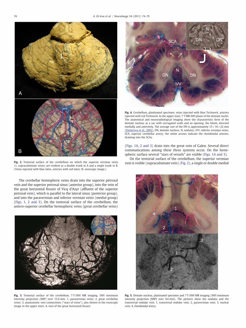
Fig. 2. Tentorial surface of the cerebellum on which the superior vermian veins(1, supraculminate veins) are evident as a double trunk in A and a single trunk in B.(Veins injected with blue latex, arteries with red latex; B: exoscopic image.)
Fig. 4. Cerebellum, plastinated specimen, veins injected with blue Technovit, arteriesinjected with red Technovit. In the upper inset, 7 T MR SWI phase of the dentate nuclei.The anatomical and neuroradiological imaging show the characteristic form of thedentate nucleus as a sac with corrugated walls and an opening, the hilum, directedmedially and anteriorly. The average size of the DN is approximately 15×16×22 mm(Dimitrova et al., 2002). DN, dentate nucleus; N, nodulus; iVV, inferior vermian veins;SCA, superior cerebellar artery; the white arrows indicate the rhomboidal arteries,draining into the SCAs.
76 A. Di Ieva et al. / NeuroImage 54 (2011) 74–79
The cerebellar hemispheric veins drain into the superior petrosalvein and the superior petrosal sinus (anterior group), into the vein ofthe great horizontal fissure of Vicq d'Azyr (affluent of the superiorpetrosal vein), which is parallel to the lateral sinus (posterior group),and into the paravermian and inferior vermian veins (medial group)(Figs. 1, 2 and 3). On the tentorial surface of the cerebellum, theantero-superior cerebellar hemispheric veins (great cerebellar veins)
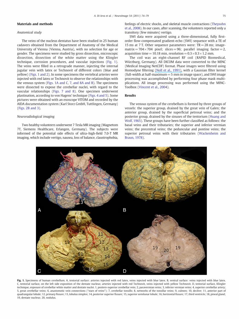
Fig. 3. Tentorial surface of the cerebellum, 7 T-SWI MR imaging. SWI minimumintensity projection (MIP) over 15.6 mm. 1, paravermian veins; 2, great cerebellarveins; 3, anastomotic vein connections (“stars of veins”), also shown in the exoscopicimage in the upper inset; 4, vein of the great horizontal fissure.
(Figs. 1A, 2 and 3) drain into the great vein of Galen. Several directcommunications among these three systems occur. On the hemi-spheric surface several “stars of vessels” are visible (Figs. 1A and 3).
On the tentorial surface of the cerebellum, the superior vermianvein is visible (supraculminate vein) (Fig. 2), a single or doublemedial
Fig. 5. Dentate nucleus, plastinated specimen and 7 T-SWI MR imaging (SWI minimumintensity projection [MIP] over 9.6 mm). The pictures show the nodulus and thetransversal nodular vein. 1, transversal nodular vein; 2, paravermian vein; 3, nuclearvein; 4, rhomboidal artery.
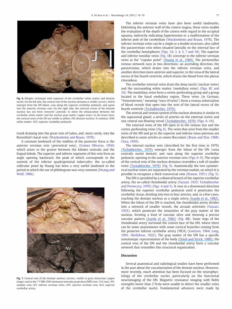
Fig. 6. Klingler technique with exposure of the cerebellar white matter and dentatenuclei. On the left side, the central vein of the nucleus dentatus is visible (arrow), whichemerges from the DN hilum, runs along the superior cerebellar peduncle, and opensinto the anterior vermian vein. On the right side, the external cortex of the dentatenucleus has not been removed (asterisk) to show the demarcation between thecerebellar white matter and the nuclear gray matter (upper inset). In the lower inset,the cortical veins of the DN are visible in yellow. DN, dentate nucleus; N, nodulus; WM,white matter; SCP, superior cerebellar peduncle.
77A. Di Ieva et al. / NeuroImage 54 (2011) 74–79
trunk draining into the great vein of Galen, and, more rarely, into theRosenthal's basal vein (Wackenheim and Braun, 1978).
A constant landmark of the midline of the posterior fossa is theanterior vermian vein (precentral vein), (Gomez Oliveros, 1950)which arises in the groove between the lobules centralis and thelingual lobule. The superior and inferior segments of this vein form anangle opening backward, the peak of which corresponds to thesummit of the inferior quadrigeminal tubercules: the so-calledcollicular point by Huang has been a very useful landmark in theperiod in which the use of phlebogramwas very common (Huang andWolf, 1966).
Fig. 7. Central vein of the dentate nucleus (arrow), visible in gross dissection (upperimage) and in the 7 TMR (SWI minimum intensity projection [MIP] over 15.6 mm). NV,nodular vein; iVV, inferior vermian veins; aVV, anterior vermian vein; SCA, superiorcerebellar artery.
The inferior vermian veins have also been useful landmarks.Delimiting the anterior wall of the cistern magna, these veins enablethe evaluation of the depth of the cistern with regard to the occipitalsquama, indirectly indicating hypertension or a malformation of theinferior pole of the cerebellum (Wackenheim and Braun, 1978). Theinferior vermian veins can be a single or a double structure, also calledthe paravermian vein when situated laterally on the internal face ofthe cerebellar hemispheres (Figs. 1A, 3, 4, 5, 7 and 10). The superiorand inferior tonsillar veins (Fig. 1B) converge in the inferior vermianveins at the “copular point” (Huang et al., 1969). The peritonsillarvenous network runs in two directions: an ascending direction, theparavermian, which drains into the inferior vermian veins, andanother directionmore anterior and superior, to the vena of the lateralrecess of the fourth ventricle, which drains the blood from the plexuschoroideus.
The cerebellar internal veins drain the deep nuclei (nuclear veins)and the surrounding white matter (medullary veins) (Figs. 8C and10). The medullary veins form a cortex-perforating group and a grouplocated in the basal medullary region. These veins (in German,“Venensternen,”meaning “stars of veins”) form a venous arborizationof blood vessels that open into the vein of the lateral recess of thefourth ventricle (Tschabitscher, 1979).
The arterial and venous system of the nucleus dentatus is similar tothe suprarenal gland; a series of arteries on the external cortex andone central out-flowing vessel (Tschabitscher, 1979) (Figs. 6–10).
The external veins of the DN open in to the venous star and thecortex-perforating veins (Fig. 6). The veins that arise from the smallerveins of the ND and go to the superior and inferior sinus petrosus aredescribed in some articles as venen floccularis (Braus and Elze, 1960;Clara, 1959).
The internal nuclear vein (described for the first time in 1979)(Tschabitscher, 1979) emerges from the hilum of the DN (venacentralis nuclei dentati) and runs along the superior cerebellarpeduncle, opening in the anterior vermian vein (Figs. 6–9). The originof the central vein of the nucleus dentatus resembles a tuft of smallerveins (Tschabitscher, 1979) (Fig. 9). Anatomically the two symmet-rical nuclear zones are separated by the vermian nodule, on which it ispossible to recognize a thick transversal vein (Krause, 1951) (Fig. 5).
The DN is sprinkled by a collateral branch of the superior cerebellarartery, the so-called rhomboidal artery (Fazzari, 1933; Tschabitscherand Perneczcy, 1976) (Figs. 4 and 5). It runs in a downward directionfollowing the superior cerebellar peduncle until it penetrates thecerebellar tissue, dividing into two to four arteries, and, in a few cases,reaching the dentate nucleus as a single artery (Icardo et al., 1982).When the hilum of the DN is reached, the rhomboidal artery dividesinto a network of smaller vessels, the arcuate arterioles (Fazzari,1933), which penetrate the sinuosities of the gray matter of thenucleus, forming a kind of vascular olive and showing a precisevascular pattern (Icardo et al., 1982) (Fig. 8B). Some wigs of therhomboidal artery surround the convex face of the DN, where therecan be some anastomoses with some cortical branches coming fromthe posterior inferior cerebellar artery (PICA) (Goetzen, 1964; Lang,1991; Shellshear, 1922). The gray matter of the DN has a specificsomatotopic representation of the body (Orioli and Strick, 1989); thecentral vein of the DN and the rhomboidal artery form a vascularnetwork that resembles this structural organization.
Discussion
Several anatomical and radiological studies have been performedin the past about the vascularization of the dentate nucleus. However,more recently, much attention has been focused on the neurophys-iology of the cerebellar nuclei, particularly on the functionalneuroimaging of the DN. Magnetic resonance imaging with fieldsstrengths lower than 3 Tesla were unable to detect the smaller veinsof the cerebellar nuclei. Fundamental advances were made by
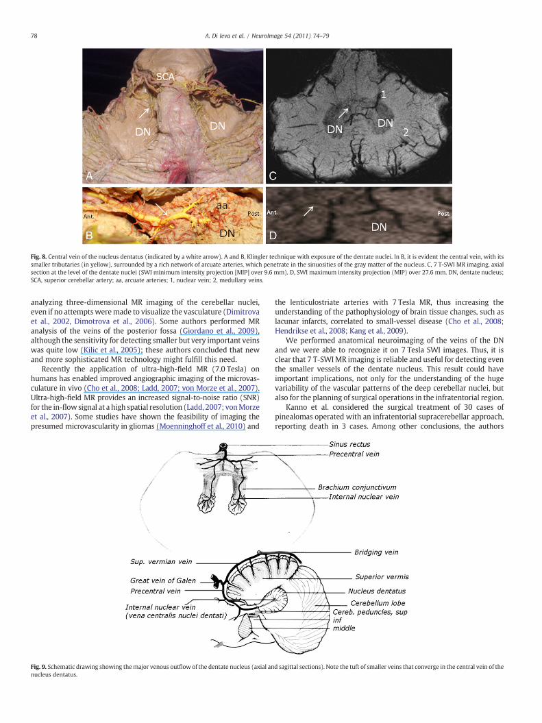
Fig. 8. Central vein of the nucleus dentatus (indicated by a white arrow). A and B, Klingler technique with exposure of the dentate nuclei. In B, it is evident the central vein, with itssmaller tributaries (in yellow), surrounded by a rich network of arcuate arteries, which penetrate in the sinuosities of the gray matter of the nucleus. C, 7 T-SWI MR imaging, axialsection at the level of the dentate nuclei (SWI minimum intensity projection [MIP] over 9.6 mm). D, SWI maximum intensity projection (MIP) over 27.6 mm. DN, dentate nucleus;SCA, superior cerebellar artery; aa, arcuate arteries; 1, nuclear vein; 2, medullary veins.
78 A. Di Ieva et al. / NeuroImage 54 (2011) 74–79
analyzing three-dimensional MR imaging of the cerebellar nuclei,even if no attempts weremade to visualize the vasculature (Dimitrovaet al., 2002, Dimotrova et al., 2006). Some authors performed MRanalysis of the veins of the posterior fossa (Giordano et al., 2009),although the sensitivity for detecting smaller but very important veinswas quite low (Kilic et al., 2005); these authors concluded that newand more sophisticated MR technology might fulfill this need.
Recently the application of ultra-high-field MR (7.0 Tesla) onhumans has enabled improved angiographic imaging of the microvas-culature in vivo (Cho et al., 2008; Ladd, 2007; von Morze et al., 2007).Ultra-high-field MR provides an increased signal-to-noise ratio (SNR)for the in-flowsignal at a high spatial resolution (Ladd, 2007; vonMorzeet al., 2007). Some studies have shown the feasibility of imaging thepresumed microvascularity in gliomas (Moenninghoff et al., 2010) and
Fig. 9. Schematic drawing showing themajor venous outflow of the dentate nucleus (axial annucleus dentatus.
the lenticulostriate arteries with 7 Tesla MR, thus increasing theunderstanding of the pathophysiology of brain tissue changes, such aslacunar infarcts, correlated to small-vessel disease (Cho et al., 2008;Hendrikse et al., 2008; Kang et al., 2009).
We performed anatomical neuroimaging of the veins of the DNand we were able to recognize it on 7 Tesla SWI images. Thus, it isclear that 7 T-SWIMR imaging is reliable and useful for detecting eventhe smaller vessels of the dentate nucleus. This result could haveimportant implications, not only for the understanding of the hugevariability of the vascular patterns of the deep cerebellar nuclei, butalso for the planning of surgical operations in the infratentorial region.
Kanno et al. considered the surgical treatment of 30 cases ofpinealomas operated with an infratentorial supracerebellar approach,reporting death in 3 cases. Among other conclusions, the authors
d sagittal sections). Note the tuft of smaller veins that converge in the central vein of the
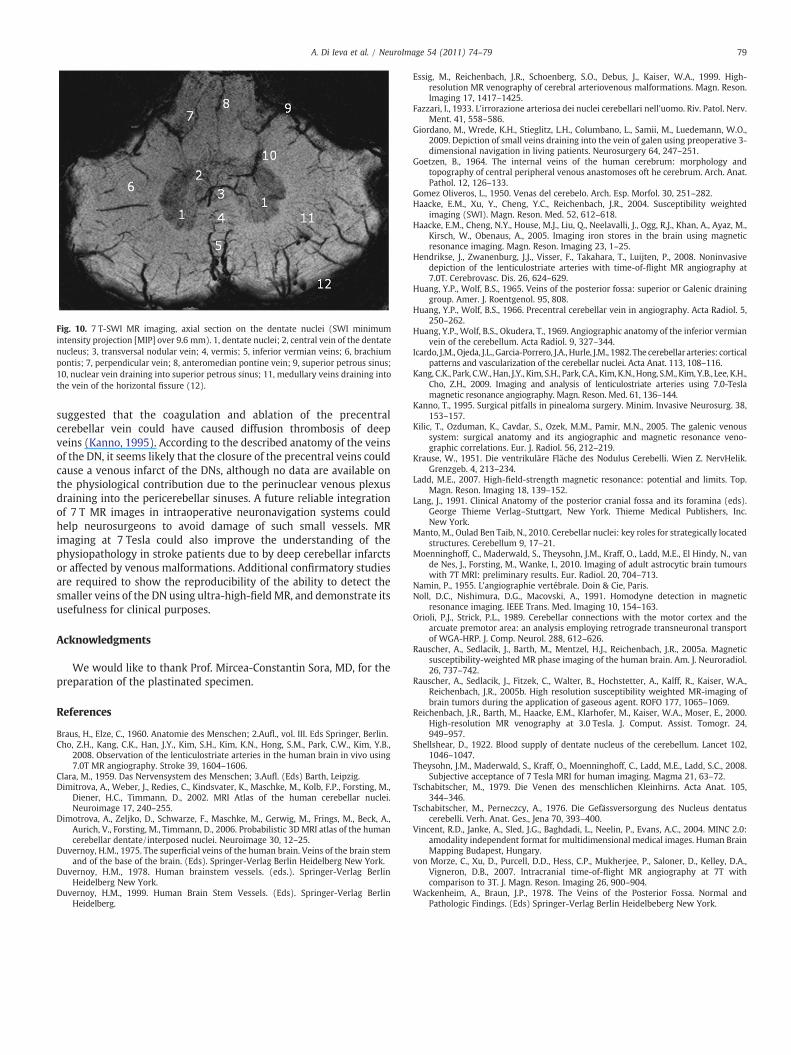
Fig. 10. 7 T-SWI MR imaging, axial section on the dentate nuclei (SWI minimumintensity projection [MIP] over 9.6 mm). 1, dentate nuclei; 2, central vein of the dentatenucleus; 3, transversal nodular vein; 4, vermis; 5, inferior vermian veins; 6, brachiumpontis; 7, perpendicular vein; 8, anteromedian pontine vein; 9, superior petrous sinus;10, nuclear vein draining into superior petrous sinus; 11, medullary veins draining intothe vein of the horizontal fissure (12).
79A. Di Ieva et al. / NeuroImage 54 (2011) 74–79
suggested that the coagulation and ablation of the precentralcerebellar vein could have caused diffusion thrombosis of deepveins (Kanno, 1995). According to the described anatomy of the veinsof the DN, it seems likely that the closure of the precentral veins couldcause a venous infarct of the DNs, although no data are available onthe physiological contribution due to the perinuclear venous plexusdraining into the pericerebellar sinuses. A future reliable integrationof 7 T MR images in intraoperative neuronavigation systems couldhelp neurosurgeons to avoid damage of such small vessels. MRimaging at 7 Tesla could also improve the understanding of thephysiopathology in stroke patients due to by deep cerebellar infarctsor affected by venous malformations. Additional confirmatory studiesare required to show the reproducibility of the ability to detect thesmaller veins of the DN using ultra-high-fieldMR, and demonstrate itsusefulness for clinical purposes.
Acknowledgments
We would like to thank Prof. Mircea-Constantin Sora, MD, for thepreparation of the plastinated specimen.
References
Braus, H., Elze, C., 1960. Anatomie des Menschen; 2.Aufl., vol. III. Eds Springer, Berlin.Cho, Z.H., Kang, C.K., Han, J.Y., Kim, S.H., Kim, K.N., Hong, S.M., Park, C.W., Kim, Y.B.,
2008. Observation of the lenticulostriate arteries in the human brain in vivo using7.0T MR angiography. Stroke 39, 1604–1606.
Clara, M., 1959. Das Nervensystem des Menschen; 3.Aufl. (Eds) Barth, Leipzig.Dimitrova, A., Weber, J., Redies, C., Kindsvater, K., Maschke, M., Kolb, F.P., Forsting, M.,
Diener, H.C., Timmann, D., 2002. MRI Atlas of the human cerebellar nuclei.Neuroimage 17, 240–255.
Dimotrova, A., Zeljko, D., Schwarze, F., Maschke, M., Gerwig, M., Frings, M., Beck, A.,Aurich, V., Forsting, M., Timmann, D., 2006. Probabilistic 3D MRI atlas of the humancerebellar dentate/interposed nuclei. Neuroimage 30, 12–25.
Duvernoy, H.M., 1975. The superficial veins of the human brain. Veins of the brain stemand of the base of the brain. (Eds). Springer-Verlag Berlin Heidelberg New York.
Duvernoy, H.M., 1978. Human brainstem vessels. (eds.). Springer-Verlag BerlinHeidelberg New York.
Duvernoy, H.M., 1999. Human Brain Stem Vessels. (Eds). Springer-Verlag BerlinHeidelberg.
Essig, M., Reichenbach, J.R., Schoenberg, S.O., Debus, J., Kaiser, W.A., 1999. High-resolution MR venography of cerebral arteriovenous malformations. Magn. Reson.Imaging 17, 1417–1425.
Fazzari, I., 1933. L'irrorazione arteriosa dei nuclei cerebellari nell'uomo. Riv. Patol. Nerv.Ment. 41, 558–586.
Giordano, M., Wrede, K.H., Stieglitz, L.H., Columbano, L., Samii, M., Luedemann, W.O.,2009. Depiction of small veins draining into the vein of galen using preoperative 3-dimensional navigation in living patients. Neurosurgery 64, 247–251.
Goetzen, B., 1964. The internal veins of the human cerebrum: morphology andtopography of central peripheral venous anastomoses oft he cerebrum. Arch. Anat.Pathol. 12, 126–133.
Gomez Oliveros, L., 1950. Venas del cerebelo. Arch. Esp. Morfol. 30, 251–282.Haacke, E.M., Xu, Y., Cheng, Y.C., Reichenbach, J.R., 2004. Susceptibility weighted
imaging (SWI). Magn. Reson. Med. 52, 612–618.Haacke, E.M., Cheng, N.Y., House, M.J., Liu, Q., Neelavalli, J., Ogg, R.J., Khan, A., Ayaz, M.,
Kirsch, W., Obenaus, A., 2005. Imaging iron stores in the brain using magneticresonance imaging. Magn. Reson. Imaging 23, 1–25.
Hendrikse, J., Zwanenburg, J.J., Visser, F., Takahara, T., Luijten, P., 2008. Noninvasivedepiction of the lenticulostriate arteries with time-of-flight MR angiography at7.0T. Cerebrovasc. Dis. 26, 624–629.
Huang, Y.P., Wolf, B.S., 1965. Veins of the posterior fossa: superior or Galenic draininggroup. Amer. J. Roentgenol. 95, 808.
Huang, Y.P., Wolf, B.S., 1966. Precentral cerebellar vein in angiography. Acta Radiol. 5,250–262.
Huang, Y.P., Wolf, B.S., Okudera, T., 1969. Angiographic anatomy of the inferior vermianvein of the cerebellum. Acta Radiol. 9, 327–344.
Icardo, J.M., Ojeda, J.L., Garcia-Porrero, J.A., Hurle, J.M., 1982. The cerebellar arteries: corticalpatterns and vascularization of the cerebellar nuclei. Acta Anat. 113, 108–116.
Kang, C.K., Park, C.W.,Han, J.Y., Kim, S.H., Park, C.A., Kim, K.N.,Hong, S.M., Kim, Y.B., Lee, K.H.,Cho, Z.H., 2009. Imaging and analysis of lenticulostriate arteries using 7.0-Teslamagnetic resonance angiography. Magn. Reson. Med. 61, 136–144.
Kanno, T., 1995. Surgical pitfalls in pinealoma surgery. Minim. Invasive Neurosurg. 38,153–157.
Kilic, T., Ozduman, K., Cavdar, S., Ozek, M.M., Pamir, M.N., 2005. The galenic venoussystem: surgical anatomy and its angiographic and magnetic resonance veno-graphic correlations. Eur. J. Radiol. 56, 212–219.
Krause, W., 1951. Die ventrikuläre Fläche des Nodulus Cerebelli. Wien Z. NervHelik.Grenzgeb. 4, 213–234.
Ladd, M.E., 2007. High-field-strength magnetic resonance: potential and limits. Top.Magn. Reson. Imaging 18, 139–152.
Lang, J., 1991. Clinical Anatomy of the posterior cranial fossa and its foramina (eds).George Thieme Verlag–Stuttgart, New York. Thieme Medical Publishers, Inc.New York.
Manto, M., Oulad Ben Taib, N., 2010. Cerebellar nuclei: key roles for strategically locatedstructures. Cerebellum 9, 17–21.
Moenninghoff, C., Maderwald, S., Theysohn, J.M., Kraff, O., Ladd, M.E., El Hindy, N., vande Nes, J., Forsting, M., Wanke, I., 2010. Imaging of adult astrocytic brain tumourswith 7T MRI: preliminary results. Eur. Radiol. 20, 704–713.
Namin, P., 1955. L'angiographie vertébrale. Doin & Cie, Paris.Noll, D.C., Nishimura, D.G., Macovski, A., 1991. Homodyne detection in magnetic
resonance imaging. IEEE Trans. Med. Imaging 10, 154–163.Orioli, P.J., Strick, P.L., 1989. Cerebellar connections with the motor cortex and the
arcuate premotor area: an analysis employing retrograde transneuronal transportof WGA-HRP. J. Comp. Neurol. 288, 612–626.
Rauscher, A., Sedlacik, J., Barth, M., Mentzel, H.J., Reichenbach, J.R., 2005a. Magneticsusceptibility-weighted MR phase imaging of the human brain. Am. J. Neuroradiol.26, 737–742.
Rauscher, A., Sedlacik, J., Fitzek, C., Walter, B., Hochstetter, A., Kalff, R., Kaiser, W.A.,Reichenbach, J.R., 2005b. High resolution susceptibility weighted MR-imaging ofbrain tumors during the application of gaseous agent. ROFO 177, 1065–1069.
Reichenbach, J.R., Barth, M., Haacke, E.M., Klarhofer, M., Kaiser, W.A., Moser, E., 2000.High-resolution MR venography at 3.0 Tesla. J. Comput. Assist. Tomogr. 24,949–957.
Shellshear, D., 1922. Blood supply of dentate nucleus of the cerebellum. Lancet 102,1046–1047.
Theysohn, J.M., Maderwald, S., Kraff, O., Moenninghoff, C., Ladd, M.E., Ladd, S.C., 2008.Subjective acceptance of 7 Tesla MRI for human imaging. Magma 21, 63–72.
Tschabitscher, M., 1979. Die Venen des menschlichen Kleinhirns. Acta Anat. 105,344–346.
Tschabitscher, M., Perneczcy, A., 1976. Die Gefässversorgung des Nucleus dentatuscerebelli. Verh. Anat. Ges., Jena 70, 393–400.
Vincent, R.D., Janke, A., Sled, J.G., Baghdadi, L., Neelin, P., Evans, A.C., 2004. MINC 2.0:amodality independent format for multidimensional medical images. Human BrainMapping Budapest, Hungary.
von Morze, C., Xu, D., Purcell, D.D., Hess, C.P., Mukherjee, P., Saloner, D., Kelley, D.A.,Vigneron, D.B., 2007. Intracranial time-of-flight MR angiography at 7T withcomparison to 3T. J. Magn. Reson. Imaging 26, 900–904.
Wackenheim, A., Braun, J.P., 1978. The Veins of the Posterior Fossa. Normal andPathologic Findings. (Eds) Springer-Verlag Berlin Heidelbeberg New York.
Copyright © 2022 FDOKUMEN