Anatomical variations of hepatic veins in Vietnamese adults
10
Anatomical variations of hepatic veins in Vietnamese adults ORIGINAL ARTICLE Eur. J. Anat. 24 (2): 111-120(2020) Hung Vinh Tran 1A , Nghia Thanh Vo 2B , Hai Van Duong 2B , Ernest F. Talarico, Jr. 3C 1 General Surgery Department, 2 Anatomy Department, 3 Department of Anatomy, Cell Biology & Physiology, A Pham Ngoc Thach University of Medicine, Vietnam, B University of Medicine and Pharmacy at Ho Chi Minh City, Vietnam, C Indiana University School of Medicine-Northwest, Gary, Indiana, USA SUMMARY The hepatic venous anatomic variations on he- patic resection and transplantation are very im- portant and the least understood aspect of the liv- er. In particular, data are lacking in the literature with reference to Vietnamese patients. The objec- tive of this study was to examine the morphologic and biometric variations of the hepatic veins in Vi- etnamese cadavers. Livers from 20 Vietnamese cadavers preserved in formalin solution were used in this study. Specimens were carefully scraped by curette to expose the branches of hepatic veins. Diameters, lengths and morphologic hepatic varia- tions were recorded. The average diameters of veins were: 34.78 mm (IVC), 20.26 mm (RHV), 14.35 mm (MHV), 14.76 mm (LHV), and 22.49 mm (common trunk). The average length of the com- mon trunk was 6.45 mm; 35% of cases in the short group (< 10 mm), and 65% in the long group (≥ 10mm). A common trunk was present in 90% of specimens. The morphology of the common trunk was comparable to that observed by other investi- gators. Only 10% of cases had accessory RHVs of Type II, with a main trunk and accessory branches to the IVC, in contrast to 90% of Type I, with a main trunk alone. The anatomical variations of the hepatic veins are very diverse. Knowledge of these variations prior to surgery is useful during both par- tial hepatectomy and segmental liver transplanta- tion. Pre-operative hepatic venous imaging can allow for assessment of venous flow and morphol- ogy, and may lessen surgical complications. Key Words: Hepatic Vein – Hepatectomy – He- patic Transplantation Abbreviations: Computerized Topography (CT) Hepatic Venous Outflow (HVO) Inferior Vena Cava (IVC) Left Hepatic Vein/Left Hepatic Veins (LHV/LHVs) Living Donor Liver Transplantation (LDLT) Magnetic Resonance Imaging (MRI) Middle Hepatic Vein/Middle Hepatic Veins (MHV/ MHVs) Right Hepatic Vein/Right Hepatic Veins (RHV/ RHVs) Split Liver Transplantation (SLT) INTRODUCTION Accurate knowledge of the anatomy of hepatic veins and their relation to the inferior vena cava (IVC) is important in liver surgery, including partial hepatic resection, hepatic trauma and liver trans- plantation (Nakamura and Tsuzuki, 1981). This understanding of hepatic veins and their anatomi- cal variations helps to prevent massive bleeding, as well as air embolism due to large tearing of the 111 Submitted: 12 November, 2019. Accepted: 21 December, 2019 Corresponding author: Hai Van Duong, M.D., Ph.D. Universi- ty of Medicine and Pharmacy at Ho Chi Minh City, 217 Hong Bang Street, District 5, Ho Chi Ming City, Vietnam. Phone: (+84) 919.669192; Fax: (+84-28) 38552304. E-mail: [email protected]
-
Upload
khangminh22 -
Category
Documents
-
view
3 -
download
0
Transcript of Anatomical variations of hepatic veins in Vietnamese adults
Anatomical variations of hepatic veins in Vietnamese adults
ORIGINAL ARTICLE Eur. J. Anat. 24 (2): 111-120(2020)
Hung Vinh Tran1A
, Nghia Thanh Vo2B
, Hai Van Duong2B
, Ernest F. Talarico,
Jr.3C
1General Surgery Department, 2Anatomy Department, 3Department of Anatomy, Cell Biology & Physiology, APham Ngoc Thach University of Medicine, Vietnam, BUniversity of Medicine and Pharmacy at Ho Chi Minh City, Vietnam,
CIndiana University School of Medicine-Northwest, Gary, Indiana, USA
SUMMARY
The hepatic venous anatomic variations on he-patic resection and transplantation are very im-portant and the least understood aspect of the liv-er. In particular, data are lacking in the literature with reference to Vietnamese patients. The objec-tive of this study was to examine the morphologic and biometric variations of the hepatic veins in Vi-etnamese cadavers. Livers from 20 Vietnamese cadavers preserved in formalin solution were used in this study. Specimens were carefully scraped by curette to expose the branches of hepatic veins. Diameters, lengths and morphologic hepatic varia-tions were recorded. The average diameters of veins were: 34.78 mm (IVC), 20.26 mm (RHV), 14.35 mm (MHV), 14.76 mm (LHV), and 22.49 mm (common trunk). The average length of the com-mon trunk was 6.45 mm; 35% of cases in the short group (< 10 mm), and 65% in the long group (≥ 10mm). A common trunk was present in 90% of specimens. The morphology of the common trunk was comparable to that observed by other investi-gators. Only 10% of cases had accessory RHVs of Type II, with a main trunk and accessory branches to the IVC, in contrast to 90% of Type I, with a main trunk alone. The anatomical variations of the hepatic veins are very diverse. Knowledge of these variations prior to surgery is useful during both par-tial hepatectomy and segmental liver transplanta-
tion. Pre-operative hepatic venous imaging can allow for assessment of venous flow and morphol-ogy, and may lessen surgical complications.
Key Words: Hepatic Vein – Hepatectomy – He-patic Transplantation Abbreviations:
Computerized Topography (CT) Hepatic Venous Outflow (HVO) Inferior Vena Cava (IVC) Left Hepatic Vein/Left Hepatic Veins (LHV/LHVs) Living Donor Liver Transplantation (LDLT) Magnetic Resonance Imaging (MRI) Middle Hepatic Vein/Middle Hepatic Veins (MHV/
MHVs) Right Hepatic Vein/Right Hepatic Veins (RHV/
RHVs) Split Liver Transplantation (SLT)
INTRODUCTION
Accurate knowledge of the anatomy of hepatic veins and their relation to the inferior vena cava (IVC) is important in liver surgery, including partial hepatic resection, hepatic trauma and liver trans-plantation (Nakamura and Tsuzuki, 1981). This understanding of hepatic veins and their anatomi-cal variations helps to prevent massive bleeding, as well as air embolism due to large tearing of the
111
Submitted: 12 November, 2019. Accepted: 21 December, 2019
Corresponding author: Hai Van Duong, M.D., Ph.D. Universi-
ty of Medicine and Pharmacy at Ho Chi Minh City, 217 Hong
Bang Street, District 5, Ho Chi Ming City, Vietnam. Phone:
(+84) 919.669192; Fax: (+84-28) 38552304.
E-mail: [email protected]
Variations of hepatic veins
112
major veins, and assists in the maintenance of ad-equate hepatic venous outflow (HVO) following surgery.
The effect of HVO on liver function is likely the least understood aspect of liver surgery. Maintain-ing HVO plays a key role in preventing hepatic dysfunction or failure, depending on the various degrees of HVO obstruction. However, the factors that yield adequate HVO after liver surgery still remain controversial.
Although hepatic vein ligation was tolerated in liver trauma patients (Ou and Hermann, 1984), human hemodynamic studies showed that, after ligation of a hepatic vein, an intrahepatic shunt occurred in the region of hepatic vein occlusion, with or without reversal of blood flow in the portal system (Ou and Hermann, 1984; Sakaguchi and Suzuki, 2010). This results in mild or moderate damage of liver function, which may lead to liver cell regeneration. In patients with cirrhosis, ob-structive jaundice, or in a liver transplant patient (where the graft is of a borderline size), there may be occlusion of HVO in a part of the liver, greatly affecting the patient outcome. Therefore, the un-derstanding of hepatic venous anatomy before surgery allows for optimal preoperative planning, and a wider margin of operative safety.
On the other hand, positive results in liver trans-plantation have extended both the quality and length of patient lives, leading to graft deficiency worldwide. Technical improvements in split liver transplantation (SLT) and living donor liver trans-plantation (LDLT) have increased the source of donor liver tissues for adults and children (Broering et al., 2008; Jin et al., 2004). LDLT is associated with risk of complications in donors, and it is im-portant to minimize complications in both donors and recipients, to increase the success rate and the application of liver transplantation techniques. Most postoperative biliary and vascular complica-tions are associated with surgical techniques and anatomical variations.
Although there are a range of modalities such as ultrasound in surgery, angiography, computed to-mography (CT), magnetic resonance imaging (MRI), that are used routinely in living donors to delineate the blood vessels and bile anatomy (Marcos, 2000), these procedures are not routinely performed in liver transplant examination. Moreo-ver, not all abnormalities can be delineated by these means, many of which are obvious only when specifically observed. Therefore, transplan-tation surgeons must understand the normal anat-omy of the liver and be able to recognize the pres-ence and implications of anatomical variants in hepatic vessels.
Restoration of hepato-venous circulation is the essential stage in liver transplantation. In these restoration techniques, preservation of the retrohe-patic IVC of the recipient is critical (Belghiti et al., 1992; Bismuth et al., 1992; Calne and Williams,
1968; Meunier et al., 1994; Tzakis et al., 1989). First described by Calne (Calne and Williams, 1968) and then named the "piggy - back” tech-nique as described by Tzakis (Tzakis et al., 1989), the “piggy - back” surgical method allows avoid-ance of the bleeding stage of retrocaval dissection and resection, especially in patients with cirrhosis (Chevalier, 1988).
Since Tzakis (Tzakis et al., 1989), several tech-niques for preserving the IVC have been de-scribed. Some of these allow for the maintenance of the caval circulation during construction of the anastomosis, avoiding total clamping of the IVC. Preservation of caval flow allows for stabilization of hemodynamics in the patient, and reduces the need for the use of an extracorporeal shunt, which has many complications (Shaw and Martin, 1984). One of these techniques, derived from the "piggy - back" technique of Tzakis (Tzakis, et al., 1889), consists of anastomosing the graft with the com-mon trunk of the middle and left hepatic veins of the recipient. The performance of this anastomosis requires favorable anatomic conditions.
A review of the literature in Vietnam and abroad showed that there are few research studies on hepatic venous anatomy applied in liver resection and transplantation surgery, as well as a lack of data from Vietnamese patients. Therefore, the pre-sent research was conducted to provide knowledge of hepatic venous anatomic variations in Vietnamese cadaveric specimens for clinicians and anatomists.
MATERIALS AND METHODS Anatomical Donors and Preservation
This study was conducted on the livers of 20 Vi-etnamese cadavers including 11 males and 9 fe-males. These anatomical donors had been pre-pared for the use in the formal course in human gross anatomy in the Department of Anatomy at the University of Medicine and Pharmacy at Ho Chi Minh City, Vietnam. The age range of subjects in this cohort was 50 to 89 years with the average age of 68.5 years-old. Livers were excluded from this cohort if the subject had hepatic disease (i.e., cirrhosis, cancer, etc.). All guidelines were fol-lowed regarding the use and care of cadaveric materials, as well as all regulations set forth by the Vietnamese Anatomical Education Program.
The embalming procedure is a 2-phase proce-dure beginning within the first 24 hours after death. The first step of the first phase of the embalming procedure is an injection of an 18 L mixture com-posed of 37% Formalin (2 L); 1 M Phenol (1 L); Glycerin 1 L, 90% Alcohol (2 L) and water (12 L). Three days following injection, cadavers are placed into 300 L of solution composed of 37% Formalin (2 L); 1 M Phenol (3 L) and water (295 L). The specimens remain submerged in the vat for a minimum of 4 months.
H. V. Duong et al.
113
Dissection Briefly, the anterior abdominal wall was cut and
opened. A vertical cut was made through the linea alba from the umbilicus to the xiphoid process, and a transverse cut, at the level of the umbilicus, per-pendicular to the vertical line. The abdominal wall was reflected in such a way that the liver could be accessed; freeing fixation ligaments (i.e., ligamen-tum teres, falciform ligaments, triangular liga-ments), and exposing the retrohepatic IVC. The liver was removed, including the retrohepatic vena cava. Cadaveric livers were carefully scraped us-ing a curette, on the diaphragmatic and visceral surfaces, to expose the branches of hepatic veins.
Measurements
Using a caliper with a center distance attach-ment, the data were saved and evaluated using SPSS 14.0. The following data were recoded: (1) biometric variables of the hepatic veins and the IVC (i.e., diameter and length); (2) morphologic variables of the common trunk of the middle and left hepatic veins. Results were expressed as mean length (or diameter) in millimeters (mm) ± mm standard deviation (SD).
Photography
Digital photography of the external features of the livers and veins was done using a NIKON D3100 SLR Camera (B&H Foto & Electronic Cor-poration, NY) equipped with an 18-55 mm VR NIK-KOR Macro lens and a Nikon 49 mm f/2.8G AF-S DX NIKKOR 2200 VR Micro lens.
RESULTS Biometric Variables
Diameter of the IVC The mean diameter of the IVC at the site of the
junction of the middle hepatic vein (MHV) and left hepatic vein (LHV) is 34.78 mm ± 4.43 mm SD, the largest is 44.83 mm, the smallest is 28.95 mm.
Diameter of Hepatic Veins The right hepatic vein (RHV) was found to have
the largest diameter (20.26 mm ± 3.93 mm SD), when compared to the LHV. However, the diame-ter of the common trunk of the MHV and LHV was larger (22.49 mm ± 4.2 mm SD). The results are shown in Tables 1 and 2.
Morphologic Variables
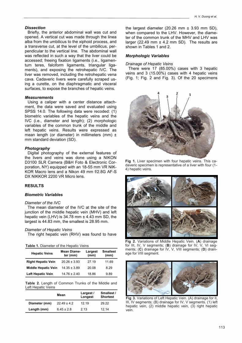
Drainage of Hepatic Veins There were 17 (85.00%) cases with 3 hepatic
veins and 3 (15.00%) cases with 4 hepatic veins (Fig. 1; Fig. 2 and Fig. 3). Of the 20 specimens
Fig 1. Liver specimen with four hepatic veins. This ca-daveric specimen is representative of a liver with four (1-4) hepatic veins.
Hepatic Veins Mean Diame-
ter (mm) Largest
(mm) Smallest
(mm)
Right Hepatic Vein 20.26 ± 3.93 27.19 11.69
Middle Hepatic Vein 14.35 ± 3.89 20.08 8.29
Left Hepatic Vein 14.76 ± 2.40 18.86 9.89
Table 1. Diameter of the Hepatic Veins
Fig 2. Variations of Middle Hepatic Vein. (A) drainage for III, IV, V segments; (B) drainage for IV, V, VI seg-ments; (C) drainage for IV, V, VIII segments; (D) drain-age for VIII segment.
Mean Largest / Longest
Smallest / Shortest
Diameter (mm) 22.49 ± 4.2 12.19 29.22
Length (mm) 6.45 ± 2.8 2.13 12.14
Table 2. Length of Common Trunks of the Middle and Left Hepatic Veins
Fig 3. Variations of Left Hepatic Vein. (A) drainage for II, III, IV segments; (B) drainage for IV, V segments. (1) left hepatic vein, (2) middle hepatic vein, (3) right hepatic vein.
Variations of hepatic veins
114
examined, 18 (90.00%) cases had a common LHV and MHV. The drainage of hepatic veins is shown in Table 3.
Anatomo-clinical classification for the common trunk of the Middle and Left Hepatic Veins
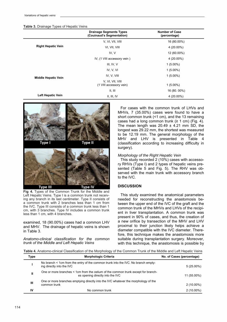
For cases with the common trunk of LHVs and MHVs, 7 (35.00%) cases were found to have a short common trunk (<1 cm), and the 13 remaining cases had a long common trunk (≥ 1 cm) (Fig. 4). The mean length was 20.49 ± 4.21 mm SD, the longest was 29.22 mm, the shortest was measured to be 12.19 mm. The general morphology of the MHV and LHV is presented in Table 4 (classification according to increasing difficulty in surgery).
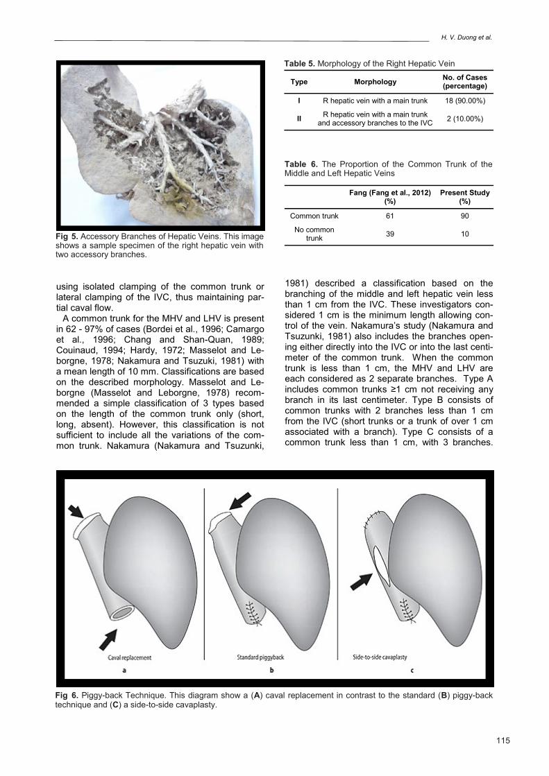
Morphology of the Right Hepatic Vein This study recorded 2 (10%) cases with accesso-
ry RHVs (Type I) and 2 types of hepatic veins pre-sented (Table 5 and Fig. 5). The RHV was ob-served with the main trunk with accessory branch to the IVC.
DISCUSSION
This study examined the anatomical parameters needed for reconstructing the anastomosis be-tween the upper end of the IVC of the graft and the common trunk of the MHVs and LHVs of the recipi-ent in liver transplantation. A common trunk was present in 90% of cases, and thus, the creation of a new orifice by transection of the MHV and LHV proximal to their junction likely helps achieve a diameter compatible with the IVC diameter. There-fore, this technique makes the anastomosis more suitable during transplantation surgery. Moreover, with this technique, the anastomosis is possible by
Drainage Segments Types
(Couinaud’s Segmentation) Number of Case
(percentage)
Right Hepatic Vein
V, VI, VII, VIII 16 (80.00%)
VI, VII, VIII 4 (20.00%)
Middle Hepatic Vein
IV, V 12 (60.00%)
IV, (1 VIII accessory vein ) 4 (20.00%)
III, IV, V 1 (5.00%)
IV, V, VI 1 (5.00%)
IV, V, VIII 1 (5.00%)
V, VI, VII, VIII (1 VIII accessory vein)
1 (5.00%)
Left Hepatic Vein
II, III 16 (80. 00%)
II, III, IV 4 (20.00%)
Table 3. Drainage Types of Hepatic Veins
Type Morphologic Criteria No. of Cases (percentage)
I No branch < 1cm from the entry of the common trunk into the IVC. No branch empty-ing directly into the IVC
5 (25.00%)
II One or more branches < 1cm from the ostium of the common trunk except for branch-
es opening directly into the IVC
11 (55.00%)
III One or more branches emptying directly into the IVC whatever the morphology of the common trunk
2 (10.00%)
IV No common trunk 2 (10.00%)
Table 4. Anatomo-clinical Classification of the Morphology of the Common Trunk of the Middle and Left Hepatic Veins
Fig 4. Types of the Common Trunk for the Middle and Left Hepatic Veins. Type I is a common trunk not receiv-ing any branch in its last centimeter. Type II consists of a common trunk with 2 branches less than 1 cm from the IVC. Type III consists of a common trunk less than 1 cm, with 3 branches. Type IV includes a common trunk less than 1 cm, with 4 branches.
H. V. Duong et al.
115
using isolated clamping of the common trunk or lateral clamping of the IVC, thus maintaining par-tial caval flow.
A common trunk for the MHV and LHV is present in 62 - 97% of cases (Bordei et al., 1996; Camargo et al., 1996; Chang and Shan-Quan, 1989; Couinaud, 1994; Hardy, 1972; Masselot and Le-borgne, 1978; Nakamura and Tsuzuki, 1981) with a mean length of 10 mm. Classifications are based on the described morphology. Masselot and Le-borgne (Masselot and Leborgne, 1978) recom-mended a simple classification of 3 types based on the length of the common trunk only (short, long, absent). However, this classification is not sufficient to include all the variations of the com-mon trunk. Nakamura (Nakamura and Tsuzunki,
1981) described a classification based on the branching of the middle and left hepatic vein less than 1 cm from the IVC. These investigators con-sidered 1 cm is the minimum length allowing con-trol of the vein. Nakamura’s study (Nakamura and Tsuzunki, 1981) also includes the branches open-ing either directly into the IVC or into the last centi-meter of the common trunk. When the common trunk is less than 1 cm, the MHV and LHV are each considered as 2 separate branches. Type A includes common trunks ≥1 cm not receiving any branch in its last centimeter. Type B consists of common trunks with 2 branches less than 1 cm from the IVC (short trunks or a trunk of over 1 cm associated with a branch). Type C consists of a common trunk less than 1 cm, with 3 branches.
Fig 5. Accessory Branches of Hepatic Veins. This image shows a sample specimen of the right hepatic vein with two accessory branches.
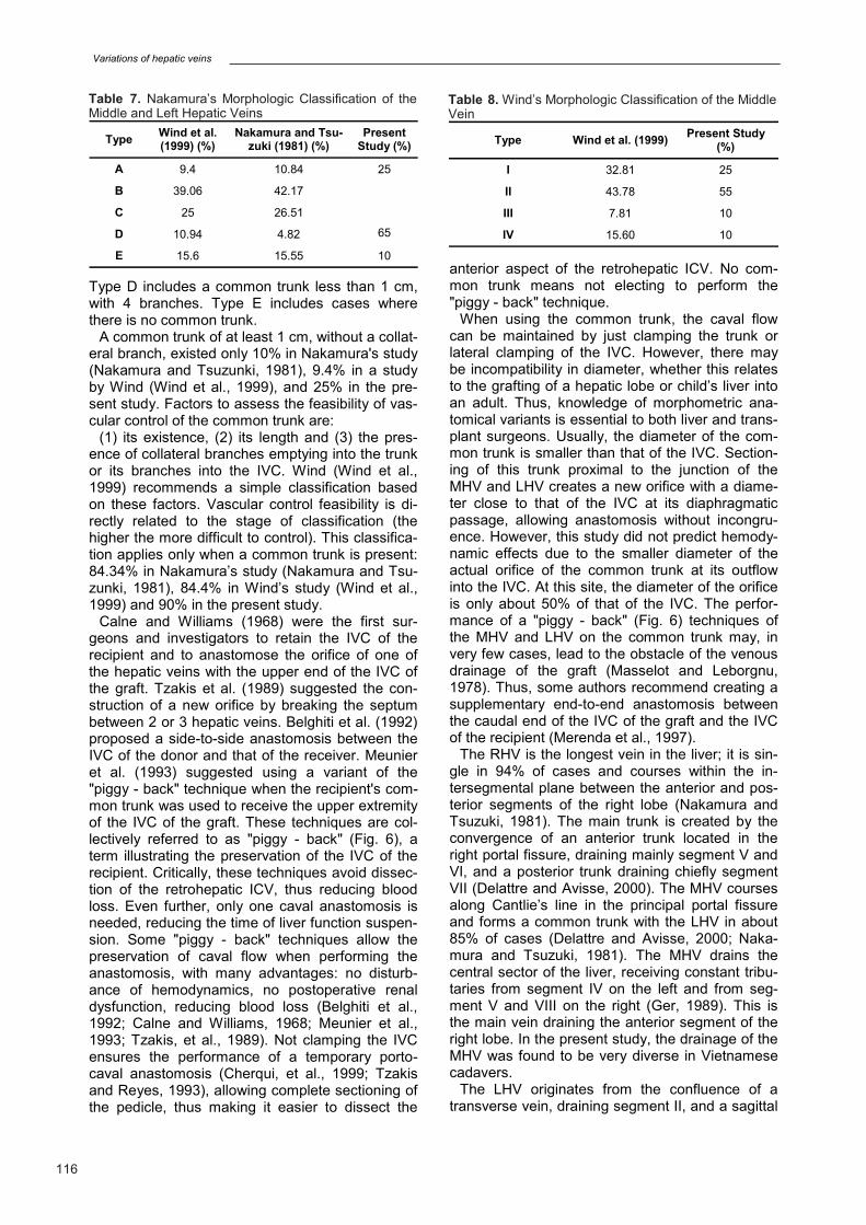
Fig 6. Piggy-back Technique. This diagram show a (A) caval replacement in contrast to the standard (B) piggy-back technique and (C) a side-to-side cavaplasty.
Fang (Fang et al., 2012)
(%) Present Study
(%)
Common trunk 61 90
No common trunk
39 10
Table 6. The Proportion of the Common Trunk of the Middle and Left Hepatic Veins
Type Morphology No. of Cases (percentage)
I R hepatic vein with a main trunk 18 (90.00%)
II R hepatic vein with a main trunk
and accessory branches to the IVC 2 (10.00%)
Table 5. Morphology of the Right Hepatic Vein
Variations of hepatic veins
116
Type D includes a common trunk less than 1 cm, with 4 branches. Type E includes cases where there is no common trunk.
A common trunk of at least 1 cm, without a collat-eral branch, existed only 10% in Nakamura's study (Nakamura and Tsuzunki, 1981), 9.4% in a study by Wind (Wind et al., 1999), and 25% in the pre-sent study. Factors to assess the feasibility of vas-cular control of the common trunk are:
(1) its existence, (2) its length and (3) the pres-ence of collateral branches emptying into the trunk or its branches into the IVC. Wind (Wind et al., 1999) recommends a simple classification based on these factors. Vascular control feasibility is di-rectly related to the stage of classification (the higher the more difficult to control). This classifica-tion applies only when a common trunk is present: 84.34% in Nakamura’s study (Nakamura and Tsu-zunki, 1981), 84.4% in Wind’s study (Wind et al., 1999) and 90% in the present study.
Calne and Williams (1968) were the first sur-geons and investigators to retain the IVC of the recipient and to anastomose the orifice of one of the hepatic veins with the upper end of the IVC of the graft. Tzakis et al. (1989) suggested the con-struction of a new orifice by breaking the septum between 2 or 3 hepatic veins. Belghiti et al. (1992) proposed a side-to-side anastomosis between the IVC of the donor and that of the receiver. Meunier et al. (1993) suggested using a variant of the "piggy - back" technique when the recipient's com-mon trunk was used to receive the upper extremity of the IVC of the graft. These techniques are col-lectively referred to as "piggy - back" (Fig. 6), a term illustrating the preservation of the IVC of the recipient. Critically, these techniques avoid dissec-tion of the retrohepatic ICV, thus reducing blood loss. Even further, only one caval anastomosis is needed, reducing the time of liver function suspen-sion. Some "piggy - back" techniques allow the preservation of caval flow when performing the anastomosis, with many advantages: no disturb-ance of hemodynamics, no postoperative renal dysfunction, reducing blood loss (Belghiti et al., 1992; Calne and Williams, 1968; Meunier et al., 1993; Tzakis, et al., 1989). Not clamping the IVC ensures the performance of a temporary porto- caval anastomosis (Cherqui, et al., 1999; Tzakis and Reyes, 1993), allowing complete sectioning of the pedicle, thus making it easier to dissect the
anterior aspect of the retrohepatic ICV. No com-mon trunk means not electing to perform the "piggy - back" technique.
When using the common trunk, the caval flow can be maintained by just clamping the trunk or lateral clamping of the IVC. However, there may be incompatibility in diameter, whether this relates to the grafting of a hepatic lobe or child’s liver into an adult. Thus, knowledge of morphometric ana-tomical variants is essential to both liver and trans-plant surgeons. Usually, the diameter of the com-mon trunk is smaller than that of the IVC. Section-ing of this trunk proximal to the junction of the MHV and LHV creates a new orifice with a diame-ter close to that of the IVC at its diaphragmatic passage, allowing anastomosis without incongru-ence. However, this study did not predict hemody-namic effects due to the smaller diameter of the actual orifice of the common trunk at its outflow into the IVC. At this site, the diameter of the orifice is only about 50% of that of the IVC. The perfor-mance of a "piggy - back" (Fig. 6) techniques of the MHV and LHV on the common trunk may, in very few cases, lead to the obstacle of the venous drainage of the graft (Masselot and Leborgnu, 1978). Thus, some authors recommend creating a supplementary end-to-end anastomosis between the caudal end of the IVC of the graft and the IVC of the recipient (Merenda et al., 1997).
The RHV is the longest vein in the liver; it is sin-gle in 94% of cases and courses within the in-tersegmental plane between the anterior and pos-terior segments of the right lobe (Nakamura and Tsuzuki, 1981). The main trunk is created by the convergence of an anterior trunk located in the right portal fissure, draining mainly segment V and VI, and a posterior trunk draining chiefly segment VII (Delattre and Avisse, 2000). The MHV courses along Cantlie’s line in the principal portal fissure and forms a common trunk with the LHV in about 85% of cases (Delattre and Avisse, 2000; Naka-mura and Tsuzuki, 1981). The MHV drains the central sector of the liver, receiving constant tribu-taries from segment IV on the left and from seg-ment V and VIII on the right (Ger, 1989). This is the main vein draining the anterior segment of the right lobe. In the present study, the drainage of the MHV was found to be very diverse in Vietnamese cadavers.
The LHV originates from the confluence of a transverse vein, draining segment II, and a sagittal
Type Wind et al. (1999) (%)
Nakamura and Tsu-zuki (1981) (%)
Present Study (%)
A 9.4 10.84 25
B 39.06 42.17
65
C 25 26.51
D 10.94 4.82
E 15.6 15.55 10
Table 7. Nakamura’s Morphologic Classification of the Middle and Left Hepatic Veins
Type Wind et al. (1999) Present Study
(%)
I 32.81 25
II 43.78 55
III 7.81 10
IV 15.60 10
Table 8. Wind’s Morphologic Classification of the Middle Vein
H. V. Duong et al.
117
vein draining segment III (Delattre and Avisse, 2000). Sometimes this vein receives a contribution from segment IV (i.e., one case in the present study). The main trunk, in the majority of cases, forms a common channel with the MHV, and opens into the suprahepatic IVC. Sometimes, it courses as an independent vein of the left lateral segment, draining separately from the MHV (i.e., 2 cases observed in the present study).
Short accessory (dorsal) RHVs (they should not be confused with the caudate lobe vein) drain the dorsal aspect of the liver (mainly segments VI and VII) and empty directly to the right of the retrohe-patic IVC. The caudate lobe is drained mainly on the left by a single vein in 50% of cases, and by 2 or 3 veins in the remaining cases. In addition, there may be up to 20 small, short venules attach-ing the caudate lobe to the retrohepatic IVC (Dodson, 1993). In the present work on Vietnam-ese, there were 2 cases with accessory RHVs draining directly into the IVC.
The MHV and LHV form a common trunk in most cases. In addition, small individual veins, draining segment III or the superior part of segment IV (segment IVa), can empty directly into the su-prahepatic IVC, close to the main LHV. This fea-ture is often not seen on the right side (Nakamura and Tsuzuki, 1981). The intersegmental area be-tween segment IV and left lateral segment consti-tutes the watershed between the drainage territo-ries of the MHV and LHV.
The results of the present study showed that RHV is the main drainage vein of segments VI, VII, VIII, and V (Figs. 1-4; Table 3). The results further demonstrated that the MHV is the main drainage vein of segments IV, V; and that the LHV is the main drainage vein of segments II, III (Fig. 3; Ta-bles 3 and 4).
Applications in Hepatic Transplantation
Trinh Hong Son (2002) commented that there are 3 remarkable variations in hepatic vein anato-my applied in liver transplantation:
2 RHV type: MHV draining segment VI (12.3%). If taking the right lobe to transplant and keeping the MHV, then 5% of this type are documented in this study.
4 RHV type: MHV draining posterior and anterior sectors (2.5%). When dividing the liver to trans-plant or procuring the right lobe in volume-reduced transplantation, it is necessary to take the IVC with accessory hepatic veins of the donor for recipient. The present work showed 15% of 4 RHV type.
6 LHV type: MHV draining segment III (13.2%). In case of procuring the left lobe to transplant, it is needed to recognize this type during transplanta-tion process to ensure the quality of the graft. The present study detected 5% of 6 LHV type. Such varieties should be known in advance to prepare for the venoplasty during transplantation (Trinh, et al., 1997; Trinh, et al., 1998).
The most common type of LST is to take the left lateral sector based on the LHV, and an extended right lobe based on MHV and LHV (Rela et al., 2013). Because the splitting line passes through the watershed zone, tributaries of the LHV and MHV are invariably encountered. These can be ligated without compromising the outflow of the left lateral sector or segment IV. However, care is re-quired when dealing with the segment III vein draining into the MHV (5% according to this study; compared to 13.2% according to Trinh Hong Son (2002)). After parenchymal division, it may be im-possible to reconstruct the outflow of the segment III and LHVs into a single opening if their orifice are wide apart, and both may need to be implanted separately to the recipient IVC.
This problem may be also encountered when a liver is being reduced, rather than split, down to a left lateral sector. Unexpected bleeding may occur during an in situ split or left lateral segment when a large branch of the LHV, draining part of segment IV, runs across the falciform ligament (Nakamura and Tsuzuki, 1981). Of the cases examined in the present study, 20% showed this variation. The presence of a transverse segment II vein, 10% in the cases examined herein, permits resection of this segment to reduce a left lateral graft down to a ‘monosegment’ for transplantation in a very small baby (Srinivasan and Vilca-Melendez, 1999), thereby overcoming a size discrepancy between donor and recipient.
Differences in the drainage patterns of the left hepatic venous system in the donor and the recipi-ent can cause anastomotic misalignment after im-plantation of the left lateral segment graft, leading to outflow obstruction. An adequate outflow is very important for cut-surface hemostasis and optimal graft function. The triangulation technique for he-patic venous anastomosis has helped to resolve this potential problem (Edmond and Heffron, 1993). Intraoperative Doppler ultrasonography af-ter liver reperfusion is useful in assessing the ade-quacy of vascular inflow and outflow, and help to determine the best position in which to anchor the graft in consideration of anatomical variants.
Techniques are evolving to split the liver into right and left lobe grafts of adequate and approxi-mately equal mass for 2 adult recipients (Strasberg and Lowell, 1999). The plane of dissection lies to the right of the MHV. This interrupts the venous outflow of segment V and VIII (10% in the present investigation), which can cause congestion and troublesome bleeding from the cut surface of the right lobe after reperfusion. This can also compro-mise the functional volume of the right lobe graft. The IVC can be saved for either lobe, but it is pre-ferred to keep it in the right lobe with large acces-sory RHV. Alternative options include opening the IVC longitudinally to take a patch containing all tributary veins, and performing a cavo- cavoplasty in the recipient (Gundlach et al., 2000). Trinh van
Variations of hepatic veins
118
Minh (1999) remarked, in most cases, the MHV developed normally and was slightly shifted to the left of the portal bifurcation. So the plane of dissec-tion should be at the right of the MHV. This tech-nique saves the common trunk of middle and left hepatic veins for the left lobe, and avoids a varia-tions of the umbilical fissure vein from the LHV into the MHV. The right lobe is drained by the RHV with abundant connecting branches (Van Minh, 1999).
Procurement of a right lobe in a living donor lobe can be also complicated by similar problems. It is helpful to map out the intrahepatic course of he-patic veins by intraoperative ultrasound (Dalattre and Avisse, 2000), especially that of the middle hepatic and accessory right hepatic veins in the donor right lobe. The length of a 1 cm extrahepatic RHV cuff is helpful for a right lobe living donor har-vest because of the ease of dissection and graft implantation to the recipient IVC. If large accesso-ry right hepatic veins are encountered, they must be implanted separately to the recipient IVC. Ex-tended right lobe living donor is relatively infre-quent for fear of insufficient liver mass in the do-nor. This can be more dangerous by inadvertent damage or torsion of the LHV at donor operation, especially when the MHV forms a common chan-nel with the LHV.
The anatomy of the MHV is the key of right lobe living donor. In about 10% of cases, when the RHV is of a small caliber, the MHV can provide the sole drainage of the anterior sector of the right lobe. This is a contraindication to right lobe dona-tion (Reichert and Renz, 2001). Congestion of the anterior sector of the right lobe occurs invariably after parenchymal transection. However, this sec-tor develops ischemia after reperfusion, rather than congestion (Cui, et al., 2001). This would not persist in the presence of an adequate vascular inflow and outflow, and a well-functioning liver mass.
CONCLUSION
Normal anatomical variants of hepatic veins are very diverse. A thorough knowledge of these varia-tions is essential for the hepatic or transplant sur-geon. Pre-operative imaging for elucidating mor-phology and the application of anatomy of these variants is suggested to be helpful for patients un-dergoing hepatic resection and segmental liver transplantation.
ACKNOWLEDGEMENTS
The authors wish to express their sincere grati-tude to the anatomical donors of Vietnam who be-queathed their bodies for medical education and basic science research. We also wish to thank all Technicians of Anatomy Department, at the Uni-versity of Medicine and Pharmacy at Ho Chi Minh City (Vietnam) for their assistance with gross ana-
tomical dissections.
NOTES ON CONTIBUTORS
HUNG VINH TRAN, M.D., Ph.D., is the Chair-man of Department of General Surgery, University of Medicine Pham Ngoc Thach at Ho Chi Minh City (461 Su Van Hanh Street, Ward 12, District 10, Ho Chi Minh City, Vietnam). He graduated in 1993 from the University of Medicine and Pharmacy at Ho Chi Minh City (217 Hong Bang Street, District 5, Ho Chi Minh City, Vietnam), and served as a General Surgeon from 1994 in People’s Hospital 115 (527 Su Van Hanh Street, Ward 12, District 10, Ho Chi Minh City, Vietnam). Currently, in addi-tion to his medical practice, he served as Director of Binh Dan Hospital at Ho Chi Minh City, a Teach-ing Hospital (371 Đien Bien Phu Street, Ward 4, District 3, Ho chi Minh City, Vietnam).
HAI VAN DUONG M.D., Ph.D., serves as Associ-ate Professor of Anatomy at the University of Med-icine and Pharmacy at Ho Chi Minh City (217 Hong Bang Street, District 5, Ho Chi Minh City, Vietnam) and is a Practicing Clinician and a Con-sulting Surgeon in charge of General Surgery in Binh Dan Hospital at Ho chi Minh City, a Teaching Hospital (371 Đien Bien Phu Street, Ward 4, Dis-trict 3, Ho Chi Minh City, Vietnam). He specialized in Surgical Anatomy and Colorectal Surgery.
NGHIA THANH VO, M.D., Ph.D., graduated in 2011 from the University of Medicine and Pharma-cy at Ho Chi Minh City (217 Hong Bang Street, District 5, Ho Chi Minh City, Vietnam) and served as a lecturer in the Anatomy Department.
ERNEST F. TALARICO, JR., Ph.D., is Associate Professor of Anatomy & Cell Biology at the Indiana University School of Medicine-Northwest (Gary, IN U.S.A.). Dr. Talarico holds a joint appointment as Associate Faculty in the Department of Radiologic Sciences at Indiana University Northwest, and an appointment as Visiting Professor of Anatomy at Tan Tao University School of Medicine (Long An, Vietnam). He created and serves as director for the International Human Cadaver Prosection Pro-gram, which in 2008 received the award for most outstanding and innovative program in undergrad-uate and continuing medical education from the AAMC Central Group on Educational Affairs. He is creator of the “Talarico Protocol for Human Gross Anatomy” and is the 2008 recipient of the Partner-ship Matters Award from the Northwest Indiana Area Health Education Center. In recognition of his work and innovations in anatomical education, in October 2010, Dr. Talarico was inducted as a fel-low into the Northwest Indiana Society of Innova-tors. Currently, Dr. Talarico also serves as the di-rector of the Anatomy Project in Vietnam and Southeast Asia.
REFERENCES BELGHITI J, PANIS Y, SAUVANET A, GAYET B, FE-
H. V. Duong et al.
119
KETE F (1992) A new technique of side to side caval anastomosis during orthotopic hepatic transplantation without inferior vena caval occlusion. Surg Gynecol Obstet, 175(3): 270-272.
BISMUTH H, CASTAING D, SHERLOCK DJ (1992) Liver transplantation by "face à face" vena- cavaplasty. Surgery, 111: 151-155.
BORDEI P, ANTOHE D (1996) Etude anatomique des veines hepatiques moyenne et gauche. Surg Radiol Anat, 18: 250.
BROERING DC, WALTER J, BRAUN F, ROGIERS X (2008) Current status of hepatic transplantation. Ana-tomical basis for liver transplantation. Curr Probl Surg, 45(9): 587-661.
CALNE RY, WILLIAMS R (1968) Liver transplantation in man - I. Observations on technique and organisation in five cases. Br Med J, 4: 535-540.
CAMARGO AM, TEIXEIRA GG, ORTALE JR (1996) Anatomy of the ostia venae hepaticae and the retrohe-patic segment of the inferior vena cava. J Anat, 188 (Pt 1): 59-64.
CHANG R, SHAN-QUAN S (1989) An applied anatomi-cal study of the ostia venae hepaticae and the retro-hepatique segment of the IVC. J Anat, 164: 41-47.
CHERQUI D, MALASSAGNE B, COLAU PI, BRUNETTI F, ROTMAN N, FAGNIEZ PL (1999) Hepatic vascular exclusion with preservation of the caval flow for liver resections. Ann Surg, 230(1): 24-30.
CHEVALIER JM (1988) Anatomic basis of vascular ex-clusion of the liver. Surg Radiol Anat, 10: 187-194.
COUINAUD C (1994) Intrahepatic anatomy. Application to liver transplantation. Ann Radiol (Paris), 37(5): 323-333.
CUI D, KIUCHI T, EGAWA H, HAVASHI M, SAKAMO-TO S, UEDA M, KAIHARA S, UEMOTO S, INOMATA Y, TANAKA K (2001) Microcirculatory changes in right lobe grafts in living-donor liver transplantation: a near-infrared spectrometry study. Transplantation, 72(2): 291-295.
DELATTRE J, AVISSE C (2000) Anatomic basis of he-patic surgery. Surg Clin North Am, 80: 345-362.
DODSON TF (1993) Surgical anatomy of hepatic trans-plantation. Surg Clin North Am, 73: 645- 659.
EMOND JC, HEFFRON TG (1993) Reconstruction of the hepatic vein in reduced sized hepatic transplanta-tion. Surg Gynecol Obstet, 176: 11-17.
FANG CH, YOU JH, LAU WY, LAI EC, FAN YF, ZHONG SZ, LI KX, CHEN ZX, SU ZH, BAO SS (2012) Anatomical variations of hepatic veins: three-dimensional computed tomography scans of 200 sub-jects. World J Surg, 36(1): 120-124.
GER R (1989) Surgical anatomy of the liver. Surg Clin North Am, 69: 179-192.
GUNDLACH M, BROERING D, TOPP S, STERNECK M, ROGIERS X (2000) Split-cava technique: liver split-ting for two adult recipients. Liver Transpl, 6(6):703-706.
HARDY KJ (1972) The hepatic veins. Aust N Z J Surg, 42: 11-14.
JIN MB, SHIMAMURA T, TANIGUCHI M, NAGASAKO Y, SUZUKI T, KAMIYAMA T, MATSUSHITA M, FU-RUKAWA H, TODO S (2004) Liver regeneration in living-donor liver transplantation. Nihon Geka Gakkai Zasshi, 105(10): 674-679.
MARCOS A (2000) Right-lobe living donor liver trans-plantation. Liver Transpl, 6(6 Suppl 2): S59-563.
MASSELOT R, LEBORGNE J (1978) Etude anatomique des veines sus-hepatiques. Anat Clin, 1: 109-125.
MERENDA R, GERUNDA GE, NERI D, BARBAZZA F, DI MARZIO E, BRUTTOCAO A, VALMASONI M, AN-GELI P, FACCIOLI AM (1997) Infrahepatic terminola-teral cavo-cavostomy as a rescue technique in compli-cated "modified" piggyback liver transplantation. J Am Coll Surg, 185(6): 576-579.
MEUNIER B, BARDAXOGLOU E, CHARETON B, LANDEN S, CAMUS C, ROUMEAS J, LAUNOIS B (1993) "Piggyback" method in hepatic transplantation. Chirurgie, 119(10): 682-685.
MEUNIER B, BARDAXOGLOU E, SPILIOPOULOS G, LANDEN S, CAMUS C, ROUMEAS J, LAUNOIS B (1994) Liver transplantation with preservation of the inferior vena cava and "piggyback" reimplantation of the liver. Ann Chir, 48(11): 986-988.
NAGORNEY D (2010) The impact of hepatic venous anatomy on the hepatic remnant: need for assess-ment? Surgery, 147: 811-812.
NAKAMURA S, TSUZUKI T (1981) Surgical anatomy of the hepatic veins and the inferior vena cava. Surg Gy-necol Obstet, 152: 43-50.
OU QJ, HERMANN ER (2001) The role of hepatic veins in liver operations. Surgery, 95: 381-391.
REICHERT PR, RENZ JF (2001) Anatomical variations hampering the use of right lobe in living donor liver transplantation. Liver Transpl, 7: C-85.
RELA M, KOTA V, SHANMUGAM V, VADEYAR H (2013) Middle hepatic vein to middle hepatic vein anastomosis in right lobe living donor liver transplanta-tion. Liver Transpl, 19(2): 229-231.
SAKAGUCHI T, SUZUKI S (2010) Analysis of intrahe-patic venovenous shunt by the hepatic venography. Surgery, 147: 805-810.
SHAW B, MARTIN DJ (1984) Venous bypass in clinical liver transplantation. Ann Surg, 200: 524-534.
SRINIVASAN P, VILCA-MELENDEZ H (1999) Liver transplantation with monosegment. Surgery, 126: 10-12.
STRASBERG SM, LOWELL JA (1999) Reducing the shortage of donor liver: what would it take to reliably split liver for transplantation into 2 adult recipients? Liver Transpl Surg, 5: 437-450.
TRINH HS (2002) Surgical anatomy of the liver in hepat-ic transplantation. Surgery, 5: 7-19 (in Vietnamese).
TRỊNH HS, DƯƠNG ĐH (1997) History of plastic sur-gery of hepatic veins. Practical Medicine, 12: 23-25.
TRỊNH HS, TÔN THẤT BÁCH (1998) Anatomical study of hepatic veins to the inferior vena cava, applied to hepatic resection, venoplasty and transplantation. Practical Medicine, 3: 37-41.

Variations of hepatic veins
120
TZAKIS A, REYES J (1993) Temporary end to side por-to - caval shunt in orthotopic hepatic transplantation in humans. Surg Gynecol Obstet, 176: 181-182.
TZAKIS A, TODO S, STARZL TE (1989) Orthotopic liver transplantation with preservation of the inferior vena cava. Ann Surg, 210(5): 649-652.
VAN MINH T (1999) The role of anatomy in hepatic re-section and transplantation (review of history and per-sonal opinion). Vietnam Medicine.
WIND P, DOUARD R, CUGNENC PH, CHEVALLIER JM (1999) Anatomy of the common trunk of the middle and left hepatic veins: application to liver transplanta-tion. Surg Radiol Anat, 21(1): 17-21.