
invasion of lymphatics, veins and nerves - NCBI
15
THE SPREAD OF CARCINOMA OF THE RECTUM: INVASION OF LYMPHATICS, VEINS AND NERVES* PHILIP H. SEEFELD, M.D. FELLOW IN SURGERY, MAYO FOUNDATION AND J. ARNOLD BARGEN, M.D. DIVIS:ON OF MEDICINE, MAYO CLINIC ROCHESTER, MINN. SINCE THE WORK OF HANDLEY,15 in I9IO, much attention has been given to the lymphatic route of spread of carcinoma of the rectum. Many extensive and painstaking studies have been made into this feature of rectal carcinoma in order to determine the prognostic implications of nodal involvement and the direction of spread of the carcinoma. The anatomic descriptions of the rectal lymphatic channels by Delamere, Poirier and Cuneo9 paved the way for later studies. Early investigations by Miles,22 Monsarrat and Williams,24 Cole,5 Cheatle,4 Pennington, 9 and others, gave impetus to more exhaustive studies by McVay,2' one of us (Bargen ) and Larsen, Gabriel, Dukes and Bussey,'2 Gilchrist and David,'3 Coller, and others,6 and Grinnell.'4 These investigators, and the large numbers of cases studied by them, have emphasized the im- portance of the lymph nodes as prognostic indicators in rectal carcinoma, and around this pathologic feature of the disease have been centered the present methods of surgical treatment. Venous invasion in rectal carcinoma has been observed for many years, Mayo,19 McArthur,20 Smith,30 Monsarrat and Williams,24 and Miles23 noting this feature and commenting on it. During the past four years, increasing attention has been given to venous invasion of rectal carcinoma as its clinical and prognostic importance has been seen to grow. The frequent development of visceral metastatic lesions after radical resection of the rectum for car- cinoma has stimulated interest in this phase of invasion by carcinoma. Brown and Warren,3 in one of the first reports during the present period of interest, expressed the opinion that the lymph nodes have been poor indicators of visceral metastatic lesions, which are often present inde- pendently of neoplastic nodes. They found that 31 per cent of patients without nodal involvement by carcinoma had visceral metastatic lesions, and they maintained that the spread of rectal carcinoma through the blood vessels is at least as important as by the lymphatic route, and that evidence of this spread is usually available in the primary growth. In the prediction of visceral metastatic lesions from the primary growth, the presence of intra- vascular invasion means as much from the prognostic standpoint as neo- plastic nodes, and its absence means much more. * Abridgment of thesis submitted by Dr. Seefeld to the Faculty of the Graduate School of the University of Minnesota in partial fulfillment of the requirements for the degree of M.S. in Surgery. 76
-
Upload
khangminh22 -
Category
Documents
-
view
0 -
download
0
Transcript of invasion of lymphatics, veins and nerves - NCBI
THE SPREAD OF CARCINOMA OF THE RECTUM:INVASION OF LYMPHATICS, VEINS AND NERVES*
PHILIP H. SEEFELD, M.D.FELLOW IN SURGERY, MAYO FOUNDATION
AND
J. ARNOLD BARGEN, M.D.DIVIS:ON OF MEDICINE, MAYO CLINIC
ROCHESTER, MINN.
SINCE THE WORK OF HANDLEY,15 in I9IO, much attention has been givento the lymphatic route of spread of carcinoma of the rectum. Many extensiveand painstaking studies have been made into this feature of rectal carcinoma inorder to determine the prognostic implications of nodal involvement and thedirection of spread of the carcinoma. The anatomic descriptions of the rectallymphatic channels by Delamere, Poirier and Cuneo9 paved the way for laterstudies. Early investigations by Miles,22 Monsarrat and Williams,24 Cole,5Cheatle,4 Pennington, 9 and others, gave impetus to more exhaustive studiesby McVay,2' one of us (Bargen ) and Larsen, Gabriel, Dukes and Bussey,'2Gilchrist and David,'3 Coller, and others,6 and Grinnell.'4 These investigators,and the large numbers of cases studied by them, have emphasized the im-portance of the lymph nodes as prognostic indicators in rectal carcinoma,and around this pathologic feature of the disease have been centered thepresent methods of surgical treatment.
Venous invasion in rectal carcinoma has been observed for many years,Mayo,19 McArthur,20 Smith,30 Monsarrat and Williams,24 and Miles23 notingthis feature and commenting on it. During the past four years, increasingattention has been given to venous invasion of rectal carcinoma as its clinicaland prognostic importance has been seen to grow. The frequent developmentof visceral metastatic lesions after radical resection of the rectum for car-cinoma has stimulated interest in this phase of invasion by carcinoma.
Brown and Warren,3 in one of the first reports during the presentperiod of interest, expressed the opinion that the lymph nodes have beenpoor indicators of visceral metastatic lesions, which are often present inde-pendently of neoplastic nodes. They found that 31 per cent of patientswithout nodal involvement by carcinoma had visceral metastatic lesions, andthey maintained that the spread of rectal carcinoma through the blood vesselsis at least as important as by the lymphatic route, and that evidence of thisspread is usually available in the primary growth. In the prediction ofvisceral metastatic lesions from the primary growth, the presence of intra-vascular invasion means as much from the prognostic standpoint as neo-plastic nodes, and its absence means much more.
* Abridgment of thesis submitted by Dr. Seefeld to the Faculty of the GraduateSchool of the University of Minnesota in partial fulfillment of the requirements for thedegree of M.S. in Surgery.
76
Volume 118Number 1 METASTASES OF CANCER OF RECTUM
Dukes and Bussey found that in i6.6 per cent of 699 specimens of rectalcarcinoma the veins were invaded and demonstrated small carcinomatousimplants along the superior hemorrhoidal vessels.
Grinnell recently found veins involved in 36 per cent of 75 cases studied.In go per cent of 30 cases in which there were visceral metastatic lesions,venous invasion was demonstrated in the primary growth. Twenty-five percent of the patients who had visceral metastatic lesions failed to shownodal involvement.
The first reported instance of neoplastic invasion of nerves was prob-ably that of Cruveilhier,8 in I842, but this phenomenon has been encoun-tered frequently since then, and almost all types of neoplasm have beenobserved to use the perineural spaces as a route of extension. Excludingreports of isolated instances of perineural invasion, no exhaustive investiga-tion of invasion of nerves had been undertaken until I936 when Warren,Harris and Graves32 were struck with the frequency of its occurrence inprostatic carcinoma. Kahler,16 in a later study, placed perineural invasionat the head of the list of criteria for the microscopic diagnosis of prostaticcarcinoma.
As early as 1770, Cotugno7 described spaces about the sciatic nervewhich he demonstrated in the cadaver. Other investigators followed withstudies to determine the anatomic nature of these spaces and the presenceor absence of a definite communication with the spinal subarachnoid space.Key and Retzius,17 Orr and Rows,2*28 Weed,33--6 Alford and Schwab,' andSullivan and Mortensen31 have contributed greatly through injection ex-periments on man and animals, to the knowledge of the nature of the neuralspaces and of the direction of flow of the fluid therein.
The fact that these spaces exist about the nerves to their smallest rami-fications, and that there is a circulating fluid medium within, gives rise to thecontemplation of another possible mode of spread of neoplastic cells, andthus suggests a possible relation between recurrence and metastatic spreadof a malignant growth and perineural invasion. This feature has been notedin rectal carcinoma but there has been little or no mention made in publishedreports of its possible relation to the recurrence and visceral spread ofrectal carcinoma.
This study concerns itself with the incidence of perineural invasion in aseries of cases, along with that of venous invasion and nodal spread, andan attempt to correlate their presence with available clinical data.
METHODS AND MATERIALS
One hundred gross operative specimens of rectal carcinoma removed bythe abdominoperineal or the abdominal route at the Mayo Clinic, duringthe year 1935 and the early part of I936, were chosen for study in theorder of their removal. Specimens removed during these particular yearswere chosen so that a sufficient interval for adequate follow-up study ofthe patients who had the growths might have elapsed.
77
SEEFELD AND BARGEN Annaas of SurgeryJ u l y. 1943
0U)404)
cov0._ u
*.~...
°t 0XU) >
0v4)0
I. 0.
CO300
O. 0."
.04)rA
4)r0C
co.
vi
_.4)44)
03
'8V).-,..D
0.b0~
0
4)40
Vr4.-co
7 is
c 0
0 o0
78
Volume 118 METASTASES OF CANCER OF RECTUMNumber 1
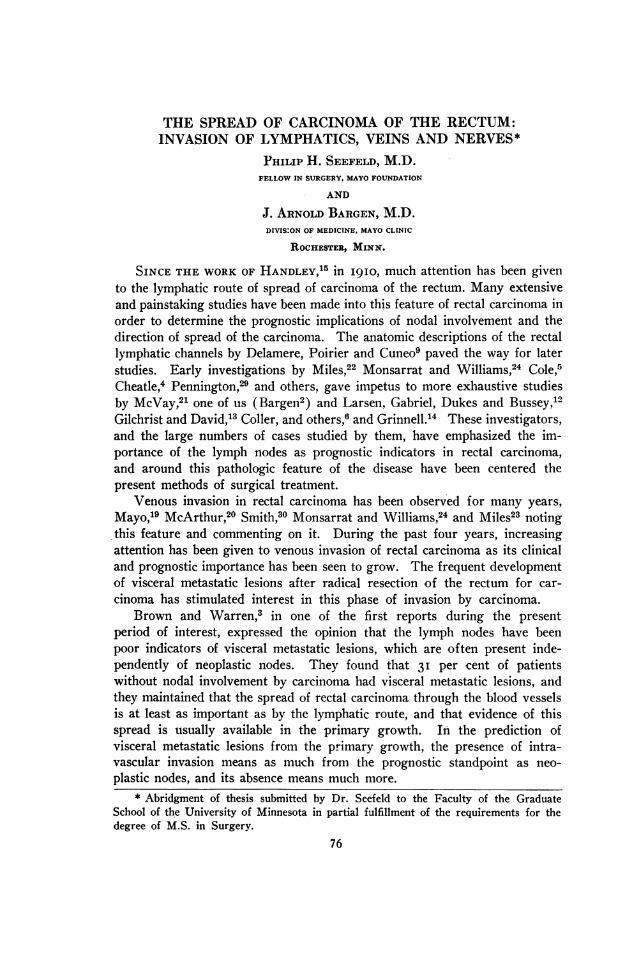
To study the involvement of veins, blocks of tissue from all points ofthe lesion and rectal wall at which microscopic evidence of venous invasioncould conceivably be present were removed. The lesion was divided intofour quadrants and a block of tissue was taken from each quadrant. Theblock included the full-thickness of the lesion and the entire thickness ofthe rectal wall as well as a certain amount of perirectal tissue (Figs. i and 2).In addition, transverse cuts were made through the perirectal fatty tissue,exposing the larger vessels leading from the vicinity of the lesion (Fig. 3),and blocks were removed for microscopic study from regions which appearedsuspicious, usually within a distance of four inches (IO cm.) above andbelow the growth.
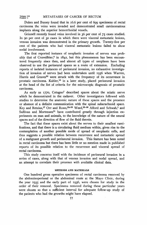
FIG. 4.-Ca) Photoni,crograph of small vein in the perirectal tissue;* the lumen is lined bymalignant cells (x I85); (b,) adenocarcinoma, Grade 4. (Broders' method), in the lumen of a smallvein in the perirectal tissue (x 140).
Preliminary microscopic examination of sections cut from the blocks,fast freezing technic being used, was facilitated by the use of an acidpolychrome methylene blue stain, which is especially adaptable for tissuesthat have been long fixed in formalin. Of those tissues in which definiteor presumptive evidence of venous or perineural invasion was found,permanent sections stained with hematoxylin and eosin were made andfurther study was carried out. We wish to thank Dr. MacCarty, in whoselaboratory the study was made, and Dr. Dockerty, who reviewed thesections.
79
SEEFELD AND BARGEN Annals of SurgeryJ u l y, 1943
After examination of all sections was completed, results were tabulatedand correlation with clinical data and follow-up records was attempted.
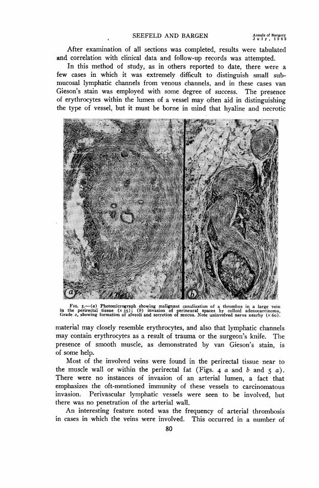
In this method of study, as in others reported to date, there were afew cases in which it was extremely difficult to distinguish small sub-niucosal lymphatic channels from venous channels, and in these cases vanGieson's stain was employed with some degree of success. The presenceof erythrocytes within the lumen of a vessel may often aid in distinguishingthe type of vessel, but it must be borne in mind that hyaline and necrotic
~~~~~~~~~~~~~~~~~~~~~~~~~~~~~~~~~~~ ...W~~~.~30, ~ A U~ ~ i~
l>'~~~~~~~~~~~~~~~~.FIG. 5.-(a) Photomicrograph showing malignant canalization of a thrombus in a large vein
in the perirectal tissue (x 35); (b) invasion of perineural spaces by colloid adenocarcinoma,Grade 2, showing formation of alveoli and secretion of mucus. Note uninvolved nerve nearby (x 6o).
material may closely resemble erythrocytes, and also that lymphatic channelsmay contain erythrocytes as a result of trauma or the surgeon's knife. Thepresence of smooth muscle, as demonstrated by van Gieson's stain, isof some help.
Most of the involved veins were found in the perirectal tissue near tothe muscle wall or within the perirectal fat (Figs. 4 a and b and 5 a).There were no instances of invasion of an arterial lumen, a fact thatemphasizes the oft-mentioned immunity of these vessels to carcinomatousinvasion. Perivascular lymphatic vessels were seen to be involved, butthere was no penetration of the arterial wall.
An interesting feature noted was the frequency of arterial thrombosisin cases in which the veins were involved. This occurred in a number of
80
Volume 118Number 1 METASTASES OF CANCER OF RECTUM
instances, and when seen on preliminary examination was taken as an indica-tor of venous involvement.
In arriving at the final microscopic diagnosis, we considered only casesin which the involved vessels were definitely venous and eliminated allcases in which the observations were indefinite.
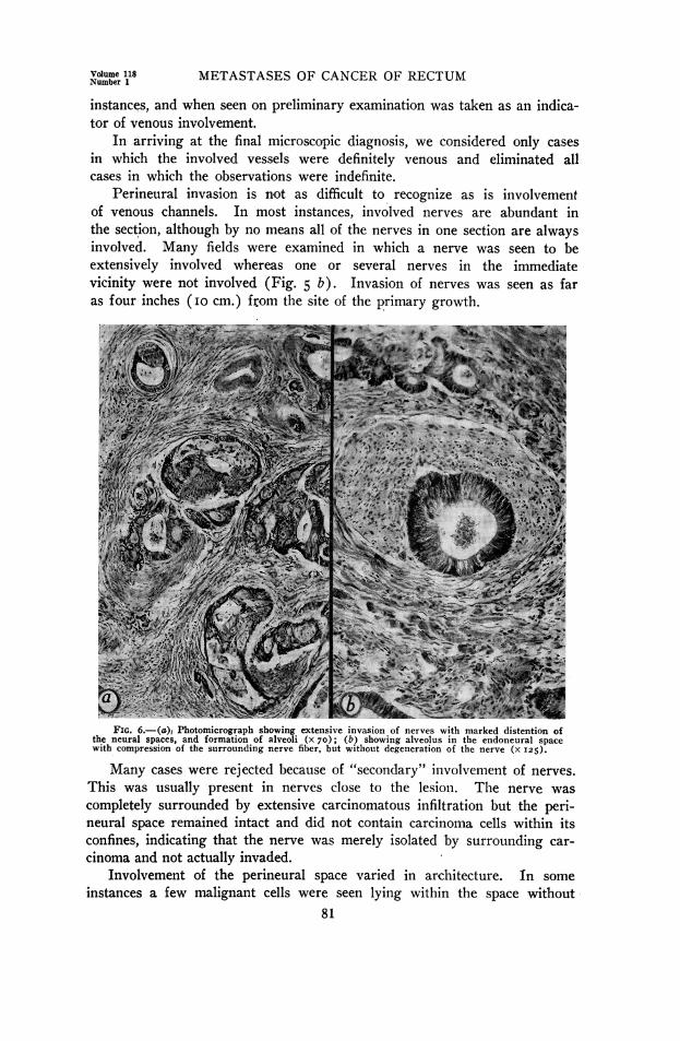
Perineural invasion is not as difficult to recognize as is involvementof venous channels. In most instances, involved nerves are abundant inthe section, although by no means all of the nerves in one section are alwaysinvolved. Many fields were examined in which a nerve was seen to beextensively involved whereas one or several nerves in the immediatevicinity were not involved (Fig. 5 b). Invasion of nerves was seen as faras four inches (IO cm.) from the site of the primary growth.g ;t 6 ..!. s~:;,s
FIG. 6.-(a), Photomicrograph showing extensive invasion of nerves with marked distention ofthe neural spaces, and formation of alveoli (x 70); (b) showing alveolus in the endoneural spacewith compression of the surrounding nerve, fiber, but witliout degeneration of the nerve (x 125).
Many cases were rejected because of "secondary" involvement of nerves.This was usually present in nerves close to the lesion. The nerve wascompletely surrounded by extensive carcinomatous infiltration but the peri-neural space remained intact and did not contain carcinoma cells within itsconfines, indicating that the nerve was merely isolated by surrounding car-cinoma and not actually invaded.
Involvement of the perineural space varied in architecture. In someinstances a few malignant cells were seen lying within the space without
81
SEEFELD AND BARGEN Annals of SurgeryJ uIy , 194 3
any apparent conformation, while in other cases the cells were arrangedin alveoli and even distended the space with their mucous secretion(Fig. 6 a).
There was little or no degeneration of nerve fibers to be seen in thenerves involved, apparently because of the distensibility of the walls of theperineural spaces, which were seen to be extremely distorted in places(Fig. 6 b).
In no cases were the plexuses of Meissner and Auerbach seen to beinvolved, although nerve fibers both proximal and distal to these structuresmight be invaded.
RESULTS
Lymphatic Involvement.-Involvement of the lymph nodes was found tobe.present in 47 per cent of the ioo cases. In previous investigations based,in total, on more than 500 specimens, the proportion of cases in which lymphnodes were involved varied from 36 to 68 per cent (Table I). We found,
TABLE I
NODAL INVOLVEMENT FOUND IN PREVIOUS STUDIES
Total No. of PercentageAuthor Cases Involved
McVay . . 100 47WoodandWilkie37 . .100 51Gabriel, Dukes and Bussey ..100 62Gilchrist and David ..25 68Coller, Kay and Maclntyre. 53 64Grinnell (1916-1932) ............. 107 36Grinnell (1938-1941) ..75 55
as others before us, that the higher the grade of malignancy (Broders' method)the higher the incidence of nodal involvement (Table II).
TABLE I I
INCIDENCE OF NODAL, PERINEURAL AND VENOUS INVASION ACCORDING TO GRADE OF MALIGNANCY (BRODERS)
Nodal Perineural VenousInvolvement Involvement Involvement
TotalGrade Cases Cases Percentage Cases Percentage Cases PercentageI...........:14 3 21.4 2 14.3 1 7.12.. 54 20 37.0 16 29.6 7 13.03.. 24 18 75.0 9 37.5 7 29.24.. 8 6 75.0 3 37.5 5 62.5
Totals. 100 47 30 20
Dukes'0 expressed the opinion that metastatic growths in lymph nodes,ire more frequently found among women than among men, a feature whichis not borne out in this study, 46.I per cent of women and 47.5 per cent ofmen having nodal involvement.
Dukes has stated that the highest incidence of nodal involvement occursamong young patients, while the tendency is less frequent among the olderpatients. This fact was also noted in this series (Table III).
Most investigators, notably Mayo and Schlicke,18 have found the highest82
Volume 118 METASTASES OF CANCER OF RECTUMNumber 1
incidence of nodal invasion in the upper part of the rectum and in the recto-sigmoid. In this series a slightly higher proportion of nodal involvementwas found in the middle third of the rectum than in the other segments, 50per cent in the middle third, and 48.8 and 40.2 per cent in the upper andlower thirds, respectively.
Of the series of patients who had nodal metastatic lesions, ten patientssurvived for three years while only two survived for five years.
Perineural Invasion.-Involvement of the perineural and endoneuralspaces was demonstrated in 30 of the ioo cases, the average age of thepatients who had perineural involvement being 51 years, almost exactly theaverage 'age in the entire series. Eighteen of the 30 patients were!men(6o per cent), and I2 were women (40 per cent).
The average preoperative duration of symptoms among those patients whohad involvement of nerves was 9.4 months, as compared with 9.7 months inthe entire series. Pain was a prominent symptom, volunteered by the patient,in 24 cases; it was not mentioned in the history of three cases; and was stated
TABLE III
INCIDENCE OF NODAL, PERINEURAL AND VENOUS INVOLVEMENT ACCORD:NG TO AGE
Nodal Perineural VenousInvolvement Involvement Involvement
TotalAge, Years Series Cases Percentage Cases Percentage Cases Percentage20-39. 11 7 63.6 3 27.3 4 36.440-59.68 34 50.0 21 30.9 13 19.160-79. : ... 21 6 28.6 6 28.5 3 14.3
Totals. 100 47 30 20
to be absent in three cases. Thus, in 89 per cent of the cases in whichinformation on this point was available, pain was a prominent symptom.In the evaluation of pain in this study, only cases in which the historyindicated that the pain might be directly neurogenic and not just dis-comfort vaguely noted by most patients with rectal carcinoma, were recorded.Terms such as "aching," "boring," "gnawing," "constant" or "steady" wereconsidered to be indicative of pain due to involvement of nerves, while tenes-mus, cramps, a feeling of fullness and the desire to defecate were not so con-sidered. Of the 70 cases in which the nerves were not involved pain was foundto be a primary symptom in only 25. Pain was not mentioned in the history of20 cases, and in 25 cases, when the patient was questioned, he stated thatit was definitely absent or was present in one form or another but notlocalized to the rectum. The incidence, then, of pain for cases without peri-neural involvement in which information was available was 50 per cent.
Of the 30 cases in which there was involvement of the nerves, the lymphnodes were also involved in 20 (67 per cent).
When involvement of nerves was considered in the light of grading,both by cellular differentiation (Broders) and by mural penetration (Dukes),it was seen that the higher the degree of malignancy, the more frequentlywas perineural involvement present (Tables II and IV).
83

SEEFELD AND BARGEN Annals of Surgery
TABLE IV
INCIDENCE OF PERINEURAL AND VENOUS INVASION ACCORDING TO DEGREE OF MURAL PENETRATION (DUKES)Perineural Involvement Venous Involvement
No. of Percentage of No. of Percentage ofClass Cases Involved Cases Cases Involved CasesA........................................... 0 0
B ............. 10 33.3 3 15C ....................................... 20 66. 7 17 85
Totals ....................................... 30 100.0 20 100
There is no apparent relation between the size of the carcinoma andthe presence of perineural invasion.
In considering the presence of perineural invasion in relation to thelocation of the growth, it seemed feasible to divide the rectum into lower,middle and upper thirds, including lesions of the rectosigmoid in the lattergroup. It was found that lesions exhibiting involvement of the nervesoccurred equally in the three locations (ten cases in each third), so that thepresence of perineural invasion is apparently not related to the location ofthe lesion. This result contrasts with involvement of lymph nodes, which,in this series, was more frequent in the middle and upper thirds of the rectumthan in the lower third, and vascular invasion, which seems to increase withthe height of the lesion in the rectum.
No relation could be observed between the location of the lesions(anterior, posterior or lateral wall) and the presence of involvement ofnerves. Sixteen (50.3 per cent) of the lesions in which nerves were in-volved were annular in type and the rest were located at different points onthe rectal wall. In the entire series, 30 lesions (43 per cent) were annular.
TABLE V
RELATION OF PERINEURAL INVASION TO LOCAL RECURRENCESNerves Involved Nerves Uninvolved
Cases Percentage Cases PercentageDefinite local recurrence ........................... 13 81.2 14 30.4Definitely no local recurrence ....................... 3 18.8 32 69.6
Totals ................................. 16 100.0 46 100.0Questionable and inadequate follow-up, and post-
operative complications .......................... 14 24
Grand Totals. 30 70
The striking feature of perineural involvement of rectal carcinoma is itsrelation to local recurrences (Table V). Only recurrences in the scar orsite of anastomosis were considered. Metastasis to nearby viscera, such asthe bladder, vagina, prostate, perineal nodes, etc., was eliminated becauseof the probability of its being due to lymphatic or venous spread rather thanto invasion of nerves. Definite local recurrences were found in 8I.2 percent of traceable cases in which there was perineural invasion, whereas, inthose without perineural invasion, recurrences occurred in only 30.4 percent. Cases in which there were definitely no local recurrences comprisedonly i8.8 per cent of those in which nerves were involved, whereas, in cases
84
Volume 118Number 1 METASTASES OF CANCER OF RECTIJM
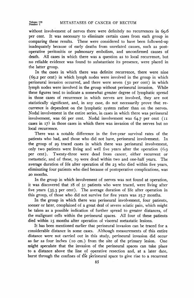
without involvement of nerves there were definitely no recurrences in 69.6per cent. It was necessary to eliminate certain cases from each group incomparing these results. These were considered to have been followed-upinadequately because of early deaths from unrelated causes, such as post-operative peritonitis or pulmonary embolism, and unconfirmed causes ofdeath. All cases in which there was a question as to local recurrence, butno reliable evidence was found to substantiate its presence, were placed inthe latter group.
In the cases in which there was definite recurrence, there were nine(69.2 per cent) in which lymph nodes were involved in the group in whichperineural invasion occurred, and there were seven (50 per cent) in whichlymph nodes were involved in the group without perineural invasion. Whilethese figures tend to indicate a somewhat greater degree of lymphatic spreadin those cases of recurrence in which nerves are involved, they are notstatistically significant, and, in any case, do not necessarily prove that re-currence is dependent on the lymphatic system rather than on the nerves.Nodal involvement in the entire series, in cases in which there was perineuralinvolvement, was 66 per cent. Nodal involvement was 64.7 per cenlt (iicases in I7) in those cases in which there was invasion of the nerves but nolocal recurrence.
There was a notable difference in the five-year survival rates of thepatients who had, and those who did not have, perineural involvement. Inthe group of 29 traced cases in which there was perineural involvement,only two patients were living and well five years after the operation (6.9per cent). Twenty-three were dead from cancer, either recurrent ormetastatic, and of these, I9 were dead within two and one-half years. Theaverage duration of life after operation of the 23 who died within five years,eliminating four patients who died because of postoperative complications, was20 months.
In the group in which involvement of nerves was not found at operation,it was discovered that i8 of 51 patients who were traced, were living afterfive years (35.3 per cent). The average duration of life after operation inthis group, of those who did not survive for five years was 25.7 months.
In the group in which there was perineural involvement, four patients,sooner or later, complained of a great deal of severe sciatic pain, which mightbe taken as a possible indication of further spread to greater distances, ofthe malignant cells within the perineural spaces. All four of these patientsdied within I5 months after operation of visceral metastatic lesions.
It has been mentioned earlier that perineural invasion can be traced for aconsiderable distance in some cases. Although measurements of this entiredistance were not carried out in this study, perineural invasion did occuras far as four inches (IO cm.) from the site of the primary lesion. Onemight speculate that the invasion of the perineural spaces can take placeto a distance above the line of operative resection and, at a later date,burst through the confines of tie perinfeural space to give rise to a recurrent
85
SEEFELD AND BARGEN Annals of SurgeryJ u l y , 1 9 4 3
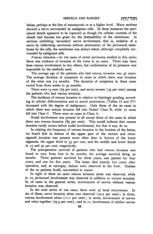
lesion, perhaps at the line of anastamosis or at a higher level. Many sectionsshowed a nerve surrounded by malignant cells. In these instances the peri-neural sheath appeared to be ruptured as though the cellular contents of thesheath had become too great for the distensibility of the membrane. Insections exhibiting 'secondary' nerve involvement, that is, isolation of anerve by infiltrating carcinoma without penetration of the perineural mem-brane by the cells, the membrane was always intact, although completely'sur-rounded by malignant cells.
Venous Invasion.-In ioo cases of rectal carcinoma studied in this seriesthere was evidence of invasion of the veins in 20 cases. There may havebeen venous involvement in two others, but confirmation of its presence wasimpossible by the methods used.
The average age of the patients who had venous invasion was 47 years.The average duration of symptoms in cases in which there was invasionof the veins was 7.9 months. The duration of symptoms in these casesvaried from three weeks to 30 months.
There were I3 men (65 per cent), and seven women (35 per cent) amongthe patients who had venous invasion.
The incidence of venous invasion in relation to histologic grading, accord-ing to cellular differentiation and to mural penetration (Tables II and IV)increased with the degree of malignancy. Only three of the 20 cases inwhich there was venous invasion fell into Dukes' Class B, while I7 casesfell into Class C. There were no cases in Class A.
Nodal involvement was present in all except three of the cases in whichthere was venous invasion (85 per cent). This would indicate that venousinvasion rarely occurs before nodal involvement, but that it may do so.
In relating the frequency of venous invasion to the location of the lesion,we found that in lesions of the upper part of the rectum and recto-sigmoid invasion was present more often than in lesions of the lowersegments, the upper third in 55 per cent, and the middle and lower thirdsin I5 and 30 per cent, respectively.
The postoperative survival of patients who had venous invasion wasfound to vary from four to 6o months, the average survival being 22months. Three patients survived for three years, one patient for fouryears, and one for five years. The latter died exactly five years afteroperation, and, at necropsy, lesions were observed in the liver. Sixteenof the 20 patients finally succumbed to cancer.
In eight of these 20 cases venous invasion alone was observed, whilein I2, perineural involvement was observed in addition to venous invasion.In i6 cases in the general series, involvement of nerves without venousinvasion was observed.
In the total series of ioo cases, there were 27 local recurrences. Insix of these, nerve invasion alone was observed (22.2 per cent); in three,venous involvement alone (iI.I per cent); in seven, involvement of nervesand veins together (25.9 per cent); and in ii, involvement of neither nerves
86
Volume 118 METASTASES OF CANCER OF RECTUMNumber 1
nor veins (40.7 per cent). In only four of the cases without involvementof nerves or veins was involvement of nodes observed. Therefore, in sevencases there was recurrence in spite of the fact that involvement of any ofthe three pathways of spread was not observed at operation.
Recurrence of rectal carcinoma, in the light of these findings, is probablynot concerned with only one of the three pathways of spread, but resultsfron different ones in different cases, and may involve any of them. However,it may be said that local recurrences are more frequent in rectal carcinomawhen nerves and veins are involved than when they are uninvolved, and thatthere are twice as many recurrences when nerves alone are involved aswhen veins alone are involved.
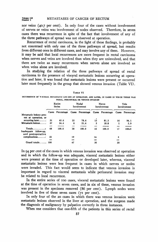
In studying the relation of the three pathways of spread of rectalcarcinoma to the presence of visceral metastatic lesions occurring at opera-tion and later, it was found that metastatic lesions were present or occurredlater most frequently in the group that showed venous invasion (Table VI).
TABLE VI
OCCURRENCE OF V;SCERAL METASTATIC LES ONS AT OPERATION, AND LATER, IN CASES IN WHICH THERE WASNODAL, PERINEURAL OR VENOUS INVASION
Entire Nodal Nerve VeinSeries Involvement Involvement Involvement
Cases Percentage Cases Percentage Cases Percentage Cases PercentageMetastatic lesions pres-
ent at operation, oroccurring later....... 31 45.6 22 78.6 13 81.2 16 94.1
Metastaticlesions...... 37 54.4 6 21.4 3 18.8 1 5.9
Totals ....... : 68 100.0 28 100.0 16 100.0 17 100:0Inadequate follow-up,and postoperativecomplications....... 32 19 14 3
Grand totals.... 100 47 30 20
In 94 per cent of the cases in which venous invasion was observed at operationand in which the follow-up was adequate, visceral metastatic lesions eitherwere present at the time of operation or developed later, whereas, visceralmetastatic lesions were less frequent in cases in which nerves or nodeswere invaded. This fact would seem to indicate that venous invasion isimportant in regard to visceral metastasis while perineural invasion maybe related to local recurrence.
In the entire series of ioo cases, visceral metastatic lesions were foundat the time of operation in seven cases, and in six of these, venous invasionwas present in the specimen removed (86 per cent). Lymph nodes wereinvolved in five of these seven cases (7I per cent).
In only four of the 20 cases in which there was venous invasion weremetastatic lesions observed in the liver at operation, and the surgeon madethe diagnosis of malignancy by palpation correctly in three instances.
When one considers that one-fifth of the patients in this series of rectal87
SEEFELD AND BARGEN Annals of SurgeryJ u l y, 1943
carcinoma had veins invaded by carcinoma, and that practically all (94per cent) of these already presented, or later acquired, visceral metastaticlesions, the prognostic importance of the presence of venous invasion in theoperative specimen is seen. The fact that four-fifths of the patients in theseries of ioo cases failed to show invasion of the veins at operation andthat visceral metastatic lesions occurred in only I8.7 per cent (I5) ofthese patients emphasizes the prognostic significance of the absence ofvenous invasion, as has been mentioned by Brown and Warren.3
CONCLUSIONS
I. In IOO cases of rectal carcinoma, lymph nodes were involved in 47per cent, nerves in 30 per cent, and veins in 20 per cent.
2. Invasion of lymph nodes, nerves and veins increases with the degreeof malignancy of the carcinoma.
3. Invasion of lymph nodes occurred equally in the two sexes, whilevenous invasion and perineural invasion were more frequent among menthan among women.
4. Venous and nodal invasion occurred more often among young thanamong old patients, while invasion of nerves was not related to age.
5. Invasion of nerves was not related to the location of the lesion,while venous invasion was most frequent in lesions of the upper part ofthe rectum, and nodal invasion was slightly more frequent in lesions of themiddle segment of the rectum than in lesions of other segments.
6. Venous invasion occurred more frequently in cases in which therewas involvement of nodes than in other cases, but it occurred in cases with-out involvement of nodes as well.
7. Nodal invasion was somewhat more frequent in cases in which therewas involvement of nerves than in those in which the nerves were notinvolved.
8. Pain was a prominent symptom in 89 per cent of cases in which therewas invasion of nerves.
9. Local recurrence was more than two and one-half times as frequentin cases in which invasion of nerves was observed as in cases in which itwas not observed.
IO. Visceral metastatic lesions at operation, or later, occurred in 94per cent of patients who had venous invasion in the primary growth, andwere five times as frequent as in patients without venous invasion in theprimary growth.
II. Venous invasion in the primary growth does not always mean thathepatic metastatic lesions are present.
I2. Eighty-six per cent of patients among whom visceral metastaticlesions were present at operation exhibited venous invasion in the primarygrowth, while 7I per cent exhibited nodal invasion.
I3. Eighty per cent of patients who had venous invasion in the primarygrowth died from carcinoma, either recurrent or metastatic.
88
Volume 118 METASTASES OF CANCER OF RECTUMNumber 1
REFERENCES1 Alford, L. B., and Schwab, S. I.: Some Experiments Bearing on the Flow of Lymph
in Nerves. J. Nerv. & Ment. Dis., 47, I i8-I25, I9I8.2 Bargen, J. A., and Larson, L. M.: The Mode of Spread of Carcinoma of the Rectum.
Minnesota Med., I6, 478-480, July, 1933.3 Brown, C. E., and Warren, Shields: Visceral Metastasis from Rectal Carcinoma.
Surg., Gynec., & Obst., 66, 6i i-621, March, I938.4 Cheatle, G. L.: The Spread of Cancer in the Lower Part of the Large Intestine.
Brit. M. J., I, 303, I9I4.5 Cole, P. P.: The Intramural Spread of Rectal Carcinoma. Brit. M. J., I, 431-
433, I9I3.6 Coller, F. A., Kay, E. B., and MacIntyre, R. S.: Regional Lymphatic Metastasis
of Carcinoma of the Rectum. Surgery, 8, 294-31I, August, I940.7 Cotugno, Dominico: Quoted by Weed, L. H.338 Cruveilhier, Jean: Maladies des nerfs. In: Anatomie pathologique du corps humain.
Paris, J. B. Bailliere, I835-I842, Vol. 2, Pt. 35, P. 3.9 Delamere, G., Poirier, P., and Cuneo, B.: The Lymphatics. Chicago, W. T. Keener
and Co., 1904, PP. 301.10 Dukes, C. E.: Cancer of the Rectum: Analysis of IOOO Cases. J. Path. & Bact., 50,
527-539, May, I940.11 Dukes, C. E., and Bussey, H. J. R.: Venous Spread in Cancer of the Rectum. Proc.
Roy. Soc. Med., 34, 57I-574, July, 194I.12 Gabriel, W. B., Dukes, Cuthbert, and Bussey, H. J. R.: Lymphatic Spread in Cancer
of the Rectum. Brit. J. S., 23, 395-4I3, October, I935.13 Gilchrist, R. K., and David, V. C.: Lymphatic Spread of Carcinoma of the Rectum.
ANNALS OF SURGERY, io8, 62i-642, October, I938.14 Grinnell, R. S.: The Lymphatic and Venous Spread of Carcinoma of the Rectum.
ANNALS OF SURGERY, ii6, 200-215, August, I942.15 Handley, W. S.: The Surgery of the Lymphatic System. Brit. M. J., i, 853-864,
April, I9IO.16 Kahler, J. E.: Carcinoma of the Prostate Gland: A Pathologic Study. J. Urol.,
41, 557-574, April, I939.17 Key, A., and Retzius, G.: Studien in der Anatomie des Nervensystemes. Arch. f.
mikr. Anat., 9, 308-386, I873.18 Mayo, C. W., and Schlicke, C. P.: Carcinoma of the Colon and Rectum. Surg.,
Gynec., & Obst., 74, 83-91, January, I942.19 Mayo, W. J.: Removal of the Rectum for Cancer. ANNALS OF SURGERY, 6o, 854-862,
I9IO.20 McArthur, L. L.: Carcinoma of the Rectum. Surg., Gynec., & Obst., 2I, 495-
497, 1915.21 McVay, J. R.: Involvement of Lymph-Nodes in Carcinoma of the Rectum. ANNALS
OF SURGERY, 76, 755-767, 1922.22 Miles, W. E.: The Pathology of the Spread of Cancer of the Rectum: Its Bearing
upon the Surgery of the Cancerous Rectum. Surg., Gynec., & Obst., 52, 350-359,February, I93I.
23 Miles, W. E.: Cancer of the Rectum. London, Harrison & Sons, Ltd., 1926, PP. 5-6.24 Monsarrat, K. W., and Williams, I. J.: Intramural Extension in Rectal Cancer. Brit.
J. Surg., I, 173-I82, 1913-I914.25 Orr, D., and Rows, R. G.: A Clinical and Experimental Investigation into the
Lymphogenous Origin of Toxic Infection of the Central Nervous System. Rev.Neurol. & Psychiat., 5, 345-360, I907.
96 Orr, D., and Rows, R. G.: Histological Evidence that Toxins Reach the Spinal89
SEEFELD AND BARGEN Aals of SurgeryJuIy , 1943
Cord wia the Spinal Roots: With Special Reference to Plasma Cells. Rev. Neurol.& Psychiat., 8, 72I-748, I9I0.
27 Orr, D., and Rows, R. G.: Lymphogenous Infection of the Central Nervous System.Brain, 36, 27I-338, May, 19I3-1914.
280rr, D., Rows, R. G., and Stephenson: The Spread of Infection by the AscendingLymph Stream of Nerves from Peripheral Inflammatory Foci to the Central NervousSystem. Rev. Neurol. & Psychiat., II, 349-366, I913.
29 Pennington, J. R.: The End-results of Operations for Cancer of the Rectum: With- Suggestions for Improving Them. J. A. M. A., 71, I892-I896, December 7, i9i8.
30 Smith, J. W.: The Operative Treatment of Carcinoma Recti. Brit. M. J., I, I036-1041, 19I I.
81 Sullivan, W. E., and Mortensen, 0. A.: Visualization of the Movement of a Bromin-ized Oil along Peripheral Nerves. Anat. Rec., 59, 493-50I, July, I934.
32 Warren, Shields, Harris, P. N., and Graves, R. C.: Osseous Metastasis of Carcinomaof the Prostate: With Special Reference to the Perineural Lymphatics. Arch. Path.,22, I39-i60, August, 1936.
88 Weed, L. H.: Studies on Cerebrospinal Fluid. J. M. Research, 31, 2I-49, I914.84 Weed, L. H.: Studies on Cerebrospinal Fluid. J. M. Research, 31, 5I-9I, I914.85 Weed, L. H.: The Development of Cerebrospinal Spaces in Pig and in Man. In Con-
tributions to Embryology, Washington, Carnegie Institution of Washington, I9I7,vol. 5, pp. 7-107.
86 Weed, L. H.: The Cerebrospinal Fluid. Physiol. Rev., 2, I7I-203, April, I922.87 Wood, W. Q., and Wilkie, D. P. D.: Carcinoma of the Rectum: An Anatomico-
Pathological Study. Edinburgh M. J., 40, 321-343, 1933.
90