University ofOslo, Norway - NCBI
22
J. Phy8iol. (1966), 183, pp. 15-36 15 With 10 text-figure8 Printed in Great Britain THE REFLEX RESPONSE TO SINUSOIDAL STRETCHING OF SOLEUS IN THE DECEREBRATE CAT BY J. K. S. JANSEN AND P. M. H. RACK* From the Laboratory of Neurophysiology, Department of Anatomy, University of Oslo, Norway (Received 6 May 1965) SUMMARY 1. Soleus muscle in the decerebrate cat was stretched sinusoidally through various distances, at various frequencies while tension and e.m.g. activity were recorded. 2. Two patterns of stretch reflex activity were seen. In one, slow stretching led to a large increase in tension, whereas in the other tension increased little during slow stretching, but rose steeply during a more rapid stretch. Intermediate states were also seen. 3. Both these reflex patterns were abolished when the fusimotor fibres were blocked with Xylocaine. 4. At low frequencies of stretching the e.m.g. activity was greatest at peak muscle length; at higher frequencies the greatest activity was found during lengthening. This angular advance of e.m.g. activity on length was greatest at 3-3-5 c/s. At the higher frequencies the e.m.g. activity was less in advance of muscle length. 5. Angular advance of e.m.g. activity was greatest in the preparations that were also the most sensitive to slow stretching. 6. Muscle contraction follows an electrical stimulus with a small delay. The delay between the end of stimulation and the end of muscle relaxation is longer, and may exceed 200 msec in cat soleus. 7. The timing of the reflex tension during sinusoidal stretching de- pended on how far the angular advance of the e.m.g. combined with the damping properties of muscle offset the time delays in the reflex pathway. 8. Changes in muscle tension generally preceded changes in muscle length. When, however, stretch amplitudes of 1 mm (peak to peak) were used, a phase delay of tension was sometimes seen. This only occurred in preparations that also showed little sensitivity to slow stretching. 9. Phase delay of tension was usually found with frequencies of * Present address: Department of Physiology, University of Birmingham.
-
Upload
khangminh22 -
Category
Documents
-
view
0 -
download
0
Transcript of University ofOslo, Norway - NCBI

J. Phy8iol. (1966), 183, pp. 15-36 15With 10 text-figure8Printed in Great Britain
THE REFLEX RESPONSE TO SINUSOIDAL STRETCHINGOF SOLEUS IN THE DECEREBRATE CAT
BY J. K. S. JANSEN AND P. M. H. RACK*
From the Laboratory of Neurophysiology, Department of Anatomy,University of Oslo, Norway
(Received 6 May 1965)
SUMMARY
1. Soleus muscle in the decerebrate cat was stretched sinusoidallythrough various distances, at various frequencies while tension and e.m.g.activity were recorded.
2. Two patterns of stretch reflex activity were seen. In one, slowstretching led to a large increase in tension, whereas in the other tensionincreased little during slow stretching, but rose steeply during a more rapidstretch. Intermediate states were also seen.
3. Both these reflex patterns were abolished when the fusimotor fibreswere blocked with Xylocaine.
4. At low frequencies of stretching the e.m.g. activity was greatest atpeak muscle length; at higher frequencies the greatest activity was foundduring lengthening. This angular advance of e.m.g. activity on length wasgreatest at 3-3-5 c/s. At the higher frequencies the e.m.g. activity was lessin advance of muscle length.
5. Angular advance of e.m.g. activity was greatest in the preparationsthat were also the most sensitive to slow stretching.
6. Muscle contraction follows an electrical stimulus with a smalldelay. The delay between the end of stimulation and the end of musclerelaxation is longer, and may exceed 200 msec in cat soleus.
7. The timing of the reflex tension during sinusoidal stretching de-pended on how far the angular advance of the e.m.g. combined withthe damping properties of muscle offset the time delays in the reflexpathway.
8. Changes in muscle tension generally preceded changes in musclelength. When, however, stretch amplitudes of 1 mm (peak to peak) wereused, a phase delay of tension was sometimes seen. This only occurred inpreparations that also showed little sensitivity to slow stretching.
9. Phase delay of tension was usually found with frequencies of* Present address: Department of Physiology, University of Birmingham.
J. K. S. JANSEN AND P. M. H. RACK
approximately 6-8 c/s, and this corresponds to the clonus frequency forsoleus.
10. The mechanisms of the two types of reflex behaviour, and thedamping properties of the system are discussed.
INTRODUCTION
Stretch reflexes in the decerebrate cat have been extensively studiedunder static conditions (Liddell & Sherrington, 1924; Granit, 1958), andduring slow stretching (Matthews, 1959a, b). The reflex response to rapidstretching has received less attention.
It is generally believed (cf. Granit, 1955; Hunt & Perl, 1960) that themuscle spindles and their primary afferent fibres are the principal sensorycomponents of this reflex, and Merton (1951) has suggested that sincethe fusimotor nerve fibres alter the response of the muscle spindles toextension, they have an indirect effect on muscle contraction whichamounts to a servo control of muscle length.
Afferent impulses from the muscle spindles do not have an instantaneouseffect on muscle tension, and the stretch reflex can be regarded as a feed-back control system with time delays. A simple position control systemwith time delays is unstable, and oscillation may occur, because correc-tions are always applied at an interval after displacement. This situationis well known to control engineers, and the instability is often overcomeby constructing a system in which the resistance to deflexion is a functionof the velocity of deflexion as well as its extent.
In skeletal muscle with an intact stretch reflex there exist mechanismswhereby the velocity of lengthening or shortening may modify the tension.Active muscle offers a resistance to changes in length that varies withvelocity (Hill, 1938; Katz, 1939). The afferent discharge from the musclespindles is also known to depend on their rate of extension as well as theiractual length (Matthews, 1933); furthermore, there is evidence that theresponse of the muscle spindles to length and to velocity of lengthening canto some extent be altered independently by fusimotor activity. Thebehaviour of the muscle spindles has recently been reviewed by Matthews(1964).In examining the performance of a control system, it is desirable to
test its response to dynamic as well as static deflexions, and controlledsinusoidal deflexions can conveniently be used for this purpose. It is thenpossible to measure the work necessary to maintain the movement, andthis gives an indication of the stability of the system. When externalwork is required to maintain the sinusoidal movement, there is a phaseadvance of force on displacement, and the system has positive dampingproperties for movement at that frequency and amplitude. When,
16
REFLEX RESPONSE TO SINUSOIDAL STRETCHING 17
however, the oscillation is maintained without external work, the systemis undamped.
This paper describes the behaviour of the decerebrate cat's soleusmuscle during sinusoidal stretching. Similar experiments have beenreported by: Lippold, Redfearn & Vuco (1958), Partridge & Glaser (1960),and by Roberts (1963). The use of higher frequencies, and smaller stretchamplitudes, with concurrent electromyogram (e.m.g.) recording has led tosome new findings.
METHODSPreparation. Twenty cats were used, but the principal results were obtained from thirteen
of them.The animals were decerebrated by transection of the mesencephalon under ether anaes-
thesia after common carotid ligation. The tendon of soleus was dissected, and the footdisarticulated. The nerve to soleus was preserved, but nerves to the other muscles of thelimb were cut. In some experiments the innervation of tibialis anterior was left intact. Inmost experiments the lumbar spinal nerve roots and cord were exposed by laminectomy.The method of application of stretch has already been described (Rack, 1966). Essen-
tially the experiments consisted of stretching the muscle sinusoidally at various frequenciesand amplitudes, while recording the tension and the e.m.g.At the end of each experiment the muscle nerve was cut, and the properties of the passive
muscle were recorded at the same muscle lengths, stretch amplitudes, and stretch fre-quencies as had been used during the earlier records.Recording was not started less than 2 hr after decerebration. The rectal temperature
was kept between 36 and 390 C during the recording periods.Electromyography. Potential changes were recorded between a silver plate placed on the
belly of the soleus muscle, and a hook which was inserted into the soft tissues near theproximal end of the muscle. A Tektronix 122 preamplifier was used, and the records weredisplayed on a Tektronix 565 oscilloscope along with the tension record.
Integration of the e.m.g. In addition to the direct display of the amplified e.m.g., the out-put of the preamplifier was fed into an integrating circuit. The circuit used was based onone described by Starr & Livingston (1963), but this was modified by shortening the timeconstant of the amplifier to 0-6 msec, increasing the time constants of the integrating CRcircuit to a possible maximum of 6j min, and inserting filters to exclude amplifier noise.Frequencies above 5000 c/s, and amplitudes less than 20 mV were excluded. The integratorhad a frequency range from 250 to 5000 c/s and integration was acceptably linear for therange of amplitudes required (up to 500 mV). The time constant of decay of the integratedpotential was never less than 100 times the duration of the cycle. The integrator was re-setto zero during alternate stretch cycles by shorting the integrating condensers through arelay operated from the driving motor.
Display of re8ults. The methods of recording and of measuring the records have beendescribed in a preceding paper (Rack, 1966). The tension was usually displayed as thevertical deflexion of an oscilloscope beam which was deflected horizontally by the lengthsignal (Fig. 1). A repeating length-tension figure was thus obtained, and it was always easyto see at once whether or not the behaviour of the preparation was stable. When this formof display was used, the e.m.g. was also displayed on a beam deflected horizontally by thelength signal, and in order to separate the traces during lengthening and shortening abiasing potential was introduced during shortening (Fig. 1). This was done through amicro-switch operated by a cam on the stretcher drive shaft.
Physiol. 1832
J. K. S. JANSEN AND P. M. H. RACK
RESULTS
The decerebrate cat is a variable and unpredictable preparation.Some animals remain in a stable state of reflex excitability for many hours,whereas others alter more or less rapidly from time to time. By takinga large number of photographs in rapid succession, it was always possibleto decide whether the reflex remained in a steady state during the re-cording period. Spontaneous changes often gave interesting information,but the main conclusions of this paper have been based on records ofactivity that remained constant for long enough to be examined bysinusoidal stretching at a number of different frequencies.
The types of reflex behaviourDifferent patterns of reflex behaviour were found in different prepara-
tions. In some there was a steep increase in e.m.g. activity and tensionwhen the muscle was slowly extended, and this activity remained as longas the muscle was held extended. These were the very rigid preparationsthat have often been described as typical decerebrate cats. In otherpreparations the tension and e.m.g. activity remained small when themuscle was slowly stretched to its physiological limit, but by contrast,rapid stretching was accompanied by a burst of electrical activity and bya large increase in tension. The preparations could be classified accordingto their response to slow or static stretch, and they varied between thosethat showed no reflex response at all, and those in which the tension roseto as much as 1800 g at the maximum physiological length. For simpli-city, the preparations will hereafter be described as being more or lessstatically sensitive.
It was not possible to discover the factors that determined which typeof reflex would occur in a particular animal. One might perhaps expectthe level at which the brain stem was cut to be important, but in facteach type of reflex was seen after both high and low decerebrations, andwe did not succeed in altering the pattern of an established reflex by re-moving more mid-brain. Variations in the magnitude of the reflex, on theother hand, were common and these occurred spontaneously as well asafter the removal of more mesencephalon.
The e.m.g. during sinusoidal stretchingChanges in muscle tension could only be explained in the light of the
accompanying electrical activity; the e.m.g. will therefore be describedfirst. The e.m.g. was usually displayed with the tension record by anoscilloscope beam which was deflected horizontally by the muscle lengthsignal; in interpreting these records it is necessary to remember that
18
REFLEX RESPONSE TO SINUSOIDAL STRETCHING 19horizontal movement does not give a linear measure of time, the time scaleis compressed at either end of the figure, and expanded in the centre, sothat evenly distributed electrical activity would appear denser at theends than in the centre.
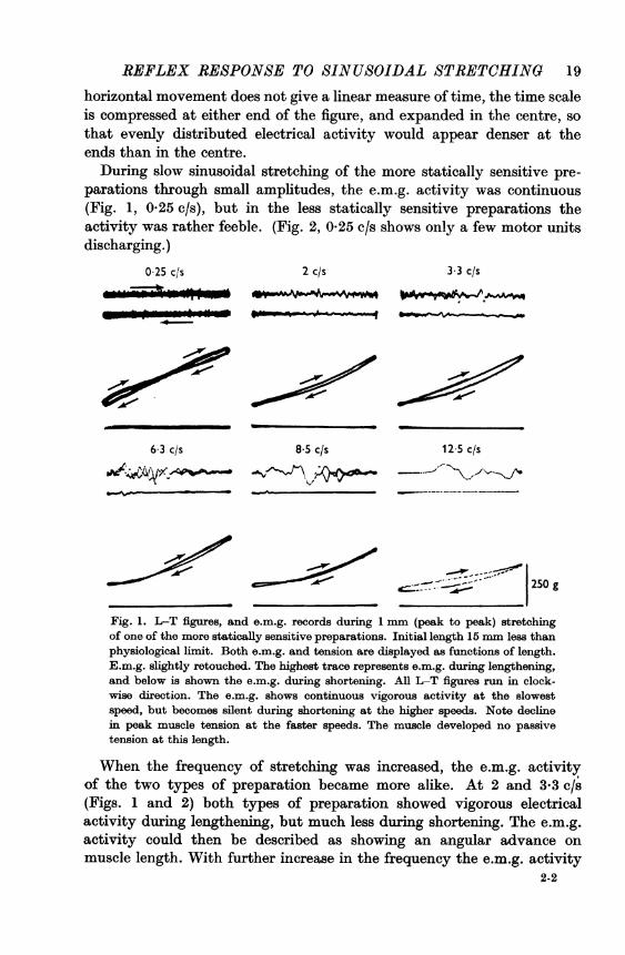
During slow sinusoidal stretching of the more statically sensitive pre-parations through small amplitudes, the e.m.g. activity was continuous(Fig. 1, 0-25 c/s), but in the less statically sensitive preparations theactivity was rather feeble. (Fig. 2, 025 c/s shows only a few motor unitsdischarging.)
0 25 c/s 2 c/s 33 c/s
63 c/s 8-5 c/s 125 c/s
- c :- 1 250 g
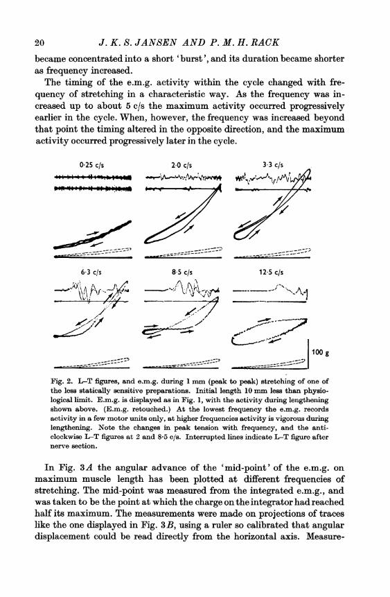
Fig. 1. L-T figures, and e.m.g. records during 1 mm (peak to peak) stretchingof one of the more statically sensitive preparations. Initial length 15 mm less thanphysiological limit. Both e.m.g. and tension are displayed as functions of length.E.m.g. slightly retouched. The highest trace represents e.m.g. during lengthening,and below is shown the e.m.g. during shortening. All L-T figures run in clock-wise direction. The e.m.g. shows continuous vigorous activity at the slowestspeed, but becomes silent during shortening at the higher speeds. Note declinein peak muscle tension at the faster speeds. The muscle developed no passivetension at this length.
When the frequency of stretching was increased, the e.m.g. activityof the two types of preparation became more alike. At 2 and 3-3 c/s(Figs. 1 and 2) both types of preparation showed vigorous electricalactivity during lengthening, but much less during shortening. The e.m.g.activity could then be described as showing an angular advance onmuscle length. With further increase in the frequency the e.m.g. activity
2-2
20 J. K. S. JANSEN AND P. M. H. RACK
became concentrated into a short 'burst', and its duration became shorteras frequency increased.The timing of the e.m.g. activity within the cycle changed with fre-
quency of stretching in a characteristic way. As the frequency was in-creased up to about 5 c/s the maximum activity occurred progressivelyearlier in the cycle. When, however, the frequency was increased beyondthat point the timing altered in the opposite direction, and the maximumactivity occurred progressively later in the cycle.
0 25 c/s 2-0 c/s 3 3 c/s
6-3 c/s 8 5 c/s 12 5 c/s
--XX.m--''''--100 g
.r ____---_ ----~.=.. I~
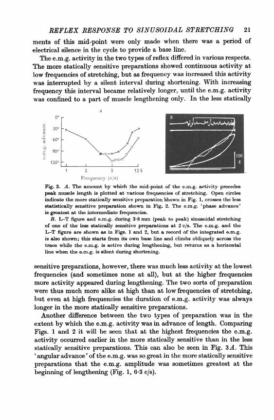
Fig. 2. LT figures, and e.m.g. during 1 mm (peak to peak) stretching of one ofthe less statically sensitive preparations. Initial length 10 mm less than physio-logical limit. E.m.g. is displayed as in Fig. 1, with the activity during lengtheningshown above. (E.m.g. retouched.) At the lowest frequency the e.m.g. recordsactivity in a few motor units only, at higher frequencies activity is vigorous duringlengthening. Note the changes in peak tension with frequency, and the anti-clockwise L-T figures at 2 and 8-5 c/s. Interrupted lines indicate L-T figure afternerve section.
In Fig. 3A the angular advance of the 'mid-point' of the e.m.g. onmaximum muscle length has been plotted at different frequencies ofstretching. The mid-point was measured from the integrated e.m.g., andwas taken to be the point at which the charge on the integrator had reachedhalf its maximum. The measurements were made on projections of traceslike the one displayed in Fig. 3B, using a ruler so calibrated that angulardisplacement could be read directly from the horizontal axis. Measure-
REFLEX RESPONSE TO SINUSOIDAL STRETCHING 21
ments of this mid-point were only made when there was a period ofelectrical silence in the cycle to provide a base line.
The e.m.g. activity in the two types of reflex differed in various respects.The more statically sensitive preparations showed continuous activity atlow frequencies of stretching, but as frequency was increased this activitywas interrupted by a silent interval during shortening. With increasingfrequency this interval became relatively longer, until the e.m.g. activitywas confined to a part of muscle lengthening only. In the less statically
A00
630 x \
1200t \W0>1 2 5 125
Frequency (c/s)
Fig. 3. A. The amount by which the mid-point of the e.m.g. activity precedespeak muscle length is plotted at various frequencies of stretching. Open circlesindicate the more statically sensitive preparation 'shown in Fig. 1, crosses the lessstatistically sensitive preparation shown in Fig. 2. The e.m.g. 'phase advance'is greatest at the intermediate frequencies.
B. L-T figure and e.m.g. during 3-8 mm (peak to peak) sinusoidal stretchingof one of the less statically sensitive preparations at 2 c/s. The e.m.g. and theL-T figure are shown as in Figs. 1 and 2, but a record of the integrated e.m.g.is also shown; this starts from its own base line and climbs obliquely across thetrace while the e.m.g. is active during lengthening, but returns as a horizontalline when the e.m.g. is silent during shortening.
sensitive preparations, however, there was much less activity at the lowestfrequencies (and sometimes none at all), but at the higher frequenciesmore activity appeared during lengthening. The two sorts of preparationwere thus much more alike at high than at low frequencies of stretching,but even at high frequencies the duration of e.m.g. activity was alwayslonger in the more statically sensitive preparations.Another difference between the two types of preparation was in the
extent by which the e.m.g. activity was in advance of length. ComparingFigs. 1 and 2 it will be seen that at the highest frequencies the e.m.g.activity occurred earlier in the more statically sensitive than in the lessstatically sensitive preparations. This can also be seen in Fig. 3A. This'angular advance' of the e.m.g. was so great in the more statically sensitivepreparations that the e.m.g. amplitude was sometimes greatest at thebeginning of lengthening (Fig. 1, 6-3 c/s).

J. K. S. JANSEN AND P. M. H. RACK
The tension during sinusoidal stretchingThe series of L-T figures displayed in Fig. 1 was obtained while
stretching sinusoidally one of the more statically sensitive preparations.In this preparation a peak tension of about 450 g was recorded, and thisremained constant at the lower frequencies of stretching. When, however,the frequency of stretching was increased beyond 6-3 c/s the tensiondeclined in spite of the increasing velocity of stretching, and at 12-5 c/sthe peak tension was only about 250 g.
Figure 2 contains a series of L-T figures obtained from one of the lessstatically sensitive preparations. The tension during slow stretching wasrather small, but at intermediate frequencies the e.m.g. activity duringlengthening was accompanied by a steep rise in tension and a higher peaktension. At higher frequencies the peak tension declined as it did in the lessstatically sensitive preparations. This decline was seen when the maximume.m.g. activity occurred less than 40 msec before the maximum musclelength. Apparently the muscle did not then develop its greatest stiffnessuntil rapid stretching was over, so that the peak tension was rather smallat the highest frequencies in either type of preparation.
In the less statically sensitive preparations the trough (minimum)tension rose when the frequency of stretching increased above about 2 c/s;the interval between the end of the e.m.g. activity, and minimum lengthwas then less than 250 msec. With a further increase in frequency thetrough tension increased, but at the highest frequencies the trough tensiondeclined. Presumably the velocity of shortening was then so great thatthe tension fell in spite of the muscle activity. A similar effect was seenin tetanized muscle (Rack, 1966).
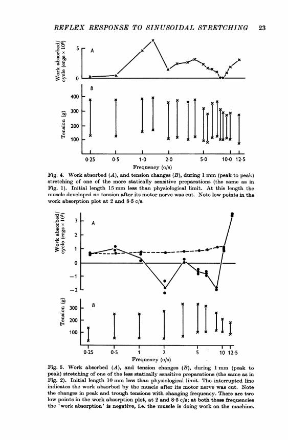
Work done on the muscle. The L-T curve during lengthening differedfrom the curve during shortening, so that in a complete cycle a figure wasdescribed. The area of this figure indicated the energy transferred betweenthe machine and the muscle. When the figure ran clockwise work was doneby the machine on the muscle, but when it ran anticlockwise the muscledid work on the machine.The work absorbed by the muscle during 1 mm stretching has been
plotted in Fig. 4 for one of the more statically sensitive preparations, andin Fig. 5 for one of the less statically sensitive preparations. In the morestatically sensitive preparations work was required to maintain themovement at all frequencies (Fig. 4), and the IT figures ran clockwise(Fig. 1). In the less statically sensitive preparations, however, the muscledid work on the machine at some frequencies (Fig. 5), and the L-Tfigures then ran predominantly anticlockwise (Fig. 2).
22
REFLEX RESPONSE TO SINUSOIDAL STRETCHINGI" x~~~~~~~~~~~~
i X/ \
400 k
300 1- lB 1 11lI I I I
0-25 0.5 1-0 20Frequency (c/s)
I IlTT*Ip
I - I ~I~_5.0 10-0 12-5
Fig. 4. Work absorbed (A), and tension changes (B), during 1 mm (peak to peak)stretching of one of the more statically sensitive preparations (the same as inFig. 1). Initial length 15 mm less than physiological limit. At this length themuscle developed no tension after its motor nerve was cut. Note low points in thework absorption plot at 2 and 8-5 c/s.
3go
4 x4, _m D
c)
2
1
0
-1
-2
A
BK
I II I I
025 0-5 1 2Frequency (c/s)
I K
I J. II I I5 10 12-5
Fig. 5. Work absorbed (A), and tension changes (B), during 1 mm (peak topeak) stretching of one of the less statically sensitive preparations (the same as inFig. 2). Initial length 10 mm less than physiological limit. The interrupted line
indicates the work absorbed by the muscle after its motor nerve was cut. Notethe changes in peak and trough tensions with changing frequency. There are twolow points in the work absorption plot, at 2 and 8-5 c/s; at both these frequenciesthe 'work absorption' is negative, i.e. the muscle is doing work on the machine.
23
bt
00H._
E-4
200
100
bo
r.00H-
3001-
200
100
0-
J. K. S. JANSEN AND P. M. H. RACK
The relation between e.m.g. and tensionThe shape and direction of the L-T figures could be correlated with the
e.m.g. activity that accompanied them. Vigorous e.m.g. activity early inlengthening was always associated with a steep rise in tension duringlengthening, and since this exceeded the tension during shortening theL-T figure then ran clockwise, and external work was required to main-tain the movement.When the e.m.g. was relatively inactive at the beginning of lengthening,
but increased later in lengthening, the tension during shortening mightexceed the tension during lengthening and the L-T figure then ran pre-dominantly anticlockwise (Fig. 2, 2 and 8-5 c/s). This state of affairs wascommonly seen during 1 mm stretching of the less statically sensitivepreparations at 5-8-5 c/s, and less often also at 2 c/s (Fig. 2). Anticlock-wise figures did not occur in the more statically sensitive preparationswith their generally greater 'angular advance' of the e.m.g. activity onlength.With frequencies of 10 c/s or more, the L-T figures always ran clock-
wise in spite of the lateness of the e.m.g. activity; the shape of the L-Tfigures suggested that the muscle then remained in an 'active state' duringa significant part of the ensuing lengthening, so that tension rose con-siderably as lengthening began.The work absorption/frequency plot from the less statically sensitive
preparation (Fig. 5) shows a low point at 2 c/s. This was not always seen.In some of the less statically sensitive preparations there was no reflexactivity during the slowest stretches, and hardly any at 2 c/s; the workabsorption/frequency plot then followed the pattern of the passive muscleat the slower speeds. Some small amount of static sensitivity seemed to benecessary for work to be done by the muscle on the machine at the slowerspeeds.Some of the tension changes are difficult to visualize when they are dis-
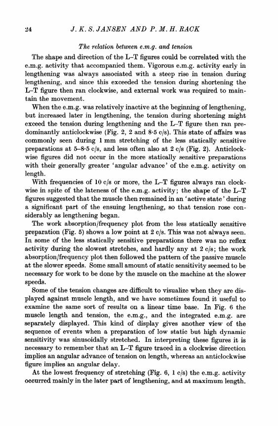
played against muscle length, and we have sometimes found it useful toexamine the same sort of results on a linear time base. In Fig. 6 themuscle length and tension, the e.m.g., and the integrated e.m.g. areseparately displayed. This kind of display gives another view of thesequence of events when a preparation of low static but high dynamicsensitivity was sinusoidally stretched. In interpreting these figures it isnecessary to remember that an L-T figure traced in a clockwise directionimplies an angular advance of tension on length, whereas an anticlockwisefigure implies an angular delay.
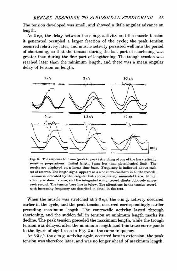
At the lowest frequency of stretching (Fig. 6, 1 c/s) the e.m.g. activityoccurred mainly in the later part of lengthening, and at maximum length.
.4
REFLEX RESPONSE TO SINUSOIDAL STRETCHING 25
The tension developed was small, and showed a little angular advance onlength.At 2 c/s, the delay between the e.m.g. activity and the muscle tension
it generated occupied a larger fraction of the cycle; the peak tensionoccurred relatively later, and muscle activity persisted well into the periodof shortening, so that the tension during the last part of shortening wasgreater than during the first part of lengthening. The trough tension wasreached later than the minimum length, and there was a mean angulardelay of tension on length.
1 c/s
5 c/s
2 c/s
6.3 c/s*tg
3 3 c/s
10 c/s
,; ' ' - -
Fig. 6. The response to 1 mm (peak to peak) stretching of one of the less staticallysensitive preparations. Initial length 9 mm less than physiological limit. Theresults are displayed on a linear time base. Frequency is indicated above eachset of records. The length signal appears as a sine curve constant in all the records.Tension is indicated by the irregular but approximately sinusoidal trace. E.m.g.activity is shown above, and the integrated e.m.g. record climbs obliquely across
each record. The tension base line is below. The alterations in the tension recordwith increasing frequency are described in detail in the text.
When the muscle was stretched at 3-3 c/s, the e.m.g. activity occurredearlier in the cycle, and the peak tension occurred correspondingly earlier
preceding maximum length. The contractile activity lasted throughshortening, and the sudden fall in tension at minimum length marks itsdecline. The peak tension preceded the maximum length, while the troughtension was delayed after the minimum length, and this trace correspondsto the figure-of-eight seen in Fig. 2 at the same frequency.At 6-3 c/s the e.m.g. activity again occurred late in extension, the peak
tension was therefore later, and was no longer ahead of maximum length.
100 g
J. K. S. JANSEN AND P. M. H. RACK
The muscle activity now persisted right through shortening, and theinflexion in the tension trace that marks the decline in contractile activityoccurred after minimum length. The trough tension was later thanminimum length, and there was a mean angular delay of tension.The highest frequency in Fig. 6 is 10 c/s. The e.m.g. activity then occurred
late in extension, and the peak tension occurred after maximum length.Activity, however, lasted well into the following lengthening, and thetension in the early part of extension was considerable, the trough tensionwas now well ahead of minimum length, and there was a mean angularadvance of tension on length.Both Figs. 2 and 6 show intermittent e.m.g. activity at the higher
frequencies, and the effect of each burst of activity can be seen in thetension record. At 2 c/s active tension lasts for at least 200 msec after theend of the electrical activity, but at 6-3 c/s an inflexion in the tensionrecord only about 85 msec after the e.m.g. activity indicates a decline inactive tension. Intermediate frequencies show intermediate time intervalsbetween the end of e.m.g. activity and the decay of active tension.The apparent discrepancy in these results is probably explicable as
follows: After the electrical impulse, contractile elements in the musclepass into an active state during which their stiffness is increased (Hill,1949; Ritchie, 1954). This stiffness then slowly declines. During thedecline a point is reached at which the contractile elements cease toshorten, and begin to be re-extended by either the series elastic elementsof the muscle, or by external movement. In an isometric muscle this pointmarks the beginning of the decline in tension, and it occurs earlier when thetension is high in an unfused tetanus than when tension is lower in asingle twitch. The interval between the end of the electrical activity andthe beginning of the decline in muscle tension is not constant, therefore,but depends on the muscle tension.When this property of muscle is taken into consideration, the results
shown in Figs. 2 and 6 present less difficulty.
Intermittent stimulation during sinusoidal stretchingThe rather complicated relation between e.m.g. activity and muscle
tension during 1 mm sinusoidal stretching was unexpected. In order toconfirm and clarify these findings some additional experiments werecarried out in which the motor activity in the stretch reflex was imitatedby interrupted tetanic stimulation of the ventral nerve roots.
Three cats were anaesthetized with sodium pentobarbitone (Nembutal). The L 7 and S 1roots were detached from the spinal cord, and their distal ends prepared for stimulation in aparaffin pool. The rest of the dissection of the leg was exactly as before, and the recordingmethods were the same. Interrupted tetani were provided by leading the output of astimulator through a contact breaker on the stretcher drive shaft. This contact breaker could
26

REFLEX RESPONSE TO SINUSOIDAL STRETCHING 27be adjusted to transmit a burst of stimuli of any desired duration in any part of the cycle.In order to approximately match the reflex tension, the stimulus was usually confined tothe Si root.
In these experiments the relation between the electrical activity andmuscle tension was found to be the same as in the stretch reflex records,and this was a useful confirmation of the earlier findings. Figure 7 showshow the direction of the L-T figure depends on the timing of the stimulus(here recorded as an e.m.g.) within the cycle. When an electrical stimulusoccurred early in extension, the tension rose early, and the L-T figure ranclockwise, but when the stimulus occurred late in extension the L-Tfigure ran anticlockwise, and the muscle did work on the machine.
Fig. 7. LIT figures, and e.m.g. during 1 mmn (peak to peak) sinusoidal stretchingat 3-3 c/s. The spinal roots have been cut, and trains of 5 impulses at 10 msec inter-vals are delivered to the S 1 root in various parts of the cycle. (The e.m.g. shows twoimperfectly superimposed traces, as the camera shutter was open for more than a
complete cycle). The L-T figure changes from a clockwise to an anticlockwisedirection as the stimuli are delivered later in the cycle.
In the isometric muscle, tension was still appreciable 250 msec after theend of a brief tetanus, and during sinusoidal stretching the interval betweenthe last stimulus and the apparent decline in active tension altered withfrequency in the same sort of way that it did in the reflex experiments.
ClonusWhen the muscle did work on the stretching machine, it was assisting
its own forced oscillation. The preparations that did this also exhibitedsustained clonus when loaded with an appropriate weight. A record ofclonus in such a muscle is shown in Fig. 8. The lower trace records themuscle length, and the upper trace the e.m.g. The clonus frequency inthis preparation was approximately 6 c/s, and this coincided with thefrequency at which the sinusoidally stretched muscle did most work on thestretching machine.In some other preparations clonus frequencies of about 8 c/s were
recorded. This value also corresponded to the frequency at which thosemuscles did most work on the stretching machine. Clonus was never seen

J. K. S. JANSEN AND P. M. H. RACKin the more statically sensitive preparations, and this was not surprisingsince work always had to be done on these preparations to maintainsinusoidal movement.Changes in magnitude of reflex activity. The magnitude of the reflex
activity varied in different decerebrate preparations, and changed fromtime to time in each preparation. The results illustrated in Figs. 1 and 2were from preparations with fairly active reflexes, other preparationswere seen in which the reflex tension was smaller, and the behaviour wascloser to that of passive muscle.
Fig. 8. Clonus in one of the less statical[ly sensitive preparations loaded with 50 gweight. Upper trace records e.m.g. Lower trace records muiscle length (recordedby measuring changes of tension in a fine spring joining the tendon to the anode pinof an RCA 5734 transducer valve). E.m.g. retouched. The clonus frequency(6 c/s) corresponded to the frequency at which this muscle did most work on themachine during sinusoidal stretching. (The higher frequency irregularities in thelength record are,probably artifacts from the recordling system.
The transfer of work between the stretcher and the muscle was alsoaffected by the magnitude of reflex activity, and when the level of reflexactivity fell the amount of work absorbed by the muscle in a cycleapproached the figure obtained for passive muscle. In the less staticallysensitive preparations the reflex activity had different effects on the workabsorbed at different frequencies of stretching (Fig. 5); at 12-5 c/s anincrease in reflex activity led to an increase in the work absorbed by themuscle, but at 6-3 c/s the effect of reflex activity was to reduce the workabsorbed below the passive level, and in the more active preparations this
28
REFLEX RESPONSE TO SINUSOIDAL STRETCHING 29
reduction was so great that the work absorbed by the muscle was negative,i.e. the muscle was doing work on the machine.
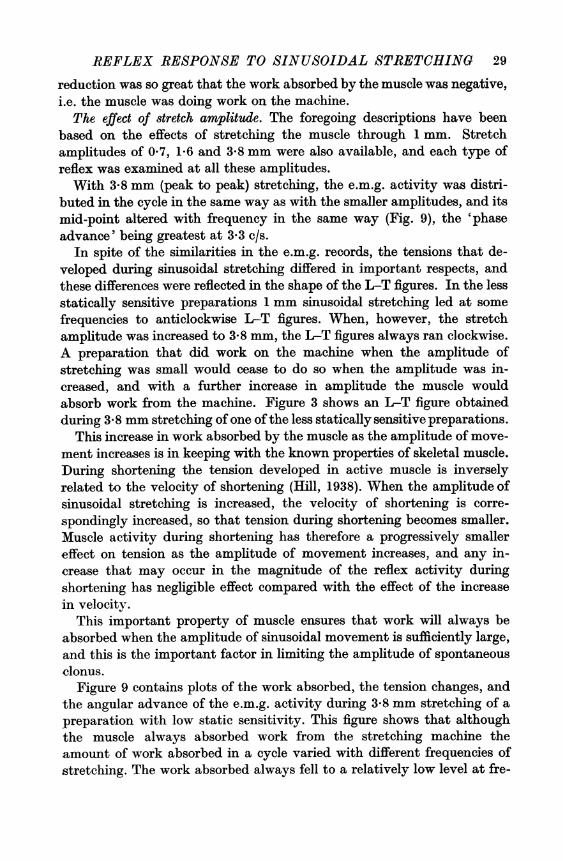
The effect of stretch amplitude. The foregoing descriptions have beenbased on the effects of stretching the muscle through 1 mm. Stretchamplitudes of 0 7, 1-6 and 3-8 mm were also available, and each type ofreflex was examined at all these amplitudes.With 3-8 mm (peak to peak) stretching, the e.m.g. activity was distri-
buted in the cycle in the same way as with the smaller amplitudes, and itsmid-point altered with frequency in the same way (Fig. 9), the 'phaseadvance' being greatest at 3-3 c/s.
In spite of the similarities in the e.m.g. records, the tensions that de-veloped during sinusoidal stretching differed in important respects, andthese differences were reflected in the shape of the L-T figures. In the lessstatically sensitive preparations 1 mm sinusoidal stretching led at somefrequencies to anticlockwise L-T figures. When, however, the stretchamplitude was increased to 3-8 mm, the L-T figures always ran clockwise.A preparation that did work on the machine when the amplitude ofstretching was small would cease to do so when the amplitude was in-creased, and with a further increase in amplitude the muscle wouldabsorb work from the machine. Figure 3 shows an L-T figure obtainedduring 3-8 mm stretching of one ofthe less statically sensitive preparations.
This increase in work absorbed by the muscle as the amplitude of move-ment increases is in keeping with the known properties of skeletal muscle.During shortening the tension developed in active muscle is inverselyrelated to the velocity of shortening (Hill, 1938). When the amplitude ofsinusoidal stretching is increased, the velocity of shortening is corre-spondingly increased, so that tension during shortening becomes smaller.Muscle activity during shortening has therefore a progressively smallereffect on tension as the amplitude of movement increases, and any in-crease that may occur in the magnitude of the reflex activity duringshortening has negligible effect compared with the effect of the increasein velocity.
This important property of muscle ensures that work will always beabsorbed when the amplitude of sinusoidal movement is sufficiently large,and this is the important factor in limiting the amplitude of spontaneousclonus.
Figure 9 contains plots of the work absorbed, the tension changes, andthe angular advance of the e.m.g. activity during 3-8 mm stretching of apreparation with low static sensitivity. This figure shows that althoughthe muscle always absorbed work from the stretching machine theamount of work absorbed in a cycle varied with different frequencies ofstretching. The work absorbed always fell to a relatively low level at fre-
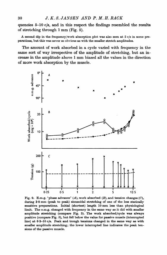
30 J. K. S. JANSEN AND P. M. H. RACKquencies 5-10 c/s, and in this respect the findings resembled the resultsof stretching through 1 mm (Fig. 5).
A second dip in the frequency/work absorption plot was also seen at 2 c/s in some pre-parations, but this was never so obvious as with the smaller stretch amplitudes.
The amount of work absorbed in a cycle varied with frequency in thesame sort of way irrespective of the amplitude of stretching, but an in-crease in the amplitude above 1 mm biased all the values in the directionof more work absorption by the muscle.
c)d
o
C)
ro
bb
1-
C.)
.),
"C o
4)C)
0
00 IAx
x
450 ,- xx
x
900 i.
201
xx
x
B
lo -
200
._
E0
100
Fig. 9. E.m.g. 'phase advance' (A), work absorbed (B), and tension changes (C),during 3*8 mm (peak to peak) sinusoidal stretching of one of the less staticallysensitive preparations. Initial (shortest) length 10 mm less than physiologicallimit. The e.m.g. changed with frequency in the same way as it did with smalleramplitude stretching (compare Fig. 3). The work absorbed/cycle was alwayspositive (compare Fig. 5), but fell below the value for passive muscle (interruptedline) at 6-3-10 c/s. Peak and trough tensions changed in the same way as withsmaller amplitude stretching; the lower interrupted line indicates the peak ten-sions of the passive muscle.

REFLEX RESPONSE TO SINUSOIDAL STRETCHING 31
Reduction of the stretch amplitude to 0 7 mm (peak to peak) reducedthe changes in the work absorption/frequency plot, so that they werealways less striking than those seen during 1 mm stretching. The workabsorbed/cycle was closer to the figure for passive muscle, and only rarelyat 6-3 c/s did the muscle do work on the machine.The rather small changes in the work absorption during stretching
through smaller amplitudes were probably associated with a reductionin the e.m.g. activity, of the less statically sensitive preparations, but noreliable measurement of total e.m.g. activity was possible in theseexperiments.
The effect of altering initial mu8cle length. The very large differences in the sensitivity ofdifferent preparations to static stretch have already been discussed. At one extreme arepreparations that develop a high tension when held extended; at the other extreme are thosethat develop a tension no greater than the denervated muscle at that length.
Although reflexes that were quite insensitive to static stretch did occur, it was more usualto find some increase in the reflex activity with an increase in muscle length even though theextra reflex tension developed was rather small. Whenever the reflex activity did increasewith length there was some alteration in the pattern of reflex behaviour also, the 'bursts'of e.m.g. activity at the higher stretch frequencies became longer, and began earlier in thecycle.
In other words, if a muscle had any sensitivity to static stretch, increasing its lengthchanged its response in the direction of the more statically sensitive preparation.
The integrity of the fusimotor fibres. During the course of these experi-ments, it seemed possible that the two types of preparation that havebeen described might merely reflect different total amounts of fusimotoractivity. To obtain further information on this point, conduction in thesmall fibres of the soleus nerve was blocked with lignocaine (Xylocaine)(Matthews & Rushworth, 1957; Critchlow & von Euler, 1963).Pieces of filter paper soaked in a solution of Xylocaine (0.25, or 0-125 g/100 ml.) were
placed on the nerve as it lay in a paraffin pool. The integrity of the alpha motor fibres wastested from time to time by measuring the isometric twitch tension during stimulation of thenerve proximal to the treated segment.
Six applications of Xylocaine were made in three different reflex preparations. One ofthese preparations showed a high sensitivity to static stretching, and the other two a lowstatic sensitivity. On each occasion there was a very active reflex before application ofXylocaine and the changes in tension and work absorption at different frequencies werecharacteristic.
Application of Xylocaine always gradually reduced the reflex activityuntil the e.m.g. became silent, and the L-T figures resembled thoseobtained after nerve section. This reduction of activity toward the passiveIstate took place without any significant blockage of conduction in thealpha motor fibres (as measured by the isometric twitch tension); thedecline in reflex activity was therefore attributed to the blocking of smallfibres in the motor nerve.

J. K. S. JANSEN AND P. M. H. RACKDuring the decline in reflex activity, there was nothing to suggest that
one type of reflex was changing into the other. The e.m.g. activity becamesteadily less, without any change in its timing, and the L-T figure becameprogressively closer to the type seen in passive muscle. In particular, thebehaviour of the more statically sensitive preparation did not at any stagecome to resemble the less statically sensitive preparations.
Control 2 min 3 min
v~~~~~~~
v15 g50Fig. 10. The effect of applying Xylocaine to the muscle nerve. One millimetrestretching of one of the less statically sensitive preparations at 3-3 c/s. BeforeXylocaine treatment the L-T figure was a figure-of-eight, and the e.m.g. wasactive during most of lengthening. A filter paper soaked in 0.25% Xylocainewas placed on the soleus nerve. Two minutes later the e.m.g. activity was less,and the tension was reduced, but the distribution of e.m.g. activity and theshape of the L-T figure were unchanged. Three minutes after Xylocaine applica-tion all e.m.g. activity had disappeared, and the L-T figure was the same as inpassive muscle. Conduction in the alpha motor neurone was unaffected. Reflexactivity returned to its previous level 10 min after removal of the Xylocaine.
Figure 10 illustrates the effect of Xylocaine on one of the less staticallysensitive preparations during sinusoidal stretching through 1 mm at3*3 c/s. Two minutes after application of Xylocaine the e.m.g. activitywas less, and the tension had declined, but the general shape of the L-Tfigures was unchanged. Three minutes after application of Xylocaine thee.m.g. activity had ceased, and the L-T figure was the same as in thepassive muscle.
This progressive reduction in reflex response was attributed to blockageof thin fibres in the soleus nerve (Matthews & IRushworth, 1957). Of thesethe fusimotor fibres were probably the most significant, since the smallafferent fibres probably play little part in the stretch reflex of extensormuscles (Hunt & Perl, 1960). It was concluded therefore that each typeof reflex behaviour depended on a background level of fusimotor activity,and the differences between the two sorts of preparation were not merelydifferences in the total amounts of fusimotor activity.
32
REFLEX RESPONSE TO SINUSOIDAL STRETCHING 33
DISCUSSION
Timing of the e.m.g. activity. The afferent discharge from innervatedmuscle spindles has not been examined in detail during sinusoidal stretch-ing. The muscle spindles are known, however, to be sensitive to thevelocity of lengthening as well as to length itself (Matthews, 1933). Duringsinusoidal stretching, therefore, the greatest afferent activity will occurin advance of peak muscle length. This angular advance will be less than900 since there is no evidence that the innervated muscle spindles aresensitive to acceleration of lengthening. The angular advance will probablyincrease as frequency (and therefore velocity of lengthening) increases.The e.m.g. during sinusoidal stretching shows an angular advance that
often reaches, and may exceed, 900; a further advance must thereforeoccur as the reflex signal passes through the spinal cord. It is possibleto envisage ways that this could occur; for example, recurrent inhibitionof the motor neurones through their axon collaterals might block trans-mission of the later part of the muscle spindle discharge, so that only itsearlier part is translated into motor neurone activity. It is unlikely thatan inhibitory effect from the soleus tendon organs plays an importantpart in this mechanism, since the tendon organ discharge is related tomuscle tension (Matthews, 1933; Jansen & Rudjord, 1964), and angularadvance was not significantly altered when the tension was reduced to alow level by an incomplete fusimotor block.The increase in the angular advance of the e.m.g. activity that occurred
with a moderate increase in frequency of stretching is in keeping with theknown properties of the muscle spindles, but the smaller angular advancethat occurred at the higher frequencies was unexpected. It was found,however, that at higher frequencies active tension persisted from theprevious cycle into the earlier part of muscle lengthening. This activitypresumably had an 'unloading' effect on the muscle spindles, diminishingtheir afferent discharge in the same way that a twitch in isometric musclecauses a silent period in the discharge of the muscle spindle afferents.Such a mechanism would delay the response of the muscle spindles toextension of the whole muscle; the effect would be more striking in theless statically sensitive preparations which change in stiffness moreobviously as the activity from the previous cycle decays. At the higherfrequencies of stretching the e.m.g. activity does in fact occur later in theless statically sensitive than in the more statically sensitive preparations.
The two types of stretch reflex. The foregoing experiments have demon-strated two different patterns of reflex activity in the decerebrate cat,neither of which can exist after fusimotor activity has been abolished.The differences in reflex behaviour may be due to differences in the3 Physiol. 183

J. K. S. JANSEN AND P. M. H. RACK
fusimotor control of the muscle spindles, or to differences in the spinalsynaptic mechanism, or to both. A good deal of information is availableabout the fusimotor control of the muscle spindles (Matthews, 1964),and about their behaviour in the soleus muscle of the decerebrate cat(Jansen & Matthews, 1962). It is therefore worth considering how far thepresent observations can be related to the known behaviour of the musclespindles.
There is convincing evidence that the sensitivity of the muscle spindlesto static stretch and to dynamic stretching can be independently in-fluenced by two different kinds of fusimotor fibres (Matthews, 1964). Inthe decerebrate cat, the response of the muscle spindle primary endingsis usually at a high level as a result of background fusimotor activity, butthe responses to static stretch and dynamic stretching vary in differentpreparations; these variations have been attributed to different mixturesof static and dynamic fusimotor activity (Jansen & Matthews, 1962).
It is tempting to assume that the different patterns of reflex behaviourseen in the present experiments correspond to the different sorts of fusi-motor activity found by Jansen & Matthews, and the fact that inter-mediate reflex states did occur presents no difficulty.
Certain features of the more statically sensitive preparations suggestvigorous activity in the static fusimotor fibres. The vigorous response ofboth tension and e.m.g. to a slow or static stretch could be a consequenceof static fusimotor activity, and the fact that the e.m.g. remains activeduring shortening at moderate speeds is very suggestive of activity in thestatic fusimotor fibres (Crowe & Matthews, 1964; Jansen & Rudjord, 1965).In contrast, the less statically sensitive preparations showed a lack ofresponse to slow stretching, and a lack of activity during shortening, whichboth suggest a low level of activity in the static fusimotor fibres.
In the less statically sensitive preparations the peak tension rose duringmoderate increases in frequency of stretching, which suggests thatdynamic fusimotor activity was enhancing the sensitivity of the musclespindles to rapid stretching. The changing angular advance of the e.m.g.activity with moderate increases in frequency was also the sort of be-haviour that could be expected during dynamic fusimotor activity; thiswas found in both types of preparation.The experimental results suggest therefore that dynamic fusimotor
activity was present in both types of preparation, but static fusimotoractivity was prominent only in the preparations that were more sensitiveto static stretch.
The relation of muscle tension to e.m.g. activity. The effect of e.m.g.activity on muscle tension during sinusoidal stretching could perhapshave been predicted from the known properties of skeletal muscle.
34
REFLEX RESPONSE TO SINUSOIDAL STRETCHING 35Electrical activity in muscle is followed by an 'active state' which
develops rapidly, but decays over a much longer period (Ritchie, 1954).E.m.g. activity early in lengthening therefore leads to muscle activitywhen the velocity of lengthening is greatest. Extension of this active, andtherefore stiff, muscle leads to a high tension during lengthening, andsince a part of the stiffness is viscous (Rack. 1966) work is required tomaintain the movement whether the activity continues during shorteningor not.
If therefore the e.m.g. activity begins sufficiently in advance of musclelength, the muscle will always absorb work from the stretching machine,whatever happens during the rest of the cycle.
Muscle damping in the decerebrate cat. When during sinusoidal stretchinga muscle absorbs work from the machine, it opposes and tends to dampmovement. When, however, the muscle does work on the machine, itassists its own movement, and the damping tendency is said to be negative.In these experiments the machine was much more powerful than the muscleso that the movement was not in fact significantly affected by the musclebehaviour. If, however, the muscle were acting on an inertial load, thenthe damping properties of the muscle at each frequency and amplitudewould govern the course of an oscillation at that frequency and amplitude.
Absorption of work, and damping, are associated with a phase advanceof tension on length, but work done by the muscle on the machine impliesphase delay, and negative damping. The phase relation between lengthand tension in any frequency-amplitude situation depends on how far theangular advance of the e.m.g. and the damping properties of the muscleitself can offset the delays in the reflex pathway.The complexity of the stretch reflex, and the non-linearities of its
component parts prevent useful mathematical description of its pro-perties. It is, however, possible to suggest the sort of effects that thedifferent parts of the reflex pathway will have on the phase relationsbetween length and tension during sinusoidal stretching:
(1) The muscle spindle discharge shows an angular advance on peaklength that first increases with frequency, but diminishes at the highestfrequencies.
(2) This reflex signal is further advanced as it passes through the spinalcord.
(3) The motor neurone discharge 'triggers' a state of contractileactivity in the muscle fibres, which slowly declines over about 250 msec.
(4) The muscle has a powerful damping effect as long as a significantpart of the contractile activity occurs during lengthening.From the functional point of view, the damping properties of the
muscle cannot be separated from the angular advance of the reflex signal,3-2
36 J. K. S. JANASEN AND P. M. H. RACK
since the muscle has important damping properties only if it is activatedearly in extension.The authors are grateful to Mr T. Rudjord for many helpful discussions, to Dr P. B. C
Matthews for his comments on results and the manuscript, and to Mr J. Rausanaksel fortechnical assistance. One of us (P. M. H. R.) was supported by the United BirminghamHospitals and Regional Hospital Board, and received a travel grant from the WellcomeTrust.
REFERENCES
CRITCHLOW, V. & VON EULER, C. (1963). Intercostal muscle spindle activity and its y-motorcontrol. J. Physiol. 168, 820-847.
CROWE, A. & MATTHEWS, P. B. C. (1964). Further studies of static and dynamic fusimotorfibres. J. Physiol. 174, 132-151.
GRANIT, R. (1955). Receptors and Sensory Perception, 366 pp. New Haven: Yale UniversityPress.
GRANIT, R. (1958). Neuromuscular interaction in postural tone of the cat's isometricsoleus muscle. J. Physiol. 143, 387-402.
HILL, A. V. (1938). Heat of shortening and dynamic constants of muscle. Proc. R. Soc.B, 126, 136-195.
HILL, A. V. (1949). The abrupt transition from rest to activitv in muscle. Proc. R. Soc. B,136, 399-420.
HUNT, C. C. & PERL, E. R. (1960). Spinal reflex mechanisms concerned with skeletalmuscle. Physiol. Rev. 40, 538-579.
JANSEN, J. K. S. & MATTHEWS, P. B. C. (1962). The central control of the dynamic responseof muscle spindle receptors. J. Physiol. 161, 357-378.
JANSEN, J. K. S. & RUDJORD, T. (1964). On the silent period and Golgi tendon organs of thesoleus muscle of the cat. Acta phy8iol. 8cand. 62, 364-379.
JANSEN, J. K. S. & RUDJORD, T. (1965). Fusimotor activity in a flexor muscle of thedecerebrate cat. Acta physiol. scand. 63, 236-246.
KATZ, B. (1939). The relation between force and speed in muscular contraction. J. Physiol.96, 45-64.
LIDDELL, E. G. T. & SHERRINGTON, C. (1924). Reflexes in response to stretch (Myotaticreflexes). Proc. R. Soc. B, 96, 212-242.
LIPPOLD, 0. C. J., REDFEARN, J. W. T. & XVuo, J. (1958). The effect of sinusoidal stretchingupon the activity of stretch receptors in voluntary muscle and their reflex responses.J. Physiol. 144, 373-386.
MATTHEWS, B. H. C. (1933). Nerve endings in mammalian muscle. J. Physiol. 78, 1-53.MATTHEWS, P. B. C. (1959a). The dependence of tension upon extension in the stretch
reflex of the soleus muscle of the decerebrate cat. J. Physiol. 147, 521-546.MATTHEWS, P. B. C. (1959b). A study of certain factors influencing the stretch reflex of the
decerebrate cat. J. Physiol. 147, 547-564.MATTHEWS, P. B. C. (1964). MIuscle spindles and their motor control. Physiol. Revs. 44,
219-288.MATTHEWS, P. B. C. & RUSHWORTH, G. (1957). The relative sensitivity of muscle nerve
fibres to procaine. J. Physiol. 135, 263-269.MERTON, P. A. (1951). The silent period in a muscle of the human hand. J. Physiol. 114,
183-198.PARTRIDGE, L. D. & GLASER, G. H. (1960). Adaptation in regulation of movement and
posture. A study of stretch responses in spastic animals. J. Neurophys8ol. 23, 257-268.RACK, P. M. H. (1966). The behaviour of a mammalian muscle during sinusoidal stretching.
J. Physiol. 183, 1-14.RITCHIE, J. M. (1954). The duration of the plateau of full activity in frog muscle. J.
Physiol. 124, 605-612.ROBERTS, T. D. M. (1963). Rhythmic excitation of a stretch reflex, revealing (a) hysteresisand (b) a difference between the response to pulling and to stretching. Q. JI exp.Physiol. 48, 328-345.
STARR, A. & LIVINGSTON, R. B. (1963). Long-lasting nervous system responses to pro-longed sound stimulation in waking cats. J. Neurophysiol. 26, 416-431.