In Central Obesity, Weight Loss Restores Platelet Sensitivity to Nitric Oxide and Prostacyclin
Upload
universityofmontrealCategory
view
0download
0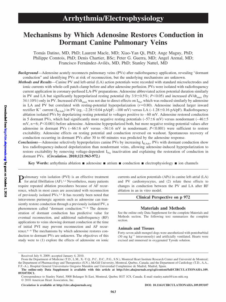
Arrhythmia/Electrophysiology
Mechanisms by Which Adenosine Restores Conduction inDormant Canine Pulmonary Veins
Tomas Datino, MD, PhD; Laurent Macle, MD; Xiao-Yan Qi, PhD; Ange Maguy, PhD;Philippe Comtois, PhD; Denis Chartier, BSc; Peter G. Guerra, MD; Angel Arenal, MD;
Francisco Fernandez-Aviles, MD, PhD; Stanley Nattel, MD
Background—Adenosine acutely reconnects pulmonary veins (PVs) after radiofrequency application, revealing “dormantconduction” and identifying PVs at risk of reconnection, but the underlying mechanisms are unknown.
Methods and Results—Canine PV and left-atrial (LA) action potentials were recorded with standard microelectrodes andionic currents with whole-cell patch clamp before and after adenosine perfusion. PVs were isolated with radiofrequencycurrent application in coronary-perfused LA-PV preparations. Adenosine abbreviated action potential duration similarlyin PV and LA but significantly hyperpolarized resting potential (by 3.90.5%; P0.05) and increased dV/dtmax (by3410%) only in PV. Increased dV/dtmax was not due to direct effects on INa, which was reduced similarly by adenosinein LA and PV but correlated with resting-potential hyperpolarization (r0.80). Adenosine induced larger inwardrectifier K current (IKAdo) in PV (eg, –2.280.04 pA/pF; –100 mV) versus LA (–1.280.16 pA/pF). Radiofrequencyablation isolated PVs by depolarizing resting potential to voltages positive to –60 mV. Adenosine restored conductionin 5 dormant PVs, which had significantly more negative resting potentials (–576 mV) versus nondormant (–465mV, n6; P0.001) before adenosine. Adenosine hyperpolarized both, but more negative resting-potential values afteradenosine in dormant PVs (–666 mV versus –566 mV in nondormant; P0.001) were sufficient to restoreexcitability. Adenosine effects on resting potential and conduction reversed on washout. Spontaneous recovery ofconduction occurring in dormant PVs after 30 to 60 minutes was predicted by the adenosine response.
Conclusions—Adenosine selectively hyperpolarizes canine PVs by increasing IKAdo. PVs with dormant conduction showless radiofrequency-induced depolarization than nondormant veins, allowing adenosine-induced hyperpolarization torestore excitability by removing voltage-dependent INa inactivation and explaining the restoration of conduction indormant PVs. (Circulation. 2010;121:963-972.)
Key Words: arrhythmia ablation adenosine atrium conduction electrophysiology ion channels
Pulmonary vein isolation (PVI) is an effective treatmentfor atrial fibrillation (AF).1,2 Nevertheless, many patients
require repeated ablation procedures because of AF recur-rence, which in most cases are associated with reconnectionof previously isolated PVs.3,4 It has recently been noted thatintravenous purinergic agonists such as adenosine can tran-siently restore conduction through a previously isolated PV, aphenomenon called “dormant conduction.”5–8 The demon-stration of dormant conduction has predictive value foreventual reconnection, and additional radiofrequency (RF)applications to veins showing dormant conduction at the timeof initial PVI may prevent reconnection and AF recur-rence.6–8 The mechanisms by which adenosine restores con-duction to dormant PVs are unknown. The objectives of thisstudy were to (1) explore the effects of adenosine on ionic
currents and action potentials (APs) in canine left-atrial (LA)and PV cardiomyocytes, and (2) relate these effects tochanges in conduction between the PV and LA after RFablation in an in vitro model.
Clinical Perspective on p 972
Materials and MethodsSee the online-only Data Supplement for the complete Materials andMethods section. The following text summarizes the completesection.
Animals and TissuesForty-seven adult mongrel dogs were anesthetized with pentobarbital(30 mg kg–1 intravenously) and artificially ventilated. Hearts wereexcised and immersed in oxygenated Tyrode solution.
Received July 9, 2009; accepted January 4, 2010.From the Department of Medicine (T.D., L.M., X.-Y.Q., P.C., D.C., P.G., S.N.), Montreal Heart Institute Research Center and Universite de Montreal;
the Department of Pharmacology and Therapeutics (S.N.), McGill University, Montreal, Quebec, Canada; and the Department of Cardiology (T.D., A.A.,F.F.-A.), Hospital General Universitario Gregorio Maranon and Universidad Complutense de Madrid, Madrid, Spain.
The online-only Data Supplement is available with this article at http://circ.ahajournals.org/cgi/content/full/CIRCULATIONAHA.109.893107/DC1.
Correspondence to Stanley Nattel, 5000 Belanger St East, Montreal, Quebec H1T 1C8, Canada. E-mail [email protected]© 2010 American Heart Association, Inc.
Circulation is available at http://circ.ahajournals.org DOI: 10.1161/CIRCULATIONAHA.109.893107
963
by guest on October 2, 2016
http://circ.ahajournals.org/D
ownloaded from
by guest on O
ctober 2, 2016http://circ.ahajournals.org/
Dow
nloaded from
by guest on October 2, 2016
http://circ.ahajournals.org/D
ownloaded from
by guest on O
ctober 2, 2016http://circ.ahajournals.org/
Dow
nloaded from
by guest on October 2, 2016
http://circ.ahajournals.org/D
ownloaded from
by guest on O
ctober 2, 2016http://circ.ahajournals.org/
Dow
nloaded from
by guest on October 2, 2016
http://circ.ahajournals.org/D
ownloaded from
by guest on O
ctober 2, 2016http://circ.ahajournals.org/
Dow
nloaded from
by guest on October 2, 2016
http://circ.ahajournals.org/D
ownloaded from
by guest on O
ctober 2, 2016http://circ.ahajournals.org/
Dow
nloaded from
by guest on October 2, 2016
http://circ.ahajournals.org/D
ownloaded from
by guest on O
ctober 2, 2016http://circ.ahajournals.org/
Dow
nloaded from
by guest on October 2, 2016
http://circ.ahajournals.org/D
ownloaded from
For standard microelectrode experiments, intact tissue prepara-tions, including LA and PVs, were perfused via the left-circumflexcoronary artery with oxygenated Krebs solution at 35°0.5°C, andAPs were recorded as previously described.9 Measurements includedresting membrane potential (RMP), AP amplitude, and AP duration(APD) at 90% of repolarization (APD90). Conduction time wasmeasured during LA pacing between peaks of differentiated (dV/dt)signals for APs recorded with 2 stable microelectrodes 1-cm apart,one in a PV and the other in the closest adjacent LA region. Cellisolation was performed as previously described.10,11 After isolation,cells were stored (4°C) and studied within 12 hours.
RF-Induced PVIPVI was conducted in LA-PV preparations subjected to microelec-trode AP recordings. One PV was isolated in each dog by ablating inthe antral region as close as possible to the PV-LA junction. Bipolarelectrodes were attached to the epicardial surface of the LA andtarget PV to evaluate PV-LA conduction. RF energy was deliveredepicardially in the unipolar mode between the standard 4-mm tip ofa 7F quadripolar ablation catheter and an indifferent peripheralelectrode, with a power limit of 25 to 35 W, for only 10 seconds ateach site to control damage. The endpoint of PVI was a bidirectionalconduction block (both from LA to PV and from the PV at 4 differentquadrants to LA, with pacing stimuli up to maximum possiblestimulation strength).
When PVI was achieved, APs were recorded in PV sleevesimmediately above the ablation line for 15 minutes after the final RFapplication. If conduction recovered, then additional RF was deliv-ered to complete PVI. Adenosine was then added, and APs wererecorded for an additional 15 minutes in 11 preparations (6 leftsuperior and 5 left inferior PVs). In preparations that recoveredconduction with adenosine, APs were recorded during a final15-minute adenosine washout period. In 6 preparations (5 leftsuperior and 1 left inferior PVs) studied to establish the time courseof spontaneous changes after PVI, the same PVI protocol wasconducted without subsequently adding adenosine, and preparationswere monitored for up to 4 hours (average monitoring time 3.40.3hours). In 9 other preparations (6 left superior and 3 left inferiorPVs), prolonged monitored was obtained after adenosine washout toevaluate whether adenosine reconnection predicts subsequent spon-taneous reconnection.
Adenosine Receptor Protein ExpressionProtein-enriched fractions were obtained from cardiomyocytes iso-lated as described above, with paired PV and LA samples from eachof 5 dogs studied. Antiadenosine A1- and A2A-receptor antibodieswere obtained from ABCAM (ab3460 and ab3461); antiadenosineA2B- and A3-receptor antibodies (A2bR23 and A3R32) were fromAlpha Diagnostic (San Antonio, TX). Donkey anti-rabbit secondaryantibodies conjugated to horseradish peroxidase were used fordetection. All of the expression data are provided relative to GAPDHstaining for the same samples on the same gels.
SolutionsDetails of solution contents for AP recording and voltage-clampstudies are provided in the online-only Data Supplement. Adenosinewas freshly prepared as a 1-mmol/L solution before each experiment.
Electrophysiology Data AcquisitionThe whole-cell patch-clamp technique was used to record currents.Electrode tip resistances were 1 to 4 Megohms, with 1 to 2 Megohmpipettes used for INa recording. Cell capacitances averaged 979 pFfor PV and 997 pF for LA cardiomyocytes. Rs averaged 2.90.3Megohms and 27918 s after compensation in LA; 2.60.2Megohms and 24213 s in PV. Currents are expressed as densities(pA/pF). K-current recording was performed at 35°0.5°C. INa
was recorded at room temperature. The maximum phase 0 upstrokevelocity (dV/dtmax) of standard microelectrode recordings was de-termined by electronic differentiation.
Statistical AnalysisData are expressed as meanSEM. Repeated-measures 1-wayANOVA was used to compare RMP at different times after PVI.Repeated-measures 2-way ANOVA was used to study the interactionbetween location (PV versus LA) and adenosine for RMP, APD90,AP amplitude, and dV/dtmax; the interaction between test potentialand treatment (adenosine versus no adenosine) for K current andINa; and the interaction between test potential and location (PVversus LA) for IKAdo. Bonferroni-adjusted comparisons were used tocompare group means when ANOVA was significant. Repeated-measures 2-way analysis was used to study interactions betweendormant/nondormant state and adenosine effect on RMP after PVI.An unpaired Student t test was used to compare ablation timebetween dormant and nondormant PVs and between control andadenosine-treated PVs. All of the data satisfied criteria for normalityof distribution (Kolmogrov-Smirnov) except for conduction time.The effect of drug on conduction time was studied with Wilcoxonsigned rank test because it was not normally distributed. Pearsoncorrelation coefficients were used to study the relation between RMPand dV/dtmax. Fisher exact test was used to compare dormant-conduction prevalence in different PVs. Whenever more than 1 cellwas obtained per region for a dog, all of the cell values in each regionwere averaged to obtain single representative values for that dog forstatistical comparison to avoid weighting values unevenly for differ-ent dogs. A 2-tailed P0.05 was considered statistically significant.The authors had full access to and take responsibility for the integrityof the data. All of the authors have read and agree to the article aswritten.
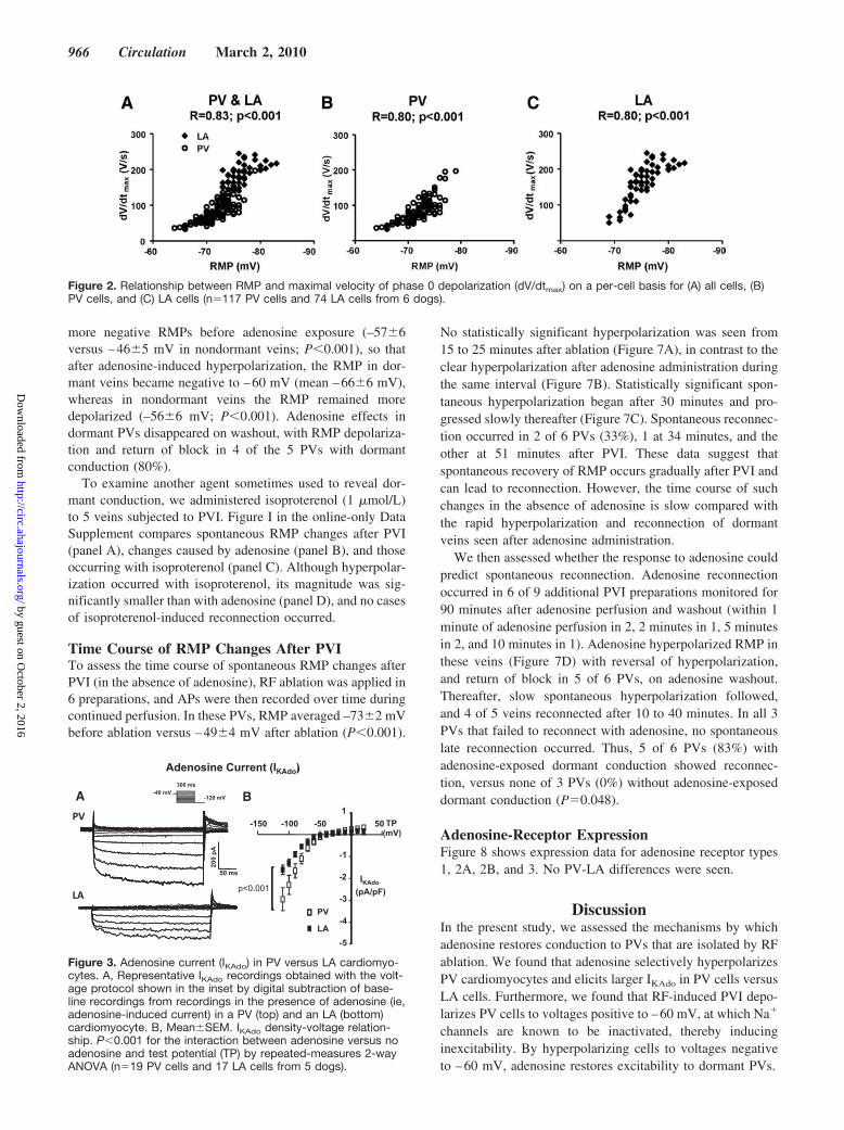
ResultsAP and Conduction-Time ChangesAP effects of adenosine were explored in 6 dogs. APrecordings before and after adenosine are illustrated in Figure1 (Top). Adenosine shortened APD in both PV (Figure 1A)and LA (Figure 1B) cells but significantly hyperpolarizedRMP and increased dV/dtmax only in PV cells. Completeresults and statistics are provided in Table 1, and mean dataare illustrated in Figure 1 (bottom). In PV sleeves but not inLA, adenosine significantly increased RMP (Figure 1C) anddV/dtmax (Figure 1D). Adenosine abbreviated APD similarlyin PV and LA (Figure 1E). AP amplitude was increased onlyin PV (Figure 1F). RMP and dV/dtmax correlated (Figure 2A),with similar and strong correlation for PV data alone (Figure2B) and LA data alone (Figure 2C), consistent with knownvoltage dependence of INa availability. Adenosine also re-duced conduction time between LA and PV electrodes from17.43.2 to 14.93.1 ms (P0.031).
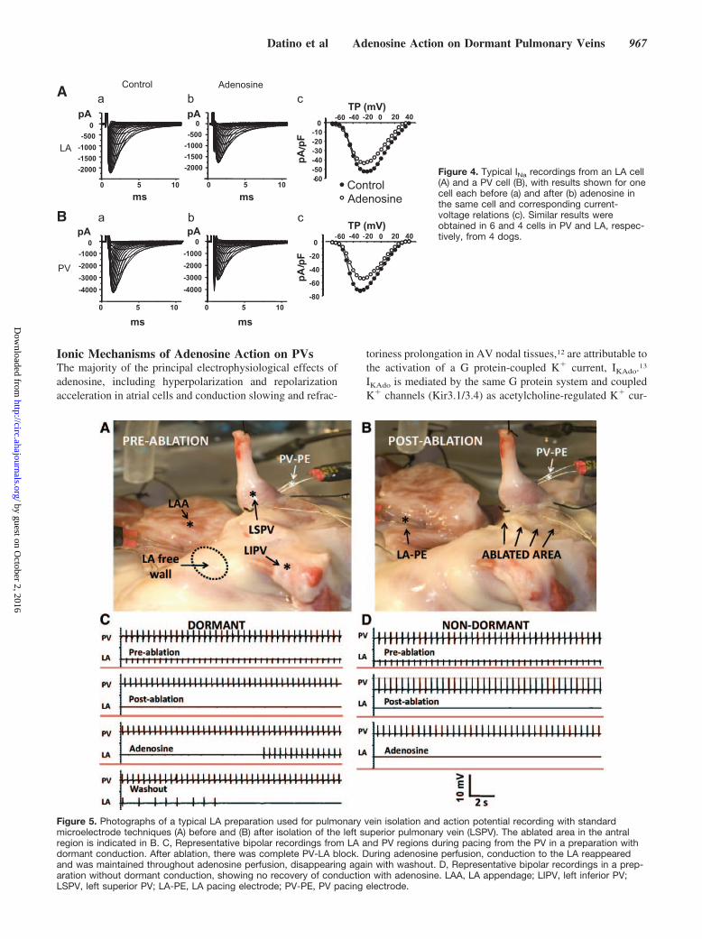
Adenosine-Induced Ion-Current ChangesIKAdo recordings from a PV and LA cardiomyocyte areillustrated in Figure 3A. Larger adenosine-induced K cur-rents were consistently seen in PVs. Overall results are shownin Figure 3B. The reversal potential was approximately –70mV (when corrected for the 15-mV mean junction potential).Location was a highly significant determinant of IKAdo, whichwas larger in PV than LA cardiomyocytes. For example, at–100 mV, IKAdo averaged –2.280.04 pA/pF in PV cellsversus –1.280.16 pA/pF in LA cells.
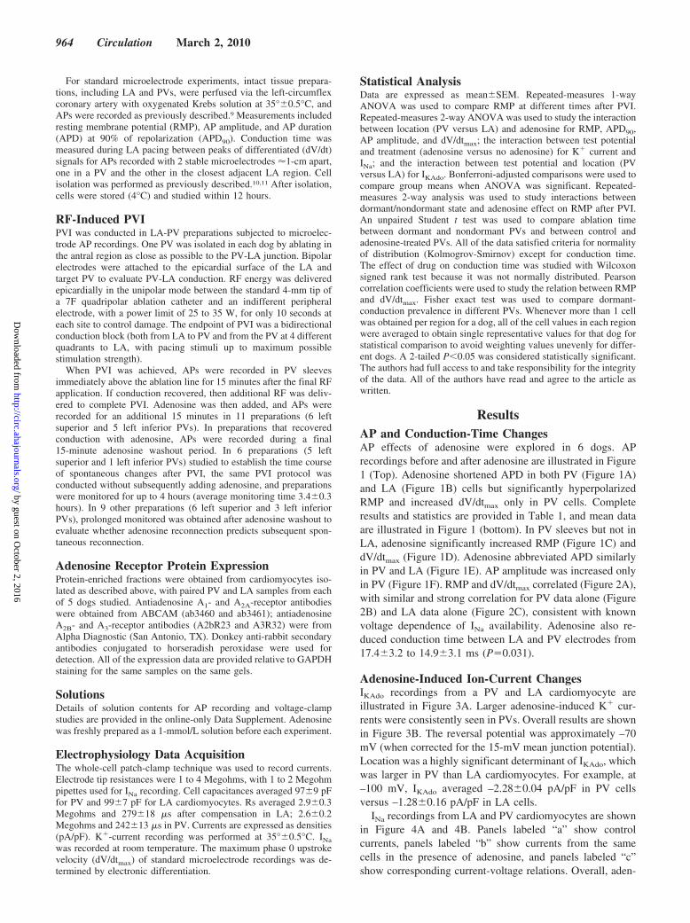
INa recordings from LA and PV cardiomyocytes are shownin Figure 4A and 4B. Panels labeled “a” show controlcurrents, panels labeled “b” show currents from the samecells in the presence of adenosine, and panels labeled “c”show corresponding current-voltage relations. Overall, aden-
964 Circulation March 2, 2010
by guest on October 2, 2016
http://circ.ahajournals.org/D
ownloaded from
osine reduced peak INa by 264% in PV cells versus 268%in LA cells (PNS). Washout resulted in up to 99.8%reversal of effect, indicating that the response was due toadenosine. These results indicate that the adenosine-induceddV/dt
maxincreases in PV cardiomyocytes are not due to direct
INa-enhancing effects.
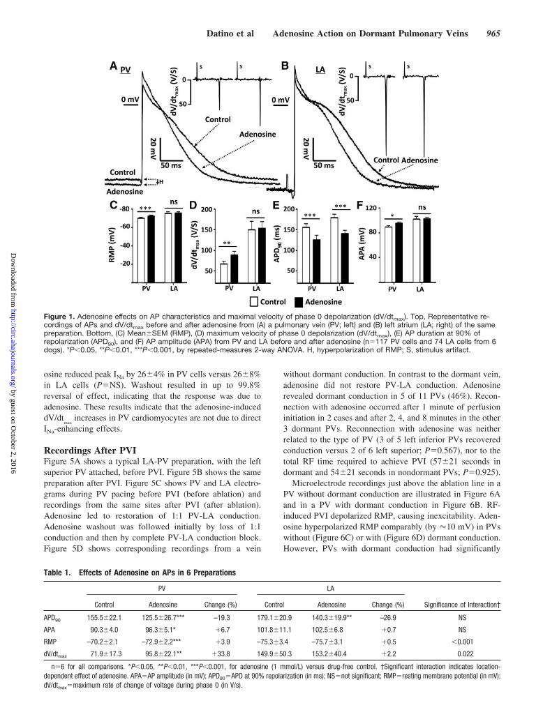
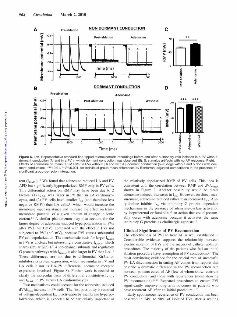
Recordings After PVIFigure 5A shows a typical LA-PV preparation, with the leftsuperior PV attached, before PVI. Figure 5B shows the samepreparation after PVI. Figure 5C shows PV and LA electro-grams during PV pacing before PVI (before ablation) andrecordings from the same sites after PVI (after ablation).Adenosine led to restoration of 1:1 PV-LA conduction.Adenosine washout was followed initially by loss of 1:1conduction and then by complete PV-LA conduction block.Figure 5D shows corresponding recordings from a vein
without dormant conduction. In contrast to the dormant vein,adenosine did not restore PV-LA conduction. Adenosinerevealed dormant conduction in 5 of 11 PVs (46%). Recon-nection with adenosine occurred after 1 minute of perfusioninitiation in 2 cases and after 2, 4, and 8 minutes in the other3 dormant PVs. Reconnection with adenosine was neitherrelated to the type of PV (3 of 5 left inferior PVs recoveredconduction versus 2 of 6 left superior; P0.567), nor to thetotal RF time required to achieve PVI (5721 seconds indormant and 5421 seconds in nondormant PVs; P0.925).
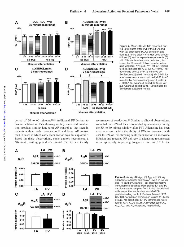
Microelectrode recordings just above the ablation line in aPV without dormant conduction are illustrated in Figure 6Aand in a PV with dormant conduction in Figure 6B. RF-induced PVI depolarized RMP, causing inexcitability. Aden-osine hyperpolarized RMP comparably (by 10 mV) in PVswithout (Figure 6C) or with (Figure 6D) dormant conduction.However, PVs with dormant conduction had significantly
Table 1. Effects of Adenosine on APs in 6 Preparations
PV LA
Significance of Interaction†Control Adenosine Change (%) Control Adenosine Change (%)
APD90 155.522.1 125.526.7*** –19.3 179.120.9 140.319.9** –26.9 NS
APA 90.34.0 96.35.1* 6.7 101.811.1 102.56.8 0.7 NS
RMP –70.22.1 –72.92.2*** 3.9 –75.33.4 –75.73.1 0.5 0.001
dV/dtmax 71.917.3 95.822.1** 33.8 149.950.3 153.240.4 2.2 0.022
n6 for all comparisons. *P0.05, **P0.01, ***P0.001, for adenosine (1 mmol/L) versus drug-free control. †Significant interaction indicates location-dependent effect of adenosine. APAAP amplitude (in mV); APD90APD at 90% repolarization (in ms); NSnot significant; RMPresting membrane potential (in mV);dV/dtmaxmaximum rate of change of voltage during phase 0 (in V/s).
Figure 1. Adenosine effects on AP characteristics and maximal velocity of phase 0 depolarization (dV/dtmax). Top, Representative re-cordings of APs and dV/dtmax before and after adenosine from (A) a pulmonary vein (PV; left) and (B) left atrium (LA; right) of the samepreparation. Bottom, (C) MeanSEM (RMP), (D) maximum velocity of phase 0 depolarization (dV/dtmax), (E) AP duration at 90% ofrepolarization (APD90), and (F) AP amplitude (APA) from PV and LA before and after adenosine (n117 PV cells and 74 LA cells from 6dogs). *P0.05, **P0.01, ***P0.001, by repeated-measures 2-way ANOVA. H, hyperpolarization of RMP; S, stimulus artifact.
Datino et al Adenosine Action on Dormant Pulmonary Veins 965
by guest on October 2, 2016
http://circ.ahajournals.org/D
ownloaded from
more negative RMPs before adenosine exposure (–576versus –465 mV in nondormant veins; P0.001), so thatafter adenosine-induced hyperpolarization, the RMP in dor-mant veins became negative to –60 mV (mean –666 mV),whereas in nondormant veins the RMP remained moredepolarized (–566 mV; P0.001). Adenosine effects indormant PVs disappeared on washout, with RMP depolariza-tion and return of block in 4 of the 5 PVs with dormantconduction (80%).
To examine another agent sometimes used to reveal dor-mant conduction, we administered isoproterenol (1 mol/L)to 5 veins subjected to PVI. Figure I in the online-only DataSupplement compares spontaneous RMP changes after PVI(panel A), changes caused by adenosine (panel B), and thoseoccurring with isoproterenol (panel C). Although hyperpolar-ization occurred with isoproterenol, its magnitude was sig-nificantly smaller than with adenosine (panel D), and no casesof isoproterenol-induced reconnection occurred.
Time Course of RMP Changes After PVITo assess the time course of spontaneous RMP changes afterPVI (in the absence of adenosine), RF ablation was applied in6 preparations, and APs were then recorded over time duringcontinued perfusion. In these PVs, RMP averaged –732 mVbefore ablation versus –494 mV after ablation (P0.001).
No statistically significant hyperpolarization was seen from15 to 25 minutes after ablation (Figure 7A), in contrast to theclear hyperpolarization after adenosine administration duringthe same interval (Figure 7B). Statistically significant spon-taneous hyperpolarization began after 30 minutes and pro-gressed slowly thereafter (Figure 7C). Spontaneous reconnec-tion occurred in 2 of 6 PVs (33%), 1 at 34 minutes, and theother at 51 minutes after PVI. These data suggest thatspontaneous recovery of RMP occurs gradually after PVI andcan lead to reconnection. However, the time course of suchchanges in the absence of adenosine is slow compared withthe rapid hyperpolarization and reconnection of dormantveins seen after adenosine administration.
We then assessed whether the response to adenosine couldpredict spontaneous reconnection. Adenosine reconnectionoccurred in 6 of 9 additional PVI preparations monitored for90 minutes after adenosine perfusion and washout (within 1minute of adenosine perfusion in 2, 2 minutes in 1, 5 minutesin 2, and 10 minutes in 1). Adenosine hyperpolarized RMP inthese veins (Figure 7D) with reversal of hyperpolarization,and return of block in 5 of 6 PVs, on adenosine washout.Thereafter, slow spontaneous hyperpolarization followed,and 4 of 5 veins reconnected after 10 to 40 minutes. In all 3PVs that failed to reconnect with adenosine, no spontaneouslate reconnection occurred. Thus, 5 of 6 PVs (83%) withadenosine-exposed dormant conduction showed reconnec-tion, versus none of 3 PVs (0%) without adenosine-exposeddormant conduction (P0.048).
Adenosine-Receptor ExpressionFigure 8 shows expression data for adenosine receptor types1, 2A, 2B, and 3. No PV-LA differences were seen.
DiscussionIn the present study, we assessed the mechanisms by whichadenosine restores conduction to PVs that are isolated by RFablation. We found that adenosine selectively hyperpolarizesPV cardiomyocytes and elicits larger IKAdo in PV cells versusLA cells. Furthermore, we found that RF-induced PVI depo-larizes PV cells to voltages positive to –60 mV, at which Na
channels are known to be inactivated, thereby inducinginexcitability. By hyperpolarizing cells to voltages negativeto –60 mV, adenosine restores excitability to dormant PVs.
)Adenosine Current (IKAdo)
A -40 mV-120 mV
300 ms
B
PV -150 -100 -50 50
1
(mV)TP
200
pA
50 ms 2
-1
LA -3
-2
PV
p<0.001 (pA/pF)IKAdo
-5
-4PV
LA
Figure 3. Adenosine current (IKAdo) in PV versus LA cardiomyo-cytes. A, Representative IKAdo recordings obtained with the volt-age protocol shown in the inset by digital subtraction of base-line recordings from recordings in the presence of adenosine (ie,adenosine-induced current) in a PV (top) and an LA (bottom)cardiomyocyte. B, MeanSEM. IKAdo density-voltage relation-ship. P0.001 for the interaction between adenosine versus noadenosine and test potential (TP) by repeated-measures 2-wayANOVA (n19 PV cells and 17 LA cells from 5 dogs).
Figure 2. Relationship between RMP and maximal velocity of phase 0 depolarization (dV/dtmax) on a per-cell basis for (A) all cells, (B)PV cells, and (C) LA cells (n117 PV cells and 74 LA cells from 6 dogs).
966 Circulation March 2, 2010
by guest on October 2, 2016
http://circ.ahajournals.org/D
ownloaded from
Ionic Mechanisms of Adenosine Action on PVsThe majority of the principal electrophysiological effects ofadenosine, including hyperpolarization and repolarizationacceleration in atrial cells and conduction slowing and refrac-
toriness prolongation in AV nodal tissues,12 are attributable tothe activation of a G protein-coupled K current, IKAdo.13
IKAdo is mediated by the same G protein system and coupledK channels (Kir3.1/3.4) as acetylcholine-regulated K cur-
pA pA0
-60 -40 -20 0 20 40
AControl Adenosine
TP (mV)
-2000-1500-1000-500
0
-2000-1500-1000-500
0
60-50-40-30-20-10
0
pA p
F
LA
0 5 10
ms ms0 5 10
-60
pA pA -60 -40 -20 0 20 40TP (mV)
a b cB
ControlAdenosine
-4000-3000-2000-1000
0
-4000-3000-2000-1000
0-60 -40 -20 0 20 40
-60
-40
-20
0
pA p
F
PV
0 5 10
ms0 5 10
ms
-80
a b c
Figure 4. Typical INa recordings from an LA cell(A) and a PV cell (B), with results shown for onecell each before (a) and after (b) adenosine inthe same cell and corresponding current-voltage relations (c). Similar results wereobtained in 6 and 4 cells in PV and LA, respec-tively, from 4 dogs.
Figure 5. Photographs of a typical LA preparation used for pulmonary vein isolation and action potential recording with standardmicroelectrode techniques (A) before and (B) after isolation of the left superior pulmonary vein (LSPV). The ablated area in the antralregion is indicated in B. C, Representative bipolar recordings from LA and PV regions during pacing from the PV in a preparation withdormant conduction. After ablation, there was complete PV-LA block. During adenosine perfusion, conduction to the LA reappearedand was maintained throughout adenosine perfusion, disappearing again with washout. D, Representative bipolar recordings in a prep-aration without dormant conduction, showing no recovery of conduction with adenosine. LAA, LA appendage; LIPV, left inferior PV;LSPV, left superior PV; LA-PE, LA pacing electrode; PV-PE, PV pacing electrode.
Datino et al Adenosine Action on Dormant Pulmonary Veins 967
by guest on October 2, 2016
http://circ.ahajournals.org/D
ownloaded from
rent (IKACh).13 We found that adenosine reduced LA and PVAPD but significantly hyperpolarized RMP only in PV cells.This differential action on RMP may have been due to 2factors: (1) IKAdo was larger in PV than in LA cardiomyo-cytes, and (2) PV cells have smaller IK1 (and therefore lessnegative RMPs) than LA cells,14 which would increase themembrane input resistance and increase the effect on trans-membrane potential of a given amount of change in ioniccurrent.15 A similar phenomenon may also account for thelarger degree of adenosine-induced hyperpolarization in PVsafter PVI (10 mV), compared with the effect in PVs notsubjected to PVI (3 mV), because PVI causes substantialPV cell depolarization. The mechanistic basis for larger IKAdo
in PVs is unclear, but interestingly constitutive IKACh, whichshares similar Kir3.1/3.4 ion-channel subunits and regulatoryG protein pathways with IKAdo, is also larger in PV than LA.16
These differences are not due to differential Kir3.x orinhibitory G protein expression, which are similar in PV andLA cells,16 nor is LA-PV differential adenosine receptorexpression involved (Figure 8). Further work is needed toclarify the molecular basis of differential constitutive IKACh
and IKAdo in PV versus LA cardiomyocytes.Two mechanisms could account for the adenosine-induced
dV/dtmax increase in PV cells. The first possibility is removalof voltage-dependent INa inactivation by membrane hyperpo-larization, which is expected to be particularly important at
the relatively depolarized RMP of PV cells. This idea isconsistent with the correlation between RMP and dV/dtmax
shown in Figure 2. Another possibility would be directadenosine-induced increases in INa. However, on direct mea-surement, adenosine reduced rather than increased INa. Ace-tylcholine inhibits INa via inhibitory G protein–dependentmechanisms in the presence of adenylate-cyclase activationby isoproterenol or forskolin,17 an action that could presum-ably occur with adenosine because it activates the sameinhibitory G proteins as cholinergic agonists.13
Clinical Significance of PV ReconnectionThe effectiveness of PVI to treat AF is well established.1,2
Considerable evidence supports the relationship betweenelectric isolation of PVs and the success of catheter ablationprocedures. The majority of the patients who fail an initialablation procedure have resumption of PV conduction.3,4 Themost convincing evidence for the crucial role of successfulPV-LA disconnection in curing AF comes from reports thatdescribe a dramatic difference in the PV reconnection ratebetween patients cured of AF (few of whom show recurrentPV conduction) and those with recurrences (most showingPV reconnection).18,19 Repeated procedures to ensure PVIsignificantly improve long-term outcomes in patients whohave recurrent AF after an initial procedure.2,20
Early spontaneous recurrence of PV conduction has beenobserved in 24% to 50% of isolated PVs after a waiting
Figure 6. Left, Representative standard fine-tipped microelectrode recordings before and after pulmonary vein isolation in a PV withoutdormant conduction (A) and in a PV in which dormant conduction was observed (B). S, stimulus artifacts with no AP response. Right,Effects of adenosine on meanSEM RMP in PVs without (C) and with (D) dormant conduction (n6 dogs without and 5 dogs with dor-mant conduction). **P0.01, ***P0.001, for individual group mean differences by Bonferroni-adjusted comparisons in the presence ofsignificant group-by-region interaction.
968 Circulation March 2, 2010
by guest on October 2, 2016
http://circ.ahajournals.org/D
ownloaded from
period of 30 to 60 minutes.21,22 Additional RF lesions toensure isolation of PVs showing acutely recovered conduc-tion provides similar long-term AF control to that seen inpatients without early reconnection23 and better AF controlthan in cases in which early reconnection was not explored.24
Based on these observations, some authors recommend a60-minute waiting period after initial PVI to detect early
recurrences of conduction.21 Similar to clinical observations,we noted that 33% of PVs reconnected spontaneously duringthe 30- to 60-minute window after PVI. Adenosine has beenused to assess rapidly the ability of PVs to reconnect, with25% to 36% of PVs showing acute reconnection on adenosineinfusion and repeated RF delivery to adenosine-reconnectedveins apparently improving long-term outcome.5–8 In the
Figure 7. MeanSEM RMP recorded dur-ing 30 minutes after PVI without (A) andwith (B) adenosine (ADO) perfusion andduring 2 hours after PVI under control con-ditions (C) and in separate experiments (D)with 15-minute adenosine perfusion, fol-lowed by 90-minute follow-up after adeno-sine washout. *P0.05, ***P0.001 versus0 to 15 minutes for A–C, D: †, P0.001 foradenosine versus 0 to 15 minutes byBonferroni-adjusted t tests; §, P0.001 foradenosine versus washout period 30 to 45minutes by Bonferroni-adjusted t tests; ¥,P0.001 for washout period 30 to 45 ver-sus washout period 60 to 120 minutes byBonferroni-adjusted t tests.
Figure 8. (A) A1, (B) A2A, (C) A2B, and (D) A3adenosine receptor expression levels in LA ver-sus PV cardiomyocytes. Top, Representativeimmunoblots obtained from paired LA and PVcardiomyocyte samples from 1 dog, hybridizedwith respective antibodies, and GAPDHprotein-loading control. Bottom, MeanSEM.GAPDH-normalized expression values (n5 pergroup). No significant LA-PV differences werefound. A1R, A2AR, A2BR, A3Radenosine A1,A2A, A2B, and A3 receptors, respectively.
Datino et al Adenosine Action on Dormant Pulmonary Veins 969
by guest on October 2, 2016
http://circ.ahajournals.org/D
ownloaded from
present study, we observed adenosine-induced reconnectionin 46% of PVs. We noted that RF lesions caused PVconduction block in association with extreme membranedepolarization that produced cellular inexcitability. We alsoobserved recovery of membrane potential rapidly on adeno-sine administration and more slowly in its absence to thepoint that excitability returned in some veins. Our study raises2 potential explanations for the relationship between adeno-sine-induced restoration of excitability and long-term successof PVI. One is that adenosine simply mimics the hyperpolar-izing effect that occurs spontaneously with time, allowing formore rapid identification of PVs in which spontaneousreconnection would be observed if they were followed longenough in the hours after the initial procedure. The secondpossible explanation is that PVs that are more stronglydepolarized (and therefore fail to reconnect with adenosine)are more severely damaged and less likely eventually torecover conduction. Further work is indicated to define moreclearly the relationship between acute and long-term PVreconnection and to understand better the significance of theextent of PV depolarization.
Novelty and Potential Clinical RelevanceOur study is the first to examine systematically the effectsof adenosine on PV cellular electrophysiology, ion cur-rents, conduction, and the response to PVI. Previously,authors have suggested that membrane hyperpolarizationand APD shortening by adenosine may facilitate electro-tonic conduction.5,7 Adenosine-induced changes in auto-nomic tone have also been implicated7,25 but were clearlynot involved in the present study because adenosine-induced reconnection was observed in the absence ofautonomic innervation in our in vitro preparations. Inhumans, dormant conduction demonstrated during sinusrhythm could have been indirectly promoted by adenosine-induced sinus cycle-length prolongation if disconnectionwas due to rate-dependent LA-PV conduction block.6
However, in the present study, all preparations were con-tinuously paced at 2 Hz, indicating that heart rate slowing isnot necessary for adenosine-induced reconnection.
Tissue injury by RF ablation is recognized to be thermallymediated.26 Hyperthermia significantly changes cardiomyo-cyte electrophysiological properties, producing potentiallyreversible (but irreversible when more severe) membranedepolarization and loss of cellular excitability.27 This obser-vation is consistent with our finding of a key role formembrane depolarization in the determination of conductionblock and reconnection.
Our observations may have relevance for the design ofimproved approaches to identify PVs at risk of reconnection.The primary mechanism for adenosine effects on dormantconduction appears to be IKAdo activation in PV cardiomyo-cytes, which occurs via stimulation of adenosine type-1 (A1)receptors. Adenosine also stimulates A2A, A2B, and A3receptors, which may not contribute to the drug’s effect onPV excitability and conduction, but rather to the drug’sadverse-effect profile.28 Novel A1 selective receptor agonistsare presently under development. Some, such as tecadenoson,selodenoson, and PJ-875, are being developed for the treat-
ment of supraventricular tachyarrhythmias.29,30 Theoretically,these drugs may have several advantages over adenosine,including more sustained and selective cardiac A1 receptoreffect and fewer adverse effects (eg, hypotension, flushing,bronchoconstriction, chest discomfort) in the assessment ofPVs at risk of reconnection after PVI.
Our findings may also have relevance for understandingadenosine-induced proarrhythmia. Purinergic agonists suchas adenosine are well recognized to induce AF episodes insome patients after intravenous administration.31 One poten-tial mechanism is acceleration and stabilization of atrialreentrant rotors by increased inward-rectifier current.32 How-ever, another possibility, based on our studies, would beimproved conduction through potential PV drivers that fail toinduce AF in the absence of adenosine because of poorPV-LA coupling.
Potential LimitationsPVI was achieved with RF delivered epicardially to multi-cellular preparations. This technique is different from clinicalpractice, in which RF is delivered by endocardially positionedtransvenous catheters. Because pulmonary veins are smallerin dogs versus humans, and because we applied RF energydirectly to the epicardial surface, we limited RF applicationsto 25 to 35 W, for only 10 seconds at each site, to controldamage. Although in humans RF applications are longer andwith higher power, the end point is similar: complete electri-cal isolation of PVs from LA. In addition, we isolated singlePVs with epicardial antral lesions adjacent to the PV-LAjunction, different from the circumferential endocardial antrallesions most commonly created in humans. Nevertheless, wedid observe conduction block between PV and LA andrecovery with adenosine, with properties similar to thosenoted after PVI in humans.
Although we observed that adenosine acutely restoresPV-LA conduction by hyperpolarizing PV cells and therebyenhancing Na-current availability, other potential mecha-nisms that we did not study could also be involved. RF-induced tissue edema or inflammation could be reversible andmay not be well tracked in all cases by RMP changes.Reversible edema or inflammation could take longer toreverse than RMP, with eventual gaps developing in theRF-induced scar that allows recovery of conduction. Suchrecurrences may not be well predicted by the acute responseto adenosine and might account for cases in which latereconnection occurs despite the absence of an acute adeno-sine response. Long-term in vivo studies of recurrent PVconduction in experimental models to assess mechanisms ofrestored PV conduction and the relationship to the acuteadenosine response would be of interest.
Ionic currents are sensitive to cell-isolation technique.Great care was therefore taken to record the same ioniccurrents from similar numbers of PV and LA cells from eachdog so that any influence of the isolation procedure would beequally distributed. When recordings could be obtained fromcells of only one of PV or LA, they were rejected for analysis.Furthermore, ionic current recordings can change over timedue to rundown. Therefore, all of the currents were recorded
970 Circulation March 2, 2010
by guest on October 2, 2016
http://circ.ahajournals.org/D
ownloaded from
after the same time intervals and with protocols applied in thesame order for both cell types.
There is wide species variability in adenosine sensitivity,and the dog is relatively insensitive,32 so we had to use arelatively high adenosine concentration (1 mmol/L) to repro-ducibly achieve significant effects. We chose to work withdogs because of the similarity of their PV cardiomyocytessleeve to humans and because of the appropriateness of theirPVs for RF isolation and conduction assessment. The elec-trophysiological effects and IKAdo properties we observed foradenosine were typical for the compound across a range ofspecies.12,13,28,32 Nevertheless, the differential adenosine sen-sitivity of canine versus human atrium could affect theapplicability of our results.
ConclusionsAdenosine selectively hyperpolarizes canine PV cardiomyo-cytes compared with LA cells, apparently by selectivelyincreasing IKAdo. Adenosine-induced hyperpolarization in-creases dV/dtmax by removing voltage-dependent INa inacti-vation. RF energy isolation of PVs is associated with severedepolarization, and adenosine significantly hyperpolarizesPV cells after PVI. RMP is less depolarized by RF applicationin PVs with dormant conduction versus PVs with fixedconduction block, allowing adenosine-induced hyperpolariza-tion to voltages negative to –60 mV to restore excitability andPV-LA conduction by removing voltage-dependent INa inac-tivation. The effects observed in our animal model couldexplain the restoration of conduction in damaged but viablePVs, thereby potentially accounting for the clinical phenom-enon of adenosine-revealed dormant conduction.
AcknowledgmentsThe authors thank Nathalie L’Heureux and Chantal St-Cyr fortechnical assistance, Roxanne Gallery and Ana Fernandez for secre-tarial help, France Theriault for excellent manuscript preparation,and Annik Fortier for statistical support.
DisclosuresThis article was supported by grants from the Canadian Institutes ofHealth Research (Awards MOP 44365, MGP 6957), the Quebec Heartand Stroke Foundation, the Mathematics of Information Technologyand Complex Systems (MITACS) Network of Centers of Excellence,the European-North American Atrial Fibrillation Research Alliance(ENAFRA) network award from Fondation Leducq (07-CVD-03), theMinisterio Espanol de Sanidad y Consumo, Instituto de Salud CarlosIII (in collaboration with Fundacion para la Investigacion Biomedicadel Hospital Gegorio Maranon), Red RECAVA, and the SpanishSociety of Cardiology.
References1. Haissaguerre M, Shah DC, Jais P, Hocini M, Yamane T, Deisenhofer I,
Chauvin M, Garrigue S, Clementy J. Electrophysiologic breakthroughsfrom the left atrium to the pulmonary veins. Circulation. 2000;102:2463–2465.
2. Oral H, Knight BP, Tada H, Ozaydin M, Chugh A, Hassan S, ScharfC, Lai SW, Greenstein R, Pelosi F Jr, Strickberger SA, Morady F.Pulmonary vein isolation for paroxysmal and persistent atrial fibril-lation. Circulation. 2002;105:1077–1081.
3. Cappato R, Negroni S, Pecora D, Bentivegna S, Lupo PP, Carolei A,Esposito C, Furlanello F, De Ambroggi L. Prospective assessment of lateconduction recurrence across radiofrequency lesions producing electricaldisconnection at the pulmonary vein ostium in patients with atrial fibril-lation. Circulation. 2003;108:1599–1604.
4. Nanthakumar K, Plumb VJ, Epstein AE, Veenhuyzen GD, Link D, KayGN. Resumption of electrical conduction in previously isolated pulmo-nary veins: rationale for a different strategy? Circulation. 2004;109:1226–1229.
5. Arentz T, Macle L, Kalusche D, Hocini M, Jais P, Shah D, HaissaguerreM. “Dormant” pulmonary vein conduction revealed by adenosine alterostial radiofrequency catheter ablation. J Cardiovasc Electrophysiol.2004;15:1041–1047.
6. Tritto M, De Ponti R, Salerno-Uriarte JA, Spadacini G, Marazzi R,Moretti P, Lanzotti M. Adenosine restores atrio-venous conduction alterapparently successful ostial isolation of the pulmonary veins. Eur Heart J.2004;25:2155–2163.
7. Hachiya H, Hirao K, Takahashi A, Nagata Y, Suzuki K, Maeda S, SasakiT, Kawabata M, Isobe M, Iesaka Y. Clinical implications of reconnectionbetween the left atrium and isolated pulmonary veins provoked by aden-osine triphosphate after extensive encircling pulmonary vein isolation.J Cardiovasc Electrophysiol. 2007;18:392–398.
8. Matsuo S, Yamane T, Date T, Inada K, Kanzaki Y, Tokuda M,Shibayama K, Miyanaga S, Miyazaki H, Sugimoto K, Mochizuki S.Reduction of AF recurrence after pulmonary vein isolation by eliminatingatp-induced transient venous re-conduction. J Cardiovasc Electrophysiol.2007;18:704–708.
9. Kneller J, Ramirez RJ, Chartier D, Courtemanche M, Nattel S. Time-dependent transients in an ionically based mathematical model of thecanine atrial action potential. Am J Physiol Heart Circ Physiol. 2002;282:H1437–H1451.
10. Li D, Zhang L, Kneller J, Nattel S. Potential ionic mechanism forrepolarization differences between canine right and left atrium. Circ Res.2001;88:1168–1175.
11. Yue L, Feng J, Gaspo R, Li GR, Wang Z, Nattel S. Ionic remodelingunderlying action potential changes in a canine model of atrial fibrillation.Circ Res. 1997;81:512–525.
12. Workman AJ, Kane KA, Rankin AC. Ionic basis of a differential effect ofadenosine on refractoriness in rabbit AV nodal and atrial isolatedmyocytes. Cardiovasc Res. 1999;43:974–984.
13. Belardinelli L, Shryock JC, Song Y, Wang D, Srinivas M. Ionic basis of theelectrophysiological actions of adenosine on cardiomyocytes. FASEB J.1995;9:359–365.
14. Ehrlich JR, Cha T-J, Zhang L, Chartier D, Hohnloser SH, Nattel S.Cellular electrophysiology of canine pulmonary vein cardiomyocytes:action potential and ionic current properties. J Physiol (Lond). 2003;551:801–813.
15. Nattel S, Maguy A, Le Bouter S, Yeh Y-H. Arrhythmogenic ion-channelremodeling in the heart: heart failure, myocardial infarction, and atrialfibrillation. Physiol Rev. 2007;87:425–456.
16. Ehrlich JR, Cha T-J, Zhang L, Chartier D, Villeneuve L, Hebert TE,Nattel S. Characterization of a hyperpolarization-activated time-dependent potassium current in canine cardiomyocytes from pulmonaryvein myocardial sleeves and left atrium. J Physiol. 2004;557:583–597.
17. Matsuda JJ, Lee HC, Shibata EF. Acetylcholine reversal of isoproterenol-stimulated sodium currents in rabbit ventricular myocytes. Circ Res.1993;72:517–525.
18. Verma A, Kilicaslan F, Pisano E, Marrouche NF, Fanelli R, Brachmann J,Geunther J, Potenza D, Martin DO, Cummings J, Burkhardt JD, Saliba W,Schweikert RA, Natale A. Response of atrial fibrillation to pulmonary veinantrum isolation is directly related to resumption and delay of pulmonaryvein conduction. Circulation. 2005;112:627–635.
19. Ouyang F, Antz M, Ernst S, Hachiya H, Mavrakis H, Deger FT, SchaumannA, Chun J, Falk P, Hennig D, Liu X, Bansch D, Kuck KH. Recoveredpulmonary vein conduction as a dominant factor for recurrent atrialtachyarrhythmias after complete circular isolation of the pulmonary veins:lessons from double Lasso technique. Circulation. 2005;111:127–135.
20. Callans DJ, Gerstenfeld EP, Dixit S, Zado E, Vanderhoff M, Ren JF,Marchlinski FE. Efficacy of repeat pulmonary vein isolation proceduresin patients with recurrent atrial fibrillation. J Cardiovasc Electrophysiol.2004;15:1050–1055.
21. Cheema A, Dong J, Dalal D, Marine JE, Henrikson CA, Spragg D, ChengA, Nazarian S, Bilchick K, Sinha S, Scherr D, Almasry I, Halperin H,Berger R, Calkins H. Incidence and time course of early recovery ofpulmonary vein conduction after catheter ablation of atrial fibrillation.J Cardiovasc Electrophysiol. 2007;18:387–391.
22. Rajappan K, Kistler PM, Earley MJ, Thomas G, Izquierdo M, SportonSC, Schelling RJ. Acute and chronic pulmonary vein reconnection afteratrial fibrillation ablation: a prospective characterization of anatomicalsites. Pacing Clin? Electrophysiol. 2008;31:1598–1605.
Datino et al Adenosine Action on Dormant Pulmonary Veins 971
by guest on October 2, 2016
http://circ.ahajournals.org/D
ownloaded from
23. Sauer WH, McKernan ML, Lin D, Gerstenfeld EP, Callans DJ, MarchlinskiFE. Clinical predictors and outcomes associated with acute return of pulmo-nary vein conduction during pulmonary vein isolation for treatment of atrialfibrillation. Heart Rhythm. 2006;3:1024–1028.
24. Wang XH, Liu X, Sun YM, Gu JN, Shi HF, Zhou L, Hu W. Earlyidentification and treatment of PV re-connections: role of observationtime and impact on clinical results of atrial fibrillation ablation.Europace. 2007;9:481–486.
25. Pelosi F Jr. The puzzle of pulmonary vein electrophysiology: the role ofadenosine. J Cardiovasc Electrophysiol. 2004;15:1048–1049.
26. Haines DE, Watson DD. Tissue heating during radiofrequency catheterablation: a thermodynamic model and observations in isolated perfusedand superfused canine right ventricular free wall. Pacing Clin? Electro-physiol. 1989;12:962–976.
27. Nath S, Lynch C III, Whayne JG, Haines DE. Cellular electrophysiolog-ical effects of hyperthermia on isolated guinea pig papillary muscle:implications for catheter ablation. Circulation. l993;88:1826–1831.
28. Shryock JC, Belardinelli L. Adenosine and adenosine receptors in thecardiovascular system: biochemistry, physiology, and pharmacology.Am J Cardiol. 1997;79:2–10.
29. Ellenbogen KA, O’Neill GO, Prystowsky EN, Camm JA, Meng L, LieuHD, Jerling M, Shreeniwas R, Belardinnelli L, Wolff AA, TEMPESTstudy group. Trial to evaluate the management of paroxysmal supraven-tricular tachycardia during an electrophysiology study with tecadenoson.Circulation. 2005;111:3202–3208.
30. Elzein E, Zablocki J. A1 adenosine receptor agonists and their potentialtherapeutic applications. Expert Opin Investig Drugs. 2008;17:1901–1910.
31. Kanei Y, Hanon S, Van-Tosh A, Schweitzer P. Adenosine-induced atrialfibrillation during pharmacologic stress testing: report of eight cases andreview of the literature. Int J Cardiol. 2008;129:e15–e17.
32. Froldi G, Belardinelli L. Species-dependent effects of adenosine on heartrate and atrioventricular nodal conduction: mechanism and physiologicalimplications. Circ Res. 1990;67:960–978.
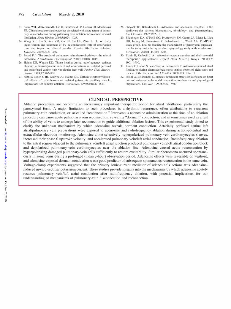
CLINICAL PERSPECTIVEAblation procedures are becoming an increasingly important therapeutic option for atrial fibrillation, particularly theparoxysmal form. A major limitation to such procedures is arrhythmia recurrence, often attributable to recurrentpulmonary-vein conduction, or so-called “reconnection.” Intravenous adenosine administration at the time of an ablationprocedure can cause acute pulmonary-vein reconnection, revealing “dormant” conduction, and is sometimes used as a testof the ability of veins to undergo later reconnection to guide additional ablation lesions. This experimental study aimed toclarify the unknown mechanism by which adenosine reveals dormant conduction. Arterially perfused canine leftatrial/pulmonary vein preparations were exposed to adenosine and radiofrequency ablation during action-potential andextracellular-electrode monitoring. Adenosine alone selectively hyperpolarized pulmonary-vein cardiomyocytes sleeves,increased their phase 0 upstroke velocity, and accelerated pulmonary vein/left atrial conduction. Radiofrequency ablationto the antral region adjacent to the pulmonary vein/left atrial junction produced pulmonary vein/left atrial conduction blockand depolarized pulmonary-vein cardiomyocytes near the ablation line. Adenosine caused acute reconnection byhyperpolarizing damaged pulmonary-vein cells sufficiently to restore excitability. Similar phenomena occurred spontane-ously in some veins during a prolonged (mean 3-hour) observation period. Adenosine effects were reversible on washout,and adenosine-exposed dormant conduction was a good predictor of subsequent spontaneous reconnection in the same vein.Voltage-clamp experiments suggested that the primary ionic-current mediator of adenosine’s actions was adenosine-induced inward-rectifier potassium current. These studies provide insights into the mechanisms by which adenosine acutelyrestores pulmonary vein/left atrial conduction after radiofrequency ablation, with potential implications for ourunderstanding of mechanisms of pulmonary-vein disconnection and reconnection.
972 Circulation March 2, 2010
by guest on October 2, 2016
http://circ.ahajournals.org/D
ownloaded from
Peter G. Guerra, Angel Arenal, Francisco Fernández-Avilés and Stanley NattelTomás Datino, Laurent Macle, Xiao-Yan Qi, Ange Maguy, Philippe Comtois, Denis Chartier,
VeinsMechanisms by Which Adenosine Restores Conduction in Dormant Canine Pulmonary
Print ISSN: 0009-7322. Online ISSN: 1524-4539 Copyright © 2010 American Heart Association, Inc. All rights reserved.
is published by the American Heart Association, 7272 Greenville Avenue, Dallas, TX 75231Circulation doi: 10.1161/CIRCULATIONAHA.109.893107
2010;121:963-972; originally published online February 16, 2010;Circulation.
http://circ.ahajournals.org/content/121/8/963World Wide Web at:
The online version of this article, along with updated information and services, is located on the
http://circ.ahajournals.org/content/suppl/2010/02/12/CIRCULATIONAHA.109.893107.DC1.htmlData Supplement (unedited) at:
http://circ.ahajournals.org//subscriptions/
is online at: Circulation Information about subscribing to Subscriptions:
http://www.lww.com/reprints Information about reprints can be found online at: Reprints:
document. Permissions and Rights Question and Answer this process is available in the
click Request Permissions in the middle column of the Web page under Services. Further information aboutOffice. Once the online version of the published article for which permission is being requested is located,
can be obtained via RightsLink, a service of the Copyright Clearance Center, not the EditorialCirculationin Requests for permissions to reproduce figures, tables, or portions of articles originally publishedPermissions:
by guest on October 2, 2016
http://circ.ahajournals.org/D
ownloaded from

SUPPLEMENTAL MATERIAL
CIRCULATIONAHA/2009/893107/R2 1
Expanded On-line Materials and Methods Section Animals and Tissues
A total of 47 adult mongrel dogs of either sex weighing 18-37 kg were anaesthetized with
pentobarbital (30 mg kg-1 I.V.) and artificially ventilated. Hearts and adjacent lung
tissues were excised via a left thoracotomy and immersed in oxygenated 2-mmol/L Ca2+-
containing Tyrode solution. Animal care procedures followed Canadian Council on
Animal Care guidelines and were approved by the Animal Research Ethics Committee of
the Montreal Heart Institute.
For standard microelectrode experiments, intact tissue preparations including the
LA and adjacent PVs were mounted in a perfusion chamber and perfused via the left-
circumflex coronary-artery with oxygenated Kreb’s solution at 35±0.5°C. Fine-tipped
microelectrodes (resistance 15-20 MΩ when filled with 3-mol/L KCl) coupled to a high
input-impedance amplifier were used to record action potentials (APs) as previously
described.1 A platinum bipolar electrode was used to stimulate the tissue at 2 Hz, with a
pulse duration of 2 ms and twice diastolic threshold current-intensity. Measurements
made from AP-recordings included: resting membrane potential (RMP, transmembrane
potential at phase-0 onset or at steady-state in quiescent preparations), AP-amplitude and
AP-duration (APD) at 90% of repolarization (APD90). Conduction-time was measured
during LA-pacing between the peaks of differentiated (dV/dt) signals for APs recorded
with 2 stable microelectrodes ~1 cm apart, one in a PV and the other in the closest
adjacent LA region.
For cell isolation, the circumflex coronary artery was cannulated and LA and
distal ends of PV myocardial sleeves were marked with silk thread to facilitate
CIRCULATIONAHA/2009/893107/R2 2
identification after enzymatic digestion, prior to ~60-minute enzyme perfusion with Ca2+-
free Tyrode solution containing collagenase (~0.5 mg/mL, CLSII, Worthington) and
0.1% bovine serum albumin (Sigma). Cell isolation was performed as previously
described.2 PVs were well-perfused and single cardiomyocytes could be isolated from all
PVs and from the LA. PV-cells were isolated as far distally from the LA-PV junction as
possible. After isolation, cells were stored at 4°C and studied within 12 hours.
RF-induced Isolation of PVs
PVI was conducted in LA-PV preparations subjected to microelectrode AP-recordings.
One PV was isolated in each dog, by ablating in the antral region as close as possible to
the PV-LA junction. Bipolar electrodes were attached to the epicardial surface of the LA
and target PV to evaluate continuously PV-LA conduction. RF-energy was delivered
epicardially at the PV-LA junction in the unipolar mode between the standard 4-mm tip
of a 7F quadripolar ablation catheter (Cordis-Webster) and an indifferent electrode in the
periphery of the preparation, with a power limit of 25-35 W, for only 10 seconds at each
site to control damage. The endpoint of PVI was the establishment of bidirectional
conduction block (block both from LA to PV and from the PV at 4 different quadrants to
LA, with pacing-stimuli up to maximum possible stimulation-strength).
When PVI had been achieved, APs were recorded in PV sleeves immediately
above the ablation line for 15 minutes following the final RF-application. If conduction
recovered, additional RF was delivered to complete PVI. Adenosine was then added and
APs recorded for an additional 15 minutes in 11 preparations (6 left superior and 5 left
inferior PVs). In preparations that recovered conduction with adenosine, APs were
CIRCULATIONAHA/2009/893107/R2 3
recorded during a final 15-minute adenosine-washout period. In 6 preparations (5 left
superior and 1 left inferior PVs) studied to establish the time-course of spontaneous
changes post-PVI, the same PVI-protocol was conducted without subsequently adding
adenosine and preparations were monitored for up to 4 hours (average monitoring time
3.4±0.3 hours). In 9 other preparations (6 left superior and 3 left inferior PVs), prolonged
monitored was obtained following adenosine-washout to evaluate whether adenosine
reconnection predicts subsequent spontaneous reconnection.
Determination of adenosine-receptor protein-expression
Protein-enriched fractions were obtained from cardiomyocytes isolated as described
above, with paired PV and LA samples from each of 5 dogs studied. Snap-frozen
cardiomyocyte-samples were homogenized in an extraction buffer containing (mmol/L):
Tris 25, NaCl 150, EDTA 5, EGTA 5, NaF 20, Na3VO4 0.2, Glycerol-2-phosphate 20,
AEBSF 0.1; Microcystin-LR 1 μmol/L, leupeptin 25 μg/ml, aprotinin 10 μg/ml, pepstatin
1 μg/ml and Triton-X-100 1%. Homogenized samples were incubated 30 minutes on ice
and centrifuged at 1000×g (10 minutes, 4°C) to pellet debris and nuclei. Supernatant
corresponding to the protein-enriched fraction was collected and protein-concentration
determined by Bradford assay (Biorad). Protein-samples (70-μg) were separated with
sodium-dodecyl-sulfate/polyacrylamide-gel electrophoresis and transferred
electrophoretically to polyvinylidene-fluoride transfer membranes. Membranes were
blocked 1 hour with TTBS (Tris-HCl 50-mmol/L/NaCl 500-mmol/L, pH 7.5, 0.1%-
Tween) containing 5% non-fat dried milk and incubated overnight with primary
antibodies in TTBS/5% non-fat dried milk. Rabbit polyclonal anti-adenosine A1 and A2A-
CIRCULATIONAHA/2009/893107/R2 4
receptor antibodies were obtained from ABCAM (ab3460 and ab3461). Rabbit
polyclonal anti-adenosine A2B and A3-receptor antibodies were obtained from Alpha-
Diagnostics (A2bR23 and A3R32). Primary antibodies were used at a dilution of 1/2000.
A donkey anti-rabbit secondary antibody (minimal cross reactions; Jackson-
Immunoresearch) conjugated to horseradish-peroxidase (HRP) was used for detection.
Staining was revealed with chemiluminescence (Western Lightning Chemiluminescence
Reagent Plus, Perkin-Elmer) and quantified with Quantity-One software (Biorad). All
expression-data are provided relative to GAPDH-staining for the same samples on the
same gels.
Solutions
The solution for cell storage contained (mmol/L): KCl 20, KH2PO4 10, dextrose 10,
mannitol 40, L-glutamic acid 70, β-hydroxybutyrate 10, taurine 20, EGTA 10 and 0.1 %
BSA (pH 7.3, KOH). Tyrode (extracellular) solution contained (mmol/L): NaCl 136,
KCl 5.4, MgCl2 1, CaCl2 1, NaH2PO4 0.33, HEPES 5 and dextrose 10 (pH 7.35, NaOH).
For K+-current recording CdCl2 (200-µmol/L), 4-aminopyridine (4AP, 2-mmol/L) and
atropine (200-nmol/L) were added to suppress Ca2+-current, transient-outward and 4AP-
dependent muscarinic K+-currents.11 The internal solution for K+-current recording
contained (mmol/L): K-aspartate 110, KCl 20, MgCl2 1, MgATP 5, GTP (lithium salt)
0.1, HEPES 10, Na-phosphocreatine 5 and EGTA 10 (pH 7.3, KOH). Inward-rectifier
K+-current was recorded upon stepping from -40 mV to voltages between -120 and +40
mV. Na+-current (INa) contamination was avoided by using a holding potential of -50
mV. Adenosine-dependent K+-current (IKado) was measured as the inward-rectifier
CIRCULATIONAHA/2009/893107/R2 5
difference-current between recordings obtained before and after adenosine in each cell.
For INa-recording, the external solution contained (mmol/L): NaCl 10, TEA-Cl 126,
MgCl2 3.0, CsCl 5.4, HEPES 10, glucose 5.5 (pH 7.35, CsOH), 4AP 2. The internal
solution contained (mmol/L): CsCl 120, TEA-Cl 20, MgCl2 1.0, HEPES 10, EGTA 10,
MgATP 5, Li-GTP 0.1 (pH 7.2, CsOH). For standard microelectrode experiments, a
solution containing (mmol/L): NaCl 120, KCl 4, KH2PO4 1.2, MgSO4 1.2, NaHCO3 25,
CaCl2 1.25 and dextrose 5 (95% O2–5% CO2, pH 7.4), was used to perfuse the tissue.
Adenosine (Sigma Chemicals) was freshly prepared as a 1-mmol/L solution before each
experiment.
Electrophysiology Data Acquisition
The whole-cell patch-clamp technique was used to record currents. Borosilicate glass
electrodes (Sutter Instrument) filled with pipette solution were connected to a patch-
clamp amplifier (Axopatch 200A, Axon). Electrodes had tip resistances of 1 to 4 MΩ,
with pipettes of 1 to 2 MΩ used for INa-recording. Cell capacitance averaged 97±9 pF for
PV and 99 ±7 pF for LA cardiomyocytes. Rs averaged 5.6±0.5 MΩ and the capacitive
time-constant 552±56 µs before, and 2.9±0.3 MΩ and 279±18 µs after, compensation in
LA; with corresponding values in PV of 5.8±0.5 MΩ and 575±59 µs before and 2.6±0.2
MΩ and 242±13 µs after compensation. Leakage-compensation was not used. Currents
are expressed as densities (pA/pF). Junction potentials between bath and pipette solution
averaged 15.0±0.7 mV and were not compensated. K+-current recording was performed
at 35±0.5°C. INa was recorded at room temperature. The maximum phase-0 upstroke-
CIRCULATIONAHA/2009/893107/R2 6
velocity (dV/dtmax) of standard microelectrode recordings was determined by electronic
differentiation.
Statistical Analysis
Data are expressed as mean±SEM. Repeated-measures 1-way ANOVA was used to
compare RMP at different times after PVI. Repeated-measures 2-way ANOVA was used
to study the interaction between location (PV vs LA) and adenosine for RMP, APD90, AP
amplitude and dV/dtmax, the interaction between test potential and treatment (adenosine
versus no adenosine) for K+-current and INa, and the interaction between test potential and
location (PV vs LA) for IKado. Bonferroni-adjusted comparisons were used to compared
group means when ANOVA was significant. Two-way repeated-measures analysis was
used to study interactions between dormant/non-dormant state and adenosine effect on
RMP after PVI. An unpaired Student t-test was used to compare ablation time between
dormant and non-dormant PVs and between control and adenosine-treated PVs. All data
satisfied criteria for normality of distribution (Kolmogrov-Smirnov) except for
conduction time. The effect of drug on conduction time was studied with Wilcoxon
Signed Rank test since it was not normally distributed. Pearson correlation-coefficients
were used to study the relation between RMP and dV/dtmax. Fisher’s exact test was used
to compare dormant-conduction prevalence in different PVs. Whenever values for more
than one cell were obtained per region for a dog, all cell-values in each region were
averaged to obtain single representative values for that dog for statistical comparison, to
avoid weighting values unevenly for different dogs. For regression analyses between
resting potential and Vmax, for which we were interested to know whether resting

CIRCULATIONAHA/2009/893107/R2 7
potential values were a determinant of Vmax values for each individual cell, irrespective of
the dog of origin, we did not average values on a per-dog basis. A two-tailed P<0.05 was
considered statistically-significant. The authors had full access to and take responsibility
for the integrity of the data. All authors have read and agree to the manuscript as written.
CIRCULATIONAHA/2009/893107/R2 8
References
1. Kneller J, Ramirez RJ, Chartier D, Courtemanche M, Nattel S. Time-dependent
transients in an ionically based mathematical model of the canine atrial action
potential. Am J Physiol Heart Circ Physiol. 2002;282:H1437-H1451.
2. Li D, Zhang L, Kneller J, Nattel S. Potential ionic mechanism for repolarization
differences between canine right and left atrium. Circ Res. 2001;88:1168-1175.
On-line Figure Legend
Figure I: Mean±SEM resting membrane potential (RMP) recorded during 30 minutes after
pulmonary vein isolation (PVI) without (A) and with (B) adenosine (ADO) perfusion, and with
isoproterenol perfusion (C). *P<0.05 and ***P<0.001 vs 0-15 minutes, for individual group
mean differences by Bonferroni-adjusted comparisons. D, Mean±SEM difference (RMPd)
between mean RMP recorded during the 15 to 30 minute period (RMP 15-30) and RMP recorded
during the 0 to 15 minute period (RMP 0-15). *** P<0.001 for individual group-mean
differences by Bonferroni-adjusted t-tests.
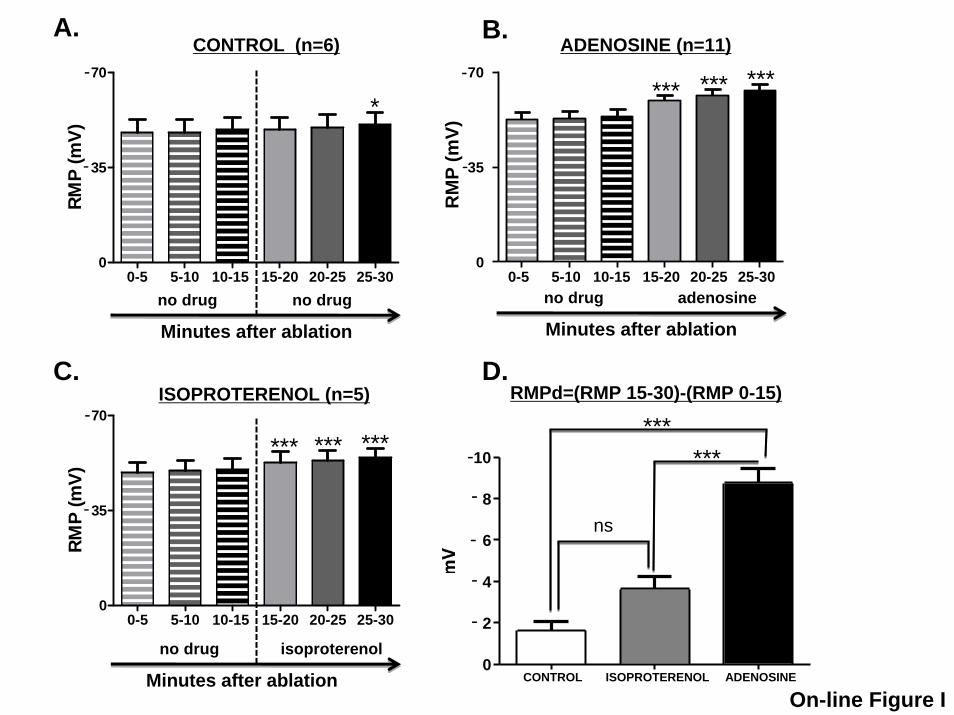
RMPd=(RMP 15-30)-(RMP 0-15)
no drug
CONTROL (n=6)
0-5 5-10 10-15 15-20 20-25 25-300
35
70
-
-RM
P (m
V)
*
Minutes after ablation
no drug
*
A. B.
D.
*** *** ***
adenosine
0-5 5-10 10-15 15-20 20-25 25-300
35
70
-
-
RMP
(mV)
C.ISOPROTERENOL (n=5)
ADENOSINE (n=11)
no drug
Minutes after ablation
no drug isoproterenol
*********
Minutes after ablation
ns
******
On-line Figure I
0-5 5-10 10-15 15-20 20-25 25-300
35
70
-
-
RM
P (m
V)
CONTROL ISOPROTERENOL ADENOSINE0
2
4
6
8
10
-
-
-
-
-
Copyright © 2022 FDOKUMEN