Textural evolution of W-Cu-Sn-bearing hydrothermal veins at ...
11
Textural evolution of W-Cu-Sn-bearing hydrothermal veins at Minas da Panasqueira, Portugal K. A. FOXFORD, R. N1CHOLSON AND D. A. POLYA Department of Geology, University of Manchester, Manchester M13 9PL Abstract Combined structural and textural studies of the quartz veins of the Panasqueira W~Cu-Sn deposit of central Portugal have revealed the presence of coeval forms of crack-seal and cavity-fill mineral textures. The textures result from the dilational and infilling histories of brittle fractures produced within the host rock by the intrusion of pressurised fluids. Crack-seal textures were produced wherever or whenever crystal growth rates matched or were greater than fracture dilation rates, whereas cavity- fill textures developed wherever or whenever dilation rates were quicker than those of crystal growth. Rates of dilation within the blade-like fissures were naturally lowest at the edges, where types of crack- seal textures were produced, and highest in the cores, where cavity-fill texture formed. During propagation the linkage of adjacent echelon blades, by the rupture of the host-rock straps (bridges) between them, enhanced dilation rates and led to the widespread development of cavity-fill textures. Within the composite fissures so produced, environments in which types of crack-seal textures could form were restricted to the fissure margins and to areas where the tensile strain was sympathetically shared between closely spaced fissures. Textures and minerals characteristic of each fracture environment were able to form simultaneously within the fracture system. KEYWORDS: crack-seal, cavity-fill, textures, blade-like fractures, bridges, dilation, hydrothermal. Introduction IN vein-hosted ore deposits, cycles of fracturing, dilation, fluid inflow and mineral deposition produce a wide variety of mineral textures which can be divided into two broad types, crack-seal and cavity-fill, though a continuum exists between them (Robert and Brown, 1986). These textures reflect the structural evolution of the cavities into which the minerals were growing. The actual minerals precipitated at a particular fracture site depend principally upon the fluid chemistry and the character of the fracture walls, which may be formed of either pre- existing vein material or the host-rock itself (Robert and Brown, 1986). Depending upon the nature of the individual fissure environment some minerals are precipitated continuously and simul- taneously but at different locations, whereas others are restricted in both time and space (Barton et al., 1977). For this reason it may not always be possible to define a conventional paragenetic sequence organised such that one Mineralogical Magazine, September 1991, Vol. 55, pp. (~ Copyright the Mineralogical Society mineral follows another in a consistent order of precipitation. At Minas da Panasqueira, Thadeu (1951) and d'Orey (1967) both commented upon the varia- bility of the mineral paragenesis within the segmented veins of the deposit. Kelly and Rye (1979) tried to overcome this difficulty and suggested that as only some minerals showed consistent relative age relationships, any parage- netic sequence should be based chiefly on those minerals. As an aid to understanding such para- genetic variations the temporal and spatial rela- tionship between the end-member textures in the Panasqueira vein system is investigated in this paper using structural and textural observations. Geological setting Minas da Panasqueira is located about 34 km west of Fund~o in the central Portuguese pro- vince of Beira Baixa (Fig. 1). It is underlain by a granitic batholith which forms part of the south- ern flank of the Hercynian granitic complex of 435-445
-
Upload
khangminh22 -
Category
Documents
-
view
1 -
download
0
Transcript of Textural evolution of W-Cu-Sn-bearing hydrothermal veins at ...

Textural evolution of W-Cu-Sn-bearing hydrothermal veins at Minas da Panasqueira,
Portugal
K. A. FOXFORD, R. N1CHOLSON AND D. A . POLYA
Department of Geology, University of Manchester, Manchester M13 9PL
Abstract
Combined structural and textural studies of the quartz veins of the Panasqueira W~Cu-Sn deposit of central Portugal have revealed the presence of coeval forms of crack-seal and cavity-fill mineral textures. The textures result from the dilational and infilling histories of brittle fractures produced within the host rock by the intrusion of pressurised fluids. Crack-seal textures were produced wherever or whenever crystal growth rates matched or were greater than fracture dilation rates, whereas cavity- fill textures developed wherever or whenever dilation rates were quicker than those of crystal growth. Rates of dilation within the blade-like fissures were naturally lowest at the edges, where types of crack- seal textures were produced, and highest in the cores, where cavity-fill texture formed. During propagation the linkage of adjacent echelon blades, by the rupture of the host-rock straps (bridges) between them, enhanced dilation rates and led to the widespread development of cavity-fill textures. Within the composite fissures so produced, environments in which types of crack-seal textures could form were restricted to the fissure margins and to areas where the tensile strain was sympathetically shared between closely spaced fissures. Textures and minerals characteristic of each fracture environment were able to form simultaneously within the fracture system.
KEYWORDS: crack-seal, cavity-fill, textures, blade-like fractures, bridges, dilation, hydrothermal.
Introduction
IN vein-hosted ore deposits, cycles of fracturing, dilation, fluid inflow and mineral deposition produce a wide variety of mineral textures which can be divided into two broad types, crack-seal and cavity-fill, though a continuum exists between them (Robert and Brown, 1986).
These textures reflect the structural evolution of the cavities into which the minerals were growing. The actual minerals precipitated at a particular fracture site depend principally upon the fluid chemistry and the character of the fracture walls, which may be formed of either pre- existing vein material or the host-rock itself (Robert and Brown, 1986). Depending upon the nature of the individual fissure environment some minerals are precipitated continuously and simul- taneously but at different locations, whereas others are restricted in both time and space (Barton et al., 1977). For this reason it may not always be possible to define a conventional paragenetic sequence organised such that one
Mineralogical Magazine, September 1991, Vol. 55, pp. (~ Copyright the Mineralogical Society
mineral follows another in a consistent order of precipitation.
At Minas da Panasqueira, Thadeu (1951) and d 'Orey (1967) both commented upon the varia- bility of the mineral paragenesis within the segmented veins of the deposit. Kelly and Rye (1979) tried to overcome this difficulty and suggested that as only some minerals showed consistent relative age relationships, any parage- netic sequence should be based chiefly on those minerals. As an aid to understanding such para- genetic variations the temporal and spatial rela- tionship between the end-member textures in the Panasqueira vein system is investigated in this paper using structural and textural observations.
Geological setting
Minas da Panasqueira is located about 34 km west of Fund~o in the central Portuguese pro- vince of Beira Baixa (Fig. 1). It is underlain by a granitic batholith which forms part of the south- ern flank of the Hercynian granitic complex of
435-445

436 K. A. FOXFORD E T A L .
Granite ~ Post Hercynian cover
[ - - ] Beira schist ~ Early Precambrian
Fault
FI(~. 1. Map of Portugal showing the location of Panasqueira and the distribution of the Hercynian granitic rocks (redrawn after Inverno and Ribeiro, 1980).
northern Portugal (Kelly and Rye, 1979). The sub-horizontal ore veins are enclosed within a tightly folded schist and greywacke complex of Pre-Cambrian to Carboniferous age, the Beira Schists, into which the granites involved were emplaced. The veins consist predominantly of quartz, with wolframite, cassiterite and chalco- pyrite present in economic concentrations. The paragenetic sequence is quite complicated (for simplified versions see Kelly and Rye, 1979; Polya, 1989).
The veins cut a greisenised granitic cupola, which crops out within the mine workings, but are not restricted to its vicinity. Above the cupola, a silica cap occurs which is continuous with the ore veins (Kelly and Rye, 1979). Using accurate 4~ 39Ar age dating techniques muscovites within the veins have been dated at between 296 and 274 Ma, whereas muscovites from the greisen give ages of 292 Ma (Snee et al . , 1988).
As intersecting fractures show a continuity of mineral textures, Kelly and Rye (1979) along with Thadeu (1951) proposed that the entire vein system was dilated before it was infilled. Heb- blethwaite and Antao (1982), however, reported evidence for incremental dilation deeper within the deposit.
From fluid inclusion evidence, Kelly and Rye (1979) report that the fluids locally attained pressures of 1 kbar within the silica cap though
elsewhere in the vein system the fluids were at pressures close to 200 bars. Fluid temperatures near 300~ and salinities less than 10 weight percent were also proposed from similar evi- dence. During the early stages of emplacement there is occasional evidence that these fluids effervesced CO2. Significant quantities of CH4 and N2 were also present which suggests that the fluids may have had some components obtained from organic matter within the Beira Schists (Bussink, 1984). These authors proposed depths of formation for the fracture system of between 1 and 2 km below the water table using fluid inclusion results and phase relationships in the system NaC1-H20-CO2 for a boiling (effer- vescing) fluid.
Structural interpretation of the vein system
Kelly and Rye (1979) recognised that the Panasqueira vein morphologies suggested that they were predominantly dilational structures formed by the forceful intrusion of fluid. They compared the structures (stepped margins and splitting veins) to those seen within magmatic dykes by Pollard (1973) and they similarly pro- posed that the fluids responsible for fracturing and dilation forced their way upwards into the schists from a reservoir below the deposit. The driving mechanism for the extension within the

THE P A N A S Q U E I R A
Beira Schists was fluid pressure. The processes causing such abnormal fluid pressures within the area have been linked to the crystallisation of granite and the release of volatiles, though the involvement of meteoric waters has also been inferred (Kelly and Rye, 1979; Polya, 1989).
Because of the structural style and shallow depth of formation, the veins at Panasqueira must represent an infilled brittle fracture system. Such brittle fracturing occurred within the Beira Schists and was caused by the intrusion into them of pressurised fluids. Those fluids counteracted the normal conditions of compressive stress present within the crust and produced effective tensile stress (Hubbert and Rubey, 1959; Jaeger, 1963; Secor, 1965). Fracture propagation is envisaged as a seismic, pulsatory process and could be very similar to the mechanism of autogenous hydraulic pulsation responsible for the early stages of lode formation in Cornwall (Halls, 1987).
Fig. 2 is a schematic diagram depicting the morphology of a section of an ore vein and can be used to describe fracture propagation and its associated cavity dilation. Each blade-like frac- ture (Fig. 2a) initially propagated as a mode I tensile crack (Pollard and Aydin, 1988) but later, as the fractures overlapped (Fig. 2b and c) the mechanical interaction between them became significant (Pollard et at., 1982) and they became mode III anti-plane shear cracks. Nicholson and Pollard (1985) have shown how dilation, in cross- sectional or profile view is enhanced once fracture overlap has occurred, and, how the straps of wallrock between adjacent fractures (seen in profile as bridges) start to bend producing sig- moidally shaped cavities (Fig. 2b and c).
The fracture dilation rate, as seen at various places in such profile views, depends upon the twist angle of the fracture array, the fracture separation and the fluid pressure, as well as location within the cavity. For each blade-like fracture segment the dilation rate towards the fracture edges and tip was naturally less than that attained within the centre of the cavity. Further propagation and dilation caused the over-flexure and rupture of distal portions of the bridges, away from the fracture tips, (Fig. 2d and Fig. 3A) between the separate blades within the echelon array. This led to the linkage of fracture seg- ments, as seen in profile, and the formation of composite fissures, each several times the width of the original fracture segment. These composite fissures attained faster dilation rates, for a given amount of deformation, than the isolated cavities could achieve alone. They too have rates of mineral-infilling faster than rates of dilation around the fracture tips.
W-Cu-SN DEPOSIT 437
The composite fissures inherited the strike and dip of the arrays rather than that of the propagat- ing fracture tips. The twist angles of the arrays are quite variable and fractures are locally stacked in the opposite sense which results in composite fissures with seemingly complex morphologies and geometries. As for discrete fractures the dila- tion rates within portions of composite fissures also depended upon the proximity of neighbour- ing fractures which sympathetically helped to relieve the local tensile stresses developing within the host-rock.
Strike-slip faults which cut the Panasqueira vein system have been investigated by Blaettler (1985) who proposed that faults with argiIIaceous fillings were active pre-, syn- and post-ore vein formation. No evidence has been found within this study to support the suggestion that any of the major faults existed prior to vein formation. The changes in vein-thickness across the faults can be explained easily by strike-slip displacements of echelon vein structures. The only fault structures cut by the veins are a series of small conjugate shears which are found within psammitic schists and are related to the Hercynian deformation. The major faults displace the vein system pas- sively and are interpreted to have formed after the main period of vein dilation and infilling. Propagation of the sub-horizontal fractures was therefore not limited by the translation of the dilation to the surface by faults. As a consequence the lateral change of dilation or the percentage vertical dilation of the schist (the dilation an- omaly) is always gradual (Hebblethwaite and Antao, 1982).
Textures
Within the ore veins forms of crack-seal, cavity- fill and coarse fibrous textures occur. A typical vein profile consists of a thin, marginal, crack-seal quartz seam separated from a central area of cavity-fill texture by a zone of fibrous texture. These textures are displayed in Figs. 3 and 4 and they have been studied using conventional mi- croscopy and cold-stage cathodoluminescence techniques. Fig. 3B shows cavity-fill texture within an ore vein and Fig. 3C early crack-seal quartz seams and their associated tourmalinisa- tion. In Fig. 4A and B varieties of fibrous texture are displayed.
Marginal crack-seal quartz seams. The first vein mineral precipitates were thin seams of quartz now found along the margins of the widely opened veins (Bussink, 1984; Polya, 1989). These quartz seams also occur along joint planes which remained essentially undilated during the rest of

438 K . A . F O X F O R D E T A L .
Section of ore-vein from the 'black' vein (L2D23R3AW25)
575m elevation
F N
s F _ _ 1 - -
Area shown schematically below lOm
Propagation direction
(Parallel to
fracture edge
Fracture edge -seal vein
lm
Bridge stub
Cavity-fill texture
Fibrous texture
FIG. 2. Top: section of the 'black' vein (L2D23R3AW25). Bottom: three-dimensional schematic of the marked section; (a)-(d) are cross-sections through it showing the fracture tip (a), sigmoidal cavities (b and c) and cross- fractured bridges (d). The sections a-d represent both lateral changes in fracture morphology and successive stages of cavity evolution with time. Near to the fracture tip (a) the mineral texture formed was of crack-seal type. As the fractures propagated and dilated the bridges began to bend which allowed more rapid crack dilation. The texture produced at this stage was a macroscopic, fibrous texture. Continued growth of the fractures (c) combined with increased bridge rotation further enhanced the rates of dilation. Fibrous texture still formed adjacent to the bridges (where the dilation is shared between adjacent fractures) but in the centre of the sigmoidal fracture segments cavity- fill texture was produced. Upon bridge rupture (d) the dilation rate increased dramatically and cavity-fill texture
predominated.
THE PANASQUEIRA W-Cu-SN DEPOSIT 439
F~6.3. Photographs of vein textures from the black vein within Level 2. (A) Portion of an ore vein, such as that shown schematically in Fig. 2, which formed through the linkage of fracture segments by bridge rupture. (B) Cavity- fill texture consisting of muscovite (M), wolframite (black mineral), quartz (Q), pyrite (p) and chalcopyrite (c), (C) Crack-seal veins cut by a later ore vein which together form a conjugate set. The dark alteration of the wallrock
associated with the crack-seal veins is tourmalinisation. Scale divisions are 10 cm.
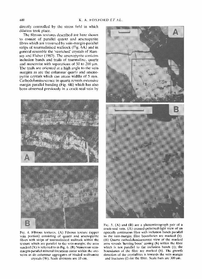
the mineralisation. Such joints are often asso- ciated with intense tourmalinisation of the rock alongside them (Marignac, 1982). In thin section these early quartz veins show a form of crack-seal growth and contain inclusion bands of muscovite, tourmaline and quartz arranged parallel to the vein margin, as well as inclusion trails of quartz and tourmaline at high angles to it (Fig. 5A). The inclusions evidently formed through syntaxial overgrowth only to be included at intervals into quartz fibres growing by an antitaxial crack-seal process (Cox, 1987). Individual dilation increments range from 50 to 200 ~tm and quartz fibres themselves can be up to 1 mm in width and 5 to 10 mm in length. Inclusion bands are defined by included crystals, generally 25 ~tm in length though tourmaline needles are often longer. Inclusion bands are frequently restricted to single quartz fibres and end before the fibre margins are reached (Fig. 5A). Tourmaline needles in the inclusion trails can attain lengths of 400 ~tm.
Using cathodoluminescence (Fig. 5B) a distinct herring-bone zoning structure is induced within each of the dilational increments. This zoning is not parallel, however, to the vein-margin. This suggests that each fibre grew towards the wallrock as a series of discrete crystallites each approxi- mately 400 ~tm wide. This variety of crack-seal growth bears a resemblance to that portrayed by Cox and Etheridge (Fig. 13, 1983), though they show no detail of the growth mechanism for the quartz fibres they describe.
Fibrous textures occur next to the early crack- seal quartz seams at the vein margins. They were first reported at Panasqueira by Hebblethwaite and Antao (1982) and were explained by Polya (1989) as being the due to progressive crystal growth under conditions of tensile stress (Philips, 1986), principally during the early stage of vein opening. This implies that at times the fractures were completely filled with mineral precipitates and that the growth and shape of crystals was
440 K . A . F O X F O R D E T A L .
directly controlled by the stress field in which dilation took place.
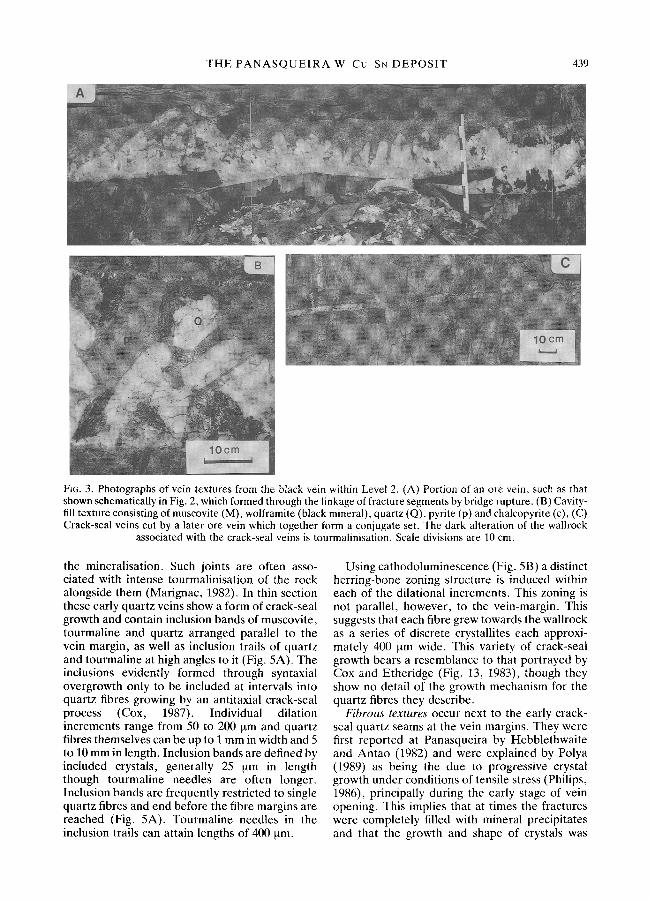
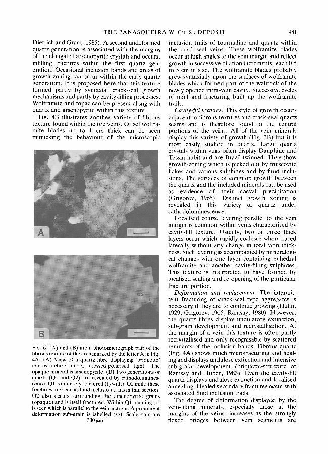
The fibrous textures described are here shown to consist of parallel quartz and arsenopyrite fibres which are traversed byvein-margin-paral lel strips of tourmalinised wallrock (Fig. 6A) and in general resemble the 's t retched' crystals of Ram- say and Huber (1983). The arsenopyrite contains inclusion bands and trails of tourmaline, quartz and muscovite with separations of 50 to 200 ~tm. The trails are oriented at a high angle to the vein margins as are the columnar quartz and arseno- pyrite crystals which can attain widths of 5 mm. Cathodoluminescence in quartz reveals extensive margin-parallel banding (Fig. 6B) which has also been observed previously in a crack-seal vein by
Fro. 4. Fibrous textures. (A) Fibrous texture (upper vein portion) consisting of quartz and arsenopyrite fibres with strips of tourmalinised wallrock within the texture which are parallel to the vein-margin; the area marked (X) is referred to in Fig. 6. (B) Numerous vein- margin-parallel mineral lineations occur within the ore- veins as do columnar aggregates of bladed wolframite
crystals (W). Scale divisions are 10 cm.
FIG. 5. (A) and (B) are a photomicrograph pair of a crack-seal vein. (A) crossed-polarised-light view of an optically continuous fibre with inclusion bands parallel to the vein-margin; fibre boundaries are marked (b). (B) Quartz cathodoluminescence view of the marked area reveals 'herring-bone' zoning (h) within the fibre which is not parallel to the inclusion bands (i); the boundaries of the fibre are marked (b). The growth direction of the crystallites is towards the vein margin and fractures (f) cut the fibre. Scale bars are 300 ~tm.
THE P A N A S Q U E I R A
Dietrich and Grant (1985). A second undeformed quartz generation is associated with the margins of the elongated arsenopyrite crystals and occurs, infilling fractures within the first quartz gen- eration. Occasional inclusion bands and areas of growth zoning can occur within the early quartz generation. It is proposed here that this texture formed partly by syntaxial crack-seal growth mechanisms and partly by cavity-filling processes. Wolframite and topaz can be present along with quartz and arsenopyrite within this texture.
Fig. 4B illustrates another variety of fibrous texture found within the ore veins. Offset wolfra- mite blades up to 1 cm thick can be seen mimicking the behaviour of the microscopic
FIG. 6. (A) and (B) are a photomicrograph pair of the fibrous texture of the area marked by the letter X in Fig. 4A. (A) View of a quartz fibre displaying 'briquette' microstructure under crossed-polarised light. The opaque mineral is arsenopyrite. (B) Two generations of quartz (Q1 and Q2) are revealed by cathodolumines- cence. Q1 is intensely fractured (O with a Q2 infill; these fractures are seen as fluid inclusion trails in thin section. Q2 also occurs surrounding the arsenopyrite grains (opaque) and is itself fractured. Within Q1 banding (z) is seen which is parallel to the vein-margin. A prominent deformation sub-grain is labelled (sg). Scale bars are
300 ~tm.
W-Cu-SN DEPOSIT 441
inclusion trails of tourmaline and quartz within the crack-seal veins. These wolframite blades occur at high angles to the vein margin and reflect growth in successive dilation increments, each 0.5 to 5 cm in size. The wolframite blades probably grew syntaxially upon the surfaces of wolframite blades which formed part of the wallrock of the newly opened intra-vein cavity. Successive cycles of infill and fracturing built up the wolframite trails.
Cavity-fill textures. This style of growth occurs adjacent to fibrous textures and crack-seal quartz seams and is therefore found in the central portions of the veins. All of the vein minerals display this variety of growth (Fig. 3B) but it is most easily studied in quartz. Large quartz crystals within vugs often display Dauphin6 and Tessin habit and are Brazil twinned. They show growth-zoning which is picked out by muscovite flakes and various sulphides and by fluid inclu- sions. The surfaces of common growth between the quartz and the included minerals can be used as evidence of their coeval precipitation (Grigorev, 1965). Distinct growth zoning is revealed in this variety of quartz under cathodoluminescence.
Localised coarse layering parallel to the vein margin is common within veins characterised by cavity-fill texture. Usually, two or three thick layers occur which rapidly coalesce when traced laterally without any change in total vein thick- ness. Such layering is accompanied by mineralogi- cal changes with one layer containing euhedral wolframite and another cavity-filling sulphides. This texture is interpreted to have formed by localised sealing and re-opening of the particular fracture portion.
Deformation and replacement. The intermit- tent fracturing of crack-seal type aggregates is necessary if they are to continue growing (Hulin, 1929; Grigorev, 1965; Ramsay, 1980). However, the quartz fibres display undulatory extinction, sub-grain development and recrystallisation. At the margin of a vein this texture is often partly recrystallised and only recognisable by scattered remnants of the inclusion bands. Fibrous quartz (Fig. 4A) shows much microfracturing and heal- ing and displays undulose extinction and intensive sub-grain development (briquette-structure of Ramsay and Huber, 1983). Even the cavity-ill[ quartz displays undulose extinction and localised annealing. Healed secondary fractures occur with associated fluid inclusion trails.
The degree of deformation displayed by the vein-filling minerals, especially those at the margins of the veins, increases as the strongly flexed bridges between vein segments are

442 K. A. FOXFORD E T A L .
approached. Throughout the veins the textures are cut by vein-margin-parallel fractures filled with later minerals such as muscovite and carbonates.
Many minerals within the veins show signs of dissolution, alteration or replacement. Especially affected are topaz and pyrrhotite which are extensively replaced. Often, minerals more fre- quently associated with later stages in the parage- nesis are found at or near the vein margins. This has led to some complication when comparing the paragenetic sequence from locality to locality.
Discussion
At Panasqueira the area of the dilation an- omaly within the Beira schists, as measured in boreholes (Hebblethwaite and Antao, 1982), shows only gentle lateral gradients, showing that the host-rock behaved coherently as a competent mass. As a consequence, the dilation can be considered as approximately invariant laterally on the 10 m scale. At this scale any sudden thinning of an individual vein segment must be offset by the thickening of an adjacent overlap- ping vein-segment (often not visible and on the hidden side of a wallrock bridge).
Within an opening fracture, the type of texture which forms (crack-seal, fibrous or cavity-fill) depends upon the relative rates of dilation and crystal growth. The scale and rate of dilation was lowest at the terminations of individual bladed segments, where crack-seal and fibrous textures are often found, whereas in the segment centres the enhanced dilation lead to large-scale cavity- fill textures. Consequently a variety of growth textures is formed within the same fracture segment. The same variation also occurs from margin to core of a composite vein. The dilation anomaly as a whole shows a progressive decrease with distance away from its centre suggesting decreased rates of dilation around the margin of the deposit. Changes in mineral textures thus reflect both contemporaneous differences in rates of opening, associated with the initial vein- segment geometries, and changes in the rate of dilation with time, such as after bridge rupture and fracture linkage.
The initial stages of dilation (synchronous with the propagation of fracture tips) gave rise to forms of crack-seal textures (Fig. 2a) and were accompanied by tourmalinisation of the schist. As propagation and dilation continued, stretched crystals showing macroscopic fibrous textures were formed (Fig. 2b). These are evidently a large-scale version of crack-seal type texture in which the fibres and inclusion trails reflect the
local shear stress orientation imposed by dilation of fractures arranged en eche lon .
Following bridge rupture, fibrous textures formed only in slowly dilating habitats, adjacent to the larger, thicker and rate-controlling bridges which separated the composite veins from one another. Cavity-fill textures formed wherever crystal growth rates were slower than rates of fracture opening (Fig. 2d).
Kelly and Rye (1979) commented upon the predominance of cavity-fill textures (Fig. 3B), and the large size of the crystals within the ore veins. They attributed these features to prolonged crystal growth in open cavities under conditions of near equilibrium saturation. Differences in crystal growth rates due to the orientation of the greatest growth vectors (Buckley, 1957; Dickson, 1983), the density of nuclei (Grigorev, 1965) and the characteristics of the specific infilling mineral species all affect crystal growth rates, as well as the degree of supersaturation (supercooling). The absence of fibrous textures, due solely to competi- tive growth (Grigorev, 1965), suggests a paucity of initial crystal nuclei. The muscovite selvedges evidently prevented heterogeneous nucleation of later minerals on wallrock grains but such nuclea- tion probably occurred upon seed fragments washed in with the fluid (Elwell and Scheel, 1975, p. 140, Berner, 1980, p. 95). Those nuclei that were present could attain large sizes irrespective of orientation and the length of the growth period. For crack-seal veins the muscovite sel- vedges are absent and heterogeneous nucleation occurred upon wallrock grains. The most favour- ably oriented quartz fibres traversed the cavity first (Cox and Etheridge, 1983) and attained the greatest widths.
Temperature changes can be important in localising minerals but such changes occur ex- tremely slowly above a cooling batholith (Norton, 1979; Helgeson and Lichtner, 1987) and they probably play only a minor role in the localised precipitation of minerals. Pressure changes and wallrock reactions are the more likely controls. Pressure fluctuations, indicated by sudden changes in the rates of dilation and therefore of fracture propagation, could be important in precipitation and dissolution of both quartz and wolframite. These minerals display reduced solu- bilities upon pressure reduction at temperatures near to 300 ~ (Fournier, 1985; Polya, 1990). One mechanism for changing pressures rapidly within vein systems is by the rupture of bridges between fracture segments. The resultant sudden increase in fracture length allows rapid dilation and a slight drop of fluid pressure. The ease of dilation, however, is controlled by the stiffness of the

THE P A N A S Q U E I R A
whole system. Pulsatory fluid release into the fracture system could be expected, however, and is a viable mechanism for overpressuring the system and promoting rapid but short-lived epi- sodes of fracture propagation, dilation and bridge rupture.
Varying fluid disequilibrium with respect to the fracture wallrock (de Caritat, 1990), caused by changing fluid-rock ratios in cavities of differing size and differing wallrock materials, could also affect the types and quantities of minerals precipi- tated. Re-opening of infilled fractures at a schist- vein contact allows the fluid access to the host schist whereas entirely intra-vein cavities confine the fluids to bathe only material which has already precipitated from them (Robert and Brown, 1986).
Tourmaline was precipitating throughout much of the time mineral infilling of the veins took place, but only achieved a macroscopic size early in the paragenesis (Kelly and Rye, 1979). In this study, sizeable tourmaline crystals are only observed within the veins in situations where they were able to overgrow early formed wallrock tourmaline. Muscovite occurs as a number of macroscopic generations within the vein minerals as well as in the form of microscopic inclusions within quartz crystals where it is often found together with tourmaline. Both of these minerals were evidently precipitating virtually continu- ously in both time and space (Kelly and Rye, 1979). Their development in a macroscopic form seems to have depended upon local fluid chemistry and the presence of the correct substrate.
In spite of a complex history of growth, tourmaline and muscovite are restricted to the main-oxide-silicate and early main-sulphide stages, and are predominantly early minerals. Indeed tourmaline, especially, formed at the fracture tips where wallrock tourmalinisation and crack-seal textures were produced. Minerals asso- ciated with later paragenetic stages formed in environments further from the propagating frac- ture tips and display textures characteristic of such environments. In this way the lateral mi- gration of growth environments as the fractures propagate and dilate can build up a vertical sequence of textural and mineral variation (Fig. 2).
About 90% of the volume of the vein system was created through dilation during the early oxide-silicate stage of the paragenesis. This stage of evolution must have been linked directly to the development of effective tensile stress within the Beira Schists. This stage also hosts the bulk of the economic mineralisation (Kelly and Rye, 1979).
W-Cu-SN DEPOSIT 443
The rate of dilation had decreased by the time the main-sulphide stage began and opening had evidently ceased by the start of the pyrrhotite alteration stage.
Numerous vein-margin-parallel trails of sui- phides and silicates (Fig. 4A and B) occur within the veins, together with similarly oriented trails of fluid inclusions (Fig. 5A and 6A). These repre- sent healed fractures caused by repeated fractur- ing and opening of the vein system. Such re- opening of cemented fractures took placed either along the margins or within the mineral infillings. Deformation of the mineral aggregates is intima- tely linked to the flexure of bridges encrusted with mineral precipitates as well as to repeated acts of dilation and infilling. The bridge flexure causes localised areas of compression and dilation. Waves of elastic strain produced by the on-going brittle deformation may have dislodged mineral aggregates from the upper surface of the cavities and stresses between growing crystals may have been important for some of the observed fractur- ing of cavity-fill texture.
These mechanisms which arose through defor- mation appear to have operated throughout the evolution of the vein system allowing fluids to permeate the mineral aggregates. Such processes may explain the widespread replacement of topaz and the occurrence of late-formed minerals within aggregates which indicate early formation.
Conclusions
The formation of fibrous and cavity-fill textures was coeval in the Panasqueira vein system as a result of the interplay between dilation rates and
r a t e s of mineral growth within the evolving fracture system. Textural facies can be traced laterally within the vein system and reflect local structural styles as well as the overall temporal cycle of increasing then decreasing dilation rates. Deformation was an essential part of the develop- ment of the system and opened channels and cavities within paragenetically early minerals. This permitted the trapping of secondary inclu- sions upon healing and allowed fluids to reach and react with previously formed precipitates. Suc- cessive intra-vein and vein-margin reopening of the fracture system as the composition of the fluids evolved, results in a complex paragenetic sequence. The paragenesis deciphered from growth relationships within individual veins shows that considerable variations exist along them. Combined structural and textural studies of such veins are essential for the construction of meaningful paragenetic diagrams.
444
Acknowledgements
This paper would not have been possible without the help and hospitality of the staff of Beralt Tin and Wolframite, (Portugal) S.A. at Barroca Grande. In particular our thanks go to A. Correia de Sa and W. Berry for making these visits possible and to M. Rotolo and his staff for all their advice and assistance. Our appreciation also goes to R. P. B. Hebblethwaite for his valuable advice and discussion. K. A. F. would especially like to acknowledge a NERC/CASE award (GT4188/GS/79) with Charter Consolidated PLC.
References
Barton, P. B., Bethke, P. M., and Roedder, E. (1977) Environment of ore deposition in the Creede mining district, San Juan mountains, Colerado: part III progress toward the interpretation of the chemistry of ore-forming fluid of the OH vein. Econ. Geol., 72, 1-24.
Berner, R. A. (1980) Diagenetic chemical processes II: precipitation, dissolution and authigenic procecces. In Earl)' diagenesis: a theoretical approach, Princeton University Press, 241pp.
Blaettler, A. J. (1985) Structural analysis of the fault pattern in the tungsten-tin mine of Panasqueira, Portugal. Unpublished thesis, University of Geneva, 77pp.
Buckley, H. E. (1957) Crystal Growth. J. Wiley & Sons, New York, 571pp.
Bussink, R. W. (1984) Geochemistry of the Panasqueira tungsten-tin deposit, Portugal. Unpublished Ph.D thesis, University of Utrecht, Netherlands.
Cox, S. F. (1987) Antitaxial crack-seal vein microstruc- tures and their relationship to displacement paths. J. Struct. Geol., 9,779-88.
- - a n d Etheridge, M. A. (1983) Crack-seal fibre growth mechanisms and their significance in the development of oriented layer silicate microstruc- tures. Tectonophys., 92, 147-70.
de Caritat, P. (1990) Persistence of quartz disequilib- rium in groundwater flows. Terra Nova, 2, 53-9.
Dickson, J. A. D. (1983) Graphical modelling of crystal aggregates and its relevance to cement diagnosis. Phil. Trans. Roy. Soc. London, A309, 465--502.
Dietrich, D. and Grant, P. R. (1985) Cathodolumin- escence petrography of syntectonic quartz fibres. J. Struct. Geol., 7,541-53.
d'Orey, F. C. (1967) Tungsten-tin mineralisation and paragenesis in the Panasqueira and Vale das Ermida mining districts, Portugal. Comunic. Serv. Geol., Portugal, 52, 117-67.
Elwell, D. and Scheel, H. J. (1975) Crystalgrowthfrom high temperature solutions. Academic Press, London, 634pp.
Fournier, R. O. (1985) The behaviour of silica in hydrothermal solutions. In: Berger, B. R. & Bethke, P. M. (eds): Reviews in Economic Geology, 2, 45-61.
Grigorev, D. P. (1965) Ontogeny of Minerals. Israel program for scientific translations, Jerusalem, 260pp.
Halls, C. 1987 A mechanistic approach to the paragen-
K. A. F O X F O R D E T A L .
etic interpretation of mineral lodes in Cornwall. Proc. Ussher Soc., 6, 548-54.
Hebblethwaite, R. P. B. and Antao, A. M. (1982) A report on the study of dilation patterns within the Panasqueira orebody. Unpublished Beralt Tin and Wolfram, Portugal report.
Helgeson, H. C. and Lichtner, P. C. (1987) Fluid flow and mineral reactions at high temperatues and pressures. J. Geol. Soc. London, 144, 313-26.
Hubbert, M. K. and Rubey, W. W. (1959) Role of fluid pressure in mechanics of overthrust faulting, 1, Mechanics of fluid filled porous solids and its appli- cation to overthrust faulting. Geol. Soc. Amer. Bull., 70, 115-66.
Hulin, C. D. 1929. Structural controls of ore deposition. Econ. Geol., 24, 15-49.
Inverno, C. and Ribeiro, M. L. (1980) Fracturacas e catejo nas Minas da Argemela (Fund~o). Comunic. Serv. Geol. Portugal, 66, 185-94.
Jaeger, J. C. 1963. Extension failures in rocks subject to fluid pressure. J. Geophys. Research, 68, 6066-7.
Kelly, W. C. and Rye, R. O. (1979) Geologic, fluid inclusion and stable isotope studies of the tin- tungsten deposits of Panasqueira, Portugal. Econ. Geol., 74, 1721-822.
Marignac, C. (1982) Geologic, fluid inclusions and stable isotope studies of the tin-tungsten deposits of Panasqueira, Portugal--a discussion. Ibid, 77, 1263-6.
Nicholson, R. and Pollard, D. D. (1985) Dilation and linkage of echelon cracks. J. Struct. Geol., 7, 583-90.
Norton, D. (1979) Transport phenomena in hydro- thermal systems: the redistribution of chemical com- ponents around cooling magmas. Bull. Mineral., 102, 471-86.
Philips, W. J. (1986) Hydraulic fracturing effects in the formation of mineral deposits. Trans. Inst. Mining MetalL, 95, 17-24.
Pollard, D. D. (1973) Derivation and evaluation of a mechanical model for sheet intrusions. Tectonophys., 19, 233-69.
- - a n d Aydin, A. (1988) Progress in understanding jointing over the past century. Geol. Soc. Amer. Bull., 100, 1181-204.
- - S e g a l , P., and Delaney, P. T. 1982 Formation and interpretation of dilatant echelon cracks. Ibid. 93, 1291-303.
Polya, D. A. (1989) Chemistry of the mainstage ore- forming fluids of the Panasqueira W-Cu(Ag)-Sn deposit, Portugal: implications for models of ore genesis. Econ. Geol., 84, 1134-52.
Polya, D. A. (1990) Pressure dependence of wolframite solubility for hydrothermal vein formation. Trans. Inst. Mining Metall., 99, B120-4.
Ramsay, J. G. (1980) The crack-seal mechanism of rock deformation. Nature, London, 284, 135-9.
and Huber, M. I. (1983) The techniques of modern structural geology, Vols. 1 and 2, Academic Press, London, p. 307 and 700.
Robert, F. and Brown, A. C. (1986) Archean gold bearing quartz veins at the Sigma mine, Abitibi

T H E P A N A S Q U E I R A W - C u - S N D E P O S I T 445
greenstone belt, Quebec: part II vein paragenesis and hydrothermal alteration. Econ. Geol., 81,593~16.
Secor, D. T. (1965) Role of fluid pressure in jointing. Amer. J. Sci., 263,633-46.
Snee, L. W., Sutter, J. F., and Kelly, W. C. (1988) Thermochronology of economic deposits: dating the stages of mineralisation at Panasqueira, Portugal, by
high precision 40Ar/39Ar age spectrum techniques on muscovite. Econ. Geol., 83,335-54.
Thadeu, D. (1951) Geologia do couto mineiro da Panasqueira, Communic. Serv. Geol. Portugal, 32, 5-64.
[Revised manuscript received 9 May 1991]