MEM-net and EchoColorDoppler assessment of veins draining the brain
8
© 2014 Mandolesi et al. This work is published by Dove Medical Press Limited, and licensed under Creative Commons Attribution – Non Commercial (unported, v3.0) License. The full terms of the License are available at http://creativecommons.org/licenses/by-nc/3.0/. Non-commercial uses of the work are permitted without any further permission from Dove Medical Press Limited, provided the work is properly attributed. Permissions beyond the scope of the License are administered by Dove Medical Press Limited. Information on how to request permission may be found at: http://www.dovepress.com/permissions.php Journal of Vascular Diagnostics 2014:2 59–66 Journal of Vascular Diagnostics Dovepress submit your manuscript | www.dovepress.com Dovepress 59 STUDY PROTOCOL open access to scientific and medical research Open Access Full Text Article http://dx.doi.org/10.2147/JVD.S60526 Using the MEM-net program to report on mapping the EchoColorDoppler assessment for chronic cerebro spinal venous insufficiency Sandro Mandolesi 1 Aldo d’Alessandro 2 Ettore Manconi 3 Tarcisio Niglio 4 Augusto Orsini 5 Dimitri Mandolesi 6 Alessandro d’Alessandro 7 Francesco Fedele 1 1 Department of Cardiovascular and Respiratory Sciences, Sapienza University Rome, Rome, Italy; 2 Department of Angiology, “T Masselli-Mascia” Hospital, San Severo (FG), Foggia, Italy; 3 Department of Cardiovascular and Neurological Sciences, University of Cagliari, Cagliari, Italy; 4 Istituto Superiore di Sanità, Rome, Italy; 5 Department of Vascular Surgery, “Gioia” Hospital, Sora (FR), Sora, Italy; 6 Medicina del lavoro Sapienza University Roma, Rome, Italy; 7 Faculty of Medicine, Catholic University “Our Lady of Good Counsel,” Tirana, Albania Correspondence: Sandro Mandolesi Via San Martino della Battaglia 25, Rome 00185, Italy Tel +39 335 6512 303 Fax +39 06 4873 984 Email [email protected] Introduction: Chronic cerebrospinal venous insufficiency (CCSVI) is characterized by multiple stenosis/obstructions affecting the principal extracranial outflow pathways of the cerebrospinal venous system. Using EchoColorDoppler (ECD) to assess chronic CCSVI is a very difficult and long examination. It takes about an hour even for an expert sonographer. Methods: Hemodynamic morphological map (MEM-Net) is a program that works on the Internet. All the morphological and hemodynamic data of the patient can be entered into the program’s anatomical scheme to create a map of the ECD report. The program also allows us to collect all the data during the ECD assessment and, using its algorithm, make the report uniform. Conclusion: Reporting on the map by using MEM-net shortens the time of ECD written reporting that is done automatically. The program also makes a blind control of the report and enables the use of it for scientific research. We hope that in the future everyone will use this data collection tool for all scientific work on this topic. Keywords: CCSVI, ECD, Map, cerebral venous system, sonography Introduction Chronic cerebrospinal venous insufficiency (CCSVI) is characterized by multiple stenosis/obstructions affecting the principal extracranial outflow pathways of the cerebrospinal venous system, the internal jugular veins (IJVs), and the azygos vein, distributed in four main hemodynamic patterns. 1 Furthermore, CCSVI determines significant changes in the cerebral venous hemo- dynamic, with a very high incidence of reflux in both intracranial and extracranial venous segments and loss of the postural regulation of cerebral venous outflow. 1,2 Recently, Zamboni et al 1 suggested five EchoColorDoppler (ECD) venous criteria that characterize this syndrome: 1) reflux constantly present in an outflow pathway of IJVs or vertebral veins (VVs) assessed in supine and standing posture (0° and 90°); 2) reflux propagated upward to the deep cerebral veins (DCVs), including superficial or internal cerebral vein, basal vein, Galen vein; 3) evidence of IJVs stenosis; 4) flow not Doppler detectable in the IJVs and VVs assessed in supine and standing posture (0° and 90°) and ; 5) negative difference in cross sectional area (CSA) of the IJV assessed in supine and standing posture (0° and 90°) in the IJV (ΔCSA). 3 The presence of two of them is enough to diagnose CCSVI. Using ECD to assess CCSVI is a very difficult and long examination, requiring about an hour of work even for an expert sonographer. It involves a venous ECD assessment of the main veins draining the brain in the supine position, a venous ECD assessment in the upright position, a Manconi breathing test, a neck rotation test,
Transcript of MEM-net and EchoColorDoppler assessment of veins draining the brain
© 2014 Mandolesi et al. This work is published by Dove Medical Press Limited, and licensed under Creative Commons Attribution – Non Commercial (unported, v3.0) License. The full terms of the License are available at http://creativecommons.org/licenses/by-nc/3.0/. Non-commercial uses of the work are permitted without any further
permission from Dove Medical Press Limited, provided the work is properly attributed. Permissions beyond the scope of the License are administered by Dove Medical Press Limited. Information on how to request permission may be found at: http://www.dovepress.com/permissions.php
Journal of Vascular Diagnostics 2014:2 59–66
Journal of Vascular Diagnostics Dovepress
submit your manuscript | www.dovepress.com
Dovepress 59
S t u Dy P r o t o c o l
open access to scientific and medical research
open Access Full text Article
http://dx.doi.org/10.2147/JVD.S60526
using the MEM-net program to report on mapping the EchocolorDoppler assessment for chronic cerebro spinal venous insufficiency
Sandro Mandolesi1
Aldo d’Alessandro2
Ettore Manconi3
tarcisio Niglio4
Augusto orsini5
Dimitri Mandolesi6
Alessandro d’Alessandro7
Francesco Fedele1 1Department of cardiovascular and Respiratory Sciences, Sapienza University Rome, Rome, Italy; 2Department of Angiology, “T Masselli-Mascia” Hospital, San Severo (FG), Foggia, Italy; 3Department of cardiovascular and Neurological Sciences, University of Cagliari, Cagliari, Italy; 4Istituto Superiore di Sanità, Rome, Italy; 5Department of Vascular Surgery, “Gioia” Hospital, Sora (FR), Sora, Italy; 6Medicina del lavoro Sapienza University Roma, Rome, Italy; 7Faculty of Medicine, Catholic University “Our Lady of Good Counsel,” Tirana, Albania
correspondence: Sandro Mandolesi Via San Martino della Battaglia 25, Rome 00185, Italy tel +39 335 6512 303 Fax +39 06 4873 984 Email [email protected]
Introduction: Chronic cerebrospinal venous insufficiency (CCSVI) is characterized by multiple
stenosis/obstructions affecting the principal extracranial outflow pathways of the cerebrospinal
venous system. Using EchoColorDoppler (ECD) to assess chronic CCSVI is a very difficult
and long examination. It takes about an hour even for an expert sonographer.
Methods: Hemodynamic morphological map (MEM-Net) is a program that works on the Internet.
All the morphological and hemodynamic data of the patient can be entered into the program’s
anatomical scheme to create a map of the ECD report. The program also allows us to collect all
the data during the ECD assessment and, using its algorithm, make the report uniform.
Conclusion: Reporting on the map by using MEM-net shortens the time of ECD written
reporting that is done automatically. The program also makes a blind control of the report and
enables the use of it for scientific research. We hope that in the future everyone will use this
data collection tool for all scientific work on this topic.
Keywords: CCSVI, ECD, Map, cerebral venous system, sonography
IntroductionChronic cerebrospinal venous insufficiency (CCSVI) is characterized by multiple
stenosis/obstructions affecting the principal extracranial outflow pathways of the
cerebrospinal venous system, the internal jugular veins (IJVs), and the azygos vein,
distributed in four main hemodynamic patterns.1
Furthermore, CCSVI determines significant changes in the cerebral venous hemo-
dynamic, with a very high incidence of reflux in both intracranial and extracranial
venous segments and loss of the postural regulation of cerebral venous outflow.1,2
Recently, Zamboni et al1 suggested five EchoColorDoppler (ECD) venous criteria
that characterize this syndrome: 1) reflux constantly present in an outflow pathway of
IJVs or vertebral veins (VVs) assessed in supine and standing posture (0° and 90°); 2)
reflux propagated upward to the deep cerebral veins (DCVs), including superficial or
internal cerebral vein, basal vein, Galen vein; 3) evidence of IJVs stenosis; 4) flow not
Doppler detectable in the IJVs and VVs assessed in supine and standing posture (0°
and 90°) and ; 5) negative difference in cross sectional area (CSA) of the IJV assessed
in supine and standing posture (0° and 90°) in the IJV (ΔCSA).3 The presence of two
of them is enough to diagnose CCSVI.
Using ECD to assess CCSVI is a very difficult and long examination, requiring
about an hour of work even for an expert sonographer. It involves a venous ECD
assessment of the main veins draining the brain in the supine position, a venous ECD
assessment in the upright position, a Manconi breathing test, a neck rotation test,
Journal of Vascular Diagnostics 2014:2submit your manuscript | www.dovepress.com
Dovepress
Dovepress
60
Mandolesi et al
V32
RVVIJVR
RDCV LDCV
IJVL LVV
J3L
J2L
J1L
V2R
V1R
BCT
V+(168)
V−(130)
BCT
SVC
IVC
LREN
AZYGOS
VPLEX
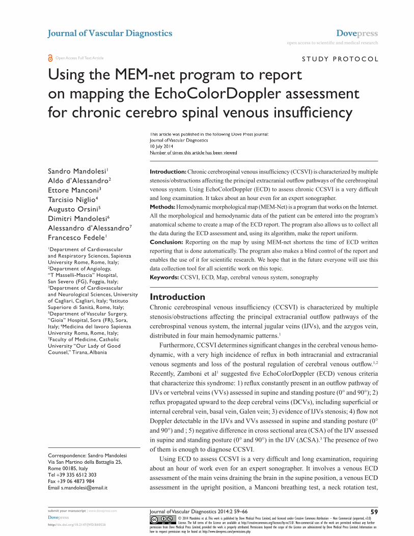
Zamboni criteria CCSVI TYPEType 1
Type 2
Type 3
Criterion 1
Criterion 2Criterion 3
Criterion 4
Criterion 5
ID patient: 756 Sex: F
EDSS: 4
ScoreCCSVI: 1VHISS: 2MEM: 15
Date: 01/27/2013Sonographer: Prof Sandro Mandolesi
CCSVI is positive if ≥2 criteriaAge: 33
Clinical type: SP First symptom: optical neuryte left eye
Duration: 13 years
Figure 1 Map on paper of an EchoColorDoppler report with morphological and hemodynamic symbols.Abbreviations: CCSVI, chronic cerebrospinal venous insufficiency; CSA, cross-sectional area; SP, secondary progressive; EDSS, Expanded Disability Status Scale; VHISS, venous hemodynamic insufficiency severity score; MEM, hemodynamic morphological map; RDCV, right deep cerebral veins; LDCV, left deep cerebral veins; J2, internal jugular vein; RVV, right vertebral vein; LVV, left vertebral vein; VPLEX, vertebral plexus; BCT, common-brachiocephalic trunk anonymous; SVC, superior cava vein; IVC, inferior cava vein; AZYGOS, Azygos vein; LREN, left renal vein; V-, valsalva negative; V+, valsalva positive; J1R, lower right internal jugular vein; J2R, middle right internal jugular vein; J3R, upper right internal jugular vein; J1L, lower left internal jugular vein; J2L, middle left internal jugular vein; J3L, upper left internal jugular vein; V1R, lower right vertebral vein; V2R, middle right vertebral vein; V3R, upper right vertebral vein; V1L, lower left vertebral vein; V2L, middle left vertebral vein; V3L, upper left vertebral vein; Dx, right; Sn, left; IJVR, right internal jugular vein; IJVL, left internal jugular vein.
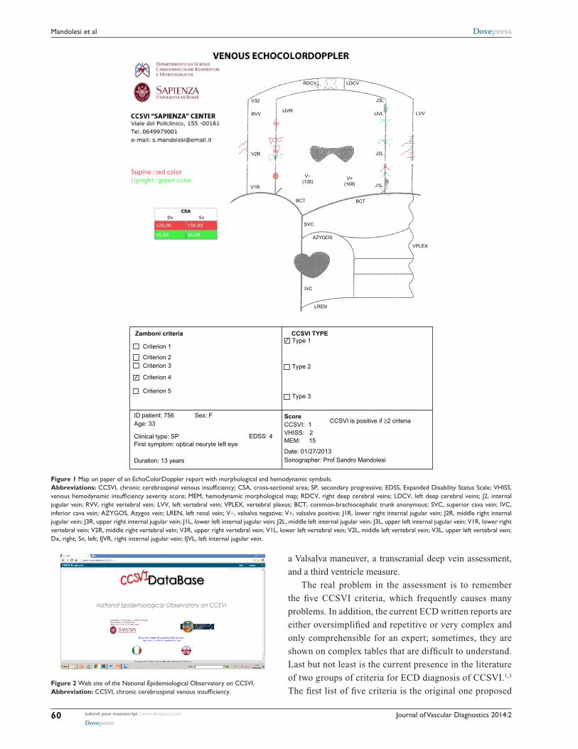
Figure 2 Web site of the National Epidemiological Observatory on CCSVI.Abbreviation: CCSVI, chronic cerebrospinal venous insufficiency.
a Valsalva maneuver, a transcranial deep vein assessment,
and a third ventricle measure.
The real problem in the assessment is to remember
the five CCSVI criteria, which frequently causes many
problems. In addition, the current ECD written reports are
either oversimplified and repetitive or very complex and
only comprehensible for an expert; sometimes, they are
shown on complex tables that are difficult to understand.
Last but not least is the current presence in the literature
of two groups of criteria for ECD diagnosis of CCSVI.1,3
The first list of five criteria is the original one proposed

Journal of Vascular Diagnostics 2014:2 submit your manuscript | www.dovepress.com
Dovepress
Dovepress
61
MEM-net and EchocolorDoppler assessment of veins draining the brain
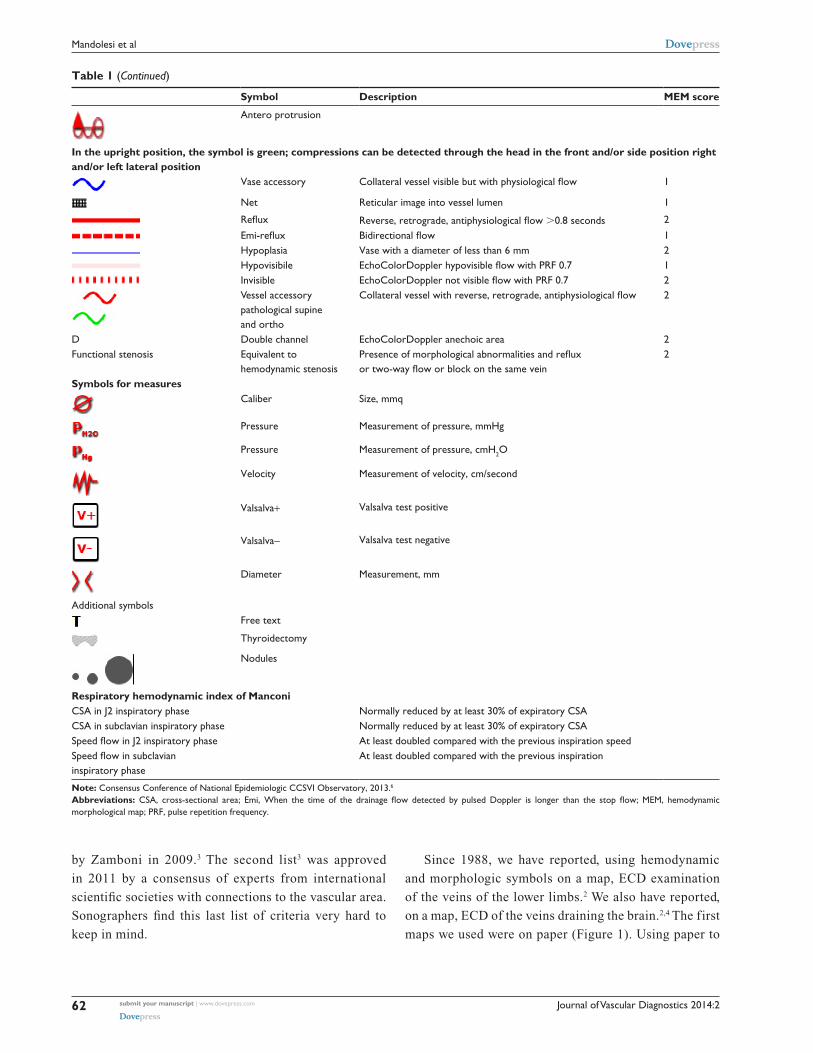
Table 1 Symbols and terminology to be used in reporting on EchoColorDoppler map examination of the veins draining the brain
Symbol Description MEM score
Calcification Endo- or extravascular 0
Full block When the block involves one or more segments with a caliber equal to or greater than the previous level or the following level
2
Empty block When the block involves one or more segments with a caliber much less than the previous level or the following level
2
Emi full block When the time of the drainage flow detected by pulsed Doppler is longer than the stop flow on one or more segments with a caliber equal to or greater than the previous level or the following level
1
Emi empty block When the time of the drainage flow detected by the pulsed Doppler is longer than the stop flow on one or more segments of size much smaller than the previous level or the following level
1
Morphological stenosis organic stenosis ,3 mmq 2
Stenosis hemodynamics Flow velocity .150 cm/second 2
Membrane Hyperechoic area endovascular 1
Septum Abnormal valve leaflet 1
Thickening Valvular thickening 1
twist twisting of the vessel 1
Vicarious Flow vicarious .45 cm/second on vertebrals (V2) in clino and .60 cm second in orto. Flow vicarious .100 cm/second on the internal jugulars (J2) both in clino and ortostatic position
1
Ectasia Vessel diameter more than 20 mm 1
Confluence Thickening of the confluence of jugular to subclavian 1
Thickening-dysplasia Thickening (dysplasia) of the vessel wall 1
thrombosis thrombosed segment 2
Recanalization Recanalization with parietal residues 1
White compression The compression is defined as white when the vein is completely compressed and we cannot detect any flow
2× each white
Frontal
right lateral
left lateral
Back protrusion
Black compression The compression is defined as black when the caliber of the vessel is less than 6 mmq and shows a flow
1× each black
Frontal
right lateral
left lateral
(Continued)
Journal of Vascular Diagnostics 2014:2submit your manuscript | www.dovepress.com
Dovepress
Dovepress
62
Mandolesi et al
Table 1 (Continued)
Symbol Description MEM score
Antero protrusion
In the upright position, the symbol is green; compressions can be detected through the head in the front and/or side position right and/or left lateral position
Vase accessory Collateral vessel visible but with physiological flow 1
Net reticular image into vessel lumen 1
Reflux Reverse, retrograde, antiphysiological flow .0.8 seconds 2Emi-reflux Bidirectional flow 1Hypoplasia Vase with a diameter of less than 6 mm 2Hypovisibile EchoColorDoppler hypovisible flow with PRF 0.7 1Invisible EchoColorDoppler not visible flow with PRF 0.7 2Vessel accessory pathological supine and ortho
Collateral vessel with reverse, retrograde, antiphysiological flow 2
D Double channel EchocolorDoppler anechoic area 2Functional stenosis Equivalent to
hemodynamic stenosisPresence of morphological abnormalities and reflux or two-way flow or block on the same vein
2
Symbols for measurescaliber Size, mmq
Pressure Measurement of pressure, mmHg
Pressure Measurement of pressure, cmH2o
Velocity Measurement of velocity, cm/second
Valsalva+ Valsalva test positive
Valsalva- Valsalva test negative
Diameter Measurement, mm
Additional symbolsFree text
Thyroidectomy
Nodules
Respiratory hemodynamic index of ManconiCSA in J2 inspiratory phase Normally reduced by at least 30% of expiratory CSACSA in subclavian inspiratory phase Normally reduced by at least 30% of expiratory CSASpeed flow in J2 inspiratory phase At least doubled compared with the previous inspiration speedSpeed flow in subclavian inspiratory phase
At least doubled compared with the previous inspiration
Note: Consensus Conference of National Epidemiologic CCSVI Observatory, 2013.6
Abbreviations: CSA, cross-sectional area; Emi, When the time of the drainage flow detected by pulsed Doppler is longer than the stop flow; MEM, hemodynamic morphological map; PRF, pulse repetition frequency.
by Zamboni in 2009.3 The second list3 was approved
in 2011 by a consensus of experts from international
scientific societies with connections to the vascular area.
Sonographers find this last list of criteria very hard to
keep in mind.
Since 1988, we have reported, using hemodynamic
and morphologic symbols on a map, ECD examination
of the veins of the lower limbs.2 We also have reported,
on a map, ECD of the veins draining the brain.2,4 The first
maps we used were on paper (Figure 1). Using paper to
Journal of Vascular Diagnostics 2014:2 submit your manuscript | www.dovepress.com
Dovepress
Dovepress
63
MEM-net and EchocolorDoppler assessment of veins draining the brain
create the map, the possibility of mistakes in the final
assessment of ECD report was lower, but human error
was always possible. After 2 years of work, in 2012,
we completed the Mappa Emodinamica e Morfologica
(MEM-Net) program, available at http://www.mem-net.
it/ (Figure 2).
MEM-net is used by the National Epidemiological
Observatory on CCSVI for data collection in its computer-
ized platform, which works on the Internet. In 2013, the
observatory organized a consensus conference involving
national scientific societies with connections to the vascular
area to share the symbols and terminology to use for MEM-
net mapping (Table 1).
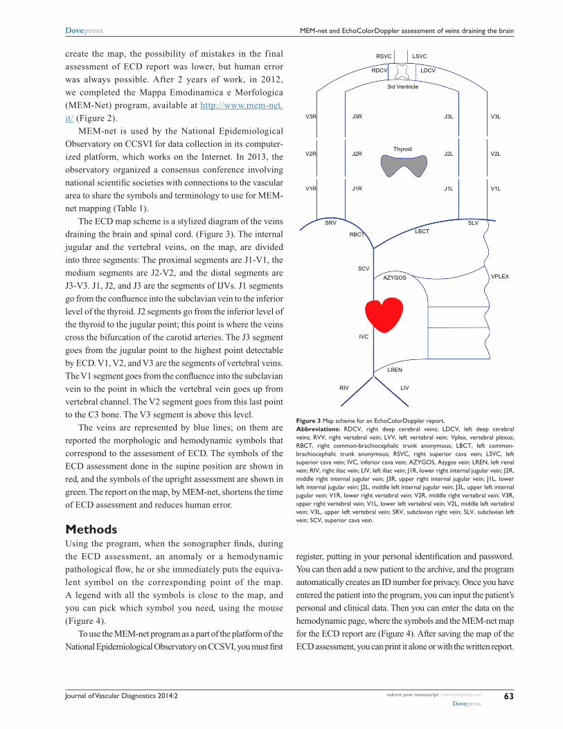
The ECD map scheme is a stylized diagram of the veins
draining the brain and spinal cord. (Figure 3). The internal
jugular and the vertebral veins, on the map, are divided
into three segments: The proximal segments are J1-V1, the
medium segments are J2-V2, and the distal segments are
J3-V3. J1, J2, and J3 are the segments of IJVs. J1 segments
go from the confluence into the subclavian vein to the inferior
level of the thyroid. J2 segments go from the inferior level of
the thyroid to the jugular point; this point is where the veins
cross the bifurcation of the carotid arteries. The J3 segment
goes from the jugular point to the highest point detectable
by ECD. V1, V2, and V3 are the segments of vertebral veins.
The V1 segment goes from the confluence into the subclavian
vein to the point in which the vertebral vein goes up from
vertebral channel. The V2 segment goes from this last point
to the C3 bone. The V3 segment is above this level.
The veins are represented by blue lines; on them are
reported the morphologic and hemodynamic symbols that
correspond to the assessment of ECD. The symbols of the
ECD assessment done in the supine position are shown in
red, and the symbols of the upright assessment are shown in
green. The report on the map, by MEM-net, shortens the time
of ECD assessment and reduces human error.
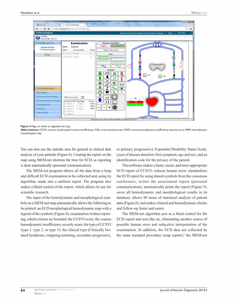
MethodsUsing the program, when the sonographer finds, during
the ECD assessment, an anomaly or a hemodynamic
pathological flow, he or she immediately puts the equiva-
lent symbol on the corresponding point of the map.
A legend with all the symbols is close to the map, and
you can pick which symbol you need, using the mouse
(Figure 4).
To use the MEM-net program as a part of the platform of the
National Epidemiological Observatory on CCSVI, you must first
RSVC
RDCV LDCV
3rd Ventricle
Thyroid
J3RV3R
J2RV2R
J1RV1R
SRV
RBCT
SCV
IVC
J3L
J2L V2L
V3L
J1L V1L
LBCT
AZYGOS
LREN
RIV LIV
SLV
VPLEX
LSVC
Figure 3 Map scheme for an EchocolorDoppler report.Abbreviations: RDCV, right deep cerebral veins; LDCV, left deep cerebral veins; RVV, right vertebral vein; LVV, left vertebral vein; Vplex, vertebral plexus; RBCT, right common-brachiocephalic trunk anonymous; LBCT, left common-brachiocephalic trunk anonymous; RSVC, right superior cava vein; LSVC, left superior cava vein; IVC, inferior cava vein; AZYGOS, Azygos vein; LREN, left renal vein; RIV, right iliac vein; LIV, left iliac vein; J1R, lower right internal jugular vein; J2R, middle right internal jugular vein; J3R, upper right internal jugular vein; J1L, lower left internal jugular vein; J2L, middle left internal jugular vein; J3L, upper left internal jugular vein; V1R, lower right vertebral vein; V2R, middle right vertebral vein; V3R, upper right vertebral vein; V1L, lower left vertebral vein; V2L, middle left vertebral vein; V3L, upper left vertebral vein; SRV, subclavian right vein; SLV, subclavian left vein; SCV, superior cava vein.
register, putting in your personal identification and password.
You can then add a new patient to the archive, and the program
automatically creates an ID number for privacy. Once you have
entered the patient into the program, you can input the patient’s
personal and clinical data. Then you can enter the data on the
hemodynamic page, where the symbols and the MEM-net map
for the ECD report are (Figure 4). After saving the map of the
ECD assessment, you can print it alone or with the written report.
Journal of Vascular Diagnostics 2014:2submit your manuscript | www.dovepress.com
Dovepress
Dovepress
64
Mandolesi et al
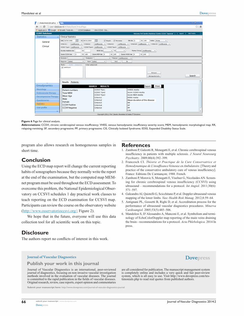
You can also use the statistic area for general or clinical data
analysis of your patients (Figure 6). Creating the report on the
map using MEM-net shortens the time for ECD, as reporting
is done automatically (personal communication).
The MEM-net program allows all the data from a long
and difficult ECD examination to be collected and, using its
algorithm, made into a uniform report. The program also
makes a blind control of the report, which allows its use for
scientific research.
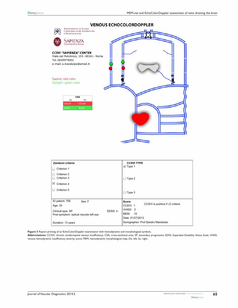
The input of the hemodynamic and morphological sym-
bols on a MEM-net map automatically allows the following to
be printed: an ECD morphological hemodynamic map with a
legend of the symbols (Figure 4); examination written report-
ing; which criteria we founded; the CCSVI score; the venous
hemodynamic insufficiency severity score; the type of CCSVI
(type 1, type 2, or type 3); the clinical type (Clinically Iso-
lated Syndrome, relapsing remitting, secondary progressive,
or primary progressive); Expanded Disability Status Scale;
years of disease duration; first symptom; age and sex; and an
identification code for the privacy of the patient.
This software makes a faster, easier, and more appropriate
ECD report of CCSVI; reduces human error; standardizes
the ECD report by using shared symbols from the consensus
conference; writes the assessment report (personal
communication); automatically prints the report (Figure 5);
saves all hemodynamic and morphological results in its
database; allows 90 items of statistical analysis of patient
data (Figure 6); and makes clinical and hemodynamic checks
and follow-up faster and easier.
The MEM-net algorithm acts as a blind control for the
ECD report and acts like an, eliminating another source of
possible human error and subjective interpretation of the
examination. In addition, the ECD data are collected by
the same standard procedure (map report);5 the MEM-net
Figure 4 Page on which to digitalize the map.Abbreviations: CCSVI, chronic cerebrospinal venous insufficiency; CSA, cross-sectional area; VHISS, venous hemodynamic insufficiency severity score; MEM, hemodynamic morphological map.
Journal of Vascular Diagnostics 2014:2 submit your manuscript | www.dovepress.com
Dovepress
Dovepress
65
MEM-net and EchocolorDoppler assessment of veins draining the brain
Zamboni criteria CCSVI TYPEType 1
Type 2
Type 3
Criterion 1
Criterion 2Criterion 3
Criterion 4
Criterion 5
ID patient: 756 Sex: F
EDSS: 4
ScoreCCSVI: 1
VHISS: 2
MEM: 15
Date: 01/27/2013
Sonographer: Prof Sandro Mandolesi
CCSVI is positive if ≥2 criteriaAge: 33
Clinical type: SP First symptom: optical neuryte left eye
Duration: 13 years
Figure 5 Report printing of an EchoColorDoppler examination with hemodynamic and morphological symbols.Abbreviations: CCSVI, chronic cerebrospinal venous insufficiency; CSA, cross-sectional area; SP, secondary progressive; EDSS, Expanded Disability Status Scale; VHISS, venous hemodynamic insufficiency severity score; MEM, hemodynamic morphological map; Dx, left; Sx, right.
Journal of Vascular Diagnostics
Publish your work in this journal
Submit your manuscript here: http://www.dovepress.com/journal-of-vascular-diagnostics-journal
Journal of Vascular Diagnostics is an international, peer-reviewed journal of diagnostics, focusing on non invasive vascular investigation methods involved in the evaluation of vascular diseases. The journal is committed to the rapid publication in the fields of vascular diseases. Original research, review, case reports, expert opinion and commentaries
are all considered for publication. The manuscript management system is completely online and includes a very quick and fair peer-review system, which is all easy to use. Visit http://www.dovepress.com/tes-timonials.php to read real quotes from published authors.
Journal of Vascular Diagnostics 2014:2submit your manuscript | www.dovepress.com
Dovepress
Dovepress
Dovepress
66
Mandolesi et al
program also allows research on homogeneous samples in
short time.
ConclusionUsing the ECD map report will change the current reporting
habits of sonographers because they normally write the report
at the end of the examination, but the computed map MEM-
net program must be used throughout the ECD assessment. To
overcome this problem, the National Epidemiological Obser-
vatory on CCSVI schedules 1 day practical work classes to
teach reporting on the ECD examination for CCSVI map.
Participants can review the course on the observatory website
(http://www.osservatorioccsvi.org/; Figure 2).
We hope that in the future, everyone will use this data
collection tool for all scientific work on this topic.
DisclosureThe authors report no conflicts of interest in this work.
References1. Zamboni P, Galeotti R, Menegatti E, et al. Chronic cerebrospinal venous
insufficiency in patients with multiple sclerosis. J Neurol Neurosurg Psychiatry. 2009;80(4):392–399.
2. Francesch CI. Théorie et Practique de la Cure Conservatrice et Hemodynamique de L’insuffisance Veineuse en Ambulatoire. [Theory and practice of the conservative ambulatory cure of venous insufficiency]. France: Editions De L’armançon; 1988. French.
3. Zamboni P, Morovic S, Menegatti E, Viselner G, Nicolaides AN. Screen-ing for chronic cerebrospinal venous insufficiency (CCSVI) using ultrasound – recommendations for a protocol. Int Angiol. 2011;30(6): 571–597.
4. Galeandro AI, Quistelli G, Scicchitano P, et al. Doppler ultrasound venous mapping of the lower limbs. Vasc Health Risk Manag. 2012;8:59–64.
5. Antignani PL, Gossetti B, Righi D, et al. Accreditation process for the performance of ultrasound vascular diagnostics procedures. Minerva Cardioangiol. 2005;53(5):485–506.
6. Mandolesi S, D’Alessandro A, Manconi E, et al. Symbolism and termi-nology of EchoColorDoppler map reporting of the main veins draining the brain - recommendations for a protocol. Acta Phlebologica. 2014 In press.
Figure 6 Page for clinical analysis. Abbreviations: CCSVI, chronic cerebrospinal venous insufficiency; VHISS, venous hemodynamic insufficiency severity score; MEM, hemodynamic morphological map; RR, relapsing-remitting; SP, secondary progressive; PP, primary progressive; CIS, Clinically Isolated Syndrome; EDSS, Expanded Disability Status Scale.