mineral, fluid and thermal evolution in veins from late orogenic ...
Upload
independentCategory
view
5download
0
EBioMedicine xxx (2014) xxx–xxx
EBIOM-00001; No of Pages 8
Contents lists available at ScienceDirect
EBioMedicine
j ourna l homepage: www.eb iomed ic ine.com
In Vivo Application of Tissue-Engineered Veins Using Autologous Peripheral WholeBlood: A Proof of Concept Study
Michael Olausson a,1, Vijay Kumar Kuna a,1, Galyna Travnikova a, Henrik Bäckdahl b, Pradeep B. Patil a,Robert Saalman c, Helena Borg c, Anders Jeppsson d, Suchitra Sumitran-Holgersson a,⁎a Department of Surgery, University of Gothenburg, Sahlgrenska University Hospital, Gothenburg, Swedenb SP Technical Research Institute of Sweden, Dept. of Chemistry, Materials and Surfaces, SE-50462 Borås, Swedenc Department of Paediatrics, University of Gothenburg, Sahlgrenska University Hospital, Gothenburg, Swedend Department of Cardiothoracic Surgery, University of Gothenburg, Sahlgrenska University Hospital, Gothenburg, Sweden
⁎ Corresponding author at: Laboratory for TransplantatSahlgrenska Science Park, Medicinaregatan 8A, 2nd Floor,
E-mail address: [email protected] (S.1 MO and VKK have contributed equally and share first
http://dx.doi.org/10.1016/j.ebiom.2014.09.0012352-3964/© 2014 Elsevier B.V. All rights reserved.
Please cite this article as: Olausson,M., et al.,Concept Study, EBioMedicine (2014), http://
a b s t r a c t
a r t i c l e i n f oArticle history:Received 2 September 2014Received in revised form 16 September 2014Accepted 16 September 2014Available online xxxx
Keywords:Vein conduitsVascular diseasesTissue-engineeringEndothelial precursors
Vascular diseases are increasing health problems affecting N25 million individuals inwesternized societies. Suchpatients could benefit from transplantation of tissue-engineered vascular grafts using autologous cells. One chal-lenge that has limited this development is the need for cell isolation, and risks associated with ex vivo expandedstem cells. Herewe demonstrate a novel approach to generate transplantable vascular grafts using decellularizedallogeneic vascular scaffolds, repopulatedwith peripheral whole blood (PWB) in vitro in a bioreactor. Circulating,VEGFR-2+/CD45+ and a smaller fraction of VEGFR-2+/CD14+ cells contributed to repopulation of the graft.SEMmicrographs showed flat cells on the luminal surface of the grafts consistent with endothelial cells. For clin-ical validation, two autologous PWB tissue-engineered vein conduitswere prepared and successfully used for by-pass procedures in two pediatric patients. These results provide a proof of principle for the generation of trans-plantable vascular grafts using a simple autologous blood sample, making it clinically feasible globally.
© 2014 Elsevier B.V. All rights reserved.
1. Introduction
Vascular diseases have been documented to be key components ofthe global burden of non-communicable diseases. Venous disease ofthe lower extremities is very common, affecting∼25% of adults inwest-ernized societies (Graham et al., 2003). Peripheral arterial disease is es-timated to affect N25 million patients in Europe and North Americaalone (Fowkes et al., 1991). These pathological conditions representan enormous burden, to both patients and healthcare systems. With anaging population, expenditure in the treatment of such diseases is likelyto increase significantly over the next 20 years (Raval and Losordo,2013). Surgical replacement of damaged vessels may include the use ofautologous vessels, synthetic grafts such as Dacron or Gore-Tex, oreven allogeneic vessels from organ donors. Problemswith these alterna-tives include stenosis, thromboembolization, calcium deposition, infec-tion, the lack of autologous vessels of the proper phenotype or quality,poor patency, donor shortage and the risk of alloimmunization or sideeffects of immunosuppressive treatment (Veith et al., 1979; O'Donnellet al., 1984; Kurobe et al., 2012). A possible intervention can be replace-mentwith tissue-engineered blood vessels from allogeneic or xenogeneic
ion and Regenerative Medicine,S-413 46 Gothenburg, Sweden.Sumitran-Holgersson).authorship.
In Vivo Application of Tissue-Edx.doi.org/10.1016/j.ebiom.2
sources, inwhich the donor's blood vessel is decellularized (DC) and usedas scaffold to repopulate with recipient's autologous stem cells. This ap-proachwouldmake it possible to produce a vessel of the right phenotypeand obviate the need for life-long immunosuppression (Olausson et al.,2012).
Studies have demonstrated that unseededDacron or Gore-Tex graftsare incorporated in the host blood vessels through a process which isgenerally referred to as graft healing resulting in endothelialization ofthe synthetic graft (Pasquinelli et al., 1990). Endothelialization canoccur from blood vessel stems at either anastomosis. However, inhumans, endothelial cell (EC) ingrowth is very limited, that is, a fewmil-limeters in years (Guidoin et al., 1993). Anothermechanism of endothe-lial healing is, EC coming from circulating cells (Shi et al., 1994; Onukiet al., 1997; Graham et al., 1980; Clowes et al., 1986; Poole-Warrenet al., 1996).We and others have shown that circulating ECs, endothelialprogenitor cells including themyeloid/monocytic lineages contribute tore-endothelialization (Nowak et al., 2004; Asahara et al., 1997; Baileyet al., 2006). Furthermore, progenitor cells for smooth muscle cells(SMCs) are also present in circulating blood (Simper et al., 2002).
In contrast to experimental animals, the flow surface of syntheticvascular grafts remains unhealed in humans, particularly in the smallcaliber conduits. Marked differences in graft healing exist between ani-mals andman; and the usualmechanisms of graft endothelialization arepartially ineffective in man (Pasquinelli et al., 1990). It has been shownthat EC seeding of synthetic grafts prior to implantation has improved
ngineered Veins Using Autologous PeripheralWhole Blood: A Proof of014.09.001
2 M. Olausson et al. / EBioMedicine xxx (2014) xxx–xxx
the patency of such grafts when compared to unseeded grafts (Wanget al., 1993; Deutsch et al., 2009). Strategies for repopulation of vascularconduits have usually used in vitro expanded cells derived from bone-marrow or peripheral blood. Recently, we successfully transplanted atissue engineered allogeneic vein with in vitro expanded autologousbone-marrow derived stem cells. The tissue-engineered graft showedgood patency in vivo, thus proving a promising and safe clinical ap-proach (Olausson et al., 2012). However, the disadvantage with this ap-proach is that the collection, isolation and large scale expansion of stemcells from tissues or bone-marrow in a limited timewith low variabilityis highly challenging.
All these observations led us to hypothesize that the use of peripher-al whole blood (known to contain circulating ECs/endothelial progeni-tor cells (EPCs)) would result in efficient recellularization (RC) andendothelialization of acellular veins, without the need to isolate and ex-pand subpopulations of angiogenic progenitor cells from bone-marrowor whole blood. Furthermore, an in vitro complete and preformed endo-thelial lining at the time of implantation would help to increase the pa-tency of these engineered vein grafts. Thus,we aimed to develop an easyprocedure that would increase the clinical use of the technique andmake it globally accessible. We report the clinical transplantation oftissue-engineered allogeneic veins using autologous peripheral wholeblood (APWB), in two pediatric patients with portal vein thrombosis.
2. Materials and Methods
All research protocols were approved by the Swedish institutionalreview boards and ethics committees.
2.1. Decellularization of Veins
Human iliac or mammary veins about 7–9 cm were retrieved from14deceased organdonors after informed consent from the relatives, im-mediately stored in sterile phosphate buffered saline (PBS) containing1% penicillin, streptomycin, amphotericin B and transported to the lab-oratory. On an average, the outer diameter of iliac veins used in the pro-cedures ranged between 1.5 and 2 cmandmammary veins ranged from0.3–0.7 cm. Decellularization (DC) was carried out as described by usearlier (Olausson et al., 2012) using 1% Triton X-100 and 1% tri-n-butylphosphate (TNBP) and 4 mg/L DNAse under sterile conditions.
2.2. Characterization of Decellularized Veins
At the end of the DC process, biopsies were taken from decellularizedveins (DV) and processed histologically with Hematoxylin–Eosin (HE),Masson's Trichrome (MT) and Verhoeff Van Gieson (VVG) staining,DNA quantification, Luminex®, scanning and transmission electron mi-croscopic analysis, tensile strength and extracellular matrix (ECM) quan-tifications. Please see Supplement 1 for further details.
2.3. Recellularization of Veins
The entire RC processwas performed under sterile conditions and allperfusions were carried out in an incubator at 37 °C supplied with 5%CO2. Before RC, the veins were perfused with heparin (Leopharma,Sweden) at a concentration of 50 IU/ml PBS for 2 h. The heparin wasdrained off and the whole blood was immediately perfused for 48 h at2 ml/min speed. For collection of blood, see Supplement 1. The bloodwas then drained off and the veins were washed with PBS containing1% penicillin–streptomycin–amphotericin for 3–5 min or until the bloodwas completely removed. The veins were subsequently perfused 4 dayswith EC and 4 dayswith SMCmedia. The complete ECmedium containedbasal medium MCDB 131, 10% heat inactivated human AB serum, 1% L-glutamine and 1% penicillin–streptomycin–amphotericin and supple-mented with EGM-2 Single Quote kit (Lonza, Walkersville, MD USA). Acommercially available SMC medium (Cascade Biologics) containing
Please cite this article as: Olausson,M., et al., In Vivo Application of Tissue-EConcept Study, EBioMedicine (2014), http://dx.doi.org/10.1016/j.ebiom.2
growth and differentiation factor supplements was used. The veinswere recellularized for a total of 10 days.
2.4. Characterization of Recellularized Veins
To visualize the presence of EC, antibodies to CD31 (1:1000)(Abcam, Germany) and von Willebrand factor (1:100) (Santa Cruz,Germany) were selected and stained by immunofluorescence, whilesmooth muscle actin (1:50) (Abcam, Germany) to visualize SMC. Thetensile strength of recellularized veins (n = 3), work and elongationwas compared to normal and decellularized veins.
2.5. Enumeration of Cells in the Repopulated Veins
Quantification of cell numbers in the lumenwas done by staining forthe endothelial cell marker VEGFR-2 and monocyte marker CD14.Single and double-positive cells were quantified manually. On average,15 images of the lumen were taken per vein covering an area of47500 μm2. The data is represented as the number of cells per squarecentimeter area of the vein and the values are represented with stan-dard error mean of the original value.
2.6. Clinical Transplantations
2.6.1. Patient 1A 4-year-old girl was referred to the children's hospital in Goth-
enburg after a history of fatigue, a slightly elevated body temperatureand a tender tumor in the left upper abdominal quadrant presentsince 1 month of age. Initial tests at 4 years showed anemia, thrombo-cytopenia, and neutropenia. A CT-angiography showed no portal circu-lation from the superior mesenteric vein to the liver (Fig. 4F) andproblems in detecting the intrahepatic portal vascular system. The um-bilical veinwas not open and the jugular veinswere estimated to be tooshort as the only source of vein graft (also see surgical procedure andpost-operative monitoring in the Supplement methods section).Splenomegaly and cavernous vein formation was found in the hilum.An endoscopy demonstrated esophagus varices from 15 to 25 cmdown to the cardiac region. The child was evaluated for a possibleMesorex procedure by a pediatric multidisciplinary team. The childweighed 17 kg and was 107 cm in height.
2.6.2. Patient 2A 24-month-old girl, weighing 7.4 kg, was referred to the children's
hospital with a history of upper abdominal pain coincidingwith feedingof the child, starting at 8months after birth. Initial investigation at a localhospital found splenomegaly and pancytopenia. A hematological workup was done and found normal. A CT-angiography revealed the absenceof the portal vein and difficulties with detection of any intrahepatic por-tal vascular system (Fig. 4G). No patent umbilical vein could be detected.Endoscopy showed prominent varices explaining the pain correlating tofood intake. The childwas evaluated for a possibleMesorex procedure bya pediatric multidisciplinary team. The child weighed 7.4 kg and was73.5 cm in height.
Since no suitable autologous veins were detected in both the pa-tients, two decellularized veins were recellularized under good man-agement practice (GMP) facilities available at The SahlgrenskaUniversity Hospital-Gothenburg-Sweden as described above with25 ml autologous blood taken from the two patients and used for theMesorex procedures.
2.6.3. The Ethical ProcessAs in the previous published case (Poole-Warren et al., 1996) in the
present two cases, a pediatric team performed the evaluation in a mul-tidisciplinary fashion, which also includes abiding to the UNICEF Con-vention on the Rights of the Child. The parents were informed severaltimes and given details of the procedure, the possible anticipated
ngineered Veins Using Autologous PeripheralWhole Blood: A Proof of014.09.001

3M. Olausson et al. / EBioMedicine xxx (2014) xxx–xxx
complications and the alternative remaining with a failed surgical cor-rection of the EHPVO. This information was given to the parents bythe responsible surgeon together with members of the pediatric team.The parents also provided informed consent, whichwas given to the pe-diatric team in the absence of the operating surgeon to avoid pressure.Moreover, permissions from the two regulatory bodies in Sweden—The National Medical Board and The Medical Products Agency weregranted. Permission from members of The Local Ethical Board (EtiskForum)was also obtained prior to proceeding with the transplantation.
Please see Supplement 1 for post-transplant monitoring and statistics.
3. Results
3.1. Decellularized Veins and Extracellular Matrix
Nine cycles of decellularization with 1% Triton and 1% tri-n-butylphosphate resulted in generation of pale and translucent veins (Supple-ment Fig. S1A, DV). The veins were successfully decellularized in12 days as evidenced by lack of nuclei when compared to normals(Fig. 1A–C, also see Supplement Fig. S1B–E) and confirmed by electronmicroscopy (Supplement Fig. S1F–H).Major ECMproteins such as colla-gen I, collagen IV, fibronectin and laminin (Fig. 1D) were preserved inDV. The quantity of GAGs (p = 0.041) but not collagen (p = 0.700)was significantly decreased in veins after DC (Supplement Fig. S2A). Asignificant decrease in DNA concentration was found in DV (15.5 ±7.93 ng/mg) as compared to 244.5 ± 65.01 ng/mg of tissue in normalveins (p= 0.007). Several growth factors albeit lowerwere still presentin the DV (Supplement Fig. S2B). Tensile strength testing of the DC veinsshowed a decrease in thework needed to deform the samples completelywhen compared to normal samples and the elongation measured at 50%
Fig. 1. A representative microscopic view of decellularized vein grafts. Masson's Trichrome staismooth muscle cells, but with abundant collagen still present (blue) and (B) normal vein s(C) Negative controls. (D) Immunohistochemical staining of normal and DV for the various extthough no cells/nuclei are seen, the ECM components stained positive indicating the retainmenreferences to color in this figure legend, the reader is referred to the web version of this article
Please cite this article as: Olausson,M., et al., In Vivo Application of Tissue-EConcept Study, EBioMedicine (2014), http://dx.doi.org/10.1016/j.ebiom.2
of total work had significantly decreased after DC (p b 0.05, SupplementFig. S2C & D).
3.2. Recellularization of Veins
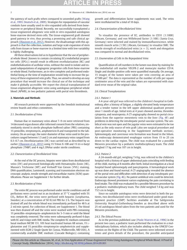
The grossmorphology of veins after RC looked pinkishwith good tu-nica externa (Supplement Fig. S1A, RV). Histological examination twodays after perfusion of DV with whole peripheral blood showed migra-tion of cells into the tissue, (Fig. 2A). Four-day perfusion with EC medi-um resulted in an almost confluent monolayer of cells on the lumen ofthe vein (Fig. 2B) and immigrated cells in themedia (Fig. 2C). This is ev-idenced by single immunofluorescent staining for CD31 (SupplementFig. S3E) and alpha smoothmuscle actin (Supplement Fig. S3D) staining.VVG and MT staining of the veins also confirmed the presence of nuclei(black), cytoplasm (red in MT) and collagen (red in VVG and blue inMT) (Fig. 2D & E respectively).
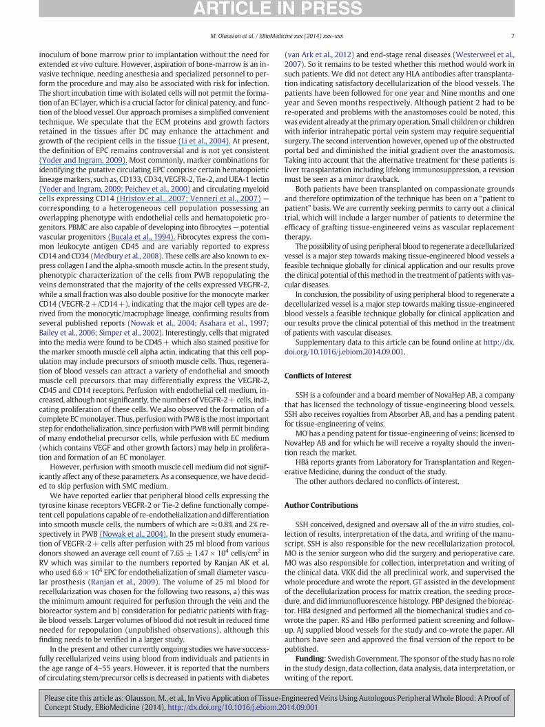
Phenotypic characterization of the peripheral blood cells involved information of neo-endothelialization demonstrated that many cellsexpressed the tyrosine kinase receptor-vascular endothelial growth fac-tor receptor-2 (VEGFR-2) (Fig. 3A; after blood & B; after endothelial cellmedium). Negative controls are shown in Supplement Fig. S3A–C. Cellspositive for the leukocyte common antigen CD45 were found mainly inthe adventitia and not the lumen of the veins (Fig. 3C), indicating thatthese cells mainly differentiated to smooth muscle cells as evidencedby positive staining for alpha smooth muscle cell actin. A smaller frac-tion of CD14+ cells (green, Fig. 3D) which also stained positive forVEGFR-2 (red, yellow indicates co-localization; arrows) contributed tothis process (Fig. 3E). A continuousmonolayer of endothelial cells stain-ing positive for VEGFR-2 and the EC marker CD31 (arrows) was ob-served after 4 days with EC medium (Fig. 3F). These findings werecorroborated by scanning electron microscopy (Fig. 4A–C) which also
ning of (A) decellularized vein (DV) showing no nuclei, indicating lack of endothelial andhowing presence of nuclei (black, arrows), cytoplasm (red/pink) and collagen (blue).racellular matrix components. Nuclei are seen only in normal but not in DV. In the DV al-t of important ECM proteins (n= 14). A–D: Scale bar = 75 μm. (For interpretation of the.)
ngineered Veins Using Autologous PeripheralWhole Blood: A Proof of014.09.001

Fig. 2.Microscopic view of a representative bioengineered vein graft with peripheral whole blood. (A) Picture showsmigration of cells (black) into the tissue after perfusion withblood, (B & C) presence of an endothelial cell monolayer on the luminal side after perfusion with endothelial cell medium and an overall improved architecture of the vein. Staining oftissue-engineered human veins recellularized with peripheral whole blood by (D) Verhoeff Van Gieson, showed presence of nuclei (black) for endothelial (black arrows) and smoothmuscle cells (yellow arrows). The cytoplasm was stained pink and elastin black. (E) Masson's Trichrome staining showed presence of black nuclei (black), blue collagen and pink cyto-plasm (n=5). A–C: Scale bar=75 μm,D& E: scale bar=25 μm. (For interpretation of the references to color in thisfigure legend, the reader is referred to theweb version of this article.)
4 M. Olausson et al. / EBioMedicine xxx (2014) xxx–xxx
revealed the presence of flattened cells along the luminal surface of theveins consistentwith endothelial cells (Fig. 4D). The cells were negativefor the markers CD3, CD19, CD68, CD133, CD34, CD56 and CD61, indi-cating that the major cell types contributing to repopulation areVEGFR-2+/CD45+ and VEGFR-2+/CD14+ cells.
Our initial results showed no difference in the numbers of cells at6 days and 10 days, therefore enumeration data is presented at twotime points i.e. immediately after perfusion with blood (48 h) andafter perfusionwith ECmedium for four days (total 6 days). On an aver-age, we found 11.5 ± 0.02 × 10⁴ DAPI + cells/cm2 in the lumen ofnormal control veins. In the recellularized veins, staining for VEGFR-2after perfusion with whole blood demonstrated an average of 5.93 ±1.26 × 10⁴ cells/cm2. The numbers of VEGFR-2+ cells were increasedafter perfusion with EC medium (7.65 ± 1.47 × 10⁴ cells/cm2). Thiswas however not statistically significant (p = 0.387). In addition, thenumber of CD14+ cells after two days of perfusion with blood showedan average of 1.11 ± 0.32 × 10⁴ cells/cm2, and the numbers of thesecells were also not significantly increased after perfusion with EC medi-um (2.63 ± 0.70 × 10⁴ cells/cm2, p = 0.132). Double staining of thecells in the lumen for CD14 and VEGFR-2 revealed that approximatelyhalf of the CD14+ cells also expressed VEGFR-2 (Fig. 4E) (0.71 ± 0.22× 10⁴ cells/cm2 with blood and 1.39 ± 0.37 × 10⁴ cells/cm2 after ECmedium).
Sterility testing showed no marked increase in absorbance during14 days ofmedia culture. The average of optical densities at 600 nmmea-sured in spectrophotometer for freshmedia (0.006) was similar to that ofperfused media (0.009) and not significantly different (p = 0.251). Theaverage values for negative control and positive control are 0.010 and0.36 respectively. Nomicrobial colonieswere seen on the agar plates. Bio-mechanical analyses showed that the work increased significantly after
Please cite this article as: Olausson,M., et al., In Vivo Application of Tissue-EConcept Study, EBioMedicine (2014), http://dx.doi.org/10.1016/j.ebiom.2
RC while no significant changes were seen in the elongation betweenRC and DC samples (Fig. S2C & D). No significant difference was foundbetween normal and RC samples, indicating that the biomechanical prop-erties of the RC veins are similar to the native veins (Supplement Fig. S2C& D).
3.3. Clinical Cases
To test the in vivo patency of these tissue-engineered veins, twopediatric patients suffering with extra hepatic portal vein obstruction(EHPVO) were transplanted with tissue engineered veins regeneratedusing 25 ml autologous peripheral blood on compassionate basis.Fig. 4F shows a CT-angiography demonstrating no portal circulationfrom the superior mesenteric vein to the liver and problems in de-tecting the intrahepatic portal vascular system in patient 1. Similarly,Fig. 4G shows a CT-angiography demonstrating the absence of theportal vein and difficulties with detection of any intrahepatic portalvascular system in patient 2.
3.4. Clinical Transplantation Data
Patient 1 received an 8 cm long vein, anastomosed to the superiormesenteric vein and the left portal vein as earlier described (Fig. 5A)Olausson et al., 2012. The central venous pressure (CVP) was 11 cmH2O and the portal pressure dropped from 22 cm to 14 cm after re-perfusion of the vein— a difference of 3 cm compared to the pressurein the right atrium. The flow velocity showed 30–40 cm/s both in theportal vein and hepatic artery (Fig. 5B). Twenty-one months post-transplantation the patient has an unchanged image as seen in a 3D re-construction of the graft (see Video in Supplement). The esophageal
ngineered Veins Using Autologous PeripheralWhole Blood: A Proof of014.09.001

Fig. 3. Phenotypic labeling of peripheral whole blood cells involved in repopulation of the bioengineered veins. (A) Cells staining positive (brown) for the tyrosine kinase receptor VEGFR-2are found adhering to the lumen of the scaffold after perfusion of peripheral blood for 48 h. (B) These VEGFR-2+ cells formed a monolayer on the luminal side of the bioengineered veinafter perfusion of endothelial cellmedium for 4 days (brown). (C) Cells expressing the common leucocytemarker CD45were foundmainly in themedia and adventitia (arrows). (D & E)Asmall fraction of cells in the lumen also stain positive for the monocyte marker CD14 (green), while double staining showed that some were also double positive (arrows) for VEGFR-2(red). (F) Double staining showing that VEGFR-2+ cells were also positive for the endothelial marker CD31. Yellow color indicates co-localization of the two markers. The repopulatingcells were negative for other markers such as CD3, CD19, CD68, CD133, CD34, CD56 and CD61 (n = 6). (For interpretation of the references to color in this figure legend, the reader isreferred to the web version of this article.)
Fig. 4. Characterization of the recellularized veins and pre-transplant clinical data of the two patients. (A & B) Scanning electronmicrographs of the recellularized veins showing binding ofcells (arrows) to the lumen and in the media after perfusion with peripheral whole blood. (C & D) Further perfusion with endothelial cells medium resulted in formation of a continoussmooth endothelial cell layer (arrows). (E) Higher numbers of VEGFR-2+ cells were found in the recellularized veins (n=6) as compared to CD14+ cells both after perfusionwith bloodand endothelial cellmedium (EM). (F & G) CT-angiographs of patients 1 & 2 respectively before transplantation. (F) Patient 1: showing varicose veins in the hilum (blue) and at the esoph-agus (black). (G) Patient 2: showing no intrahepatic portal system. In both cases, no portal circulation is observed.
5M. Olausson et al. / EBioMedicine xxx (2014) xxx–xxx
Please cite this article as: Olausson,M., et al., In Vivo Application of Tissue-Engineered Veins Using Autologous PeripheralWhole Blood: A Proof ofConcept Study, EBioMedicine (2014), http://dx.doi.org/10.1016/j.ebiom.2014.09.001

Fig. 5. Post-transplant clinical data for the two transplanted patients. (A) Patient 1: Postoperative CT-angiograph, showing the new graft (blue arrow) with good anastomotic sites (blackarrows) and well established intrahepatic portal system. (B) The postoperative period was uneventful with good intrahepatic blood flows. The flow velocity showed 30–40 cm/s both inthe portal vein and in the hepatic artery. (C) Patient 2: CT-angiography after first transplantation, showing a revascularized portal flow and intrahepatic portal system (blue arrow). At thesix-month checkup, the patient had a reduced diameter at both anastomotic sites, (black arrows), however the intrahepatic portal vein system was significantly developed (D). Sevenmonths later the patient received a second graft. (E) CT angiography day after surgery of the second graft revealed poor perfusion of the right portal system. (F) However, after a percu-taneous angiography the right portal systemwaswell perfused. (For interpretation of the references to color in this figure legend, the reader is referred to theweb version of this article.)
6 M. Olausson et al. / EBioMedicine xxx (2014) xxx–xxx
varices have diminished (endoscopy) and clinical laboratory parametersare normal. No surgical or other complications have been observed dur-ing frequent regular controls and ultrasounds.
Patient 2 received a 6–7 cm long vein, initially anastomosed tothe patient's own 5 cm long umbilical vein. This had to be adjustedperioperatively due to thrombosis in the umbilical vein section,resulting in an anastomosis between the vein graft and the left portalvein as earlier described (Olausson et al., 2012). The gradient betweenthe cava and the intrahepatic system was b1 cm H2O, indicating goodliver parenchyma, whereas the gradient between the portal vein andthe mesenteric system remained 6 cm, indicating a partial stricture.On the second day after surgery the patient was revised due to anarrowing at the site of anastomosis in the liver (Fig. 5C). After revision,both anastomoses were patent, but difficulties to lower the differencebetween the CVP and themesenteric pressure below8 cmH2Owere en-countered. The postoperative period was otherwise uneventful withgood intrahepatic blood flows. At the 6-month checkup, the patienthad a reduced diameter at both anastomotic sites, however theintrahepatic portal vein system was significantly developed (Fig. 5D).An attempt to dilate the narrow anastomosis on themesenteric side re-sulted in thrombosis of the graft. Sevenmonths after the first procedurea decision to revise the graft was taken and the patient received a sec-ond vein, prepared exactly as the first. During this procedure difficultiesto open up the right portal vascular bed was evident. The right portalsystemwas difficult to reach with a surgical microprobe. After reperfu-sion a remaining gradient of 10 cm H2O was detected. CT angiographythe day after the surgery revealed poor perfusion of the right portal sys-tem (Fig. 5E). Four days later a percutaneous angiography was per-formed with measurements of the gradient over the anastomoses. Theright portal system was now well perfused and the gradient was only3 cm H2O (Fig. 5F). Further controls have been uneventful, (Nineteen
Please cite this article as: Olausson,M., et al., In Vivo Application of Tissue-EConcept Study, EBioMedicine (2014), http://dx.doi.org/10.1016/j.ebiom.2
months after the procedure). Both patients received heparin infusionsfor 2 weeks followed by warfarin for 1 year as anti-coagulationtherapy.
For both patients INR improved from (1.6–1.8) prior to surgery to(1.0–1.2) 2 weeks after surgery. This was similar to the experience pre-viously reported in thefirst patient. Platelet count aswell as hemoglobinand leucocytes also improved. At 1.5 years post-implantation, both pa-tients are less tired and symptoms related to food intake for patient 2disappeared after surgery. Furthermore, no antibodies to HLA class I orII antigens were found. Patient 1 now weighs 20.3 kg and is 118.7 cmlong, while patient 2 weighs 10 kg and is 87 cm long.
4. Discussion
Our study is a proof-of-concept clinical report of the successfulrecellularization of decellularized human blood vessels with autologousperipheral whole blood, which were subsequently used for a bypassprocedure in two patients with portal vein thrombosis without theneed for immunosuppression. The work is important conceptually be-cause it provides early evidence for generating clinically useful person-alized blood vessels using a simple blood sample from the patient. Thework also establishes the feasibility and safety of a novel paradigm fortreatment, in cases of venous insufficiency, obstructed veins or inade-quate autologous veins within a short period of time. This method ofrecellularization is an easy procedure that will increase the clinical ap-plication of the technique and make it globally feasible.
Recellularization using blood is advantageous over the use of in vitroexpanded autologous stem cells as onemay reduce the risk of spontane-ous mutations that may be associated with expanded cells. One clinicalstudy (Hibino et al., 2010) reports successful outcomes in their thoracicvenous conduit using a bio-absorbable graft incubated for 2 h with an
ngineered Veins Using Autologous PeripheralWhole Blood: A Proof of014.09.001
7M. Olausson et al. / EBioMedicine xxx (2014) xxx–xxx
inoculum of bone marrow prior to implantation without the need forextended ex vivo culture. However, aspiration of bone-marrow is an in-vasive technique, needing anesthesia and specialized personnel to per-form the procedure and may also be associated with risk for infection.The short incubation time with isolated cells will not permit the forma-tion of an EC layer, which is a crucial factor for clinical patency, and func-tion of the blood vessel. Our approach promises a simplified convenienttechnique. We speculate that the ECM proteins and growth factorsretained in the tissues after DC may enhance the attachment andgrowth of the recipient cells in the tissue (Li et al., 2004). At present,the definition of EPC remains controversial and is not yet consistent(Yoder and Ingram, 2009). Most commonly, marker combinations foridentifying the putative circulating EPC comprise certain hematopoieticlineagemarkers, such as, CD133, CD34, VEGFR-2, Tie-2, andUEA-1 lectin(Yoder and Ingram, 2009; Peichev et al., 2000) and circulating myeloidcells expressing CD14 (Hristov et al., 2007; Venneri et al., 2007) —corresponding to a heterogeneous cell population possessing anoverlapping phenotype with endothelial cells and hematopoietic pro-genitors. PBMC are also capable of developing into fibrocytes— potentialvascular progenitors (Bucala et al., 1994). Fibrocytes express the com-mon leukocyte antigen CD45 and are variably reported to expressCD14 and CD34 (Medbury et al., 2008). These cells are also known to ex-press collagen I and the alpha-smoothmuscle actin. In the present study,phenotypic characterization of the cells from PWB repopulating theveins demonstrated that the majority of the cells expressed VEGFR-2,while a small fraction was also double positive for the monocyte markerCD14 (VEGFR-2+/CD14+), indicating that the major cell types are de-rived from the monocytic/macrophage lineage, confirming results fromseveral published reports (Nowak et al., 2004; Asahara et al., 1997;Bailey et al., 2006; Simper et al., 2002). Interestingly, cells that migratedinto the media were found to be CD45+which also stained positive forthe marker smooth muscle cell alpha actin, indicating that this cell pop-ulation may include precursors of smooth muscle cells. Thus, regenera-tion of blood vessels can attract a variety of endothelial and smoothmuscle cell precursors that may differentially express the VEGFR-2,CD45 and CD14 receptors. Perfusion with endothelial cell medium, in-creased, althoughnot significantly, the numbers of VEGFR-2+cells, indi-cating proliferation of these cells. We also observed the formation of acomplete ECmonolayer. Thus, perfusionwith PWB is themost importantstep for endothelialization, since perfusionwith PWBwill permit bindingof many endothelial precursor cells, while perfusion with EC medium(which contains VEGF and other growth factors) may help in prolifera-tion and formation of an EC monolayer.
However, perfusionwith smoothmuscle cell mediumdid not signif-icantly affect any of these parameters. As a consequence,we have decid-ed to skip perfusion with SMC medium.
We have reported earlier that peripheral blood cells expressing thetyrosine kinase receptors VEGFR-2 or Tie-2 define functionally compe-tent cell populations capable of re-endothelialization anddifferentiationinto smooth muscle cells, the numbers of which are ≈0.8% and 2% re-spectively in PWB (Nowak et al., 2004). In the present study enumera-tion of VEGFR-2+ cells after perfusion with 25 ml blood from variousdonors showed an average cell count of 7.65 ± 1.47 × 10⁴ cells/cm2 inRV which was similar to the numbers reported by Ranjan AK et al.who used 6.6 × 10⁴ EPC for endothelialization of small diameter vascu-lar prosthesis (Ranjan et al., 2009). The volume of 25 ml blood forrecellularization was chosen for the following two reasons, a) this wasthe minimum amount required for perfusion through the vein and thebioreactor system and b) consideration for pediatric patients with frag-ile blood vessels. Larger volumes of blood did not result in reduced timeneeded for repopulation (unpublished observations), although thisfinding needs to be verified in a larger study.
In the present and other currently ongoing studies we have success-fully recellularized veins using blood from individuals and patients inthe age range of 4–55 years. However, it is reported that the numbersof circulating stem/precursor cells is decreased in patients with diabetes
Please cite this article as: Olausson,M., et al., In Vivo Application of Tissue-EConcept Study, EBioMedicine (2014), http://dx.doi.org/10.1016/j.ebiom.2
(van Ark et al., 2012) and end-stage renal diseases (Westerweel et al.,2007). So it remains to be tested whether this method would work insuch patients. We did not detect any HLA antibodies after transplanta-tion indicating satisfactory decellularization of the blood vessels. Thepatients have been followed for one year and Nine months and oneyear and Seven months respectively. Although patient 2 had to bere-operated and problems with the anastomoses could be noted, thiswas evident already at the primary operation. Small children or childrenwith inferior intrahepatic portal vein system may require sequentialsurgery. The second intervention however, opened up of the obstructedportal bed and diminished the initial gradient over the anastomosis.Taking into account that the alternative treatment for these patients isliver transplantation including lifelong immunosuppression, a revisionmust be seen as a minor drawback.
Both patients have been transplanted on compassionate groundsand therefore optimization of the technique has been on a “patient topatient” basis. We are currently seeking permits to carry out a clinicaltrial, which will include a larger number of patients to determine theefficacy of grafting tissue-engineered veins as vascular replacementtherapy.
The possibility of using peripheral blood to regenerate a decellularizedvessel is a major step towards making tissue-engineered blood vessels afeasible technique globally for clinical application and our results provethe clinical potential of thismethod in the treatment of patients with vas-cular diseases.
In conclusion, the possibility of using peripheral blood to regenerate adecellularized vessel is a major step towards making tissue-engineeredblood vessels a feasible technique globally for clinical application andour results prove the clinical potential of this method in the treatmentof patients with vascular diseases.
Supplementary data to this article can be found online at http://dx.doi.org/10.1016/j.ebiom.2014.09.001.
Conflicts of Interest
SSH is a cofounder and a board member of NovaHep AB, a companythat has licensed the technology of tissue-engineering blood vessels.SSH also receives royalties from Absorber AB, and has a pending patentfor tissue-engineering of veins.
MO has a pending patent for tissue-engineering of veins; licensed toNovaHep AB and for which he will receive a royalty should the inven-tion reach the market.
HBä reports grants from Laboratory for Transplantation and Regen-erative Medicine, during the conduct of the study.
The other authors declared no conflicts of interest.
Author Contributions
SSH conceived, designed and oversaw all of the in vitro studies, col-lection of results, interpretation of the data, and writing of the manu-script. SSH is also responsible for the new recellularization protocol.MO is the senior surgeon who did the surgery and perioperative care.MO was also responsible for collection, interpretation and writing ofthe clinical data. VKK did the all preclinical work, and supervised thewhole procedure and wrote the report. GT assisted in the developmentof the decellularization process for matrix creation, the seeding proce-dure, and did immunofluorescence histology. PBP designed the bioreac-tor. HBä designed and performed all the biomechanical studies and co-wrote the paper. RS and HBo performed patient screening and follow-up. AJ supplied blood vessels for the study and co-wrote the paper. Allauthors have seen and approved the final version of the report to bepublished.
Funding: SwedishGovernment. The sponsor of the study has no rolein the study design, data collection, data analysis, data interpretation, orwriting of the report.
ngineered Veins Using Autologous PeripheralWhole Blood: A Proof of014.09.001

8 M. Olausson et al. / EBioMedicine xxx (2014) xxx–xxx
Please cite thisConcept Study
Acknowledgments
This study was financed by the Swedish Govern-ment LUA ALF grants to SSH and MO, The SwedishHeart and Lung Foundation 20130505, The SwedishResearch Council K2013-65X-22347-01-3 and TheInga Britt and Arne Lundberg's Foundation 2009-362
to SSH.
References
Asahara, T., Murohara, T., Sullivan, A., et al., 1997. Isolation of putative progenitor endo-thelial cells for angiogenesis. Science 275 (5302), 964–967.
Bailey, A.S., Willenbring, H., Jiang, S., et al., 2006. Myeloid lineage progenitors give rise tovascular endothelium. Proc. Natl. Acad. Sci. U. S. A. 103 (35), 13156–13161.
Bucala, R., Spiegel, L.A., Chesney, J., Hogan, M., Cerami, A., 1994. Circulating fibrocytes de-fine a new leukocyte subpopulation that mediates tissue repair. Mol. Med. 1 (1),71–81.
Clowes, A.W., Kirkman, T.R., Reidy, M.A., 1986. Mechanisms of arterial graft healing. Rapidtransmural capillary ingrowth provides a source of intimal endothelium and smoothmuscle in porous PTFE prostheses. Am. J. Pathol. 123 (2), 220–230.
Deutsch, M., Meinhart, J., Zilla, P., et al., 2009. Long-term experience in autologous in vitroendothelialization of infrainguinal ePTFE grafts. J. Vasc. Surg. 49 (2), 352–362 (discus-sion 62).
Fowkes, F.G., Housley, E., Cawood, E.H., Macintyre, C.C., Ruckley, C.V., Prescott, R.J., 1991.Edinburgh Artery Study: prevalence of asymptomatic and symptomatic peripheralarterial disease in the general population. Int. J. Epidemiol. 20 (2), 384–392.
Graham, L.M., Vinter, D.W., Ford, J.W., Kahn, R.H., Burkel, W.E., Stanley, J.C., 1980. Endo-thelial cell seeding of prosthetic vascular grafts: early experimental studies with cul-tured autologous canine endothelium. Arch. Surg. 115 (8), 929–933.
Graham, I.D., Harrison, M.B., Nelson, E.A., Lorimer, K., Fisher, A., 2003. Prevalence of lower-limb ulceration: a systematic review of prevalence studies. Adv. SkinWound Care 16(6), 305–316.
Guidoin, R., Chakfe, N., Maurel, S., et al., 1993. Expanded polytetrafluoroethylene arterialprostheses in humans: histopathological study of 298 surgically excised grafts. Bio-materials 14 (9), 678–693.
Hibino, N., McGillicuddy, E., Matsumura, G., et al., 2010. Late-term results of tissue-engineered vascular grafts in humans. J. Thorac. Cardiovasc. Surg. 139 (2), 431–436(6 e1-2).
Hristov, M., Zernecke, A., Bidzhekov, K., et al., 2007. Importance of CXC chemokine recep-tor 2 in the homing of human peripheral blood endothelial progenitor cells to sites ofarterial injury. Circ. Res. 100 (4), 590–597.
Kurobe, H., Maxfield, M.W., Breuer, C.K., Shinoka, T., 2012. Concise review: tissue-engineered vascular grafts for cardiac surgery: past, present, and future. Stem CellsTransl. Med. 1 (7), 566–571.
article as: Olausson,M., et al., In Vivo Application of Tissue-E, EBioMedicine (2014), http://dx.doi.org/10.1016/j.ebiom.2
Li, F., Li, W., Johnson, S., Ingram, D., Yoder, M., Badylak, S., 2004. Low-molecular-weightpeptides derived from extracellular matrix as chemoattractants for primary endothe-lial cells. Endothelium 11 (3–4), 199–206.
Medbury, H.J., Tarran, S.L., Guiffre, A.K., et al., 2008. Monocytes contribute to the athero-sclerotic cap by transformation into fibrocytes. Int. Angiol. 27 (2), 114–123.
Nowak, G., Karrar, A., Holmen, C., et al., 2004. Expression of vascular endothelial growthfactor receptor-2 or Tie-2 on peripheral blood cells defines functionally competentcell populations capable of reendothelialization. Circulation 110 (24), 3699–3707.
O'Donnell Jr., T.F., Mackey, W., McCullough Jr., J.L., et al., 1984. Correlation of operativefindings with angiographic and noninvasive hemodynamic factors associated withfailure of polytetrafluoroethylene grafts. J. Vasc. Surg. 1 (1), 136–148.
Olausson, M., Patil, P.B., Kuna, V.K., et al., 2012. Transplantation of an allogeneic veinbioengineered with autologous stem cells: a proof-of-concept study. Lancet 380(9838), 230–237.
Onuki, Y., Hayashida, N., Wu, M.H., et al., 1997. Accelerated endothelialization model forthe study of Dacron graft healing. Ann. Vasc. Surg. 11 (2), 141–148.
Pasquinelli, G., Freyrie, A., Preda, P., Curti, T., D'Addato, M., Laschi, R., 1990. Healing ofprosthetic arterial grafts. Scanning Microsc. 4 (2), 351–362.
Peichev, M., Naiyer, A.J., Pereira, D., et al., 2000. Expression of VEGFR-2 and AC133 by cir-culating human CD34(+) cells identifies a population of functional endothelial pre-cursors. Blood 95 (3), 952–958.
Poole-Warren, L.A., Schindhelm, K., Graham, A.R., Slowiaczek, P.R., Noble, K.R., 1996. Per-formance of small diameter synthetic vascular prostheses with confluent autologousendothelial cell linings. J. Biomed. Mater. Res. 30 (2), 221–229.
Ranjan, A.K., Kumar, U., Hardikar, A.A., Poddar, P., Nair, P.D., Hardikar, A.A., 2009. Humanblood vessel-derived endothelial progenitors for endothelialization of small diametervascular prosthesis. PLoS One 4 (11), e7718.
Raval, Z., Losordo, D.W., 2013. Cell therapy of peripheral arterial disease: from experimen-tal findings to clinical trials. Circ. Res. 112 (9), 1288–1302.
Shi, Q., Wu, M.H., Hayashida, N., Wechezak, A.R., Clowes, A.W., Sauvage, L.R., 1994. Proofof fallout endothelialization of impervious Dacron grafts in the aorta and inferiorvena cava of the dog. J. Vasc. Surg. 20 (4), 546–556 (discussion 56–7).
Simper, D., Stalboerger, P.G., Panetta, C.J., Wang, S., Caplice, N.M., 2002. Smooth muscleprogenitor cells in human blood. Circulation 106 (10), 1199–1204.
van Ark, J., Moser, J., Lexis, C.P., et al., 2012. Type 2 diabetes mellitus is associated with animbalance in circulating endothelial and smooth muscle progenitor cell numbers.Diabetologia 55 (9), 2501–2512.
Veith, F.J., Moss, C.M., Sprayregen, S., Montefusco, C., 1979. Preoperative saphenous ve-nography in arterial reconstructive surgery of the lower extremity. Surgery 85 (3),253–256.
Venneri, M.A., De Palma, M., Ponzoni, M., et al., 2007. Identification of proangiogenic TIE2-expressing monocytes (TEMs) in human peripheral blood and cancer. Blood 109(12), 5276–5285.
Wang, Z.G., Li, G., Wu, J., et al., 1993. Enhanced patency of venous Dacron grafts by endo-thelial cell sodding. Ann. Vasc. Surg. 7 (5), 429–436.
Westerweel, P.E., Hoefer, I.E., Blankestijn, P.J., et al., 2007. End-stage renal disease causesan imbalance between endothelial and smooth muscle progenitor cells. Am. J. Phys-iol. Renal Physiol. 292 (4), F1132–F1140.
Yoder, M.C., Ingram, D.A., 2009. The definition of EPCs and other bone marrow cells con-tributing to neoangiogenesis and tumor growth: is there common ground for under-standing the roles of numerous marrow-derived cells in the neoangiogenic process?Biochim. Biophys. Acta 1796 (1), 50–54.
ngineered Veins Using Autologous PeripheralWhole Blood: A Proof of014.09.001
Copyright © 2022 FDOKUMEN