Selective photothermolysis of spider veins and reticular ...
6
Selective photothermolysis of spider veins and reticular varices with the long-pulsed Nd:YAG laser Selektive Photothermolyse der Besenreiser und retikulären Varizen mit dem langgepulsten Nd:YAG-Laser Authors A. A. M. Fratila 1 , G. G. Gauglitz 2 , A. Strohbücker 1 , D. Radu 3 Affiliations 1 Jungbrunnen-Klinik GmbH, Bonn 2 Klinik and Poliklinik für Dermatologie and Allergologie, Ludwig-Maximilians-Universität München 3 Clinica 1 Chirurgie, Universitatea de Medicina si Farmacie Victor Babes, Timisoara, Romania Key words spider veins, telangiectasia, reticular veins, long pulsed (LP) Nd:YAG laser, photosclerosis Schlüsselwörter Besenreiser, Teleangiektasien, retikuläre Varizen, langgepulster (LP) Nd:YAG-Laser, Photosklerose received 28.06.2018 accepted 16.01.2019 Bibliography DOI https://doi.org/10.1055/a-0865-5296 Phlebologie 2020; 49: 16–21 © Georg Thieme Verlag KG, Stuttgart · New York ISSN 0939-978X Correspondence Prof. Dr.-medic (RO) Alina Fratila [email protected] ABSTRACT The therapy of spider veins, telangiectasia and reticular veins of lower extremities can be successfully performed with sclero- therapy or by using the long pulsed (LP) Nd:YAG laser. A matter of discussion, however, is how should laser parameters – such as wavelength, fluence, pulse duration, number of pulses – be utilized for effective and selective photothermolysis treatment without any side effects. The selective photothermolysis was introduced in 1983 by Anderson and Parrish [1] as a concept in laser treatment, meaning the selective thermal destruction of the target tissue (the chromophores – the light-absorption molecule is here the blood vessel) using a specific laser light wavelength, with minimal injury to surrounding tissue (the skin). The effectiveness of the selective photothermolysis pro- cess using an LP Nd:YAG laser at 1064nm for the treatment of leg veins telangiectasias up to 2 mm in diameter, is the result of 30-years clinical experience sustained by patient satisfaction and photo documentation. The use of double and triple pulses seems to be the key of success in treating even larger vessels and has demonstrated superior safety and efficacy. Even bigger telangiectasias, reticular veins or other dilated veins on neck- line, upper abdomen or in the face can be successfully treated with the LP Nd:YAG laser. ZUSAMMENFASSUNG Die Behandlung von Besenreisern, Teleangiektasien und retiku- lären Varizen der unteren Extremitäten kann erfolgreich entwe- der mit der Sklerotherapie erfolgen oder durch Einsatz des langgepulsten (LP) Nd:YAG-Lasers. Gegenstand der Diskussion ist, wie die Lasereinstellungen zu erfolgen haben – also die Wellenlänge, die Energiedensität, die Puls-Länge und die Puls- Zahl – für eine effektive selektive Photothermolyse-Behand- lung möglichst ohne jegliche Nebenwirkungen. Die selektive Photothermolyse, ein Begriff, der 1983 von Anderson und Par- rish etabliert wurde [1], ist ein physikalisches Wirkprinzip, das in der Lasertherapie die gezielte thermische Zerstörung einer bestimmten Gewebestruktur (auch Ziel-Chromophor genannt – hier Blutgefäße) mithilfe einer spezifischen Wellenlänge bedeutet, ohne die umliegenden Gewebe (hier Haut) zu beschädigen. Die Effektivität der selektiven Photothermolyse für die Behandlung von Besenreisern und retikulären Varizen bis zu 2 mm im Durchmesser unter Anwendung eines LP-Nd: YAG-Lasers mit einer Wellenlänge von 1064 nm ist Ergebnis einer 30-jährigen Erfahrung, gestützt sowohl auf die geäußerte Zufriedenheit der Patienten als auch unterstützt durch Photo- dokumentation. Die Anwendung von Doppel- bzw. 3-Fach- pulsen scheint der Schlüssel bei der Behandlung auch von größeren Gefäßen zu sein und hat sich als hochgradig sicher und wirkungsvoll erwiesen. Auch größere Teleangiektasien, retikuläre Varizen bzw. allgemein Venektasien im Bereich von Dekolleté, Oberbauch oder auch im Gesicht können erfolgreich mit dem LP-Nd:YAG-Laser behandelt werden. Review 16 Fratila AAM et al. Selective photothermolysis of… Phlebologie 2020; 49: 16–21 This document was downloaded for personal use only. Unauthorized distribution is strictly prohibited. Published online: 2019-06-07
-
Upload
khangminh22 -
Category
Documents
-
view
3 -
download
0
Transcript of Selective photothermolysis of spider veins and reticular ...
Selective photothermolysis of spider veins and reticular variceswith the long-pulsed Nd:YAG laser
Selektive Photothermolyse der Besenreiser und retikulärenVarizen mit dem langgepulsten Nd:YAG-Laser
Authors
A. A. M. Fratila1, G. G. Gauglitz2, A. Strohbücker1, D. Radu3
Affiliations
1 Jungbrunnen-Klinik GmbH, Bonn
2 Klinik and Poliklinik für Dermatologie and Allergologie,
Ludwig-Maximilians-Universität München
3 Clinica 1 Chirurgie, Universitatea de Medicina si Farmacie
Victor Babes, Timisoara, Romania
Key words
spider veins, telangiectasia, reticular veins, long pulsed (LP)
Nd:YAG laser, photosclerosis
Schlüsselwörter
Besenreiser, Teleangiektasien, retikuläre Varizen,
langgepulster (LP) Nd:YAG-Laser, Photosklerose
received 28.06.2018
accepted 16.01.2019
Bibliography
DOI https://doi.org/10.1055/a-0865-5296
Phlebologie 2020; 49: 16–21
© Georg Thieme Verlag KG, Stuttgart · New York
ISSN 0939-978X
Correspondence
Prof. Dr.-medic (RO) Alina Fratila
ABSTRACT
The therapy of spider veins, telangiectasia and reticular veins of
lower extremities can be successfully performed with sclero-
therapy or by using the long pulsed (LP) Nd:YAG laser. A matter
of discussion, however, is how should laser parameters – such
as wavelength, fluence, pulse duration, number of pulses – be
utilized for effective and selective photothermolysis treatment
without any side effects. The selective photothermolysis was
introduced in 1983 by Anderson and Parrish [1] as a concept
in laser treatment, meaning the selective thermal destruction
of the target tissue (the chromophores – the light-absorption
molecule is here the blood vessel) using a specific laser light
wavelength, with minimal injury to surrounding tissue (the
skin). The effectiveness of the selective photothermolysis pro-
cess using an LP Nd:YAG laser at 1064 nm for the treatment of
leg veins telangiectasias up to 2mm in diameter, is the result of
30-years clinical experience sustained by patient satisfaction
and photo documentation. The use of double and triple pulses
seems to be the key of success in treating even larger vessels
and has demonstrated superior safety and efficacy. Even bigger
telangiectasias, reticular veins or other dilated veins on neck-
line, upper abdomen or in the face can be successfully treated
with the LP Nd:YAG laser.
ZUSAMMENFASSUNG
Die Behandlung von Besenreisern, Teleangiektasien und retiku-
lären Varizen der unteren Extremitäten kann erfolgreich entwe-
der mit der Sklerotherapie erfolgen oder durch Einsatz des
langgepulsten (LP) Nd:YAG-Lasers. Gegenstand der Diskussion
ist, wie die Lasereinstellungen zu erfolgen haben – also die
Wellenlänge, die Energiedensität, die Puls-Länge und die Puls-
Zahl – für eine effektive selektive Photothermolyse-Behand-
lung möglichst ohne jegliche Nebenwirkungen. Die selektive
Photothermolyse, ein Begriff, der 1983 von Anderson und Par-
rish etabliert wurde [1], ist ein physikalisches Wirkprinzip, das
in der Lasertherapie die gezielte thermische Zerstörung einer
bestimmten Gewebestruktur (auch Ziel-Chromophor genannt
– hier Blutgefäße) mithilfe einer spezifischen Wellenlänge
bedeutet, ohne die umliegenden Gewebe (hier Haut) zu
beschädigen. Die Effektivität der selektiven Photothermolyse
für die Behandlung von Besenreisern und retikulären Varizen
bis zu 2mm im Durchmesser unter Anwendung eines LP-Nd:
YAG-Lasers mit einer Wellenlänge von 1064 nm ist Ergebnis
einer 30-jährigen Erfahrung, gestützt sowohl auf die geäußerte
Zufriedenheit der Patienten als auch unterstützt durch Photo-
dokumentation. Die Anwendung von Doppel- bzw. 3-Fach-
pulsen scheint der Schlüssel bei der Behandlung auch von
größeren Gefäßen zu sein und hat sich als hochgradig sicher
und wirkungsvoll erwiesen. Auch größere Teleangiektasien,
retikuläre Varizen bzw. allgemein Venektasien im Bereich von
Dekolleté, Oberbauch oder auch im Gesicht können erfolgreich
mit dem LP-Nd:YAG-Laser behandelt werden.
Review
16 Fratila AAM et al. Selective photothermolysis of… Phlebologie 2020; 49: 16–21
Thi
s do
cum
ent w
as d
ownl
oade
d fo
r pe
rson
al u
se o
nly.
Una
utho
rized
dis
trib
utio
n is
str
ictly
pro
hibi
ted.
Published online: 2019-06-07
Introduction
Telangiectasias, known colloquially as spider veins, and reticularveins are more common in women (about 40%) than men (about15%). They are primarily an aesthetic problem, but they also haveunderlying genetic and hormonal factors. Standing for long peri-ods at work may also be a reason for the development of spiderveins. Overweight is another reason [2, 3]. Even when the patientpresents to the doctor mainly for aesthetic reasons, there shouldbe a thorough examination of the venous system to rule out anyunderlying incompetence of trunk or perforating veins [4].
Anatomical considerations
Reticular veins are small blue varicose veins within the skin thatusually measure less than 3mm in diameter [5]. Spider veins ortelangiectasias, which may be dark red or dark blue in colour,also lie within the skin but are smaller, with a diameter of 0.2–2mm, and tend to be more superficial. They have the appearanceof a spider’s web or a starburst. The very small telangiectasias lieimmediately below the surface of the skin and are therefore ideal-ly suited to laser therapy. These dilated vessels, whether red orblue or very fine or somewhat coarser, are the main reason forthe patient to visit a phlebologist and may also be present evenwhen there is no venous insufficiency [6]. If valve incompetencein the trunk vein is diagnosed on investigation, it is recommendedthat the larger veins are treated first, either with endovenousthermal ablation (radiofrequency or laser) or high saphenofemor-al ligation and stripping in more advanced cases. The reticularveins and spider veins are then dealt with, either by means ofsclerotherapy or an LP Nd:YAG laser [7]. Sclerotherapy is a goodmethod for treating dilated veins, which is both effective andcost-effective, as has been demonstrated in numerous studies [3].
Long-pulsed (LP) Nd:YAG laser
Various Laser and Intense Pulsed Light (IPL) devices have beentested in the treatment of telangiectasias of the legs over the last30 years (long-pulsed Alexandrite laser, long-pulsed dye laser, KTPlaser, diode laser, IPL). The results with the laser devices first usedwere not at all satisfactory or only moderately so [8, 9]. Thesecomparative studies showed that the LP Nd:YAG laser was muchmore effective than diode lasers (810 nm) and Alexandrite lasers(755 nm). Numerous studies [10–14] have confirmed that the LPNd:YAG laser with 3–100ms pulses is a very efficient and safemethod of treatment for telangiectasias measuring 0.2–3mm indiameter [6]. When treating blood vessels with laser light, thethree most important chromophores (light-absorbing molecules)are haemoglobin in the blood and melanin and water in the skin.When a specific laser wavelength is absorbed by a chromophore,all the energy is transmitted to the target chromophore. Light ofdifferent wavelengths is absorbed by different target chromo-phores. In the case of blood vessels, the target chromophore ishaemoglobin. Ideally, the wavelength selected should be onethat is absorbed exclusively by haemoglobin and not by otherchromophores such as water or melanin. This is the case with the
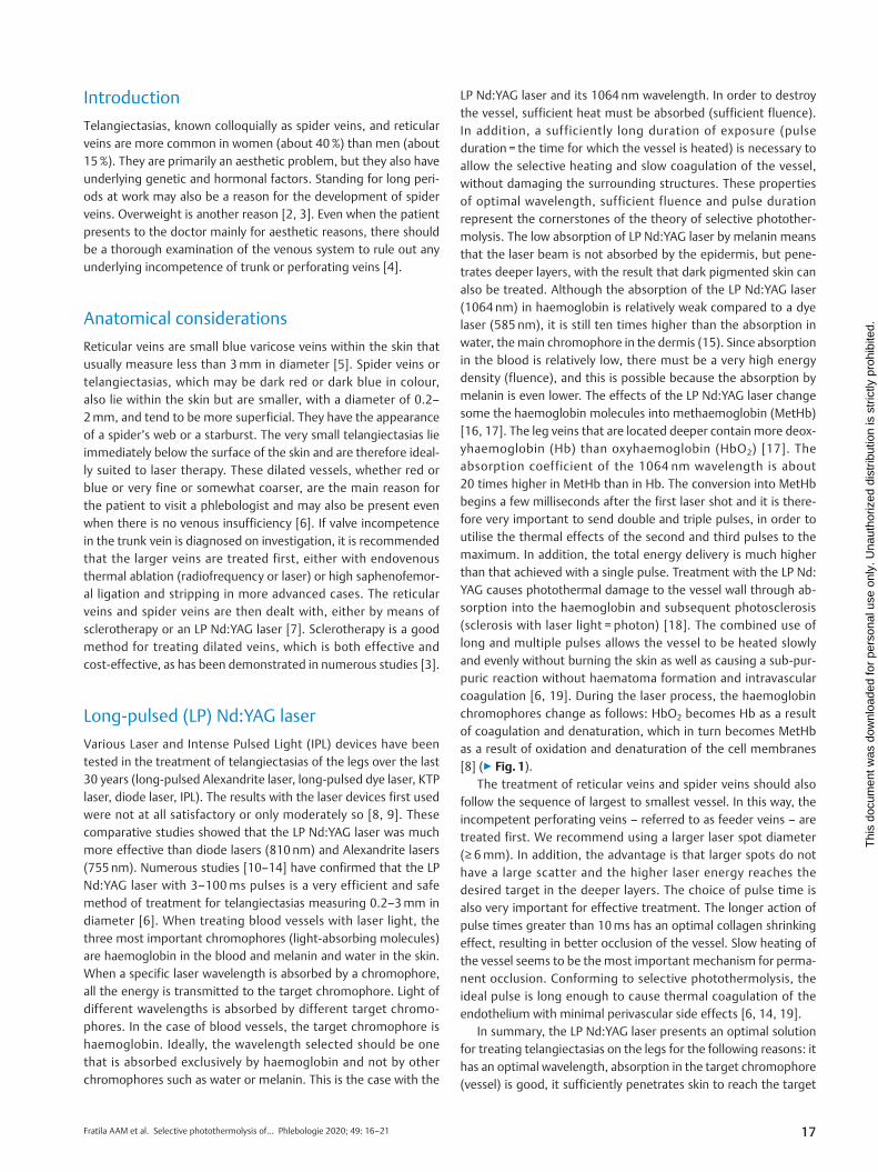
LP Nd:YAG laser and its 1064 nm wavelength. In order to destroythe vessel, sufficient heat must be absorbed (sufficient fluence).In addition, a sufficiently long duration of exposure (pulseduration = the time for which the vessel is heated) is necessary toallow the selective heating and slow coagulation of the vessel,without damaging the surrounding structures. These propertiesof optimal wavelength, sufficient fluence and pulse durationrepresent the cornerstones of the theory of selective photother-molysis. The low absorption of LP Nd:YAG laser by melanin meansthat the laser beam is not absorbed by the epidermis, but pene-trates deeper layers, with the result that dark pigmented skin canalso be treated. Although the absorption of the LP Nd:YAG laser(1064 nm) in haemoglobin is relatively weak compared to a dyelaser (585 nm), it is still ten times higher than the absorption inwater, the main chromophore in the dermis (15). Since absorptionin the blood is relatively low, there must be a very high energydensity (fluence), and this is possible because the absorption bymelanin is even lower. The effects of the LP Nd:YAG laser changesome the haemoglobin molecules into methaemoglobin (MetHb)[16, 17]. The leg veins that are located deeper contain more deox-yhaemoglobin (Hb) than oxyhaemoglobin (HbO2) [17]. Theabsorption coefficient of the 1064 nm wavelength is about20 times higher in MetHb than in Hb. The conversion into MetHbbegins a few milliseconds after the first laser shot and it is there-fore very important to send double and triple pulses, in order toutilise the thermal effects of the second and third pulses to themaximum. In addition, the total energy delivery is much higherthan that achieved with a single pulse. Treatment with the LP Nd:YAG causes photothermal damage to the vessel wall through ab-sorption into the haemoglobin and subsequent photosclerosis(sclerosis with laser light = photon) [18]. The combined use oflong and multiple pulses allows the vessel to be heated slowlyand evenly without burning the skin as well as causing a sub-pur-puric reaction without haematoma formation and intravascularcoagulation [6, 19]. During the laser process, the haemoglobinchromophores change as follows: HbO2 becomes Hb as a resultof coagulation and denaturation, which in turn becomes MetHbas a result of oxidation and denaturation of the cell membranes[8] (▶ Fig. 1).
The treatment of reticular veins and spider veins should alsofollow the sequence of largest to smallest vessel. In this way, theincompetent perforating veins – referred to as feeder veins – aretreated first. We recommend using a larger laser spot diameter(≥ 6mm). In addition, the advantage is that larger spots do nothave a large scatter and the higher laser energy reaches thedesired target in the deeper layers. The choice of pulse time isalso very important for effective treatment. The longer action ofpulse times greater than 10ms has an optimal collagen shrinkingeffect, resulting in better occlusion of the vessel. Slow heating ofthe vessel seems to be the most important mechanism for perma-nent occlusion. Conforming to selective photothermolysis, theideal pulse is long enough to cause thermal coagulation of theendothelium with minimal perivascular side effects [6, 14, 19].
In summary, the LP Nd:YAG laser presents an optimal solutionfor treating telangiectasias on the legs for the following reasons: ithas an optimal wavelength, absorption in the target chromophore(vessel) is good, it sufficiently penetrates skin to reach the target
17Fratila AAM et al. Selective photothermolysis of… Phlebologie 2020; 49: 16–21
Thi
s do
cum
ent w
as d
ownl
oade
d fo
r pe
rson
al u
se o
nly.
Una
utho
rized
dis
trib
utio
n is
str
ictly
pro
hibi
ted.
vessels (small perforating veins can also be treated effectively), ithas the greatest effect out of all types of lasers thanks to its mini-mal absorption in the epidermis and lowest absorption by melanin– which means that post-inflammatory hyperpigmentation (PIH)is extremely rare. The three pulse sequences with their differentdelay times provide dynamic cooling of the epidermis (withoutsubstantially cooling the dermis), so that sufficient fluence andpulse duration are achieved in the target tissues to allow selectiveheating and slow coagulation of the target vessel and its intima,without causing any damage to the surrounding tissues (theskin). The pulse duration should correspond to the thermal relaxa-tion time of the target structure (shorter or the same length). Thethermal relaxation time is the time that the target tissues requireto pass the absorbed heat on to the surrounding structures.
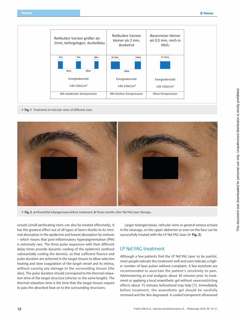
Larger telangiectasias, reticular veins or general venous ectasiain the cleavage, on the upper abdomen or even on the face can besuccessfully treated with the LP Nd:YAG laser (▶ Fig. 2).
LP Nd:YAG treatment
Although a few patients find the LP Nd:YAG laser to be painful,most people tolerate the treatment well and even tolerate a high-er number of laser pulses without complaint. A few testshots arerecommended to ascertain the patient’s sensitivity to pain.Administering an oral analgesic about 30 minutes prior to treat-ment or applying a local anaesthetic gel without vasoconstrictingeffects about 15 minutes beforehand may help [7]. Immediatelybefore treatment, the anaesthetic gel should be carefullyremoved and the skin degreased. A cooled transparent ultrasound
▶ Fig. 2 a Infraorbital telangiectasia before treatment. b Three months after Nd:YAG laser therapy.
Re�kuläre Varizen größer als 2mm, �efergelegen, dunkelblau
Re�kuläre Varizenkleiner als 2 mm,
dunkelrot
Energiedensität
140-150J/cm²
Besenreiser kleinerals 0,5 mm, reich in
HbO2
Energiedensität
140-150J/cm²
Energiedensität
120-130J/cm²
Mit moderater Kompression Mit leichter Kompression Ohne Kompression
▶ Fig. 1 Treatment of reticular veins of different sizes.
18 Fratila AAM et al. Selective photothermolysis of… Phlebologie 2020; 49: 16–21
Review
Thi
s do
cum
ent w
as d
ownl
oade
d fo
r pe
rson
al u
se o
nly.
Una
utho
rized
dis
trib
utio
n is
str
ictly
pro
hibi
ted.
gel is then applied to the area to be treated. If the gel becomesmilky during treatment, the laser head and the skin should becleaned. New gel is then applied [7].
There are four lightguides that can be attached to the LP Nd:YAG laser handpiece: round with diameters of 1.5mm, 6mm,and 9mm and a rectangular guide measuring 2 × 4mm [7]. Thetreating physician must first select the spot size depending onthe size of the vessel to be treated: a smaller spot for fine super-ficial veins and larger spots for thicker and more deeply lyingveins. Several recommendations for use are stored in the systemof our LP Nd:YAG laser, which are very helpful for the beginner[7]. We start with the larger deeper reticular veins and use a triplepulse with pulse times of 7ms and corresponding delays of 25ms:7ms-25ms-7ms-25ms-6ms ▶ Fig. 1). The selected fluence is140–150 J/cm2. Telangiectasias that are smaller than 2mm indiameter and from dark red to purple in colour, are treated witha double pulse: 8–11ms for the first pulse and 7–8ms for thesecond, with a delay of 25ms and the same fluence of 140–150 J/cm2. Finally, we treat the very fine spider veins of less than0.5mm in diameter, which are bright red and rich in HbO2. Thepresent article presents a treatment method that is always carriedout with the Nd:YAG laser handpiece of the M22 multiapplicationdevice from Lumenis®. The treatment parameters given in this ar-ticle are to be considered as recommendations only and must notbe extrapolated to other Nd:YAG lasers. In general, treatmentwith the LP Nd:YAG laser depends less on the technical affinitythan on the selected settings and the treatment skills of the doc-tor. After each applied shot, the treated vessel should be mas-saged with the highly cooled lightguide, to massage away the in-travascular coagulum and render any later removal of haematomaunnecessary [7]. Before the start of treatment, the area to betreated should be cooled by a back-and-forth movement of thecooled laser head for 1–2 seconds. This massage in the prepara-tory phase allows the direction of blood flow to be determined.Before using the LP Nd:YAG laser, the position of the laser head isorientated to the direction of blood flow, in order to squeeze theblood away. To ensure a good contact, the laser head is positionedobliquely against the skin and slight to moderate pressure is ap-
plied. The exerted contact pressure is proportional to the sizeand depth of the vein, in order to reduce the overall diameter ofthe vein and maintain a good density of action. The effects of theheat are diminished by the reduction in blood volume. As a result,the treatment will of course be less painful, especially in the caseof the largest veins, and will thereby also prevent the formation oflarge intravascular haematomas. If the blood return flow is slow,the direction of the lightguide is not so important. Irrespective ofthe spot size, closure of thinner blood vessels is less painful than itis for larger vessels. Positioning of the LP Nd:YAG lightguide at anoblique angle and treating the entire length of the blood vesselcan also reduce the likelihood of the lumen reopening. It goeswithout saying that heavily tanned skin and skin treated withself-tanning agents should not be treated. Slightly hyperpigmen-ted skin can be treated and we are therefore prepared to treatthese patients throughout the summer as well. It is then recom-mended to reduce the fluence by about 10% to 15% and to useonly double or triple pulse therapy, whereby the interval betweenthe pulses should be increased to 30ms.
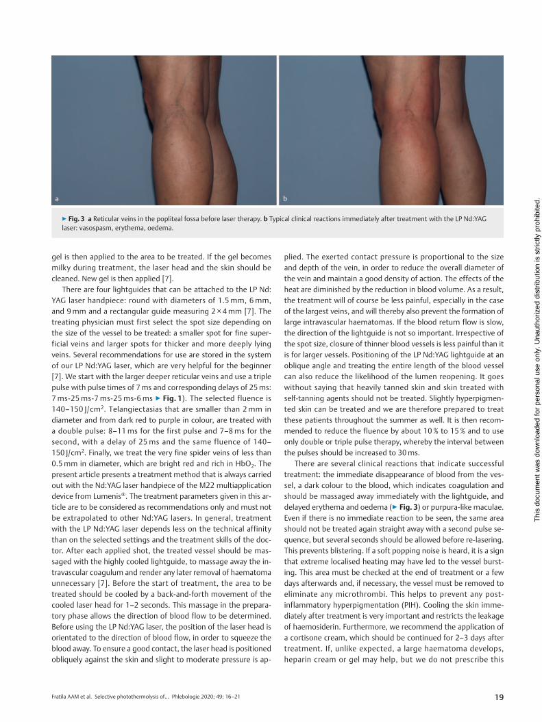
There are several clinical reactions that indicate successfultreatment: the immediate disappearance of blood from the ves-sel, a dark colour to the blood, which indicates coagulation andshould be massaged away immediately with the lightguide, anddelayed erythema and oedema (▶ Fig. 3) or purpura-like maculae.Even if there is no immediate reaction to be seen, the same areashould not be treated again straight away with a second pulse se-quence, but several seconds should be allowed before re-lasering.This prevents blistering. If a soft popping noise is heard, it is a signthat extreme localised heating may have led to the vessel burst-ing. This area must be checked at the end of treatment or a fewdays afterwards and, if necessary, the vessel must be removed toeliminate any microthrombi. This helps to prevent any post-inflammatory hyperpigmentation (PIH). Cooling the skin imme-diately after treatment is very important and restricts the leakageof haemosiderin. Furthermore, we recommend the application ofa cortisone cream, which should be continued for 2–3 days aftertreatment. If, unlike expected, a large haematoma develops,heparin cream or gel may help, but we do not prescribe this
▶ Fig. 3 a Reticular veins in the popliteal fossa before laser therapy. b Typical clinical reactions immediately after treatment with the LP Nd:YAGlaser: vasospasm, erythema, oedema.
19Fratila AAM et al. Selective photothermolysis of… Phlebologie 2020; 49: 16–21
Thi
s do
cum
ent w
as d
ownl
oade
d fo
r pe
rson
al u
se o
nly.
Una
utho
rized
dis
trib
utio
n is
str
ictly
pro
hibi
ted.
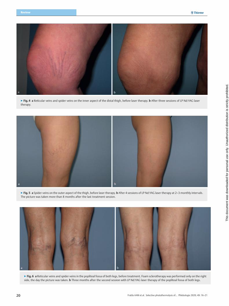
▶ Fig. 4 a Reticular veins and spider veins on the inner aspect of the distal thigh, before laser therapy. b After three sessions of LP Nd:YAG lasertherapy.
▶ Fig. 5 a Spider veins on the outer aspect of the thigh, before laser therapy. b After 4 sessions of LP Nd:YAG laser therapy at 2–3 monthly intervals.The picture was taken more than 8 months after the last treatment session.
▶ Fig. 6 a Reticular veins and spider veins in the popliteal fossa of both legs, before treatment. Foam sclerotherapy was performed only on the rightside, the day the picture was taken. b Three months after the second session with LP Nd:YAG laser therapy of the popliteal fossa of both legs.
20 Fratila AAM et al. Selective photothermolysis of… Phlebologie 2020; 49: 16–21
Review
Thi
s do
cum
ent w
as d
ownl
oade
d fo
r pe
rson
al u
se o
nly.
Una
utho
rized
dis
trib
utio
n is
str
ictly
pro
hibi
ted.
routinely. Subsequent treatment sessions should be planned atintervals of 6–8 weeks (▶ Fig. 4–6). Patients must be told to avoidany excessive exposure to the sun until the next treatment sessionand not to use any self-tanning agents before then. Sunscreenshould be used consistently during holidays [7].
Advantages of treating reticular veins andspider veins with the LP Nd:YAG laser
There are no allergic reactions either during or after laser treat-ment. Pain is minimised with the use of a local anaesthetic gel ororal analgesics. So far, thrombosis has not been observed as a sideeffect, so there is no limit to the number of veins or areas that canbe treated. All reticular veins and spider veins can be treated inone session, providing the patient’s pain tolerance allows it. Reti-cular veins on the foot, below the medial or lateral malleolus, canalso be treated without the risk of a deep vein thrombosis. Hypo-pigmentation and hyperpigmentation are extremely rare withcorrect use of the laser. However, a very long learning curve forthe user is normal. Compression bandages are not necessary.Wearing compression stockings is also not really necessary, butwe do recommend that class I compression stockings are wornfor 1–2 weeks, if larger reticular veins have been treated [7].Busy people particularly appreciate the laser method, becausethey only need to come for treatment every two months – with atotal of 3–5 sessions on average. Combination with IPL is possibleand advisable when there is matting, as our laser devices areappropriately equipped for such use.
For patients, who are set on having laser therapy, this is theirmain reason for seeking out your practice.
Summary
The LP Nd:YAG with its 1064 nm wavelength allows the deepestpenetration of all non-invasive vascular lasers. The high energy al-lows penetration into the dermis and also the destruction of largevessels. The LP Nd:YAG with larger spots penetrates deeper. Themultiple pulses also penetrate deeper and allow greater thermalrelaxation times. The long pulse duration leads to a slower andmore uniform heating of the vessel, without causing it to burstthus reducing the possibility of later purpura and hyperpigmenta-tion.
Conflict of interest
The first two authors are key opinion leaders of Lumenis®. They receivehonoraria from this company for presentations made at weekend work-shops. They have not received any fee for the present article.
References
[1] Anderson RR, Parrish JA. Selective photothermolysis: precise microsur-gery by selective absorption of pulse radiation. Science 1983; 220:524–727
[2] Parlar B, Blazek C, Cazzaniga S et al. Treatment of lower extremitytelangiectasias in women by foam sclerotherapy vs. Nd:YAG laser:a prospective, comparative, randomized, open-label trial. JEADV 2014
[3] Lohr JM, Bush RL. Venous disease in women: Epidemiology, manifesta-tions, and treatment. J Vasc Surg 2013; 57: 37–45
[4] Munia MA, Wolosker N, Munia CG et al. Comparison of laser versussclerotherapy in the treatment of lower extremity teleangiectases: aprospective study. Dermatol Surg 2012; 38: 635–639
[5] Meissner MH. Lower extremity venous anatomy. Semin Intervent Radiol2005; 22 (3): 147–156
[6] Ross EV, Domankevitz Y. laser Treatment of Leg Veins: Physical Mecha-nisms and Theoretical Considerations lasers Surg. Med 2005; 36: 105–116
[7] Lumenis®, Wittig C. Leg veins – Long-pulsed (LP) Nd:YAG Guidelines/CD-1008960 Rev A 2017.
[8] Gloviczki P, Comerota AJ, Dalsing MC et al. Society for Vascular Surgery;American Venous Forum. The care of patients with varicose veins andassociated chronic venous diseases: Clinical practice guidelines of theSociety for Vascular Surgery and the American Venous Forum. J VascSurg 2011; 53: 2–48
[9] Eremia S, Li C, Umar SH. A side-by-side comparative study of 1064 nmNd:YAG, 810 nm diode and 755 nm alexandrite lasers for treatment of0.3–3mm leg veins. Dermatologic Surgery 2002; 28 (3): 224–230
[10] Dover JS, Sadick NS, Goldman MP. The role of laser s and light sources inthe treatment of leg veins. Dermatol Surg 1999; 25: 328–335
[11] Adamic M, Troilius A, Adatto M et al. Vascular laser s and IPLS: Guidelinesfor care from the European Society for Laser Dermatology. J CosmetLaser Ther 2007; 9: 113–124
[12] Sadick NS, Weiss RA, Goldman MP. Advances in laser surgery for legveins: Bimodal wavelength approach to lower extremity vessels, newcooling techniques and longer pulse durations. Dermatol Surg 2002; 28:16–20
[13] Levy JL, Elbahr C, Jouve E et al. Comparison and sequential study of longpulsed Nd:YAG 1,064 nm laser and sclerotherapy in leg telangiectasiastreatment. Lasers in Surgery and Medicine 2004; 34: 273–276
[14] Trelles MA, Allones I, Martín-Vázquez MJ et al. Long pulse Nd:YAG laserfor treatment of leg veins in 40 Patients with assessments at 6 and12 months. Lasers in Surgery and Medicine 2004; 35: 68–76
[15] Bäumler W, Ulrich H, Hartl A et al. Optimal parameters for the treatmentof leg veins using Nd:YAG lasers at 1064 nm. British Journal of Derma-tology 2006; 155: pp364–pp371
[16] Randeberg LL, Bonesrønning JH, Dalaker M et al. Methaemoglobin for-mation during laser-induced photothermolysis of vascular skin lesions.Lasers Surg Med 2004; 34: 414–419
[17] Black JF, Barton JK. Chemical and structural changes in blood under-going laser photocoagulation. Photochemistry and Photobiology 2004;80: 89–97
[18] Mordon S, Brisot D, Fournier N. Using a “non uniform pulse sequence”can improve selective coagulation with a Nd:YAG laser (1.06mm) thanksto Met-hemoglobin absorption: a clinical study on blue leg veins. Lasersin Surgery and Medicine 2003; 32: 160–170
[19] Parlette EC, Groff WF, Kinshella MJ et al. Optimal pulse durations for thetreatment of legtelangiectasias with a Neodymium YAG laser. Lasers inSurgery and Medicine 2006; 38: 98–105
21Fratila AAM et al. Selective photothermolysis of… Phlebologie 2020; 49: 16–21
Thi
s do
cum
ent w
as d
ownl
oade
d fo
r pe
rson
al u
se o
nly.
Una
utho
rized
dis
trib
utio
n is
str
ictly
pro
hibi
ted.