Microevolution of Candida albicans in macrophages restores filamentation in a nonfilamentous mutant
18
Microevolution of Candida albicans in Macrophages Restores Filamentation in a Nonfilamentous Mutant Anja Wartenberg 1 , Jo ¨ rg Linde 2 , Ronny Martin 3 , Maria Schreiner 1 , Fabian Horn 2 , Ilse D. Jacobsen 1,4 , Sabrina Jenull 5 , Thomas Wolf 2 , Karl Kuchler 5 , Reinhard Guthke 2 , Oliver Kurzai 3 , Anja Forche 6 , Christophe d’Enfert 7,8 , Sascha Brunke 1,9. , Bernhard Hube 1,9,10 * . 1 Department of Microbial Pathogenicity Mechanisms, Leibniz Institute for Natural Product Research and Infection Biology – Hans Knoell Institute Jena (HKI), Jena, Germany, 2 Research Group Systems Biology & Bioinformatics, Leibniz Institute for Natural Product Research and Infection Biology – Hans Knoell Institute Jena (HKI), Jena, Germany, 3 Septomics Research Center, Friedrich Schiller University and Leibniz Institute for Natural Product Research and Infection Biology –Hans Knoell Institute, Jena, Germany, 4 Research Group Microbial Immunology, Leibniz Institute for Natural Product Research and Infection Biology – Hans Knoell Institute Jena (HKI), Jena, Germany, 5 Medical University Vienna, Max F. Perutz Laboratories, Department of Medical Biochemistry, Vienna, Austria, 6 Department of Biology, Bowdoin College, Brunswick, Maine, United States of America, 7 Institut Pasteur, Unite ´ Biologie et Pathoge ´nicite ´ Fongiques, De ´partement Ge ´ nomes et Ge ´ne ´ tique, Paris, France, 8 INRA, USC2019, Paris, France, 9 Integrated Research and Treatment Center, Sepsis und Sepsisfolgen, Center for Sepsis Control and Care (CSCC), Universita ¨tsklinikum Jena, Germany, 10 Friedrich Schiller University, Jena, Germany Abstract Following antifungal treatment, Candida albicans, and other human pathogenic fungi can undergo microevolution, which leads to the emergence of drug resistance. However, the capacity for microevolutionary adaptation of fungi goes beyond the development of resistance against antifungals. Here we used an experimental microevolution approach to show that one of the central pathogenicity mechanisms of C. albicans, the yeast-to-hyphae transition, can be subject to experimental evolution. The C. albicans cph1D/efg1D mutant is nonfilamentous, as central signaling pathways linking environmental cues to hyphal formation are disrupted. We subjected this mutant to constant selection pressure in the hostile environment of the macrophage phagosome. In a comparatively short time-frame, the mutant evolved the ability to escape macrophages by filamentation. In addition, the evolved mutant exhibited hyper-virulence in a murine infection model and an altered cell wall composition compared to the cph1D/efg1D strain. Moreover, the transcriptional regulation of hyphae-associated, and other pathogenicity-related genes became re-responsive to environmental cues in the evolved strain. We went on to identify the causative missense mutation via whole genome- and transcriptome-sequencing: a single nucleotide exchange took place within SSN3 that encodes a component of the Cdk8 module of the Mediator complex, which links transcription factors with the general transcription machinery. This mutation was responsible for the reconnection of the hyphal growth program with environmental signals in the evolved strain and was sufficient to bypass Efg1/Cph1-dependent filamentation. These data demonstrate that even central transcriptional networks can be remodeled very quickly under appropriate selection pressure. Citation: Wartenberg A, Linde J, Martin R, Schreiner M, Horn F, et al. (2014) Microevolution of Candida albicans in Macrophages Restores Filamentation in a Nonfilamentous Mutant. PLoS Genet 10(12): e1004824. doi:10.1371/journal.pgen.1004824 Editor: Geraldine Butler, University College Dublin, Ireland Received May 8, 2014; Accepted October 15, 2014; Published December 4, 2014 Copyright: ß 2014 Wartenberg et al. This is an open-access article distributed under the terms of the Creative Commons Attribution License, which permits unrestricted use, distribution, and reproduction in any medium, provided the original author and source are credited. Data Availability: The authors confirm that all data underlying the findings are fully available without restriction. Data were deposited at the Gene Expression Omnibus (GSE56174) and can be found in S2 Table. Funding: This work was supported by the German Federal Ministry of Education and Health (BMBF, www.bmbf.de) Germany, FKZ: 01EO1002 - Integrated Research and Treatment Center, Center for Sepsis Control and Care (CSSC, www.cscc.uniklinikum-jena.de) and the DACH program of the DFG and FWF (DFG HU 528/17-1 & FWF-I-746-B11). AW and TW were also supported by the excellence graduate school Jena School for Microbial Communication (JSMC; www.jsmc.uni- jena.de) and the International Leibniz Research School for Microbial and Biomolecular Interactions (ILRS, www.ilrs.hki-jena.de), respectively. JL was supported by the Deutsche Forschungsgemeinschaft (DFG, www.dfg.de) CRC/Transregio 124 ‘FungiNet-Pathogenic fungi and their human host: Networks of interaction’ (www. funginet.de), subproject INF. IDJ was supported by BMBF 0314108. AF was supported by the National Institute of Allergy and Infectious Diseases, grant R15 AI090633 02 (NIAID, www.niaid.nih.gov). Cd has received funding from the French Government’s Investissement d’Avenir program, Laboratoire d’Excellence ‘‘Integrative Biology of Emerging Infectious Diseases’’ (Grant #ANR-10-LABX-62-IBEID). Work in the KK laboratory was supported by the Austrian Science Foundation FWF by a grant from the Christian Doppler Society. SJ was additionally supported by the FWF (project P-25333-B22). The funders had no role in study design, data collection and analysis, decision to publish, or preparation of the manuscript. Competing Interests: The authors have declared that no competing interests exist. * Email: [email protected] . These authors contributed equally to this work. Introduction The incidence of invasive fungal infections has steadily increased within the past decades, largely because of a growing population of susceptible individuals, reflecting the progress of modern medicine in prolonging life even with severe underlying diseases and the increasing rate of immuno-deficient patients. One of the most frequently isolated fungi is Candida albicans, an ubiquitous and normally harmless commensal of the alimentary tract and mucocutaneous membranes. As an opportunistic pathogen, it can cause superficial infections like oropharyngeal candidiasis, especially in HIV patients, as well as life-threatening PLOS Genetics | www.plosgenetics.org 1 December 2014 | Volume 10 | Issue 12 | e1004824
-
Upload
independent -
Category
Documents
-
view
0 -
download
0
Transcript of Microevolution of Candida albicans in macrophages restores filamentation in a nonfilamentous mutant

Microevolution of Candida albicans in MacrophagesRestores Filamentation in a Nonfilamentous MutantAnja Wartenberg1, Jorg Linde2, Ronny Martin3, Maria Schreiner1, Fabian Horn2, Ilse D. Jacobsen1,4,
Sabrina Jenull5, Thomas Wolf2, Karl Kuchler5, Reinhard Guthke2, Oliver Kurzai3, Anja Forche6,
Christophe d’Enfert7,8, Sascha Brunke1,9., Bernhard Hube1,9,10*.
1 Department of Microbial Pathogenicity Mechanisms, Leibniz Institute for Natural Product Research and Infection Biology – Hans Knoell Institute Jena (HKI), Jena,
Germany, 2 Research Group Systems Biology & Bioinformatics, Leibniz Institute for Natural Product Research and Infection Biology – Hans Knoell Institute Jena (HKI), Jena,
Germany, 3 Septomics Research Center, Friedrich Schiller University and Leibniz Institute for Natural Product Research and Infection Biology –Hans Knoell Institute, Jena,
Germany, 4 Research Group Microbial Immunology, Leibniz Institute for Natural Product Research and Infection Biology – Hans Knoell Institute Jena (HKI), Jena, Germany,
5 Medical University Vienna, Max F. Perutz Laboratories, Department of Medical Biochemistry, Vienna, Austria, 6 Department of Biology, Bowdoin College, Brunswick,
Maine, United States of America, 7 Institut Pasteur, Unite Biologie et Pathogenicite Fongiques, Departement Genomes et Genetique, Paris, France, 8 INRA, USC2019, Paris,
France, 9 Integrated Research and Treatment Center, Sepsis und Sepsisfolgen, Center for Sepsis Control and Care (CSCC), Universitatsklinikum Jena, Germany, 10 Friedrich
Schiller University, Jena, Germany
Abstract
Following antifungal treatment, Candida albicans, and other human pathogenic fungi can undergo microevolution, whichleads to the emergence of drug resistance. However, the capacity for microevolutionary adaptation of fungi goes beyondthe development of resistance against antifungals. Here we used an experimental microevolution approach to show thatone of the central pathogenicity mechanisms of C. albicans, the yeast-to-hyphae transition, can be subject to experimentalevolution. The C. albicans cph1D/efg1D mutant is nonfilamentous, as central signaling pathways linking environmental cuesto hyphal formation are disrupted. We subjected this mutant to constant selection pressure in the hostile environment ofthe macrophage phagosome. In a comparatively short time-frame, the mutant evolved the ability to escape macrophagesby filamentation. In addition, the evolved mutant exhibited hyper-virulence in a murine infection model and an altered cellwall composition compared to the cph1D/efg1D strain. Moreover, the transcriptional regulation of hyphae-associated, andother pathogenicity-related genes became re-responsive to environmental cues in the evolved strain. We went on toidentify the causative missense mutation via whole genome- and transcriptome-sequencing: a single nucleotide exchangetook place within SSN3 that encodes a component of the Cdk8 module of the Mediator complex, which links transcriptionfactors with the general transcription machinery. This mutation was responsible for the reconnection of the hyphal growthprogram with environmental signals in the evolved strain and was sufficient to bypass Efg1/Cph1-dependent filamentation.These data demonstrate that even central transcriptional networks can be remodeled very quickly under appropriateselection pressure.
Citation: Wartenberg A, Linde J, Martin R, Schreiner M, Horn F, et al. (2014) Microevolution of Candida albicans in Macrophages Restores Filamentation in aNonfilamentous Mutant. PLoS Genet 10(12): e1004824. doi:10.1371/journal.pgen.1004824
Editor: Geraldine Butler, University College Dublin, Ireland
Received May 8, 2014; Accepted October 15, 2014; Published December 4, 2014
Copyright: � 2014 Wartenberg et al. This is an open-access article distributed under the terms of the Creative Commons Attribution License, which permitsunrestricted use, distribution, and reproduction in any medium, provided the original author and source are credited.
Data Availability: The authors confirm that all data underlying the findings are fully available without restriction. Data were deposited at the Gene ExpressionOmnibus (GSE56174) and can be found in S2 Table.
Funding: This work was supported by the German Federal Ministry of Education and Health (BMBF, www.bmbf.de) Germany, FKZ: 01EO1002 - IntegratedResearch and Treatment Center, Center for Sepsis Control and Care (CSSC, www.cscc.uniklinikum-jena.de) and the DACH program of the DFG and FWF (DFG HU528/17-1 & FWF-I-746-B11). AW and TW were also supported by the excellence graduate school Jena School for Microbial Communication (JSMC; www.jsmc.uni-jena.de) and the International Leibniz Research School for Microbial and Biomolecular Interactions (ILRS, www.ilrs.hki-jena.de), respectively. JL was supported bythe Deutsche Forschungsgemeinschaft (DFG, www.dfg.de) CRC/Transregio 124 ‘FungiNet-Pathogenic fungi and their human host: Networks of interaction’ (www.funginet.de), subproject INF. IDJ was supported by BMBF 0314108. AF was supported by the National Institute of Allergy and Infectious Diseases, grant R15AI090633 02 (NIAID, www.niaid.nih.gov). Cd has received funding from the French Government’s Investissement d’Avenir program, Laboratoire d’Excellence‘‘Integrative Biology of Emerging Infectious Diseases’’ (Grant #ANR-10-LABX-62-IBEID). Work in the KK laboratory was supported by the Austrian ScienceFoundation FWF by a grant from the Christian Doppler Society. SJ was additionally supported by the FWF (project P-25333-B22). The funders had no role in studydesign, data collection and analysis, decision to publish, or preparation of the manuscript.
Competing Interests: The authors have declared that no competing interests exist.
* Email: [email protected]
. These authors contributed equally to this work.
Introduction
The incidence of invasive fungal infections has steadily
increased within the past decades, largely because of a growing
population of susceptible individuals, reflecting the progress of
modern medicine in prolonging life even with severe underlying
diseases and the increasing rate of immuno-deficient patients. One
of the most frequently isolated fungi is Candida albicans, an
ubiquitous and normally harmless commensal of the alimentary
tract and mucocutaneous membranes. As an opportunistic
pathogen, it can cause superficial infections like oropharyngeal
candidiasis, especially in HIV patients, as well as life-threatening
PLOS Genetics | www.plosgenetics.org 1 December 2014 | Volume 10 | Issue 12 | e1004824

systemic infections with mortality rates up to 40%, even with
current antifungal treatment options [1].
The transition from the commensal to a pathogenic state
depends on the microbiota, the host response, and C. albicansactivities, such as adhesion, secretion of hydrolases, metabolic
adaptation, biofilm formation and, importantly, morphological
plasticity, which includes the yeast-to-filament transition [2–7]. To
survive and thrive in the many different niches inside the host, C.albicans must be able to adapt to changing environments and
different stresses. In the short term, this occurs primarily by
changes in gene expression and translation, and via post-
translational modifications, but ultimately microevolutionary
processes will play an important role. As a prominent example,
White et al. [8] have shown that microevolution is the driving
force behind the emergence of antifungal drug resistance. They
demonstrated the de novo appearance of fluconazole resistance in
evolving C. albicans strains in vivo [8]. Furthermore, clinical
isolates generally exhibit large genetic variations, and microevo-
lution can be observed both in vitro and in vivo [9,10], indicating
that this process plays an important role in host-pathogen
interactions. Therefore, microevolution provides a source of
variation for the adaptive response of C. albicans to challenging
(host) environments.
Different mechanisms account for the generation of new
genotypic variants, including point mutations, amplification or
deletion of chromosomal segments, chromosomal translocation or
inversion, and whole chromosome aneuploidy. These genetic
variations can affect expression of single genes or the structure of
their encoded proteins as well as whole transcriptional networks
via a mechanism known as transcriptional rewiring. In this
process, the interaction between promoter regions and their
corresponding regulators can be switched to different pairings,
which in turn cause new connections to be formed between a
signal and a transcriptional response [11,12]. Whereas many
studies have explored the underlying mechanisms of drug
resistance, the role that microevolution plays in host-pathogen
interactions has rarely been investigated: Forche et al. [13] found
that a C. albicans strain, passaged through a mouse host,
responded by undergoing chromosome-level genetic variations,
which were sufficient to generate new variants of C. albicans.The yeast-to-hyphae transition of C. albicans is central for
pathogenicity [14,15]. Filamentation plays a pivotal role for
adhesion to, invasion into and damage of epithelial and
endothelial cells [2,16,17]. Upon internalization by macrophages,
C. albicans induces host cell death by triggering pyroptosis, a form
of programmed cell death [18,19]. However, later in the infection
process the yeast-to-hyphae transition contributes to escape from
the phagosome [19,20]. Morphology also plays a key role in host
recognition [21]. Given the importance of morphology of C.albicans for pathogenicity, it is not surprising that the yeast-to-
filament transition is induced by a wide range of environmental
factors and conditions like high pH, host body temperature, CO2,
starvation and presence of serum, all of which act via several
signaling pathways. Among them, the cAMP-dependent protein
kinase A (cAMP-PKA) and the mitogen-activated protein kinase
(MAPK) pathways, which target the transcription factors Efg1 and
Cph1, respectively, play a central role in hyphal formation
[22,23]. This is demonstrated by a cph1D/efg1D double mutant,
which is unable to form hyphae under almost all hyphae-inducing
conditions in vitro (except agar embedded conditions) and which is
probably the most commonly used mutant of C. albicans in a wide
range of experiments [14,15,22,24].
Due to the central role of the yeast-to-filament transition for C.albicans virulence, we used the cph1D/efg1D double mutant as a
model for evolutionary adaptation. To this end, we performed a
series of co-culture passages of this mutant with macrophages. We
expected that the hostile environment of the phagosome imposes a
high selective pressure on the fungus favoring either intracellular
adaptation or return to filamentation in order to escape. We
performed phenotypic, transcriptomic and genomic analyses of the
pre- and post-passaged strains to elucidate the degree of genetic
plasticity of C. albicans when facing host stresses. We show that
adaptation to macrophages leads to distinct phenotypic differences
between the pre- and post-passaged strains with regained
filamentation in the latter. As the causative mutation, we identified
a heterozygous, non-synonymous single nucleotide exchange in
the gene SSN3, which encodes the cyclin-dependent kinase of a
regulatory module of the Mediator complex. Our results
demonstrate that the regulation of the morphological switch in
C. albicans can be subject to microevolution.
Results
Experimental microevolution causes a reversion of thenonfilamentous phenotype of the cph1D/efg1D mutantstrain
To determine the ability of C. albicans to adapt to stresses inside
phagocytes and to test the adaptability of the hyphal regulatory
network, we first screened for mutants which are unable to escape
from macrophages via filamentation response. We tested multiple
C. albicans deletion strains with known defects in hyphal
formation: strains lacking RAS1, RIM101, DFG16, TEC1,
HGC1, EED1, or UME6 and the avirulent double deletion
mutant lacking CPH1 and EFG1 [22]. Of these, only the cph1D/
efg1D double mutant was completely unable to escape from
macrophages even after 24 hours, while all other mutants still
formed filaments inside the host cell and pierced the phagocyte
membrane to some extent (S1A Figure). Microscopy with FITC-
labeled cph1D/efg1D cells revealed that these cells were viable and
Author Summary
Pathogenic microbes often evolve complex traits to adaptto their respective hosts, and this evolution is ongoing: forexample, microorganisms are developing resistance toantimicrobial compounds in the clinical setting. The abilityof the common human pathogenic fungus, Candidaalbicans, to switch from yeast to hyphal (filamentous)growth is considered a central virulence attribute. Forexample, hyphal formation allows C. albicans to escapefrom macrophages following phagocytosis. A well-investi-gated signaling network integrates different environmen-tal cues to induce and maintain hyphal growth. In fact,deletion of two central transcription factors in this networkresults in a mutant that is both nonfilamentous andavirulent. We used experimental evolution to study theadaptation capability of this mutant by continuous co-incubation within macrophages. We found that thisselection regime led to a relatively rapid re-connection ofsignaling between environmental cues and the hyphalgrowth program. Indeed, the evolved mutant regained theability to filament and its virulence in vivo. This bypass ofcentral transcription factors was based on a singlenucleotide exchange in a gene encoding a componentof the general transcription regulation machinery. Ourresults show that even a complex regulatory network, suchas the transcriptional network which governs hyphalgrowth, can be remodeled via microevolution.
Adaptation of a Nonfilamentous C. albicans Mutant to Macrophages
PLOS Genetics | www.plosgenetics.org 2 December 2014 | Volume 10 | Issue 12 | e1004824
still able to replicate in the yeast form after ingestion by
macrophages (S1B Figure).
Therefore, we chose the cph1D/efg1D strain for the following
microevolution experiment. Cells of the murine macrophage cell
line J774A.1 were infected with the cph1D/efg1D double mutant
at a macrophage-to-fungal ratio of 2:1 and co-incubated. Every
24 hours, non-phagocytosed cells were removed and macrophages
were lysed to harvest the phagocytosed cells. A defined fraction of
this population was then transferred to a fresh macrophage
population.
After 19 passages, a significant morphological alteration became
visible, as several phagocytosed cells started to form filaments.
These filamenting cells became fixed in the population after
additional 23 rounds of co-incubation. This morphologically
distinct variant, evolutionary derived from the cph1D/efg1Dmutant, was termed Evo. The absence of CPH1 and EFG1 in the
Evo strain was verified by Southern blot analysis (S1C Figure). To
exclude temporary or epigenetic effects, the Evo strain was
repassaged daily in liquid rich (YPD) medium without any
selection pressure by host cells for 14 passages. The phenotype
remained stable and no reversal was detected.
To test whether the regained ability to form filaments was
restricted to macrophage interactions or observed under additional
hypha-inducing conditions, we analyzed the morphology of the
Evo strain in the absence of host cells. In the cell culture medium
DMEM with 10% serum at 37uC and 5% CO2, clear filament
formation of the Evo strain, but not the cph1D/efg1D strain, was
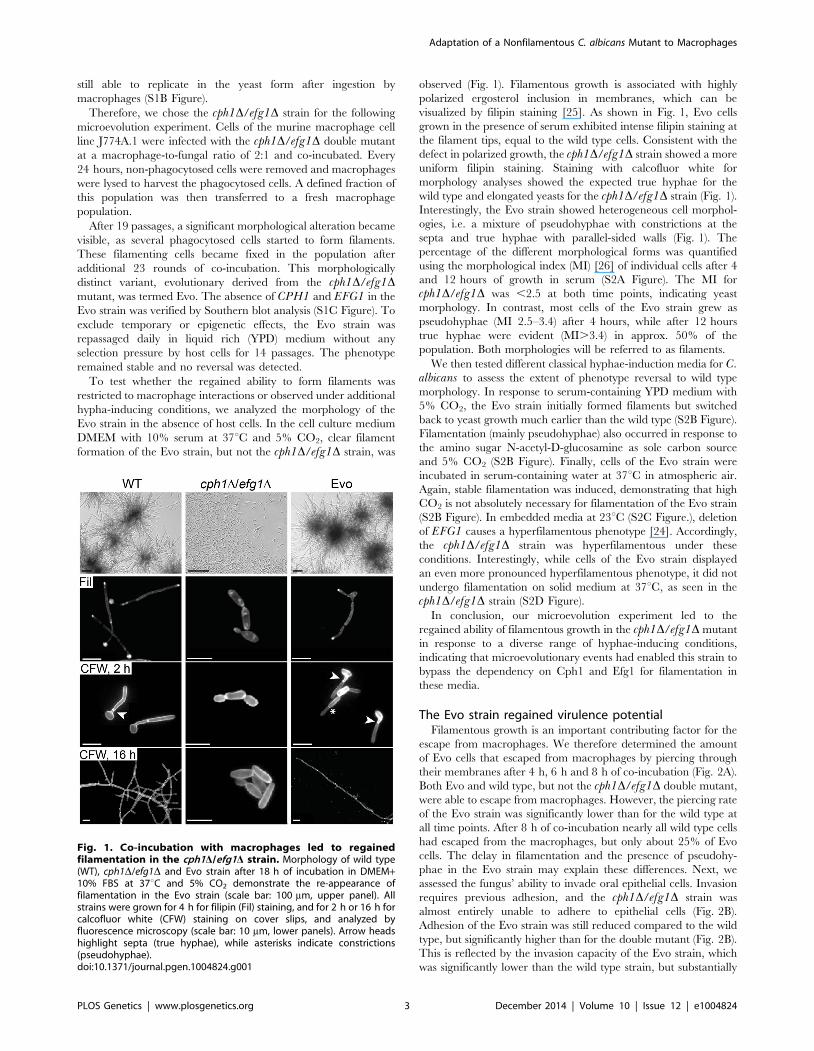
observed (Fig. 1). Filamentous growth is associated with highly
polarized ergosterol inclusion in membranes, which can be
visualized by filipin staining [25]. As shown in Fig. 1, Evo cells
grown in the presence of serum exhibited intense filipin staining at
the filament tips, equal to the wild type cells. Consistent with the
defect in polarized growth, the cph1D/efg1D strain showed a more
uniform filipin staining. Staining with calcofluor white for
morphology analyses showed the expected true hyphae for the
wild type and elongated yeasts for the cph1D/efg1D strain (Fig. 1).
Interestingly, the Evo strain showed heterogeneous cell morphol-
ogies, i.e. a mixture of pseudohyphae with constrictions at the
septa and true hyphae with parallel-sided walls (Fig. 1). The
percentage of the different morphological forms was quantified
using the morphological index (MI) [26] of individual cells after 4
and 12 hours of growth in serum (S2A Figure). The MI for
cph1D/efg1D was ,2.5 at both time points, indicating yeast
morphology. In contrast, most cells of the Evo strain grew as
pseudohyphae (MI 2.5–3.4) after 4 hours, while after 12 hours
true hyphae were evident (MI.3.4) in approx. 50% of the
population. Both morphologies will be referred to as filaments.
We then tested different classical hyphae-induction media for C.albicans to assess the extent of phenotype reversal to wild type
morphology. In response to serum-containing YPD medium with
5% CO2, the Evo strain initially formed filaments but switched
back to yeast growth much earlier than the wild type (S2B Figure).
Filamentation (mainly pseudohyphae) also occurred in response to
the amino sugar N-acetyl-D-glucosamine as sole carbon source
and 5% CO2 (S2B Figure). Finally, cells of the Evo strain were
incubated in serum-containing water at 37uC in atmospheric air.
Again, stable filamentation was induced, demonstrating that high
CO2 is not absolutely necessary for filamentation of the Evo strain
(S2B Figure). In embedded media at 23uC (S2C Figure.), deletion
of EFG1 causes a hyperfilamentous phenotype [24]. Accordingly,
the cph1D/efg1D strain was hyperfilamentous under these
conditions. Interestingly, while cells of the Evo strain displayed
an even more pronounced hyperfilamentous phenotype, it did not
undergo filamentation on solid medium at 37uC, as seen in the
cph1D/efg1D strain (S2D Figure).
In conclusion, our microevolution experiment led to the
regained ability of filamentous growth in the cph1D/efg1D mutant
in response to a diverse range of hyphae-inducing conditions,
indicating that microevolutionary events had enabled this strain to
bypass the dependency on Cph1 and Efg1 for filamentation in
these media.
The Evo strain regained virulence potentialFilamentous growth is an important contributing factor for the
escape from macrophages. We therefore determined the amount
of Evo cells that escaped from macrophages by piercing through
their membranes after 4 h, 6 h and 8 h of co-incubation (Fig. 2A).
Both Evo and wild type, but not the cph1D/efg1D double mutant,
were able to escape from macrophages. However, the piercing rate
of the Evo strain was significantly lower than for the wild type at
all time points. After 8 h of co-incubation nearly all wild type cells
had escaped from the macrophages, but only about 25% of Evo
cells. The delay in filamentation and the presence of pseudohy-
phae in the Evo strain may explain these differences. Next, we
assessed the fungus’ ability to invade oral epithelial cells. Invasion
requires previous adhesion, and the cph1D/efg1D strain was
almost entirely unable to adhere to epithelial cells (Fig. 2B).
Adhesion of the Evo strain was still reduced compared to the wild
type, but significantly higher than for the double mutant (Fig. 2B).
This is reflected by the invasion capacity of the Evo strain, which
was significantly lower than the wild type strain, but substantially
Fig. 1. Co-incubation with macrophages led to regainedfilamentation in the cph1D/efg1D strain. Morphology of wild type(WT), cph1D/efg1D and Evo strain after 18 h of incubation in DMEM+10% FBS at 37uC and 5% CO2 demonstrate the re-appearance offilamentation in the Evo strain (scale bar: 100 mm, upper panel). Allstrains were grown for 4 h for filipin (Fil) staining, and for 2 h or 16 h forcalcofluor white (CFW) staining on cover slips, and analyzed byfluorescence microscopy (scale bar: 10 mm, lower panels). Arrow headshighlight septa (true hyphae), while asterisks indicate constrictions(pseudohyphae).doi:10.1371/journal.pgen.1004824.g001
Adaptation of a Nonfilamentous C. albicans Mutant to Macrophages
PLOS Genetics | www.plosgenetics.org 3 December 2014 | Volume 10 | Issue 12 | e1004824

Fig. 2. Characterization of Evo strain interaction with host cells and virulence potential. (A) Escape of C. albicans cells by piercing ofmacrophages (J774A.1) after different timepoints (left). Micrographs of strains after 6 h of co-incubation with J774A.1 cells (right). Intracellular C.albicans appears blue (CFW), extracellular section of the cells red (Concanavalin A, ConA). Cells of the cph1D/efg1D strain cannot escape frommacrophages, while Evo cells regained this property during the evolution experiment. (B) Adhesion to and invasion of oral epithelial cells (TR-146).Adhesion values are given as percentage of adherent WT cells (left). Micrographs show filamentation of C. albicans WT and Evo strains after 6 h ofincubation with TR-146 cells (right). The regained ability to filament enabled the Evo strain to invade epithelial cells. Staining was performed asdescribed in (A). (C) Damage to macrophages and epithelial monolayers, determined by lactate dehydrogenase (LDH) assay after 32 h of co-incubation (LC = low control, medium only). WT and Evo strain, but not the cph1D/efg1D strain caused clear damage to both cell types. For piercing,adhesion, invasion and cell damage assay results are given as mean+SD of three independent experiments (*p,0.05). (D) Survival of BALB/c micechallenged intravenously (left; n = 10/strain). Nearly all mice infected with the Evo strain succumbed to the infection, while almost all animals infectedwith cph1D/efg1D strain survived (*p,0.05). PAS-hematoxylin-stained kidney sections from different days (d) post challenge (right) show fungal cells(arrows) either in the filamentous form (WT and Evo strain) or yeast form (cph1D/efg1D strain).doi:10.1371/journal.pgen.1004824.g002
Adaptation of a Nonfilamentous C. albicans Mutant to Macrophages
PLOS Genetics | www.plosgenetics.org 4 December 2014 | Volume 10 | Issue 12 | e1004824

higher than the cph1D/efg1D strain. Finally, we also investigated
the potential of the Evo strain to damage macrophages and
epithelial cells by measuring the release of lactate dehydrogenase
(LDH). After 32 hours of co-incubation, the Evo strain had
damaged macrophages to the same extent as the wild type strain,
and epithelial cells to a significantly higher degree than the cph1D/
efg1D strain (Fig. 2C).
The Evo strain had thus regained abilities putatively relevant for
systemic infections. Hence, the virulence of the Evo strain was
tested in a murine model of hematogenously disseminated
candidiasis. Survival was monitored over a period of 21 days. As
predicted, mice infected with the Evo strain showed an interme-
diate and significantly different survival rate compared to mice
infected with the wild type and cph1D/efg1D strains (Fig. 2D).
Histological examination of kidneys from infected animals
revealed that the Evo strain retained its filamentous morphology
in vivo, even though filaments formed by the Evo strain were
shorter than by the wild type, and invasion into deeper layers of
the kidney tissue was less pronounced (Fig. 2D).
In summary, the evolved changes in response to macrophages
enabled the Evo strain not only to form filaments in vitro, but also
in contact with host cells, which correlated with a higher virulence
potential both in vitro and in vivo.
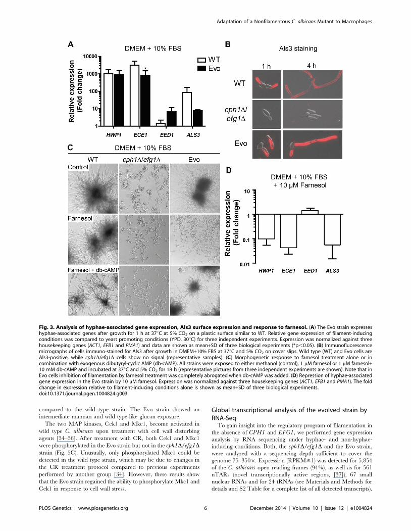
The Evo strain expresses hyphal-associated genes andresponds to farnesol
Hyphal-associated virulence of C. albicans is not only due to
filamentation per se, but also to the expression of hyphae-
associated genes. In order to monitor the expression of typical
hyphae-associated genes in the Evo strain, we measured the
mRNA levels of HWP1, ECE1 and ALS3, all encoding hyphal
cell surface proteins, and of EED1, a gene that is associated with
hyphal cell elongation [27]. An upregulation of all four genes in
the Evo strain was confirmed by qRT-PCR after 1 hour of
incubation in DMEM+10% FBS at 37uC and 5% CO2 (Fig. 3A).
HWP1 expression was similar in the Evo and WT strain, whereas
ECE1 and ALS3 were higher expressed in the WT strain, and
EED1 was more strongly upregulated in the Evo strain.
Furthermore, we observed Als3 exposure on the surface of wild
type and Evo cells by immunofluorescence, but not on the cph1D/
efg1D strain (Fig. 3B). This regained cell-surface exposure of the
Als3 adhesin [28] is in accordance with the increased adhesion
potential of the Evo strain.
We were next interested if the filamentation program can be
blocked by the quorum-sensing molecule farnesol. Very low
concentrations (1 mM) of farnesol in the medium resulted in a
complete repression of filament formation in the Evo strain,
whereas wild type cells still formed hyphae (Fig. 3C). Consistently,
farnesol treatment led to a dramatic repression of filament-
associated gene expression (Fig. 3D). By addition of exogenous
dibutyryl-cyclic AMP (db-cAMP) to the farnesol-containing
medium, filamentation was rescued in the Evo strain (Fig. 3C).
These data suggest a critical role for cAMP signaling in the
filamentation process of the Evo strain.
The Evo strain shows wild type levels of filamentation-associated transcription factor gene expression
The yeast-to-filament regulatory network comprises many
different transcription factors (TFs). The filament-associated
biofilm formation is controlled by a network formed by Bcr1,
Tec1, Brg1, Rob1, Ndt80 and Efg1 [29]. Efg1 positively regulates
all other TF genes in this network except ROB1. We measured the
transcription of these central TF genes at 30 min and 60 min after
filament induction. As shown in Fig. 4A, we found an at least 1.5-
fold upregulation of ROB1 and TEC1 after 30 min, and of BCR1and BRG1 at both timepoints in the Evo strain. The wild type,
however, showed only an increased expression of TEC1 at both
timepoints and of BRG1 after 30 min. In contrast, most of these
TF genes were down- or scarcely upregulated in the cph1D/efg1Dstrain (Fig. 4A).
Formation of wild type filaments is also regulated in part by
CZF1 under certain conditions [30]. An increased expression of
CZF1, however, is not the cause for filamentation in the Evo
strain. The CZF1 mRNA levels under serum induction did not
greatly differ from the mRNA level in the cph1D/efg1D strain
(Fig. 4B). In addition, the mRNA level of UME6, a key TF gene
necessary for the maintenance of filamentation [31], was
upregulated in wild type cells at both time points but not in the
cph1D/efg1D strain (Fig. 4B). Interestingly, UME6 expression was
more than 4-fold upregulated in the Evo strain after 60 min
growth in serum-containing medium.
C. albicans possesses an EFG1 homolog, EFH1, and overex-
pression of this gene is known to induce pseudohyphal growth. In
addition, like EFG1, EFH1 is involved in the regulation of
expression of filament-associated genes [24]. We found that EFH1showed the strongest upregulation (7.4-fold) among the tested TF
genes in the Evo strain. However, deletion of EFH1 did not
abolish filamentation of an Evo strain derivative (Fig. 4C). Hence,
the filamentation phenotype of the Evo strain was not linked to
this TF.
In summary, the Evo strain has regained most of the
transcriptional hallmarks of filament production, including the
upregulation of the central transcription factor genes TEC1,
BRG1 and UME6. The few discrepancies to the wild type may
partially explain the remaining differences in morphology.
However, the late-phase upregulation of UME6 indicates that
the filament maintenance of the Evo strain is similar to the wild
type at the transcriptional level. Furthermore, the function of Efg1
was not replaced by Efh1 in the Evo strain.
The cell wall defects of the cph1D/efg1D mutant arereverted in the Evo strain
Our data indicated that the Evo strain regained the potential to
produce hyphae, showed upregulation of transcription factor genes
involved in filamentous growth and other hyphal associated genes,
and regained a high virulence potential. The reduced virulence of
the cph1D/efg1D strain is likely predominantly caused by the
filamentation defects, however, Efg1 has also an important role in
cell wall architecture [32] and the cell wall is essential for adhesion
and invasive growth and thus for pathogenicity [33]. We therefore
tested the Evo strain for cell wall defects by treatment with cell wall
perturbing agents, i.e. congo red (CR), calcofluor white (CFW) and
sodium dodecyl sulfate (SDS). As shown in Fig. 5A, the cph1D/
efg1D strain was hypersensitive to all tested agents. In contrast, the
Evo strain was as resistant as the wild type to CR and CFW, agents
that disturb glucan and chitin architecture, respectively. The same
phenotypic reversal was observed for the cell membrane disturbing
agent SDS, suggesting a loose structure of the cell wall only in the
cph1D/efg1D strain.
These results indicate that the altered cell wall composition of
the cph1D/efg1D strain was at least partially restored in the Evo
strain. We therefore stained exposed mannan and b-1,3-glucan
with fluorescently labeled concanavalin A (ConA) and anti-b-1,3-
glucan antibody, respectively (Fig. 5B). Quantification by FACS
analysis displayed significantly reduced mannan and increased b-
1,3-glucan signals on the surface of the cph1D/efg1D strain
Adaptation of a Nonfilamentous C. albicans Mutant to Macrophages
PLOS Genetics | www.plosgenetics.org 5 December 2014 | Volume 10 | Issue 12 | e1004824
compared to the wild type strain. The Evo strain showed an
intermediate mannan and wild type-like glucan exposure.
The two MAP kinases, Cek1 and Mkc1, become activated in
wild type C. albicans upon treatment with cell wall disturbing
agents [34–36]. After treatment with CR, both Cek1 and Mkc1
were phosphorylated in the Evo strain but not in the cph1D/efg1Dstrain (Fig. 5C). Unusually, only phosphorylated Mkc1 could be
detected in the wild type strain, which may be due to changes in
the CR treatment protocol compared to previous experiments
performed by another group [34]. However, these results show
that the Evo strain regained the ability to phosphorylate Mkc1 and
Cek1 in response to cell wall stress.
Global transcriptional analysis of the evolved strain byRNA-Seq
To gain insight into the regulatory program of filamentation in
the absence of CPH1 and EFG1, we performed gene expression
analysis by RNA sequencing under hyphae- and non-hyphae-
inducing conditions. Both, the cph1D/efg1D and the Evo strain,
were analyzed with a sequencing depth sufficient to cover the
genome 75–3506. Expression (RPKM$1) was detected for 5,854
of the C. albicans open reading frames (94%), as well as for 561
nTARs (novel transcriptionally active regions, [37]), 67 small
nuclear RNAs and for 24 tRNAs (see Materials and Methods for
details and S2 Table for a complete list of all detected transcripts).
Fig. 3. Analysis of hyphae-associated gene expression, Als3 surface expression and response to farnesol. (A) The Evo strain expresseshyphae-associated genes after growth for 1 h at 37uC at 5% CO2 on a plastic surface similar to WT. Relative gene expression of filament-inducingconditions was compared to yeast promoting conditions (YPD, 30uC) for three independent experiments. Expression was normalized against threehousekeeping genes (ACT1, EFB1 and PMA1) and data are shown as mean+SD of three biological experiments (*p,0.05). (B) Immunofluorescencemicrographs of cells immuno-stained for Als3 after growth in DMEM+10% FBS at 37uC and 5% CO2 on cover slips. Wild type (WT) and Evo cells areAls3-positive, while cph1D/efg1D cells show no signal (representative samples). (C) Morphogenetic response to farnesol treatment alone or incombination with exogenous dibutyryl-cyclic AMP (db-cAMP). All strains were exposed to either methanol (control), 1 mM farnesol or 1 mM farnesol+10 mM db-cAMP and incubated at 37uC and 5% CO2 for 18 h (representative pictures from three independent experiments are shown). Note that inEvo cells inhibition of filamentation by farnesol treatment was completely abrogated when db-cAMP was added. (D) Repression of hyphae-associatedgene expression in the Evo strain by 10 mM farnesol. Expression was normalized against three housekeeping genes (ACT1, EFB1 and PMA1). The foldchange in expression relative to filament-inducing conditions alone is shown as mean+SD of three biological experiments.doi:10.1371/journal.pgen.1004824.g003
Adaptation of a Nonfilamentous C. albicans Mutant to Macrophages
PLOS Genetics | www.plosgenetics.org 6 December 2014 | Volume 10 | Issue 12 | e1004824

Differential expression of selected genes was subsequently validat-
ed by qRT-PCR using biological replicates (S3A Figure).
After the transfer to filament-inducing conditions, 379 tran-
scripts were significantly upregulated ($2-fold, p,0.01) and 279
downregulated in the Evo strain. In the cph1D/efg1D strain, 255
transcripts were up- and 252 downregulated under the same
condition. Within the group of upregulated transcripts, 209 genes
were induced in both strains, while 46 transcripts were specifically
induced in the cph1D/efg1D strain and 170 transcripts specifically
in the Evo strain. 186 of the downregulated transcripts were
repressed in both strains, whereas 66 and 93 transcripts were
specifically repressed in the cph1D/efg1D and Evo strains,
respectively (S3B Figure).
We investigated the expression of individual marker genes for
filamentation [38] more closely (S3C Figure). As expected, all
eight genes of the core filamentation response (ALS3, ECE1,
DCK1, HGT2, HWP1, IHD1, RBT1 and orf19.2457) were
significantly upregulated in the Evo strain under filament-inducing
conditions. Four of these genes (ECE1, HWP1, IHD1 and RBT1)
and further filament-associated genes, like ALS1, BRG1 and
HGC1 were also upregulated in the non-filamenting cph1D/efg1Dstrain. Expression of filament-associated genes independent of any
morphological transition has previously been described in the
cph1D/efg1D mutant [38–40]. However, these genes were
expressed at a significantly higher level in the Evo strain compared
to the cph1D/efg1D strain under filament-inducing condition (S2
DS5 Table).
Overall, genes most highly expressed ($5-fold) in the Evo strain
under filament-inducing condition are mainly hyphal-associated
genes (HWP1, ECE1, ALS3, RBT1, FRG2, ALS1 and IHD1).
Furthermore, the expression of YWP1, encoding a yeast-form cell
wall protein, is downregulated in the Evo strain, while its
expression did not change in the cph1D/efg1D strain. These
results suggest that genes associated with C. albicans hyphae
formation are also associated with filamentation of the Evo strain.
Upregulation (.1.5-fold, p,0.01; S2 DS1 and DS4 Table) of
DCK1, LMO1 and CEK1, which are required for filamentation
under embedded conditions and for cell wall integrity [41], was
found solely in the Evo strain. This provides a possible explanation
for the hyper-filamentous phenotype under embedded conditions
as well as the increased resistance to cell wall perturbants
compared to the double mutant (Figs. 1+5).
To determine whether changes in the regulation of effector
genes are reflected by an upregulation of specific TF genes, we also
Fig. 4. Expression of transcription factors under filament-inducing conditions. (A+B) Relative expression of nine central transcription factorgenes in the analyzed strains after growth in DMEM+10% FBS at 37uC and 5% CO2 on a plastic surface. Fold change between filament-inducing andyeast promoting conditions (YPD, 30uC) is shown, normalized to three housekeeping genes (ACT1, EFB1 and PMA1). Means+SD of n = 3 (dotted lineindicates threshold at 1.5; *p,0.05). (C) Deletion of EFH1 in the Evo strain did not affect hyphal growth. Cells were incubated for 18 h at 37uC and 5%CO2 in DMEM+10% FBS (representative pictures).doi:10.1371/journal.pgen.1004824.g004
Adaptation of a Nonfilamentous C. albicans Mutant to Macrophages
PLOS Genetics | www.plosgenetics.org 7 December 2014 | Volume 10 | Issue 12 | e1004824

analyzed the expression levels of TF genes in the cph1D/efg1Dand Evo strains under filament-inducing conditions in more depth
(S2 DS7 Table). A significantly higher expression of 21 TF genes
was shared by both strains, and only five TF genes were
specifically upregulated in the cph1D/efg1D strain as compared
to the levels in the Evo strain. Interestingly, 17 TF genes had
significantly higher expression specifically in the Evo strain and
not in cph1D/efg1D, including three genes known to be important
hyphal morphogenesis regulators: UME6 (in agreement with
previous qRT-PCR results), RIM101 and HAC1. Eight of the
higher expressed TF genes in the Evo strain have unknown
biological functions.
In the cph1D/efg1D strain, but not in the Evo strain, CPH2,
TEC1 and ACE2, which encode TFs involved in hyphal growth,
were significantly downregulated under filament-inducing con-
ditions. Finally, a significantly lower expression was observed for
NRG1 in the Evo strain, which codes for a repressor of hyphal
development [42]. Hence, we scanned for Nrg1 binding sites
(A/C)(A/C/G)C3T [43] in the putative promoter regions of
genes specifically upregulated twofold in the Evo strain and
detected the sequence motifs in 70% of these promoter regions
(S2 DS8 Table). With this, the Nrg1 binding motif is statistically
overrepresented in promoters of upregulated genes (p,0.01)
when compared to promoters of all other genes. The downreg-
ulation of NRG1 in the Evo strain may therefore facilitate
expression of filament-associated genes and hence filament
formation.
Further analyses indicated a significant upregulation of genes
encoding for secreted aspartyl proteases (SAP5, SAP6, SAP10). In
addition, significant differences in expression of genes associated
with cell wall biosynthesis (CHK1, KRE6, GLC3, MP65, ALG11
and MNT2), alkalinisation (ACH1) as well as of genes involved in
glucose and galactose interconversion and uptake (GAL10, GAL1,
HGT2, HGT4, HGT12 and GSY1) were observed.
In summary, our transcriptional analysis indicated that serial
passage through macrophages led to substantial alterations of the
global transcriptional profile. The programs and pattern we found
differed clearly from the cph1D/efg1D mutant, and resembled
more the well-known programs of the wild type strain. This is
concomitant with and likely correlated with the regained ability of
the Evo strain to induce filaments and to induce damage to host
cells in vitro and in vivo.
Comparative whole genome re-sequencing identifiesmutations potentially linked to Cph1/Efg1-independentfilamentation
We went on to determine the genetic basis for the observed
phenotypical differences. No obvious large-scale structural varia-
tions were detectable between the karyotypes of wild type, the
cph1D/efg1D and Evo strains using pulsed field gel electrophoresis
(PFGE; S4A Figure). To detect possible loss of heterozygosity
(LOH) events [44], we analyzed four SNP-restriction fragment
length polymorphism (RFLP) markers per chromosome [45]. No
differences were detected between double mutant and Evo strain
(S3 DS1 Table). Taken together, these data show that no gross
chromosomal rearrangements have occurred in the Evo strain.
We re-sequenced the genomes of the Evo and the cph1D/efg1Dstrains to identify single nucleotide polymorphisms (SNP) that may
have arisen during the microevolution experiment. Sequencing
depth for cph1D/efg1D and Evo were 996and 1086 in average,
respectively, with 98.8% of the C. albicans SC5314 reference
genome covered in both cases. Comparison of both sequences
Fig. 5. Microevolution led to decreased sensitivity of the Evo strain to different cell wall perturbing agents. (A) Resistance of analyzedstrains against different cell wall stressors. The cph1D/efg1D strain was sensitive to all stresses while the Evo strain regained WT resistance(representative pictures of three experiments are shown). (B) Flow cytometry analysis of mannan and b-glucan exposure on the surface of live cells.Differences in fluorescence intensity between cph1D/efg1D strain and Evo strain point to an altered cell wall composition. Mean fluorescenceintensity+SD of n = 3 (*p,0.05). (C) Western blot analysis to identify phosphorylated Mkc1 and Cek1 in C. albicans strains grown under non-stressconditions (control) or conditions of cell wall stress (450 mg/ml congo red) for 4 hours. Cell wall stress triggered phosphorylation of Mkc1 and Cek1 inthe Evo strain, but not in the cph1D/efg1D strain. Tubulin served as loading control.doi:10.1371/journal.pgen.1004824.g005
Adaptation of a Nonfilamentous C. albicans Mutant to Macrophages
PLOS Genetics | www.plosgenetics.org 8 December 2014 | Volume 10 | Issue 12 | e1004824

revealed a chromosome 7 trisomy in the cph1D/efg1D strain, an
aneuploidy that appears to have been lost during the evolution
experiment (S4B Figure). This is also reflected by a 1.56 higher
mean transcription level of genes on chromosome 7 in the cph1D/
efg1D strain (S4C Figure). In addition, an amplification of URA3on chromosome 3 was observed. URA3 was originally used as a
marker to delete CPH1 and EFG1 in the cph1D/efg1D strain, and
is now present in three copies in this mutant. The Evo strain
contained 7–8 copies (S4B Figure). A qPCR analysis on isolated
gDNA supported these findings (S4D Figure). PFGE and
subsequent hybridization with a URA3 specific probe further
revealed that all copies were located on the same chromosome
(S4E Figure). To exclude any possible contribution of multiple
URA3 gene copies to the filamentous phenotype, the Evo strain
was cured from URA3 with 5-fluoroorotic acid treatment [46].
This Evo Ura2 strain was still able to filament, showing that
URA3 copy number is not responsible for the filamentous
phenotype (S4F Figure). Additionally, after re-introduction of a
single URA3 using the standard CIp10 plasmid at the RPS10locus [47], these strains exhibited the same adhesion, invasion and
macrophage damage properties as their multi-URA3 counterparts
(S4G Figure). This indicates that the excessive URA3 copies do
not have an influence on classical virulence properties of C.albicans.
We observed a high number of SNPs in the cph1D/efg1Dstrain: altogether, 70,197 heterozygous and 3,156 homozygous
SNPs were identified in cph1D/efg1D relative to the C. albicansSC5314 consensus reference genome (Assembly 21, [48]).
Similarly, 72,315 heterozygous and 3,294 homozygous SNPs
were identified in the Evo strain. These figures are consistent with
those achieved when reads obtained by sequencing the genome of
C. albicans SC5314 are aligned on the reference genome and
reflect the high level of heterozygosity in C. albicans as well as
putative sequencing errors and ambiguous positions in the
reference genome (homozygous SNPs). After combining these
sets and filtering, only 329 putative SNPs were found to
distinguish the cph1D/efg1D and Evo strains. Notably, polymor-
phisms at 209 of these positions are observed in the genomes of
19 clinical isolates, distributed over several C. albicans phyloge-
netic groups (CdE, unpublished data). This suggests that they
were most likely not responsible for the restoration of filamenta-
tion. Of the 120 remaining positions, 83 were in non-coding
regions, 22 resulted in synonymous changes and 15 resulted in
non-synonymous changes (S3 DS2-4 Table). Finally, the RNA-
Seq dataset was used as an additional source to detect SNPs
specifically in expressed genes (see Materials and Methods & S3
DS6&7 Table,): A total of 65 putative transcribed SNPs, both
heterozygous and homozygous, were found in the Evo strain, of
which 21 were located in non-coding regions. Inside ORFs, 26
caused a synonymous and 13 a non-synonymous nucleotide
exchange. Of all 39 SNPs detected in coding regions, 24 were
located in genes of the ALS gene family (ALS2 and ALS4),
although these are likely false positives, as genes of the ALSfamily possess a very high sequence similarity and tandem repeat
regions complicating read-mapping and SNP resolution [49].
Comparison of SNPs detected by RNA-Seq and Whole-Genome
Sequencing revealed three SNPs shared by both detection
methods. One SNP was located in a non-coding region between
two uncharacterized genes (orf19.351 and orf19.352), while the
other two were located inside ORFs. A SNP in ATP18(orf19.2066.1) resulted in a synonymous amino acid exchange,
while the second SNP in SSN3 (orf19.794) resulted in a
heterozygous, non-synonymous Arg/Arg to Arg/Gln amino acid
change.
A Mutation in SSN3 is essential for the filamentousphenotype in the Evo strain
As the SNP at nucleotide position 1,055 in the SSN3 ORF
(Fig. 6A) was detected in both analyses, we focused our
investigation on this specific mutation. Ssn3 has been well
characterized in Saccharomyces cerevisiae as an RNA polymerase
II holoenzyme-associated cyclin-dependent kinase of the Mediator
complex contributing to transcriptional control [50]. It was shown
that Ssn3 promotes the degradation of the transcription factor
Ste12 by phosphorylation and thereby regulates S. cerevisiaefilamentous growth [51]. As depicted in Fig. 6B, the heterozygous
Arg352Gln mutation of Ssn3 in the Evo strain is located within the
activation segment of the protein kinase catalytic domain. An
amino acid sequence comparison of C. albicans Ssn3 to sequences
from S. cerevisiae, Cryptococcus neoformans, Mus musculus and
Homo sapiens demonstrated this arginine residue to be conserved
from fungi to mammals. The activation segment comprises several
conserved structural features: the magnesium binding loop, the
activation loop and the P+1 loop, in which the mutation occurred.
While the activation loop is the site of regulatory phosphorylation
in many kinases, the P+1 loop forms a pocket that recognizes the
substrate protein [52].
To ascertain the impact of the SNP on filamentation induction,
we selectively deleted either the mutated or the wild type SSN3allele in the Evo strain, using the dominant selection marker SAT1[53]. Sanger sequencing confirmed the exclusive presence of either
one allele in the genome (Fig. 7A). Strikingly, when incubated in
DMEM with 10% serum at 37uC and 5% CO2 only the strain
with the mutated allele still present (Evo ssn3D/SSN3m) was able
to induce and maintain filamentation. The mutant containing only
the wild type allele (Evo SSN3/ssn3mD) remained in the elongated
yeast form, and thus presented the typical ancestral (cph1D/efg1D)
phenotype (Fig. 7A). In addition, only the Evo ssn3D/SSN3m
strain could escape from macrophages by forming filaments like
the wild type (Fig. 7A). The damage capacity correlated with this
ability to produce filaments: While Evo and Evo ssn3D/SSN3m
strains showed the same levels of phagocyte lysis, the Evo SSN3/
ssn3mD strain caused significantly less damage during co-
incubation with macrophages. In fact, damage was indistinguish-
able from the original cph1D/efg1D strain (Fig. 7B). In contrast,
the deletion of the mutated allele had no influence on the hyphal
development defect on solid medium (S5A Figure) and sensitivity
to cell wall disturbing agents (S5B Figure).
To further ascertain that the SSN3 mutation alone is sufficient
to allow filamentation in a cph1D/efg1D background, we created
an independent cph1D/efg1D double mutant using the dominant
selection marker SAT1 (see Protocol S1). Importantly, this cph1D/
efg1DSAT1 strain contained neither the URA3 amplification nor
the trisomy of chromosome 7 or other genetic alterations of the
original cph1D/efg1D strain. In all our filamentation assays, this
mutant behaved identical to the original cph1D/efg1D strain by
not forming any hyphae (Fig. 7A) and hence not escaping from or
damaging macrophages (Figs. 7A & 7B). To isolate the effect of
the mutated SSN3, we followed several strategies with this new
mutant: SSN3 overexpression strains were created of both the wild
type and mutated (SSN3m) allele under the control of the strong
ADH1 promoter (see S1 Protocol). Strikingly, only the mutated
allele allowed hyphae formation under inducing conditions in the
cph1D/efg1DSAT1 strain (Fig. 7A, lower left corner), even in the
presence of the two native SSN3 alleles. Similarly, macrophage
lysis was increased in the SSN3m overexpressing strain, but not
under SSN3 overexpression (Fig. 7B, right panel). Finally, we
integrated the mutated SSN3 (together with a SAT1 cassette) into
the SSN3 locus of cph1D/efg1DSAT1, replacing one SSN3 allele
Adaptation of a Nonfilamentous C. albicans Mutant to Macrophages
PLOS Genetics | www.plosgenetics.org 9 December 2014 | Volume 10 | Issue 12 | e1004824

and essentially reproducing the heterozygous situation of the Evo
strain. Again, this strain behaved virtually identical to the Evo
strain, both in forming hyphae (Fig. 7A) and in damaging
macrophages (Fig. 7B, right panel).
In summary, these data show that a non-synonymous mutation
in SSN3 that arose during our microevolution experiment is alone
sufficient for regaining the ability to filament even in the absence
of Efg1 and Cph1.
Discussion
Previous experimental studies on the acquisition of antifungal
drug resistance and on stress-induced chromosome rearrange-
ments have elegantly demonstrated the adaptive potential of C.albicans [44,54]. Here, we demonstrate – to our knowledge for the
first time – that a complex trait such as the hyphal formation
program of C. albicans can be subject to microevolution in the
laboratory.
The yeast-to-hyphae transition is of crucial importance for
full C. albicans pathogenicity, which is reflected by its complex
regulation [14]. Multiple overlapping as well as separate
signaling pathways are activated by various environmental
signals to regulate hyphae formation. Wild type hyphae are an
important contributor to the fungus’ ability to escape from
engulfing macrophages. In contrast, the cph1D/efg1D mutant
strain cannot escape by filament formation, yet is able to
replicate inside macrophages and to block phagosome matu-
ration. Therefore, we expected that the mutant strain would
survive in the phagosome, albeit with reduced fitness
compared to the wild type. We monitored the phenotypic
changes of the cph1D/efg1D strain co-passaged with macro-
phages for 42 passages. On a comparatively short evolutionary
timescale our experiment resulted in a strain which not only
regained the ability to filament, but also re-acquired other
important characteristics, like a more wild type-like cell wall
structure and increased virulence. We were able to show that a
minimal sequence alteration accounts for the striking pheno-
typic reversal to wild type-like filamentation: a single missense
mutation in SSN3. SSN3 encodes a fungal protein kinase,
which phosphorylates various regulators in S. cerevisiae. Our
data shows that it can become important for bypassing the
requirements of Efg1 and Cph1 for filamentation in C.albicans.
The in-depth characterization of the evolved strain revealed
that the hyphal morphogenesis program can be induced by
certain, but not all conditions which induce filamentation in the
wild type strain. The fact that the Evo strain filaments in liquid,
but not on solid media indicates an involvement of cAMP
signaling and hence argues for a bypass of Efg1 functions rather
than Cph1 [55–57]. This was further supported by three
additional findings. First, the yeast-to-filament switch occurred in
response to either serum, GlcNAc or CO2, stimuli all known to
trigger the activation of PKA signaling [23,58–60]. Second,
filamentation was entirely blocked by the addition of the quorum-
sensing molecule farnesol which represses both cAMP-PKA and
MAPK signaling pathways [61,62]. The full restoration of
filamentation when cAMP was added supports the involvement
of the cAMP-PKA pathway. Third, the repressor of hyphae
formation, Nrg1 is normally downregulated by the cAMP-PKA
pathway, except in the presence of farnesol [63]. Transcriptome
analysis showed NRG1 expression to be downregulated in the Evo
strain, but not in the cph1D/efg1D mutant. As 70% of the
upregulated genes in the Evo strain contain an Nrg1 binding site,
these data emphasize the likely importance of Nrg1 levels on
filamentation of the Evo strain.
Given that the cph1D/efg1D mutant is strongly reduced in
virulence [22], the almost wild type-level virulence in the Evo
strain in our murine model was striking. Examination of kidney
sections revealed filament formation of the Evo strain in vivo.
Compared to wild type filaments, these were shorter and resulted
in less pronounced tissue invasion, which is likely associated with
the lower overall virulence compared to the wild type.
Three factors are likely to have contributed to the increased
virulence of the Evo strain in the absence of Efg1 and Cph1: First,
its ability to escape from macrophages like the wild type; second,
its adhesion to host cells which was significantly higher than the
cph1D/efg1D strain; and third, the ability to form filaments upon
Fig. 6. Single nucleotide polymorphism in SSN3 of the Evo strain and location of the mutated amino acid. (A) Partial SSN3 sequence forcph1D/efg1D and Evo strains flanking SNP 1055 (marked with an arrow). Notice the heterozygosity in the Evo strain. (B) Schematic view of thecatalytic domain of Ssn3 (STK = serine/threonine kinase) with the position of the activation segment highlighted in brown and the amino acidexchange indicated by an arrow (top). Sequence alignment of the Ssn3 activation segment in different species (H. s. Homo sapiens [NP_001251.1], M.m. Mus musculus [NP_705827.2], C. n. Cryptococcus neoformans [XP_568416.1], C. a. C. albicans [XP_720918.1] and S. c. Saccharomyces cerevisiae[NP_015283.1]). The arrow indicates the amino acid exchange in the Evo strain. The Mg-binding loop is highlighted in yellow, the activation loop inblue and the P+1 loop in purple. Amino acids that are known to abrogate kinase activity when mutated are colored in green [51,98]. Asterisksunderneath the alignment indicate positions with conserved amino acids and colons indicate highly similar residues (bottom). The mutated arginine(red) is part of the highly conserved P+1 substrate recognition loop.doi:10.1371/journal.pgen.1004824.g006
Adaptation of a Nonfilamentous C. albicans Mutant to Macrophages
PLOS Genetics | www.plosgenetics.org 10 December 2014 | Volume 10 | Issue 12 | e1004824
Fig. 7. A single nucleotide polymorphism in SSN3 is essential for filamentation. (A) Distinct impact on morphology by: deleting either themutated (SSN3/ssn3mD) or the wild type SSN3 allele (ssn3D/SSN3m) in the Evo strain, by overexpressing either the wild type SSN3 allele (cph1D/efg1DSAT1SSN3OE) or the mutated SSN3 allele (cph1D/efg1DSAT1SSN3m
OE) or by expressing the mutated SSN3 allele from its native locus (cph1D/efg1DSAT1SSN3m) in a newly generated cph1D/efg1DSAT1 strain. The partial SSN3 sequences demonstrate the homozygosity or heterozygosity of theSSN3 allele (left). Filamentous growth is visible in the Evo ssn3D/SSN3m, cph1D/efg1DSAT1SSN3m
OE and cph1D/efg1DSAT1SSN3m strains after growth for18 h at 37uC and 5% CO2 in DMEM+10% FBS and during co-incubation with macrophages, but not with the Evo SSN3/ssn3mD strain, cph1D/efg1DSAT1
and cph1D/efg1DSAT1SSN3OE strains (scale bars: 18 h, 50 mm and MW, 20 mm; representative pictures are shown) (right). (B) Cell damage ofmacrophages caused by the different strains, as determined by lactate dehydrogenase (LDH) assay after 32 h of co-incubation. Robust host celldamage depends on the presence of the mutated allele SSN3m. Mean and SD of n = 4 (*p,0.05; compared to cph1D/efg1D and cph1D/efg1DSAT1
respectively; LC = low control, medium only).doi:10.1371/journal.pgen.1004824.g007
Adaptation of a Nonfilamentous C. albicans Mutant to Macrophages
PLOS Genetics | www.plosgenetics.org 11 December 2014 | Volume 10 | Issue 12 | e1004824

contact with epithelial cells, which is a prerequisite for both active
penetration into and induced endocytosis by host cells [64].
Wachtler et al. [17] showed that filamentation alone is insufficient
to cause damage of host cells. We therefore compared the damage
capacities of the cph1D/efg1D and the Evo strains. The Evo strain
exhibited a significantly increased potential to damage both
macrophages and epithelial cells compared to the double mutant.
The adaptation to macrophages was accompanied by differences
in additional traits, such as resistance to cell wall stresses. In the
cph1D/efg1D strain, the higher sensitivity to cell wall disturbing
agents, as well as the modified exposure of cell wall components,
likely reflect an altered cell wall organization which was restored in
the Evo strain. This is supported by findings from a recent study
by Zavrel et al. [32] which showed that deletion of EFG1 alone
affects cell wall architecture. In our strains, these modifications of
the cell wall seemed to be mediated by the kinases Mkc1 and
Cek1. Previous analyses carried out in cek1D and mkc1D mutants
already indicated their direct relationship to cell wall composition
and integrity [35,65,66].
By analyzing the differences in gene expression acquired during
co-culture passaging with macrophages, we found that all genes
belonging to the core filamentation network [38] were upregulated
in the Evo strain. This suggests that during filamentation the Evo
strain transcriptionally utilizes the complete filamentation pro-
gram. The transcription factors Tec1, Brg1, Ume6, Rim101, Hac1
and Efh1, which are known to be involved in regulation of
filamentation [24,67–71], were also upregulated in the Evo strain.
Together with Nrg1, they likely orchestrate filament formation in
the Evo strain. For UME6, it has been shown that its transcription
is repressed by Nrg1-Tup1 and that ectopic Ume6 expression in
cph1D/efg1D can rescue the filamentation defect under certain
conditions [69].
For the maintenance of hyphal extension, both UME6 and
EED1 are central [27,31] and both showed an increased
expression in the evolved strain. Thus, the mechanisms of hyphal
extension seems similar between Evo and wild type cells [27].
Hence, the transcriptional conditions for initiation and mainte-
nance of filamentation, which comprise the release of repression
and the upregulation of positive regulators of filamentation, are
met in the Evo strain. Furthermore, a considerable number of
transcripts specifically up- and downregulated in the Evo strain are
both Candida-specific and uncharacterized. It is feasible, there-
fore, that these uncharacterized transcripts assumed a novel role
specifically during filament formation in the Evo strain. This is
especially true as the morphological switch is one of the best-
investigated characteristics in C. albicans, and genes involved in
this process are generally well studied. However, differential
regulation of genes not clearly linked to the yeast-to-hyphal switch,
including these genes, but also WOR1 and NAT4 (both involved
in the white-opaque switching) and SST2 (involved in the mating
response pathway), could have been caused by the mutated Ssn3
kinase (see below). Finally, it also should be noted that, even in the
absence of filamentation, cph1D/efg1D was able to upregulate
certain genes described as hyphae-associated under the condition
tested here (incubation in DMEM+10% FBS at 37uC and 5%
CO2 on a plastic surface). This is in disagreement with previous
data showing that EFG1 is required for expression of several
hyphae-associated genes [23,72]. It is possible, however, that
alternative pathway(s), such as the Rim101 pH response pathway,
are involved, as the cells were simultaneously exposed to diverse
stimuli for filamentation. However, these genes still showed an
increased induction in the Evo strain compared to the cph1D/
efg1D strain, which argues for a further adaptation-induced,
filament-associated change in regulation.
It has been demonstrated that acquired drug resistance in C.albicans is often accompanied by aneuploidy and/or isochromo-
some formation [54,73] and that several stress conditions can
enhance the rates of LOH events likely by mitotic recombination
[44]. However, we did not detect any LOH events between the
cph1D/efg1D and the Evo strain. The chromosome 7 trisomy was
present initially in the cph1D/efg1D strain [74] and the Evo strain
restored disomy by loss of one copy. The remaining gross genetic
difference, an URA3 amplification in the Evo strain can be
explained by an insufficient Ura3 expression from the EFG1 locus.
An amplification of the gene may have increased fungal fitness
during our experiment by ensuring more transcripts and hence
more efficient growth. Prior studies suggested that ectopic
expression of URA3 influences the phenotypes of a diverse range
of mutants [75,76], and duplication of a hisG-URA3-hisG cassette
resulted in restored filamentation of an hwp1D mutant [77]. We
were able to exclude these Ura3 effects as causes for the Evo strain
filamentation, as the acquired filamentation phenotype was
maintained after removal of the multiple URA3 copies. Further-
more, after re-introduction of a single copy of URA3, no
differences in virulence traits, like adhesion or invasion, were
detectable as compared to the multi-copy strains. Overall, these
data and the fact that the observed filamentation and other
phenotypes persisted even after repassaging in rich (YPD)
medium, argued for small-scale genomic alterations, rather than
epigenetic changes, acquired by cph1D/efg1D cells adapting to
macrophages.
Comparative genome sequencing (by WGS and RNA-Seq) of
cph1D/efg1D and Evo strains allowed us to pinpoint the
microevolutionary changes in the Evo strain at the single
nucleotide level. By combining the different approaches, we
detected an expressed SNP in SSN3, which resulted in an Arg-to-
Gln change at a highly conserved position within the presumable
protein kinase domain. This SNP and thus gain of heterozygosity
was found to be central for the yeast-to-filament transition of the
Evo strain. Deletion of the mutated SSN3 allele prevented the
morphological switch in the Evo strain during growth under
filament-inducing conditions and interaction with several types of
host cells. Other phenotypes specific to the Evo strain were not
affected by the deletion of the mutated SSN3 allele, suggesting
that they evolved independently from filamentation.
Importantly, introduction of a single mutated allele into an
independent efg1D/cph1D strain fully copied the filamentation
and host cell damage phenotype of the Evo strain. This strain
contained neither the multiple URA3 copies nor the trisomy of
chromosome 7 or any other possible genetic alterations of the
original efg1D/cph1D strain. Hence, the SSN3 mutation alone
bypassed the lack of the central transcription factors Cph1 and
Efg1 and restored the ability to cause host cell damage in vitro,
and likely to induce higher virulence in vivo.
Ssn3 itself (also referred as Srb10 or Cdk8) is part of the CDK
(cyclin-dependent kinase) module (SRB10/11) of the Mediator
complex, which is a regulator of RNA-polymerase II (RNAP II)
activity [78,79]. This CDK module phosphorylates the largest
subunit of RNAP II, and Ssn3 additionally has roles in both
transcriptional activation and repression in response to physiolog-
ical signals, coordinating gene expression. By regulating the
stability of the two important regulators, Ste12 (ortholog of Cph1)
and Phd1, Ssn3 in S. cerevisiae is involved in the differentiation of
yeasts into pseudohyphae under nutrient-limiting conditions
[51,80]. Interestingly, a kinase-deficient Asp290Ala Ssn3 only
weakly phosphorylates Ste12 in vitro, and the lack of phosphor-
ylation increases its stability [51]. Moreover, the catalytic activity
of Ssn3 contributes to the repression of a subset of Tup1-regulated
Adaptation of a Nonfilamentous C. albicans Mutant to Macrophages
PLOS Genetics | www.plosgenetics.org 12 December 2014 | Volume 10 | Issue 12 | e1004824

genes [81–83] in S. cerevisiae. Tup1 is recruited to promoters by
Nrg1 [84], a factor which was downregulated in the Evo strain.
Although the precise signaling pathway(s) controlling Ssn3
remain to be determined, Chang et al. [85] showed that the
activity of Srb9, another subunit of the CDK kinase module, is
regulated by the PKA signaling pathway in S. cerevisiae. Based on
our data it is tempting to speculate that the activation of the
cAMP-PKA pathway results in activation of Ssn3 kinase activity,
and the observed filament-specific transcriptional changes may
thus depend on either a reduced or absent substrate recognition or
on impaired substrate phosphorylation activity due to the
Arg352Gln substitution. It is supposable that a loss of the
substrate-specific kinase activity increases the stability of positive
regulator(s) of filamentation by reducing their phosphorylation.
Alternatively (or in addition), the impaired kinase activity could
lead to a derepression of genes associated with positive regulation
of filamentous growth. Importantly, in this model the kinase-
deficient Ssn3 remains part of the Mediator complex, and could
fulfill any additional function it may have (e.g. in the structure or
recruitment of additional proteins). In both models, a decreased
kinase activity would reduce inhibitory effects on filamentation,
and hence would increase the sensitivity of the filamentation
network to external stimuli. This would likely allow to bypass the
need for additional Efg1 signaling. Additional genes outside of the
immediate filamentation network may also be affected, as this
model implies a pleiotropic effect of the Ssn3 mutation, with
several transcription factors as possible clients.
Thus, it seems that not the disrupted cAMP-PKA signaling
pathway itself evolved in our microevolution experiment, but
instead a regulatory hub for filamentation which the pathway
probably targets in addition to Efg1. In this hub, even single or
few mutations seem to be able to lead to striking phenotypic
alterations, as many filament-associated genes are directly or
indirectly targeted. Finally, it is interesting to speculate why
only one SSN3 allele was mutated, and we did not observe any
LOH event to homozygosity at this locus. It seems possible that
one mutated allele alone was sufficient to promote filamenta-
tion in macrophages, while the other wild type allele, still
capable of full phosphorylation activity, was still required for
additional functions of Ssn3. This is somewhat supported by
the observation that overexpression of the mutated SSN3 allele
in a background with the native SSN3 alleles still in place was
sufficient to allow hyphae formation. In our model, the
mutated Ssn3 competes with the wild type Ssn3, and
overexpression allows the mutated protein to gain entry into
a sufficient number of Mediator complexes.
In conclusion, using the nonfilamentous mutant cph1D/efg1D,
we have shown that C. albicans can rescue one of its key virulence
traits, the yeast-to-hyphal switch, with a single nucleotide change
when put under adequate selection pressure. A mutation in the
transcriptional regulator Ssn3 adaptively rewired the transcription
network to enable filamentation in response to external cues while
bypassing the need for Efg1 and Cph1. This shows an unexpected
robustness of the whole filamentation system even to severe
disruptions, and a high degree of adaptability. The selection
scenario we used - co-incubation with macrophages - clearly
reflects a condition C. albicans encounters in the host and thus
might be an evolutionary pressure that can shape the infection
biology of this fungus. In fact, this hypothesis is supported by
another evolution experiment, which analyzed the adaptation of
C. glabrata to macrophages. There, the selection pressure resulted
in the appearance of a strain with pseudohyphae-like structures
and increased virulence again by a single nucleotide mutation
[86]. This demonstrates that during interaction with the host or
host cells, significant changes in morphology and virulence are
possible on a very short evolutionary time-scale.
Materials and Methods
Ethic statementAll animal experiments were in compliance with the German
animal protection law and were approved by the responsible
Federal State authority (Thuringer Landesamt fur Lebensmittel-
sicherheit und Verbraucherschutz) and ethics committee (bera-
tende Kommission nach 1 15 Abs. 1 Tierschutzgesetz; permit
no. 03-007/07).
Body surface temperature and body weight were recorded daily
and animals were monitored twice a day for disease progression.
Mice showing severe signs of illness (isolation from the group,
apathy, hypothermia and drastic weight loss) were humanely
sacrificed by ketamine/xylazine overdose and exsanguination.
Strains and growth conditionsCandida albicans strains and mutants used in this study are
listed in S1 Table. Strains were grown in YPD medium (1%
peptone, 1% yeast extract, 2% glucose and optionally 2% agar) or
SD medium (2% dextrose, 0.17% yeast nitrogen base, 0.5%
ammonium sulfate and optionally 2% agar) at 30uC. Uridine
(50 mg/ml) or nourseothricin (NAT; 100 mg/ml) were added as
required. If not stated otherwise, stationary phase cells were used
in the experiments. Mutants were constructed as described in
Protocol S1.
Cell linesThe murine peritoneal macrophage-like cell line J774A.1
(DSMZ) and the human buccal carcinoma epithelial cell line
TR-146 (Cancer Research Technology) were grown in Dulbecco’s
Modified Eagle’s Medium (DMEM, PAA) supplemented with
10% FBS (PAA) and routinely cultured until passage 20. Both cell
lines were maintained at 37uC under 5% CO2. J774A.1 cells were
removed from tissue-culture flasks by gentle scraping, while TR-
146 cells were enzymatically harvested by Accutase (PAA)
treatment.
Evolution experimentAbout 86106 J774A.1 macrophages were seeded into a 75 cm2
cell culture flask with DMEM supplemented with 10% FBS and
1% Penicillin/Streptomycin (PAA). For the evolution experiment,
macrophages were initially infected with 46106 cells of the cph1D/
efg1D strain. After that, 46106 re-isolated C. albicans cells were
transferred to a fresh macrophage culture. After 24 h of co-
incubation, infected macrophages were washed (36with PBS) and
lysed with 2 ml lysis buffer (50 mM Tris, 5 mM EDTA, 150 mM
NaCl and 0.5% Nonidet P40 [Sigma-Aldrich]). The lysate was
transferred to a 2 ml reaction tube and fungal cells were collected
by centrifugation. The C. albicans cells were washed two times
with DMEM and counted before infection of fresh macrophages.
To verify the absence of EFG1 and CPH1, Southern blot
analysis was performed for the Evo strain as described previously
[22]. Briefly, genomic DNA (gDNA) was digested with AvaII or
KpnI to verify EFG1 or CPH1 deletion, respectively. DIG-labeled
probes were generated (Roche) using genomic DNA from the
strain SC5314 and primers EFG-A/EFG-B and P33/CPH-B (S1
Table).
Phenotypic characterizationA detailed description of the phenotypic analyses can be found
in Protocol S1.
Adaptation of a Nonfilamentous C. albicans Mutant to Macrophages
PLOS Genetics | www.plosgenetics.org 13 December 2014 | Volume 10 | Issue 12 | e1004824

Staining procedures and detection of b-1,3-glucans andmannans
Fungal cells were grown in DMEM+10% FBS on glass coverslips in
a 24 well microtiter plate for filipin (Sigma), calcofluor white (CFW)
and Als3 immunostaining. Flow cytometry was used to quantify
mannan and b-1,3-glucan exposure on the surface of stationary C.albicans cells after staining with concanavalin A and anti- b-1,3-glucan.
Piercing and invasion rate were determined by differential staining. All
staining procedures are described in Protocol S1. Epifluorescence
(Leica DM5500B, Leica DFC360 FX) was used to detect CFW and
filipin (DAPI filter), Alexa Fluor 488 (FITC filter) and Alexa Fluor 647
(Cy5 filter). Micrographs were taken with a Leica Digital Camera
DFC360 FX or a Zeiss AxiCam ICc3.
Replication and piercing assayTwo times 105 J774A.1 macrophages were seeded onto glass
cover slips placed in 24 well microtiter plates and allowed to adhere
overnight. Non-adherent macrophages were removed by washing
with PBS. To monitor intracellular replication, C. albicans cells
were labeled with 100 mg/ml fluorescein isothiocyanate (FITC,
Sigma-Aldrich) in carbonate buffer (0.1 M Na2CO3, 0.15 M NaCl,
pH 9.0) for 30 min at 37uC and washed 36with PBS. To quantify
piercing rates, cells were washed without prior staining. Two times
105 fungal cells were added to macrophages in DMEM+10% FBS.
The plates were incubated for indicated timepoints (see figure
legends). Cells were then washed once with PBS and fixed with 4%
paraformaldehyde. Intracellular replication was detected by fluo-
rescence microscopy after mounting the samples in ProLong Gold
Antifade Reagent with DAPI (Invitrogen). After co-incubation,
piercing of macrophages by filaments was quantified by differential
staining. The assays were performed in biological triplicates.
Adherence and invasion assayTwo times 105 TR-146 epithelial cells were seeded onto glass
cover slips placed in 24 well microtiter plates and cultured for 2–3
days to 95%-100% confluency. Adherence and invasion assays
were performed as previously described [17]. Briefly, to determine
the adhesion rate, TR-146 monolayers were infected with 16106
C. albicans cells. After one hour of co-incubation, non-adherent
yeast cells were removed by rinsing 36with PBS. Cells were fixed
with 4% paraformaldehyde, permeabilized with 0.5% Triton X-
100 and adherent C. albicans cells were stained with CFW for
fluorescence microscopy. Invasion rates were determined by
infecting TR-146 monolayers with 16105 C. albicans cells. After
incubation, cells were fixed and differentially stained for fluores-
cence microscopy. Both assays were repeated at least three times.
Quantification of damage to host cellsFive times 104 host cells (J774A.1 or TR-146) were seeded in 96
well microtiter plates. J774A.1 macrophages were cultured for 1
day before use, while TR-146 epithelial cells were cultured for 2
days to 95%–100% confluency. Damage of macrophages and
epithelial cells was determined by measuring the release of lactate
dehydrogenase (LDH) with the Cytotoxicity Detection Kit (Roche
Applied Science) following 32 h of co-incubation with 56104 C.albicans cells according to the manufacturer’s protocol. The
experiments were performed as previously described [17] and
repeated at least three times.
Murine infection modelFor survival studies the intravenous challenge model for
disseminated C. albicans infection was used. Six to eight weeks
old female BALB/c mice (18–20 g) purchased from Charles River
were used for the experiments. Mice were challenged intrave-
nously with 56105 C. albicans cells in 200 ml PBS via the lateral
tail vein. All mice surviving to day 20 were humanely sacrificed.
For histology, kidneys were collected and fixed with buffered
formalin and paraffin-embedded sections were stained with
Periodic acid-Schiff (PAS) according to standard protocols.
Western blot analysisTo detect phosphorylated Mkc1 and Cek1 as well as a-tubulin,
cells of an overnight culture were adjusted to an OD600 of 0.5 in
SD medium (control) or SD medium supplemented with 450 mg/
ml congo red, and incubated for 4 hours at 30uC. Cell disruption,
protein extraction and western blot analysis using anti-phospho-
p44/42 MAP kinase antibody (Cell Signalling Technology) and rat
anti-a-tubulin antibody (AbD Serotec), respectively, were per-
formed as previously described [87].
RNA sample preparation and isolationC. albicans cells from an overnight culture were diluted to
OD600 = 0.2 in YPD medium and grown to log-phase for 4 h at
30uC. Cells were collected by centrifugation and a zero time point
sample was frozen in liquid nitrogen until RNA extraction. In
addition, 16107 cells were incubated one hour under filament-
inducing conditions (DMEM+10% FBS at 37uC and 5% CO2 in a
75 cm2 cell culture flask). For farnesol experiments, 10 mM
farnesol was added to the medium just prior to the experiment.
After incubation, medium and non-adherent cells were removed
and 5 ml ice-cold PBS was added. The cells were collected by
scraping and then centrifuged for 5 min at 6,000 g at 4uC. Cell
pellets were snap frozen in liquid nitrogen. Total RNA was
isolated using the Ribopure-Yeast Kit (Ambion) and treated with
Turbo DNase (Ambion). RNA quality was determined in a
Bioanalyzer with an RNA 6000 Nano LabChip Kit (Agilent
Technologies) according to the manufacturer’s protocol. RNA
concentration was determined with a Nanodrop ND1000 (Peqlab).
Copy number determination and quantitative geneexpression analysis
Copy number and expression levels of selected genes were
analyzed with a my-Budget 56 EvaGreen QPCR Mix II
(Bio&Sell) in a C1000TM Thermal Cycler (BioRad) using gene-
specific primers (S1 Table). For expression analysis, 600 ng of total
RNA was reversely transcribed with the SuperScript III First-
Strand Synthesis Kit (Invitrogen) according to the manufacturer’s
instructions. URA3 gene copy number was determined from
100 ng of gDNA with primers URA3-fw and URA3-re (S1 Table).
PCR conditions were as followed: 95uC for 15 min, 40 cycles of
each 95uC for 15 s, 60uC for 40 s and 72uC for 15 s. A melting
profile was generated to confirm PCR product specificity. Relative
gene expression levels were determined by the 2DDCt method [88]
with ACT1, EFB1 and PMA1 as internal controls. URA3 copy
number was calculated with ACT1 internal control and gDNA
from SC5314 (containing two copies of URA3) as reference. Three
independent experiments were performed.
Pulsed-field gel electrophoresis (PFGE) and SNP-RFLPanalysis
PFGE and SNP-RFLP are described in Protocol S1.
RNA sequencing, transcriptional profiling and SNPdiscovery from RNA-Seq data
In order to use only high quality reads, trimming was performed
using Btrim (window size = 15, average quality score = 20) [89].
Adaptation of a Nonfilamentous C. albicans Mutant to Macrophages
PLOS Genetics | www.plosgenetics.org 14 December 2014 | Volume 10 | Issue 12 | e1004824

For differential gene expression analysis high quality trimmed
reads were mapped against the sequence Assembly 21 of strain
SC5314 [48] using the spliced read mapper TopHat 2.0.6 [90]
with the ‘‘known transcripts’’ (-G option) and uniquely mapped
reads were counted using HTSeq [86]. Raw counts for each gene
were loaded into R and differentially expressed genes were
identified using the packages edgeR and DESeq [91,92] and
filtered by adjusted p-values (,0.01) and RPKM value ($1). Data
were deposited at the Gene Expression Omnibus (GSE56174) and
can be found in S2 Table. Nrg1 binding sites (A/C)(A/C/G)C3T
in putative promoter regions (usually 21000 bp/+50 bp) of all C.albicans genes were determined by SiTaR [93] allowing no
mismatch. Fisher’s exact test was used to determine if the Nrg1
motif-containing promoters were overrepresented in genes specif-
ically upregulated twofold in the Evo strain, as compared to all
remaining genes. For SNP calling quality trimmed reads from all
samples of each strain were merged and the protocol of GATK
[94] with slight changes was followed (i.e. reads were mapped
using BWA algorithm [95], duplicates were removed and
realignment around indels and base recalibration was performed).
Next, we used bam-readcount (www.github.com/genome/bam-
readcount), which determines the nucleotide distribution at each
single base. Heterozygous SNPs were defined as positions where
25% or more of the reads showed an alternative nucleotide.
Homozygous SNPs were defined as positions where more than
90% of the reads differed from the reference. Minimum nucleotide
sequence depth was 20. Clustal Omega [96] was used for multiple
sequence alignments.
Whole genome sequencing and sequence analysisGenomic DNA isolated from the cph1D/efg1D and Evo strains
were processed to prepare libraries for Illumina sequencing, and the
TruSeq DNA Sample Prep kit (Illumina) was used according to the
manufacturer’s recommendations. DNAs were randomly fragmented
by sonication to an average fragment length of 500 bp and Illumina
adapters were blunt-end ligated to the fragments. The final libraries
were amplified by PCR followed by sequencing on an Illumina
Genome Analyzer platform (Illumina GAII). 60 nt single-end reads
were aligned to the C. albicans strain SC5314 reference genome [48]
downloaded on 02/24/2012 using shore 5.0 [97]. Sequencing depth
scores were computed for each 1 kb region across the genomes and for
ORFs using sequencing depth data for each nucleotide located within
the 1 kb region or the ORF. Sequencing depth scores were normalized
based on the overall sequencing depth obtained for each genome.
Single nucleotide polymorphisms were identified using shore 5.0 [97]
at positions covered at least 30 times with a minimum quality of 25.
Homozygous SNPs were defined as positions where 90% of the reads
meeting these criteria differed from the reference genome. Heterozy-
gous SNPs were defined as positions where 20% or more of the reads
showed one allele and 80% or less of the reads showed a second allele.
Statistical analysisData were visualized and statistically analyzed using GraphPad
Prism version 5.00 (GraphPad Software, USA). Statistical analyses
were performed by 1-way ANOVA (mannan and b-1,3-glucan
exposure) or 2-way ANOVA (piercing, adhesion, invasion, damage
and gene expression) followed by a Bonferroni correction. Differences
in survival of mice were evaluated by Log-rank (Mantel-Cox) test.
Supporting Information
S1 Figure Screening of C. albicans deletion mutants for defects
in hyphal formation during interaction with macrophages and
verification of the cph1D/efg1D genotype in the Evo strain. (A)
Morphologies of different C. albicans mutants and the corre-
sponding wild type strains upon phagocytosis by macrophages
(J774A.1). Note that only the double mutant strain cannot escape
from macrophages. Figures show overlay of DIC and fluorescent
images. C. albicans appears blue (CFW) and extracellular section
of hypha red (ConA). Arrows highlight piercing of macrophage
membrane by C. albicans cells (scale bar: 10 mm). (B) The WT
(CAI4+CIp10) forms hyphae after phagocytosis (white arrows),
while the cph1D/efg1D strain replicates intracellularly in J774A.1
cells (black arrows). Yeast cells were stained with FITC prior to
infection. Six hours after infection samples were fixed and stained
with DAPI for analysis by fluorescence microscopy. FITC is not
transferred to hypha or new daughter cells during cell division
(scale bar: 10 mm, representative picture). (C) Southern blots of
wild type (WT), cph1D/efg1D and Evo strains confirm the
deletion of EFG1 (left) and CPH1 (right) in the Evo strain.
Genomic DNAs were digested with either AvaII or KpnI, and
DNA molecular weight marker III, DIG labeled (Roche) was used
as size standard.
(TIF)
S2 Figure Morphological index and morphology of the Evo
strain under different conditions. (A) Overnight cultures of cph1D/
efg1D and Evo strains were diluted into DMEM+10% FBS and
incubated for the indicated time points at 37uC and 5% CO2 on
cover slips. After fixation the percentage of yeast, pseudohyphal
and hyphal cells was quantified using the morphological index
(MI) [26]. Mean+SD of at least 100 cells in two experiments. (B)
Morphology of cph1D/efg1D and Evo strains in phase contrast
under different filament-inducing conditions. The Evo strain
formed filaments in response to stimuli other than used during the
evolution experiment. (C) Colony morphology of strains under
embedded conditions. The Evo strain exceeds the hyperfilamenta-
tion phenotype of the cph1D/efg1D strain. (D) Colony morphol-
ogy of analyzed strains grown on solid YPD agar supplemented
with 10% FBS for 6 days, on solid Spider and Lee’s medium for 9
days, and on solid YNB agar supplemented with 2% glucose and
10 mM urea for 11 days at 37uC (scale bar: 1 mm; representative
pictures are shown). Only WT forms filaments under these
conditions.
(TIF)
S3 Figure Results of the RNA-Seq analysis. (A) RNA-Seq results
are in good agreement with qRT-PCR analyses of four selected
genes. Expression was normalized against three housekeeping
genes (ACT1, EFB1 and PMA1). Fold change gene expression of
filament-inducing condition versus yeast promoting condition
(YPD, 30uC) is shown (mean+SD). (B) Venn diagrams of
differentially expressed genes of cph1D/efg1D and Evo strains
during growth in DMEM+10% FBS at 37uC and 5% CO2 on a
plastic surface compared to yeast promoting condition (YPD,
30uC). (C) Expression heat map of the eight genes of the core
filamentation response in the cph1D/efg1D and Evo strains during
growth in DMEM+10% FBS at 37uC and 5% CO2 with white
boxes indicating no differential expression (NDE).
(TIF)
S4 Figure Analysis of the genome architecture of the Evo strain.
(A) Whole genome profiles of wild type (WT), cph1D/efg1D and
Evo chromosomes separated by PFGE and stained with ethidium
bromide are identical among all strains analyzed. (B) Whole-
genome sequencing of cph1D/efg1D and Evo strains revealed a
chromosome 7 (Chr 7) trisomy in the cph1D/efg1D strain (top)
and an amplification of URA3 on chromosome 3 (Chr 3) in the
Evo strain (bottom). Normalized log2 read depth per 1 kb region
along the chromosomes are shown. (C) Relative expression from
Adaptation of a Nonfilamentous C. albicans Mutant to Macrophages
PLOS Genetics | www.plosgenetics.org 15 December 2014 | Volume 10 | Issue 12 | e1004824
the RNA-Seq experiment between cph1D/efg1D and Evo. The
chromosome 7 trisomy of cph1D/efg1D is reflected by a mean
1.56 higher expression of genes specifically on this chromosome.
Other chromosomes showed no change in mean expression
(shown here for chromosome 6 as an example) (D) Verification of
URA3 amplification by qPCR analysis using genomic DNA
isolated from the cph1D/efg1D and Evo strains. Copy number of
URA3 was normalized to ACT1. Mean+SD. (E) The URA3 gene
was successfully deleted from the Evo Ura2 strain. Whole
chromosomes of the Evo strain and 5-fluoroorotic acid (FOA)
treated Evo cells (Evo Ura2) were separated by PFGE, stained
with ethidium bromide (left) and subjected to Southern hybrid-
ization with a URA3 probe to verify successful deletion of all
URA3 copies in the genome (right). (F) Phenotype of Evo and Evo
Ura2 strains after growth for 18 h at 37uC and 5% CO2 in
DMEM+10% FBS (representative pictures). Removal of all URA3copies has no influence on the regained filamentation properties of
the Evo strain. (G) Virulence traits of the efg1D/cph1D (DD) and
Evo strains cured of URA3 (Ura-) and after subsequent re-
introduction of a single URA3 copy using the CIp10 plasmid for
genomic integration. No difference can be detected between the
multi-copy and single-copy strains in adhesion or invasion of
epithelial cells, or damage to macrophages. Absence of URA3reduces macrophage damage, probably due to the low uridine
concentration inside the phagosome.
(TIF)
S5 Figure Characterization of the Evo SSN3/ssn3mD and Evo
ssn3D/SSN3m strains. (A) Colony morphology of the Evo ssn3D/
SSN3m strain grown on solid Lee’s medium, Spider medium and
YPD medium supplemented with 10% FBS for 6 days at 37uC(scale bar: 1 mm; representative pictures are shown). Observed
morphologies resemble those of the Evo strain. (B) Stress resistance
of cph1D/efg1D, Evo, Evo SSN3/ssn3mD and Evo ssn3D/SSN3m
strains against different cell wall perturbing agents. The presence
of either SSN3 allele has no influence on the increased stress
resistance of the Evo strain. Experiments yielded similar results for
at least three replicates.
(TIF)
S1 Table Strains and primers used in the study.
(DOC)
S2 Table Excel data sheets contain data for differentially
expressed genes in the cph1D/efg1D and the Evo strains during
incubation under filament-inducing condition (DMEM+10% FBS
at 37uC and 5% CO2 on a plastic surface) identified by RNA-Seq
analysis (log2 RPKM data and raw read counts for all annotated
transcripts and novel transcripts identified by Bruno et al. [37]).
(XLS)
S3 Table SNP-RFLP analysis of 32 SNP-RFLP markers (two
per chromosome arm) to detect large-scale changes in the genomes
of the cph1D/efg1D and Evo strains and predicted SNPs
discovered by whole-genome sequencing and RNA-Seq of
cph1D/efg1D and Evo strains.
(XLS)
S1 Protocol Additional methods used in this manuscript and for
generation of the supplementary figures.
(DOC)
Acknowledgments
We thank G. Fink for providing the original efg1D/cph1D mutant. We
thank U. Stockel and B. Weber for their assistance with the mouse infection
experiment, B. Hebecker and J. Voigt for their help with flow cytometry,
and N. Jablonowski and D. Schulz for their invaluable assistance with the
experiments. We also thank M. von der Heide for help with PFGE and S.
G. Filler for kindly providing the Als3 antibody. We are grateful to L.
Kasper, B. Hebecker, K. Seider, M. Polke and D. Wilson for their
suggestions and helpful discussions.
Author Contributions
Conceived and designed the experiments: AW RM IDJ KK OK AF Cd SB
BH. Performed the experiments: AW RM MS IDJ SJ AF Cd. Analyzed the
data: AW JL RM FH IDJ SJ TW AF Cd SB. Contributed reagents/
materials/analysis tools: IDJ AF RG OK Cd SB BH. Wrote the paper: AW
RM SJ AF Cd SB RG BH.
References
1. Pfaller MA, Diekema DJ (2007) Epidemiology of invasive candidiasis: a
persistent public health problem. Clin Microbiol Rev 20: 133–163.
2. Kumamoto CA, Vinces MD (2005) Contributions of hyphae and hypha-co-
regulated genes to Candida albicans virulence. Cell Microbiol 7: 1546–1554.
3. Ene IV, Brunke S, Brown AJ, Hube B (2014) Metabolism in FungalPathogenesis. Cold Spring Harb Perspect Med.
4. Kvaal CA, Srikantha T, Soll DR (1997) Misexpression of the white-phase-
specific gene WH11 in the opaque phase of Candida albicans affects switching
and virulence. Infect Immun 65: 4468–4475.
5. Mayer FL, Wilson D, Hube B (2013) Candida albicans pathogenicity
mechanisms. Virulence 4: 119–128.
6. van de Veerdonk FL, Plantinga TS, Hoischen A, Smeekens SP, Joosten LA, etal. (2011) STAT1 mutations in autosomal dominant chronic mucocutaneous
candidiasis. N Engl J Med 365: 54–61.
7. van Enckevort FH, Netea MG, Hermus AR, Sweep CG, Meis JF, et al. (1999)Increased susceptibility to systemic candidiasis in interleukin-6 deficient mice.
Med Mycol 37: 419–426.
8. White TC, Pfaller MA, Rinaldi MG, Smith J, Redding SW (1997) Stable azole
drug resistance associated with a substrain of Candida albicans from an HIV-infected patient. Oral Dis 3 Suppl 1: S102–109.
9. Rustchenko-Bulgac EP, Sherman F, Hicks JB (1990) Chromosomal rearrange-
ments associated with morphological mutants provide a means for genetic
variation of Candida albicans. J Bacteriol 172: 1276–1283.
10. Shin JH, Park MR, Song JW, Shin DH, Jung SI, et al. (2004) Microevolution of
Candida albicans strains during catheter-related candidemia. J Clin Microbiol
42: 4025–4031.
11. Tsong AE, Tuch BB, Li H, Johnson AD (2006) Evolution of alternative
transcriptional circuits with identical logic. Nature 443: 415–420.
12. Tuch BB, Galgoczy DJ, Hernday AD, Li H, Johnson AD (2008) The evolution
of combinatorial gene regulation in fungi. PLoS Biol 6: e38.
13. Forche A, Magee PT, Selmecki A, Berman J, May G (2009) Evolution in
Candida albicans populations during a single passage through a mouse host.
Genetics 182: 799–811.
14. Sudbery PE (2011) Growth of Candida albicans hyphae. Nat Rev Microbiol 9:
737–748.
15. Jacobsen ID, Wilson D, Wachtler B, Brunke S, Naglik JR, et al. (2012) Candidaalbicans dimorphism as a therapeutic target. Expert Rev Anti Infect Ther 10:
85–93.
16. Filler SG, Sheppard DC (2006) Fungal invasion of normally non-phagocytic host
cells. PLoS Pathog 2: e129.
17. Wachtler B, Wilson D, Haedicke K, Dalle F, Hube B (2011) From attachment to
damage: defined genes of Candida albicans mediate adhesion, invasion and
damage during interaction with oral epithelial cells. PLoS One 6: e17046.
18. Wellington M, Koselny K, Sutterwala FS, Krysan DJ (2014) Candida albicanstriggers NLRP3-mediated pyroptosis in macrophages. Eukaryot Cell 13: 329–
340.
19. Uwamahoro N, Verma-Gaur J, Shen HH, Qu Y, Lewis R, et al. (2014) The
pathogen Candida albicans hijacks pyroptosis for escape from macrophages.
MBio 5: e00003–00014.
20. Lorenz MC, Bender JA, Fink GR (2004) Transcriptional response of Candidaalbicans upon internalization by macrophages. Eukaryot Cell 3: 1076–1087.
21. Gow NA, van de Veerdonk FL, Brown AJ, Netea MG (2011) Candida albicansmorphogenesis and host defence: discriminating invasion from colonization. Nat
Rev Microbiol 10: 112–122.
22. Lo HJ, Kohler JR, DiDomenico B, Loebenberg D, Cacciapuoti A, et al. (1997)
Nonfilamentous C. albicans mutants are avirulent. Cell 90: 939–949.
23. Stoldt VR, Sonneborn A, Leuker CE, Ernst JF (1997) Efg1p, an essential
regulator of morphogenesis of the human pathogen Candida albicans, is a
member of a conserved class of bHLH proteins regulating morphogenetic
processes in fungi. Embo J 16: 1982–1991.
Adaptation of a Nonfilamentous C. albicans Mutant to Macrophages
PLOS Genetics | www.plosgenetics.org 16 December 2014 | Volume 10 | Issue 12 | e1004824

24. Doedt T, Krishnamurthy S, Bockmuhl DP, Tebarth B, Stempel C, et al. (2004)APSES proteins regulate morphogenesis and metabolism in Candida albicans.Mol Biol Cell 15: 3167–3180.
25. Martin SW, Konopka JB (2004) Lipid raft polarization contributes to hyphalgrowth in Candida albicans. Eukaryot Cell 3: 675–684.
26. Merson-Davies LA, Odds FC (1989) A morphology index for characterization of
cell shape in Candida albicans. J Gen Microbiol 135: 3143–3152.
27. Martin R, Moran GP, Jacobsen ID, Heyken A, Domey J, et al. (2011) TheCandida albicans-specific gene EED1 encodes a key regulator of hyphal
extension. PLoS One 6: e18394.
28. Zhao X, Oh SH, Cheng G, Green CB, Nuessen JA, et al. (2004) ALS3 and
ALS8 represent a single locus that encodes a Candida albicans adhesin;
functional comparisons between Als3p and Als1p. Microbiology 150: 2415–2428.
29. Nobile CJ, Fox EP, Nett JE, Sorrells TR, Mitrovich QM, et al. (2012) A recently
evolved transcriptional network controls biofilm development in Candidaalbicans. Cell 148: 126–138.
30. Brown DH, Jr., Giusani AD, Chen X, Kumamoto CA (1999) Filamentous
growth of Candida albicans in response to physical environmental cues and itsregulation by the unique CZF1 gene. Mol Microbiol 34: 651–662.
31. Banerjee M, Thompson DS, Lazzell A, Carlisle PL, Pierce C, et al. (2008)UME6, a novel filament-specific regulator of Candida albicans hyphal extension
and virulence. Mol Biol Cell 19: 1354–1365.
32. Zavrel M, Majer O, Kuchler K, Rupp S (2012) Transcription factor Efg1 showsa haploinsufficiency phenotype in modulating the cell wall architecture and
immunogenicity of Candida albicans. Eukaryot Cell 11: 129–140.
33. Gow NA, Hube B (2012) Importance of the Candida albicans cell wall duringcommensalism and infection. Curr Opin Microbiol 15: 406–412.
34. Eisman B, Alonso-Monge R, Roman E, Arana D, Nombela C, et al. (2006) The
Cek1 and Hog1 mitogen-activated protein kinases play complementary roles incell wall biogenesis and chlamydospore formation in the fungal pathogen
Candida albicans. Eukaryot Cell 5: 347–358.
35. Navarro-Garcia F, Alonso-Monge R, Rico H, Pla J, Sentandreu R, et al. (1998)A role for the MAP kinase gene MKC1 in cell wall construction and
morphological transitions in Candida albicans. Microbiology 144 (Pt 2): 411–424.
36. Roman E, Nombela C, Pla J (2005) The Sho1 adaptor protein links oxidative
stress to morphogenesis and cell wall biosynthesis in the fungal pathogenCandida albicans. Mol Cell Biol 25: 10611–10627.
37. Bruno VM, Wang Z, Marjani SL, Euskirchen GM, Martin J, et al. (2010)
Comprehensive annotation of the transcriptome of the human fungal pathogenCandida albicans using RNA-seq. Genome Res 20: 1451–1458.
38. Martin R, Albrecht-Eckardt D, Brunke S, Hube B, Hunniger K, et al. (2013) A
core filamentation response network in Candida albicans is restricted to eightgenes. PLoS One 8: e58613.
39. Pierce JV, Dignard D, Whiteway M, Kumamoto CA (2013) Normal adaptation
of Candida albicans to the murine gastrointestinal tract requires Efg1p-dependent regulation of metabolic and host defense genes. Eukaryot Cell 12:
37–49.
40. Samaranayake YH, Cheung BP, Yau JY, Yeung SK, Samaranayake LP (2013)Human serum promotes Candida albicans biofilm growth and virulence gene
expression on silicone biomaterial. PLoS One 8: e62902.
41. Hope H, Schmauch C, Arkowitz RA, Bassilana M (2010) The Candida albicansELMO homologue functions together with Rac1 and Dck1, upstream of the
MAP Kinase Cek1, in invasive filamentous growth. Mol Microbiol 76: 1572–1590.
42. Braun BR, Kadosh D, Johnson AD (2001) NRG1, a repressor of filamentous
growth in C.albicans, is down-regulated during filament induction. Embo J 20:4753–4761.
43. Murad AM, Leng P, Straffon M, Wishart J, Macaskill S, et al. (2001) NRG1represses yeast-hypha morphogenesis and hypha-specific gene expression inCandida albicans. Embo J 20: 4742–4752.
44. Forche A, Abbey D, Pisithkul T, Weinzierl MA, Ringstrom T, et al. (2011) Stressalters rates and types of loss of heterozygosity in Candida albicans. MBio 2.
45. Forche A, Steinbach M, Berman J (2009) Efficient and rapid identification of
Candida albicans allelic status using SNP-RFLP. FEMS Yeast Res 9: 1061–1069.
46. Fonzi WA, Irwin MY (1993) Isogenic strain construction and gene mapping in
Candida albicans. Genetics 134: 717–728.
47. Murad AM, Lee PR, Broadbent ID, Barelle CJ, Brown AJ (2000) CIp10, anefficient and convenient integrating vector for Candida albicans. Yeast 16: 325–
327.
48. Inglis DO, Arnaud MB, Binkley J, Shah P, Skrzypek MS, et al. (2012) TheCandida genome database incorporates multiple Candida species: multispecies
search and analysis tools with curated gene and protein information for Candidaalbicans and Candida glabrata. Nucleic Acids Res 40: D667–674.
49. Hoyer LL (2001) The ALS gene family of Candida albicans. Trends Microbiol 9:
176–180.
50. Kuchin S, Yeghiayan P, Carlson M (1995) Cyclin-dependent protein kinase and
cyclin homologs SSN3 and SSN8 contribute to transcriptional control in yeast.
Proc Natl Acad Sci U S A 92: 4006–4010.
51. Nelson C, Goto S, Lund K, Hung W, Sadowski I (2003) Srb10/Cdk8 regulates
yeast filamentous growth by phosphorylating the transcription factor Ste12.
Nature 421: 187–190.
52. Nolen B, Taylor S, Ghosh G (2004) Regulation of protein kinases; controllingactivity through activation segment conformation. Mol Cell 15: 661–675.
53. Reuss O, Vik A, Kolter R, Morschhauser J (2004) The SAT1 flipper, an
optimized tool for gene disruption in Candida albicans. Gene 341: 119–127.
54. Selmecki AM, Dulmage K, Cowen LE, Anderson JB, Berman J (2009)
Acquisition of aneuploidy provides increased fitness during the evolution ofantifungal drug resistance. PLoS Genet 5: e1000705.
55. Liu H, Kohler J, Fink GR (1994) Suppression of hyphal formation in Candidaalbicans by mutation of a STE12 homolog. Science 266: 1723–1726.
56. Bockmuhl DP, Ernst JF (2001) A potential phosphorylation site for an A-type
kinase in the Efg1 regulator protein contributes to hyphal morphogenesis ofCandida albicans. Genetics 157: 1523–1530.
57. Leberer E, Harcus D, Broadbent ID, Clark KL, Dignard D, et al. (1996) Signal
transduction through homologs of the Ste20p and Ste7p protein kinases cantrigger hyphal formation in the pathogenic fungus Candida albicans. Proc Natl
Acad Sci U S A 93: 13217–13222.
58. Jung WH, Stateva LI (2003) The cAMP phosphodiesterase encoded by CaPDE2is required for hyphal development in Candida albicans. Microbiology 149:
2961–2976.
59. Castilla R, Passeron S, Cantore ML (1998) N-acetyl-D-glucosamine induces
germination in Candida albicans through a mechanism sensitive to inhibitors of
cAMP-dependent protein kinase. Cell Signal 10: 713–719.
60. Klengel T, Liang WJ, Chaloupka J, Ruoff C, Schroppel K, et al. (2005) Fungal
adenylyl cyclase integrates CO2 sensing with cAMP signaling and virulence.Curr Biol 15: 2021–2026.
61. Roman E, Alonso-Monge R, Gong Q, Li D, Calderone R, et al. (2009) The
Cek1 MAPK is a short-lived protein regulated by quorum sensing in the fungalpathogen Candida albicans. FEMS Yeast Res 9: 942–955.
62. Davis-Hanna A, Piispanen AE, Stateva LI, Hogan DA (2008) Farnesol anddodecanol effects on the Candida albicans Ras1-cAMP signalling pathway and
the regulation of morphogenesis. Mol Microbiol 67: 47–62.
63. Lu Y, Su C, Wang A, Liu H (2011) Hyphal development in Candida albicansrequires two temporally linked changes in promoter chromatin for initiation and
maintenance. PLoS Biol 9: e1001105.
64. Zakikhany K, Naglik JR, Schmidt-Westhausen A, Holland G, Schaller M, et al.(2007) In vivo transcript profiling of Candida albicans identifies a gene essential
for interepithelial dissemination. Cell Microbiol 9: 2938–2954.
65. Navarro-Garcia F, Eisman B, Fiuza SM, Nombela C, Pla J (2005) The MAP
kinase Mkc1p is activated under different stress conditions in Candida albicans.Microbiology 151: 2737–2749.
66. Galan-Diez M, Arana DM, Serrano-Gomez D, Kremer L, Casasnovas JM, et al.
(2010) Candida albicans beta-glucan exposure is controlled by the fungal CEK1-mediated mitogen-activated protein kinase pathway that modulates immune
responses triggered through dectin-1. Infect Immun 78: 1426–1436.
67. Schweizer A, Rupp S, Taylor BN, Rollinghoff M, Schroppel K (2000) TheTEA/ATTS transcription factor CaTec1p regulates hyphal development and
virulence in Candida albicans. Mol Microbiol 38: 435–445.
68. Cleary IA, Lazzell AL, Monteagudo C, Thomas DP, Saville SP (2012) BRG1and NRG1 form a novel feedback circuit regulating Candida albicans hypha
formation and virulence. Mol Microbiol 85: 557–573.
69. Zeidler U, Lettner T, Lassnig C, Muller M, Lajko R, et al. (2009) UME6 is a
crucial downstream target of other transcriptional regulators of true hyphaldevelopment in Candida albicans. FEMS Yeast Res 9: 126–142.
70. Davis D, Wilson RB, Mitchell AP (2000) RIM101-dependent and-independent
pathways govern pH responses in Candida albicans. Mol Cell Biol 20: 971–978.
71. Wimalasena TT, Enjalbert B, Guillemette T, Plumridge A, Budge S, et al. (2008)
Impact of the unfolded protein response upon genome-wide expression patterns,and the role of Hac1 in the polarized growth, of Candida albicans. Fungal Genet
Biol 45: 1235–1247.
72. Sharkey LL, McNemar MD, Saporito-Irwin SM, Sypherd PS, Fonzi WA (1999)HWP1 functions in the morphological development of Candida albicansdownstream of EFG1, TUP1, and RBF1. J Bacteriol 181: 5273–5279.
73. Selmecki A, Gerami-Nejad M, Paulson C, Forche A, Berman J (2008) Anisochromosome confers drug resistance in vivo by amplification of two genes,
ERG11 and TAC1. Mol Microbiol 68: 624–641.
74. Arbour M, Epp E, Hogues H, Sellam A, Lacroix C, et al. (2009) Widespread
occurrence of chromosomal aneuploidy following the routine production of
Candida albicans mutants. FEMS Yeast Res 9: 1070–1077.
75. Cheng S, Nguyen MH, Zhang Z, Jia H, Handfield M, et al. (2003) Evaluation of
the roles of four Candida albicans genes in virulence by using gene disruptionstrains that express URA3 from the native locus. Infect Immun 71: 6101–6103.
76. Sundstrom P, Cutler JE, Staab JF (2002) Reevaluation of the role of HWP1 in
systemic candidiasis by use of Candida albicans strains with selectable markerURA3 targeted to the ENO1 locus. Infect Immun 70: 3281–3283.
77. Sharkey LL, Liao WL, Ghosh AK, Fonzi WA (2005) Flanking direct repeats ofhisG alter URA3 marker expression at the HWP1 locus of Candida albicans.Microbiology 151: 1061–1071.
78. Myers LC, Kornberg RD (2000) Mediator of transcriptional regulation. AnnuRev Biochem 69: 729–749.
79. Lewis BA, Reinberg D (2003) The mediator coactivator complex: functional andphysical roles in transcriptional regulation. J Cell Sci 116: 3667–3675.
80. Raithatha S, Su TC, Lourenco P, Goto S, Sadowski I (2012) Cdk8 regulates
stability of the transcription factor Phd1 to control pseudohyphal differentiationof Saccharomyces cerevisiae. Mol Cell Biol 32: 664–674.
Adaptation of a Nonfilamentous C. albicans Mutant to Macrophages
PLOS Genetics | www.plosgenetics.org 17 December 2014 | Volume 10 | Issue 12 | e1004824

81. Schuller J, Lehming N (2003) The cyclin in the RNA polymerase holoenzyme is
a target for the transcriptional repressor Tup1p in Saccharomyces cerevisiae.J Mol Microbiol Biotechnol 5: 199–205.
82. Green SR, Johnson AD (2004) Promoter-dependent roles for the Srb10 cyclin-
dependent kinase and the Hda1 deacetylase in Tup1-mediated repression inSaccharomyces cerevisiae. Mol Biol Cell 15: 4191–4202.
83. Kuchin S, Carlson M (1998) Functional relationships of Srb10-Srb11 kinase,carboxy-terminal domain kinase CTDK-I, and transcriptional corepressor Ssn6-
Tup1. Mol Cell Biol 18: 1163–1171.
84. Park SH, Koh SS, Chun JH, Hwang HJ, Kang HS (1999) Nrg1 is atranscriptional repressor for glucose repression of STA1 gene expression in
Saccharomyces cerevisiae. Mol Cell Biol 19: 2044–2050.85. Chang YW, Howard SC, Herman PK (2004) The Ras/PKA signaling pathway
directly targets the Srb9 protein, a component of the general RNA polymerase IItranscription apparatus. Mol Cell 15: 107–116.
86. Brunke S, Seider K, Fischer D, Jacobsen I, Kasper L, et al. (2014) Brunke S,
Seider K, Fischer D, Jacobsen ID, Kasper L, et al. (2014) One Small Step for aYeast - Microevolution within Macrophages Renders Candida glabrata
Hypervirulent Due to a Single Point Mutation. PLoS Pathog 10(10):e1004478. doi:10.1371/journal.ppat.1004478.
87. Mayer FL, Wilson D, Jacobsen ID, Miramon P, Grosse K, et al. (2012) The
novel Candida albicans transporter Dur31 Is a multi-stage pathogenicity factor.PLoS Pathog 8: e1002592.
88. Livak KJ, Schmittgen TD (2001) Analysis of relative gene expression data usingreal-time quantitative PCR and the 22DCT Method. Methods 25: 402–408.
89. Kong Y (2011) Btrim: a fast, lightweight adapter and quality trimming program
for next-generation sequencing technologies. Genomics 98: 152–153.90. Trapnell C, Pachter L, Salzberg SL (2009) TopHat: discovering splice junctions
with RNA-Seq. Bioinformatics 25: 1105–1111.
91. Robinson MD, McCarthy DJ, Smyth GK (2010) edgeR: a Bioconductorpackage for differential expression analysis of digital gene expression data.
Bioinformatics 26: 139–140.92. Anders S, Huber W (2010) Differential expression analysis for sequence count
data. Genome Biol 11: R106.
93. Fazius E, Shelest V, Shelest E (2011) SiTaR: a novel tool for transcription factorbinding site prediction. Bioinformatics 27: 2806–2811.
94. Van der Auwera GA, Mauricio OC, Hatl C, Poplin R, del Angel G, et al. (2013)From FastQ Data to High-Confidence Variant Calls: The Genome Analysis
Toolkit Best Practices Pipeline. Current Protocols in Bioinformatics 11.10.1–11.10.33.
95. Li H, Durbin R (2009) Fast and accurate short read alignment with Burrows-
Wheeler transform. Bioinformatics 25: 1754–1760.96. Sievers F, Wilm A, Dineen D, Gibson TJ, Karplus K, et al. (2011) Fast, scalable
generation of high-quality protein multiple sequence alignments using ClustalOmega. Mol Syst Biol 7: 539.
97. Ossowski S, Schneeberger K, Clark RM, Lanz C, Warthmann N, et al. (2008)
Sequencing of natural strains of Arabidopsis thaliana with short reads. GenomeRes 18: 2024–2033.
98. Akoulitchev S, Chuikov S, Reinberg D (2000) TFIIH is negatively regulated bycdk8-containing mediator complexes. Nature 407: 102–106.
Adaptation of a Nonfilamentous C. albicans Mutant to Macrophages
PLOS Genetics | www.plosgenetics.org 18 December 2014 | Volume 10 | Issue 12 | e1004824