Bryostatin-1 Restores Blood Brain Barrier Integrity following Blast-Induced Traumatic Brain Injury
16
Bryostatin-1 Restores Blood Brain Barrier Integrity following Blast-Induced Traumatic Brain Injury Brandon P. Lucke-Wold & Aric F. Logsdon & Kelly E. Smith & Ryan C. Turner & Daniel L. Alkon & Zhenjun Tan & Zachary J. Naser & Chelsea M. Knotts & Jason D. Huber & Charles L. Rosen Received: 29 July 2014 /Accepted: 24 September 2014 # Springer Science+Business Media New York 2014 Abstract Recent wars in Iraq and Afghanistan have accounted for an estimated 270,000 blast exposures among military personnel. Blast traumatic brain injury (TBI) is the ‘signature injury’ of modern warfare. Blood brain barrier (BBB) disruption following blast TBI can lead to long-term and diffuse neuroinflammation. In this study, we investigate for the first time the role of bryostatin-1, a specific protein kinase C (PKC) modulator, in ameliorating BBB breakdown. Thirty seven Sprague–Dawley rats were used for this study. We utilized a clinically relevant and validated blast model to expose animals to moderate blast exposure. Groups included: control, single blast exposure, and single blast exposure + bryostatin-1. Bryostatin-1 was administered i.p. 2.5 mg/kg after blast exposure. Evan’ s blue, immunohistochemistry, and western blot analysis were performed to assess injury. Evan’ s blue binds to albumin and is a marker for BBB dis- ruption. The single blast exposure caused an increase in per- meability compared to control (t =4.808, p <0.05), and a re- duction back toward control levels when bryostatin-1 was administered (t =5.113, p <0.01). Three important PKC iso- zymes, PKCα, PKCδ, and PKCε, were co-localized primarily with endothelial cells but not astrocytes. Bryostatin-1 admin- istration reduced toxic PKCα levels back toward control levels (t =4.559, p <0.01) and increased the neuroprotective isozyme PKCε (t =6.102, p <0.01). Bryostatin-1 caused a significant increase in the tight junction proteins VE- cadherin, ZO-1, and occludin through modulation of PKC activity. Bryostatin-1 ultimately decreased BBB breakdown potentially due to modulation of PKC iso- zymes. Future work will examine the role of bryostatin- 1 in preventing chronic neurodegeneration following repetitive neurotrauma. Keywords Blood brain barrier . Bryostatin-1 . Protein kinase C . Tight junction proteins Abbreviations CTRL Control GFAP Glial fibrillary acidic protein IHC Immunohistochemistry PKC Protein kinase C SB Single blast SB + Bryo Single blast + bryostatin-1 VWF Von Willebrand factor Electronic supplementary material The online version of this article (doi:10.1007/s12035-014-8902-7) contains supplementary material, which is available to authorized users. B. P. Lucke-Wold : R. C. Turner : Z. Tan : Z. J. Naser : C. M. Knotts : C. L. Rosen Department of Neurosurgery, West Virginia University School of Medicine, Morgantown, WV 26506, USA B. P. Lucke-Wold : A. F. Logsdon : K. E. Smith : R. C. Turner : Z. Tan : Z. J. Naser : J. D. Huber : C. L. Rosen The Center for Neuroscience, West Virginia University School of Medicine, Morgantown, WV 26506, USA A. F. Logsdon : K. E. Smith : J. D. Huber Department of Basic Pharmaceutical Sciences, West Virginia University School of Pharmacy, Morgantown, WV 26506, USA D. L. Alkon Blanchette Rockefeller Neurosciences Institute, Morgantown, WV 26506, USA Z. J. Naser Office of Professional Studies in Health Sciences, Drexel University College of Medicine, Philadelphia, PA 19102, USA C. L. Rosen (*) Department of Neurosurgery, West Virginia University School of Medicine, One Medical Center Drive, Suite 4300, Health Sciences Center, PO Box 9183, Morgantown, WV 26506-9183, USA e-mail: [email protected] Mol Neurobiol DOI 10.1007/s12035-014-8902-7
Transcript of Bryostatin-1 Restores Blood Brain Barrier Integrity following Blast-Induced Traumatic Brain Injury

Bryostatin-1 Restores Blood Brain Barrier Integrity followingBlast-Induced Traumatic Brain Injury
Brandon P. Lucke-Wold & Aric F. Logsdon & Kelly E. Smith &
Ryan C. Turner & Daniel L. Alkon & Zhenjun Tan & Zachary J. Naser &
Chelsea M. Knotts & Jason D. Huber & Charles L. Rosen
Received: 29 July 2014 /Accepted: 24 September 2014# Springer Science+Business Media New York 2014
Abstract Recent wars in Iraq and Afghanistan haveaccounted for an estimated 270,000 blast exposures amongmilitary personnel. Blast traumatic brain injury (TBI) is the‘signature injury’ of modern warfare. Blood brain barrier(BBB) disruption following blast TBI can lead to long-termand diffuse neuroinflammation. In this study, we investigatefor the first time the role of bryostatin-1, a specific proteinkinase C (PKC) modulator, in ameliorating BBB breakdown.Thirty seven Sprague–Dawley rats were used for this study.We utilized a clinically relevant and validated blast model to
expose animals to moderate blast exposure. Groups included:control, single blast exposure, and single blast exposure +bryostatin-1. Bryostatin-1 was administered i.p. 2.5 mg/kgafter blast exposure. Evan’s blue, immunohistochemistry,and western blot analysis were performed to assess injury.Evan’s blue binds to albumin and is a marker for BBB dis-ruption. The single blast exposure caused an increase in per-meability compared to control (t=4.808, p<0.05), and a re-duction back toward control levels when bryostatin-1 wasadministered (t=5.113, p<0.01). Three important PKC iso-zymes, PKCα, PKCδ, and PKCε, were co-localized primarilywith endothelial cells but not astrocytes. Bryostatin-1 admin-istration reduced toxic PKCα levels back toward controllevels (t=4.559, p<0.01) and increased the neuroprotectiveisozyme PKCε (t=6.102, p<0.01). Bryostatin-1 caused asignificant increase in the tight junction proteins VE-cadherin, ZO-1, and occludin through modulation ofPKC activity. Bryostatin-1 ultimately decreased BBBbreakdown potentially due to modulation of PKC iso-zymes. Future work will examine the role of bryostatin-1 in preventing chronic neurodegeneration followingrepetitive neurotrauma.
Keywords Blood brain barrier . Bryostatin-1 . Protein kinaseC . Tight junction proteins
AbbreviationsCTRL ControlGFAP Glial fibrillary acidic proteinIHC ImmunohistochemistryPKC Protein kinase CSB Single blastSB + Bryo Single blast + bryostatin-1VWF Von Willebrand factor
Electronic supplementary material The online version of this article(doi:10.1007/s12035-014-8902-7) contains supplementary material,which is available to authorized users.
B. P. Lucke-Wold : R. C. Turner : Z. Tan : Z. J. Naser :C. M. Knotts : C. L. RosenDepartment of Neurosurgery, West Virginia University School ofMedicine, Morgantown, WV 26506, USA
B. P. Lucke-Wold :A. F. Logsdon :K. E. Smith : R. C. Turner :Z. Tan : Z. J. Naser : J. D. Huber :C. L. RosenThe Center for Neuroscience, West Virginia University School ofMedicine, Morgantown, WV 26506, USA
A. F. Logsdon :K. E. Smith : J. D. HuberDepartment of Basic Pharmaceutical Sciences, West VirginiaUniversity School of Pharmacy, Morgantown, WV 26506, USA
D. L. AlkonBlanchette Rockefeller Neurosciences Institute, Morgantown,WV 26506, USA
Z. J. NaserOffice of Professional Studies in Health Sciences, Drexel UniversityCollege of Medicine, Philadelphia, PA 19102, USA
C. L. Rosen (*)Department of Neurosurgery, West Virginia University School ofMedicine, One Medical Center Drive, Suite 4300, Health SciencesCenter, PO Box 9183, Morgantown, WV 26506-9183, USAe-mail: [email protected]
Mol NeurobiolDOI 10.1007/s12035-014-8902-7
Introduction
Each year, 1.7 million Americans experience a traumatic braininjury (TBI) in the USA [1]. Over 70 % of those patientssustain some form of disability following TBI [2]. The mech-anisms by which TBI contributes to permanent homeostaticchanges within the central nervous system (CNS) remainpoorly understood. Recent evidence implicates disruption ofthe blood brain barrier (BBB) as an important indicator ofbrain injury [3]. Blast TBI, in particular, contributes to avascular pressure surge that challenges BBB integrity [4].Blast TBI also causes significant alterations in ZO-1,occludin, and VE-cadherin, important tight junction proteins[5]. The disruption is not universal across vasculature, how-ever, with scattered lesions spread across the brain at specificmicrofoci [6]. Oxidative stress from damaged tissue causesincreased BBB permeability at these microfoci [7]. Theresulting BBB dysfunction contributes to inflammatory cas-cades and injury expansion [8]. The increase in blast-inducedBBB permeability is transient with a return of tight junctionfunction within 3 days, but the subsequent injury response islong-lasting [9].
One possible reason for these long-lasting effects is chang-es in protein kinase C (PKC) isozyme activity after injury.Some PKC isozymes, PKCα, PKCβ, and PKCε, translocateto the plasma membrane within 3 h after TBI and remainactive for days [10–12]. PKCα activity has been linked toneuronal apoptosis following TBI [13] and is closely tied withglutamate receptor signaling [14]. PKCα and PKCδ activitycan cause an uncoupling of NMDA receptors from spectrinmediated through sigma-1 receptor activation leading to cal-cium oscillations [15–17]. The calcium oscillations contributeto mitochondrial dysfunction and cell death [18]. PKCα andPKCδ can also hyperphosphorylate structural proteins such astau and TBI61 within the hippocampus following injury [19].Interestingly, PKC activity also decreases cerebral edemafollowing TBI [20]. Three important isozymes play importantroles in cerebral vasculature. PKCα regulates ZO-1 andoccludin-tight junction proteins [21]. PKCδ activity regulatesvessel constriction and vascular tone [22]. PKCε decreasesvessel tone and provides neuroprotection [23]. Despite theseassociations, the role of specific PKC isozymes in BBB dis-ruption following blast TBI has yet to be elucidated.
Although PKC activity has not been well studied in TBI,available evidence suggests that PKC isozymes play an im-portant role in neural injury. An 80% increase in PKC activitywas reported 3 h after TBI and contributes to secondaryneuronal injury [10]. Compounds that modulate PKC iso-zymes therefore warrant further investigation. The potentPKC modulator, bryostatin-1, has profound neuroprotectiveeffects in animal models of neural injury. The single studyusing bryostatin-1 for treatment of mild TBI showed profoundprotection against learning and memory deficits in mice by
increasing the α-secretase, ADAM10 [24]. Although PKCmodulation has not been extensively studied in TBI models,in an Alzheimer’s disease model it increased PKCε, whichfacilitated amyloid β plaque degradation by increasing theenzyme neprilysin [25]. It also enhanced gabaergic signalingimproving learning and memory through PKCα modulationand decreased synaptic loss [26, 27]. Following ischemicstroke, bryostatin-1 modulated PKCα and PKCε to enhancesurvival and reduce edema in aged Sprague–Dawley rats [28].Although Alzheimer’s disease and stroke are vastly differentthan TBI, they do share similar secondary mechanisms ofinjury, such as PKC and microglia activation, that should becarefully investigated
Herein, we show for the first time that PKC isozymes areincreased after blast-induced TBI, and are correlated withacute BBB breakdown and tight junction protein changes.Using our clinically relevant and validated blast model [29],we observed statistically significant acute changes in BBBpermeability 6 h after a single blast exposure. Interestingly, thePKC modulator bryostatin-1 reduced BBB permeability, de-creased levels of the detrimental PKCα isozyme, and in-creased the beneficial PKCε isozyme. The mechanism ofbryostatin-1’s beneficial effects is closely linked to regulationof tight junction proteins through specific modulation of PKCisozymes. Occludin closely interacts with the scaffolding pro-teins ZO-1 and VE-Cadherin [30], and our data show thatbryostatin-1 significantly elevated levels of occludin, VE-Cadherin, and ZO-1. Increasing the levels of tight junctionproteins has been shown to help maintain BBB integrity [31].Modulation of PKC expression may therefore be a promisingtreatment strategy for preventing BBB disruption followingacute neurotrauma.
Materials and Methods
Animals and Treatments
Thirty-seven (37) young adult (300–350 g) male Sprague–Dawley rats (Hilltop Lab Animals, Inc.) were used for thisstudy. Animals were housed in pairs with 12 h:12 h dark tolight cycle and food available ad libitum. The InstitutionalAnimal Care and Use Committee of West Virginia Universityapproved all procedures and experiments involving animals.The experiments were performed in accordance with theGuide for the Care and Use of Laboratory Animals. Ten(10) animals were used for Evan’s Blue (EB) absorbanceana lys i s . Eighteen (18) animals were used forimmunohistochemisty (IHC) staining and western blot analy-sis. Nine (9) animals were used for microvessel isolation andsubsequent western blot analysis (Fig. 1). Bryostatin-1 wasadministered by intraperitoneal injection (2.5 mg/kg) [28],5 min after blast exposure for all blast + bryostatin-1 groups.
Mol Neurobiol
Bryostatin-1 was dissolved in vehicle (10 % ethanol in 0.9 %normal saline). Bryostatin-1 was obtained by Dr. DanielAlkon from the National Cancer Institute, National Institutesof Health, Bethesda, MD, USA. The dose of bryostatin-1 wasselected in accordance with animal protocols and according toa dose response curve showing that bryostatin-1 enters thebrain but is maintained well below the maximal tolerated dose[32, 33]. Control animals were anesthetized and injected withvehicle. Bryostatin-1 is a potent modulator of PKC isozymeswith selectivity for PKCα and PKCε [34].
Blast Exposure
The animal was oriented perpendicular to the blast apparatus,with the driver section placed near the right side of the skull. Apolyvinyl chloride shield protected the rest of the body. Weutilized a membrane with a 0.005-in. thickness to generate ablast exposure with peak reflected overpressure of ~50 PSIand peak incident overpressure of ~15 PSI (Fig. 1b-c). Theexposure produced moderate blast injury as previously char-acterized. [29]. The primary extent of injury has been local-ized to the left hemisphere due to coup and counter-coupacceleration/deceleration mechanisms [35]. Briefly, nitrogengas builds pressure behind a clear polyester film that subse-quently ruptures to produce a blast wave. Piezoelectric sensors(Model 102AO5; PCB Piezotronics) were placed in thereflected and incident positions at the exit of the shock tubeand data was recorded with a data acquisition board (DAQ23GF, National Instruments) and sensor signal conditioner(482C Series; PCB Piezotronics). LabView software version12.0 (National Instruments) was used to reconstruct accuratepressure recording graphs (Fig. 1d). The short driver sectiongenerates brief-duration waves comparable to those seen fromimprovised explosive devices when scaled properly for therodent size based on principles previously highlighted by Bassand colleagues [36, 37].
Evan’s Blue
Groups for Evan’s Blue (EB) included control (n=3),single blast (n=4), and single blast + bryostatin-1 (n=3). BBB permeability was assessed 6 h after blastexposure using EB as a tracer molecule. EB binds toalbumin, and albumin will only enter the brain if theBBB is compromised. Animals were anesthetized withinhaled 4 % isoflurane (Halocarbon) and maintainedwith 2 % isoflurane before normal saline containingEB (2 %, 5 mL/kg) (2 %w/v in saline) was adminis-tered intravenously (femoral vein) 30 min prior to per-fusion. The rats were transaortally perfused with normalsaline for 10 min. The brains were excised; meningesand ependymal organs removed, hemispheres separated,and left prefrontal cortex sectioned. The prefrontal cor-tex is one of the brain regions most susceptible to BBBdisruption following injury [38]. The left prefrontal cor-tex samples were then weighed, and homogenized in1 mL of 50 % trichloroacetic acid (TCA). The resultingsuspensions were placed in 0.5 mL aliquots. The ali-quots were incubated for 24 h at 37°, and centrifuged at10,000 × g for 10 min. The supernatant was collectedand measured by absorbance spectroscopy at 620 nmfor EB determination. Calculations were based on exter-nal standard readings (50 μg/mL of EB dissolved insaline with eight serial dilutions). The extravasated dyein brain tissue was expressed as ng EB/mg of braintissue.
Microvessel Isolation
Rats were anesthetized with 4 % isoflurane, decapitated, andthe brain was removed. The brain was placed in 10 mL ofMVI Buffer two (6.02 g/L NaCl, 0.35 g/L KCl, 0.37 g/LCaCl2, 0.16 g/L KH2PO4, 0.3 g/L MgSO4, and 3.57 g/LHEPES) with protease inhibitor cocktail. The meninges andchoroid plexus were removed and the cerebral hemisphereswere placed in a glass homogenization tube with 4 mL ofMVIBuffer one (6.02 g/L NaCl, 0.35 g/L KCl, 0.37 g/L CaCl2,0.16 g/L KH2PO4, 0.3 g/L MgSO4, 3.57 g/L HEPES, 2.1 g/LNaHCO3, 1.80 g/L Glucose, 0.11 g/L Na Pyruvate, and 10 g/LDextran (64 K)) and protease inhibitor cocktail. MVIs one andtwo were pH adjusted to 7.4. With a Teflon pestle, the tissuewas homogenized and filtrate placed in a 16-mL tube with4mL of 26% dextran. The tubes were centrifuged at 5,800 × gfor 10 min at 4 °C. The supernatant was aspirated and thepellet was re-suspended in 5 mL of MVI buffer two withprotease inhibitor cocktail. The homogenate was filteredthrough 100 μm filter, and centrifuged at 5,800 × g for5 min at 4 °C. The supernatant was decanted and microvesselpellet saved for western blot analysis.
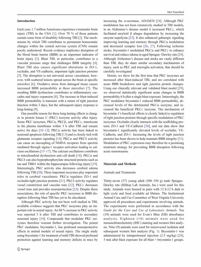
Fig. 1 Table showing the experimental techniques used for each timepoint
Mol Neurobiol

Western Blot
Western blot analysis was performed similar to work previous-ly published by our laboratory [39]. Briefly, 24 h after blastexposure, animals were anesthetized with 4 % isoflurane,brains were removed by decapitation, and the brains weresubsequently sectioned. Groups included control, single blast,and single blast + bryostatin-1. Protein samples from the leftprefrontal cortex were dissolved in 0.5 mL hot (85–95 °C) 1 %sodium docecyl sulfate (SDS), sonicated, and a BicinchoninicAcid (BCA) protein assay kit (Thermo Fisher Scientific,Rockfield, IL, USA) was used to determine protein concentra-tion. The pre-stained standard SeeBlue® Plus2 (Life Technol-ogies, Carlsbad, CA, USA) was used. 2X Lammeli buffer
combined with 30 μg of protein was loaded per well and runon a Bolt® Mini tank system (Life Technologies) with pre-castBolt® Bis-Tris Plus 10 % 12-well gels (Life Technologies).PVDF membranes (Bio-Rad, Contra Costa, CA, USA) weresoaked inmethanol and used for wet transfer (Bio-Rad) at 60 Vfor 2.5 h. Primary antibodies used were occludin rabbit 1:1,000(Life Technologies), PKCα, PKCδ, PKCεmouse 1:200 (SantaCruz Biotechnology, Santa Cruz, CA, USA), and VE-Cadherin rabbit 1:1,000 (Cell Signaling, Danvers, MA,USA). Microvessel isolation tissue was used to detect changesin PKC isozymes. LI-COR secondary antibodies IRDye®800CW and IRDye® 680RD (LI-COR, Lincoln, NE, USA)were used with an Odyssey fluorescent scanner at wavelengths800 or 700, intensity 6.0, and 84 resolution with high image
Fig. 2 Bryostatin-1 decreases PKCα and increases PKCε 24 h after blastexposure. Our data show that bryostatin-1 has a profound effect after blasttraumatic brain injury using fluorescent IHC. Scale bar=100 μm in leftprefrontal cortex. PKCα control (a) with inlay (b) compared to singleblast exposure (c) with inlay (d), and single blast exposure + bryostatin-1(e) with inlay (f) showed significant difference between groups. Post-hoccomparison between control and single blast (**p<0.01) and betweensingle blast and single blast + bryostatin-1 (##p<0.01) as depicted in bargraph (g). PKCδ control (h) with inlay (i) compared to single blastexposure (j) with inlay (k), and single blast exposure + bryostatin-1 (l)
with inlay (m) showed significant difference between groups. Post-hoccomparison between control and single blast (***p<0.001) and betweencontrol and single blast + bryostatin-1 (**p<0.01) as depicted in bargraph (n). PKCε control (o) with inlay (p) compared to single blastexposure (q) with inlay (r), and single blast exposure + bryostatin-1 (s)with inlay (t) showed a significant difference between groups. Post-hoccomparison between control and single blast + bryostatin-1(***p<0.001) and between single blast and single blast + bryostatin-1(!!p<0.01) as depicted in bar graph (u)
Mol Neurobiol

quality. Images were analyzed after background subtraction,and normalized to β-actin to give relative overall intensity.
Immunohistochemistry
Experimental groups included control (n=3), single blast (n=3), and single blast + bryostatin-1 (n=3). Rats were anesthe-tized with 4 % isoflurane and transcardially perfused with0.9 % saline followed by 4 % paraformaldehyde at 24 h afterblast exposure. The rats were decapitated, and brains wereremoved and stored in 4 % paraformaldehyde until process-ing. The tissue was sectioned and paraffin embedded. Six-micrometer slices from the left prefrontal cortex were cut witha Leica RM2235 microtome (Leica Microsystems, Wetzlar,Germany) and mounted on slides. Slides were washed inxylene, 100 % EtOH and 95 % EtOH for 5 min each in orderto remove the paraffin, followed by rehydration in dH2O for5 min. Using 10 % methanol and 10 % H2O2 in Dulbecco’sphosphate buffered saline (DPBS), slides were quenched for15 min, followed by three rinses in DPBS for 5 min each. Theslides were then added to permeabilizing solution (1.8 % L-Lysine, 4 % horse serum, and 0.2 % Triton X-100 in DPBS)for 30min. Slices were circumscribed and incubated overnightwith a primary antibody and 4 % horse serum. The next day,slides were rinsed and incubated for 3 h with a fluorescentsecondary antibody. A second set of primary and secondaryantibodies were used in the case of co-localization staining.Slides were then fixed with Permount mounting media. Pri-mary antibodies include: PKCα, PKCδ, and PKCε mouse1:500 (Santa Cruz Biotechnology), Claudin-5 mouse 1:200(Life Technologies), occludin rabbit 1:200 (Life
Technologies), VE-Cadherin rabbit 1:150 (Cell Signaling),ZO-1 Alexa Fluor® 488 mouse 1:200 (Life Technologies),Glial Fibrillary Acidic Protein (GFAP) mouse 1:150 (CellSignaling), GFAP rabbit 1:500 (DAKO, Glostrup, Denmark),Neuronal/glia proteoglycan 2 (NG-2) rabbit 1:500 (Santa CruzBiotechnology) and Von Willebrand Factor (VWF) rabbit1:200 (Sigma Aldrich, St. Louis, MO, USA). Secondary anti-bodies include Alexa Fluor 594 goat anti-mouse 1:1,000 (LifeTechnologies), Alexa Fluor 594 goat anti-rabbit 1:500 (LifeTechnologies), Alexa Fluor 488 donkey anti-mouse 1:500 (LifeTechnologies), and Alexa 488 donkey anti-rabbit 1:1,000 (LifeTechnologies). Twenty cells per slide were randomly selected,outlined, and measured with ImageJ software (NIH) by anobserver blinded to experimental group. Density was adjustedper mean area to give corrected total cell fluorescence normal-ized to background for claudin-5, occludin, VE-Cadherin, ZO-1, and PKCα, PKCδ, and PKCε. Co-localization was quanti-fied with the ImageJ plugin Just Another Co-localizationplugin to give Pearson’s coefficient for controls and overlapcoefficient for treatment groups [40]. For percentage of co-localization, an observer blinded to experimental group count-ed 100 cells and the ratio of positive cells to total cells wasrecorded. χ2 analyses was used to compare between groups.
Statistical Analysis
Biochemical assay data were analyzed using GraphPad Prism6.0 (GraphPad Software, Inc., La Jolla, CA, USA) by anobserver blinded to experimental group. A one-way ANOVAwith Tukey’s post-hoc was used to compare across groups forEB absorbance, western blot analysis, and IHC quantification.
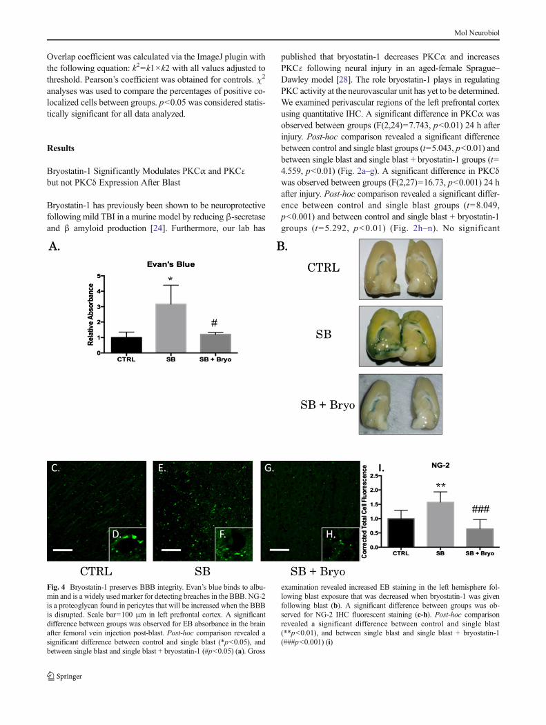
Fig. 3 Bryostatin-1 is a potent PKC modulator. It has been used toregulate PKC activity in a time-specific manner for multiple neural injurymodels. Protein concentrations were measured in the left prefrontal cortex24 h after blast expsoure using western blot analysis. A significantdifference between groups was observed for PKCα. Post-hoc comparisonbetween control and single blast (*p<0.05), and between single blast andsingle blast + bryostatin-1 (##p<0.01) (a). A significant difference
between groups was observed for PKCδ. Post-hoc comparison betweencontrol and single blast (**p<0.01), and control and single blast +bryostatin-1 (*p<0.05) (b). A significant difference between groupswas observed for PKCε. Post-hoc comparison between control and singleblast (*p<0.05), and between control and single blast + bryostatin-1(**p<0.01) (c). Bryostatin-1 significantly decreased PKCα levels andincreased PKCε levels when administered after blast exposure
Mol Neurobiol
Overlap coefficient was calculated via the ImageJ plugin withthe following equation: k2=k1×k2 with all values adjusted tothreshold. Pearson’s coefficient was obtained for controls. χ2
analyses was used to compare the percentages of positive co-localized cells between groups. p<0.05 was considered statis-tically significant for all data analyzed.
Results
Bryostatin-1 Significantly Modulates PKCα and PKCεbut not PKCδ Expression After Blast
Bryostatin-1 has previously been shown to be neuroprotectivefollowing mild TBI in a murine model by reducing β-secretaseand β amyloid production [24]. Furthermore, our lab has
published that bryostatin-1 decreases PKCα and increasesPKCε following neural injury in an aged-female Sprague–Dawley model [28]. The role bryostatin-1 plays in regulatingPKC activity at the neurovascular unit has yet to be determined.We examined perivascular regions of the left prefrontal cortexusing quantitative IHC. A significant difference in PKCα wasobserved between groups (F(2,24)=7.743, p<0.01) 24 h afterinjury. Post-hoc comparison revealed a significant differencebetween control and single blast groups (t=5.043, p<0.01) andbetween single blast and single blast + bryostatin-1 groups (t=4.559, p<0.01) (Fig. 2a–g). A significant difference in PKCδwas observed between groups (F(2,27)=16.73, p<0.001) 24 hafter injury. Post-hoc comparison revealed a significant differ-ence between control and single blast groups (t=8.049,p<0.001) and between control and single blast + bryostatin-1groups (t=5.292, p<0.01) (Fig. 2h–n). No significant
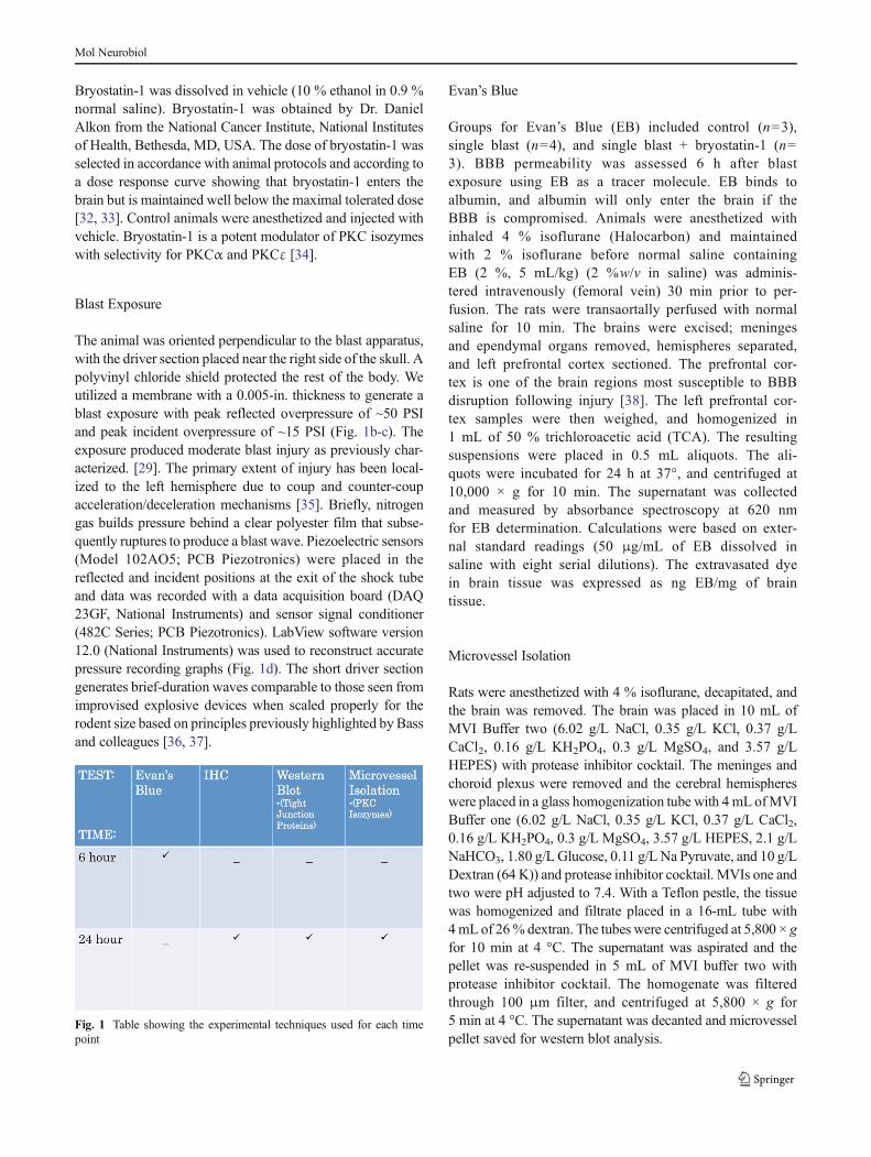
Fig. 4 Bryostatin-1 preserves BBB integrity. Evan’s blue binds to albu-min and is a widely usedmarker for detecting breaches in the BBB. NG-2is a proteoglycan found in pericytes that will be increased when the BBBis disrupted. Scale bar=100 μm in left prefrontal cortex. A significantdifference between groups was observed for EB absorbance in the brainafter femoral vein injection post-blast. Post-hoc comparison revealed asignificant difference between control and single blast (*p<0.05), andbetween single blast and single blast + bryostatin-1 (#p<0.05) (a). Gross
examination revealed increased EB staining in the left hemisphere fol-lowing blast exposure that was decreased when bryostatin-1 was givenfollowing blast (b). A significant difference between groups was ob-served for NG-2 IHC fluorescent staining (c-h). Post-hoc comparisonrevealed a significant difference between control and single blast(**p<0.01), and between single blast and single blast + bryostatin-1(###p<0.001) (i)
Mol Neurobiol
difference was seen between single blast and single blast +bryostatin-1, consistent with the notion that bryostatin-1 negli-gibly alters PKCδ expression. A significant difference in PKCεwas observed between groups (F(2,27)=14.73, p<0.001) 24 hafter injury. Post-hoc comparison revealed a significant differ-ence between control and single blast + bryostatin-1 groups (t=7.509, p<0.001) and between single blast and single blast +bryostatin-1 groups (t=5.133, p<0.01) (Fig. 2o–u). We alsoexamined changes in PKC expression using western blot anal-ysis. A significant difference between groups was observed forPKCα (F(2,14)=7.672, p<0.01). Post-hoc comparison re-vealed a significant difference between control and single blast(t=4.576, p<0.05), and between single blast and single blast +bryostatin-1 (t=5.113, p<0.01) (Fig. 3a). A significant differ-ence between groups was observed for PKCδ (F(2,6)=13.25,p<0.01). Post-hoc comparison revealed a significant differencebetween control and single blast (t=7.197, p<0.01), and be-tween control and single blast + bryostatin-1 (t=4.549, p<0.05)(Fig. 3b). A significant difference between groups was ob-served for PKCε (F(2,15)=10.05, p<0.01). Post-hoc compar-ison revealed a significant difference between control and
single blast (t=4.546, p<0.05), and between control and singleblast + bryostatin-1 (t=6.102, p<0.01) (Fig. 3 c).
Bryostatin-1 Maintained BBB Integrity Following BlastExposure
BBB integrity can be measured by EB extravasation into thebrain parenchyma [7]. EB binds to albumin, and albumin doesnot readily penetrate an intact BBB. NG-2 is a pericyte pro-teoglycan that is increased during BBB disruption and subse-quent neovascularization [41]. Disruption of the BBB hasmultiple effects including neuroinflammation and formationof reactive oxygen species [42]. A significant difference be-tween groups was observed for EB extravasation 6 h afterblast exposure (F(2,7)=7.38, p<0.05) for the left prefrontalcortex (Fig. 4a).Post-hoc comparison was significant betweencontrol and single blast (t=4.808, p<0.05) and between singleblast and single blast + bryostatin-1 (t=4.347, p<0.05). Nosignificant difference was seen between control and singleblast + bryostatin-1. Bryostatin-1 successfully preserved theintegrity of the BBB apparent on gross exam of the left
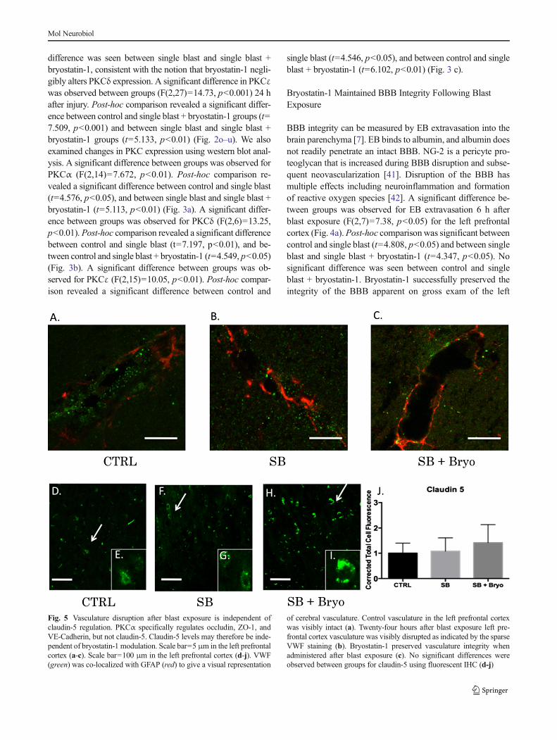
Fig. 5 Vasculature disruption after blast exposure is independent ofclaudin-5 regulation. PKCα specifically regulates occludin, ZO-1, andVE-Cadherin, but not claudin-5. Claudin-5 levels may therefore be inde-pendent of bryostatin-1 modulation. Scale bar=5 μm in the left prefrontalcortex (a-c). Scale bar=100 μm in the left prefrontal cortex (d-j). VWF(green) was co-localized with GFAP (red) to give a visual representation
of cerebral vasculature. Control vasculature in the left prefrontal cortexwas visibly intact (a). Twenty-four hours after blast exposure left pre-frontal cortex vasculature was visibly disrupted as indicated by the sparseVWF staining (b). Bryostatin-1 preserved vasculature integrity whenadministered after blast exposure (c). No significant differences wereobserved between groups for claudin-5 using fluorescent IHC (d-j)
Mol Neurobiol

hemisphere (Fig. 4b). A significant difference in NG-2 wasobserved 24 h after blast exposure (F(2,27)=20.42, p<0.001)for the left prefrontal cortex (Fig. 4c–h). Post-hoc comparisonwas significant between control and single blast (t=5.504,p<0.01), and between single blast and single blast +bryostatin-1 (t=8.961, p<0.001), but not between controland single blast + bryostatin-1.
PKC Modulation Alters Tight Junction Proteins at the BloodBrain Barrier
Activation of PKC isozymes is thought to play a crucial role inBBB disruption [43]. PKCα, in particular, has been linked toredistribution of tight junction proteins leading to increasedvascular permeability [44]. PKCα is also known to activate
sonic hedgehog protein leading to dysfunctional synthesis oftight junction proteins [45, 46]. Alternatively, increasingPKCε facilitates tight junction protein translocation from thenucleus to the plasma membrane [47]. Gross examination ofthe neurovascular unit (VWF co-localized with GFAP) re-vealed histological disruption of the BBB following blastinjury but maintenance of barrier integrity when bryostatin-1was administered following blast exposure (Fig. 5a–c). Asignificant difference between groups was not observed forClaudin-5 (Fig. 5d–j) using fluorescent IHC. A significantdifference between groups was observed for VE-Cadherin(F(2,6)=18.58, p<0.01) using western blot. Post-hoc compar-ison revealed a significant difference between control andsingle blast (t=4.571, p<0.05), and between control and sin-gle blast + bryostatin-1 (t=8.616, p<0.01) (Fig. 6a). A
Fig. 6 Bryostatin-1 significantly increases tight junction proteins. Ourdata show that bryostatin-1 significantly upregulated tight junction pro-teins leading to maintenance of BBB integrity following blast TBI. Scalebar=100 μm in left prefrontal cortex. A significant difference was ob-served between groups using western blot for VE-Cadherin with post hoccomparison showing significance between control and single blast(*p<0.05), and between control and single blast + bryostatin-1(**p<0.01) 24 h after blast exposure (a). A significant difference wasobserved between groups using western blot for occludin with post hoc
comparison showing significance between control and single blast(**p<0.01), between control and single blast + bryostatin-1(***p<0.001), and between single blast and single blast + bryostatin-1(##p<0.01) 24 h after blast exposure (b). A significant difference wasobserved between groups using fluorescent IHC for ZO-1 with post hoccomparison showing significance between control and single blast +bryostatin-1 (***p<0.001), and between single blast and single blast +bryostatin-1 (###p<0.001) 24 h after blast exposure (c)
Mol Neurobiol
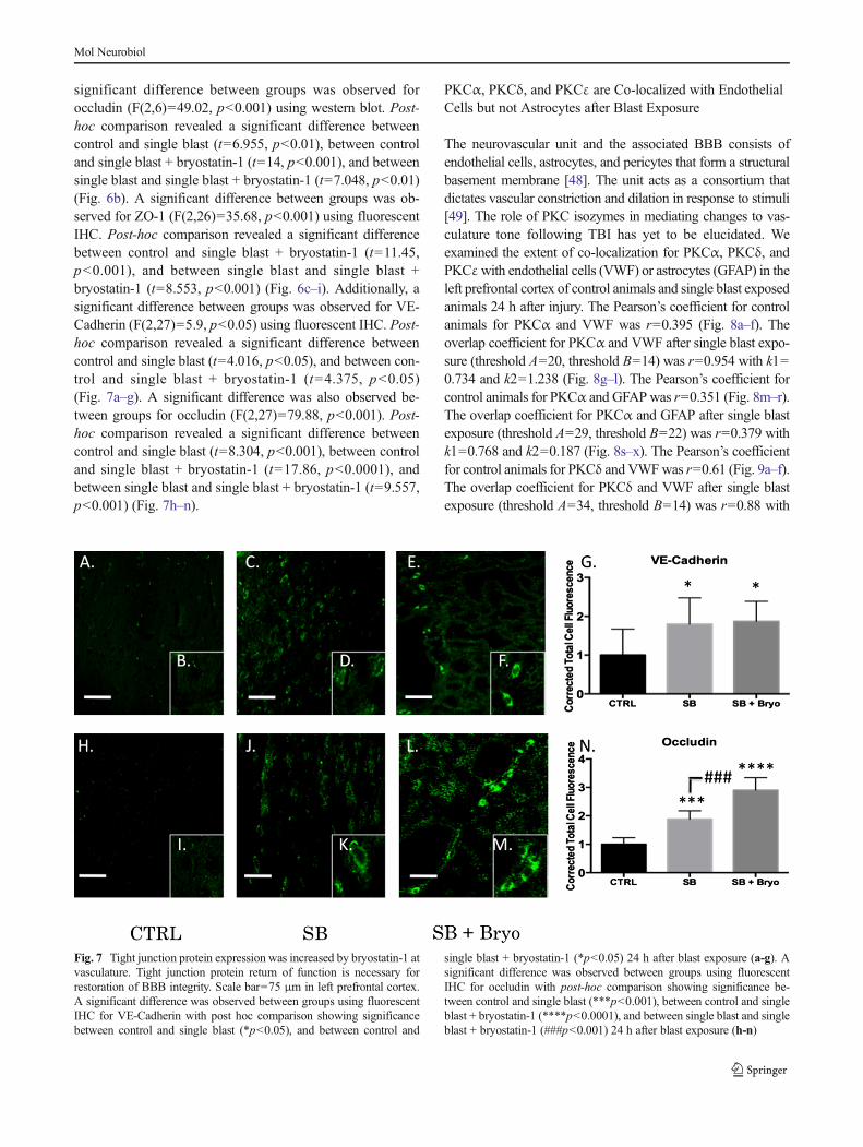
significant difference between groups was observed foroccludin (F(2,6)=49.02, p<0.001) using western blot. Post-hoc comparison revealed a significant difference betweencontrol and single blast (t=6.955, p<0.01), between controland single blast + bryostatin-1 (t=14, p<0.001), and betweensingle blast and single blast + bryostatin-1 (t=7.048, p<0.01)(Fig. 6b). A significant difference between groups was ob-served for ZO-1 (F(2,26)=35.68, p<0.001) using fluorescentIHC. Post-hoc comparison revealed a significant differencebetween control and single blast + bryostatin-1 (t=11.45,p<0.001), and between single blast and single blast +bryostatin-1 (t=8.553, p<0.001) (Fig. 6c–i). Additionally, asignificant difference between groups was observed for VE-Cadherin (F(2,27)=5.9, p<0.05) using fluorescent IHC. Post-hoc comparison revealed a significant difference betweencontrol and single blast (t=4.016, p<0.05), and between con-trol and single blast + bryostatin-1 (t=4.375, p<0.05)(Fig. 7a–g). A significant difference was also observed be-tween groups for occludin (F(2,27)=79.88, p<0.001). Post-hoc comparison revealed a significant difference betweencontrol and single blast (t=8.304, p<0.001), between controland single blast + bryostatin-1 (t=17.86, p<0.0001), andbetween single blast and single blast + bryostatin-1 (t=9.557,p<0.001) (Fig. 7h–n).
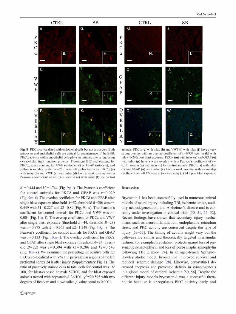
PKCα, PKCδ, and PKCε are Co-localized with EndothelialCells but not Astrocytes after Blast Exposure
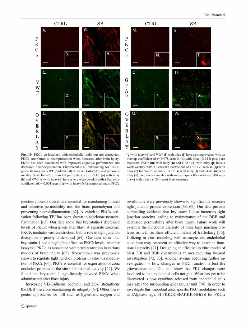
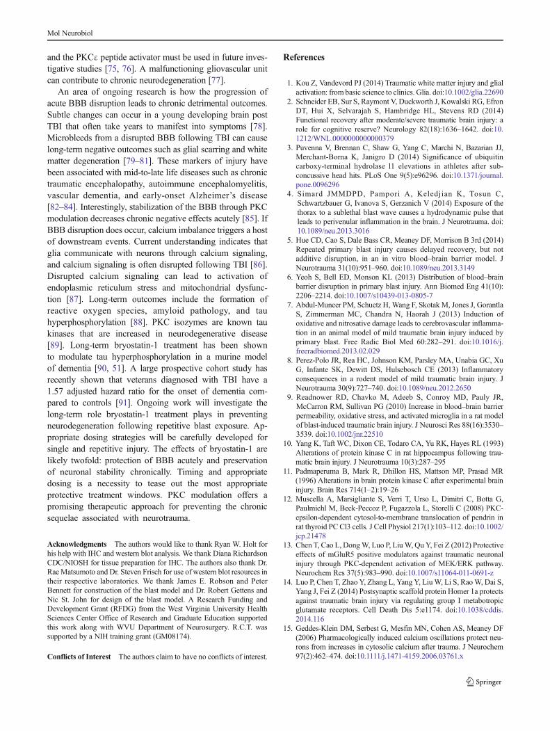
The neurovascular unit and the associated BBB consists ofendothelial cells, astrocytes, and pericytes that form a structuralbasement membrane [48]. The unit acts as a consortium thatdictates vascular constriction and dilation in response to stimuli[49]. The role of PKC isozymes in mediating changes to vas-culature tone following TBI has yet to be elucidated. Weexamined the extent of co-localization for PKCα, PKCδ, andPKCεwith endothelial cells (VWF) or astrocytes (GFAP) in theleft prefrontal cortex of control animals and single blast exposedanimals 24 h after injury. The Pearson’s coefficient for controlanimals for PKCα and VWF was r=0.395 (Fig. 8a–f). Theoverlap coefficient for PKCα and VWF after single blast expo-sure (threshold A=20, threshold B=14) was r=0.954 with k1=0.734 and k2=1.238 (Fig. 8g–l). The Pearson’s coefficient forcontrol animals for PKCα and GFAPwas r=0.351 (Fig. 8m–r).The overlap coefficient for PKCα and GFAP after single blastexposure (threshold A=29, threshold B=22) was r=0.379 withk1=0.768 and k2=0.187 (Fig. 8s–x). The Pearson’s coefficientfor control animals for PKCδ and VWFwas r=0.61 (Fig. 9a–f).The overlap coefficient for PKCδ and VWF after single blastexposure (threshold A=34, threshold B=14) was r=0.88 with
Fig. 7 Tight junction protein expression was increased by bryostatin-1 atvasculature. Tight junction protein return of function is necessary forrestoration of BBB integrity. Scale bar=75 μm in left prefrontal cortex.A significant difference was observed between groups using fluorescentIHC for VE-Cadherin with post hoc comparison showing significancebetween control and single blast (*p<0.05), and between control and
single blast + bryostatin-1 (*p<0.05) 24 h after blast exposure (a-g). Asignificant difference was observed between groups using fluorescentIHC for occludin with post-hoc comparison showing significance be-tween control and single blast (***p<0.001), between control and singleblast + bryostatin-1 (****p<0.0001), and between single blast and singleblast + bryostatin-1 (###p<0.001) 24 h after blast exposure (h-n)
Mol Neurobiol
k1=0.444 and k2=1.744 (Fig. 9g–l). The Pearson’s coefficientfor control animals for PKCδ and GFAP was r=0.029(Fig. 9m–r). The overlap coefficient for PKCδ and GFAP aftersingle blast exposure (thresholdA=52, thresholdB=20) was r=0.449 with k1=0.227 and k2=0.89 (Fig. 9s–x). The Pearson’scoefficient for control animals for PKCε and VWF was r=0.004 (Fig. 10a–f). The overlap coefficient for PKCε and VWFafter single blast exposure (threshold A=44, threshold B=23)was r=0.978 with k1=0.765 and k2=1.249 (Fig. 10g–l). ThePearson’s coefficient for control animals for PKCε and GFAPwas r=0.133 (Fig. 10m–r). The overlap coefficient for PKCεand GFAP after single blast exposure (threshold A=24, thresh-old B=22) was r=0.394 with k1=0.286 and k2=0.543(Fig. 10s–x). We examined the percentage of positive cells forPKCα co-localized with VWF in perivascular regions of the leftprefrontal cortex 24 h after injury (Supplementary Fig. 1). Theratio of positively stained cells to total cells for control was 18/100, for blast-exposed animals 57/100, and for blast exposedanimals treated with bryostatin-1 36/100. χ2=20.595 with twodegrees of freedom and a two-tailed p value equal to 0.0001.
Discussion
Bryostatin-1 has been successfully used in numerous animalmodels of neural injury including TBI, ischemic stroke, audi-tory neurodegeneration, and Alzheimer’s disease and is cur-rently under investigation in clinical trials [50, 51, 24, 52].Recent findings have shown that secondary injury mecha-nisms such as neuroinflammation, endoplasmic reticulumstress, and PKC activity are conserved despite the type ofinjury [53–55]. The timing of activity might vary but thepathways are similar and theoretically targeted in a similarfashion. For example, bryostatin-1 protects against loss of pre-synaptic synaptophysin and loss of post-synaptic spinophylinfollowing TBI in mice [24]. In an aged-female Sprague–Dawley stroke model, bryostatin-1 improved survival andreduced ischemic damage [28]. Likewise, bryostatin-1 de-creased apoptosis and prevented deficits in synaptogenesisin a global model of cerebral ischemia [50, 56]. Despite thedifferent injury models bryostatin-1 was a successful thera-peutic because it upregulates PKC activity early and
Fig. 8 PKCα co-localized with endothelial cells but not astrocytes. Bothastrocytes and endothelial cells are critical for maintenance of the BBB.PKCα activity within endothelial cells plays an intimate role in regulatingextracellular tight junction proteins. Fluorescnt IHC red staining forPKCα, green staining for VWF (endothelial) or GFAP (astocyte), andyellow is overlay. Scale bar=20 μm in left prefrontal cortex. PKCα (a)with inlay (b) and VWF (c) with inlay (d) have a weak overlay with aPearson’s coefficient of r=0.395 seen in (e) with inlay (f) for control
animals. PKCα (g) with inlay (h) and VWF (i) with inlay (j) have a verystrong overlay with an overlap coefficient of r=0.954 seen in (k) withinlay (l) 24 h post blast exposure. PKCα (m) with inlay (n) and GFAP (o)with inlay (p) have a weak overlay with a Pearson’s coefficient of r=0.351 seen in (q) with inlay (r) for control animals. PKCα (s) with inlay(t) and GFAP (u) with inlay (v) have a weak overlay with an overlapcoefficient of r=0.379 seen in (w) with inlay (x) 24 h post blast exposure
Mol Neurobiol

downregulates activity when used at excess doses or at chron-ic time points [57]. At the dose used in this study, it is possiblethat an early activation of PKCα is followed by a drasticdownregulation observed at 24 h. PKCε may also be in-creased not only due to upregulation but also from increasedsynthesis [27]. Future studies will be required to look at PKCchanges at both earlier time points and also the beneficialeffects of bryostatin-1 when administered at subacute timepoints. In Alzheimer’s disease, early upregulation of PKCactivity enhances neuronal repair and decreases amyloid-mediated degeneration while maintaining long-term potentia-tion [58, 59]. In addition, bryostatin-1 facilitates neurite out-growth, maintains cognitive function [52], and enhances syn-aptic connectivity and spatial learning [34]. For affectivedisorders, bryostatin-1 downregulates ERK-dependent PKCactivity at late time points, which can have beneficial effectsfor treating substance abuse, depression, and aversive emo-tional memories [51, 60]. Similar molecular PKC targetsappear to be altered in TBI [14].The potential for bryostatin-
1 as a treatment for TBI warrants further investigation. Thenecessity for different dosing strategies as well as altering thetiming of dosing must be carefully considered in a series offuture studies. Furthermore, specific activation of select iso-zymes will offer compelling insight into their protective prop-erties following TBI. In this study, we examined how a single2.5 mg/kg dose of bryostatin-1 affects BBB integrity acutelyfollowing blast-induced TBI.
We found that a single moderate TBI significantly in-creased EB extravasation into the left prefrontal cortex 6 hafter injury. Bryostatin-1 administration ameliorated this dis-ruption. A potential mechanism by which this occurs isthrough regulation of PKCα and PKCε, which subsequentlyincreases the tight junction proteins VE-Cadherin, occludin,and ZO-1 at the BBB. It has been previously shown in anischemic stroke model that inhibiting PKCα increases thetight junction proteins ZO-1, occludin, and VE-Cadherin[61]. PKCα has limited regulatory function for claudin-5accounting for the non-significant findings observed. Tight
Fig. 9 PKCδ co-localized with endothelial cells but not astrocytes.PKCδ plays an important role in mediating vascular tone. Its role inregulation of tight junction proteins is not completely understood. Fluo-rescent IHC red staining for PKCδ, green staining for VWF (endothelial)or GFAP (astocyte), and yellow is overlay. Scale bar=20 μm in leftprefrontal cortex. PKCδ (a) with inlay (b) and VWF (c) with inlay (d)have a moderate overlay with a Pearson’s coefficient of r=0.61 seen in (e)with inlay (f) for control animals. PKCδ (g) with inlay (h) and VWF (i)
with inlay (j) have a strong overlay with an overlap coefficient of r=0.88seen in (k) with inlay (l) 24 h post blast exposure. PKCδ (m) with inlay(n) and GFAP (o) with inlay (p) have a very weak overlay with aPearson’s coefficient of r=0.029 seen in (q) with inlay (r) for controlanimals. PKCδ (s) with inlay (t) and GFAP (u) with inlay (v) have amoderate overlay with an overlap coefficient of r=0.449 seen in (w) withinlay (x) 24 h post blast exposure
Mol Neurobiol
junction proteins overall are essential for maintaining limitedand selective permeability into the brain parenchyma andpreventing neuroinflammation [62]. A switch to PKCα acti-vation following TBI has been shown to accelerate neuroin-flammation [63]. Our data show that bryostatin-1 decreasedlevels of PKCα when given after blast. A separate isozyme,PKCδ, mediates vasoconstriction, but its role in tight junctiondisruption is poorly understood [64]. Our data show thatbryostatin-1 had a negligible effect on PKCδ levels. Anotherisozyme, PKCε, is associated with neuroprotection in variousmodels of brain injury [65]. Bryostatin-1 was previouslyshown to regulate tight junction proteins in vitro via modula-tion of PKCε [66]. PKCε is essential for exportation of zonaoccluden proteins to the site of functional activity [47]. Wefound that bryostatin-1 significantly elevated PKCε whenadministered after blast injury.
Increasing VE-Cadherin, occludin, and ZO-1 strengthensthe BBB therefore maintaining its integrity [67]. Other thera-peutic approaches for TBI such as hyperbaric oxygen and
sevoflurane were previously shown to significantly increasetight junction protein expression [68, 69]. Our data providecompelling evidence that bryostatin-1 also increases tightjunction proteins leading to maintenance of the BBB anddecreased permeability after blast injury. Future work willexamine the functional capacity of these tight junction pro-teins as well as their efficient means of trafficking [70].Utilizing in vitro modeling with astrocyte and endothelialco-culture may represent an effective way to examine func-tional capacity [71]. Designing an effective in vitro model ofblast TBI and BBB dynamics is an area requiring focusedinvestigation [72, 73]. Another avenue requiring further in-vestigation is how changes in PKC function affect thegliovascular unit. Our data show that PKC changes werelocalized to the endothelial cells not glia. What has yet to bediscovered is how cytokines released from endothelial cellsmay alter the surrounding gliovascular unit [74]. In order toinvestigate this important area, specific PKC modulators suchas (Alphatomega; H-FKKQGSFAKKK-NH(2)) for PKCα
Fig. 10 PKCε co-localized with endothelial cells but not astrocytes.PKCε contributes to neuroprotection when increased after brain injury.PKCε has been associated with improved cognitive performance anddecreased neurodegeneration. Fluorescent IHC red staining for PKCε,green staining for VWF (endothelial) or GFAP (astocyte), and yellow isoverlay. Scale bar=20 μm in left prefrontal cortex. PKCε (a) with inlay(b) and VWF (c) with inlay (d) have a very weak overlay with a Pearson’scoefficient of r=0.004 seen in (e) with inlay (f) for control animals. PKCε
(g) with inlay (h) and VWF (i) with inlay (j) have a strong overlay with anoverlap coefficient of r=0.978 seen in (k) with inlay (l) 24 h post blastexposure. PKCε (m) with inlay (n) and GFAP (o) with inlay (p) have aweak overlay with a Pearson’s coefficient of r=0.133 seen in (q) withinlay (r) for control animals. PKCε (s) with inlay (t) and GFAP (u) withinlay (v) have a weak overlay with an overlap coefficient of r=0.394 seenin (w) with inlay (x) 24 h post blast exposure
Mol Neurobiol
and the PKCε peptide activator must be used in future inves-tigative studies [75, 76]. A malfunctioning gliovascular unitcan contribute to chronic neurodegeneration [77].
An area of ongoing research is how the progression ofacute BBB disruption leads to chronic detrimental outcomes.Subtle changes can occur in a young developing brain postTBI that often take years to manifest into symptoms [78].Microbleeds from a disrupted BBB following TBI can causelong-term negative outcomes such as glial scarring and whitematter degeneration [79–81]. These markers of injury havebeen associated with mid-to-late life diseases such as chronictraumatic encephalopathy, autoimmune encephalomyelitis,vascular dementia, and early-onset Alzheimer’s disease[82–84]. Interestingly, stabilization of the BBB through PKCmodulation decreases chronic negative effects acutely [85]. IfBBB disruption does occur, calcium imbalance triggers a hostof downstream events. Current understanding indicates thatglia communicate with neurons through calcium signaling,and calcium signaling is often disrupted following TBI [86].Disrupted calcium signaling can lead to activation ofendoplasmic reticulum stress and mitochondrial dysfunc-tion [87]. Long-term outcomes include the formation ofreactive oxygen species, amyloid pathology, and tauhyperphosphorylation [88]. PKC isozymes are known taukinases that are increased in neurodegenerative disease[89]. Long-term bryostatin-1 treatment has been shownto modulate tau hyperphosphorylation in a murine modelof dementia [90, 51]. A large prospective cohort study hasrecently shown that veterans diagnosed with TBI have a1.57 adjusted hazard ratio for the onset of dementia com-pared to controls [91]. Ongoing work will investigate thelong-term role bryostatin-1 treatment plays in preventingneurodegeneration following repetitive blast exposure. Ap-propriate dosing strategies will be carefully developed forsingle and repetitive injury. The effects of bryostatin-1 arelikely twofold: protection of BBB acutely and preservationof neuronal stability chronically. Timing and appropriatedosing is a necessity to tease out the most appropriateprotective treatment windows. PKC modulation offers apromising therapeutic approach for preventing the chronicsequelae associated with neurotrauma.
Acknowledgments The authors would like to thank Ryan W. Holt forhis help with IHC and western blot analysis. We thank Diana RichardsonCDC/NIOSH for tissue preparation for IHC. The authors also thank Dr.RaeMatsumoto and Dr. Steven Frisch for use of western blot resources intheir respective laboratories. We thank James E. Robson and PeterBennett for construction of the blast model and Dr. Robert Gettens andNic St. John for design of the blast model. A Research Funding andDevelopment Grant (RFDG) from the West Virginia University HealthSciences Center Office of Research and Graduate Education supportedthis work along with WVU Department of Neurosurgery. R.C.T. wassupported by a NIH training grant (GM08174).
Conflicts of Interest The authors claim to have no conflicts of interest.
References
1. Kou Z, Vandevord PJ (2014) Traumatic white matter injury and glialactivation: from basic science to clinics. Glia. doi:10.1002/glia.22690
2. Schneider EB, Sur S, Raymont V, Duckworth J, Kowalski RG, EfronDT, Hui X, Selvarajah S, Hambridge HL, Stevens RD (2014)Functional recovery after moderate/severe traumatic brain injury: arole for cognitive reserve? Neurology 82(18):1636–1642. doi:10.1212/WNL.0000000000000379
3. Puvenna V, Brennan C, Shaw G, Yang C, Marchi N, Bazarian JJ,Merchant-Borna K, Janigro D (2014) Significance of ubiquitincarboxy-terminal hydrolase l1 elevations in athletes after sub-concussive head hits. PLoS One 9(5):e96296. doi:10.1371/journal.pone.0096296
4. Simard JMMDPD, Pampori A, Keledjian K, Tosun C,Schwartzbauer G, Ivanova S, Gerzanich V (2014) Exposure of thethorax to a sublethal blast wave causes a hydrodynamic pulse thatleads to perivenular inflammation in the brain. J Neurotrauma. doi:10.1089/neu.2013.3016
5. Hue CD, Cao S, Dale Bass CR, Meaney DF, Morrison B 3rd (2014)Repeated primary blast injury causes delayed recovery, but notadditive disruption, in an in vitro blood–brain barrier model. JNeurotrauma 31(10):951–960. doi:10.1089/neu.2013.3149
6. Yeoh S, Bell ED, Monson KL (2013) Distribution of blood–brainbarrier disruption in primary blast injury. Ann Biomed Eng 41(10):2206–2214. doi:10.1007/s10439-013-0805-7
7. Abdul-Muneer PM, Schuetz H,Wang F, Skotak M, Jones J, GorantlaS, Zimmerman MC, Chandra N, Haorah J (2013) Induction ofoxidative and nitrosative damage leads to cerebrovascular inflamma-tion in an animal model of mild traumatic brain injury induced byprimary blast. Free Radic Biol Med 60:282–291. doi:10.1016/j.freeradbiomed.2013.02.029
8. Perez-Polo JR, Rea HC, Johnson KM, Parsley MA, Unabia GC, XuG, Infante SK, Dewitt DS, Hulsebosch CE (2013) Inflammatoryconsequences in a rodent model of mild traumatic brain injury. JNeurotrauma 30(9):727–740. doi:10.1089/neu.2012.2650
9. Readnower RD, Chavko M, Adeeb S, Conroy MD, Pauly JR,McCarron RM, Sullivan PG (2010) Increase in blood–brain barrierpermeability, oxidative stress, and activated microglia in a rat modelof blast-induced traumatic brain injury. J Neurosci Res 88(16):3530–3539. doi:10.1002/jnr.22510
10. Yang K, Taft WC, Dixon CE, Todaro CA, Yu RK, Hayes RL (1993)Alterations of protein kinase C in rat hippocampus following trau-matic brain injury. J Neurotrauma 10(3):287–295
11. Padmaperuma B, Mark R, Dhillon HS, Mattson MP, Prasad MR(1996) Alterations in brain protein kinase C after experimental braininjury. Brain Res 714(1–2):19–26
12. Muscella A, Marsigliante S, Verri T, Urso L, Dimitri C, Botta G,Paulmichl M, Beck-Peccoz P, Fugazzola L, Storelli C (2008) PKC-epsilon-dependent cytosol-to-membrane translocation of pendrin inrat thyroid PC Cl3 cells. J Cell Physiol 217(1):103–112. doi:10.1002/jcp.21478
13. Chen T, Cao L, DongW, Luo P, LiuW, QuY, Fei Z (2012) Protectiveeffects of mGluR5 positive modulators against traumatic neuronalinjury through PKC-dependent activation of MEK/ERK pathway.Neurochem Res 37(5):983–990. doi:10.1007/s11064-011-0691-z
14. Luo P, Chen T, Zhao Y, Zhang L, Yang Y, LiuW, Li S, RaoW, Dai S,Yang J, Fei Z (2014) Postsynaptic scaffold protein Homer 1a protectsagainst traumatic brain injury via regulating group I metabotropicglutamate receptors. Cell Death Dis 5:e1174. doi:10.1038/cddis.2014.116
15. Geddes-Klein DM, Serbest G, Mesfin MN, Cohen AS, Meaney DF(2006) Pharmacologically induced calcium oscillations protect neu-rons from increases in cytosolic calcium after trauma. J Neurochem97(2):462–474. doi:10.1111/j.1471-4159.2006.03761.x
Mol Neurobiol

16. Roh DH, Yoon SY, Seo HS, Kang SY, Moon JY, Song S, Beitz AJ,Lee JH (2010) Sigma-1 receptor-induced increase in murine spinalNR1 phosphorylation is mediated by the PKCalpha and epsilon, butnot the PKCzeta, isoforms. Neurosci Lett 477(2):95–99. doi:10.1016/j.neulet.2010.04.041
17. Giordano G, Sanchez-Perez AM, Burgal M, Montoliu C, Costa LG,Felipo V (2005) Chronic exposure to ammonia induces isoform-selective alterations in the intracellular distribution and NMDAreceptor-mediated translocation of protein kinase C in cerebellarneurons in culture. J Neurochem 92(1):143–157. doi:10.1111/j.1471-4159.2004.02852.x
18. Pandya JD, Nukala VN, Sullivan PG (2013) Concentration depen-dent effect of calcium on brain mitochondrial bioenergetics andoxidative stress parameters. Front Neuroenerg 5:10. doi:10.3389/fnene.2013.00010
19. Yang K, Taft WC, Dixon CE, Yu RK, Hayes RL (1994)Endogenous phosphorylation of a 61,000 dalton hippocampalprotein increases following traumatic brain injury. J Neurotrauma11(5):523–532
20. Karasu A, Aras Y, Sabanci PA, Saglam G, Izgi N, Biltekin B, BarakT, Hepgul KT, KayaM, Bilir A (2010) The effects of protein kinase Cactivator phorbol dibutyrate on traumatic brain edema and aquaporin-4 expression. Ulusal travma ve acil cerrahi dergisi. Turk J TraumaEmerg Surg TJTES 16(5):390–394
21. Kim YA, Park SL, Kim MY, Lee SH, Baik EJ, Moon CH, Jung YS(2010) Role of PKCbetaII and PKCdelta in blood–brain barrierpermeability during aglycemic hypoxia. Neurosci Lett 468(3):254–258. doi:10.1016/j.neulet.2009.11.007
22. Kizub IV, Klymenko KI, Soloviev AI (2014) Protein kinase C inenhanced vascular tone in diabetes mellitus. Int J Cardiol 174(2):230–242. doi:10.1016/j.ijcard.2014.04.117
23. Lin HW, Della-Morte D, Thompson JW, Gresia VL, Narayanan SV,Defazio RA, Raval AP, Saul I, Dave KR, Morris KC, Si ML, Perez-Pinzon MA (2012) Differential effects of delta and epsilon proteinkinase C inmodulation of postischemic cerebral blood flow. Adv ExpMed Biol 737:63–69. doi:10.1007/978-1-4614-1566-4_10
24. Zohar O, Lavy R, Zi X, Nelson TJ, Hongpaisan J, Pick CG, AlkonDL (2011) PKC activator therapeutic for mild traumatic brain injuryin mice. Neurobiol Dis 41(2):329–337. doi:10.1016/j.nbd.2010.10.001
25. Lim CS, Alkon DL (2014) PKCepsilon promotes HuD-mediatedneprilysin mRNA stability and enhances neprilysin-induced Abetadegradation in brain neurons. PLoS One 9(5):e97756. doi:10.1371/journal.pone.0097756
26. Xu C, Liu QY, Alkon DL (2014) PKC activators enhanceGABAergic neurotransmission and paired-pulse facilitation in hip-pocampal CA1 pyramidal neurons. Neuroscience 268:75–86. doi:10.1016/j.neuroscience.2014.03.008
27. Hongpaisan J, Sun MK, Alkon DL (2011) PKC epsilon activationprevents synaptic loss, Abeta elevation, and cognitive deficits inAlzheimer's disease transgenic mice. J Neurosci Off J Soc Neurosci31(2):630–643. doi:10.1523/JNEUROSCI.5209-10.2011
28. Tan Z, Turner RC, Leon RL, Li X, Hongpaisan J, ZhengW, LogsdonAF, Naser ZJ, Alkon DL, Rosen CL, Huber JD (2013) Bryostatinimproves survival and reduces ischemic brain injury in aged rats afteracute ischemic stroke. Stroke J Cereb Circ 44(12):3490–3497. doi:10.1161/STROKEAHA.113.002411
29. Turner RC, Naser ZJ, Logsdon AF, DiPasquale KH, Jackson GJ,Robson MJ, Gettens RT, Matsumoto RR, Huber JD, Rosen CL(2013)Modeling clinically relevant blast parameters based on scalingprinciples produces functional histological deficits in rats. ExpNeurol 248:520–529. doi:10.1016/j.expneurol.2013.07.008
30. Morrow CM, Mruk D, Cheng CY, Hess RA (2010) Claudin andoccludin expression and function in the seminiferous epithelium.Philos Trans R Soc Lond B Biol Sci 365(1546):1679–1696. doi:10.1098/rstb.2010.0025
31. Goncalves A, Leal E, Paiva A, Teixeira Lemos E, Teixeira F, RibeiroCF, Reis F, Ambrosio AF, Fernandes R (2012) Protective effects ofthe dipeptidyl peptidase IV inhibitor sitagliptin in the blood-retinalbarrier in a type 2 diabetes animal model. Diabetes Obes Metab14(5):454–463. doi:10.1111/j.1463-1326.2011.01548.x
32. Sun MK, Alkon DL (2006) Bryostatin-1: pharmacology and thera-peutic potential as a CNS drug. CNS Drug Rev 12(1):1–8. doi:10.1111/j.1527-3458.2006.00001.x
33. Berkow RL, Schlabach L, Dodson R, Benjamin WH Jr, Pettit GR,Rustagi P, Kraft AS (1993) In vivo administration of the anticanceragent bryostatin 1 activates platelets and neutrophils and modulatesprotein kinase C activity. Cancer Res 53(12):2810–2815
34. Sun MK, Hongpaisan J, Lim CS, Alkon DL (2014) Bryostatin-1restores hippocampal synapses and spatial learning and memory inadult fragile x mice. J Pharmacol Exp Ther 349(3):393–401. doi:10.1124/jpet.114.214098
35. Turner RC, Naser ZJ, Bailes JE, Smith DW, Fisher JA, Rosen CL(2012) Effect of slosh mitigation on histologic markers of traumaticbrain injury: laboratory investigation. J Neurosurg 117(6):1110–1118. doi:10.3171/2012.8.JNS12358
36. Bass CR, Panzer MB, Rafaels KA, Wood G, Shridharani J, CapehartB (2012) Brain injuries from blast. Ann Biomed Eng 40(1):185–202.doi:10.1007/s10439-011-0424-0
37. Panzer MB, Wood GW, Bass CR (2014) Scaling in neurotrauma:how do we apply animal experiments to people? Exp Neurol. doi:10.1016/j.expneurol.2014.07.002
38. Yen LF, Wei VC, Kuo EY, Lai TW (2013) Distinct patterns ofcerebral extravasation by Evans blue and sodium fluorescein in rats.PLoS One 8(7):e68595. doi:10.1371/journal.pone.0068595
39. Dinapoli VA, Benkovic SA, Li X, Kelly KA, Miller DB, Rosen CL,Huber JD, O'Callaghan JP (2010) Age exaggerates proinflammatorycytokine signaling and truncates signal transducers and activators oftranscription 3 signaling following ischemic stroke in the rat.Neuroscience 170(2):633–644. doi:10.1016/j.neuroscience.2010.07.011
40. Bolte S, Cordelieres FP (2006) A guided tour into subcellularcolocalization analysis in light microscopy. J Microsc 224(Pt 3):213–232. doi:10.1111/j.1365-2818.2006.01706.x
41. Girolamo F, Dallatomasina A, Rizzi M, Errede M, Walchli T,Mucignat MT, Frei K, Roncali L, Perris R, Virgintino D (2013)Diversified expression of NG2/CSPG4 isoforms in glioblastomaand human foetal brain identifies pericyte subsets. PLoS One 8(12):e84883. doi:10.1371/journal.pone.0084883
42. Abdul-Muneer PM, Chandra N, Haorah J (2014) Interactions ofoxidative stress and neurovascular inflammation in the pathogenesisof traumatic brain injury. Mol Neurobiol. doi:10.1007/s12035-014-8752-3
43. Shao B, Bayraktutan U (2014) Hyperglycaemia promotes humanbrain microvascular endothelial cell apoptosis via induction of pro-tein kinase C-ssI and prooxidant enzyme NADPH oxidase. RedoxBiol 2:694–701. doi:10.1016/j.redox.2014.05.005
44. Stamatovic SM, Dimitrijevic OB, Keep RF, Andjelkovic AV (2006)Protein kinase Calpha-RhoA cross-talk in CCL2-induced alterationsin brain endothelial permeability. J Biol Chem 281(13):8379–8388.doi:10.1074/jbc.M513122200
45. Xia YP, He QW, Li YN, Chen SC, HuangM,Wang Y, Gao Y, HuangY, Wang MD, Mao L, Hu B (2013) Recombinant human sonichedgehog protein regulates the expression of ZO-1 and occludin byactivating angiopoietin-1 in stroke damage. PLoS One 8(7):e68891.doi:10.1371/journal.pone.0068891
46. Guo D, Standley C, Bellve K, Fogarty K, Bao ZZ (2012) Proteinkinase Calpha and integrin-linked kinase mediate the negative axonguidance effects of Sonic hedgehog. Mol Cell Neurosci 50(1):82–92.doi:10.1016/j.mcn.2012.03.008
47. Chamorro D, Alarcon L, Ponce A, Tapia R, Gonzalez-Aguilar H,Robles-Flores M, Mejia-Castillo T, Segovia J, Bandala Y, Juaristi E,
Mol Neurobiol

Gonzalez-Mariscal L (2009) Phosphorylation of zona occludens-2 byprotein kinase C epsilon regulates its nuclear exportation. Mol BiolCell 20(18):4120–4129. doi:10.1091/mbc.E08-11-1129
48. Amtul Z, Hepburn JD (2014) Protein markers of cerebrovasculardisruption of neurovascular unit: immunohistochemical and imagingapproaches. Rev Neurosci. doi:10.1515/revneuro-2013-0041
49. Muoio V, Persson PB, SendeskiMM (2014) The neurovascular unit -concept review. Acta Physiol 210(4):790–798. doi:10.1111/apha.12250
50. Sun MK, Hongpaisan J, Alkon DL (2009) Postischemic PKC acti-vation rescues retrograde and anterograde long-term memory. ProcNatl Acad Sci U S A 106(34):14676–14680. doi:10.1073/pnas.0907842106
51. Sun MK, Alkon DL (2014) The "Memory Kinases": roles of PKCisoforms in signal processing and memory formation. Prog Mol BiolTransl Sci 122:31–59. doi:10.1016/B978-0-12-420170-5.00002-7
52. Lallemend F, Hadjab S, Hans G,Moonen G, Lefebvre PP, MalgrangeB (2005) Activation of protein kinase CbetaI constitutes a newneurotrophic pathway for deafferented spiral ganglion neurons. JCell Sci 118(Pt 19):4511–4525. doi:10.1242/jcs.02572
53. Wu J, Zhao Z, Sabirzhanov B, Stoica BA, Kumar A, Luo T, SkoviraJ, Faden AI (2014) Spinal cord injury causes brain inflammationassociated with cognitive and affective changes: role of cell cyclepathways. J Neurosci Off J Soc Neurosci 34(33):10989–11006. doi:10.1523/JNEUROSCI.5110-13.2014
54. Lucke-Wold BP, Turner RC, Logsdon AF, Simpkins JW, Alkon DL,Smith KE, Chen YW, Tan Z, Huber JD, Rosen CL (2014) Commonmechanisms of Alzheimer's disease and ischemic stroke: the role ofprotein kinase C in the progression of age-related neurodegeneration.J Alzheimers Dis JAD. doi:10.3233/JAD-141422
55. Hoozemans JJ, Scheper W (2012) Endoplasmic reticulum: the un-folded protein response is tangled in neurodegeneration. Int JBiochem Cell Biol 44(8):1295–1298. doi:10.1016/j.biocel.2012.04.023
56. Sun MK, Hongpaisan J, Nelson TJ, Alkon DL (2008) Poststrokeneuronal rescue and synaptogenesis mediated in vivo by proteinkinase C in adult brains. Proc Natl Acad Sci U S A 105(36):13620–13625. doi:10.1073/pnas.0805952105
57. Sun MK, Alkon DL (2013) Cerebral ischemia-induced difference insensitivity to depression and potential therapeutics in rats. BehavPharmacol 24(3):222–228. doi:10.1097/FBP.0b013e3283618afe
58. KimH, Han SH, Quan HY, JungYJ, An J, Kang P, Park JB, Yoon BJ,Seol GH, Min SS (2012) Bryostatin-1 promotes long-term potentia-tion via activation of PKCalpha and PKCepsilon in the hippocampus.Neuroscience 226:348–355. doi:10.1016/j.neuroscience.2012.08.055
59. Nelson TJ, Alkon DL (2009) Neuroprotective versus tumorigenicprotein kinase C activators. Trends Biochem Sci 34(3):136–145. doi:10.1016/j.tibs.2008.11.006
60. Mogha A, Guariglia SR, Debata PR, Wen GY, Banerjee P (2012)Serotonin 1A receptor-mediated signaling through ERK andPKCalpha is essential for normal synaptogenesis in neonatal mousehippocampus. Transl Psychiatry 2:e66. doi:10.1038/tp.2011.58
61. Yu H, Wang P, An P, Xue Y (2012) Recombinant humanangiopoietin-1 ameliorates the expressions of ZO-1, occludin, VE-cadherin, and PKCalpha signaling after focal cerebral ischemia/reperfusion in rats. J Mol Neurosci MN 46(1):236–247. doi:10.1007/s12031-011-9584-5
62. Chodobski A, Zink BJ, Szmydynger-Chodobska J (2011) Blood–brain barrier pathophysiology in traumatic brain injury. Transl StrokeRes 2(4):492–516. doi:10.1007/s12975-011-0125-x
63. Dai SS, Zhou YG, Li W, An JH, Li P, Yang N, Chen XY, Xiong RP,Liu P, Zhao Y, Shen HY, Zhu PF, Chen JF (2010) Local glutamatelevel dictates adenosine A2A receptor regulation of neuroinflamma-tion and traumatic brain injury. J Neurosci Off J Soc Neurosci 30(16):5802–5810. doi:10.1523/JNEUROSCI.0268-10.2010
64. Novokhatska T, Tishkin S, Dosenko V, Boldyriev A, Ivanova I,Strielkov I, Soloviev A (2013) Correction of vascularhypercontractility in spontaneously hypertensive rats usingshRNAs-induced delta protein kinase C gene silencing. Eur JPharmacol 718(1–3):401–407. doi:10.1016/j.ejphar.2013.08.003
65. Feng S, Li D, Li Y, Yang X, Han S, Li J (2013) Insight into hypoxicpreconditioning and ischemic injury through determination ofnPKCepsilon-interacting proteins in mouse brain. Neurochem Int63(2):69–79. doi:10.1016/j.neuint.2013.04.011
66. Yoo J, Nichols A, Mammen J, Calvo I, Song JC, Worrell RT, MatlinK,Matthews JB (2003) Bryostatin-1 enhances barrier function in T84epithelia through PKC-dependent regulation of tight junction pro-teins. Am J Physiol Cell Physiol 285(2):C300–C309. doi:10.1152/ajpcell.00267.2002
67. LiuW,Wang P, Shang C, Chen L, Cai H, Ma J, Yao Y, Shang X, XueY (2014) Endophilin-1 regulates blood–brain barrier permeability bycontrolling ZO-1 and occludin expression via the EGFR-ERK1/2pathway. Brain Res. doi:10.1016/j.brainres.2014.05.022
68. Thal SC, Luh C, Schaible EV, Timaru-Kast R, Hedrich J, LuhmannHJ, Engelhard K, Zehendner CM (2012) Volatile anesthetics influ-ence blood–brain barrier integrity by modulation of tight junctionprotein expression in traumatic brain injury. PLoSOne 7(12):e50752.doi:10.1371/journal.pone.0050752
69. Chen X, Duan XS, Xu LJ, Zhao JJ, She ZF, Chen WW, Zheng ZJ,Jiang GD (2014) Interleukin-10 mediates the neuroprotection ofhyperbaric oxygen therapy against traumatic brain injury in mice.Neuroscience 266:235–243. doi:10.1016/j.neuroscience.2013.11.036
70. McCaffrey G, Davis TP (2012) Physiology and pathophysiology ofthe blood–brain barrier: P-glycoprotein and occludin trafficking astherapeutic targets to optimize central nervous system drug delivery. JInvestig Med Off Publ Am Fed Clin Res 60(8):1131–1140
71. Yang L, Shah KK, Abbruscato TJ (2012) An in vitro model ofischemic stroke. Methods Mol Biol 814:451–466. doi:10.1007/978-1-61779-452-0_30
72. Effgen GB, Vogel EW 3rd, Lynch KA, Lobel A, Hue CD, MeaneyDF, Bass CR, Morrison B 3rd (2014) Isolated primary blast altersneuronal function with minimal cell death in organotypic hippocam-pal slice cultures. J Neurotrauma 31(13):1202–1210. doi:10.1089/neu.2013.3227
73. Arun P, Abu-Taleb R, Valiyaveettil M, Wang Y, Long JB, NambiarMP (2013) Extracellular cyclophilin A protects against blast-inducedneuronal injury. Neurosci Res 76(1–2):98–100. doi:10.1016/j.neures.2013.02.009
74. Alves JL (2014) Blood–brain barrier and traumatic brain injury. JNeurosci Res 92(2):141–147. doi:10.1002/jnr.23300
75. Kang JH, Asai D, Yamada S, Toita R, Oishi J, Mori T, Niidome T,Katayama Y (2008) A short peptide is a protein kinase C (PKC)alpha-specific substrate. Proteomics 8(10):2006–2011. doi:10.1002/pmic.200701045
76. Teng JC, Kay H, Chen Q, Adams JS, Grilli C, Guglielmello G,Zambrano C, Krass S, Bell A, Young LH (2008) Mechanisms relatedto the cardioprotective effects of protein kinase C epsilon (PKCepsilon) peptide activator or inhibitor in rat ischemia/reperfusioninjury. Naunyn Schmiedeberg's Arch Pharmacol 378(1):1–15. doi:10.1007/s00210-008-0288-5
77. Barzilai A (2013) The interrelations between malfunctioning DNAdamage response (DDR) and the functionality of the neuro-glio-vascular unit. DNA Repair 12(8):543–557. doi:10.1016/j.dnarep.2013.04.007
78. Adelson PD, Fellows-Mayle W, Kochanek PM, Dixon CE (2013)Morris water maze function and histologic characterization of twoage-at-injury experimental models of controlled cortical impact in theimmature rat. Childs Nerv Syst CHNS Off J Int Soc PediatrNeurosurg 29(1):43–53. doi:10.1007/s00381-012-1932-4
79. Glushakova OY, Johnson D, Hayes RL (2014) Delayed increases inmicrovascular pathology after experimental traumatic brain injury are
Mol Neurobiol

associated with prolonged inflammation, blood–brain barrier disrup-tion, and progressive white matter damage. J Neurotrauma. doi:10.1089/neu.2013.3080
80. Lucke-Wold BP, Turner RC, Logsdon AF, Bailes JE, Huber JD,Rosen CL (2014) Linking traumatic brain injury to chronic traumaticencephalopathy: identification of potential mechanisms leading toneurofibrillary tangle development. J Neurotrauma. doi:10.1089/neu.2013.3303
81. Wu J, Pajoohesh-Ganji A, Stoica BA, Dinizo M, Guanciale K, FadenAI (2012) Delayed expression of cell cycle proteins contributes toastroglial scar formation and chronic inflammation after rat spinalcord contusion. J Neuroinflammation 9:169. doi:10.1186/1742-2094-9-169
82. Stein TD, Alvarez VE,McKee AC (2014) Chronic traumatic enceph-alopathy: a spectrum of neuropathological changes following repet-itive brain trauma in athletes and military personnel. Alzheimers ResTher 6(1):4. doi:10.1186/alzrt234
83. Turner RC, Lucke-Wold BP, Robson MJ, Omalu BI, Petraglia AL,Bailes JE (2012) Repetitive traumatic brain injury and developmentof chronic traumatic encephalopathy: a potential role for biomarkersin diagnosis, prognosis, and treatment? Front Neurol 3:186. doi:10.3389/fneur.2012.00186
84. Marcelo A, Bix G (2014) The potential role of perlecan domain Vasnovel therapy in vascular dementia. Metab Brain Dis. doi:10.1007/s11011-014-9576-6
85. Lanz TV, Becker S, Osswald M, Bittner S, Schuhmann MK, OpitzCA, Gaikwad S, Wiestler B, Litzenburger UM, Sahm F, Ott M,
Iwantscheff S, Grabitz C, Mittelbronn M, von Deimling A, WinklerF, Meuth SG, Wick W, Platten M (2013) Protein kinase Cbeta as atherapeutic target stabilizing blood–brain barrier disruption in exper-imental autoimmune encephalomyelitis. Proc Natl Acad Sci U S A110(36):14735–14740. doi:10.1073/pnas.1302569110
86. De Bock M, Decrock E, Wang N, Bol M, Vinken M, Bultynck G,Leybaert L (2014) The dual face of connexin-based astroglial Cacommunication: a key player in brain physiology and a prime targetin pathology. Biochim Biophys Acta. doi:10.1016/j.bbamcr.2014.04.016
87. Naviaux RK (2014) Metabolic features of the cell danger response.Mitochondrion 16:7–17. doi:10.1016/j.mito.2013.08.006
88. Eckert A, Nisbet R, Grimm A, Gotz J (2013) March separate, striketogether - role of phosphorylated TAU in mitochondrial dysfunctionin Alzheimer's disease. Biochim Biophys Acta. doi:10.1016/j.bbadis.2013.08.013
89. Kamat PK, Rai S, Swarnkar S, Shukla R, Nath C (2014) Molecularand cellular mechanism of okadaic acid (OKA)-induced neurotoxic-ity: a novel tool for Alzheimer's disease therapeutic application. MolNeurobiol. doi:10.1007/s12035-014-8699-4
90. Ekinci FJ, Shea TB (1997) Selective activation by bryostatin-1 dem-onstrates unique roles for PKC epsilon in neurite extension and tauphosphorylation. Int J Dev Neurosci Off J Int Soc Dev Neurosci15(7):867–874
91. Barnes DE, Kaup A, Kirby KA, Byers AL, Diaz-Arrastia R, Yaffe K(2014) Traumatic brain injury and risk of dementia in older veterans.Neurology 83(4):312–319. doi:10.1212/WNL.0000000000000616
Mol Neurobiol