Induction of Monocyte Chemoattractant Protein-1 In the Small Veins of the Ischemic and Reperfused...
20
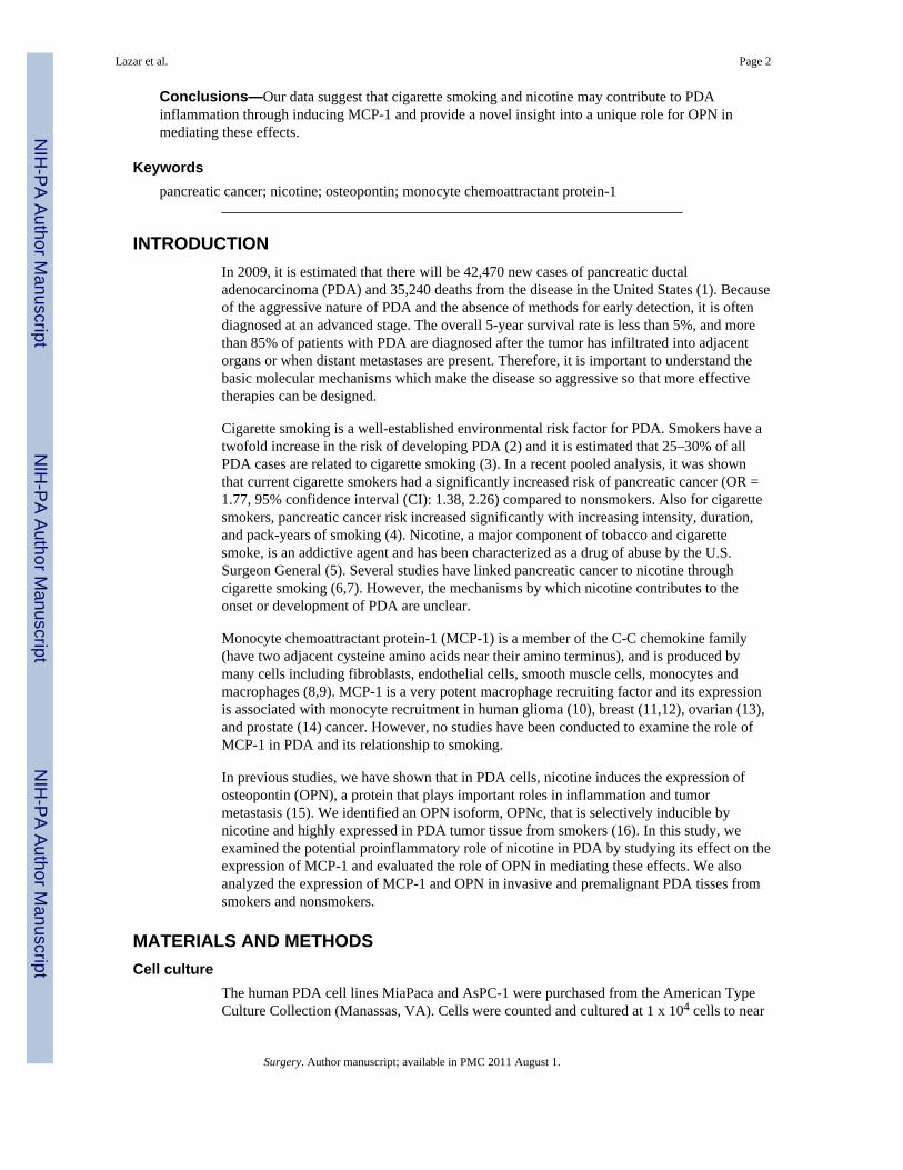
INDUCTION OF MONOCYTE CHEMOATTRACTANT PROTEIN-1 BY NICOTINE IN PANCREATIC DUCTAL ADENOCARCINOMA CELLS: ROLE OF OSTEOPONTIN Melissa Lazar 1 , Jennifer Sullivan 1 , Galina Chipitsyna 1 , Tamer Aziz, Ahmed F Salem, Qiaoke Gong 1 , Agnes Witkiewicz 2 , David T. Denhardt 3 , Charles J. Yeo 1 , and Hwyda A. Arafat 1,2 1 Department of Surgery, Jefferson Pancreatic, Biliary and Related Cancer Center, Thomas Jefferson University, Philadelphia, PA 2 Department of Pathology Anatomy and Cell Biology, Thomas Jefferson University, Philadelphia, PA 3 Department of Cell Biology and Neuroscience, Rutgers University, New Brunswick, NJ Abstract Introduction—Cigarette smoke and nicotine are among the leading environmental risk factors for developing pancreatic ductal adenocarcinoma (PDA). We showed recently that nicotine induces osteopontin (OPN), a protein that plays critical roles in inflammation and tumor metastasis. We identified an OPN isoform, OPNc, that is selectively inducible by nicotine and highly expressed in PDA tissue from smokers. In this study, we explored the potential proinflammatory role of nicotine in PDA through studying its effect on the expression of monocyte chemoattractant protein- (MCP)-1 and evaluated the role of OPN in mediating these effects. Methods—MCP-1 mRNA and protein in PDA cells treated with or without nicotine (3–300 nM) or OPN (0.15–15 nM) were analyzed by real time PCR and ELISA. Luciferase-labeled promoter studies evaluated the effects of nicotine and OPN on MCP-1 transcription. Intracellular and tissue colocalization of OPN and MCP-1 were examined by immunofluorescence and immunohistochemistry. Results—Nicotine treatment significantly increased MCP-1 expression in PDA cells. Interestingly, blocking OPN with siRNA or OPN antibody abolished these effects. Transient transfection of the OPNc gene in PDA cells or their treatment with recombinant OPN protein significantly (P<0.05) increased MCP-1 mRNA and protein and induced its promoter activity. MCP-1 was found in 60% of invasive PDA lesions, of which 66% were smokers. MCP-1 colocalized with OPN in PDA cells and in the malignant ducts, and correlated well with higher expression levels of OPN in the tissue from patients with invasive PDA. Corresponding author: Hwyda A. Arafat, MD, PhD, Department of Surgery, Thomas Jefferson University, 1015 Walnut Street, Suite 618 Curtis Philadelphia, PA 19107, Tel. 215 955-6383, [email protected]. Publisher's Disclaimer: This is a PDF file of an unedited manuscript that has been accepted for publication. As a service to our customers we are providing this early version of the manuscript. The manuscript will undergo copyediting, typesetting, and review of the resulting proof before it is published in its final citable form. Please note that during the production process errors may be discovered which could affect the content, and all legal disclaimers that apply to the journal pertain. NIH Public Access Author Manuscript Surgery. Author manuscript; available in PMC 2011 August 1. Published in final edited form as: Surgery. 2010 August ; 148(2): 298–309. doi:10.1016/j.surg.2010.05.002. NIH-PA Author Manuscript NIH-PA Author Manuscript NIH-PA Author Manuscript
-
Upload
mississippimedical -
Category
Documents
-
view
0 -
download
0
Transcript of Induction of Monocyte Chemoattractant Protein-1 In the Small Veins of the Ischemic and Reperfused...
INDUCTION OF MONOCYTE CHEMOATTRACTANT PROTEIN-1BY NICOTINE IN PANCREATIC DUCTAL ADENOCARCINOMACELLS: ROLE OF OSTEOPONTIN
Melissa Lazar1, Jennifer Sullivan1, Galina Chipitsyna1, Tamer Aziz, Ahmed F Salem,Qiaoke Gong1, Agnes Witkiewicz2, David T. Denhardt3, Charles J. Yeo1, and Hwyda A.Arafat1,21 Department of Surgery, Jefferson Pancreatic, Biliary and Related Cancer Center, ThomasJefferson University, Philadelphia, PA2 Department of Pathology Anatomy and Cell Biology, Thomas Jefferson University, Philadelphia,PA3 Department of Cell Biology and Neuroscience, Rutgers University, New Brunswick, NJ
AbstractIntroduction—Cigarette smoke and nicotine are among the leading environmental risk factorsfor developing pancreatic ductal adenocarcinoma (PDA). We showed recently that nicotineinduces osteopontin (OPN), a protein that plays critical roles in inflammation and tumormetastasis. We identified an OPN isoform, OPNc, that is selectively inducible by nicotine andhighly expressed in PDA tissue from smokers. In this study, we explored the potentialproinflammatory role of nicotine in PDA through studying its effect on the expression ofmonocyte chemoattractant protein- (MCP)-1 and evaluated the role of OPN in mediating theseeffects.
Methods—MCP-1 mRNA and protein in PDA cells treated with or without nicotine (3–300 nM)or OPN (0.15–15 nM) were analyzed by real time PCR and ELISA. Luciferase-labeled promoterstudies evaluated the effects of nicotine and OPN on MCP-1 transcription. Intracellular and tissuecolocalization of OPN and MCP-1 were examined by immunofluorescence andimmunohistochemistry.
Results—Nicotine treatment significantly increased MCP-1 expression in PDA cells.Interestingly, blocking OPN with siRNA or OPN antibody abolished these effects. Transienttransfection of the OPNc gene in PDA cells or their treatment with recombinant OPN proteinsignificantly (P<0.05) increased MCP-1 mRNA and protein and induced its promoter activity.MCP-1 was found in 60% of invasive PDA lesions, of which 66% were smokers. MCP-1colocalized with OPN in PDA cells and in the malignant ducts, and correlated well with higherexpression levels of OPN in the tissue from patients with invasive PDA.
Corresponding author: Hwyda A. Arafat, MD, PhD, Department of Surgery, Thomas Jefferson University, 1015 Walnut Street, Suite618 Curtis Philadelphia, PA 19107, Tel. 215 955-6383, [email protected]'s Disclaimer: This is a PDF file of an unedited manuscript that has been accepted for publication. As a service to ourcustomers we are providing this early version of the manuscript. The manuscript will undergo copyediting, typesetting, and review ofthe resulting proof before it is published in its final citable form. Please note that during the production process errors may bediscovered which could affect the content, and all legal disclaimers that apply to the journal pertain.
NIH Public AccessAuthor ManuscriptSurgery. Author manuscript; available in PMC 2011 August 1.
Published in final edited form as:Surgery. 2010 August ; 148(2): 298–309. doi:10.1016/j.surg.2010.05.002.
NIH
-PA Author Manuscript
NIH
-PA Author Manuscript
NIH
-PA Author Manuscript
Conclusions—Our data suggest that cigarette smoking and nicotine may contribute to PDAinflammation through inducing MCP-1 and provide a novel insight into a unique role for OPN inmediating these effects.
Keywordspancreatic cancer; nicotine; osteopontin; monocyte chemoattractant protein-1
INTRODUCTIONIn 2009, it is estimated that there will be 42,470 new cases of pancreatic ductaladenocarcinoma (PDA) and 35,240 deaths from the disease in the United States (1). Becauseof the aggressive nature of PDA and the absence of methods for early detection, it is oftendiagnosed at an advanced stage. The overall 5-year survival rate is less than 5%, and morethan 85% of patients with PDA are diagnosed after the tumor has infiltrated into adjacentorgans or when distant metastases are present. Therefore, it is important to understand thebasic molecular mechanisms which make the disease so aggressive so that more effectivetherapies can be designed.
Cigarette smoking is a well-established environmental risk factor for PDA. Smokers have atwofold increase in the risk of developing PDA (2) and it is estimated that 25–30% of allPDA cases are related to cigarette smoking (3). In a recent pooled analysis, it was shownthat current cigarette smokers had a significantly increased risk of pancreatic cancer (OR =1.77, 95% confidence interval (CI): 1.38, 2.26) compared to nonsmokers. Also for cigarettesmokers, pancreatic cancer risk increased significantly with increasing intensity, duration,and pack-years of smoking (4). Nicotine, a major component of tobacco and cigarettesmoke, is an addictive agent and has been characterized as a drug of abuse by the U.S.Surgeon General (5). Several studies have linked pancreatic cancer to nicotine throughcigarette smoking (6,7). However, the mechanisms by which nicotine contributes to theonset or development of PDA are unclear.
Monocyte chemoattractant protein-1 (MCP-1) is a member of the C-C chemokine family(have two adjacent cysteine amino acids near their amino terminus), and is produced bymany cells including fibroblasts, endothelial cells, smooth muscle cells, monocytes andmacrophages (8,9). MCP-1 is a very potent macrophage recruiting factor and its expressionis associated with monocyte recruitment in human glioma (10), breast (11,12), ovarian (13),and prostate (14) cancer. However, no studies have been conducted to examine the role ofMCP-1 in PDA and its relationship to smoking.
In previous studies, we have shown that in PDA cells, nicotine induces the expression ofosteopontin (OPN), a protein that plays important roles in inflammation and tumormetastasis (15). We identified an OPN isoform, OPNc, that is selectively inducible bynicotine and highly expressed in PDA tumor tissue from smokers (16). In this study, weexamined the potential proinflammatory role of nicotine in PDA by studying its effect on theexpression of MCP-1 and evaluated the role of OPN in mediating these effects. We alsoanalyzed the expression of MCP-1 and OPN in invasive and premalignant PDA tisses fromsmokers and nonsmokers.
MATERIALS AND METHODSCell culture
The human PDA cell lines MiaPaca and AsPC-1 were purchased from the American TypeCulture Collection (Manassas, VA). Cells were counted and cultured at 1 x 10⁴ cells to near
Lazar et al. Page 2
Surgery. Author manuscript; available in PMC 2011 August 1.
NIH
-PA Author Manuscript
NIH
-PA Author Manuscript
NIH
-PA Author Manuscript
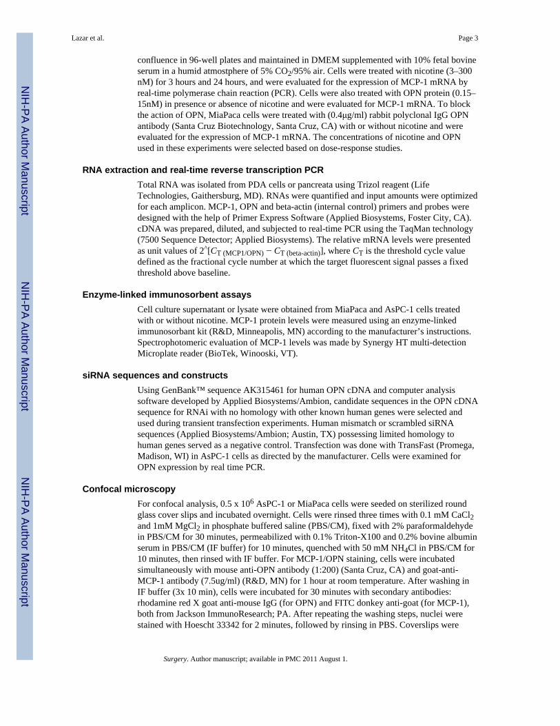
confluence in 96-well plates and maintained in DMEM supplemented with 10% fetal bovineserum in a humid atmostphere of 5% CO2/95% air. Cells were treated with nicotine (3–300nM) for 3 hours and 24 hours, and were evaluated for the expression of MCP-1 mRNA byreal-time polymerase chain reaction (PCR). Cells were also treated with OPN protein (0.15–15nM) in presence or absence of nicotine and were evaluated for MCP-1 mRNA. To blockthe action of OPN, MiaPaca cells were treated with (0.4μg/ml) rabbit polyclonal IgG OPNantibody (Santa Cruz Biotechnology, Santa Cruz, CA) with or without nicotine and wereevaluated for the expression of MCP-1 mRNA. The concentrations of nicotine and OPNused in these experiments were selected based on dose-response studies.
RNA extraction and real-time reverse transcription PCRTotal RNA was isolated from PDA cells or pancreata using Trizol reagent (LifeTechnologies, Gaithersburg, MD). RNAs were quantified and input amounts were optimizedfor each amplicon. MCP-1, OPN and beta-actin (internal control) primers and probes weredesigned with the help of Primer Express Software (Applied Biosystems, Foster City, CA).cDNA was prepared, diluted, and subjected to real-time PCR using the TaqMan technology(7500 Sequence Detector; Applied Biosystems). The relative mRNA levels were presentedas unit values of 2^[CT (MCP1/OPN) − CT (beta-actin)], where CT is the threshold cycle valuedefined as the fractional cycle number at which the target fluorescent signal passes a fixedthreshold above baseline.
Enzyme-linked immunosorbent assaysCell culture supernatant or lysate were obtained from MiaPaca and AsPC-1 cells treatedwith or without nicotine. MCP-1 protein levels were measured using an enzyme-linkedimmunosorbant kit (R&D, Minneapolis, MN) according to the manufacturer’s instructions.Spectrophotomeric evaluation of MCP-1 levels was made by Synergy HT multi-detectionMicroplate reader (BioTek, Winooski, VT).
siRNA sequences and constructsUsing GenBank™ sequence AK315461 for human OPN cDNA and computer analysissoftware developed by Applied Biosystems/Ambion, candidate sequences in the OPN cDNAsequence for RNAi with no homology with other known human genes were selected andused during transient transfection experiments. Human mismatch or scrambled siRNAsequences (Applied Biosystems/Ambion; Austin, TX) possessing limited homology tohuman genes served as a negative control. Transfection was done with TransFast (Promega,Madison, WI) in AsPC-1 cells as directed by the manufacturer. Cells were examined forOPN expression by real time PCR.
Confocal microscopyFor confocal analysis, 0.5 x 10⁶ AsPC-1 or MiaPaca cells were seeded on sterilized roundglass cover slips and incubated overnight. Cells were rinsed three times with 0.1 mM CaCl2and 1mM MgCl2 in phosphate buffered saline (PBS/CM), fixed with 2% paraformaldehydein PBS/CM for 30 minutes, permeabilized with 0.1% Triton-X100 and 0.2% bovine albuminserum in PBS/CM (IF buffer) for 10 minutes, quenched with 50 mM NH4Cl in PBS/CM for10 minutes, then rinsed with IF buffer. For MCP-1/OPN staining, cells were incubatedsimultaneously with mouse anti-OPN antibody (1:200) (Santa Cruz, CA) and goat-anti-MCP-1 antibody (7.5ug/ml) (R&D, MN) for 1 hour at room temperature. After washing inIF buffer (3x 10 min), cells were incubated for 30 minutes with secondary antibodies:rhodamine red X goat anti-mouse IgG (for OPN) and FITC donkey anti-goat (for MCP-1),both from Jackson ImmunoResearch; PA. After repeating the washing steps, nuclei werestained with Hoescht 33342 for 2 minutes, followed by rinsing in PBS. Coverslips were
Lazar et al. Page 3
Surgery. Author manuscript; available in PMC 2011 August 1.
NIH
-PA Author Manuscript
NIH
-PA Author Manuscript
NIH
-PA Author Manuscript
mounted with ProLong Gold anti-fade (Molecular Probes, OR) and left overnight in thedark. The laser confocal microscope LSM 510 microscope (Carl Zeiss GmbH, Thornwood,NY) was used to image the cells in the respective channels at a magnification of 60X.
Transient Transfection of OPNcpDest-490 vector containing a truncated splice variant OPN-c (base pairs 1–93, 175–942)was a generous gift from Dr. X Wang, Center for Cancer Research, NCI, Bethesda, MD(17). MiaPaca cells were transfected with 0.5 μg OPNc plasmid DNA using TransFast(Promega, Madison, WI), and lysates were harvested after 24 h for initial real time PCRtesting of expression of the OPNc. For subsequent experiments to determine the levels ofMCP-1 by real time PCR, MiaPaca cells (1X 10⁶) were transfected with 0.5 μg OPNcplasmid DNA using optimized nucleofection conditions (60–80% efficiency by pGEM4/enhanced green fluorescent protein (EGFP) visualization). We determined the levels ofMCP-1 by real time PCR after 3 and 24 h of transfection.
ELISAHuman OPN concentration in the cell culture media were measured using human-specificELISA kit (Assay Design, Ann Arbor, MI). Spectrophotometric evaluation of OPN levelswere made by Synergy HT multi-detection microplate reader (BioTeck, Winooski, VT).
Promoter studiesTo evaluate the effect of nicotine and OPN on MCP-1 transcription, we used the MCP-1gene promoter (GenBankTM accession number AF079313) in luciferase expression vectorpGL3 basic (Promega), kindly provided by Dr. Decio Ezirik, Free University Brussels,Belgium (18). Cells were seeded into 24-well culture plates (105). At ~80% confluence theywere co transfected by TransFast reagent (Promega) and 0.5 μg of pGL3 vectors containingthe luciferase-labeled MCP-1 promoter and 0.1 μg of green fluorescence protein (GFP) astransfection control. Two hours later, serum-containing medium was overlaid and the cellswere incubated for an additional 24 h. The cells then were incubated with serum freemedium for 18 h, after which nicotine or OPN were added for 3h. Luciferase activities wereassayed with the Dual-Luciferase Reporter Assay System (Promega) in a Veritas MicroplateLuminometer (Turner Designs, Sunnyvale, Calif., USA). Transfection efficiency wasnormalized using the total protein concentration of the cell lysates. The results for nicotine-treated cells were expressed as a fold-induction of the luciferase activity of the sameconstruct in the control condition, taking the control (no treatment) value as 100.
Human Tissue Acquisition and AnalysisHuman PDA (n=73) and premalignant specimens (n=6) and normal pancreatic tissue wereobtained from patients who underwent surgical resection at Thomas Jefferson UniversityHospital between 2005 and 2007. All patients signed an appropriate consent for tissueacquisition and study. The study was approved by the Institutional Review Board of ThomasJefferson University. Patients’ smoking history was examined and correlated with MCP-1and OPN expression levels.
Tissue samples were stored in RNA Later for RNA analysis or fixed in neutral formaline forhistological processing. Sections at 5 μm were stained with H&E. To localize MCP-1 andOPN, sections from the different tissues were analyzed by immunohistochemistry using theOPN and MCP-1 antibodies. A vectastain universal elite ABC kit and 3,3'-diaminobenzidinetetrahydrochloride chromogenic substrate (Vector Laboratories Inc.) was used according tothe manufacturer protocol to visualize the tissue reaction. Antibody specificity was validated
Lazar et al. Page 4
Surgery. Author manuscript; available in PMC 2011 August 1.
NIH
-PA Author Manuscript
NIH
-PA Author Manuscript
NIH
-PA Author Manuscript
with nonimmune isotype serum. Negative control sections, where the primary or secondaryantibodies were omitted were also prepared.
Statistical analysesAll experiments were performed 3 to 5 times. Data were analyzed for statistical significanceby ANOVA with post-hoc Student t test analysis. Data are presented as mean ± SEM.Continuous, normally distributed variables were analyzed by Student-t-test. Spearman's rankcorrelation test was performed to analyze the correlation between OPN, and MCP-1 mRNAsexpression. Analyses were performed with the assistance of a computer program (JMP 5Software SAS Campus Drive, Cary, NC). Differences were considered significant at P ≤0.05.
RESULTSNicotine stimulates MCP-1 mRNA accumulation and protein secretion in cultured PDAcells
PDA cells express different basal levels of OPN (15,16). To investigate whether nicotine candirectly increase MCP-1 mRNA accumulation in PDA cells, we used MiaPaca and AsPC-1cells, which express low and high levels of basal OPN, respectively. Cells were treated withor without nicotine (3–300 nM) for 3 and 24 h. Significant induction of MCP-1 mRNAexpression was seen with a maximum increase at 24 h in MiaPaca cells. (Fig 1A). InAsPC-1 cells, only the higher doses of nicotine (30 and 300 nM) significantly increasedMCP-1 mRNA levels after 3 h of nicotine stimulation. To examine whether the increase inMCP-1 mRNA levels in response to nicotine is associated with MCP-1 production, MCP-1protein levels in the media were determined by ELISA. MCP-1 levels were increased byalmost 3-fold after 48h of nicotine stimulation in MiaPaca cells (Fig 2A). In AsPC-1 cells,MCP-1 protein concentration increased by more than 2-fold after 24 hours then levelsreturned to basal levels by 48 hours after nicotine treatment (Fig 2B). These data indicatethat MCP-1 induction by nicotine is a general phenomenon seen in the tested PDA cellslines.
Nicotine does not induce MCP-1 promoter activityHowever, nicotine did not induce a significant increase in MCP-1 promoter activity when itwas added (3–300 nM) to PDA cells that were transfected with luciferase-labeled MCP-1promoter for 1, 3, 6,12, 24 and 48h (data not shown). This suggested that MCP-1 promoterdoes not respond directly to nicotine. Since we showed previously that nicotine directlyinduces OPN transcription in PDA cells (15,16), and since OPN was shown to increaseMCP-1 expression in other cells (19), we tested the hypothesis that OPN mediates theupregulation of MCP-1 mRNA by nicotine.
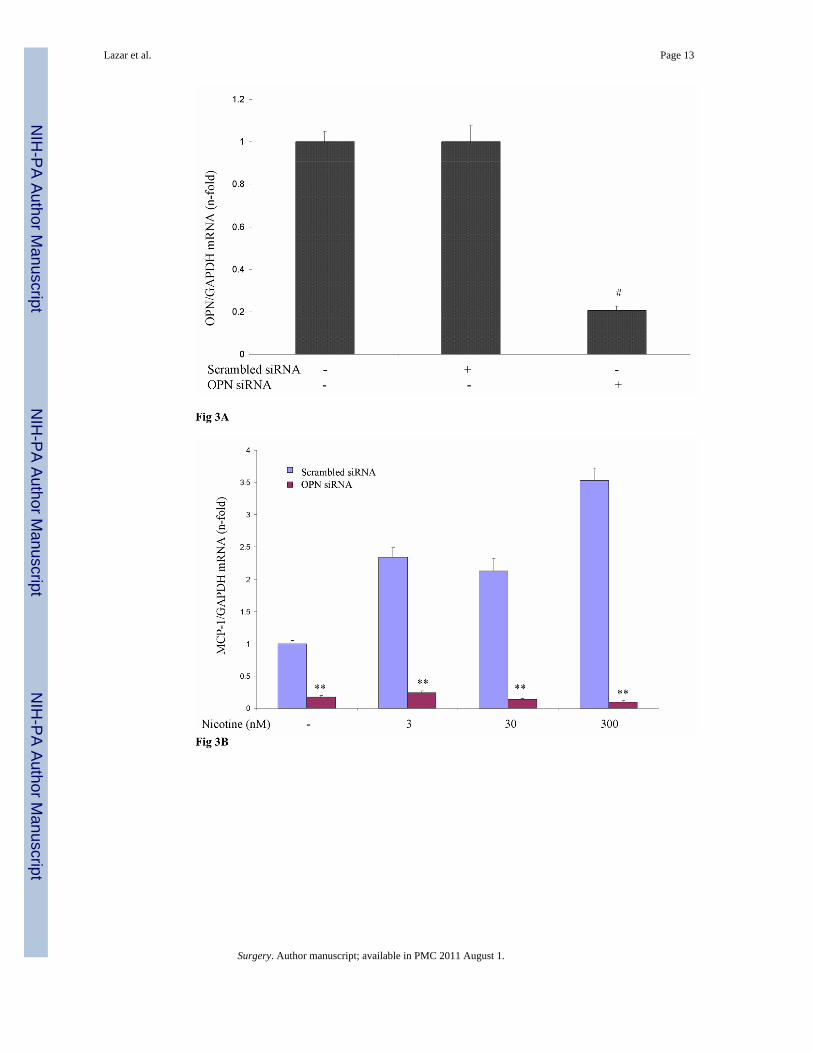
RNAi decreases OPN expression and reduce MCP-1-mediated upregulation of nicotineAsPC-1 cells are highly invasive and normally constitutively express OPN at high levels. Tosuppress OPN expression in AsPC-1 cells, we selected two 21-nt targets within the OPNcDNA for RNAi. Based on these targets, double-stranded 21-nt siRNA constructs weredesigned encoding sense and antisense siRNA, and the OPN levels were measured usingreal time PCR. As shown in Fig 3A, OPN mRNA expression level was significantlyinhibited by ~ 80%. We added nicotine (3–300 nM) to these cells and evaluated MCP-1expression by real time PCR. As seen in Fig 3B, addition of nicotine was unable to elicit anincrease in MCP-1 mRNA. It also was apparent that cells lacking OPN also expressed lessendogenous MCP-1 mRNA. These data indicate that there is a relationship between thepresence of intracellular OPN and the cell response to nicotine to produce MCP-1.
Lazar et al. Page 5
Surgery. Author manuscript; available in PMC 2011 August 1.
NIH
-PA Author Manuscript
NIH
-PA Author Manuscript
NIH
-PA Author Manuscript
To confirm our data in MiaPaca cells, we blocked OPN function by adding OPN rabbitpolyclonal antibody (0.4 μg/ml) 1 h before addition of nicotine (30 nM) for 24 h. As seen infigure 3C, blocking OPN resulted in complete prevention of the nicotine-mediated increasein MCP-1 mRNA. Cells that express normal levels of OPN expressed higher levels of basalMCP-1 when compared with cells that express no OPN.
This indicates that the presence of OPN in PDA cells is essential for the nicotine-mediatedinduction of MCP-1. Next, we asked whether OPN itself could induce MCP-1 transcriptionin PDA cells.
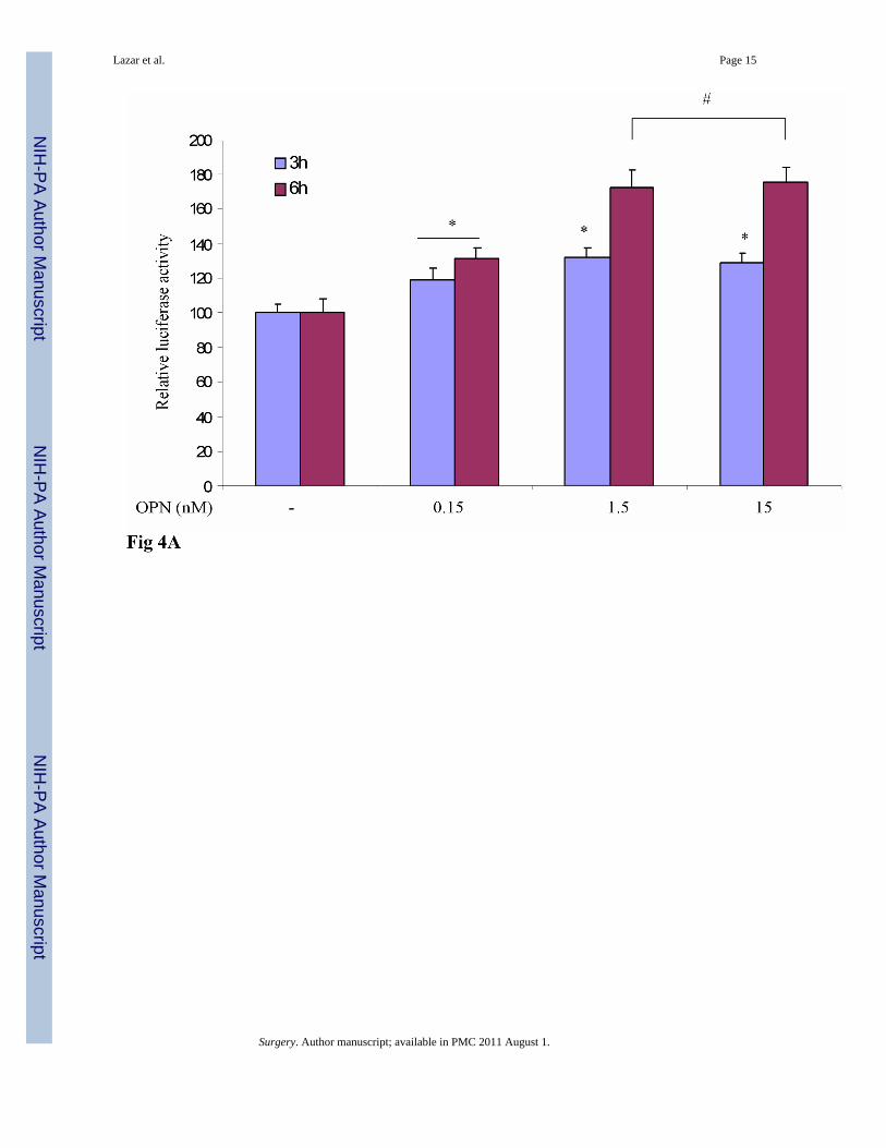
OPN induces MCP-1 transcription in PDA cellsRecombinant OPN protein (0.15–15 nM) was added for 3 and 6 h to MiaPaca cells that weretransfected with MCP-1 luciferase-labeled promoter. OPN significantly and dose-dependently activated MCP-1 promoter (Fig 4A). In other experiments, OPNc plasmid wastransiently transfected into MiaPaca cells. Real time PCR and ELISA confirmedoverexpression of OPN (Fig 4B). Real time PCR analysis showed more than 3-fold increasein MCP-1 mRNA levels in the cells that overexpress OPNc vs. control cells (Fig 4C).Interestingly, addition of nicotine to OPNc overexpressing PDA cells induced furtherupregulation of MCP-1 mRNA (Fig 4C), suggesting that initial stimulation of OPN isnecessary for further upregulation of MCP-1 by nicotine. Next, we analyzed the localizationof OPN and MCP-1 in PDA cells in vitro
Colocalization of OPN and MCP-1 in PDA cellsConfocal microscopy after immunofluorescence staining of MiaPaca cells (Fig 5A) andASPC-1 cells (Fig 5B) with antibodies against OPN and MCP-1 show colocalization of bothproteins in the cytosol of PDA cells. OPN (red) was expressed in the form of granules thatcould be localized in the cytosol and cell membrane of PDA cells, whereas MCP-1 showed amore homogenous pattern and was present in the cytosol. We next examined theendogenous levels and localization of MCP-1 and OPN in premalignant and malignant PDAlesions.
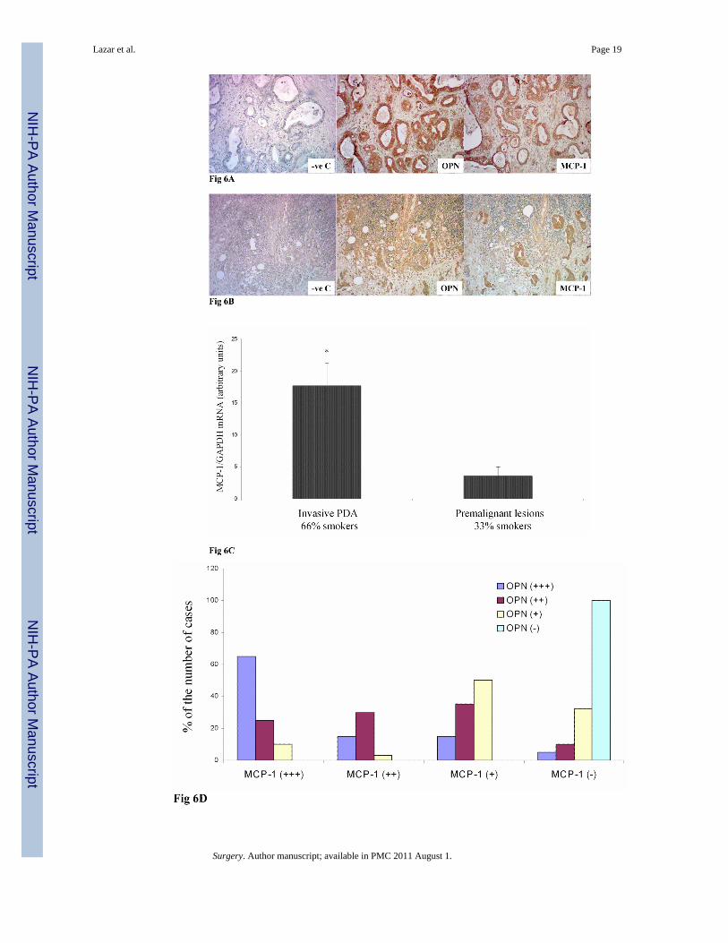
Expression of OPN in human PDAUsing immunohistochemical staining, we found that MCP-1 was absent from the normalpancreatic ducts, whereas OPN was focally present, mostly on the apical surface of theductal epithelium (15). In PDA (Fig 6A), OPN ductal epithelial staining was intensified andlocalized to the cell membrane and cytoplasm of the tumor cells. MCP-1 colocalized withOPN in the malignant ducts. Interestingly, both MCP-1 and OPN were also intensely stainedin the metastatic lesions in the lymph nodes (Fig 6B).
Analysis of quantitative PCR data of MCP-1 mRNA corrected with GAPDH as an internalcontrol, invasive PDA lesions showed significantly higher levels of MCP-1 mRNA whencompared to IPMN premalignant lesions (Fig 6C). Interestingly, only 33% of the patientswith premalignant lesions were smokers (2 of 6), whereas of the examined malignantlesions, 66% belonged to patients who were smokers (48 of 73) (Fig 6C).
To correlate the expression levels of MCP-1 and OPN in PDA tissue, the level of mRNA foreach gene was recorded. Relative quantification (RQ) values of MCP-1 and OPN /GAPDHof >1 indicated high levels and were labeled (+++), values of 0.5 – 1 were labeled (++), of0.1-0.5 were labeled (+), and of <0.1 was labeled (−). Expression of MCP-1 correlated wellwith OPN expression levels (Fig 6D). These data suggest that increased OPN expression insmokers is associated with the expression of MCP-1.
Lazar et al. Page 6
Surgery. Author manuscript; available in PMC 2011 August 1.
NIH
-PA Author Manuscript
NIH
-PA Author Manuscript
NIH
-PA Author Manuscript
DISCUSSIONPDA is an aggressive disease that has a strong etiological association with cigarette smokingand high blood levels of nicotine (2–4). MCP-1 is the most frequently found CC chemokinein several tumors, including those of the pancreas (10–14,20,21). Although several studiesinvestigated the role of MCP-1 in cancer progression, very few have investigated itsrelationship to smoking and the upstream factors involved in its regulation in PDA cells. Inthis study, we show for the first time that nicotine treatment both dose- and time-dependently increased MCP-1 expression in PDA cells. We also demonstrate a previouslyundescribed role for OPN as a mediator for these effects.
Our data show that nicotine induced MCP-1 accumulation rapidly in PDA cells and withsignificant magnitude. Dose-response studies demonstrated a significant induction ofMCP-1 mRNA and protein levels at the physiological range of blood levels of nicotine insmokers (3–300 nM). The maximal effective concentration (at 30 nM) is similar to othernicotine actions that have been reported (22).
Our data show that the MCP-1 promoter was not activated by nicotine when it was added at(3–300 nM) to PDA cells at different time points. Since we showed previously that nicotinedirectly induces OPN transcription in PDA cells (15,16), and since OPN was shown toincrease MCP-1 expression in other cells (19), we tested the hypothesis that OPN mediatesthe upregulation of MCP-1 by nicotine.
We conducted four sets of experiments to evaluate the role of OPN. First, we inhibited OPNsynthesis by siRNA (Fig 3A,B) or blocked its function by a polyclonal antibody againsthuman OPN (Fig 3C). In both studies, nicotine was unable to increase MCP-1 expression inPDA cells, suggesting that OPN may play a role in mediating the effects of nicotine. Next,we treated the PDA cells with recombinant human OPN protein (Fig 4A) or overexpressedan OPN isoform (OPNc) (Fig 4B), which has been shown to promote metastasis in cancercells (23,24). In both experiments, we show that increased constitutive OPN or addition ofexogenous OPN allowed the increase of MCP-1 expression by nicotine (Fig 4C).Furthermore, OPN also increased the basal levels of MCP-1 expression in PDA cells (Fig4C) and increased its transcription by activating its promoter activity (Fig 4A). Previousstudies have shown that nicotine induces the expression of total OPN (15) and selectivelyinduce the expression of OPNc in PDA cells (16). This is the first report to demonstrate arelationship between nicotine, OPN/OPNc and MCP-1. Confocal microscopy analysis alsorevealed intracytoplasmic colocalization of OPN and MCP-1 in PDA cells (Fig 5), providingmore evidence for the paracrine/autocrine relationship between the two molecules.Additional studies are now required to delineate the details of this relationship and thesignaling pathways involved in mediating the increase of MCP-1 by OPN. Furthermore, theeffect of the nicotine-mediated increase of OPNc on PDA cell inflammatory behavior andfunction is the subject of our currently ongoing studies in the laboratory.
Numerous studies have correlated high levels of MCP-1 expression with tumor progressionin many cancers, including pancreatic cancer (20,21). MCP-1 promotes cell survival (25)and induces the expression of vascular endothelial growth factor (26). These functions aresimilar to those reported to be mediated by OPN (27,28). Our in vitro data suggest that OPNmight be acting upstream of MCP-1 to mediate these effects.
Studies in colorectal cancer have correlated higher levels of MCP-1 with tumor stage andhigh metastatic potential (26). Our analyses reported here have found that MCP-1 was foundin 60% of invasive PDA lesions, of which 66% were smokers. This is the first report toexamine the relationship between tumor MCP-1 and the status of smoking in PDA patients.Our analysis also reveals that higher levels of MCP-1 were seen in invasive PDA as
Lazar et al. Page 7
Surgery. Author manuscript; available in PMC 2011 August 1.
NIH
-PA Author Manuscript
NIH
-PA Author Manuscript
NIH
-PA Author Manuscript
compared to premalignant lesions. Immunohistochemical analysis of PDA showed thatMCP-1 colocalizes with OPN in the malignant ducts and its mRNA levels correlated wellwith higher expression levels of OPN in the tissue from patients with invasive PDA (Fig 6).It remains to be determined, however, whether MCP-1 levels correlate with pathologicstage, survival or recurrence. Furthermore, additional studies are required to determinewhether similar findings could be obtained from Endoscopic Ultrasound and Fine NeedleAspiration (EUS FNA) samples.
Interestingly, our studies show that high levels of MCP-1 and OPN exist in invasive lesionsfrom non-smokers. Factors such as second hand smoke could contribute to this. In addition,a previous history of pancreatic inflammation (pancreatitis), which has been linked topancreatic carcinogenesis (29,30) could create a tumor microenvironment with higher levelsof OPN and MCP-1. Additional studies addressing these possibilities are currently ongoingin our laboratory.
In summary, our study suggests that cigarette smoking and nicotine may contribute to PDAinflammation through inducing MCP-1 and provides a novel insight into a unique role forOPN in mediating these effects. Although the role of OPN-mediated induction of MCP-1 inpancreatic carcinogenesis remains to be defined, the existence of OPN as a downstreameffector of nicotine, capable of mediating proinflammatory effects in PDA cells is novel andcould provide a unique potential target to control pancreatic cancer aggressiveness,especially in the cigarette smoking population.
AcknowledgmentsThis work was supported by NIH grant 1R21 CA133753-02
References1. Pancreatic Cancer homepage. National Cancer Institute; [Online]
http://www.cancer.gov/cancertopics/types/pancreatic2. Wang XL, Wang J. Smoking-gene interaction and disease development: relevance to pancreatic
cancer and atherosclerosis. World Journal of Surgery 2005;29:344–53. [PubMed: 15696395]3. Lowenfels AB, Maisonneuve P. Epidemiology and risk factors for pancreatic cancer. Best Prac Res
Clin Gastroenterology 2006;20:197–209.4. Lynch SM, Vrieling A, Lubin JH, Kraft P, Mendelsohn JB, Hartge P, et al. Cigarette smoking and
pancreatic cancer: a pooled analysis from the pancreatic cancer cohort consortium. Am J Epidemiol2009;170:403–13. [PubMed: 19561064]
5. US Department of Health and Human Services. The health consequences of using smokelesstobacco: a report of the advisory committee to the Surgeon General [NIH Publication #86-2874].Bethesda, MD: USDHHS; Apr. 1996
6. Farrow DC, Davis S. Risk of pancreatic cancer in relation to medical history and use of tobacco,alcohol and coffee. Intl J Cancer 1990;45:816–20.
7. Silverman DT, Dunn JA, Hoover RN, Schiffman M, Lillemoe KD, Schoenberg JB, et al. Cigarettesmoking and pancreas cancer: a case control study based on direct interviews. J Natl Cancer Inst1994;86:1510–6. [PubMed: 7932805]
8. Oppenheim JJ, Zacchariae CO, Mukaida N, Matsushima K. Properties of the novel proinflammatorysupergene “intercrine” cytokine family. Annu Rev Immunol 1991;9:617–48. [PubMed: 1910690]
9. Gu L, Tseng C, Rollins BJ. Monocyte chemoattractant protein-1. Chem Immunol 1999;72:7–29.[PubMed: 10550927]
10. Takeshima H, Kuratsu J, Takeya M, Yoshimura T, Ushio Y. Expression and localization ofmessenger RNA and protein for monocyte chemoattractant protein-1 in human malignant glioma.J Neurosurg 1994;80:1056–62. [PubMed: 8189261]
Lazar et al. Page 8
Surgery. Author manuscript; available in PMC 2011 August 1.
NIH
-PA Author Manuscript
NIH
-PA Author Manuscript
NIH
-PA Author Manuscript
11. Ueno T, Toi M, Saji H, Muta M, Bando H, Kuroi K, Koike M, Inadera H, Matsushima K.Significance of macrophage chemoattractant protein-1 in macrophage recruitment, angiogenesisand survival in human breast cancer. Clin Cancer Res 2000;6:3282–9. [PubMed: 10955814]
12. Goede V, Brogelli L, Ziche M, Augustin HG. Induction of inflammatory angiogenesis bymonocyte chemoattractant protein-1. Int J Cancer 1999;82:765–70. [PubMed: 10417778]
13. Negus RP, Stamp GW, Hadley J, Balkwill FR. Quantitative assessment of the leukocyte infiltratein ovarian cancer and its relationship to the expression of C-C chemokines. Am J Pathol1997;50:1723–34. [PubMed: 9137096]
14. Mazzucchelli L, Loetscher P, Kappeler A, Uguccioni M, Baggiolini M, Laissue JA, Mueller C.Monocyte chemoattractant protein-1 gene expression in prostatic hyperplasia and prostateadenocarcinoma. Am J Pathol 1996;149:501–9. [PubMed: 8701989]
15. Chipitsyna G, Gong Q, Anandanadesan R, Alnajar A, Batra SK, Denhardt DT, et al. Induction ofosteopontin (OPN) expression by nicotine and cigarette smoke in the pancreas and pancreaticductal adenocarcinoma cells. Int J Cancer 2009;125:276–85. [PubMed: 19358273]
16. Sullivan J, Blair L, Alnajar A, Aziz T, Ng CY, et al. Expression of a prometastatic splice variant ofosteopontin, OPNc, in human pancreatic ductal adenocarcinoma. Surgery 2009;146:232–240.[PubMed: 19628079]
17. Takafuji V, Forgues M, Unsworth E, Goldsmith P, Wang XW. An osteopontin fragment isessential for tumor cell invasion in hepatocellular carcinoma. Oncogene 2007;26(44):6361–71.[PubMed: 17452979]
18. Kutlu B, Darville MI, Cardozo AK, Eizirik DL. Molecular regulation of monocyte chemoattractantprotein-1 expression in pancreatic beta-cells. Diabetes 2003;52(2):348–55. [PubMed: 12540607]
19. Zheng W, Li R, Pan H, He D, Xu R, Guo TB, Guo Y, Zhang J. Role of osteopontin in induction ofmonocyte chemoattractant protein 1 and macrophage inflammatory protein 1β through the NF-κBand MAPK pathways in rheumatoid arthritis. Arthritis and Rheumatism 2009;60:1957–1965.[PubMed: 19565503]
20. Bottazzi B, Polentarutti N, Acero R, Balsari A, Boraschi D, Ghezzi P, Salmona M, Mantovani A.Regulation of the macrophage content of neoplasms by chemoattractants. Science (Wash DC)1983;220:210–212.
21. Monti P, Leone BE, Marchesi F, Balzano G, Zerbi A, Scaltrini F, Pasquali C, Calori G, Pessi F,Sperti C, Di Carlo V, Allavena P, Piemonti L. The CC chemokine MCP-1/CCL2 in pancreaticcancer progression: regulation of expression and potential mechanisms of Antimalignant activity.Cancer Research 2003;63:7451–7461. [PubMed: 14612545]
22. Hukkanen J, Jacob P 3rd, Benowitz NL. Metabolism and disposition kinetics of nicotine.Pharmacol Rev 2005;57:79–115. [PubMed: 15734728]
23. Takafuji V, Forgues M, Unsworth E, Goldsmith P, Wang XW. An osteopontin fragment isessential for tumor cell invasion in hepatocellular carcinoma. Oncogene 2007;26:6361–71.[PubMed: 17452979]
24. Mirza M, Shaughnessy E, Hurley JK, Vanpatten KA, Pestano GA, He B, et al. Osteopontin-c is aselective marker of breast cancer. Int J Cancer 2008;122:889–897. [PubMed: 17960616]
25. Salcedo R, Ponce ML, Young HA, Wasserman K, Ward JM, Kleinman HK, Oppenheim JJ,Murphy WJ. Human endothelial cells express CCR2 and respond to MCP-1: direct role of MCP-1in angiogenesis and tumor progression. Blood 2009;96:34–40.
26. Yoshidome H, Kohno H, Shida T, Kimura F, Shimizu H, Ohtsuka M, Nakatani Y, Miyazaki M.Significance of monocyte chemoattractant protein-1 in angiogenesis and survival in colorectalliver metastates. Int J Oncology 2009;34:923–930.
27. Denhardt DT, Noda M, O'Regan AW, Pavlin D, Berman JS. Osteopontin as a means to cope withenvironmental insults: regulation of inflammation, tissue remodeling, and cell survival. J ClinInvest 2001;107:1055–61. [PubMed: 11342566]
28. Chakraborty G, Jain S, Kundu GC. Osteopontin promotes vascular endothelial growth factor-dependent breast tumor growth and angiogenesis via autocrine and paracrine mechanisms. CancerRes 2008;68:152–61. [PubMed: 18172307]
29. Rocca G, Gaia E, Iuliano R. Increased incidence of cancer in chronic pancreatitis. J ClinGastroenterol 1987;9:175–179. [PubMed: 3571892]
Lazar et al. Page 9
Surgery. Author manuscript; available in PMC 2011 August 1.
NIH
-PA Author Manuscript
NIH
-PA Author Manuscript
NIH
-PA Author Manuscript

30. Maisonnneuve P, Lowenfels AB. Chronic pancreatitis and pancreatic cancer. Dig Dis 2002;20:32–37.
Lazar et al. Page 10
Surgery. Author manuscript; available in PMC 2011 August 1.
NIH
-PA Author Manuscript
NIH
-PA Author Manuscript
NIH
-PA Author Manuscript
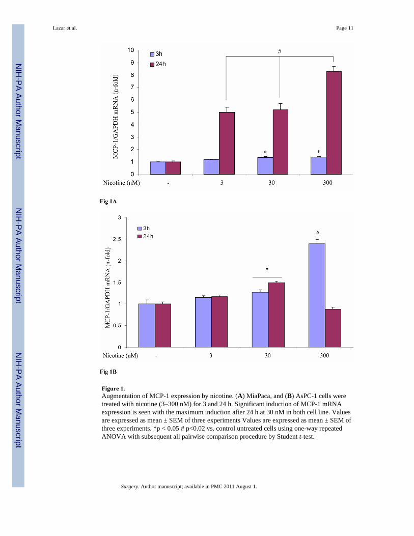
Figure 1.Augmentation of MCP-1 expression by nicotine. (A) MiaPaca, and (B) AsPC-1 cells weretreated with nicotine (3–300 nM) for 3 and 24 h. Significant induction of MCP-1 mRNAexpression is seen with the maximum induction after 24 h at 30 nM in both cell line. Valuesare expressed as mean ± SEM of three experiments Values are expressed as mean ± SEM ofthree experiments. *p < 0.05 # p<0.02 vs. control untreated cells using one-way repeatedANOVA with subsequent all pairwise comparison procedure by Student t-test.
Lazar et al. Page 11
Surgery. Author manuscript; available in PMC 2011 August 1.
NIH
-PA Author Manuscript
NIH
-PA Author Manuscript
NIH
-PA Author Manuscript
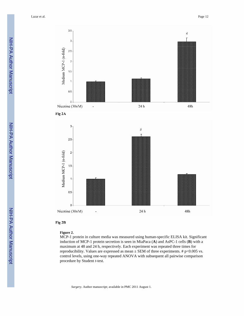
Figure 2.MCP-1 protein in culture media was measured using human-specific ELISA kit. Significantinduction of MCP-1 protein secretion is seen in MiaPaca (A) and AsPC-1 cells (B) with amaximum at 48 and 24 h, respectively. Each experiment was repeated three times forreproducibility. Values are expressed as mean ± SEM of three experiments. # p<0.005 vs.control levels, using one-way repeated ANOVA with subsequent all pairwise comparisonprocedure by Student t-test.
Lazar et al. Page 12
Surgery. Author manuscript; available in PMC 2011 August 1.
NIH
-PA Author Manuscript
NIH
-PA Author Manuscript
NIH
-PA Author Manuscript
Lazar et al. Page 13
Surgery. Author manuscript; available in PMC 2011 August 1.
NIH
-PA Author Manuscript
NIH
-PA Author Manuscript
NIH
-PA Author Manuscript
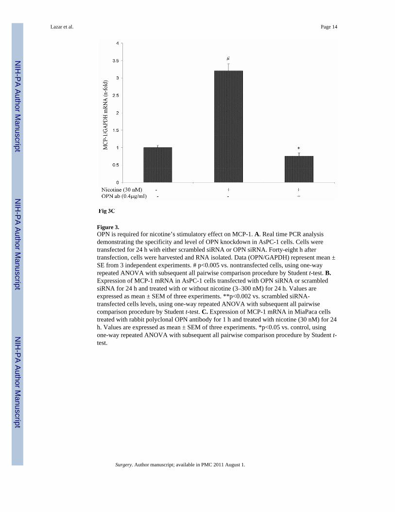
Figure 3.OPN is required for nicotine’s stimulatory effect on MCP-1. A. Real time PCR analysisdemonstrating the specificity and level of OPN knockdown in AsPC-1 cells. Cells weretransfected for 24 h with either scrambled siRNA or OPN siRNA. Forty-eight h aftertransfection, cells were harvested and RNA isolated. Data (OPN/GAPDH) represent mean ±SE from 3 independent experiments. # p<0.005 vs. nontransfected cells, using one-wayrepeated ANOVA with subsequent all pairwise comparison procedure by Student t-test. B.Expression of MCP-1 mRNA in AsPC-1 cells transfected with OPN siRNA or scrambledsiRNA for 24 h and treated with or without nicotine (3–300 nM) for 24 h. Values areexpressed as mean ± SEM of three experiments. **p<0.002 vs. scrambled siRNA-transfected cells levels, using one-way repeated ANOVA with subsequent all pairwisecomparison procedure by Student t-test. C. Expression of MCP-1 mRNA in MiaPaca cellstreated with rabbit polyclonal OPN antibody for 1 h and treated with nicotine (30 nM) for 24h. Values are expressed as mean ± SEM of three experiments. *p<0.05 vs. control, usingone-way repeated ANOVA with subsequent all pairwise comparison procedure by Student t-test.
Lazar et al. Page 14
Surgery. Author manuscript; available in PMC 2011 August 1.
NIH
-PA Author Manuscript
NIH
-PA Author Manuscript
NIH
-PA Author Manuscript
Lazar et al. Page 15
Surgery. Author manuscript; available in PMC 2011 August 1.
NIH
-PA Author Manuscript
NIH
-PA Author Manuscript
NIH
-PA Author Manuscript
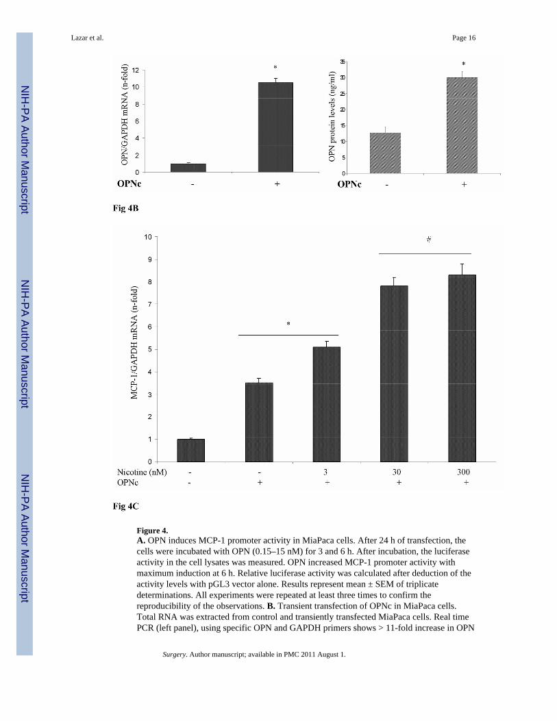
Figure 4.A. OPN induces MCP-1 promoter activity in MiaPaca cells. After 24 h of transfection, thecells were incubated with OPN (0.15–15 nM) for 3 and 6 h. After incubation, the luciferaseactivity in the cell lysates was measured. OPN increased MCP-1 promoter activity withmaximum induction at 6 h. Relative luciferase activity was calculated after deduction of theactivity levels with pGL3 vector alone. Results represent mean ± SEM of triplicatedeterminations. All experiments were repeated at least three times to confirm thereproducibility of the observations. B. Transient transfection of OPNc in MiaPaca cells.Total RNA was extracted from control and transiently transfected MiaPaca cells. Real timePCR (left panel), using specific OPN and GAPDH primers shows > 11-fold increase in OPN
Lazar et al. Page 16
Surgery. Author manuscript; available in PMC 2011 August 1.
NIH
-PA Author Manuscript
NIH
-PA Author Manuscript
NIH
-PA Author Manuscript

mRNA. ELISA analysis of culture media from these cells (right panel) shows significantincrease in OPN protein expression in the media. * p < 0.05 vs. control cells. C. Nicotineinduces higher levels of MCP-1 mRNA accumulation in OPNc expressing cells. OPNcexpressing MiaPaca cells were treated with or without nicotine (3–300 nM) for 24 h.Significant increase in basal MCP-1 mRNA levels can also be seen. Values are expressed asmean ± SEM of three experiments. * p < 0.05 # p<0.005 vs. control levels, using one-wayrepeated ANOVA with subsequent all pairwise comparison procedure by student t-test.
Lazar et al. Page 17
Surgery. Author manuscript; available in PMC 2011 August 1.
NIH
-PA Author Manuscript
NIH
-PA Author Manuscript
NIH
-PA Author Manuscript
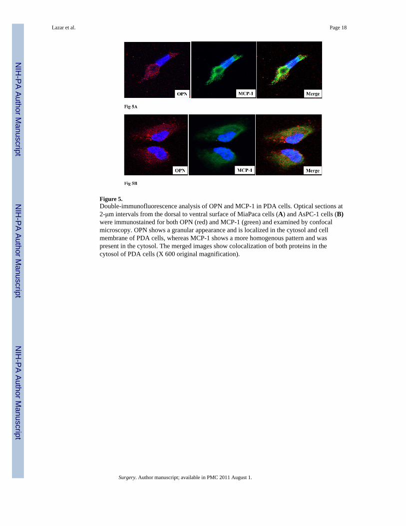
Figure 5.Double-immunofluorescence analysis of OPN and MCP-1 in PDA cells. Optical sections at2-μm intervals from the dorsal to ventral surface of MiaPaca cells (A) and AsPC-1 cells (B)were immunostained for both OPN (red) and MCP-1 (green) and examined by confocalmicroscopy. OPN shows a granular appearance and is localized in the cytosol and cellmembrane of PDA cells, whereas MCP-1 shows a more homogenous pattern and waspresent in the cytosol. The merged images show colocalization of both proteins in thecytosol of PDA cells (X 600 original magnification).
Lazar et al. Page 18
Surgery. Author manuscript; available in PMC 2011 August 1.
NIH
-PA Author Manuscript
NIH
-PA Author Manuscript
NIH
-PA Author Manuscript
Lazar et al. Page 19
Surgery. Author manuscript; available in PMC 2011 August 1.
NIH
-PA Author Manuscript
NIH
-PA Author Manuscript
NIH
-PA Author Manuscript
Figure 6.A. Representative immunohistochemical staining for OPN and MCP-1 in malignant PDA.Serial sections of paraffin embedded PDA sections were stained with OPN and MCP-1antibodies. MCP-1 colocalized with OPN in the malignant ducts ductal epithelial stainingand in stromal cells. (X200 original magnification). B. Lymph node metastatic lesionsshowed intense OPN and MCP-1 staining in the ductal metastasis. (X 100 originalmagnification). Negative control (−ve C) sections where the primary antibody was notadded did not show non-specific reaction. C. Expression of MCP-1 in human tissue. Realtime PCR analysis of MCP-1 mRNA in premalignant lesions, and invasive PDA.Significantly higher MCP-1 mRNA levels are seen in invasive PDA. Analysis of patienthistory of the samples used for MCP-1 analysis shows that invasive PDA patients weremostly (66%) smokers, while a the majority (67%) of patients with premalignant lesionswere non smokers* p<0.05 vs. non-malignant levels using one-way repeated ANOVA withsubsequent all pairwise comparison procedure by student t-test. D. Significant correlation(p<0.05) between tissue OPN and MCP-1 mRNAs. High MCP-1 (+++) was found in 65% ofthe invasive PDA samples that expressed high OPN (+++). No MCP-1 was found in IPMNlesions that expressed very low levels of OPN.
Lazar et al. Page 20
Surgery. Author manuscript; available in PMC 2011 August 1.
NIH
-PA Author Manuscript
NIH
-PA Author Manuscript
NIH
-PA Author Manuscript