Sonic hedgehog is a potent chemoattractant for human monocytes: diabetes mellitus inhibits Sonic...
11
ORIGINAL CONTRIBUTION Sonic hedgehog is a potent chemoattractant for human monocytes: diabetes mellitus inhibits Sonic hedgehog-induced monocyte chemotaxis Marina Dunaeva Stefan Voo Carolien van Oosterhoud Johannes Waltenberger Received: 5 January 2009 / Revised: 29 June 2009 / Accepted: 10 July 2009 / Published online: 24 July 2009 Ó The Author(s) 2009. This article is published with open access at Springerlink.com Abstract The aim of the present study was to evaluate the expression of hedgehog (Hh) signaling molecules and the chemotactic activity of Sonic hedgehog (Shh) in monocytes from control (CTR) and diabetic patients with or without coronary artery disease (CAD). Previously several studies demonstrated that exogenous administration of Shh can induce angiogenesis and accelerate repair of ischemic myocardium and skeletal muscles. Blood samples were collected from (1) CTR (n = 25); (2) patients with stable CAD without diabetes mellitus (CAD-DM, n = 10); and (3) with stable CAD with DM (CAD?DM, n = 15). Monocytes were isolated by Percoll gradient and subjected to PCR and chemotaxis analysis. Hh signaling molecules were expressed in human monocytes, and Shh- induced monocyte chemotaxis. Shh-stimulated migration of monocytes from CTR measured 172.5 ± 90% and a maximal stimulation was observed at Shh concentration of 1 lg/ml. However, Shh failed to induce migration of monocytes from CAD?DM (94.3 ± 27%, P \ 0.001 vs. CTR). The impaired response to Shh was associated with strong transcriptional upregulation of the receptor Ptc, while expression of downstream molecules was not altered. Moreover, Ptc is strongly expressed in macrophages of human aortic atherosclerotic plaque. Thus, Shh is a potent chemoattractant for monocytes and it activates classical signaling pathways related to migration. The Shh signaling was negatively affected by DM which might be involved in the pathogenesis of DM-related complications. Keywords Monocytes Hedgehog Signal transduction Chemotaxis Atherosclerosis Introduction The Hedgehog (Hh) pathway is highly conserved in evo- lution and Hh proteins are essential for cell fate decisions during development and homeostasis of adult tissues. Three proteins compose the Hh family: Sonic (Shh), Indian (Ihh) and Desert (Dhh) hedgehogs. All Hh proteins bind to a surface receptor complex comprised of two transmem- brane proteins Patched (Ptc), a receptor for Hh proteins and Smoothened (Smo), a signal transducer. It has been dem- onstrated that all Hh members Shh, Ihh and Dhh proteins bind the receptor Ptc with similar affinity, however, they vary with regard to their potency. The rank order of potency was generally Shh C Ihh C Dhh [27]. The Hh signal is then mediated by the Gli family transcription factors (Gli 1–3), which regulate the expression of different target genes [10]. There is increasing evidence for the role of Hh signaling in vascularization and neovascularization. Recently, it has been shown that activation of Hh signaling is critical for coronary development and sufficient to promote formation of coronary vessels in the embryonic and adult heart [13, 22, 29]. Shh gene therapy could promote both angiogenesis and arteriogenesis and protect from ischemic injury in rodent and large animal models [21]. However, the mechanism how Shh induces neovascularization is still unknown. It has been shown that blood circulating mono- nuclear cells play an important role in both artheriogenesis M. Dunaeva S. Voo C. van Oosterhoud J. Waltenberger (&) Department of Cardiology, University Hospital of Maastricht, Cardiovascular Research Institute of Maastricht (CARIM), P.Debyelaan 25, P.O. Box 5800, 6202 AZ Maastricht, The Netherlands e-mail: [email protected] 123 Basic Res Cardiol (2010) 105:61–71 DOI 10.1007/s00395-009-0047-x
-
Upload
kunstraum44 -
Category
Documents
-
view
0 -
download
0
Transcript of Sonic hedgehog is a potent chemoattractant for human monocytes: diabetes mellitus inhibits Sonic...

ORIGINAL CONTRIBUTION
Sonic hedgehog is a potent chemoattractant for human monocytes:diabetes mellitus inhibits Sonic hedgehog-induced monocytechemotaxis
Marina Dunaeva Æ Stefan Voo ÆCarolien van Oosterhoud Æ Johannes Waltenberger
Received: 5 January 2009 / Revised: 29 June 2009 / Accepted: 10 July 2009 / Published online: 24 July 2009
� The Author(s) 2009. This article is published with open access at Springerlink.com
Abstract The aim of the present study was to evaluate
the expression of hedgehog (Hh) signaling molecules and
the chemotactic activity of Sonic hedgehog (Shh) in
monocytes from control (CTR) and diabetic patients with
or without coronary artery disease (CAD). Previously
several studies demonstrated that exogenous administration
of Shh can induce angiogenesis and accelerate repair of
ischemic myocardium and skeletal muscles. Blood samples
were collected from (1) CTR (n = 25); (2) patients with
stable CAD without diabetes mellitus (CAD-DM,
n = 10); and (3) with stable CAD with DM (CAD?DM,
n = 15). Monocytes were isolated by Percoll gradient and
subjected to PCR and chemotaxis analysis. Hh signaling
molecules were expressed in human monocytes, and Shh-
induced monocyte chemotaxis. Shh-stimulated migration
of monocytes from CTR measured 172.5 ± 90% and a
maximal stimulation was observed at Shh concentration of
1 lg/ml. However, Shh failed to induce migration of
monocytes from CAD?DM (94.3 ± 27%, P \ 0.001 vs.
CTR). The impaired response to Shh was associated with
strong transcriptional upregulation of the receptor Ptc,
while expression of downstream molecules was not altered.
Moreover, Ptc is strongly expressed in macrophages of
human aortic atherosclerotic plaque. Thus, Shh is a potent
chemoattractant for monocytes and it activates classical
signaling pathways related to migration. The Shh signaling
was negatively affected by DM which might be involved in
the pathogenesis of DM-related complications.
Keywords Monocytes � Hedgehog � Signal transduction �Chemotaxis � Atherosclerosis
Introduction
The Hedgehog (Hh) pathway is highly conserved in evo-
lution and Hh proteins are essential for cell fate decisions
during development and homeostasis of adult tissues.
Three proteins compose the Hh family: Sonic (Shh), Indian
(Ihh) and Desert (Dhh) hedgehogs. All Hh proteins bind to
a surface receptor complex comprised of two transmem-
brane proteins Patched (Ptc), a receptor for Hh proteins and
Smoothened (Smo), a signal transducer. It has been dem-
onstrated that all Hh members Shh, Ihh and Dhh proteins
bind the receptor Ptc with similar affinity, however, they
vary with regard to their potency. The rank order of
potency was generally Shh C Ihh C Dhh [27]. The Hh
signal is then mediated by the Gli family transcription
factors (Gli 1–3), which regulate the expression of different
target genes [10].
There is increasing evidence for the role of Hh signaling
in vascularization and neovascularization. Recently, it has
been shown that activation of Hh signaling is critical for
coronary development and sufficient to promote formation
of coronary vessels in the embryonic and adult heart [13,
22, 29]. Shh gene therapy could promote both angiogenesis
and arteriogenesis and protect from ischemic injury in
rodent and large animal models [21]. However, the
mechanism how Shh induces neovascularization is still
unknown. It has been shown that blood circulating mono-
nuclear cells play an important role in both artheriogenesis
M. Dunaeva � S. Voo � C. van Oosterhoud �J. Waltenberger (&)
Department of Cardiology, University Hospital of Maastricht,
Cardiovascular Research Institute of Maastricht (CARIM),
P.Debyelaan 25, P.O. Box 5800,
6202 AZ Maastricht, The Netherlands
e-mail: [email protected]
123
Basic Res Cardiol (2010) 105:61–71
DOI 10.1007/s00395-009-0047-x

and angiogenesis [15, 32]. They adhere to the arterial wall,
infiltrate and stimulate vessel growth in the ischemic tissue
by the release of cytokines, growth factors and proteases.
Several recent studies have suggested a role of Hh sig-
naling in the control of motility and migration of multiple
cell types. Hedgehog proteins act as a chemoattractant on
isolated axons, guide neuronal migration during embryonic
development [6], serves as an attractive cue to guide germ
cell migration through the embryo to form primitive gonad
in Drosophila [12], promote the migration of endothelial
progenitor cells [1], rat activated pancreatic stellate cells
[33], embryonic endothelial cells and fibroblasts [18] as
well as optic nerve oligodendrocyte precursors [24].
However, nothing is known about migration control of
immune cells by Hh proteins. Here, we tested the
hypothesis whether Shh stimulates monocyte chemotaxis.
Our results demonstrate that Shh is a potent chemoattrac-
tant for peripheral monocytes and it activates classical
signaling pathways related to cellular migration such as
G-proteins or PI3K. Moreover, we provide the first piece of
evidence that pathological conditions such as diabetes
mellitus (DM) significantly impair Shh-induced chemo-
taxis, which is accompanied by elevated expression levels
of Ptc. Thus, these data indicate that the Hh signaling
pathway (1) is involved in monocyte biology and (2) is
negatively affected by the cardiovascular risk factor DM.
Methods
Study population
This study was performed with the approval of the medical
ethics committee of the University Hospital of Maastricht
(The Netherlands), and conforms to the principals outlined
in the Declaration of Helsinki. All enrolled subjects gave
their written informed consent. Three groups of subjects
were studied: (1) Patients with stable coronary artery dis-
ease without diabetes mellitus (CAD-DM, n = 10). Stable
CAD was defined as history of stable angina pectoris or
history of PCI and/or CABG. Patients with acute myocar-
dial infarction and/or recent surgical intervention (less then
6 months) were excluded. All patients had a recent coro-
nary angiogram. (2) CAD patients with diabetes mellitus
(CAD?DM, n = 15). The diagnosis of DM was verified
by history of DM, elevated fasting glucose level and/or
increased concentration of glycosylated HbA1c. (3) Control
subjects (CTR) without any history or clinical signs of
Table 1 Clinical characteristics
of control subjects and patients
Results are expressed as the
mean ± SD. P value indicated
for CAD-DM versus
CAD?DM
CAD Coronary artery disease,
DM type 2 diabetes mellitus,
ACE angiotensin converting
enzyme, ARB angiotensin
receptor blockers, FPG fasting
plasma glucose, ND not
determined, OHD oral
hypoglycemic drugs
CTR, n = 25 CAD-DM, n = 10 CAD?DM, n = 15 P value
Age (years) 59.9 ± 12.7 63.7 ± 9.9 66 ± 11 0.579
BMI ND 26.8 ± 2 26.3 ± 5
Male gender, n (%) 12 (48) 7 (70) 9 (60) 0.470
Cardiovascular risk factors
CAD history (years) 0 10 ± 8 9 ± 8 0.578
Family history of CAD, n (%) 11 (44) 7 (70) 7 (47) 0.231
Hypertension, n (%) 12 (48) 4 (40) 9 (60) 0.284
Smoking, n (%) 6 (24) 3 (30) 6 (40) 0.607
Diabetes, n (%) 0 (0) 0 (0) 15 (100)
Duration of DM (years) 0 0 8 ± 7
Patients receiving insulin (%) 0 0 40
Patients receiving OHD (%) 0 0 100
FPG (mmol/l) 5.5 ± 0.7 6.3 ± 0.7 8.3 ± 4.6 0.048
HbA1c (%) ND ND 7.3 ± 0.6
Laboratory parameters
Cholesterol, total (mmol/l) 5.9 ± 1.3 4.4 ± 0.8 4.0 ± 1.4 0.283
LDL-cholesterol (mmol/l) 4.0 ± 1.2 2.1 ± 0.8 2.1 ± 0.9 0.16
HDL-cholesterol (mmol/l) 1.8 ± 1.0 1.2 ± 0.6 1.0 ± 0.3 0.127
Triglycerides (mmol/l) 1.8 ± 1.2 1.8 ± 0.4 2.1 ± 1.1 0.638
Medication at admission
Antiaggregatory therapy
(antiplatelet drugs) n (%)
11 (44) 7 (70) 7 (47) 0.231
Beta-blockers, n (%) 16 (64) 7 (70) 10 (67) 0.222
ACE-inhibitors/ARB, n (%) 12 (48) 6 (60) 7 (47) 0.688
Statins, n (%) 12 (48) 8 (80) 12 (80) 0.687
62 Basic Res Cardiol (2010) 105:61–71
123

CAD, metabolic disease or chronic illness (n = 25).
Baseline clinical features of CTR, CAD-DM and
CAD?DM patients are presented in Table 1. There were
no significant differences between three groups with
regards to age, gender, BMI, presence of cardiovascular
risk factors and medication. CAD?DM patients exhibited
higher fasting glucose level compared to CTR (P = 0.019)
and compared to CAD-DM group (P = 0.048).
Monocyte isolation and migration
Blood samples (100 ml) were collected from all subjects.
Monocytes were isolated by Percoll gradient centrifugation
based on a previously described protocol [40]. The purity
of isolated monocytes was C90% as determined by flow
cytometry. Monocytes were subjected to chemotaxis assay
using the modified Boyden chamber [2]. Briefly, different
concentrations of Shh or Ihh were placed in the bottom
well and monocytes were placed in the top well. Polycar-
bonate membranes with a pore diameter of 5 lm (Nucle-
pore) were used. The chambers were incubated at 37�C in
the presence of 5% CO2 for 90 min. Afterwards filters were
removed, fixed, stained with Giemsa dye before scraping
off cells at the upper side of the filter membrane and five
high-power fields were counted for each sample (primary
magnification 209). To differentiate between chemotaxis
and chemokinesis, checkerboard analysis was performed
by placing various dilutions of Shh in both the lower and
upper wells of the modified Boyden chamber. To study the
effect of different inhibitors on Shh-induced migration,
monocytes were pretreated with a specific inhibitor of Hh
signaling, cyclopamine (CP, 10 lM), the PI3K inhibitor,
LY294002 (10 lM), or a specific inhibitor of Gai/o, per-
tussis toxin (PTX, 100 ng/ml) for 15 min before the che-
motaxis assay.
RT-PCR analysis
Total RNA from monocytes or HUVEC (used as a
control) was isolated using RNeasy Protect Mini Kit
from QIAGEN. For reverse transcription, 1 lg of total
RNA was converted into cDNA by AMV reverse
transcriptase at 37�C for 1 h in a 20 ll RT reaction. All
the primers used in this study and PCR conditions are
listed in Table 2.
Flow cytometry analysis
Expression of Ptc on leukocytes was analyzed by flow
cytometry. Leukocytes were stained with anti-Ptc primary
antibodies (Santa Cruz Biotechnology). Double staining
with CD14-FITC (Becton Dickinson) and Ptc/anti-goat-PE
antibody was performed to confirm the expression of both
proteins on monocytes. The analysis was carried out on a
FACSCalibur flow cytometer using the CellQuest software
(Becton Dickinson).
Table 2 Primers and conditions used for RT-PCR
Gene Sequence (50–30) Annealing temperature
and cycle number
Fragment size (bp)
Shh FW: ACT GGG TGT ACT ACG AGT CCA AGG 63�C, 50 cycles 211
RV: AAA GTG AGG AAG TCG CTG TAG AGC
Ihh FW: CTA CGC CCC GCT CAC AAA G 60�C, 30 cycles 376
RV: GGC AGA GGA GAT GGC AGG AG
Dhh FW: ACCAATCTACTGCCCCTGTG 62�C, 30 cycles 246
RV: GTTGTAGTTGGGCACGAGGT
Smo FW: CAG GAC ATG CAC AGC TAC ATC G 65�C, 30 cycles 380
RV: CCA CAA AGA AGC ACG CAT TGA C
Ptc FW: CCA TGT TCC AGT TAA TGA CTC 55�C, 30 cycles 462
RV: ACA TCA TCC ACA CCA ACA
SUFU FW: CCT CCA GAT CGT TGG TGT CT 55�C, 30 cycles 195
RV: CTG TCT CGA TGC CTT TGT CA
Gli-1 FW: GGG ATG ATC CCA CAT CCT CAG TC 60�C, 50 cycles 386
RV: CTG GAG CAG CCC CCC CAG T
Gli-2 FW: ACC GCT GCT CAA AGA GAA TG 64�C, 50 cycles 507
RV: CCC ACT GCC ACT GAA GTT TTC C
Gli3 FW: CCT CAA AGC GGG CCG CCT GC 64�C, 50 cycles 406
RV: CAG GTT GTT GTT GGA CTG TGT GC
Basic Res Cardiol (2010) 105:61–71 63
123

Immunohistochemistry
Cross sections of human aortic atherosclerotic plaque were
deparaffinized, dehydrated, and permeabilized with 0.05%
Tween in citrate buffer, and then blocked with normal
horse serum. Further, they were incubated with primary
polyclonal antibodies for Ptc (1:25, goat polyclonals; Santa
Cruz Biotechnology Inc) or CD68 (1:100, mouse mono-
clonal; DakoCytomation). A biotinylated horse anti-mouse
and anti-goat IgG (1:1,000) was incubated followed by
incubation with streptavidin/HRP antibodies and visualized
with AEC staining solution. Consecutive sections were
stained and the results of Ptc staining and CD68 staining
were merged for demonstration of coexpression. The nega-
tive controls included substitution of primary antiserum for
PBS.
Statistical analysis
Statistical analysis was performed with SPSS12.0.1 statis-
tical software. Categorical variables are presented as per-
centage of patients and compared by Fisher’s exact test.
Data are presented as the mean ± standard deviation (SD).
In addition, the medians (in the figure: median as line,
25th–75th percentiles as box, 5th–95th percentiles as
whiskers) are given in case of non-normally distributed
results. CTR, CAD-DM and CAD?DM patients were
compared using a Mann–Whitney U test with Bonferroni
correction. Values of P \ 0.05 were considered statisti-
cally significant.
Results
Human peripheral monocytes express components
of Hh signaling
Initially, we examined human monocytes for the expres-
sion of components involved in Hh signaling including the
ligands Shh, Dhh and Ihh, the receptor Ptc, co-receptor
Smoothened (Smo), hedgehog interacting protein (Hip),
suppressor of Fused (SUFU), transcription factors Gli 1, 2
and 3. We carried out RT-PCR on RNAs prepared from
isolated human peripheral monocytes. RNA from HUVEC
was used as a positive control. We confirmed the expres-
sion of Ihh, Dhh, Ptc, Smo, SUFU and Gli3 genes on
monocytes, but could not detect the expression of Shh, Hip,
Gli1 and Gli2 (Fig. 1a).
To confirm the surface expression of Hh receptor Ptc
protein, flow cytometric analysis was performed. Double
staining of the monocyte fraction with the monocyte mar-
ker CD14 and Ptc antibodies demonstrated that 95% of the
monocytes were positive for Ptc (Fig. 1b). These results
suggest that monocytes can be susceptible to Hh protein
stimulation.
Chemotactic properties of Shh
Monocytes isolated from the blood of control subjects were
assessed for their ability to migrate to Shh. Shh-induced
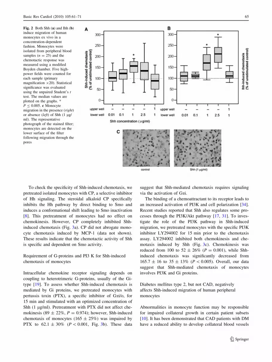
monocyte chemotaxis with a bell-shaped dose–response
curve, whereby stimulation with 0.01 lg/ml Shh stimulated
migration to 123.8 ± 34% (P = 0.622), stimulation with
0.1 lg/ml Shh led to 132.4 ± 26% (P = 0.093), while
stimulation with 1 lg/ml resulted in 172.5 ± 90% of
baseline (P \ 0.005), and 2.5 lg/ml Shh in 153.1 ± 60%
(P = 0.006) (Fig. 2a, c). Based on these results, we used
1 lg/ml Shh for all further experiments presented. Similar
results were observed when Ihh was used to induce
monocyte migration (Fig. 2B). However, the optimal
concentration for Ihh was 2.5 lg/ml. In the checkerboard
control experiment, when Shh was added to both upper and
lower wells, monocyte chemotaxis was not observed
(Fig. 2a). These data indicate that a concentration gradient
of Shh is required for monocyte chemotaxis.
Fig. 1 Expression of Hedgehog signaling components in human
monocytes. a RT-PCR analysis of the Shh, Ihh, Dhh, Smo, Ptc,
SUFU, Hip, Gli1, Gli2, Gli3, and actin gene expression on human
peripheral monocytes and HUVEC (used as a positive control). The
results are representative of three independent experiments. b Flow
cytometric analysis of Ptc expression on monocytes. Left panelIsotype (negative) control. Right panel Purified monocytes were
double-stained with anti-Ptc antibodies and CD14 antibodies as a
marker for monocytes
64 Basic Res Cardiol (2010) 105:61–71
123
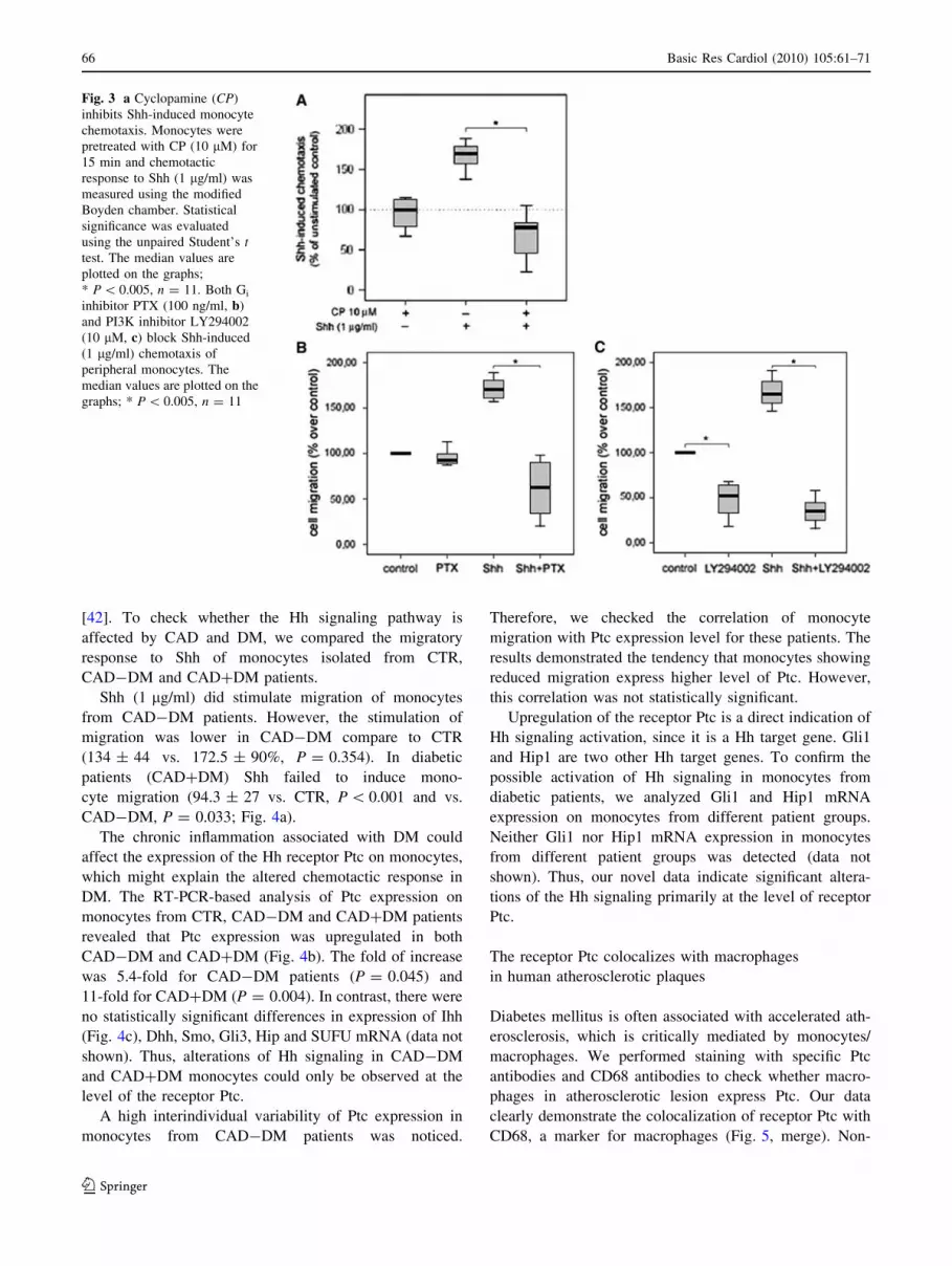
To check the specificity of Shh-induced chemotaxis, we
pretreated isolated monocytes with CP, a selective inhibitor
of Hh signaling. The steroidal alkaloid CP specifically
inhibits the Hh pathway by direct binding to Smo and
induces a conformational shift leading to Smo inactivation
[8]. This pretreatment of monocytes had no effect on
chemokinesis. However, CP completely inhibited Shh-
induced chemotaxis (Fig. 3a). CP did not abrogate mono-
cyte chemotaxis induced by MCP-1 (data not shown).
These results indicate that the chemotactic activity of Shh
is specific and dependent on Smo activity.
Requirement of G-proteins and PI3 K for Shh-induced
chemotaxis of monocytes
Intracellular chemokine receptor signaling depends on
coupling to heterotrimeric G-proteins, usually of the Gi-
type [19]. To assess whether Shh-induced chemotaxis is
mediated by Gi proteins, we pretreated monocytes with
pertussis toxin (PTX), a specific inhibitor of Gai/o, for
15 min and stimulated with an optimized concentration of
Shh (1 lg/ml). Pretreatment with PTX did not affect che-
mokinesis (89 ± 22%, P = 0.974); however, Shh-induced
chemotaxis of monocytes (165 ± 25%) was impaired by
PTX to 62.1 ± 30% (P \ 0.001, Fig. 3b). These data
suggest that Shh-mediated chemotaxis requires signaling
via the activation of Gai.
The binding of a chemoattractant to its receptor leads to
an increased activation of PI3K and cell polarization [34].
Recent studies reported that Shh also regulates some pro-
cesses through the PI3K/Akt pathway [17, 31]. To inves-
tigate the role of the PI3K pathway in Shh-induced
migration, we pretreated monocytes with the specific PI3K
inhibitor LY294002 for 15 min prior to the chemotaxis
assay. LY294002 inhibited both chemokinesis and che-
motaxis induced by Shh (Fig. 3c). Chemokinesis was
reduced from 100 to 52 ± 26% (P = 0.001), while Shh-
induced chemotaxis was significantly decreased from
165.7 ± 16 to 35 ± 13% (P \ 0.005). Overall, our data
suggest that Shh-mediated chemotaxis of monocytes
involves PI3K and Gi proteins.
Diabetes mellitus type 2, but not CAD, negatively
affects Shh-induced migration of human peripheral
monocytes
Abnormalities in monocyte function may be responsible
for impaired collateral growth in certain patient subsets
[10]. It has been demonstrated that CAD patients with DM
have a reduced ability to develop collateral blood vessels
Fig. 2 Both Shh (a) and Ihh (b)
induce migration of human
monocytes ex vivo in a
concentration-dependent
fashion. Monocytes were
isolated from peripheral blood
samples (n = 25) and the
chemotactic response was
measured using a modified
Boyden chamber. Five high-
power fields were counted for
each sample (primary
magnification 920). Statistical
significance was evaluated
using the unpaired Student’s ttest. The median values are
plotted on the graphs. *
P B 0.005. c Monocyte
migration in the presence (right)or absence (left) of Shh (1 lg/
ml). The representative
photograph of the stained filter;
monocytes are detected on the
lower surface of the filter
following migration through the
pores
Basic Res Cardiol (2010) 105:61–71 65
123
[42]. To check whether the Hh signaling pathway is
affected by CAD and DM, we compared the migratory
response to Shh of monocytes isolated from CTR,
CAD-DM and CAD?DM patients.
Shh (1 lg/ml) did stimulate migration of monocytes
from CAD-DM patients. However, the stimulation of
migration was lower in CAD-DM compare to CTR
(134 ± 44 vs. 172.5 ± 90%, P = 0.354). In diabetic
patients (CAD?DM) Shh failed to induce mono-
cyte migration (94.3 ± 27 vs. CTR, P \ 0.001 and vs.
CAD-DM, P = 0.033; Fig. 4a).
The chronic inflammation associated with DM could
affect the expression of the Hh receptor Ptc on monocytes,
which might explain the altered chemotactic response in
DM. The RT-PCR-based analysis of Ptc expression on
monocytes from CTR, CAD-DM and CAD?DM patients
revealed that Ptc expression was upregulated in both
CAD-DM and CAD?DM (Fig. 4b). The fold of increase
was 5.4-fold for CAD-DM patients (P = 0.045) and
11-fold for CAD?DM (P = 0.004). In contrast, there were
no statistically significant differences in expression of Ihh
(Fig. 4c), Dhh, Smo, Gli3, Hip and SUFU mRNA (data not
shown). Thus, alterations of Hh signaling in CAD-DM
and CAD?DM monocytes could only be observed at the
level of the receptor Ptc.
A high interindividual variability of Ptc expression in
monocytes from CAD-DM patients was noticed.
Therefore, we checked the correlation of monocyte
migration with Ptc expression level for these patients. The
results demonstrated the tendency that monocytes showing
reduced migration express higher level of Ptc. However,
this correlation was not statistically significant.
Upregulation of the receptor Ptc is a direct indication of
Hh signaling activation, since it is a Hh target gene. Gli1
and Hip1 are two other Hh target genes. To confirm the
possible activation of Hh signaling in monocytes from
diabetic patients, we analyzed Gli1 and Hip1 mRNA
expression on monocytes from different patient groups.
Neither Gli1 nor Hip1 mRNA expression in monocytes
from different patient groups was detected (data not
shown). Thus, our novel data indicate significant altera-
tions of the Hh signaling primarily at the level of receptor
Ptc.
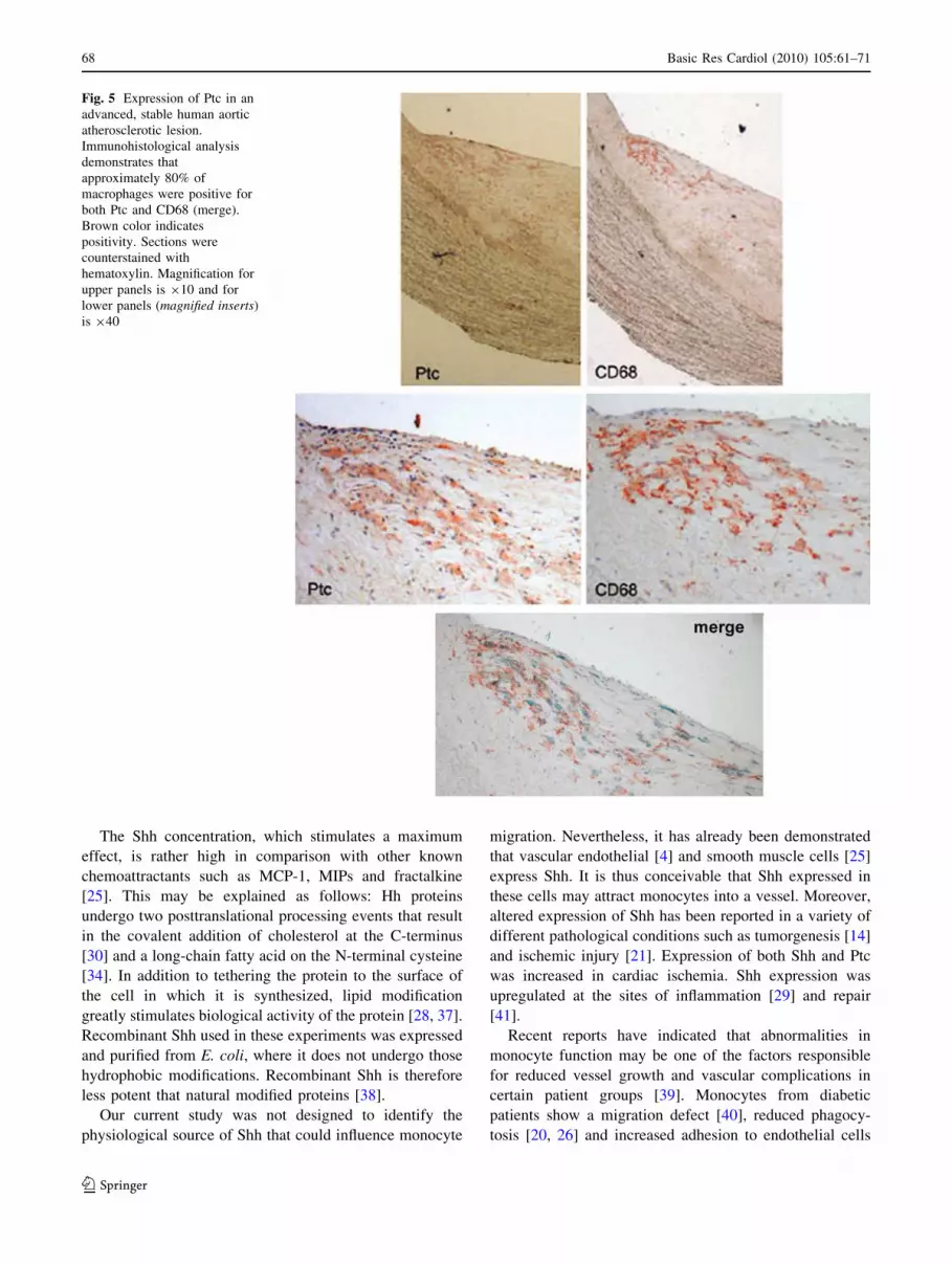
The receptor Ptc colocalizes with macrophages
in human atherosclerotic plaques
Diabetes mellitus is often associated with accelerated ath-
erosclerosis, which is critically mediated by monocytes/
macrophages. We performed staining with specific Ptc
antibodies and CD68 antibodies to check whether macro-
phages in atherosclerotic lesion express Ptc. Our data
clearly demonstrate the colocalization of receptor Ptc with
CD68, a marker for macrophages (Fig. 5, merge). Non-
Fig. 3 a Cyclopamine (CP)
inhibits Shh-induced monocyte
chemotaxis. Monocytes were
pretreated with CP (10 lM) for
15 min and chemotactic
response to Shh (1 lg/ml) was
measured using the modified
Boyden chamber. Statistical
significance was evaluated
using the unpaired Student’s ttest. The median values are
plotted on the graphs;
* P \ 0.005, n = 11. Both Gi
inhibitor PTX (100 ng/ml, b)
and PI3K inhibitor LY294002
(10 lM, c) block Shh-induced
(1 lg/ml) chemotaxis of
peripheral monocytes. The
median values are plotted on the
graphs; * P \ 0.005, n = 11
66 Basic Res Cardiol (2010) 105:61–71
123

immune IgG did not show any positive staining (data not
shown).
Discussion
In this study, we present data supporting a role for Shh/Ihh
in stimulation of monocyte chemotaxis. We have not used
Dhh for our experiments, because up to now there is no
evidence that Dhh is expressed in vascular endothelial cell
or vascular smooth muscle cells. Our data demonstrate that
both Shh and Ihh stimulate monocyte chemotaxis at the
optimal concentration for Shh 1 lg/ml and for Ihh 2.5 lg/
ml. Chemotaxis induced by Shh was specific and Smo-
dependent. Moreover, Shh activates classical intracellular
signal transduction pathways related to cellular migration
such as G-protein-coupled specific receptor pathways or
PI3K.
Fig. 4 a Shh-induced (1 lg/ml) chemotaxis of monocytes isolated
from CTR, CAD-DM or CAD?DM patients. The median values are
plotted; ns; nonsignificant versus unstimulated control. The median
(line), 25th and 75th percentiles (box), 5th and 95th percentiles
(whiskers) are presented. * P \ 0.005. The level of Ptc (b), and Ihh
(c) gene expression on monocytes isolated from CTR, CAD-DM and
CAD?DM patients. RNA isolated from monocytes was subjected to
RT-PCR. b-actin cDNA product served as internal standard to
normalize Ptc and Ihh. Signal intensity of Ptc and Ihh mRNA bands
was quantified by densitometry and compared with the internal
standard b-actin
Basic Res Cardiol (2010) 105:61–71 67
123
The Shh concentration, which stimulates a maximum
effect, is rather high in comparison with other known
chemoattractants such as MCP-1, MIPs and fractalkine
[25]. This may be explained as follows: Hh proteins
undergo two posttranslational processing events that result
in the covalent addition of cholesterol at the C-terminus
[30] and a long-chain fatty acid on the N-terminal cysteine
[34]. In addition to tethering the protein to the surface of
the cell in which it is synthesized, lipid modification
greatly stimulates biological activity of the protein [28, 37].
Recombinant Shh used in these experiments was expressed
and purified from E. coli, where it does not undergo those
hydrophobic modifications. Recombinant Shh is therefore
less potent that natural modified proteins [38].
Our current study was not designed to identify the
physiological source of Shh that could influence monocyte
migration. Nevertheless, it has already been demonstrated
that vascular endothelial [4] and smooth muscle cells [25]
express Shh. It is thus conceivable that Shh expressed in
these cells may attract monocytes into a vessel. Moreover,
altered expression of Shh has been reported in a variety of
different pathological conditions such as tumorgenesis [14]
and ischemic injury [21]. Expression of both Shh and Ptc
was increased in cardiac ischemia. Shh expression was
upregulated at the sites of inflammation [29] and repair
[41].
Recent reports have indicated that abnormalities in
monocyte function may be one of the factors responsible
for reduced vessel growth and vascular complications in
certain patient groups [39]. Monocytes from diabetic
patients show a migration defect [40], reduced phagocy-
tosis [20, 26] and increased adhesion to endothelial cells
Fig. 5 Expression of Ptc in an
advanced, stable human aortic
atherosclerotic lesion.
Immunohistological analysis
demonstrates that
approximately 80% of
macrophages were positive for
both Ptc and CD68 (merge).
Brown color indicates
positivity. Sections were
counterstained with
hematoxylin. Magnification for
upper panels is 910 and for
lower panels (magnified inserts)
is 940
68 Basic Res Cardiol (2010) 105:61–71
123

[9]. The present study demonstrates that monocytes from
diabetic patients do not respond to Shh at the most efficient
concentration in CTR, i.e. 1 lg/ml. This impaired chemo-
tactic response was associated with a 11-fold increase of
Ptc mRNA expression in diabetic monocytes. The increase
of Ptc expression could be also observed for some CAD-
DM patients; however, the difference in expression was not
statistically significant in comparison to control subjects (3
patients from 10 had higher level of Ptc expression com-
pared to control). However, there was no significant dif-
ference in mRNA expression between different groups for
ligands Ihh and Dhh and downstream Hh signaling com-
ponents Smo and SUFU. Previously, impaired chemotactic
response of monocytes associated with increased surface
expression of receptors was observed for patients with DM
type 1 [5]. Similar effect was observed for blood mono-
cytes from epithelial ovarian cancer. Notwithstanding the
increased levels of CCR2 and CCR5 detected on ascitic
monocytes, in comparison to that of normal donors,
migration response to their ligand RANTES was impaired
[16].
Diabetes mellitus type 2 is often associated with obesity.
It has been reported that the Hh signaling pathway was less
active in obese compared to lean mice. Two models have
been tested, mice invalidated in leptine (ob/ob) and high-
fat diet induced obesity. So far all experiments leading to a
stimulation of Hh signaling in vivo result in an enlarged
body mass and conversely, an inhibition of the pathway
prevents weight gain [11]. We did not observe significant
differences between CAD-DM and CAD?DM with
regards to BMI. Therefore, it is unlikely that an excess of
adipose mass, which characterizes obesity and is often
associated with diabetes, may play a role in disregulation
of Hh pathway.
Moreover, diabetes mellitus is also accompanied by a
systemic low-grade inflammation and it is an important
factor in accelerating atherosclerosis. It has been reported
that LPS-induced Ptc expression in macrophages after 18 h
in culture [41]. Thus, low-grade inflammation could
upregulate expression of Ptc on circulating monocytes of
diabetes patients. In addition, it has been previously shown
in Drosophila that to achieve maximal pathway activity,
large excess (more than 50-fold) of Smo over Ptc is
required [36]. If similar regulation of Hh signaling applies
to the human system, the strong upregulation of Ptc and
preserved expression level of Smo in diabetic patients
could explain the observed defect in Hh signaling.
Monocytes/macrophages are recognized as one of the
principal cell types in the pathology of atherosclerosis [43,
44]. Accumulating evidence suggests that the Hh pathway
is involved in peripheral immunity and tissue remodeling
[21, 23]. Expression of Shh and Ptc is upregulated in small
bowel allografts undergoing chronic rejection. Systemic
treatment with a neutralizing anti-Shh antibody reduced
tissue remodeling, fibrosis and vascular occlusion of the
intestinal grafts [7]. The cells that expressed Ptc in the
intestinal grafts undergoing chronic rejection were mainly
macrophages. In a recent study using an ApoE-/--based
mouse model of atherosclerosis, inhibition of Shh using
specific antibodies resulted in a pro-atherosclerotic phe-
notype secondary to enhanced lipid uptake in macrophages
[3]. Our novel data do support the idea that inhibition of
Shh may be pro-atherogenic. Macrophages in human ath-
erosclerotic plaques express high levels of receptor Ptc.
The diabetes-related inhibition of macrophages may pre-
vent their clearance from atherosclerotic plaques. This
could be a novel molecular mechanism promoting athero-
sclerosis; however, its functional relevance and extent in
the pathogenesis of human atherosclerosis remains to be
determined.
Thus, our data represent the first example of an endo-
genous metabolic abnormality, namely DM, which is
associated with a functional inhibition of the Hh signaling
pathway. Likewise, this impaired monocyte function is
likely to be associated with impaired arteriogenesis and
impaired wound healing [35, 39]. Identification of novel
signal transduction pathways regulating monocyte chemo-
taxis can indicate unique targets for preventive therapies
for treatment of chronic inflammatory diseases.
Acknowledgments We wish to acknowledge the support from
Moniek Baggen in patient recruitment. This study was supported in
part by grant VEGF therapies (QLRT-2001-01955) from the Euro-
pean Commission, and by the Cardiovascular Research Institute
Maastricht (CARIM).
Conflict of interest statement None
Open Access This article is distributed under the terms of the
Creative Commons Attribution Noncommercial License which per-
mits any noncommercial use, distribution, and reproduction in any
medium, provided the original author(s) and source are credited.
References
1. Asai J, Takenaka H, Kusano K, Ii M, Luedemann C, Curry C,
Eaton E, Iwakura A, Tsutsumi Y, Hamada H, Kishimoto S,
Thorne T, Kishore R, Losordo D (2006) Topical sonic hedgehog
gene therapy accelerates wound healing in diabetes by enhancing
endothelial progenitor cell-mediated microvascular remodeling.
Circulation 113:2413–2424
2. Barleon B, Sozzani S, Zhou D, Weich H, Mantovani A, Marme D
(1996) Migration of human monocytes in response to vascular
endothelial growth factor (VEGF) is mediated via the VEGF
receptor flt-1. Blood 87:3336–3343
3. Beckers L, Heeneman S, Wang L, Burkly L, Rousch M, David-
son N, Gijbels M, de Winther M, Daemen M, Lutgens E (2007)
Disruption of hedgehog signalling in ApoE-/- mice reduces
plasma lipid levels, but increases atherosclerosis due to enhanced
lipid uptake by macrophages. J Pathol 212:420–428
Basic Res Cardiol (2010) 105:61–71 69
123
4. Bhardwaj G, Murdoch B, Wu D, Baker D, Williams K, Chadwick
K, Ling L, Karanu F, Bhatia M (2001) Sonic hedgehog induces
the proliferation of primitive human hematopoietic cells via BMP
regulation. Nat Immunol 2:172–180
5. Bouma G, Coppens J, Lam-Tse W, Luini W, Sintnicolaas K,
Levering W, Sozzani S, Drexhage H, Versnel M (2005) An
increased MRP8/14 expression and adhesion, but a decreased
migration towards proinflammatory chemokines of type 1 dia-
betes monocytes. Clin Exp Immunol 141:509–517
6. Charron F, Stein E, Jeong J, McMahon AP, Tessier-Lavigne M
(2003) The morphogen sonic hedgehog is an axonal chemoat-
tractant that collaborates with netrin-1 in midline axon guidance.
Cell 113:11–23
7. Chen Y, Li X, Tian L, Lui V, Dallman M, Lamb J, Tam P (2007)
Inhibition of sonic hedgehog signaling reduces chronic rejection
and prolongs allograft survival in a rat orthotopic small bowel
transplantation model. Transplantation 83:1351–1357
8. Chen J, Taipale J, Cooper M, Beachy P (2002) Inhibition of
Hedgehog signaling by direct binding of cyclopamine to
Smoothened. Genes Dev 16:2743–2748
9. Cipolletta C, Ryan K, Hanna E, Trimble E (2005) Activation of
peripheral blood CD14? monocytes occurs in diabetes. Diabetes
549:2779–2786
10. Cohen M (2003) The hedgehog signaling network. Am J Med
Genetics 123:5–28
11. Cousin W, Fontaine C, Dani C, Peraldi P (2007) Hedgehog and
adipogenesis: fat and fiction. Biochimie 89:1447–1453
12. Deshpande G, Swanhart L, Chiang P, Schedl P (2001) Hedgehog
signaling in germ cell migration. Cell 106:759–769
13. Dyer M, Farrington S, Mohn D, Munday J, Baron M (2001)
Indian hedgehog activates hematopoiesis and vasculogenesis and
can respecify prospective neurectodermal cell fate in the mouse
embryo. Development 128:1717–1730
14. Fan L, Pepicelli C, Dibble C, Catbagan W, Zarycki J, Laciak R,
Gipp J, Shaw A, Lamm M, Munoz A, Lipinski R, Thrasher J,
Bushman W (2004) Hedgehog signaling promotes prostate
xenograft tumor growth. Endocrinology 145:3961–3970
15. Frantz S, Vincent K, Feron O, Kelly R (2005) Innate immunity
and angiogenesis. Circ Res 96:15–26
16. Freedman R, Ma Q, Wang E, Gallardo S, Gordon I, Shin J, Jin P,
Stroncek D, Marincola F (2008) Migration deficit in monocyte-
macrophages in human ovarian cancer. Cancer Immunol Im-
munother 57:635–645
17. Fu J, Liu W, Zhou J, Sun H, Xu H, Luo L, Zhang H, Zhou YF
(2006) Sonic hedgehog protein promotes bone marrow-derived
endothelial progenitor cell proliferation, migration and VEGF
production via PI 3-kinase/Akt signaling pathways. Acta Phar-
macol Sin 27:685–693
18. Hochman E, Castiel A, Jacob-Hirsch J, Amariglio N, Izraeli S
(2006) Molecular pathways regulating pro-migratory effects of
Hedgehog signaling. J Biol Chem 281:33860–33870
19. Kamps A, Coffman C (2005) G protein-coupled receptor roles in
cell migration and cell death decisions. Ann N Y Acad Sci
1049:17–23
20. Katz S, Klein B, Fishman P, Djaldetti M (1983) Phagocytotic
activity on monocytes from diabetic patients. Diabetes Care
6:479–482
21. Kusano K, Pola R, Murayama T, Curry C, Kawamoto A, Iwakura
A, Shintani S, Ii M, Asai J, Tkebuchava T, Thorne T, Takenaka
H, Aikawa R, Goukassian D, von Samson P, Hamada H, Yoon
YS, Silver M, Eaton E, Ma H, Heyd L, Kearney M, Munger W,
Porter JA, Kishore R, Losordo DW (2005) Sonic hedgehog
myocardial gene therapy: tissue repair through transient recon-
stitution of embryonic signaling. Nat Med 11:1197–1204
22. Lavine K, White A, Park C, Smith C, Choi K, Long F, Hui C,
Ornitz D (2006) Fibroblast growth factor signals regulate a wave
of Hedgehog activation that is essential for coronary vascular
development. Genes Dev 20:1651–1666
23. Lowrey J, Stewart G, Lindey S, Hoyne G, Dallman M, Howie S,
Lamb J (2002) Sonic hedgehog promotes cell cycle progression
in activated peripheral CD4(?) T lymphocytes. J Immunol
169:1869–1875
24. Merchan P, Bribian A, Sanchez-Camacho C, Lezameta M, Bo-
volenta P, de Castro F (2007) Sonic hedgehog promotes the
migration and proliferation of optic nerve oligodendrocyte pre-
cursors. Mol Cell Neurosci 36:355–368
25. Morrow D, Sweeney C, Birney Y, Guha S, Collins N, Cummins
P, Murphy R, Walls D, Redmond E, Cahill P (2007) Biome-
chanical regulation of hedgehog signaling in vascular smooth
muscle cells in vitro and in vivo. Am J Physiol Cell Physiol
292:C488–C496
26. O’Brien B, Huang Y, Geng X, Dutz J, Finegood D (2002)
Phagocytosis of apoptotic cells by macrophages from NOD mice
is reduced. Diabetes 51:2481–2488
27. Pathi S, Pagan-Westphal S, Baker D, Garber E, Rayhorn P,
Bumcrot D, Tabin CJ, Blake Pepinsky R, Williams KP (2001)
Comparative biological responses to human Sonic, Indian, and
Desert hedgehog. Mech Dev 106:107–117
28. Pepinsky R, Zeng C, Wen D, Rayhorn P, Baker D, Williams K,
Bixler S, Ambrose C, Garber E, Miatkowski K, Taylor F, Wang
E, Galdes A (1998) Identification of a palmitic acid-modified
form of human Sonic hedgehog. J Biol Chem 273:14037–14045
29. Pola R, Ling L, Silver M, Corbley M, Kearney M, Blake Pe-
pinsky R, Shapiro R, Taylor F, Baker D, Asahara T, Isner J
(2001) The morphogen Sonic hedgehog is an indirect angiogenic
agent upregulating two families of angiogenic growth factors. Nat
Med 7:706–711
30. Porter J, Young K, Beachy P (1996) Cholesterol modification of
hedgehog signaling proteins in animal development. Science
274:255–259
31. Riobo N, Lu K, Ai X, Haines G, Emerson C (2006) Phosphoin-
ositide 3-kinase and Akt are essential for Sonic hedgehog sig-
naling. Proc Natl Acad Sci USA 103:4505–4510
32. Schaper W, Scholz D (2003) Factors regulating arteriogenesis.
Arterioscler Thromb Vasc Biol 23:1143–1151
33. Shinozaki S, Ohnishi H, Hama K, Kita H, Yamamoto H, Os-
awa H, Sato K, Tamada K, Mashima H, Sugano K (2008)
Indian hedgehog promotes the migration of rat activated pan-
creatic stellate cells by increasing membrane type-1 matrix
metalloproteinase on the plasma membrane. J Cell Physiol
216:38–46
34. Stephens L, Ellson C, Hawkins P (2002) Roles of PI3Ks in leu-
kocyte chemotaxis and phagocytosis. Curr Opin Cell Biol
14:203–213
35. Stewart G, Hoyne G, Ahmad S, Jarman E, Wallace W, Harrison
D, Haslett C, Lamb J, Howie S (2003) Expression of the devel-
opmental Sonic hedgehog (Shh) signalling pathway is up-regu-
lated in chronic lung fibrosis and the Shh receptor patched 1 is
present in circulating T lymphocytes. J Pathol 199:488–495
36. Taipale J, Cooper M, Maiti T, Beachy P (2002) Patched acts
catalytically to suppress the activity of Smoothened. Nature
418:892–897
37. Taylor F, Wen D, Garber E, Carmillo A, Baker D, Arduini R,
Williams KP, Weinreb PH, Rayhorn P, Hronowski X, Whitty A,
Day ES, Boriack-Sjodin A, Shapiro RI, Galdes A, Pepinsky RB
(2001) Enhanced potency of human Sonic hedgehog by hydro-
phobic modification. Biochemistry 40:4359–4371
38. Wakelin S, Forsythe J, Garden O, Howie S (2008) Commercially
available recombinant sonic hedgehog up-regulates Ptc and
modulates the cytokine and chemokine expression of human
macrophages: an effect mediated by endotoxin contamination?
Immunobiology 213:25–38
70 Basic Res Cardiol (2010) 105:61–71
123
39. Waltenberger J (2001) Impaired collateral development in dia-
betes: potential cellular mechanisms and therapeutic implications.
Cardiovasc Res 49:554–560
40. Waltenberger J, Lange J, Kranz A (2000) Vascular endothelial
growth factor-A-induced chemotaxis of monocytes is attenuated in
patients with diabetes mellitus: a potential predictor for the indi-
vidual capacity to develop collaterals. Circulation 102:185–190
41. Watkins D, Berman D, Burkholder S, Wang B, Beachy P, Baylin
S (2003) Hedgehog signalling within airway epithelial progeni-
tors and in small-cell lung cancer. Nature 422:313–317
42. Werner G, Richartz B, Heinke S, Ferrari M, Figulla H (2003)
Impaired acute collateral recruitment as a possible mechanism for
increased cardiac adverse events in patients with diabetes melli-
tus. Eur Heart J 24:1134–1142
43. Zernecke A, Weber C (2005) Inflammatory mediators in ath-
erosclerotic vascular disease. Basic Res Cardiol 100:93–101
44. Zhang C (2008) The role of inflammatory cytokines in endo-
thelial dysfunction. Basic Res Cardiol 103:398–406
Basic Res Cardiol (2010) 105:61–71 71
123


![u[sonic] Modbus - Lambrecht meteo](https://static.fdokumen.com/doc/165x107/6334bd04a6138719eb0b33dc/usonic-modbus-lambrecht-meteo.jpg)