Geology and morphology of the rock niches from the Eastern Rhodopes
Upload
independentCategory
view
1download
0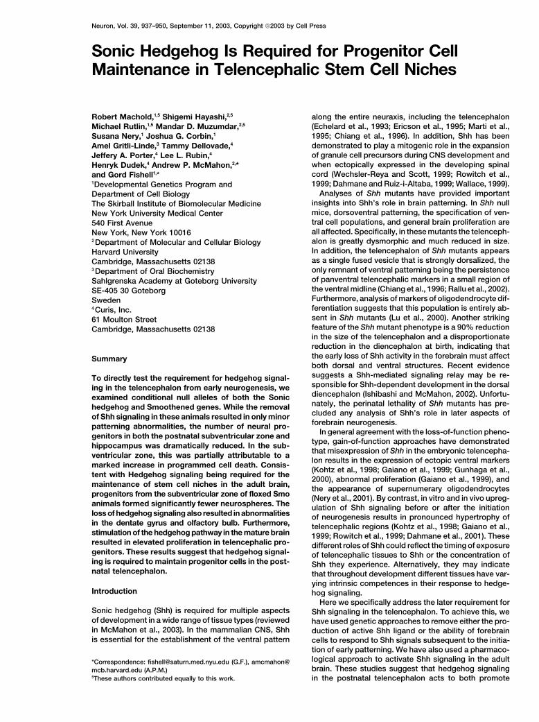
Neuron, Vol. 39, 937–950, September 11, 2003, Copyright 2003 by Cell Press
Sonic Hedgehog Is Required for Progenitor CellMaintenance in Telencephalic Stem Cell Niches
along the entire neuraxis, including the telencephalon(Echelard et al., 1993; Ericson et al., 1995; Marti et al.,1995; Chiang et al., 1996). In addition, Shh has been
Robert Machold,1,5 Shigemi Hayashi,2,5
Michael Rutlin,1,5 Mandar D. Muzumdar,2,5
Susana Nery,1 Joshua G. Corbin,1
demonstrated to play a mitogenic role in the expansionAmel Gritli-Linde,3 Tammy Dellovade,4
of granule cell precursors during CNS development andJeffery A. Porter,4 Lee L. Rubin,4
when ectopically expressed in the developing spinalHenryk Dudek,4 Andrew P. McMahon,2,*cord (Wechsler-Reya and Scott, 1999; Rowitch et al.,and Gord Fishell1,*1999; Dahmane and Ruiz-i-Altaba, 1999; Wallace, 1999).1Developmental Genetics Program and
Analyses of Shh mutants have provided importantDepartment of Cell Biologyinsights into Shh’s role in brain patterning. In Shh nullThe Skirball Institute of Biomolecular Medicinemice, dorsoventral patterning, the specification of ven-New York University Medical Centertral cell populations, and general brain proliferation are540 First Avenueall affected. Specifically, in these mutants the telenceph-New York, New York 10016alon is greatly dysmorphic and much reduced in size.2 Department of Molecular and Cellular BiologyIn addition, the telencephalon of Shh mutants appearsHarvard Universityas a single fused vesicle that is strongly dorsalized, theCambridge, Massachusetts 02138only remnant of ventral patterning being the persistence3 Department of Oral Biochemistryof panventral telencephalic markers in a small region ofSahlgrenska Academy at Goteborg Universitythe ventral midline (Chiang et al., 1996; Rallu et al., 2002).SE-405 30 GoteborgFurthermore, analysis of markers of oligodendrocyte dif-Swedenferentiation suggests that this population is entirely ab-4 Curis, Inc.sent in Shh mutants (Lu et al., 2000). Another striking61 Moulton Streetfeature of the Shh mutant phenotype is a 90% reductionCambridge, Massachusetts 02138in the size of the telencephalon and a disproportionatereduction in the diencephalon at birth, indicating thatthe early loss of Shh activity in the forebrain must affectSummaryboth dorsal and ventral structures. Recent evidencesuggests a Shh-mediated signaling relay may be re-To directly test the requirement for hedgehog signal-sponsible for Shh-dependent development in the dorsaling in the telencephalon from early neurogenesis, wediencephalon (Ishibashi and McMahon, 2002). Unfortu-examined conditional null alleles of both the Sonicnately, the perinatal lethality of Shh mutants has pre-hedgehog and Smoothened genes. While the removalcluded any analysis of Shh’s role in later aspects ofof Shh signaling in these animals resulted in only minorforebrain neurogenesis.patterning abnormalities, the number of neural pro-
In general agreement with the loss-of-function pheno-genitors in both the postnatal subventricular zone andtype, gain-of-function approaches have demonstratedhippocampus was dramatically reduced. In the sub-that misexpression of Shh in the embryonic telencepha-ventricular zone, this was partially attributable to alon results in the expression of ectopic ventral markers
marked increase in programmed cell death. Consis-(Kohtz et al., 1998; Gaiano et al., 1999; Gunhaga et al.,
tent with Hedgehog signaling being required for the2000), abnormal proliferation (Gaiano et al., 1999), and
maintenance of stem cell niches in the adult brain, the appearance of supernumerary oligodendrocytesprogenitors from the subventricular zone of floxed Smo (Nery et al., 2001). By contrast, in vitro and in vivo upreg-animals formed significantly fewer neurospheres. The ulation of Shh signaling before or after the initiationloss of hedgehog signaling also resulted in abnormalities of neurogenesis results in pronounced hypertrophy ofin the dentate gyrus and olfactory bulb. Furthermore, telencephalic regions (Kohtz et al., 1998; Gaiano et al.,stimulation of the hedgehog pathway in the mature brain 1999; Rowitch et al., 1999; Dahmane et al., 2001). Theseresulted in elevated proliferation in telencephalic pro- different roles of Shh could reflect the timing of exposuregenitors. These results suggest that hedgehog signal- of telencephalic tissues to Shh or the concentration ofing is required to maintain progenitor cells in the post- Shh they experience. Alternatively, they may indicatenatal telencephalon. that throughout development different tissues have var-
ying intrinsic competences in their response to hedge-Introduction hog signaling.
Here we specifically address the later requirement forSonic hedgehog (Shh) is required for multiple aspects Shh signaling in the telencephalon. To achieve this, weof development in a wide range of tissue types (reviewed have used genetic approaches to remove either the pro-in McMahon et al., 2003). In the mammalian CNS, Shh duction of active Shh ligand or the ability of forebrainis essential for the establishment of the ventral pattern cells to respond to Shh signals subsequent to the initia-
tion of early patterning. We have also used a pharmaco-logical approach to activate Shh signaling in the adult*Correspondence: [email protected] (G.F.), amcmahon@brain. These studies suggest that hedgehog signalingmcb.harvard.edu (A.P.M.)
5These authors contributed equally to this work. in the postnatal telencephalon acts to both promote
Neuron938
proliferation and maintain populations of neural progeni-tors. Hence, Shh signaling in the mammalian telenceph-alon may participate in the maintenance of a neural stemcell niche akin to the action of the hedgehog pathwayin Drosophila ovaries and testes (Forbes et al., 1996a,1996b; Zhang and Kalderon, 2000).
Results
In this study, we have used a conditional loss-of-func-tion approach to examine the role of Shh signaling dur-ing telencephalic development after the initiation of neu-rogenesis. This was achieved through the use of aconditional Shh allele and a transgenic line in which theneural-specific Nestin promoter/enhancer directs theexpression of Cre recombinase (NCre) in neural progenitorpopulations. The existence of two other hedgehog pro-teins (Echelard et al., 1993) raises the possibility that inaddition to Shh these other ligands may also have a rolein telencephalic development. Indeed, such a mecha-nism has been proposed to reconcile the finding that,while telencephalic tissue from Shh null animals cangenerate oligodendrocytes in vitro (Nery et al., 2001),cyclopamine-treated telencephalic tissue cannot (Tekki-Kessaris et al., 2001). To rule out the possibility of com-pensatory hedgehog signaling, we have also examinedthe phenotype generated by the removal of Smo genefunction in NCre mice. The single mammalian Smo gene,like its Drosophila counterpart, is essential for transduc-tion of all hedgehog signaling (Zhang et al., 2001).
Loss of Hedgehog Signaling in Shhn/c;Nestincre
or Smon/c;Nestincre AnimalsNestin, a ubiquitous marker of neural progenitors, isactivated after the initiation of early patterning events
Figure 1. Shh Signaling Is Abolished in the Forebrain of Shhn/c;NCrein the mammalian neural tube (Zimmerman et al., 1994;and Smon/c;NCre Embryos by E12.5Lendahl et al., 1990). To examine the distribution of cells(A) A R26R;NCre brain is shown. Here, �-galactosidase histochemistryin which Cre is expressed, the Ncre animals were crossedprovides an indirect assay of the extent of Cre activity in theseto a R26R line in which Cre-mediated recombinationanimals. Note that the entire telencephalon appears to be blue. (B)
transcriptionally activates a LacZ reporter allele (Sori- and (C) show in situ hybridizations of wild-type (B) and Shhn/c;NCre
ano, 1999; Graus-Porta et al., 2001). These results con- (C) animals using an exon2 Shh probe that recognizes the regionsfirmed the complex spatial and temporal activation of removed in Cre-mediated recombination. Note in (C) the absence
of exon2-containing transcripts in telencephalic tissue, indicatingthe Nestin allele and demonstrated that most if not allthe successful deletion of Shh exon2 in Shhn/c;NCre animals by E12.5.telencephalic neural precursors had undergone a Cre-In situ hybridization of targets of the Shh signaling pathway, Gli1mediated recombination by E12.5 (Figure 1A; Graus-(D and E), and Ptch1 (F and G) on E12.5 wild-type (D and F) and
Porta et al., 2001). To evaluate the effectiveness of this Smon/c;NCre (E and G) embryo brains. Note that the expression oftransgene in the removal of Shh itself in Shhn/c;Ncre em- Gli1 and Ptch1 in the forebrain was completely abolished in thebryos, we used an exon2-specific probe that recognizes mutants (E and G). Instead, ectopic expression of Gli1 and Ptch1
was observed in the dorsal midline of the diencephalon of mutantthe region of the Shh transcript that is deleted after Cre-embryos (arrowhead in [E] and [G]). This occurs due to the combinedmediated recombination. Expression of an active Shhresult of the removal of Ptch1-mediated inhibition in more ventraltranscript was greatly reduced by E10.5, as was Shhregions and the lack of Nestin-mediated Cre activity in the dorsal
signaling in target cells in Shhn/c;Ncre brains (Figures 1B diencephalon.and 1C and data not shown). Similarly, when Smon/c;Ncre
embryos were examined for the expression of Patched1(Ptch1) and Gli1, whose expression provides a direct sion of the floxed allele was directed by Cre expression
from the Foxg1 loci (Hebert and McConnell, 2000). Inindication of Shh signaling (reviewed in Ingham andMcMahon, 2001), we observed that Shh signaling was these mice removal of Smo gene function begins at E8.5
and is complete by E10 (Hebert and McConnell, 2000;almost completely absent from its normal telencephalicdomain by E12.5 in these animals (Figures 1D–1G). In a G.F., unpublished data). In contrast to Smon/c;Ncre brains,
the telencephalon of Smon/c;Foxg1cre/� mice is stronglyseparate study, we have examined the phenotype re-sulting from earlier removal of hedgehog signaling within dorsalized (G.F., unpublished data). This demonstrates
that hedgehog’s requirement in regionalization of thethe telencephalon by examining Smon/c mice where exci-
Shh in Forebrain Development939
brain precedes the ages studied in the present experi- In addition to oligodendrocytes, interneurons are theother major population that arises from the MGE (Ander-ments.
Interestingly in Smon/c;Ncre mutants, we observed ec- son et al., 1997; Wichterle et al., 2001). Consistent withthe observation that Dlx2 expression is unchanged intopic activation of Ptch1 and Gli1 in the dorsal dienceph-
alon, in an area where Shh signaling is preserved in these mutants (data not shown), we detected no propor-tional decrease in the numbers of GABAergic cells inmutant animals as a result of the absence of Nestin-
mediated Cre expression in this region (Figures 1E and the cortex, hippocampus, or striatum (SupplementalFigure S1 at http://www.neuron.org/cgi/content/full/39/1G). This reflects a dramatically increased range of Shh
action in the absence of Ptch1-mediated inhibition of 6/937/DC1).Shh movement, as observed in other systems (e.g., Chenand Struhl, 1996; Burke et al., 1999; Long et al., 2001). The Expression of Shh, Ptch1, and Gli1Given the similarities in the loss of Ptch1 and Gli1 in in Postnatal TelencephalonShhn/c;Ncre and Smon/c;Ncre animals and the observation In order to explore which populations in the postnatalthat morphologically the phenotypes of these alleles (P15) brain might be affected by the loss of hedgehogwere indistinguishable, we focused our more detailed signaling, we examined the distribution of Shh and itsanalysis on Smon/c;Ncre animals. transcriptional targets Ptch1 and Gli1 (reviewed in Ing-
ham and McMahon, 2001). Immunostaining using a Shh-specific antibody revealed that Shh is broadly distrib-Telencephalic Patterning at E12.5 in Smon/c;Ncre
Mice Appeared Largely Normal uted in the P15 telencephalon (Figures 3A and 3B). Themost prominent areas of Shh immunoreactivity are inIn Shh null mutants, most aspects of ventral patterning
in the telencephalon are abrogated (Chiang et al., 1996; the ventral telencephalon where embryonic Shh expres-sion is observed. Specifically, we see Shh-positive cellsRallu et al., 2002). By contrast, analysis of both telen-
cephalic morphology and the distribution of regional within the ventral accumbens, entorhinal cortex, andventral septum, along the path transited by cells in themarkers indicated that patterning appeared to be rela-
tively unperturbed in Smon/c;Ncre animals at E12.5. The rostral migratory stream. Furthermore, within layer 3 ofthe cortex and along the corpus callosum we also ob-distribution of dorsally expressed markers such as Pax6
and Ngn2 was indistinguishable in mutant and wild- serve Shh immunoreactivity (asterisks in Figure 3A anddata not shown). Finally, a population of axons in thetype animals (Figures 2A and 2B and data not shown).
Similarly, the expression domain of Gsh2 and Dlx2 (Fig- medial septum is also immunopositive for Shh (arrow inFigure 3A). This observation is consistent with a recentures 2A and 2B and data not shown), which demarcate
the entire ventral telencephalon (Corbin et al., 2000; Yun report from the Schaffer laboratory (Lai et al., 2003),which reported that the septal (i.e., fimbria) fibers pro-et al., 2001; Toresson et al., 2000), was unchanged in
distribution, although in some cases levels of expression jecting to the hippocampus are a source of Shh withinthis structure. Further supporting the observations ofwere moderately reduced (data not shown).
As might be expected from studies to date, it was the Lai et al. (2003), we observe Shh staining in the whitematter tracks in the CA3 and hilus regions of the hippo-ventral-most structure in the telencephalon, the medial
ganglionic eminence (MGE), that was most affected in campus (arrows in Figure 3B). Examination of mice thatare heterozygous for a LacZ reporter under the controlthese mutants. In Smon/c;Ncre embryos, the MGE was
reduced in size by 50% and was often fused with the of the endogenous Shh promoter displayed a similarpattern of cellular Shh expression (Figures 3C and 3D). InLGE, the structure immediately lateral to the MGE (com-
pare Figures 2E and 2H), a phenotype resembling that addition, they revealed the existence of Shh-expressingcells within the hilus of the dentate gyrus, adjacent to theobserved in Nkx2.1 mutants. However, the expressivity
of this phenotype varied (compare Figures 2H and 2L). hippocampal stem cell population (arrowhead in Figure3D). As this staining was not evident in antibody visual-Notably, even in the most severely affected mutants, the
remaining Nkx2.1 expression had a normal distribution ization of Shh, it seems likely that these cells only ex-press low levels of Shh and require the enzymatic ampli-(Figures 2C–2E and 2F–2H). Furthermore, the MGE in
Smon/c;Ncre brains maintained a normal pattern of gene fication provided by LacZ staining to visualize. Theexpression of LacZ directed through either the Ptch1 orexpression, as shown by the persistence of the normal
domain of Lhx6 expression (data not shown). This sug- Gli1 loci was generally consistent in indicating in whichpopulations the hedgehog pathway was activated. Thegests that the defect observed at E12.5 in the Smon/c;Ncre
telencephalon resulted from a failure in the expansion or expression of LacZ directed by the Ptch1 loci wasbroader than that seen from the Gli1 loci. The observa-survival of MGE progenitors within this region rather
than a misspecification of their regional identity. One tion that Gli1 expression more closely matched sourcesof endogenous Shh suggests that Gli1 is the more accu-population that was affected in these mutants was oligo-
dendrocyte precursors, as reflected by the loss or re- rate readout of Shh activation. Furthermore, embryonicstudies indicate that all Gli1 expression is Shh depen-duction (depending on the anteroposterior level exam-
ined) of the preoligodendrocyte marker Sox10 (Figures dent, whereas Ptch1, the Shh receptor, is expressedat basal levels in the absence of any direct Hedgehog2I and 2L). The underlying cause of the reduced MGE
size and decreased oligodendrocyte precursor numbers signaling input (Bai et al., 2002).Gli1-LacZ and Ptch1-LacZ expression was observedin affected mutants is unclear. We observed no obvious
differences in the levels of proliferation or cell survival in regions where proliferation persisted in the telenceph-alon (Figures 3G, 3J, 3L, and 3O). Specifically, within thewithin the MGE of wild-type versus mutant embryos at
E12.5 (Figure 2J, 2K, 2M, and 2N). SVZ, rostral migratory stream, and subcallosal region,
Neuron940
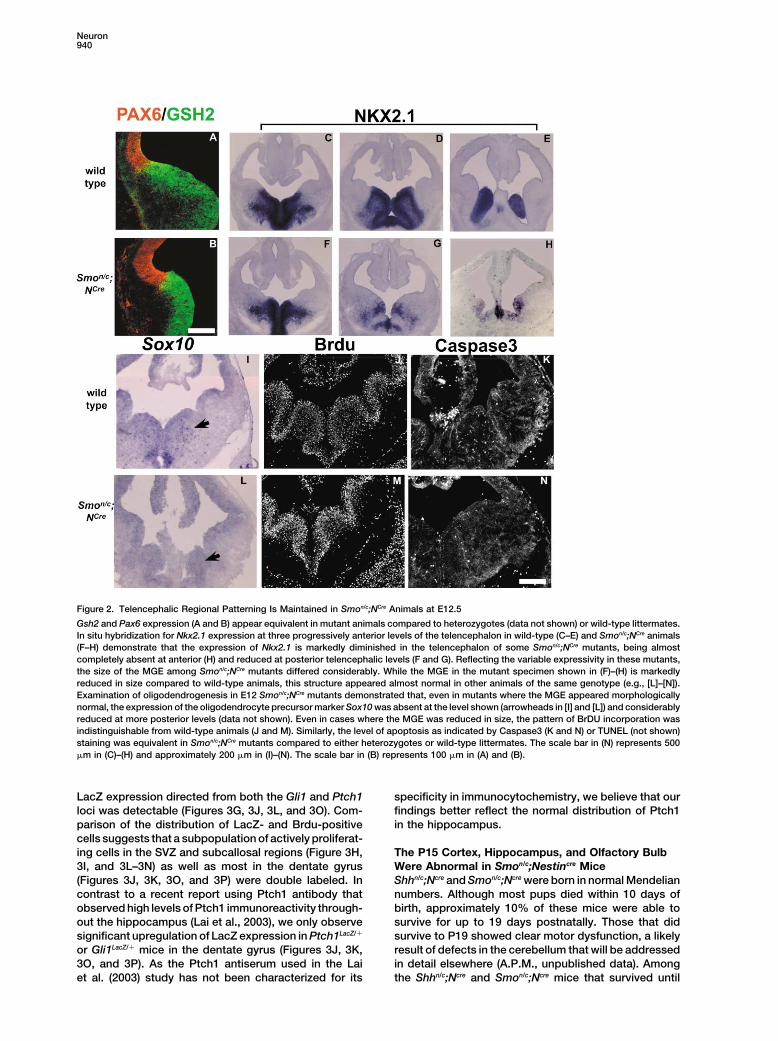
Figure 2. Telencephalic Regional Patterning Is Maintained in Smon/c;NCre Animals at E12.5
Gsh2 and Pax6 expression (A and B) appear equivalent in mutant animals compared to heterozygotes (data not shown) or wild-type littermates.In situ hybridization for Nkx2.1 expression at three progressively anterior levels of the telencephalon in wild-type (C–E) and Smon/c;NCre animals(F–H) demonstrate that the expression of Nkx2.1 is markedly diminished in the telencephalon of some Smon/c;NCre mutants, being almostcompletely absent at anterior (H) and reduced at posterior telencephalic levels (F and G). Reflecting the variable expressivity in these mutants,the size of the MGE among Smon/c;NCre mutants differed considerably. While the MGE in the mutant specimen shown in (F)–(H) is markedlyreduced in size compared to wild-type animals, this structure appeared almost normal in other animals of the same genotype (e.g., [L]–[N]).Examination of oligodendrogenesis in E12 Smon/c;NCre mutants demonstrated that, even in mutants where the MGE appeared morphologicallynormal, the expression of the oligodendrocyte precursor marker Sox10 was absent at the level shown (arrowheads in [I] and [L]) and considerablyreduced at more posterior levels (data not shown). Even in cases where the MGE was reduced in size, the pattern of BrDU incorporation wasindistinguishable from wild-type animals (J and M). Similarly, the level of apoptosis as indicated by Caspase3 (K and N) or TUNEL (not shown)staining was equivalent in Smon/c;NCre mutants compared to either heterozygotes or wild-type littermates. The scale bar in (N) represents 500�m in (C)–(H) and approximately 200 �m in (I)–(N). The scale bar in (B) represents 100 �m in (A) and (B).
LacZ expression directed from both the Gli1 and Ptch1 specificity in immunocytochemistry, we believe that ourfindings better reflect the normal distribution of Ptch1loci was detectable (Figures 3G, 3J, 3L, and 3O). Com-
parison of the distribution of LacZ- and Brdu-positive in the hippocampus.cells suggests that a subpopulation of actively proliferat-ing cells in the SVZ and subcallosal regions (Figure 3H, The P15 Cortex, Hippocampus, and Olfactory Bulb
Were Abnormal in Smon/c;Nestincre Mice3I, and 3L–3N) as well as most in the dentate gyrus(Figures 3J, 3K, 3O, and 3P) were double labeled. In Shhn/c;Ncre and Smon/c;Ncre were born in normal Mendelian
numbers. Although most pups died within 10 days ofcontrast to a recent report using Ptch1 antibody thatobserved high levels of Ptch1 immunoreactivity through- birth, approximately 10% of these mice were able to
survive for up to 19 days postnatally. Those that didout the hippocampus (Lai et al., 2003), we only observesignificant upregulation of LacZ expression in Ptch1LacZ/� survive to P19 showed clear motor dysfunction, a likely
result of defects in the cerebellum that will be addressedor Gli1LacZ/� mice in the dentate gyrus (Figures 3J, 3K,3O, and 3P). As the Ptch1 antiserum used in the Lai in detail elsewhere (A.P.M., unpublished data). Among
the Shhn/c;Ncre and Smon/c;Ncre mice that survived untilet al. (2003) study has not been characterized for its

Shh in Forebrain Development941
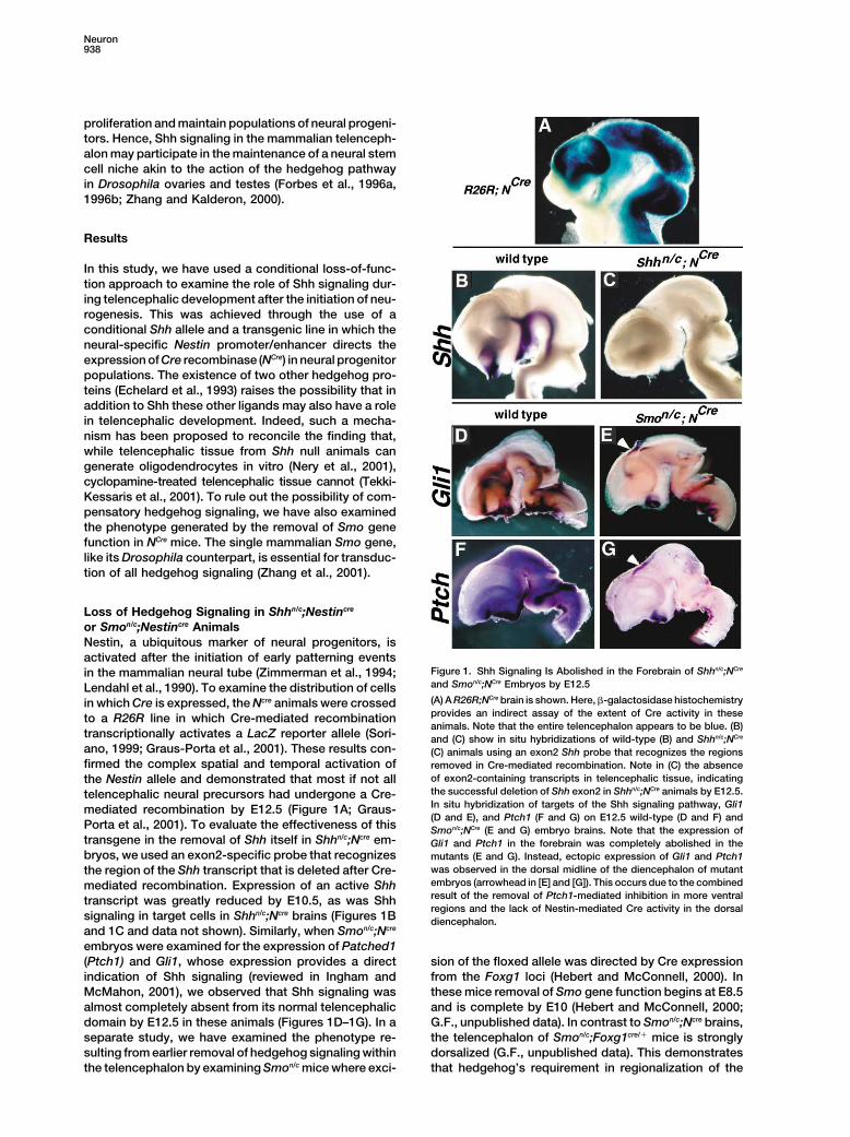
Figure 3. Postnatal Expression of Shh, Gli1, and Ptch1 in Proliferating and Postmitotic Telencephalon
(A) and (B) show immunocytochemical localization of Shh in the adult telencephalon. Arrowhead in (A) shows the expression of Shh in theventral accumbens, entorhinal cortex, and ventral septum. Arrow in (A) shows axonal labeling in the medial septum. Asterisks in (A) showexpression of Shh beneath the corpus callosum (lower two) and in layer 3 of the cortex (upper asterisk). Arrows in (B) show the Shh expressionin the CA3 and hilus regions of the hippocampus (this Shh presumably originates from the efferent fimbria fibers, including the medial septalfibers shown in [A]). (C) illustrates that a similar pattern of LacZ expression is observed in ShhLacZ/� animals as seen with immunocytochemistry.(D) shows that LacZ-positive cells (arrowhead) are observed in the hilus of the hippocampus in ShhLacZ/� animals (dashed line indicates theapical surface of the dentate gyrus). (E) is a photomicrograph of the striatum and SVZ of a P15 Ptch1LacZ/� mouse double labeled for BrdU(green) and LacZ (red). (F) is an enlargement of the boxed region in (E). In these animals, only a few cells in the SVZ are double labeled. (G)and (J) show the striatum and associated SVZ (G) and the hippocampus (J) of P15 Gli1LacZ/� mice. (L) and (O) show the striatum and associatedSVZ (L) and the hippocampus (O) of P15 Ptch1LacZ/� mice double labeled for LacZ (green) and BrDU (brown). (H), (I), and (K) show enlargementsof the double labeling for LacZ and BrDU in adjacent sections from those shown in (G) or (J), respectively, from the approximate areas shownin (G) or (J). (H) is the area shown in the upper box and (I) is the lower box of (G), while (K) is the area shown in the box in (J). (M), (N), and(P) show enlargements of the double labeling for LacZ and BrDU in sections shown in (L) or (O), respectively. (M) is the area shown in theupper box and (N) is the lower box of (L), while (P) is the area shown in the box in (O). The small rip in the tissue in (A) is the unfortunateconsequence of the dehydration step necessary for optimal Shh immunostaining. The scale bar in (F) represents 750 �m in (A), 200 �m in(B), 2000 �m in (C) and (E), 150 �m in (D) and (F). The scale bar in (P) represents 1500 �m in (G) and (L), 200 �m in (J) and (O), and 75 �m in(H), (I), (K), (M), (N), and (P).
Neuron942
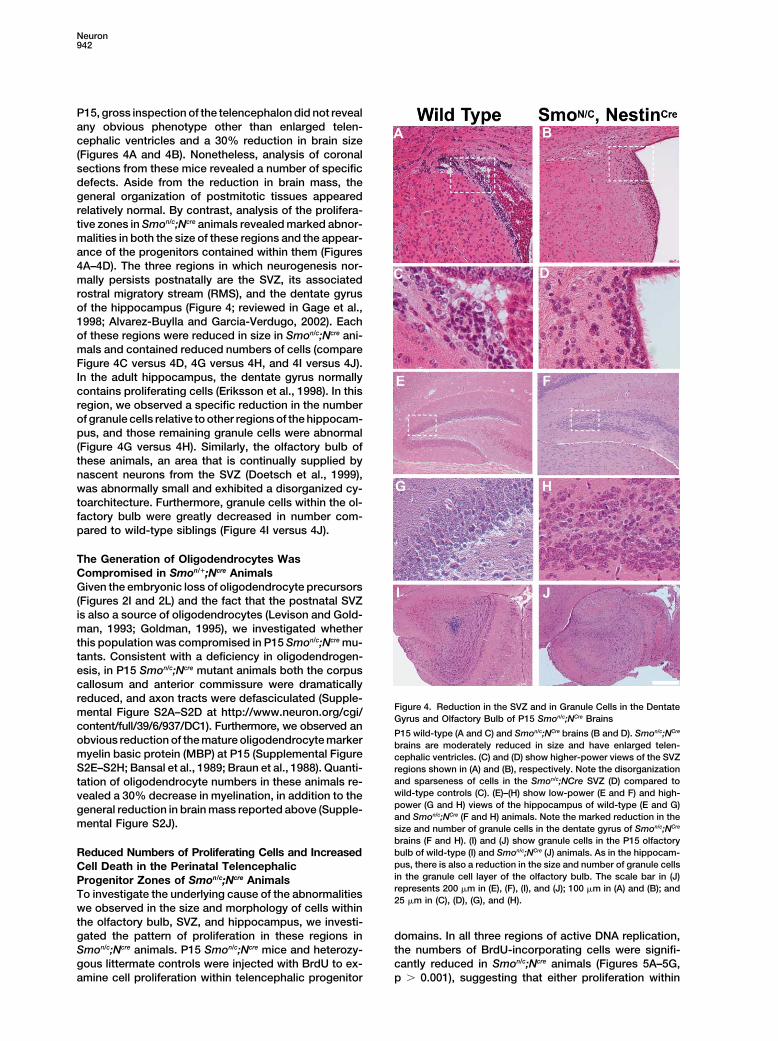
P15, gross inspection of the telencephalon did not revealany obvious phenotype other than enlarged telen-cephalic ventricles and a 30% reduction in brain size(Figures 4A and 4B). Nonetheless, analysis of coronalsections from these mice revealed a number of specificdefects. Aside from the reduction in brain mass, thegeneral organization of postmitotic tissues appearedrelatively normal. By contrast, analysis of the prolifera-tive zones in Smon/c;Ncre animals revealed marked abnor-malities in both the size of these regions and the appear-ance of the progenitors contained within them (Figures4A–4D). The three regions in which neurogenesis nor-mally persists postnatally are the SVZ, its associatedrostral migratory stream (RMS), and the dentate gyrusof the hippocampus (Figure 4; reviewed in Gage et al.,1998; Alvarez-Buylla and Garcia-Verdugo, 2002). Eachof these regions were reduced in size in Smon/c;Ncre ani-mals and contained reduced numbers of cells (compareFigure 4C versus 4D, 4G versus 4H, and 4I versus 4J).In the adult hippocampus, the dentate gyrus normallycontains proliferating cells (Eriksson et al., 1998). In thisregion, we observed a specific reduction in the numberof granule cells relative to other regions of the hippocam-pus, and those remaining granule cells were abnormal(Figure 4G versus 4H). Similarly, the olfactory bulb ofthese animals, an area that is continually supplied bynascent neurons from the SVZ (Doetsch et al., 1999),was abnormally small and exhibited a disorganized cy-toarchitecture. Furthermore, granule cells within the ol-factory bulb were greatly decreased in number com-pared to wild-type siblings (Figure 4I versus 4J).
The Generation of Oligodendrocytes WasCompromised in Smon/�;Ncre AnimalsGiven the embryonic loss of oligodendrocyte precursors(Figures 2I and 2L) and the fact that the postnatal SVZis also a source of oligodendrocytes (Levison and Gold-man, 1993; Goldman, 1995), we investigated whetherthis population was compromised in P15 Smon/c;Ncre mu-tants. Consistent with a deficiency in oligodendrogen-esis, in P15 Smon/c;Ncre mutant animals both the corpuscallosum and anterior commissure were dramaticallyreduced, and axon tracts were defasciculated (Supple-
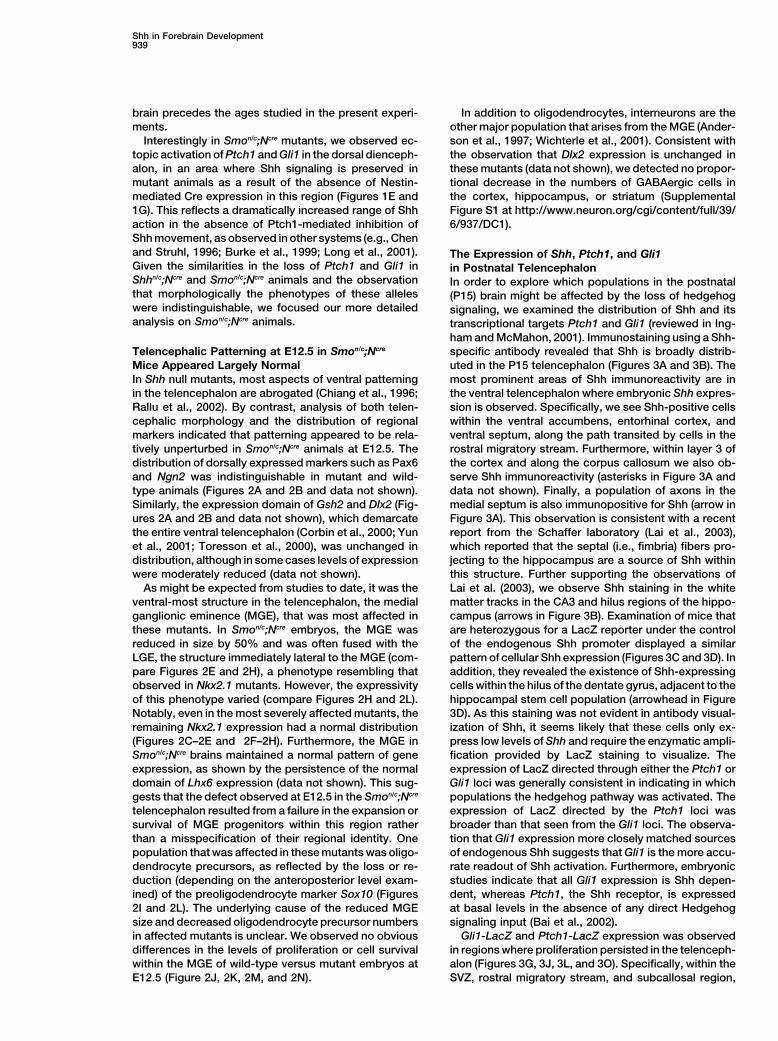
Figure 4. Reduction in the SVZ and in Granule Cells in the Dentatemental Figure S2A–S2D at http://www.neuron.org/cgi/Gyrus and Olfactory Bulb of P15 Smon/c;NCre Brains
content/full/39/6/937/DC1). Furthermore, we observed anP15 wild-type (A and C) and Smon/c;NCre brains (B and D). Smon/c;NCre
obvious reduction of the mature oligodendrocyte marker brains are moderately reduced in size and have enlarged telen-myelin basic protein (MBP) at P15 (Supplemental Figure cephalic ventricles. (C) and (D) show higher-power views of the SVZS2E–S2H; Bansal et al., 1989; Braun et al., 1988). Quanti- regions shown in (A) and (B), respectively. Note the disorganization
and sparseness of cells in the Smon/c;NCre SVZ (D) compared totation of oligodendrocyte numbers in these animals re-wild-type controls (C). (E)–(H) show low-power (E and F) and high-vealed a 30% decrease in myelination, in addition to thepower (G and H) views of the hippocampus of wild-type (E and G)general reduction in brain mass reported above (Supple-and Smon/c;NCre (F and H) animals. Note the marked reduction in the
mental Figure S2J). size and number of granule cells in the dentate gyrus of Smon/c;NCre
brains (F and H). (I) and (J) show granule cells in the P15 olfactorybulb of wild-type (I) and Smon/c;NCre (J) animals. As in the hippocam-Reduced Numbers of Proliferating Cells and Increasedpus, there is also a reduction in the size and number of granule cellsCell Death in the Perinatal Telencephalicin the granule cell layer of the olfactory bulb. The scale bar in (J)Progenitor Zones of Smon/c;Ncre Animalsrepresents 200 �m in (E), (F), (I), and (J); 100 �m in (A) and (B); andTo investigate the underlying cause of the abnormalities25 �m in (C), (D), (G), and (H).
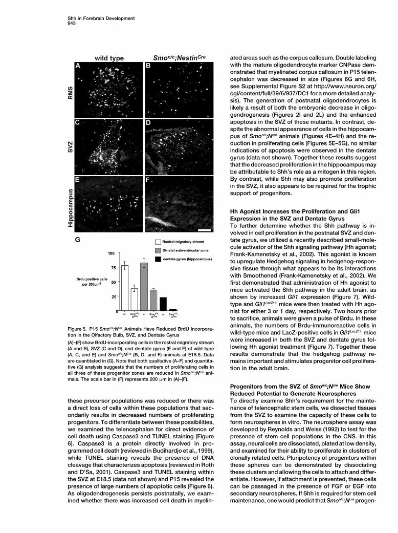
we observed in the size and morphology of cells withinthe olfactory bulb, SVZ, and hippocampus, we investi-gated the pattern of proliferation in these regions in domains. In all three regions of active DNA replication,
the numbers of BrdU-incorporating cells were signifi-Smon/c;Ncre animals. P15 Smon/c;Ncre mice and heterozy-gous littermate controls were injected with BrdU to ex- cantly reduced in Smon/c;Ncre animals (Figures 5A–5G,
p � 0.001), suggesting that either proliferation withinamine cell proliferation within telencephalic progenitor
Shh in Forebrain Development943
ated areas such as the corpus callosum. Double labelingwith the mature oligodendrocyte marker CNPase dem-onstrated that myelinated corpus callosum in P15 telen-cephalon was decreased in size (Figures 6G and 6H,see Supplemental Figure S2 at http://www.neuron.org/cgi/content/full/39/6/937/DC1 for a more detailed analy-sis). The generation of postnatal oligodendrocytes islikely a result of both the embryonic decrease in oligo-gendrogenesis (Figures 2I and 2L) and the enhancedapoptosis in the SVZ of these mutants. In contrast, de-spite the abnormal appearance of cells in the hippocam-pus of Smon/c;Ncre animals (Figures 4E–4H) and the re-duction in proliferating cells (Figures 5E–5G), no similarindications of apoptosis were observed in the dentategyrus (data not shown). Together these results suggestthat the decreased proliferation in the hippocampus maybe attributable to Shh’s role as a mitogen in this region.By contrast, while Shh may also promote proliferationin the SVZ, it also appears to be required for the trophicsupport of progenitors.
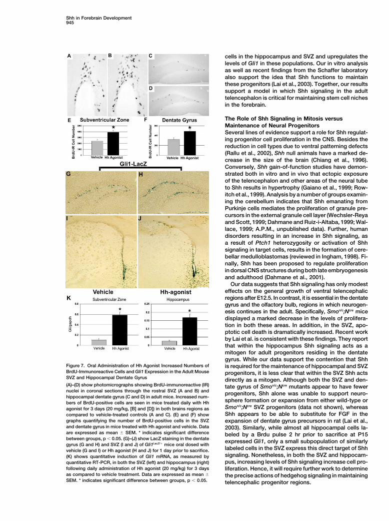
Hh Agonist Increases the Proliferation and Gli1Expression in the SVZ and Dentate GyrusTo further determine whether the Shh pathway is in-volved in cell proliferation in the postnatal SVZ and den-tate gyrus, we utilized a recently described small-mole-cule activator of the Shh signaling pathway (Hh agonist;Frank-Kamenetsky et al., 2002). This agonist is knownto upregulate Hedgehog signaling in hedgehog-respon-sive tissue through what appears to be its interactionswith Smoothened (Frank-Kamenetsky et al., 2002). Wefirst demonstrated that administration of Hh agonist tomice activated the Shh pathway in the adult brain, asshown by increased Gli1 expression (Figure 7). Wild-type and Gli1LacZ/� mice were then treated with Hh ago-nist for either 3 or 1 day, respectively. Two hours priorto sacrifice, animals were given a pulse of Brdu. In theseanimals, the numbers of Brdu-immunoreactive cells in
Figure 5. P15 Smon/c;NCre Animals Have Reduced BrdU Incorpora-wild-type mice and LacZ-positive cells in Gli1LacZ/� micetion in the Olfactory Bulb, SVZ, and Dentate Gyruswere increased in both the SVZ and dentate gyrus fol-(A)–(F) show BrdU-incorporating cells in the rostral migratory streamlowing Hh agonist treatment (Figure 7). Together these(A and B), SVZ (C and D), and dentate gyrus (E and F) of wild-typeresults demonstrate that the hedgehog pathway re-(A, C, and E) and Smon/c;NCre (B, D, and F) animals at E18.5. Data
are quantitated in (G). Note that both qualitative (A–F) and quantita- mains important and stimulates progenitor cell prolifera-tive (G) analysis suggests that the numbers of proliferating cells in tion in the adult brain.all three of these progenitor zones are reduced in Smon/c;NCre ani-mals. The scale bar in (F) represents 200 �m in (A)–(F).
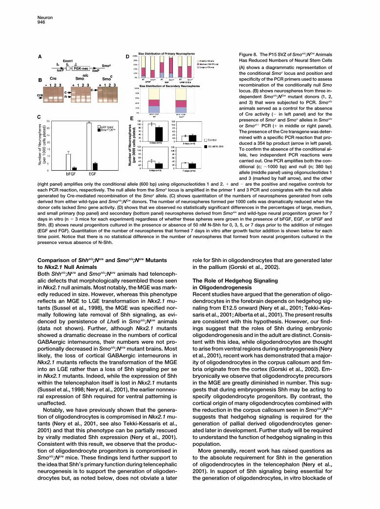
Progenitors from the SVZ of Smon/c;Ncre Mice ShowReduced Potential to Generate NeurospheresTo directly examine Shh’s requirement for the mainte-these precursor populations was reduced or there was
a direct loss of cells within these populations that sec- nance of telencephalic stem cells, we dissected tissuesfrom the SVZ to examine the capacity of these cells toondarily results in decreased numbers of proliferating
progenitors. To differentiate between these possibilities, form neurospheres in vitro. The neurosphere assay wasdeveloped by Reynolds and Weiss (1992) to test for thewe examined the telencephalon for direct evidence of
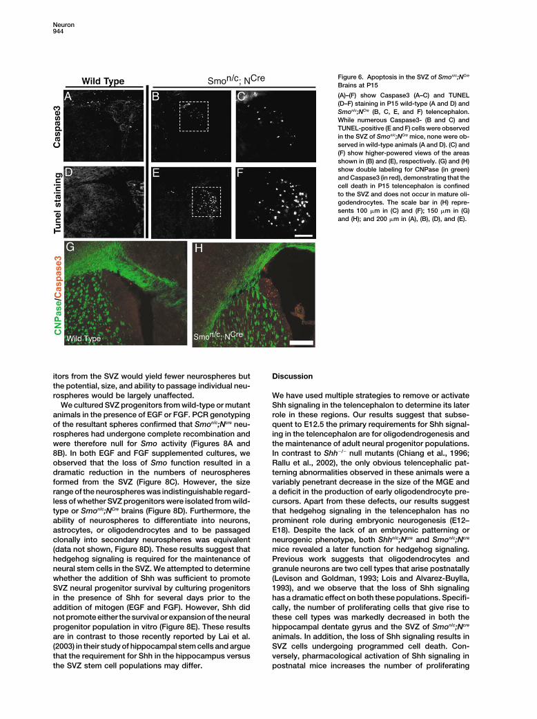
cell death using Caspase3 and TUNEL staining (Figure presence of stem cell populations in the CNS. In thisassay, neural cells are dissociated, plated at low density,6). Caspase3 is a protein directly involved in pro-
grammed cell death (reviewed in Budihardjo et al., 1999), and examined for their ability to proliferate in clusters ofclonally related cells. Pluripotency of progenitors withinwhile TUNEL staining reveals the presence of DNA
cleavage that characterizes apoptosis (reviewed in Roth these spheres can be demonstrated by dissociatingthese clusters and allowing the cells to attach and differ-and D’Sa, 2001). Caspase3 and TUNEL staining within
the SVZ at E18.5 (data not shown) and P15 revealed the entiate. However, if attachment is prevented, these cellscan be passaged in the presence of FGF or EGF intopresence of large numbers of apoptotic cells (Figure 6).
As oligodendrogenesis persists postnatally, we exam- secondary neurospheres. If Shh is required for stem cellmaintenance, one would predict that Smon/c;Ncre progen-ined whether there was increased cell death in myelin-
Neuron944
Figure 6. Apoptosis in the SVZ of Smon/c;NCre
Brains at P15
(A)–(F) show Caspase3 (A–C) and TUNEL(D–F) staining in P15 wild-type (A and D) andSmon/c;NCre (B, C, E, and F) telencephalon.While numerous Caspase3- (B and C) andTUNEL-positive (E and F) cells were observedin the SVZ of Smon/c;NCre mice, none were ob-served in wild-type animals (A and D). (C) and(F) show higher-powered views of the areasshown in (B) and (E), respectively. (G) and (H)show double labeling for CNPase (in green)and Caspase3 (in red), demonstrating that thecell death in P15 telencephalon is confinedto the SVZ and does not occur in mature oli-godendrocytes. The scale bar in (H) repre-sents 100 �m in (C) and (F); 150 �m in (G)and (H); and 200 �m in (A), (B), (D), and (E).
itors from the SVZ would yield fewer neurospheres but Discussionthe potential, size, and ability to passage individual neu-rospheres would be largely unaffected. We have used multiple strategies to remove or activate
Shh signaling in the telencephalon to determine its laterWe cultured SVZ progenitors from wild-type or mutantanimals in the presence of EGF or FGF. PCR genotyping role in these regions. Our results suggest that subse-
quent to E12.5 the primary requirements for Shh signal-of the resultant spheres confirmed that Smon/c;Ncre neu-rospheres had undergone complete recombination and ing in the telencephalon are for oligodendrogenesis and
the maintenance of adult neural progenitor populations.were therefore null for Smo activity (Figures 8A and8B). In both EGF and FGF supplemented cultures, we In contrast to Shh�/� null mutants (Chiang et al., 1996;
Rallu et al., 2002), the only obvious telencephalic pat-observed that the loss of Smo function resulted in adramatic reduction in the numbers of neurospheres terning abnormalities observed in these animals were a
variably penetrant decrease in the size of the MGE andformed from the SVZ (Figure 8C). However, the sizerange of the neurospheres was indistinguishable regard- a deficit in the production of early oligodendrocyte pre-
cursors. Apart from these defects, our results suggestless of whether SVZ progenitors were isolated from wild-type or Smon/c;NCre brains (Figure 8D). Furthermore, the that hedgehog signaling in the telencephalon has no
prominent role during embryonic neurogenesis (E12–ability of neurospheres to differentiate into neurons,astrocytes, or oligodendrocytes and to be passaged E18). Despite the lack of an embryonic patterning or
neurogenic phenotype, both Shhn/c;Ncre and Smon/c;Ncreclonally into secondary neurospheres was equivalent(data not shown, Figure 8D). These results suggest that mice revealed a later function for hedgehog signaling.
Previous work suggests that oligodendrocytes andhedgehog signaling is required for the maintenance ofneural stem cells in the SVZ. We attempted to determine granule neurons are two cell types that arise postnatally
(Levison and Goldman, 1993; Lois and Alvarez-Buylla,whether the addition of Shh was sufficient to promoteSVZ neural progenitor survival by culturing progenitors 1993), and we observe that the loss of Shh signaling
has a dramatic effect on both these populations. Specifi-in the presence of Shh for several days prior to theaddition of mitogen (EGF and FGF). However, Shh did cally, the number of proliferating cells that give rise to
these cell types was markedly decreased in both thenot promote either the survival or expansion of the neuralprogenitor population in vitro (Figure 8E). These results hippocampal dentate gyrus and the SVZ of Smon/c;Ncre
animals. In addition, the loss of Shh signaling results inare in contrast to those recently reported by Lai et al.(2003) in their study of hippocampal stem cells and argue SVZ cells undergoing programmed cell death. Con-
versely, pharmacological activation of Shh signaling inthat the requirement for Shh in the hippocampus versusthe SVZ stem cell populations may differ. postnatal mice increases the number of proliferating
Shh in Forebrain Development945
cells in the hippocampus and SVZ and upregulates thelevels of Gli1 in these populations. Our in vitro analysisas well as recent findings from the Schaffer laboratoryalso support the idea that Shh functions to maintainthese progenitors (Lai et al., 2003). Together, our resultssupport a model in which Shh signaling in the adulttelencephalon is critical for maintaining stem cell nichesin the forebrain.
The Role of Shh Signaling in Mitosis versusMaintenance of Neural ProgenitorsSeveral lines of evidence support a role for Shh regulat-ing progenitor cell proliferation in the CNS. Besides thereduction in cell types due to ventral patterning defects(Rallu et al., 2002), Shh null animals have a marked de-crease in the size of the brain (Chiang et al., 1996).Conversely, Shh gain-of-function studies have demon-strated both in vitro and in vivo that ectopic exposureof the telencephalon and other areas of the neural tubeto Shh results in hypertrophy (Gaiano et al., 1999; Row-itch et al., 1999). Analysis by a number of groups examin-ing the cerebellum indicates that Shh emanating fromPurkinje cells mediates the proliferation of granule pre-cursors in the external granule cell layer (Wechsler-Reyaand Scott, 1999; Dahmane and Ruiz-i-Altaba, 1999; Wal-lace, 1999; A.P.M., unpublished data). Further, humandisorders resulting in an increase in Shh signaling, asa result of Ptch1 heterozygosity or activation of Shhsignaling in target cells, results in the formation of cere-bellar medulloblastomas (reviewed in Ingham, 1998). Fi-nally, Shh has been proposed to regulate proliferationin dorsal CNS structures during both late embryogenesisand adulthood (Dahmane et al., 2001).
Our data suggests that Shh signaling has only modesteffects on the general growth of ventral telencephalicregions after E12.5. In contrast, it is essential in the dentategyrus and the olfactory bulb, regions in which neurogen-esis continues in the adult. Specifically, Smon/c;Ncre micedisplayed a marked decrease in the levels of prolifera-tion in both these areas. In addition, in the SVZ, apo-ptotic cell death is dramatically increased. Recent workby Lai et al. is consistent with these findings. They reportthat within the hippocampus Shh signaling acts as amitogen for adult progenitors residing in the dentategyrus. While our data support the contention that Shh
Figure 7. Oral Administration of Hh Agonist Increased Numbers of is required for the maintenance of hippocampal and SVZBrdU-Immunoreactive Cells and Gli1 Expression in the Adult Mouse progenitors, it is less clear that within the SVZ Shh actsSVZ and Hippocampal Dentate Gyrus directly as a mitogen. Although both the SVZ and den-(A)–(D) show photomicrographs showing BrdU-immunoreactive (IR) tate gyrus of Smon/c;Ncre mutants appear to have fewernuclei in coronal sections through the rostral SVZ (A and B) and progenitors, Shh alone was unable to support neuro-hippocampal dentate gyrus (C and D) in adult mice. Increased num-
sphere formation or expansion from either wild-type orbers of BrdU-positive cells are seen in mice treated daily with HhSmon/c;Ncre SVZ progenitors (data not shown), whereasagonist for 3 days (20 mg/kg, [B] and [D]) in both brains regions asShh appears to be able to substitute for FGF in thecompared to vehicle-treated controls (A and C). (E) and (F) show
graphs quantifying the number of BrdU-positive cells in the SVZ expansion of dentate gyrus precursors in rat (Lai et al.,and dentate gyrus in mice treated with Hh agonist and vehicle. Data 2003). Similarly, while almost all hippocampal cells la-are expressed as mean � SEM. * indicates significant difference beled by a Brdu pulse 2 hr prior to sacrifice at P15between groups, p � 0.05. (G)–(J) show LacZ staining in the dentate expressed Gli1, only a small subpopulation of similarlygyrus (G and H) and SVZ (I and J) of Gli1LacZ/� mice oral dosed with
labeled cells in the SVZ express this direct target of Shhvehicle (G and I) or Hh agonist (H and J) for 1 day prior to sacrifice.signaling. Nonetheless, in both the SVZ and hippocam-(K) shows quantitative induction of Gli1 mRNA, as measured bypus, increasing levels of Shh signaling increase cell pro-quantitative RT-PCR, in both the SVZ (left) and hippocampus (right)
following daily administration of Hh agonist (20 mg/kg) for 3 days liferation. Hence, it will require further work to determineas compared to vehicle treatment. Data are expressed as mean � the precise actions of hedgehog signaling in maintainingSEM. * indicates significant difference between groups, p � 0.05. telencephalic progenitor regions.
Neuron946
Figure 8. The P15 SVZ of Smon/c;NCre AnimalsHas Reduced Numbers of Neural Stem Cells
(A) shows a diagrammatic representation ofthe conditional Smoc locus and position andspecificity of the PCR primers used to assessrecombination of the conditionally null Smolocus. (B) shows neurospheres from three in-dependent Smon/c;NCre mutant donors (1, 2,and 3) that were subjected to PCR. Smon/c
animals served as a control for the absenceof Cre activity (� in left panel) and for thepresence of Smoc and Smon alleles in Smon/c
or Smoc/� PCR (� in middle or right panel).The presence of the Cre transgene was deter-mined with a specific PCR reaction that pro-duced a 354 bp product (arrow in left panel).To confirm the absence of the conditional al-lele, two independent PCR reactions werecarried out. One PCR amplifies both the con-ditional (c; �1000 bp) and null (n; 380 bp)allele (middle panel) using oligonucleotides 1and 3 (marked by half arrow), and the other
(right panel) amplifies only the conditional allele (600 bp) using oligonucleotides 1 and 2. � and � are the positive and negative controls foreach PCR reaction, respectively. The null allele from the Smon locus is amplified in the primer 1 and 3 PCR and comigrates with the null allelegenerated by Cre-mediated recombination of the Smoc allele. (C) shows quantitation of the numbers of neurospheres generated from cellsderived from either wild-type and Smon/c;NCre donors. The number of neurospheres formed per 1000 cells was dramatically reduced when thedonor cells lacked Smo gene activity. (D) shows that we observed no statistically significant differences in the percentages of large, medium,and small primary (top panel) and secondary (bottom panel) neurospheres derived from Smon/n and wild-type neural progenitors grown for 7days in vitro (n � 3 mice for each experiment) regardless of whether these spheres were grown in the presence of bFGF, EGF, or bFGF andShh. (E) shows neural progenitors cultured in the presence or absence of 50 nM N-Shh for 0, 3, 5, or 7 days prior to the addition of mitogen(EGF and FGF). Quantitation of the number of neurospheres that formed 7 days in vitro after growth factor addition is shown below for eachtime point. Notice that there is no statistical difference in the number of neurospheres that formed from neural progenitors cultured in thepresence versus absence of N-Shh.
Comparison of Shhn/c;Ncre and Smon/c;Ncre Mutants role for Shh in oligodendrocytes that are generated laterin the pallium (Gorski et al., 2002).to Nkx2.1 Null Animals
Both Shhn/c;Ncre and Smon/c;Ncre animals had telenceph-alic defects that morphologically resembled those seen The Role of Hedgehog Signaling
in Oligodendrogenesisin Nkx2.1 null animals. Most notably, the MGE was mark-edly reduced in size. However, whereas this phenotype Recent studies have argued that the generation of oligo-
dendrocytes in the forebrain depends on hedgehog sig-reflects an MGE to LGE transformation in Nkx2.1 mu-tants (Sussel et al., 1998), the MGE was specified nor- naling from E12.5 onward (Nery et al., 2001; Tekki-Kes-
saris et al., 2001; Alberta et al., 2001). The present resultsmally following late removal of Shh signaling, as evi-denced by persistence of Lhx6 in Smon/c;Ncre animals are consistent with this hypothesis. However, our find-
ings suggest that the roles of Shh during embryonic(data not shown). Further, although Nkx2.1 mutantsshowed a dramatic decrease in the numbers of cortical oligodendrogenesis and in the adult are distinct. Consis-
tent with this idea, while oligodendrocytes are thoughtGABAergic interneurons, their numbers were not pro-portionally decreased in Smon/c;Ncre mutant brains. Most to arise from ventral regions during embryogenesis (Nery
et al., 2001), recent work has demonstrated that a major-likely, the loss of cortical GABAergic interneurons inNkx2.1 mutants reflects the transformation of the MGE ity of oligodendrocytes in the corpus callosum and fim-
bria originate from the cortex (Gorski et al., 2002). Em-into an LGE rather than a loss of Shh signaling per sein Nkx2.1 mutants. Indeed, while the expression of Shh bryonically we observe that oligodendrocyte precursors
in the MGE are greatly diminished in number. This sug-within the telencephalon itself is lost in Nkx2.1 mutants(Sussel et al., 1998; Nery et al., 2001), the earlier nonneu- gests that during embryogenesis Shh may be acting to
specify oligodendrocyte progenitors. By contrast, theral expression of Shh required for ventral patterning isunaffected. cortical origin of many oligodendrocytes combined with
the reduction in the corpus callosum seen in Smon/c;NCreNotably, we have previously shown that the genera-tion of oligodendrocytes is compromised in Nkx2.1 mu- suggests that hedgehog signaling is required for the
generation of pallial derived oligodendrocytes gener-tants (Nery et al., 2001, see also Tekki-Kessaris et al.,2001) and that this phenotype can be partially rescued ated later in development. Further study will be required
to understand the function of hedgehog signaling in thisby virally mediated Shh expression (Nery et al., 2001).Consistent with this result, we observe that the produc- population.
More generally, recent work has raised questions astion of oligodendrocyte progenitors is compromised inSmon/c;Ncre mice. These findings lend further support to to the absolute requirement for Shh in the generation
of oligodendrocytes in the telencephalon (Nery et al.,the idea that Shh’s primary function during telencephalicneurogenesis is to support the generation of oligoden- 2001). In support of Shh signaling being essential for
the generation of oligodendrocytes, in vitro blockade ofdrocytes but, as noted below, does not obviate a later
Shh in Forebrain Development947
sors of neurons and glia starting around embryonic day E10.5, con-Shh signaling using pharmacological means completelysistent with the known properties of the neural-specific enhancerblocks oligodendrocyte formation. The application ofof the nestin promoter (Zimmerman et al., 1994; Graus-Porta et al.,cyclopamine, a general inhibitor of the hedgehog signal-2001). Conversely, this transgene does not result in Cre expression
ing pathway, to dissociated telencephalic cells isolated in nonneural sources of Shh signaling, such as the limb and noto-at E10.5 and cultured for 8 days in vitro prevents chord.oligodendrocyte formation (Tekki-Kessaris et al., 2001).
Generation and Usage of Shhires-LacZ;Ptch1LacZ and Gli1LacZHowever, Shh null mutants generate oligodendrocytesReporter Strains(Nery et al., 2001), implying that other hedgehog proteinsHeterozygous Shhires-LacZ (gift of Andrea S. Kottman and Thomas(Dhh or Ihh) may compensate. In our study, oligodendro-Jessell; A.P.M., unpublished data), Ptch1LacZ (Jackson Labs; Good-
cytes formed but in reduced numbers following the re- rich et al., 1997), and Gli1LacZ (gift of A. Joyner; Bai et al., 2002) weremoval of Smo activity by E12.5. Thus, if there is an collected at E18 and P15. Two hours prior to sacrifice, either theabsolute requirement for hedgehog signaling in the pregnant mother or the postnatal pup was injected with a pulse of
BrDU. Animals were fixed with 4% paraformaldhyde, sectioned, andspecification of oligodendrocyte precursors, this musthistochemically stained for �-galactosidase activity. In some cases,occur during the early patterning phases of develop-animals were then processed to visualize cells that incorporatedment. The analysis of Smo mutant cells in chimerasBrDU using immunofluorescence or Dab as a histochemical sub-
generated between Smo and wild-type cells may resolve strate.this issue.
By birth, neurogenesis in the telencephalon becomes Tissue Preparation and Histologyrestricted to a few specialized regions adjacent to the E12.5, E18.5, and P15 embryos were fixed overnight in 4% paraform-
aldehyde at 4C, rinsed in PBS, dehydrated in methanol, and storedependyma, in the SVZ and dentate gyrus (Gage et al.,at �20C prior to whole-mount in situ hybridization. For section in1998; Alvarez-Buylla and Garcia-Verdugo, 2002). Thesesitu hybridization, heads of embryos or postnatal pups were fixedareas become specialized stem cell niches akin to thoseby immersion in 4% paraformaldehyde (PFA) for 2–3 hr at 4C. Then
in the gonads, skin, and gut (Parisi and Lin, 1998). Nota- all samples were cryoprotected in 30% sucrose in PBS, embeddedbly, in Drosophila, hedgehog signaling regulates the in Histoprep (Fisher Scientific), and frozen. All tissue was seriallystem cell property of cells that give rise to somatic cells sectioned at 20 �m prior to RNA in situ hybriziation or immunofluo-
rescence analysis.in the ovary and germline in males (Forbes et al., 1996a,1996b; Tworoger et al., 1999; Zhang and Kalderon, 2000;
In Situ HybridizationKing et al., 2001). Taken together with our findings, thisSection in situ hybrizations were carried out as described insuggests that hedgehog signaling is intimately involvedSchaeren-Wiemers and Gerfin-Moser (1993) and Wilkinson and
in the maintenance of stem cell niches across widely Nieto (1993) using nonradioactive DIG-labeled probes. The cDNAdivergent species and cell types. Of immediate interest probes used were Shh (Echelard et al., 1993), Gli1 (Kinzler et al.,
1988), Ptch1 (Goodrich et al., 1997), Nkx2.1 (Kimura et al., 1996),will be to determine the precise somatic cells within thePDGFR (Mercola et al., 1990), Gsh2 (Szucsik et al., 1997), Dlx2telencephalic SVZ and dentate gyrus that produce Shh(Bulfone et al., 1993), Ngn2 (Ma et al., 1999), Sox10 (Zhou et al.,through to adulthood and to further explore the in vivo2001), and Olig2 (Lu et al., 2000).relationship between the cells producing the Shh ligand
and the neural cells that respond to the signaling input. ImmunohistochemistryFinally, it may be possible to take advantage of the The following primary antibodies were used: rabbit -Pax6 (Ameri-continued importance of Shh signaling in adult progeni- can Research Products, Belmont, MA, 1:200), rabbit -Gsh2 (Tores-
son et al., 2000; gift of K. Campbell), mouse -TuJ1 (Berkeley Anti-tor zones, to develop new therapeutic approaches forbody Company, Richmond, CA, 1:200 on sections, 1:500 onneural disorders. We have shown that activation of theneurospheres), rabbit -GFAP (Accurate, Westbury, NY, 1:200 onShh signaling pathway in situ, using an orally adminis-neurospheres), rabbit -GABA (Sigma, St. Louis, MO, 1:500), rabbit
tered Hh agonist, leads to proliferation of endogenous-GAD7 (Sigma, 1:2000), rabbit -MBP (SMI99 and SMI94, IgG2b,
progenitor cells. If these cells can be recruited to repop- 1:1000, Sternberger Monoclonals Inc), rabbit -Caspase3 (NEB, MA,ulate areas of damage or cell loss in the adult brain, 1:200), rabbit -Shh (Ab80; Bumcrot et al., 1995; 1:200). Most sec-
ondary antibodies were obtained from Jackson ImmunoResearchthis strategy may allow the body’s own developmentalLaboratories (West Grove, PA) and were raised in goat. Standardpathways and resident progenitor cells to be utilized forimmunostaining procedures were used. Antibodies were diluted inregenerative therapies.PBS plus 1% normal bovine serum albumin plus 0.2% Triton X-100.TUNEL staining was performed using the ApopTag kit (IntergenCompany, Purchase, NY).Experimental Procedures
Immunohistochemistry with Ab80Generation and Breeding of Floxed Shh and FloxedBrains from 15 days postpartum (dpp) and adult male mice wereSmo Micefixed in Sainte Marie’s solution and processed for paraffin embed-For a description of the conditional Shhc and Smoc alleles, recombi-ding and immunohistochemistry with anti-Shh antibody (Bumcrotnation at this loci, and genotyping procedures, see Lewis et al.et al., 1995), as described (Gritli-Linde et al., 2001). Other specimens(2001) and Long et al. (2001). To remove Shh or Smo activity fromwere snap-frozen in liquid nitrogen-cooled isopentane and im-neural precursors, NestinCre (NCre) (Graus-Porta et al., 2001) animalsmersed in successive methanol baths at �80C, �20C, and �4Cwere first intercrossed to mice carrying the Shh null (Shhn;for 3 days each. They were then embedded in paraffin and pro-St-Jacques et al., 1998) or Smo null (Smon; Zhang et al., 2001) allelescessed for immunohistochemistry as above.to generate Shhn/�;NCre or Smon/�;NCre males. These were then mated
to either Shhc/c or Smoc/c females to generate embryos or pups ofthe informative genotypes Shhn/c;Ncre and Smon/c;Ncre (approximately Cell Dissociation, Isolation, and In Vitro Culturing
The telencephalic subventricular zone (SVZ) was dissected from25% of pups in litters). The NCre allele used for directing removal ofShh or Smo expresses the Cre recombinase from the neural-specific P9–P14 wt, Smon/c;NCre embryos. The tissue to be dissociated was
minced, washed twice in PBS with 1 mM EDTA, and incubated inenhancer of the nestin promoter (Tronche et al., 1999). This nestin-Cre allele induces efficient and widespread recombination in precur- papain (20 U/mL in EBSS, 1 mM cysteine, 0.5 mM EDTA) (Worthing-
Neuron948
ton) at 37C for 45 min. Ovomucoid inhibitor and albumin (both 10 immunoreactive cells were sometimes in small clusters. In thesecases, only those BrdU-positive cells in which the entire nucleusmg/mL) were used to inhibit papain and 100 U/mL DNase (Worthing-
ton) was included. Samples were triturated using a fire-polished outline was visible were counted. Cell counts from each brain regionwere totaled for each animal to give a total number of BrdU-positivePasteur pipet, and cells were run over a buoyant-density gradient.
Cells were washed twice with DMEM/F12 media, resuspended in cells/animal in the SVZ and DG (n � 5/treatment group). Groupmeans were analyzed by two-tailed Student t test, and p � 0.05DMEM/F12 (GIBCO), and run through a 40 �m cell strainer (Falcon)
prior to sorting. The SVZ was dissected away from postmitotic tis- was considered significant.sues using Lumsden bioscissors. Cells were transferred to 6-welldishes and cultured at clonal density (�1–2 cells/�l) in DMEM/F12 Acknowledgmentsand N2 supplements (Gibco), 2 mM glutamine (Gibco), and 2 �g/mlheparin (Sigma). When FGF2 and EGF (R&D Systems) were included We thank Nicholas Gaiano for critical reading of this manuscript andin the media, these factors were added at 20 ng/ml and 10–20 help with in situ hybridizations, as well as Fred Gage for discussion ofng/ml, respectively. When Shh (Bumcrot et al., 1995; Biogen) was his recent findings concerning the role of Shh in the hippocampusincluded in the media, it was used at 50 nM. Primary neurospheres and advice on hippocampal neuroprogenitor culture. We are alsowere isolated after 7–10 days of culture. These were then either grateful to Ruediger Klein for the gift of NestinCre mice and to Pauladifferentiated or passaged to make secondary spheres. To promote Lewis for analysis of Shh expression in Shhn/c;NCre experiments. Wedifferentiation, neurospheres were cultured for 5 days on poly-D- also thank the following people for the gifts of mice, probes, orlysine coated LabTek II chamber slides (Nunc) in DMEM/F12, 2 mM reagents: A. Joyner (Gli1LacZ/� mice), B. Vogelstein (Gli1 probe), M.L-glutamine, and penicillin/streptomycin with or without 50 nM Shh, Scott (Ptch1 probe), F. Guillemot (Mash1; Ngn2 probes), S. Kimuraand with or without 5% fetal bovine serum. Single spheres were (Nkx2.1 probe), B. Richardson (PDGFR probe), S. Potter (Gsh2dissociated and plated at clonal densities in 48-well plates. probe), C. Stiles (Olig2 probe), A. Kawakami (anti-Pax6), K. Campbell
(anti-Gsh2), and D. Anderson (Sox10 probe). We also acknowledgethe contribution of Yuan Yuan Huang for her technical assistance
Oral Treatment with Hh Agonistand Joanne Gensert for her invaluable suggestions concerning our
Adult (8 weeks) male C57Bl/6 mice (Charles River) were housed andanalysis of myelination. Work in G.F.’s laboratory was supported by
maintained on a 12/12 hr light/dark cycle (lights on at 6 am) anda NIH grant (NS39007), a March of Dimes basic research grant, and
provided with ad libitum access to food and water. All experimentsa Children’s Brain Tumor Foundation grant to G.F., by a NIH grant to
described here were reviewed and approved by the Curis AnimalJ.G.C. (NS10962-01) and R.M. (1F32NS42525-01). Work in A.P.M.’s
Care and Use Committee. Mice were treated for 3 days (once/day)laboratory was supported by a grant from the NIH (NS 33642). A.G.L.
with either vehicle (0.5% methyl cellulose, 0.2% Tween-80/dH2O,is supported by the Swedish Medical Research Council grants 2789
po; n � 10) or Hh agonist (20 mg/kg n � 10). Approximately 24 hrand 14100. M.D.M. acknowledges support from the Clarke Fund
after the third dose, five mice/treatment group were injected withand a gift from the Zewinski family.
BrdU (0.1 ml/10 g b.w., ip of BrdU labeling reagent, Zymed Labora-tories, Inc) and 2 hr later they were transcardially perfused with
Received: August 21, 2002saline followed by 4% paraformaldehyde (pH 7.4; n � 5/treatmentRevised: May 15, 2003group). Brains were then removed, postfixed overnight, cryopro-Accepted: July 17, 2003tected in 30% sucrose/PB (pH 7.4), frozen in cold isopentane, andPublished: September 10, 2003tissue stored at �80C until processing for BrdU immunohistochem-
istry. The remaining five animals/treatment group were euthanizedReferencesby C02 overdose, brains rapidly removed, SVZ and hippocampal
tissues isolated, and frozen on dry ice. For analysis of Gli1 mRNAAlberta, J.A., Park, S.K., Mora, J., Yuk, D., Pawlitzky, I., Iannarelli,levels, tissues were homogenized and RNA isolated using TRIzolP., Vartanian, T., Stiles, C.D., and Rowitch, D.H. (2001). Sonic hedge-(Life Technologies, Inc).hog is required during an early phase of oligodendrocyte develop-In a second experiment, Gli1LacZ heterozygous mice (8 weeks ofment in mammalian brain. Mol. Cell. Neurosci. 18, 434–441.age) were given a single oral dose of Hh agonist (20 mg/kg, n � 4
for each group; Frank-Kamenetsky et al., 2002) . The following day, Alvarez-Buylla, A., and Garcia-Verdugo, J.M. (2002). Neurogenesismice were injected with BrdU 2 hr prior to sacrifice. Animals were in adult subventricular zone. J. Neurosci. 22, 629–634.sacrificed and processed for �-galactosidase histochemistry. Anderson, S.A., Eisenstat, D.D., Shi, L., and Rubenstein, J.L. (1997).
For BrdU analysis, every sixth coronal section (20 �m), beginning Interneuron migration from basal forebrain to neocortex: depen-immediately behind the olfactory bulbs and continuing through the dence on Dlx genes. Science 278, 474–476.entire rostral-caudal extent of the hippocampus, was processed for
Bai, C.B., Auerbach, W., Lee, J.S., Stephen, D., and Joyner, A.L.BrdU immunohistochemistry. Sections were sequentially pretreated(2002). Gli2, but not Gli1, is required for initial Shh signaling andwith 0.1 M glycine, 0.5% sodium borohydride and then denaturedectopic activation of the Shh pathway. Development 129, 4753–in 2 N HCl, at 40C. Sections were rinsed well and then incubated4761.in a blocking solution containing 5% normal donkey serum (NDS),Bansal, R., Warrington, A.E., Gard, A.L., Ranscht, B., and Pfeiffer,0.4% triton-X, and 1% hydrogen peroxide in PBS. Sections wereS.E. (1989). Multiple and novel specificities of monoclonal antibodiesthen incubated in rat anti-BrdU (1:300; Harlan Sera-Labs) for 36–48O1, O4, and R-mAb used in the analysis of oligodendrocyte develop-hr at 4C. Sections were rinsed well and incubated in biotinylatedment. J. Neurosci. Res. 24, 548–557.rat secondary antibodies (1:2000, Jackson Laboratories), followed
by avidin-biotin-horseradish peroxidase (ABC Elite, Vector Labora- Braun, P.E., Sandillon, F., Edwards, A., Matthieu, J.M., and Privat,tories). Immunoreactivity was visualized by incubation in 0.04% di- A. (1988). Immunocytochemical localization by electron microscopyaminobenzidine (DAB) in Tris (pH 7.2) containing 0.1% nickel ammo- of 2�3�-cyclic nucleotide 3�-phosphodiesterase in developing oligo-nium sulfate and activated by 0.02% hydrogen peroxide. dendrocytes of normal and mutant brain. J. Neurosci. 8, 3057–3066.
Numbers of BrdU-immunoreactive cells were counted in region-Budihardjo, I., Oliver, H., Lutter, M., Luo, X., and Wang, X. (1999).
matched coronal sections through the SVZ and hippocampal den-Biochemical pathways of caspase activation during apoptosis.
tate gyrus by an observer blind to the treatment of the animals. ForAnnu. Rev. Cell Dev. Biol. 15, 269–290.
the SVZ, immunoreactive cells were counted on one side of theBulfone, A., Kim, H.J., Puelles, L., Porteus, M.H., Grippo, J.F., andbrain, in eight sections (approximately 240 �m apart) through theRubenstein, J.L. (1993). The mouse Dlx-2 (Tes-1) gene is expressedrostral SVZ. For the DG, immunoreactive cells were counted on onein spatially restricted domains of the forebrain, face and limbs inside of the brain in ten matched sections (approximately 120 �mmidgestation mouse embryos. Mech. Dev. 40, 129–140.apart) through the entire rostral-caudal extent of the hippocampus.
For both brain regions, cells were counted at 600� using an oil Bumcrot, M.J., Takada, R., and McMahon, A.P. (1995). Proteolyticprocessing yields two secreted forms of sonic hedgehog. Mol. Cell.objective, and sections were sufficiently separated (120–240 �m) to
ensure that cells were not counted twice. In both the SVZ and DG, Biol. 15, 2294–2303.
Shh in Forebrain Development949
Burke, R., Nellen, D., Bellotto, M., Hafen, E., Senti, K.A., Dickson, long-range activity of hedgehog signaling peptides. Dev. Biol. 236,364–386.B.J., and Basler, K. (1999). Dispatched, a novel sterol-sensing do-
main protein dedicated to the release of cholesterol-modified Gunhaga, L., Jessell, T.M., and Edlund, T. (2000). Sonic hedgehoghedgehog from signaling cells. Cell 99, 803–815. signaling at gastrula stages specifies ventral telencephalic cells in
the chick embryo. Development 127, 3283–3293.Chen, Y., and Struhl, G. (1996). Dual roles for patched in sequesteringand transducing Hedgehog. Cell 87, 553–563. Hebert, J.M., and McConnell, S.K. (2000). Targeting of cre to the
Foxg1 (BF-1) locus mediates loxP recombination in the telencepha-Chiang, C., Litingtung, Y., Lee, E., Young, K.E., Corden, J.L., West-lon and other developing head structures. Dev. Biol. 222, 296–306.phal, H., and Beachy, P.A. (1996). Cyclopia and defective axial pat-
terning in mice lacking Sonic hedgehog gene function. Nature 383, Ingham, P.W. (1998). The patched gene in development and cancer.407–413. Curr. Opin. Genet. Dev. 8, 88–94.Corbin, J.G., Gaiano, N., Machold, R.P., Langston, A., and Fishell, Ingham, P.W., and McMahon, A.P. (2001). Hedgehog signaling inG. (2000). The Gsh2 homeodomain gene controls multiple aspects animal development: paradigms and principles. Genes Dev. 15,of telencephalic development. Development 127, 5007–5020. 3059–3087.Dahmane, N., and Ruiz-i-Altaba, A. (1999). Sonic hedgehog regu- Ishibashi, M., and McMahon, A.P. (2002). A Sonic hedgehog-depen-lates the growth and patterning of the cerebellum. Development dent signaling relay regulates growth of diencephalic and mesen-126, 3089–3100. cephalic primordia in the early mouse embryo. Development 129,
4807–4819.Dahmane, N., Sanchez, P., Gitton, Y., Palma, V., Sun, T., Beyna, M.,Weiner, H., and Ruiz i Altaba, A. (2001). The Sonic hedgehog-Gli Kimura, S., Hara, Y., Pineau, T., Fernandez-Salguero, P., Fox, C.H.,pathway regulates dorsal brain growth and tumorigenesis. Develop- Ward, J.M., and Gonzalez, F.J. (1996). The T/ebp null mouse: thyroid-ment 128, 5201–5212. specific enhancer-binding protein is essential for the organogenesis
of the thyroid, lung, ventral forebrain, and pituitary. Genes Dev. 10,Doetsch, F., Caille, I., Lim, D.A., Garcia-Verdugo, J.M., and Alvarez-60–69.Buylla, A. (1999). Subventricular zone astrocytes are neural stem
cells in the adult mammalian brain. Cell 97, 703–716. King, F.J., Szakmary, A., Cox, D.N., and Lin, H. (2001). Yb modulatesthe divisions of both germline and somatic stem cells through piwi-Echelard, Y., Epstein, D.J., St-Jacques, B., Shen, L., Mohler, J.,and hh-mediated mechanisms in the Drosophila ovary. Mol. Cell 7,McMahon, J.A., and McMahon, A.P. (1993). Sonic hedgehog, a497–508.member of a family of putative signaling molecules, is implicated
in the regulation of CNS polarity. Cell 75, 1417–1430. Kinzler, K.W., Ruppert, J.M., Bigner, S.H., and Vogelstein, B. (1988).The GLI gene is a member of the Kruppel family of zinc fingerEricson, J., Muhr, J., Placzek, M., Lints, T., Jessell, T.M., and Edlund,proteins. Nature 332, 371–374.T. (1995). Sonic hedgehog induces the differentiation of ventral fore-
brain neurons: a common signal for ventral patterning within the Kohtz, J.D., Baker, D.P., Corte, G., and Fishell, G. (1998). Regional-neural tube. Cell 81, 747–756. ization within the mammalian telencephalon is mediated by changes
in responsiveness to Sonic hedgehog. Development 125, 5079–Eriksson, P.S., Perfilieva, E., Bjork-Eriksson, T., Alborn, A.M., Nord-5089.borg, C., Peterson, D.A., and Gage, F.H. (1998). Neurogenesis in the
adult human hippocampus. Nat. Med. 4, 1313–1317. Lai, K., Kaspar, B.K., Gage, F.H., and Schaffer, D.V. (2003). Sonichedgehog regulates adult neural progenitor proliferation in vitro andForbes, A.J., Spradling, A.C., Ingham, P.W., and Lin, H. (1996a). Thein vivo. Nat. Neurosci. 6, 21–27.role of segment polarity genes during early oogenesis in Drosophila.
Development 122, 3283–3294. Lendahl, U., Zimmerman, L.B., and McKay, R.D. (1990). CNS stemcells express a new class of intermediate filament protein. Cell 60,Forbes, A.J., Lin, H., Ingham, P.W., and Spradling, A.C. (1996b).585–595.Hedgehog is required for the proliferation and specification of ovar-
ian somatic cells prior to egg chamber formation in Drosophila. Levison, S.W., and Goldman, J.E. (1993). Both oligodendrocytes andDevelopment 122, 1125–1135. astrocytes develop from progenitors in the subventricular zone of
postnatal rat forebrain. Neuron 10, 201–212.Frank-Kamenetsky, M., Zhang, X.M., Bottega, S., Guicherit, O.,Wichterle, H., Dudek, H., Bumcrot, D., Wang, F.Y., Jones, S., Shulok, Lewis, P.M., Dunn, M.P., McMahon, J.A., Logan, M., Martin, J.F.,J., et al. (2002). Small-molecule modulators of Hedgehog signaling: St-Jacques, B., and McMahon, A.P. (2001). Cholesterol modificationidentification and characterization of Smoothened agonists and an- of sonic hedgehog is required for long-range signaling activity andtagonists. J. Biol. 1, 1–10. effective modulation of signaling by Ptch. Cell 105, 599–612.
Gage, F.H., Kempermann, G., Palmer, T.D., Peterson, D.A., and Ray, Lois, C., and Alvarez-Buylla, A. (1993). Proliferating subventricularJ. (1998). Multipotent progenitor cells in the adult dentate gyrus. J. zone cells in the adult mammalian forebrain can differentiate intoNeurobiol. 36, 249–266. neurons and glia. Proc. Natl. Acad. Sci. USA 90, 2074–2077.
Gaiano, N., Kohtz, J.D., Turnbull, D.H., and Fishell, G. (1999). A Long, F., Zhang, X.M., Karp, S., Yang, Y., and McMahon, A.P. (2001).method for rapid gain-of-function studies in the mouse embryonic Genetic manipulation of hedgehog signaling in the endochondralnervous system. Nat. Neurosci. 2, 812–819. skeleton reveals a direct role in the regulation of chondrocyte prolif-
eration. Development 128, 5099–5108.Goldman, J.E. (1995). Lineage, migration, and fate determinationof postnatal subventricular zone cells in the mammalian CNS. J. Lu, Q.R., Yuk, D., Alberta, J.A., Zhu, Z., Pawlitzky, I., Chan, J., McMa-Neurooncol. 24, 61–64. hon, A.P., Stiles, C.D., and Rowitch, D.H. (2000). Sonic hedgehog-
regulated oligodendrocyte lineage genes encoding bHLH proteinsGoodrich, L.V., Milenkovic, L., Higgins, K.M., and Scott, M.P. (1997).in the mammalian central nervous system. Neuron 25, 317–329.Altered neural cell fates and medulloblastoma in mouse patched
mutants. Science 277, 1109–1113. Ma, Q., Fode, C., Guillemot, F., and Anderson, D.J. (1999). Neuro-genin1 and neurogenin2 control two distinct waves of neurogenesisGorski, J.A., Talley, T., Qiu, M., Puelles, L., Rubenstein, J.L., andin developing dorsal root ganglia. Genes Dev. 13, 1717–1728.Jones, K.R. (2002). Cortical excitatory neurons and glia, but not
GABAergic neurons, are produced in the Emx1-expressing lineage. Marti, E., Bumcrot, D.A., Takada, R., and McMahon, A.P. (1995).J. Neurosci. 22, 6309–6314. Requirement of 19K form of Sonic hedgehog for induction of distinct
ventral cell types in CNS explants. Nature 375, 322–325.Graus-Porta, D., Blaess, S., Senften, M., Littlewood-Evans, A., Dam-sky, C., Huang, Z., Orban, P., Klein, R., Schittny, J.C., and Muller, McMahon, A.P., Ingham, P.W., and Tabin, C. (2003). The develop-U. (2001). Beta1-class integrins regulate the development of laminae mental roles and clinical significance of hedgehog signaling. Curr.and folia in the cerebral and cerebellar cortex. Neuron 31, 367–379. Top. Dev. Biol. 53, 1–114.
Mercola, M., Wang, C.Y., Kelly, J., Brownlee, C., Jackson-Grusby,Gritli-Linde, A., Lewis, P., McMahon, A.P., and Linde, A. (2001). Thewhereabouts of a morphogen: Direct evidence for short- and graded L., Stiles, C., and Bowen-Pope, D. (1990). Selective expression of
Neuron950
PDGF and its receptor during early mouse embryogenesis. Dev. complementary roles in dorsoventral patterning of the mammaliantelencephalon. Development 128, 193–205.Biol. 138, 114–122.
Zhang, Y., and Kalderon, D. (2000). Regulation of cell proliferationNery, S., Wichterle, H., and Fishell, G. (2001). Sonic hedgehog con-and patterning in Drosophila oogenesis by hedgehog signaling. De-tributes to oligodendrocyte specification in the mammalian telen-velopment 127, 2165–2176.cephalon. Development 128, 527–540.
Zhang, X.M., Ramalho-Santos, M., and McMahon, A.P. (2001).Parisi, M.J., and Lin, H. (1998). The role of the hedgehog/patchedSmoothened mutants reveal redundant roles for Shh and Ihh signal-signaling pathway in epithelial stem cell proliferation: from fly toing including regulation of L/R symmetry by the mouse node. Cellhuman. Cell Res. 8, 15–21.106, 781–792.Rallu, M., Machold, R., Gaiano, N., Corbin, J.G., McMahon, A.P.,Zhou, Q., Choi, G., and Anderson, D.J. (2001). The bHLH transcrip-and Fishell, G. (2002). Dorso-ventral patterning is established in thetion factor Olig2 promotes oligodendrocyte differentiation in collab-telencephalon of mutants lacking both Gli3 and hedgehog signaling.oration with Nkx2.2. Neuron 31, 791–807.Development 129, 4963–4974.Zimmerman, L., Parr, B., Lendahl, U., Cunningham, M., McKay, R.,Reynolds, B.A., and Weiss, S. (1992). Generation of neurons andGavin, B., Mann, J., Vassileva, G., and McMahon, A. (1994). Indepen-astrocytes from isolated cells of the adult mammalian central ner-dent regulatory elements in the nestin gene direct transgene expres-vous system. Science 255, 1707–1710.sion to neural stem cells or muscle precursors. Neuron 12, 11–24.
Roth, K.A., and D’Sa, C. (2001). Apoptosis and brain development.Ment. Retard Dev. Disabil. Res. Rev. 7, 261–266.
Rowitch, D.H., St-Jacques, B., Lee, S.M., Flax, J.D., Snyder, E.Y.,and McMahon, A.P. (1999). Sonic hedgehog regulates proliferationand inhibits differentiation of CNS precursor cells. J. Neurosci. 19,8954–8965.
Schaeren-Wiemers, N., and Gerfin-Moser, A. (1993). A single proto-col to detect transcripts of various types and expression levels inneural tissue and cultured cells: in situ hybridization using digoxi-genin-labelled cRNA probes. Histochemistry 100, 431–440.
Soriano, P. (1999). Generalized lacZ expression with the ROSA26Cre reporter strain. Nat. Genet. 21, 70–71.
St-Jacques, B., Dassule, H.R., Karavanova, I., Botchkarev, V.A., Li,J., Danielian, P.S., McMahon, J.A., Lewis, P.M., Paus, R., and McMa-hon, A.P. (1998). Sonic hedgehog signaling is essential for hair devel-opment. Curr. Biol. 8, 1058–1068.
Sussel, L., Kalamaras, J., Hartigan-O’Connor, D.J., Meneses, J.J.,Pedersen, R.A., Rubenstein, J.L., and German, M.S. (1998). Micelacking the homeodomain transcription factor Nkx2.2 have diabetesdue to arrested differentiation of pancreatic beta cells. Development125, 2213–2221.
Szucsik, J.C., Witte, D.P., Li, H., Pixley, S.K., Small, K.M., and Potter,S.S. (1997). Altered forebrain and hindbrain development in micemutant for the Gsh-2 homeobox gene. Dev. Biol. 191, 230–242.
Tekki-Kessaris, N., Woodruff, R., Hall, A.C., Gaffield, W., Kimura, S.,Stiles, C.D., Rowitch, D.H., and Richardson, W.D. (2001). Hedgehog-dependent oligodendrocyte lineage specification in the telencepha-lon. Development 128, 2545–2554.
Toresson, H., Potter, S.S., and Campbell, K. (2000). Genetic controlof dorsal-ventral identity in the telencephalon: opposing roles forPax6 and Gsh2. Development 127, 4361–4371.
Tronche, F., Kellendonk, C., Kretz, O., Gass, P., Anlag, K., Orban,P.C., Bock, R., Klein, R., and Schutz, G. (1999). Disruption of theglucocorticoid receptor gene in the nervous system results in re-duced anxiety. Nat. Genet. 23, 99–103.
Tworoger, M., Larkin, M.K., Bryant, Z., and Ruohola-Baker, H. (1999).Mosaic analysis in the drosophila ovary reveals a common hedge-hog-inducible precursor stage for stalk and polar cells. Genetics151, 739–748.
Wallace, V.A. (1999). Purkinje-cell-derived Sonic hedgehog regu-lates granule neuron precursor cell proliferation in the developingmouse cerebellum. Curr. Biol. 9, 445–448.
Wechsler-Reya, R.J., and Scott, M.P. (1999). Control of neuronalprecursor proliferation in the cerebellum by Sonic hedgehog. Neuron22, 103–114.
Wichterle, H., Turnbull, D.H., Nery, S., Fishell, G., and Alvarez-Buylla,A. (2001). In utero fate mapping reveals distinct migratory pathwaysand fates of neurons born in the mammalian basal forebrain. Devel-opment 128, 3759–3771.
Wilkinson, D.G., and Nieto, M.A. (1993). Detection of messengerRNA by in situ hybridization to tissue sections and whole mounts.Methods Enzymol. 225, 361–373.
Yun, K., Potter, S., and Rubenstein, J.L. (2001). Gsh2 and Pax6 play
Copyright © 2022 FDOKUMEN