Bmp2 antagonizes sonic hedgehog-mediated proliferation of cerebellar granule neurones through Smad5...
10
3159 Introduction The cerebellum, comprising the deep cerebellar nuclei, the white matter and the cerebellar cortex, is well described as playing several important roles in the control of motor coordination. The mature cerebellar cortex arises late in development and is organized in three layers: an outer molecular layer (ML) containing granule cell axons and Purkinje cell dendrites; a layer of Purkinje cell bodies (PcL); and an inner layer containing granule neurones, the internal granular layer (IGL). Neurogenesis of the Purkinje cells occurs within the ventricular zone during the early phases of cerebellar development, Purkinje cell precursors migrate to form the first cell layer and thereby set up the framework for the cerebellar cortex. Concomitantly, granule cell precursors appear in an area of the neuroepithelium, which is known as the rhombic lip, just dorsal to the zone where Purkinje cells are generated. Precursor cells within the rhombic lip segregate from the adjacent neuroepithelium, and migrate up onto the surface of the cerebellar anlage. The layer of proliferating cells, which spreads across the roof of the anlage is called the external germinal layer (EGL) (reviewed by Hatten et al., 1997). The EGL exists transiently on the surface of the cerebellar anlage and contains many mitotically active cells (granule cell precursors) (Altman and Bayer, 1997; Ramón y Cajal, 1911). After clonal expansion in the superficial EGL, granule cells migrate through the field of differentiating Purkinje neurones to settle in the IGL. Recent studies have provided insights into the molecular nature of the signals directing the subsequent steps of cerebellar cortex development. Dorsal midline-derived bone morphogenetic proteins (BMPs), which are members of the transforming growth factor β (TGFβ) superfamily, appear to act on a regionalized cerebellar anlage to induce the generation of granule neuron progenitors that migrate from the rhombic lip to populate the EGL (Alder et al., 1999). Furthermore, sonic hedgehog (Shh) within the EGL and secreted from adjacent Purkinje neurones acts as a potent mitogenic signal to expand the granule cell progenitor population (Dahmane and Ruiz i Altaba, 1999; Kenney and Rowitch, 1999; Pons et al., 2001; Wallace, 1999; Wechsler-Reya and Scott, 1999). Subsequent steps of granule cell differentiation require exit from the cell cycle, initiation of differentiation and migration through the Purkinje cell layer, events that occur within a Shh-rich environment. Therefore, termination of granule cell proliferation is probably not due to reduced exposure to Shh, rather it is likely to result from signals that can suppress the proliferative response to Shh. Extracellular matrix glycoproteins (Pons et al., 2001) and fibroblast growth factors During development of the cerebellum, sonic hedgehog (Shh) is directly responsible for the proliferation of granule cell precursors in the external germinal layer. We have looked for signals able to regulate a switch from the Shh- mediated proliferative response to one that directs differentiation of granule neurones. Bone morphogenetic proteins (BMPs) are expressed in distinct neuronal populations within the developing cerebellar cortex. Bmp2 and Bmp4 are expressed in the proliferating precursors and subsequently in differentiated granule neurones of the internal granular layer, whereas Bmp7 is expressed by Purkinje neurones. In primary cultures, Bmp2 and Bmp4, but not Bmp7, are able to prevent Shh-induced proliferation, thereby allowing granule neuron differentiation. Furthermore, Bmp2 treatment downregulates components of the Shh pathway in proliferating granule cell precursors. Smad proteins, the only known BMP receptor substrates capable of transducing the signal, are also differentially expressed in the developing cerebellum: Smad1 in the external germinal layer and Smad5 in newly differentiated granule neurones. Among them, only Smad5 is phosphorylated in vivo and in primary cultures treated with Bmp2, and overexpression of Smad5 is sufficient to induce granule cell differentiation in the presence of Shh. We propose a model in which Bmp2- mediated Smad5 signalling suppresses the proliferative response to Shh by downregulation of the pathway, and allows granule cell precursor to enter their differentiation programme. Key words: Cerebellar development, Granule neuron, Sonic hedgehog, Bone morphogenetic proteins, Smad proteins, Mouse, Chick Summary Bmp2 antagonizes sonic hedgehog-mediated proliferation of cerebellar granule neurones through Smad5 signalling Iria Rios 1, *, Rubén Alvarez-Rodríguez 2, *, Elisa Martí 1,† and Sebastián Pons 2,† 1 Instituto de Biología Molecular de Barcelona (CSIC), Parc Cientific de Barcelona, C/Josep Samitier 1-5, Barcelona 08028, Spain 2 Instituto de Investigaciones Biomédicas de Barcelona (IDIBAPS-CSIC), C/Rosselló 161, Barcelona 08036, Spain *These authors contributed equally to this work † Authors for correspondence (e-mail: [email protected] and [email protected]) Accepted 23 March 2004 Development 131, 3159-3168 Published by The Company of Biologists 2004 doi:10.1242/dev.01188 Research article
-
Upload
independent -
Category
Documents
-
view
0 -
download
0
Transcript of Bmp2 antagonizes sonic hedgehog-mediated proliferation of cerebellar granule neurones through Smad5...
3159
IntroductionThe cerebellum, comprising the deep cerebellar nuclei, thewhite matter and the cerebellar cortex, is well described asplaying several important roles in the control of motorcoordination. The mature cerebellar cortex arises late indevelopment and is organized in three layers: an outermolecular layer (ML) containing granule cell axons andPurkinje cell dendrites; a layer of Purkinje cell bodies (PcL);and an inner layer containing granule neurones, the internalgranular layer (IGL). Neurogenesis of the Purkinje cells occurswithin the ventricular zone during the early phases ofcerebellar development, Purkinje cell precursors migrate toform the first cell layer and thereby set up the framework forthe cerebellar cortex. Concomitantly, granule cell precursorsappear in an area of the neuroepithelium, which is known asthe rhombic lip, just dorsal to the zone where Purkinje cells aregenerated. Precursor cells within the rhombic lip segregatefrom the adjacent neuroepithelium, and migrate up onto thesurface of the cerebellar anlage. The layer of proliferating cells,which spreads across the roof of the anlage is called theexternal germinal layer (EGL) (reviewed by Hatten et al.,1997). The EGL exists transiently on the surface of thecerebellar anlage and contains many mitotically active cells(granule cell precursors) (Altman and Bayer, 1997; Ramón y
Cajal, 1911). After clonal expansion in the superficial EGL,granule cells migrate through the field of differentiatingPurkinje neurones to settle in the IGL.
Recent studies have provided insights into the molecularnature of the signals directing the subsequent steps ofcerebellar cortex development. Dorsal midline-derived bonemorphogenetic proteins (BMPs), which are members of thetransforming growth factor β (TGFβ) superfamily, appear toact on a regionalized cerebellar anlage to induce the generationof granule neuron progenitors that migrate from the rhombiclip to populate the EGL (Alder et al., 1999). Furthermore, sonichedgehog (Shh) within the EGL and secreted from adjacentPurkinje neurones acts as a potent mitogenic signal to expandthe granule cell progenitor population (Dahmane and Ruiz iAltaba, 1999; Kenney and Rowitch, 1999; Pons et al., 2001;Wallace, 1999; Wechsler-Reya and Scott, 1999). Subsequentsteps of granule cell differentiation require exit from the cellcycle, initiation of differentiation and migration through thePurkinje cell layer, events that occur within a Shh-richenvironment. Therefore, termination of granule cellproliferation is probably not due to reduced exposure to Shh,rather it is likely to result from signals that can suppressthe proliferative response to Shh. Extracellular matrixglycoproteins (Pons et al., 2001) and fibroblast growth factors
During development of the cerebellum, sonic hedgehog(Shh) is directly responsible for the proliferation of granulecell precursors in the external germinal layer. We havelooked for signals able to regulate a switch from the Shh-mediated proliferative response to one that directsdifferentiation of granule neurones. Bone morphogeneticproteins (BMPs) are expressed in distinct neuronalpopulations within the developing cerebellar cortex. Bmp2and Bmp4 are expressed in the proliferating precursorsand subsequently in differentiated granule neurones of theinternal granular layer, whereas Bmp7 is expressed byPurkinje neurones. In primary cultures, Bmp2 and Bmp4,but not Bmp7, are able to prevent Shh-inducedproliferation, thereby allowing granule neurondifferentiation. Furthermore, Bmp2 treatmentdownregulates components of the Shh pathway inproliferating granule cell precursors. Smad proteins,
the only known BMP receptor substrates capable oftransducing the signal, are also differentially expressed inthe developing cerebellum: Smad1 in the external germinallayer and Smad5 in newly differentiated granule neurones.Among them, only Smad5 is phosphorylated in vivo and inprimary cultures treated with Bmp2, and overexpression ofSmad5 is sufficient to induce granule cell differentiation inthe presence of Shh. We propose a model in which Bmp2-mediated Smad5 signalling suppresses the proliferativeresponse to Shh by downregulation of the pathway, andallows granule cell precursor to enter their differentiationprogramme.
Key words: Cerebellar development, Granule neuron, Sonichedgehog, Bone morphogenetic proteins, Smad proteins, Mouse,Chick
Summary
Bmp2 antagonizes sonic hedgehog-mediated proliferation ofcerebellar granule neurones through Smad5 signallingIria Rios 1,*, Rubén Alvarez-Rodríguez 2,*, Elisa Martí 1,† and Sebastián Pons 2,†
1Instituto de Biología Molecular de Barcelona (CSIC), Parc Cientific de Barcelona, C/Josep Samitier 1-5, Barcelona 08028, Spain2Instituto de Investigaciones Biomédicas de Barcelona (IDIBAPS-CSIC), C/Rosselló 161, Barcelona 08036, Spain*These authors contributed equally to this work†Authors for correspondence (e-mail: [email protected] and [email protected])
Accepted 23 March 2004
Development 131, 3159-3168Published by The Company of Biologists 2004doi:10.1242/dev.01188
Research article
3160
(FGFs) (Wechsler-Reya and Scott, 1999; Pons et al., 2001) areable to differentially modulate but not to totally suppress Shh-mediated proliferation of granule cell precursors, making themunlikely candidates to account completely for the suppressionof the proliferative response. However, hedgehog proteins andBMPs are co-expressed at many sites of cell-cell interactionduring development (Bitgood and McMahon, 1995) and areknown to have opposing activities in many developmentalparadigms (Lee and Jessell, 1999; Mekki-Dauriac et al., 2002;Patten and Placzek, 2002; Zhu et al., 1999). We have thereforeundertaken an analysis of BMP protein expression and functionin the developing cerebellum, as putative antagonists of Shh-mediated proliferation.
Shh is a secreted protein that signals through its receptorpatched (Ptch1), an eleven-pass transmembrane receptor. In theabsence of Shh, Ptch1 associates with and sequesters theactivity of smoothened (Smo) (for reviews, see Ingham andMcMahon, 2001; Ho and Scott, 2002; Nybakken and Perrimon,2002; Martí and Bovolenta, 2002). In response to the bindingof Shh, Ptch1 releases Smo inhibition, which then activates aGαi subunit to inhibit cAMP production within the cell. TheGli family of zinc-finger transcription factors act at the lastknown step in the Shh signal-transduction pathway (reviewedby Ruiz i Altaba et al., 2002a; Ruiz i Altaba et al., 2002b).Within the Shh-receiving cell, Gli proteins are regulated in thecytoplasm via multiple distinct molecular mechanisms. Thecyclic AMP-dependent protein kinase (PKA) acts as a commonnegative regulator such that Gli repressor forms are generatedby PKA-mediated phosphorylation and that inhibition of PKAactivity releases Gli activated forms. Gli proteins then move tothe nucleus where, by acting together with co-activators(Goodman and Smolik, 2000) or with co-repressors (Dai et al.,2002), they regulate transcription of target genes.
BMPs are also secreted proteins that use a relatively simplemechanism to signal to the nucleus (for reviews, see Massagué,2000; Massagué et al., 2000; Shi and Massagué, 2003). BMPligands bring together members from two families of receptorserine/threonine kinases, known as the type I and type IIreceptors. Type II receptors activate type I receptors that thenpropagate the signal by phosphorylating Smads, which are theonly known BMP receptor substrates capable of signaltransduction. Phosphorylation causes Smads to move to thenucleus where they assemble complexes that directly controlgene expression. Each different ligand may have a choice ofseveral type I and type II receptors, and a given cell may expressdifferent receptor forms; however, in the case of BMPs, thevarious type I receptors funnel their activities through one ofthree different Smads (Smad1 or the closely related Smad5 andSmad8). Phosphorylation of Smad1/5/8 increases their affinityfor a particular member of the family, Smad4, that functions asa shared partner (co-Smad), and is required for the assembly ofactive transcriptional complexes. Activated Smad1/5/8 movesto the nucleus where, acting together with co-activators(Goodman and Smolik, 2000) or with co-repressors (Wang etal., 2000), it activates/represses target genes transcription.
We describe the expression of several BMPs in thedeveloping cerebellar cortex. In search of a putative functionalantagonism of Shh activity by BMPs, we show that Bmp2 andBmp4, but not Bmp7, are able to totally overcome Shh-inducedproliferation of granule cell precursors. Furthermore, we showthat Bmp2-mediated differentiation of cerebellar granule
neurones is mediated by Smad5 signalling and that Smad5expression is sufficient to trigger granule cell precursordifferentiation, thus providing a strong basis for understandingthe molecular control of granule neuron proliferation/differentiation.
Materials and methodsChick and mouse embryosEggs from White-Leghorn chickens were incubated at 38.5°C in anatmosphere of 70% humidity. Embryos were staged as describedpreviously (Hamburger and Hamilton, 1951). BALB/c mice from aninbred colony were kept under standard housing, feeding and lightingconditions (22°C, 12 hours light/12 hour dark). The day of birth wastaken as day 0 of postnatal life (P0). All experiments were performedin accordance with the National Institutes of Health Guide for the Careand Use of Laboratory Animals (NIH Publication Number 85-23).
Antibodies and chemicalsThe monoclonal antibody anti-bromodeoxyuridine (BrdU) wasobtained from the Developmental Studies Hybridoma Bank (Iowa).The monoclonal antibody against a unique βΙΙΙ -Tubulin (Tuj-1) wasobtained from MEDPASS (Grand Duché de Luxembourg) and usedto identify postmitotic neurons. The rabbit polyclonal anti-GFAP(AB1980) was purchased from CHEMICON. The rabbit polyclonalanti-calbindin-D28K was purchased from Swant. The rabbitpolyclonal anti-phospho-histone 3 (P-H3) was purchased fromUpstate Biochemicals.
Anti-Smad1, anti-Smad5 and anti-Smad8 were purchased fromSanta Cruz. A phospho-specific antibody that recognizes the activatedforms of Smad1/5/8 was purchased from Cell Signaling Technologies.
TheE. coli-produced 19 kDa N-terminal fragment of recombinantsonic hedgehog (based on the human sequence) used in this study wasa gift from Biogen (Cambridge, MA).
Bmp2 was obtained from Genetic Institute (Cambridge, MA),Bmp4 was purchased from R&D Systems (Minneapolis, MN) andBmp7 from Creative BioMolecules (Boston, MA).
In situ hybridisation, immunohistochemistry and RT-PCRIn situ hybridisation was performed on 50 µm vibratome sectionsfollowing standard procedures. Hybridisation was revealed byalkaline phosphatase-coupled anti-digoxigenin Fab fragments(Boehringer Mannheim).
The following probes have been previously described: chick sonichedgehog(Riddle et al., 1993); mouse Shh (Echelard el al., 1993);chick bmp2 and bmp4 (Francis-West et al., 1994); chick bmp7(Houston et al., 1994); mouse Bmp2, Bmp4 and Bmp7 (Piedra andRos, 2002). Chick Smad1and Smad5probes were obtained from DrJuan Hurlé (University of Cantabria, Spain), and Smad8probe wasobtained from the chicken EST project (UK-HGMP RC). MouseSmad1 and Smad5probes were obtained from Dr E. Robertson(Harvard University, MA).
Immunohistochemistry was performed on free-floating vibratomesections (50 µm) based on standard procedures. After single or doublestaining, sections were mounted, analysed and photographed using aLeica Confocal microscope.
RNA extractions were done following the user manual of theNucleoSpin RNA purification kit from BD Biosciences. RT-PCRswere performed following the user manual of the Titanium One-StepRT-PCR kit from as BD Biosciences. Primer sequences are availableon request.
Primary culture of granule cellsCerebellar cultures were performed using a modification of theprocedure described by Meyer-Franke et al. (Meyer-Franke et al.,1995). Chemicals and incubation times were optimised for the
Development 131 (13) Research article

3161BMPs and sonic hedgehog in cerebellar development
simultaneously processing of four P6 mouse cerebellae. Cerebellaewere aseptically removed, washed once in Earl’s Balanced SaltSolution (EBSS) (Invitrogen), cut into small pieces (1 mm) andtransferred to a 50 ml screw cap tube. Tissue fragments were allowedto settle, the excess EBSS aspirated and 4 ml of EBSS containing 100U/ml of DNAse (Worthington, Lake Wood, NJ), 1 mM CaCl2 and1 mM MgCl2 was added and gently mixed with tissue fragments.Finally, 100 U of papain (Worthington) that was pre-activated for 30minutes at 37°C in 1 ml of activation buffer (EBSS containing 5 mML-Cys, 2 mM EDTA and 0.067 mM β-mercaptoethanol) was added,air was displaced with 95%O2/5%CO2 and the sample was incubatedfor 90 minutes on a shaking platform at 37°C. At the end of thisperiod, the tube was vortexed at low speed for 1 minute andundigested fragments were allowed to settle, the supernatant was thentransferred to a fresh 15 ml screw cup tube and centrifuged at 600 gfor 5 minutes. The supernatant was aspirated and the pelletresuspended in 3 ml of EBSS containing 3 mg of ovomucoid proteaseinhibitor (Worthington), layered onto an albumin cushion consistingof 5 ml of EBSS containing ovomucoid protease inhibitor andovoalbumin (Worthington) each at 10 mg/ml and centrifuged at 600gfor 5 minutes. The resulting pellet was resuspended in neurobasalmedium (Invitrogen), and the cell number and viability was assessedusing a haemocytometer. The typical yield from this protocol is 12-15 million cells per cerebellum and cell viability is higher than 90%.Cells were plated onto laminin (GIBCO, BRL)-coated tissue culturedishes or glass coverslips at 100,000 cells/cm2 in neurobasal mediumsupplemented with B27 (Invitrogen) containing 20 mM KCl andwere maintained in a humidified incubator at 37°C in a 5% CO2atmosphere. This culture medium has been optimized to supportneuronal survival and minimize glial proliferation; more than 95%of the cells displaying neuronal markers after 48 hours in culture.Twelve hours before harvesting cultures were pulsed-labelled with[3H]thymidine (1 µCi/ml) for incorporation of radioactivity. Culturesplated onto glass coverslips were fixed in 4% paraformaldehyde andprocessed for immunostaining. TUNEL staining was performed usingthe In Situ Cell Death detection Kit (Roche).
Organotypic slice culture experimentsCerebellar slice cultures were prepared either from mouse P4-P6 orfrom chick stage 38-40 [embryonic day (E) 12-13] cerebellae. Brainswere aseptically removed and the cerebellae were excised. Tissuepieces were cut into sagittal slices (50 µm) using a tissue chopper(McIlwain Tissue Chopper, Vibrotome 800) and maintained in icecold Earl’s Balanced Salt Solution (EBSS) (Invitrogen). Cerebellarslices were placed onto 1 µm polycarbonate filters (Costar) and thefilters were supported by stainless steel grids on the surface ofthe culture medium (Dulbecco’s Modified Eagles Medium/F12supplemented with 2 mM glutamine, penicillin/streptomycin and B27(Invitrogen). The medium was changed every 24 hours.
Heparin acrylic beads (Sigma, 80 µm) were used for exogenousapplication of proteins. Beads were rinsed with PBS and incubatedwith 5 µl of protein solution at room temperature for 1 hour beforeuse. Beads were soaked either in PBS (control beads) or inrecombinant purified human Shh (1 µg/ml), recombinant humanBmp2 (0.1µg/ml), Bmp4 (0.1µg/ml) or Bmp7 (0.1µg/ml). Fourhours prior to fixation, cultures were treated with 50 ng/mlBrdU, fixed in 4% paraformaldehyde and processed forimmunohistochemistry.
Western blottingPrimary cultures were lysed in 1×SDS loading buffer [10% glycerol,2% SDS, 100 mM DTT and 60 mM Tris-HCl (pH 6.8)] and the DNAdisrupted by sonication. Samples were then separated by SDS-PAGEgel electrophoresis and transferred to nitrocellulose membranes;blocked with 8% non-fat dry milk in TTBS (150 mM NaCl; 0.05Tween-20 and 20 mM Tris-HCl pH 7.4) and probed with the differentantibodies used. The blots were developed using anti-rabbit coupled
peroxidase plus the ECL system (Amersham). Quantifications wereperformed using a Molecular Dynamics Densitometer.
Transfection of granule cell precursorsAfter 24 hours in culture, granular cell preparations were transfectedusing FuGENE6 reagent (Roche). Briefly, P6 granular cells plattedonto PLL/LN coated cover-slips were grown in neurobasal-B27containing 3 µg/ml of Shh for 24 hours. For the transfection, 50 µl ofneurobasal media containing 2 µl of FuGENE plus 0.5 µg of DNAvectors were added to each well of a 12-well culture dish. Full codingregions of Smad1, Smad5 and Smad8 were subcloned in the bi-cistronic vector pCIG (Megason and McMahon, 2001) that containsnuclear EGFP. The cultures were allowed to grow for 48 hours in thesame media, fixed in 4% paraformaldehyde and processed forimmunocytochemistry.
ResultsBMPs are expressed in the developing cerebellarcortexWe first analysed the expression of Bmp2, Bmp4 andBmp7inthe developing cerebellum as a prerequisite for a functionalinteraction with Shh. Bmp2and Bmp4are expressed in the EGL,at HH38 in the chick and P0 in the mouse, with Bmp2 beingexpressed more weakly (Fig. 1A,E). As development proceeds,Bmp2expression in the EGL is reduced concomitantly with anincreased expression in the IGL (Fig. 1B,C). Double labellingwith the Purkinje cell marker calbindin revealed that Bmp2expression in the IGL is strong in cells just beneath the Purkinjecell layer (Fig. 1D). Bmp4, however, is expressed in the EGLat all stages analysed and only weak expression is seen in theIGL (Fig. 1E-G). Bmp7 in both chick (Fig. 1H-J) and mouse(not shown) cerebellae is expressed in migrating and settledPurkinje neurones in a similar pattern to that previously shownfor Shh (Fig. 1K-M). These studies demonstrate that BMPs areexpressed in all three layers of the developing cerebellar cortex:in the superficial EGL where proliferation of granule cellprecursors occurs, in the Pukinje cell layer and the IGL wheregranule neuron differentiation is terminated, thus raising thepossibility of an interaction between Shh and BMPs duringcerebellar development.
BMPs differentially regulate Shh-inducedproliferation of granule cell precursors in primaryculturesTo assess the putative activity of BMPs in the developingcerebellum, granule cell precursors were cultured on lamininfor 48 hours in the presence or the absence of purified Shh (3µg/ml), Bmp2 (100 ng/ml), Bmp4 (100 ng/ml) or Bmp7 (100ng/ml). Proliferative activity was analysed by pulsing cultureswith either [3H]thymidine or BrdU (50 ng/ml) for a further12 hours before analysis of incorporated radioactivity, orimmunocytochemistry. Under these culture conditions,purified Shh induces a potent and long-lasting proliferativeresponse in granule cell precursors as reported previously(Pons et al., 2001). However, Bmp2, Bmp4 and Bmp7 do notexert any effect on [3H]thymidine incorporation above controllevels (Fig. 2A). To assess the phenotype of BMP-treatedcultures, at the end of the culture period cells were fixed andimmunostained for the pan-neuronal marker βIII-Tubulin andfor the glial marker glial fibrillary acidic protein (Gfap). Ourculture conditions have been optimized to support neuronal

3162
survival and minimize glial proliferation (Pons et al., 2001);therefore, after 48 hours in control cultures, more than 95%of the cells exhibit a neural phenotype and express of βIII-tubulin whereas fewer than 5% of total cells show anastroglial phenotype expressing Gfap (Fig. 2B). In culturesgrown in the presence of Shh, the percentage of
undifferentiated cells is significantly higher, asindicated by groups of nuclei that are notlabelled with either neural or glial markers (Fig.2C). These cells are actively proliferating asrevealed by immunostaining with anti-BrdU(data not shown) (Pons et al., 2001). However,cultures grown in the presence of Bmp2 (Fig.2D), Bmp4 (not shown) or Bmp7 (Fig. 2E)showed no differences in either the percentagesor the phenotypes of neural and glial cells, withrespect to the control cultures.
To test whether BMPs exert any effect on theShh-induced proliferation of granule precursors,cells were grown in the presence of Shh together
with Bmp2 (100 ng/ml), Bmp4 (100 ng/ml) or Bmp7 (100ng/ml). Even in the presence of a highly proliferativeconcentration of Shh (3 µg/ml) and in cultures plated onlaminin (Pons et al., 2001), Bmp2 and Bmp4 totally abolishedShh-induced [3H]thymidine incorporation which returned tocontrol levels. Furthermore, Bmp2/4 suppression of Shh-
Development 131 (13) Research article
Fig. 1.Expression analysis of BMPs in the developingcerebellum. In situ hybridisation was performed onvibratome sections of mouse and chick cerebellae withprobes against Bmp2, Bmp4, Bmp7and sonichedgehog (Shh). (A) Hybridisation of a postnatal day0 (P0) mouse with a Bmp2 probe shows expression inthe EGL but weak expression in the IGL. (B,C) In achick HH42 cerebellum, Bmp2 expression is weak inthe EGL and strong in the IGL. (D) Double labellingwith the Purkinje cell marker calbindin revealed thatBmp2 expression is located just beneath the Purkinjecell layer. (E) Hybridisation with a Bmp4 probe atHH38, shows expression in the EGL and weakexpression in the IGL. (F,G) A similar pattern ofBmp4 expression is maintained throughout cerebellardevelopment. (H) Hybridisation with a Bmp7 probe atHH38 reveals expression in migrating Purkinje cells.(I,J) Bmp7 expression is maintained in settled Purkinjeneurones at HH42. (K) Hybridisation with a Shh probeat HH38 shows expression in migrating Purkinje cells.(L,M) Shh expression is maintained in settled Purkinjeneurones at HH42.
Fig. 2.Effects of treatment with Shh andBMPs on the proliferation of granule cellprecursors. Cerebellar granule cells werecultured in medium without additionalgrowth factors (control), or with Shh(3 µg/ml), Bmp2 (100 ng/ml), Bmp4(100 ng/ml) or Bmp7 (100 ng/ml).(A) After 60 hours in culture, cells werepulsed-labelled with [3H]thymidine for 12hours, harvested and analysed forincorporation of radioactivity. BMPs hadno effect on proliferation, whereas Shhinduces a high rate of [3H]thymidineincorporation. (B-E) After 72 hour inculture, cells were immunostained for theneural-specific marker βIII Tubulin (red)and the astroglial marker Gfap (green) in control cultures (B) and cultures treated with Shh (C), with Bmp2 (D) or with Bmp7 (E). Culturestreated with BMPs were not phenotypically different from control cultures. Shh-treated cultures showed the presence of big clumps of precursorcells that showed no immunoreactivity to either neural or glial marker. All cultures are counterstained with the nuclear marker DAPI (blue).

3163BMPs and sonic hedgehog in cerebellar development
mediated proliferation is as efficient as treatment withdibutiryl-cyclic-AMP (DBA) that causes a direct inhibition ofthe Shh pathway through the intracellular increase of cAMPlevels and PKA activation (Fig. 3A). However, Bmp7, whichis co-expressed with Shh in Purkinje neurones, has nosignificant effect on the Shh-mediated proliferation of granulecells (Fig. 3A).
As the lack of [3H]thymidine incorporation could simplyreflect an increase in cell death, we compared the rate ofapoptosis in the different culture conditions and found nosignificant changes in the number of TUNEL positive cells incultures treated either with or without Shh alone or with acombination of Shh and Bmp2 (Fig. 3B,C). This is in contrastto the effect of Bmp2 in medulloblastoma cells in which it is
reported to mediate retinoid-induced apoptosis (Hallahan et al.,2003).
Furthermore, cultures treated with Shh together with Bmp2or Bmp4 showed the same phenotype as control cultures inwhich more than 95% of the cells are βIII-Tubulin positive andless than 5% cells are GFAP positive (Fig. 3D). However, cellsgrown in the presence of Shh and Bmp7, in which proliferationis still high, show the presence of groups of undifferentiatedcells that are not labelled with either neural or glial markers(Fig. 3E), similar to the phenotype of cultures treated with Shhalone (Fig. 2C). A moderate phenotypic change occurs in glialcells grown in the presence of DBA, either alone or togetherwith Shh, in which astrocyte processes are longer and thinnerthan controls (data not quantified, Fig. 3F,G). The role of Bmp2
Fig. 3. Modulation of Shh inducedproliferation by BMPs. Cerebellargranule cells were cultured in the absenceor presence of purified Shh alone(3 µg/ml), or the same concentration ofShh together with Bmp2 (100 ng/ml),Bmp4 (100 ng/ml), Bmp7 (100 ng/ml),or with the PKA activator dibutyrilcAMP (DBA). (A) After 60 hours inculture, cells were pulsed labelled with[3H]thymidine for a further 12 hours,harvested and analysed for incorporationof radioactivity. Bmp2 and Bmp4 cause astrong reduction in [3H]thymidineincorporation, equal to the reductioncaused by DBA. Bmp7 had no effect onthe Shh-induced proliferation.(B,C) After 72 hour in culture, apoptosiswas assessed in cultures using theTUNEL reaction (green) andimmunostained for the neural specificmarker βIII Tubulin (red). There was nosignificant difference between thenumbers of apoptotic cells in culturesgrown with Shh alone (B) or with Shhand Bmp2 (C). (D-G) Cells wereimmunostained for the neural-specificmarker βIII Tubulin (red) and theastroglial marker Gfap (green), andcounterstained with the nuclear markerDAPI (blue). (D) Cultures treated withShh and Bmp2 show a phenotype similarto that of control cultures (not shown).(E) Cultures grown in the presence ofShh and Bmp7 show a similar phenotypeto those grown with Shh alone (notshown). (F,G) Cultures grown in thepresence of DBA, either alone (F) ortogether with Shh (G) show longastrocytic processes. (H,I) Culturesgrown in the presence of Bmp2, eitheralone (H) or together with Shh (I) showearly differentiated astrocytes thatexpress GFAP, even though they have aprecursor cell morphology. (J) RT-PCRanalysis of RNA isolated from culturestreated with Shh for 48 hours (lane 1),with Shh for 72 hours (lane 2), with Shhfor 48 hours and with Bmp2 for theindicated periods of time (lanes 3-6).

3164
and Bmp4 in astrocytic differentiation has been wellcharacterised both in vitro (Angley et al., 2003; Nakashima etal., 2001; Zhu et al., 1999) and in vivo (Gomes et al., 2003)and, consistent with this role, in our culture conditions weobserve the presence of progenitor cells expressing GFAP incultures grown in the presence of Bmp2 or Bmp4 alone (Fig.3H), which are more abundant in cultures treated with Shh andBmp2/4 (Fig. 3I).
We next tested the effect of Bmp2 treatment on componentsof the Shh pathway. Cultures were grown for 48 hours in thepresence of Shh, Bmp2 (200 ng/ml) was then added fordifferent time points (Fig. 3J). RT-PCR analysis of RNAisolated from cultures treated solely with Shh show highexpression of key components of the pathway Ptch1, Smo andGli1, either after 48 hours (lane 1) or after 72 hours (lane 2) inculture. Smo and Gli1 expression are downregulated after 24hours of Bmp2 treatment (lane 6), whereas Ptch1 expression isnot regulated by Bmp2. This is in concordance to the in vivosituation where Smo and Gli1 expression are downregulatedalong cerebellum development, whereas Ptch1 and Shhexpression are maintained to adulthood (Traiffort et al., 1999).Experiment shown reflects observations made in three separateexperiments.
Bmp2 and Bmp4 inhibit proliferation of granule cellprecursors in the EGLOur data supports the suggestion that Bmp2 and Bmp4 exert apotent antagonistic activity on the Shh-induced proliferation ofgranule cell precursors in vitro. However, cell proliferation/differentiation of cerebellar granule neurones is also finelyregulated by extracellular matrix glycoproteins (Graus-Porta etal., 2001; Pons et al., 2001) and by proteoglycans (Rubin et al.,2002). Thus, in order to test the in vivo relevance of BMPactivity seen in primary cultures, we adapted an organotypicslice culture of cerebellum that maintains cell-cell and cell-matrix interactions occurring during normal granule neurondevelopment. Beads soaked either in PBS (control beads), Shh(1 µg/ml), Bmp2 (0.1 µg/ml), Bmp4 (0.1 µg/ml) or Bmp7 (0.1µg/ml), were implanted close to the EGL (Fig. 4A), andorganotypic slices were maintained for 3 days in culture. Fourhours prior to fixation cultures were pulsed-labelled withBrdU (50 ng/ml) to label proliferating cells, fixed and co-immunostained with anti-BrdU antibody and with an anti-calbindin antibody to label Purkinje neurones. Only those beadsthat at the end of the culture period were properly placed eitherabove or within the Purkinje cell layer (Fig. 4B) were furtheranalysed. At least eight to ten beads loaded with each purifiedprotein were analysed in three independent experiments. Thetotal numbers of BrdU-immunostained nuclei, in a fixed areasurrounding the beads, were counted on a Leica confocalmicroscope and quantitative data were expressed as mean±s.e.m.
Under these culture conditions, Shh beads induce asignificant increase in BrdU-positive cells (Shh; 236±37,P<0.001 Turkey’s test) above control beads (PBS; 212±31),whereas Bmp2 and Bmp4 beads induce a significant reductionin BrdU-positive cells (Bmp2; 131±30, P<0.001 and Bmp4;150±24, P<0.01) below control. Bmp7 beads induced a slightbut non-significant increase in the number of BrdU-positivecells (Bmp7; 220±27, P<0.05) above control beads, aproliferative effect that we do not observe in primary culturestreated with Bmp7 alone (Fig. 2A). This might reflect the
combined activity of an exogenous Bmp7 protein source withthe endogenous Shh protein secreted from the Purkinjeneurones, similar to the cooperation of Bmp7 and Shh observed
Development 131 (13) Research article
Fig. 4. Modulation of granule cell precursor proliferation in the EGL byShh and BMPs. Organotypic slice cultures of cerebellum weremaintained for 72 hours and analysed for granule cell precursorproliferation by BrdU incorporation. (A) Cerebellar slices wereobtained (300 µm), cultured on polycarbonate filters. Beads soaked ineither PBS, Shh, Bmp2, Bmp4 or Bmp7 were implanted close to theEGL. (B) Confocal images of cultured slices that were in totoimmunostained with anti-calbindin antibody (green cells) and anti-BrdU (red nuclei). White dots indicate the location of the bead.Analysis of each culture was performed by counting total numbers ofBrdU-positive cells in a fixed area surrounding the beads.(C) Quantification of proliferation in the EGL of organotypic slicecultures with different beads implanted. Values are expressed asmean±s.e.m. A significant increase in the total number of BrdU-positivecells is observed in slices with Shh beads (**P<0.001 Turkey’s Test)when compared with PBS-soaked beads. A significant decrease on thetotal number of BrdU-positive cells is observed in slices treated withBmp2 (**P<0.001) and Bmp4 (*P<0.01) when compared with PBSbeads. Although Bmp4 seams less active in these experiments,differences between Bmp2 and Bmp4 activities have no statisticalsignificance (Bmp2 versus Bmp4 P<0.05). Bmp7-loaded beads induceda slight but not statistically significant increase on the total number ofBrdU-positive cells (P<0.05) when compared with PBS beads.
3165BMPs and sonic hedgehog in cerebellar development
during the induction of forebrain ventral midline cells (Dale etal., 1997). These results support the suggestion that the in vivoantagonists of Shh-induced proliferation of granule cellprecursors are Bmp2 and/or Bmp4, and raise the question ofhow these different BMP signals are read and interpretedwithin the developing cerebellum.
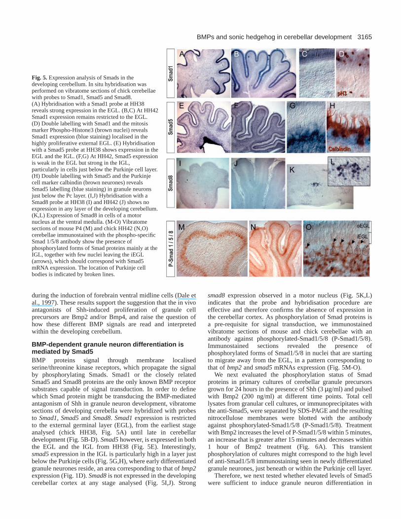
BMP-dependent granule neuron differentiation ismediated by Smad5BMP proteins signal through membrane localisedserine/threonine kinase receptors, which propagate the signalby phosphorylating Smads. Smad1 or the closely relatedSmad5 and Smad8 proteins are the only known BMP receptorsubstrates capable of signal transduction. In order to definewhich Smad protein might be transducing the BMP-mediatedantagonism of Shh in granule neuron development, vibratomesections of developing cerebella were hybridized with probesto Smad1, Smad5and Smad8. Smad1expression is restrictedto the external germinal layer (EGL), from the earliest stageanalysed (chick HH38, Fig. 5A) until late in cerebellardevelopment (Fig. 5B-D). Smad5however, is expressed in boththe EGL and the IGL from HH38 (Fig. 5E). Interestingly,smad5expression in the IGL is particularly high in a layer justbelow the Purkinje cells (Fig. 5G,H), where early differentiatedgranule neurones reside, an area corresponding to that ofbmp2expression (Fig. 1D). Smad8is not expressed in the developingcerebellar cortex at any stage analysed (Fig. 5I,J). Strong
smad8expression observed in a motor nucleus (Fig. 5K,L)indicates that the probe and hybridisation procedure areeffective and therefore confirms the absence of expression inthe cerebellar cortex. As phosphorylation of Smad proteins isa pre-requisite for signal transduction, we immunostainedvibratome sections of mouse and chick cerebellae with anantibody against phosphorylated-Smad1/5/8 (P-Smad1/5/8).Immunostained sections revealed the presence ofphosphorylated forms of Smad1/5/8 in nuclei that are startingto migrate away from the EGL, in a pattern corresponding tothat of bmp2and smad5mRNAs expression (Fig. 5M-O).
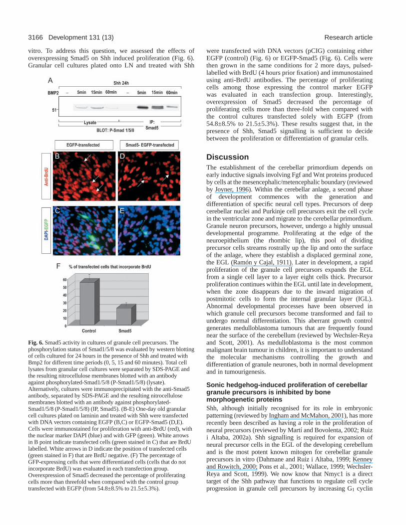
We next evaluated the phosphorylation status of Smadproteins in primary cultures of cerebellar granule precursorsgrown for 24 hours in the presence of Shh (3 µg/ml) and pulsedwith Bmp2 (200 ng/ml) at different time points. Total celllysates from granular cell cultures, or immunoprecipitates withthe anti-Smad5, were separated by SDS-PAGE and the resultingnitrocellulose membranes were blotted with the antibodyagainst phosphorylated-Smad1/5/8 (P-Smad1/5/8). Treatmentwith Bmp2 increases the level of P-Smad1/5/8 within 5 minutes,an increase that is greater after 15 minutes and decreases within1 hour of Bmp2 treatment (Fig. 6A). This transientphosphorylation of cultures might correspond to the high levelof anti-Smad1/5/8 immunostaining seen in newly differentiatedgranule neurones, just beneath or within the Purkinje cell layer.
Therefore, we next tested whether elevated levels of Smad5were sufficient to induce granule neuron differentiation in
Fig. 5.Expression analysis of Smads in thedeveloping cerebellum. In situ hybridisation wasperformed on vibratome sections of chick cerebellaewith probes to Smad1, Smad5 and Smad8.(A) Hybridisation with a Smad1 probe at HH38reveals strong expression in the EGL. (B,C) At HH42Smad1 expression remains restricted to the EGL.(D) Double labelling with Smad1 and the mitosismarker Phospho-Histone3 (brown nuclei) revealsSmad1 expression (blue staining) localised in thehighly proliferative external EGL. (E) Hybridisationwith a Smad5 probe at HH38 shows expression in theEGL and the IGL. (F,G) At HH42, Smad5 expressionis weak in the EGL but strong in the IGL,particularly in cells just below the Purkinje cell layer.(H) Double labelling with Smad5 and the Purkinjecell marker calbindin (brown neurones) revealsSmad5 labelling (blue staining) in granule neuronsjust below the Pc layer. (I,J) Hybridisation with aSmad8 probe at HH38 (I) and HH42 (J) shows noexpression in any layer of the developing cerebellum.(K,L) Expression of Smad8 in cells of a motornucleus at the ventral medulla. (M-O) Vibratomesections of mouse P4 (M) and chick HH42 (N,O)cerebellae immunostained with the phospho-specificSmad 1/5/8 antibody show the presence ofphosphorylated forms of Smad proteins mainly at theIGL, together with few nuclei leaving the iEGL(arrows), which should correspond with Smad5mRNA expression. The location of Purkinje cellbodies is indicated by broken lines.
3166
vitro. To address this question, we assessed the effects ofoverexpressing Smad5 on Shh induced proliferation (Fig. 6).Granular cell cultures plated onto LN and treated with Shh
were transfected with DNA vectors (pCIG) containing eitherEGFP (control) (Fig. 6) or EGFP-Smad5 (Fig. 6). Cells werethen grown in the same conditions for 2 more days, pulsed-labelled with BrdU (4 hours prior fixation) and immunostainedusing anti-BrdU antibodies. The percentage of proliferatingcells among those expressing the control marker EGFPwas evaluated in each transfection group. Interestingly,overexpression of Smad5 decreased the percentage ofproliferating cells more than three-fold when compared withthe control cultures transfected solely with EGFP (from54.8±8.5% to 21.5±5.3%). These results suggest that, in thepresence of Shh, Smad5 signalling is sufficient to decidebetween the proliferation or differentiation of granular cells.
DiscussionThe establishment of the cerebellar primordium depends onearly inductive signals involving Fgf and Wnt proteins producedby cells at the mesencephalic/metencephalic boundary (reviewedby Joyner, 1996). Within the cerebellar anlage, a second phaseof development commences with the generation anddifferentiation of specific neural cell types. Precursors of deepcerebellar nuclei and Purkinje cell precursors exit the cell cyclein the ventricular zone and migrate to the cerebellar primordium.Granule neuron precursors, however, undergo a highly unusualdevelopmental programme. Proliferating at the edge of theneuroepithelium (the rhombic lip), this pool of dividingprecursor cells streams rostrally up the lip and onto the surfaceof the anlage, where they establish a displaced germinal zone,the EGL (Ramón y Cajal, 1911). Later in development, a rapidproliferation of the granule cell precursors expands the EGLfrom a single cell layer to a layer eight cells thick. Precursorproliferation continues within the EGL until late in development,when the zone disappears due to the inward migration ofpostmitotic cells to form the internal granular layer (IGL).Abnormal developmental processes have been observed inwhich granule cell precursors become transformed and fail toundergo normal differentiation. This aberrant growth controlgenerates medulloblastoma tumours that are frequently foundnear the surface of the cerebellum (reviewed by Wechsler-Reyaand Scott, 2001). As medulloblastoma is the most commonmalignant brain tumour in children, it is important to understandthe molecular mechanisms controlling the growth anddifferentiation of granule neurones, both in normal developmentand in tumourigenesis.
Sonic hedgehog-induced proliferation of cerebellargranule precursors is inhibited by bonemorphogenetic proteinsShh, although initially recognised for its role in embryonicpatterning (reviewed by Ingham and McMahon, 2001), has morerecently been described as having a role in the proliferation ofneural precursors (reviewed by Martí and Bovolenta, 2002; Ruizi Altaba, 2002a). Shh signalling is required for expansion ofneural precursor cells in the EGL of the developing cerebellumand is the most potent known mitogen for cerebellar granuleprecursors in vitro (Dahmane and Ruiz i Altaba, 1999; Kenneyand Rowitch, 2000; Pons et al., 2001; Wallace, 1999; Wechsler-Reya and Scott, 1999). We now know that Nmyc1 is a directtarget of the Shh pathway that functions to regulate cell cycleprogression in granule cell precursors by increasing G1 cyclin
Development 131 (13) Research article
Fig. 6.Smad5 activity in cultures of granule cell precursors. Thephosphorylation status of Smad1/5/8 was evaluated by western blottingof cells cultured for 24 hours in the presence of Shh and treated withBmp2 for different time periods (0, 5, 15 and 60 minutes). Total celllysates from granular cell cultures were separated by SDS-PAGE andthe resulting nitrocellulose membranes blotted with an antibodyagainst phosphorylated-Smad1/5/8 (P-Smad1/5/8) (lysate).Alternatively, cultures were immunoprecipitated with the anti-Smad5antibody, separated by SDS-PAGE and the resulting nitrocellulosemembranes blotted with an antibody against phosphorylated-Smad1/5/8 (P-Smad1/5/8) (IP, Smad5). (B-E) One-day old granularcell cultures plated on laminin and treated with Shh were transfectedwith DNA vectors containing EGFP (B,C) or EGFP-Smad5 (D,E).Cells were immunostained for proliferation with anti-BrdU (red), withthe nuclear marker DAPI (blue) and with GFP (green). White arrowsin B point indicate transfected cells (green stained in C) that are BrdUlabelled. White arrows in D indicate the position of transfected cells(green stained in F) that are BrdU negative. (F) The percentage ofGFP-expressing cells that were differentiated cells (cells that do notincorporate BrdU) was evaluated in each transfection group.Overexpression of Smad5 decreased the percentage of proliferatingcells more than threefold when compared with the control grouptransfected with EGFP (from 54.8±8.5% to 21.5±5.3%).
3167BMPs and sonic hedgehog in cerebellar development
expression (Kenney and Rowitch, 2000; Kenney et al., 2003).Furthermore, mutations in several components of the Shhpathway seem to account for most cases of desmoplasticmedulloblastomas (reviewed by Rubin and Rowitch, 2002;Wechsler-Reya and Scott, 2001). Thus, the control of Sonicsignalling is likely to be a key point in the normal developmentalprogramme of cerebellar granule neurones.
We, and others, have previously reported that extracellularmatrix glycoproteins (Pons et al., 2001) and FGFs (Wechsler-Reya and Scott, 1999; Pons et al., 2001) are able to differentiallymodulate but not to totally suppress Shh-mediated proliferationof granule cell precursors. TGFβ signalling generally has anegative effect on cell growth such that inactivation of thispathway contributes to tumourigenesis (reviewed by Shi andMassagué, 2003). Among the TGFβ superfamily, BMPs haveopposing activities to hedgehogs in many developmentalparadigms (Lee and Jessell, 1999; Mekki-Dauriac et al., 2002;Patten and Placzek, 2002; Zhu et al., 1999). Thus the expressionof Bmp2 and Bmp4 in proliferating and early differentiatedgranule neurones of the cerebellum shown here, indicates thepossibility of a functional interaction with Shh in the regulationof granule cell development. In primary cultures of granule cellprecursors, Bmp2 and Bmp4 are able to totally overcome Shh-induced proliferation. Furthermore, in a pseudo-in vivo situation,such as organotypic slice cultures, Bmp2 and Bmp4 significantlyreduce granule cell precursor proliferation at the EGL,proliferation induced by endogenous Shh. These results stronglysuggest that Bmp2 and/or Bmp4 are potent inhibitors of the Shhpathway during normal development of the cerebellum and raisethe interesting point of whether these molecules could alsocontrol hedgehog pathway activity in medulloblastoma growth.In accordance with this, a recent publication has suggested thatBmp2 may mediate retinoid-induced apoptosis inmedulloblastoma cells (Hallahan et al., 2003).
Bmp7, a slightly more divergent member of the BMP family,is expressed in a different cell type population and exhibits anapparently different role. Similar to previous reports for Shh(Dahmane and Ruiz i Altaba, 1999; Traiffort et al., 1999;Wallace, 1999; Wechsler-Reya and Scott, 1999), Bmp7 isexpressed in migrating and settled Purkinje neurones. Bmp7had no significant effect on the Shh-mediated proliferationof granule cell precursors, either in primary cultures or inorganotypic slice cultures, thus leaving the role that Bmp7might be playing in cerebellar development unresolved.
Bmp2 activity in the developing cerebellum ismediated by Smad5Although the diverse TGFβ ligands elicit quite differentcellular responses, they all share a highly conserved signallingpathway. Ligand binding to type I and type II receptorserine/threonine kinases at the cell surface initiates signallingthrough phosphorylation of the Smad proteins. There areeight distinct Smad proteins among which Smad1, Smad5and Smad8 are directly phosphorylated and activated byBmp signalling. Phosphorylated Smad1/5/8 undergoeshomotrimerization and formation of heteromeric complexeswith Smad4. The activated Smad complexes are thentranslocated into the nucleus and, in conjunction with othernuclear co-factors, regulate the transcription of target genes(Massagué, 2000; Massagué et al., 2000). We asked whichSmad protein might be transducing the Bmp-mediated
antagonism of Shh activity in the developing cerebellum. Weshow that Smad1 and Smad5 are both expressed in thedeveloping cerebellar cortex, although in different cellpopulations. Whereas Smad1expression is restricted to theEGL, where granular cell precursors proliferate, Smad5 isexpressed in early differentiated granule neurones. We used aphospho-specific antibody to Smad1/5/8 (anti-P-Smad1/5/8) toevaluate the activation of Smad1 and Smad5, and found that atthese developmental stages anti-P-Smad1/5/8 only labelsnuclei in a pattern overlapping to that of Smad5mRNAexpression. This expression analysis strongly suggests that thesignalling activity of Bmp2 is mediated by Smad5. Bmp4,however, which is expressed in an overlapping pattern to thatof Smad1at the EGL, seems not to be signalling at thisdevelopmental stages as Smad1 is not being phosphorylated.We favour the hypothesis that expression of Bmp4at the EGLmay be inherited from earlier developmental stages, at whichBmp4 mediates determination of granule cell precursor (Alderet al., 1999), and that later Bmp4 is apparently not active duringclonal expansion and/or final differentiation of granuleneurones. However, the fact that Bmp receptor (Bmpr) 1a andBmpr1b are highly expressed in the EGL (Ming et al., 2002)suggests that Bmp4 might be alternatively using a non-canonical Smad1/5/8 signalling that needs to be investigated.
In primary cultures of granule cell precursors grown in thepresence of Shh, Bmp2 treatment induces strong and transientSmad5 phosphorylation. Furthermore, we show that Smad5overexpression is sufficient to suppress the proliferativeresponse to Shh and allow granule cell precursor to enter thedifferentiation programme. Whether this is achieved by a directinteraction Smad/Gli (Liu et al., 1998), by the competition forcommon transcriptional co-activators (Goodman and Smolik,2000) or co-repressors (Dai et al., 2002; Wang et al., 2000) ofthe Smad and the Gli pathways, or by different mechanisms,remains to be elucidated.
Shh-mediated proliferation of granule cell precursors is aswell regulated by components of the extracellular matrix(Graus-Porta et al., 2001; Pons et al., 2001; Rubin et al., 2002).We have previously described that the extracellular matrixglycoprotein vitronectin stimulates CREB phosphorylationusing a pathway not involving MAPK, and that CREBsignalling was sufficient to induce differentiation of granulecells. These results revealed CREB as an essential signal forgranule neuron differentiation (Pons et al., 2001). We describedthe role of Bmp2 as a potent inhibitor of Shh-inducedproliferation, and show that the BMP pathway in thedeveloping cerebellum activates Smad5 phosphorylation.Whether CREB- and Smad5-mediated transcription of targetgenes are two parallel pathways leading to granule neurondifferentiation, or whether there are points of crosstalk betweenthese two pathways remain to be determined, althougha cooperation between Smads and CREB to activatetranscription in response to TGFβ signalling has already beenreported in a different cell context (Zhang and Derynck., 2000).
The authors thank Dr Iain K. Patten for improvements in themanuscript and Monica Pons for aid in confocal microscopy. We alsothank Dr Marian Ros (University of Cantabria, Spain) for the mouseBmp2, Bmp4 and Bmp7 probes; Dr Juan Hurlé (University ofCantabria, Spain) for the chick Smad1 and Smad5 probes; and Dr E.Robertson (Harvard University, USA) for the mouse Smad1 andSmad5 probes. We also thank Biogen for the purified human N-Shh
3168
and the Genetic Institute for the purified Bmp2 protein. I.R. issupported by a FPI Predoctoral Fellowship (FP-2001-1740) from theSpanish Ministry of Science and Technology. R.A. is supported by aPredoctoral Fellowship from Grant SAS2002-03430 to S.P. Thisresearch was supported by Grants BMC2001-1345 and EU QLGC-CT-2002-01141 to E.M. and SAS2002-03430 to S.P.
ReferencesAlder, J., Lee, K. L., Jessell, T. M. and Hatten, M. E. (1999). Generation of
cerebellar granule neurons in vivo by transplantation of BMP-treated neuralprogenitor cells. Nat. Neurosci.2, 535-540.
Altman, J. and Bayer, S. A. (1997). Development of the Cerebellar System:In Relation to its Revolution, Structure and Functions. Boca Raton, FL: CRCPress.
Angley, C., Kumar, M., Dinsio, K. J., Hall, A. K. and Siegel, R. E. (2003).Signalling by bone morphogenetic proteins and smad1 modulates thepostnatal differentiation of cerebellar cells. J. Neurosci. 23, 260-268.
Bitgood, M. J. and McMahon, A. P. (1995). Hedgehog and Bmp genes arecoexpressed at many diverse sites of cell-cell interaction in the mouseembryo.Dev. Biol. 172, 126-138.
Dahmane, N. and Ruiz i Altaba, A. (1999). Sonic hedgehog regulates thegrowth and patterning of the cerebellum. Development 126, 3089-3100.
Dai, P., Shinagawa, T., Nomura, T., Harada, J., Kaul, S. C., Wadhwa, R.,Khan, Md. M., Akimura, H., Colmenares, C. and Ishii, S. (2002). Ski isinvolved in transcriptional regulation by the repressor and full-length formsof Gli3. Genes Dev. 16, 2843-2848.
Dale, J. K., Vesque, C., Lints, T. J., Sampath, T. K., Furley, A., Dodd, J.and Placzek, M. (1997). Cooperation of BMP7 and SHH in the inductionof forebrain ventral midline cells by prechordal mesoderm.Cell 90, 257-269.
Echelard, Y., Epstein, D. J., St-Jacques, B., Shen, L., Mohler, J.,McMahon, J. A. and McMahon, A. P. (1993). Sonic hedgehog, a memberof a family of putative signalling molecules, is implicated in regulation ofCNS polarity. Cell 75, 1417-1430.
Francis-West, P. H., Tatla, T. and Brickell, P. M. (1994). Expression patternsof the bone morphogenetic protein genes Bmp-4 and Bmp-2 in thedeveloping chick face suggest a role in outgrowth of the primordia. Dev.Dyn. 201, 168-178.
Goodman, R. H. and Smolik, S. (2000). CBP/p300 in cell growth,transformation, and development. Genes Dev. 14, 1553-1577.
Gomes, W. A., Mehler, M. F. and Kessler, J. A.(2003). Transgenicoverexpression of BMP4 increases astroglial and decreases oligodendrogliallineage commitment. Dev Biol. 255, 164-177.
Graus-Porta, D., Blaess, S., Senften, M., Littlewood-Evans, A., Damsky,C., Huang, Z., Orban, P., Klein, R., Schittny, J. C. and Muller, U. (2001).Beta1-class integrins regulate the development of laminae and folia in thecerebral and cerebellar cortex. Neuron31, 367-367.
Hallahan, A. R., Pritchard, J. I., Chandraratna, R. A. S., Ellenbogen, R.E., Geyer, J. R., Overland, R., Strand, A. D., Tapscott, S. J. and Olson,J. M. (2003). BMP-2 mediates retinoid-induced apoptosis inmedulloblastoma cells through a paracrine effect. Nat. Med.9, 1033-1038.
Hamburger, V. and Hamilton, H. L. (1951). A series of normal stages in thedevelopment of chick embryo. J. Morphol. 88, 49-92.
Hatten, M. E., Alder, J., Zimmerman, K. and Heintz, N. (1997). Genesinvolved in cerebellar cell specification and differentiation. Curr. Opin.Neurobiol. 7, 40-47.
Ho, K. S. and Scott, M. P. (2002). Sonic hedgehog in the nervous system:functions, modifications and mechanisms. Curr. Opin. Neurobiol. 12, 57-63.
Houston, B., Thorp, B. H. and Burt, D. W. (1994). Molecular cloning andexpression of bone morphogenetic protein-7 in the chick epiphyseal growthplate.J. Mol. Endocrinol. 13, 289-301.
Ingham, P. W. and McMahon, A. P. (2001). Hedgehog signaling in animaldevelopment: paradigms and principles. Genes Dev. 15, 3059-3087.
Joyner, A. L. (1996). Engrailed, wnt and pax genes regulate midbrain-hindbrain development. Trends Genet. 12, 15-20.
Kenney, A. M. and Rowitch, D. H. (2000). Sonic hedgehog promotes G1cyclin expression and sustained cell cycle progression in mammalianneuronal precursors. Mol. Cell. Biol.20, 9055-9067.
Kenney, A. M., Cole, M. D. and Rowitch, D. H. (2003). Nmyc upregulationby sonic hedgehog signaling promotes proliferation in developing cerebellargranule neuron precursors. Development130, 15-28.
Lee, K. J. and Jessell, T. M.(1999). The specification of dorsal cell fates inthe vertebrate central nervous system. Ann. Rev. Neurosci. 22, 261-294.
Liu, F., Massagué, J. and Ruiz i Altaba, A. (1998). Carboxy-terminallytruncated Gli3 proteins associate with Smads. Nat. Genet. 20, 325-326.
Massagué, J. (2000). How cells read TGF-β signals. Nat. Mol. Cell Rev.1,169-178.
Massagué, J., Blain, S. W. and Lo, R. S. (2000). TGFβ signaling in growthcontrol, cancer and heritable disorders. Cell 103, 295-309.
Martí, E. and Bovolenta, P. (2002). Sonic hedgehog in CNS development:one signal, multiple outputs. Trends Neurosci. 25, 89-96.
Megason, S. G. and McMahon, A. P. (2002). A mitogen gradient of dorsalmidline Wnts organizes growth in the CNS.Development129, 2087-2098.
Mekki-Dauriac, S., Agius, E., Kan, P. and Cochard, P. (2002). Bonemorphogenetic proteins negatively control oligodendrocyte precursorspecification in the chick spinal cord. Development 129, 5117-5130.
Meyer-Franke, A., Kaplan, M. R., Pfrieger, F. W. and Barres, B. A. (1995).Characterisation of the signalling interactions that promote the survival andgrowth of developing retinal ganglion cells in culture. Neuron 15, 805-819.
Ming, J. E., Elkan, M., Tang, K. and Golden, J. A. (2002). Type I bonemorphogenetic protein receptors are expressed on cerebellar granularneurons and a constitutively active form of the type IA receptor inducescerebellar abnormalities. Neuroscience114, 849-857.
Nakashima, K., Takizawa, T., Ochiai, W., Yanagisawa, M., Hisatsune, T.,Nakafuku, M., Miyazono, K., Kishimoto, T., Kageyama, R. and Taga,T. (2001). BMP2-mediated alteration in the developmental pathway of fetalmouse brain cells from neurogenesis to astrocytogenesis.Proc. Natl. Acad.Sci. USA 98, 5868-5873.
Nybakken, K. and Perrimon, N. (2002). Hedgehog signal transduction:recent findings. Curr. Opin. Genet. Dev.12, 503-511.
Patten, I. and Placzek, M. (2002). Opponent activities of Shh and BMPsignaling during floor plate induction in vivo. Curr. Biol. 12, 47-52.
Piedra, E. and Ros, M. A. (2002). BMP signaling positively regulates Nodalexpression during left right specification in the chick embryo.Development129, 3431-3440.
Pons, S., Trejo, J. L., Martínez-Morales, J. R. and Martí, E.(2001).Vitronectin regulates Sonic hedgehog activity during cerebellumdevelopment through CREB phosphorilation. Development128, 1481-1492.
Ramón y Cajal, S. (1911). Histologie du Sisteme Nerveux de l’Homme et desVertebrates. Paris: Malonie (Reprinted by Consejo Superior deInvestigaciones Científicas, Madrid 1955).
Riddle, R. D., Jonson, R. L., Laufer, E. and Tabin, C. (1993). Sonichedgehog mediates the polarising activity of the ZPA. Cell 75, 1401-1416.
Rubin, J. B., Choi, Y. and Segal, R. A. (2002). Cerebellar proteoglycansregulate Sonic hedgehog responses during development.Development129,2223-2232.
Rubin, J. B. and Rowitch, D. H. (2002). Medulloblastoma: a problem ofdevelopmental biology. Cancer Cell2, 7-8.
Ruiz i Altaba, A., Palma, V. and Dahmane, N. (2002a). Hedgehog-Glisignalling and the growth of the brain. Nat. Rev. Neurosci. 3, 24-33.
Ruiz i Altaba, A., Sanchez, P. and Dahmane, N. (2002b). Gli and hedgehogin cancer: tumours, embryos and stem cells. Nat. Rev. Cancer2, 361-372.
Shi, Y. and Massagué, J. (2003). Mechanisms of TGF-β signalling from cellmembrane to the nucleus. Cell 113, 658-700.
Traiffort, E., Charytoniuk, D., Watroba, L., Faure, H. and Ruat, M.(1999). Discrete localisation of hedgehog signalling components in thedeveloping and adult rat nervous system. Eur. J. Neurosci. 11, 3199-3214.
Wallace, V. A. (1999). Purkinje-cell-derived Sonic hedgehog regulates granuleneurone precursor cell proliferation in the developing mouse cerebellum.Curr. Biol. 9, 445-448.
Wang, W., Mariani, F. V., Harland, R. M. and Luo, K. (2000). Ski repressesbone morphogenetic protein signalling in Xenopus and mammalian cells.Proc. Natl. Acad. Sci. USA97, 14394-14399.
Wechsler-Reya, R. J. and Scott, M. P. (1999). Control of neuronal precursorproliferation in the cerebellum by sonic hedgehog. Neuron22, 103-114.
Wechsler-Reya, R. J. and Scott, M. P. (2001). Developmental biology ofbrain tumors. Annu. Rev. Neurosci.24, 385-428.
Zhang, Y. and Derynk, R. (2000). Transcriptional regulation of thetransforming growth factor-β-inducible mouse germ line Ig α constantregion gene by functional cooperation of Smad, CREB, and AML familymembers. J. Biol. Chem. 275, 16979-16985.
Zhu, G., Mehler, M. F., Zhao, J., Yung, S. Y. and Kessler, J. A. (1999). Sonichedgehog and BMP2 exert opposing actions on proliferation anddifferentiation of embryonic neural progenitor cells. Dev. Biol. 215, 118-129.
Development 131 (13) Research article