High temperature (>350 °C) thermochronology and mechanisms of Pb loss in apatite
Upload
independentCategory
view
1download
0
Adsorption and Release of BMP-2 on NanocrystallineApatite-Coated and Uncoated Hydroxyapatite/b-TricalciumPhosphate Porous Ceramics
Helene Autefage,1,5 Fabienne Briand-Mesange,2 Sophie Cazalbou,1 Christophe Drouet,1 Daniel Fourmy,3
Stephane Goncalves,4 Jean-Pierre Salles,2 Christele Combes,1 Pascal Swider,5 Christian Rey1
1 Universite de Toulouse, CIRIMAT, UPS-INPT-CNRS, ENSIACET, 31077 Toulouse Cedex 4, France
2 INSERM U563, Centre de physiopathologie de Toulouse Purpan, Dept. Lipoproteines et Mediateurs Lipidiques,CHU Purpan, 31024 Toulouse Cedex 3, France
3 INSERM U858, Institut Louis Bugnard, 31432 Toulouse Cedex 4, France
4 Teknimed, Z.I. de Montredon, 31240 L’Union, France
5 Universite de Toulouse, Biomechanics Laboratory EA3697, CHU Purpan, 31059 Toulouse Cedex, France
Received 31 July 2008; revised 19 January 2009; accepted 15 April 2009Published online 6 July 2009 in Wiley InterScience (www.interscience.wiley.com). DOI: 10.1002/jbm.b.31447
Abstract: The association of bone morphogenetic proteins (BMPs) with calcium phosphate
bioceramics is known to confer them osteoinductive properties. The aim of this study was to
evaluate the surface properties, especially regarding recombinant human BMP-2 (rhBMP-2)
adsorption and release, of commercial sintered biphasic calcium phosphate ceramics after
coating with biomimetic nanocrystalline apatite. The raw and coated ceramics exhibited
similar macroporous structures but different nanometer-sized pores contents. Both types of
ceramics showed Langmuir-type adsorption isotherms of rhBMP-2. The coating noticeably
increased the rate of adsorption and the total amount of growth factor taken up, but the
maximum coverage per surface area unit as well as the affinity constant appeared lower for
coated ceramics compared with raw ceramic surfaces. The limited advantage gained by
coating the ceramics can be assigned to a lower accessibility of the surface adsorption sites
compared with the raw ceramics. The quantity of rhBMP-2 spontaneously released in cell
culture medium during the first weeks was lower for coated samples than for uncoated
ceramics and represented a minor fraction of the total adsorbed amount. In conclusion, the
nanocrystalline apatite coating was found to favor the adsorption of rhBMP-2 while providing
a mean to fine tune the release of the growth factor. ' 2009 Wiley Periodicals, Inc. J Biomed Mater
Res Part B: Appl Biomater 91B: 706–715, 2009
Keywords: bone morphogenetic protein 2 (BMP-2); bioceramics; biphasic calcium
phosphate (BCP); nanocrystalline apatite; protein adsorption and release
INTRODUCTION
One of the most efficient ways to improve the bone form-
ing ability of biomaterials is their association with bone
morphogenetic proteins (BMPs). Such growth factors,
BMP-2 and BMP-7, the two most efficient variants of BMP
growth factors available, have been associated with differ-
ent types of biomaterials for orthopedic applications (col-
lagen, calcium phosphate (Ca-P) cements, polymers,
ceramics).1 Although calcium phosphates are among the
most frequently used biomaterials for bone reconstruction,
little is known about their association with BMP.2,3
Regarding porous ceramics, the growth factors are gener-
ally associated with the implants by simple impregnation
and drying and the type of bonding with the substrate and
the release rate are often undetermined.4–7 This lack of
knowledge could explain divergences in the reported effect
of such associations. Most studies testify to an improve-
ment of bone formation with an acceleration of repair4–7;
however, a few studies have mentioned a resorptive process
of bone attributed to a stimulation of osteoclast activity.8,9
Recently, a few studies have been published on the adsorp-
tion characteristics of BMP-2 on apatitic calcium phos-
Correspondence to: H. Autefage (e-mail: [email protected])Contract grant sponsor: Midi-Pyrenees Region of France; Contract grant number:
06001852.Contract grant sponsor: Teknimed S.A. (L’Union, France)
' 2009 Wiley Periodicals, Inc.
706

phate.2,3 In a first study,2 the adsorption isotherms have
been drawn and the adsorption parameters determined. The
data show a strong affinity of BMP-2 for apatite surfaces,
enhanced in the presence of calcium ions as in the case of
the adsorption of other proteins.10–12
The second work on this topic3 is based on the use of
theoretical modeling of the interactions of surfaces (in this
case, the (001) apatite plane) with functional groups of
BMP-2. The results indicate a preferential interaction
between carboxylic functional groups from the protein and
calcium ions from the apatitic surface.
These experiments however do not give information on
the behavior of sintered bioceramics and do not evoke the
crucial question of the release of the growth factor in bio-
logical medium. Biphasic ceramics composed of hydroxyap-
atite (HA) and b-tricalcium phosphate (b2TCP) have been
commercialized for more than 15 years as bone substitute
materials. They have been shown to combine an excellent
biointegration with a bioresorption capable of being modu-
lated according to the b-TCP content.13 Because of high sin-
tering temperatures, these ceramics generally exhibit a very
low surface reactivity. Although natural osteoinductive prop-
erties have been suggested for these compounds,14,15 they
occur erratically and do not seem to be related to identified
surface characteristics.15 However, microporosity has been
suggested to be an important factor likely to favor the bind-
ing of circulating growth factor on the material surface.16,17
This contribution aims at investigating the potential
increase of the surface reactivity of sintered ceramics
obtained by coating their surface with highly reactive bio-
mimetic nanocrystalline apatites.18 A very thin layer, a few
micrometers thick, should not alter the porous structure of
the ceramic and could enhance its adsorption capabilities
with regard to growth factors such as rhBMP-2. This first
report has particularly focused on comparing the adsorption
properties of rhBMP-2 on sintered biphasic ceramics before
and after coating with nanocrystalline apatite, while keep-
ing similar macroporous structures and different nanome-
ter-sized pore contents. The release of rhBMP-2 was
evaluated in a biological fluid model, namely Dulbecco’s
modified Eagle’s medium, in anticipation of subsequent
studies on in vitro and in vivo behavior of rhBMP-2-con-
taining bioceramics.
MATERIALS AND METHODS
Materials
Biphasic calcium phosphate (BCP) ceramics (hydroxyapa-
tite/b-tricalcium phosphate, 65/35) were produced by
Teknimed S.A. (l’Union, France). These highly porous
BCP blocks (65% porosity) were crushed and sieved to
obtain randomly shaped 1–1.4 mm diameter granules (12/
10 powder, CERAFORM).
These granules were coated, when needed, with a nano-
crystalline apatite layer, a few micrometers thick, using a
process being patented (Teknimed, l’Union, France). This
layer represented 1.6% 6 0.1% of the samples weight. The
nanocrystalline apatite deposit was found to settle on both
the external surface of the granules and the inner pore
surface, shown by scanning electron microscopy (SEM)
observations (Figure 1).
Characterization of the Nanocrystalline Apatite
Although many applications and materials containing nano-
crystalline apatite are tested and produced nowadays,19
they are generally not thoroughly characterized from a
physicochemical point of view, despite the demonstration
that adsorption properties20,21 and other properties such as
dissolution22 depend strongly on the type of nanocrystals
considered. The composition of the nanocrystalline apatite
used to coat the granules in this work was determined by
chemical analysis: calcium was titrated by complexation
with ethylene diamineteraacetic acid (EDTA)23; phosphate
ions were quantified by spectrophotometry using a phos-
phovanadomolybdic complex23; the carbonate content was
measured by coulometry (UIC.Inc CM5014). The nanocrys-
tals were also characterized by X-ray diffraction (XRD,
Inel CPS 120 diffractometer) using a Co anticathode (k 5
1.78892 A) and Fourier transform infrared (FTIR) spectros-
copy (Nicolet 5700), by transmission, using the KBr pellet
method. Decomposition of m3CO3 and m4PO4 was per-
formed from FTIR spectra using GRAMS curve-fitting
software.24 Crystal morphology was observed by high-reso-
lution transmission electron microscopy (TEM) at 480 and
3200 kV using a Jeol Jem 2100F microscope.
Characterization of the Granules
Morphological characterization of the coated and uncoated
granules was carried out by SEM using a Leo 435 VP
microscope. The specific surface area was measured by the
Brunauer-Emmett-Teller (BET) method using nitrogen
adsorption (Quantachrome Nova 1000). The porosity of
coated and uncoated granules was analyzed using a mer-
cury intrusion porosimetrer (Micromeretics Autopore III)
between 8 and 60,000 psi. Considering the complex shapes
of the granules, the adhesion strength of the coating could
not be determined by usual methods (ISO 2409 standards).
rhBMP-2 Labeling
The recombinant human BMP-2 (rhBMP-2), expressed in
E. coli, was supplied by Wurtsburg University (Pr. W.
Sebald).
rhBMP-2 was labeled by 125I using a modified chloram-
ine T method previously described.25 Briefly, 10 lg of
rhBMP-2 was dissolved in 50 lL of a 0.25M phosphate
buffer pH 7.5 and mixed with 10 lL of Na125I (1 mCi).
The reaction was initiated by adding 5 lL of a chloramine
T solution (0.1 mg/mL of phosphate buffer). After an incu-
bation period of 2 min at room temperature, a second 5 lL
aliquot of chloramine T solution was added, and after 1.5
707NANOCRYSTALLINE APATITE-COATED BCP, BMP-2 ADSORPTION AND RELEASE
Journal of Biomedical Materials Research Part B: Applied Biomaterials

min a last 5 lL aliquot was added and left for 1 min. The
reaction was stopped with 20 lL of 50 mM L-tyrosine, 200
lL of potassium iodide (60 mM), and 200 lL of ultrapure
urea (1.2 mg/mL in acetic acid 1M). 125I-rhBMP-2 separa-
tion was carried out using a previously equilibrated chro-
matography Sephadex G25 column. Labeled proteins were
eluted with a 4 mM HCl, 75 mM sodium chloride, and
0.1% bovine serum albumin (BSA) solution.
rhBMP-2 Adsorption on BCP Granules
Adsorption Isotherms. The adsorption protocol used in
these experiments was based on preliminary results show-
ing an increase of the maximum adsorbed amount of sev-
eral proteins, including BMP, in the presence of calcium
ions.2,10–12 The solutions used for the adsorption experi-
ments were prepared just before their incubation with gran-
ules. They were obtained by diluting a rhBMP-2 solution
with a buffer solution (NaCl 1M, Tris 25 mM, and CaCl210 mM, pH 7.4). The 125I-rhBMP-2 was then added to the
solution to reach a 125I-labeled-to-unlabeled protein ratio
of 1:2000. The adsorption experiments were performed at
378C in batches containing 20 mg of coated or uncoated
ceramic granules immersed into 40 lL of rhBMP-2 solu-
tion. After 24 h, the supernatant was removed and the free
radiolabeled molecules in solution were counted. The
amount of rhBMP-2 adsorbed was determined from the
quantity of 125I-rhBMP-2 remaining in solutions. One of
the main problems using BMP-2 solutions at physiologic
pH is precipitation. The concentrations of rhBMP-2 varied
from 1000 to 150 lg/mL (39.9–6 lM). In this range, no
precipitate was observed in the solutions. To confirm that
the decrease of the amount of BMP-2 in solution was
solely due to adsorption and not to precipitation from an
unstable solution, a second control of the amount adsorbed
was performed on the ceramic itself: the granules were
washed three times with 1 mL of a solution saturated with
respect to b-TCP (the most soluble phase of the BCP
ceramics) to eliminate nonadsorbed 125I-rhBMP-2
(including possibly precipitated BMP-2) without dissolving
the granules and the coating and dried at ambient tempera-
ture for 72 h. These granules were then dissolved in
perchloric acid 6N and the 125I-rhBMP-2 present in the sol-
utions was counted. Both methods gave very close results,
thus confirming the bonding of BMP-2 on the ceramic
surfaces and the absence of bias related to a possible
BMP-2 precipitation. We will report only the data obtained
by analysis of the supernatant solutions carried out in
duplicate. Results were expressed as mean 6 standard
deviation (SD).
Figure 1. SEM micrographs of uncoated [magnification 3100 (a) and 31000 (c)] and coated granules
[magnification 3100 (b) and 1000 (d)]. The 2- to 5-lm-thick coating settles on the porous surface.
708 AUTEFAGE ET AL.
Journal of Biomedical Materials Research Part B: Applied Biomaterials
Adsorption Kinetics. The adsorption experiments were
performed at 378C in batches containing 30 mg of coated
or uncoated ceramics immersed into 60 lL of rhBMP-2 so-
lution at 300 lg/mL (adsorption solution). After 15 min
and up to 24 h, the supernatant was removed and the free
radiolabeled molecules in solution were counted. Each
assay was carried out in triplicate. Results were expressed
as mean 6 standard error of the mean (SEM).
rhBMP-2 Release. After removal of the radiolabeled
adsorption solution, the granules were washed and dried as
described earlier. Two samples were dissolved in perchloric
acid, and the 125I-rhBMP-2 contained in the dissolution sol-
utions was counted to determine the initial amount of
adsorbed rhBMP-2. Three other samples were used for the
release experiment. The desorption kinetics were monitored
in 1 mL of culture medium (Dulbecco’s modified Eagle’s
medium) complemented with 10% of fetal bovine serum
and 1% of penicillin and streptomycin at 378C. The me-
dium was removed and replaced by a fresh one after 2 min
and at irregular intervals (12 times) over a period of 21
days. The radiolabeled molecules released in solution were
counted. The loss of radioactivity because of the half-time
life of 125I was considered in the determination of the
amount of protein released. Assays were carried out in trip-
licate. Results were expressed as mean 6 SEM. It shall be
noticed that the amount of BMP released was always much
lower than the solubility limit of nonglycosylated rhBMP-2
in physiologic conditions.26
Statistical Analysis. The experiments performed in tripli-
cate were compared using the Student’s t-test. A probability
value (p-value) of less than 0.05 was considered significant.
RESULTS
Characterization of the Nanocrystals
The XRD pattern of the nanocrystals used for coating the
samples (Figure 2) was characteristic of a poor crystalline
apatite with broad diffraction peaks. The average crystal
size was estimated using Scherrer’s formula. The nanocrys-
tals appeared elongated along the c-axis of the hexagonal
structure (245 6 5 A average length determined from the
(002) peak at 2y 5 30.48). The average width thickness
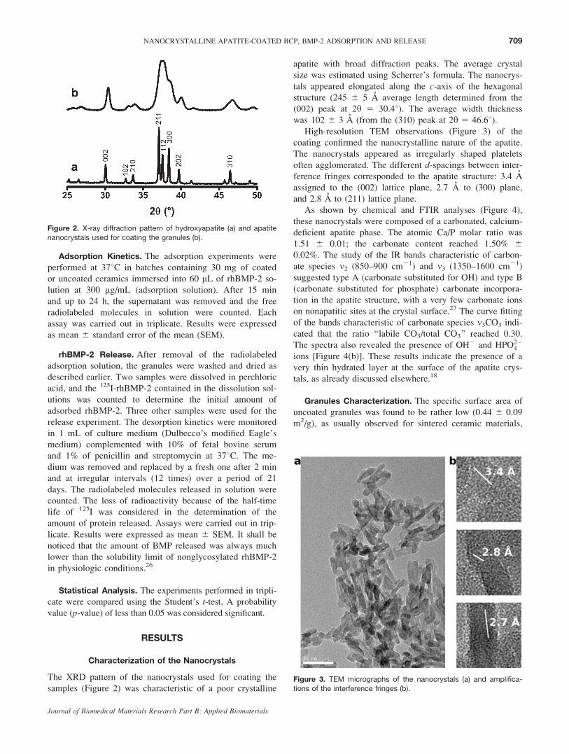
was 102 6 3 A (from the (310) peak at 2y 5 46.68).High-resolution TEM observations (Figure 3) of the
coating confirmed the nanocrystalline nature of the apatite.
The nanocrystals appeared as irregularly shaped platelets
often agglomerated. The different d-spacings between inter-
ference fringes corresponded to the apatite structure: 3.4 A
assigned to the (002) lattice plane, 2.7 A to (300) plane,
and 2.8 A to (211) lattice plane.
As shown by chemical and FTIR analyses (Figure 4),
these nanocrystals were composed of a carbonated, calcium-
deficient apatite phase. The atomic Ca/P molar ratio was
1.51 6 0.01; the carbonate content reached 1.50% 6
0.02%. The study of the IR bands characteristic of carbon-
ate species m2 (850–900 cm21) and m3 (1350–1600 cm21)
suggested type A (carbonate substituted for OH) and type B
(carbonate substituted for phosphate) carbonate incorpora-
tion in the apatite structure, with a very few carbonate ions
on nonapatitic sites at the crystal surface.27 The curve fitting
of the bands characteristic of carbonate species m3CO3 indi-
cated that the ratio ‘‘labile CO3/total CO3’’ reached 0.30.
The spectra also revealed the presence of OH2 and HPO2�4
ions [Figure 4(b)]. These results indicate the presence of a
very thin hydrated layer at the surface of the apatite crys-
tals, as already discussed elsewhere.18
Granules Characterization. The specific surface area of
uncoated granules was found to be rather low (0.44 6 0.09
m2/g), as usually observed for sintered ceramic materials,
Figure 2. X-ray diffraction pattern of hydroxyapatite (a) and apatite
nanocrystals used for coating the granules (b).
Figure 3. TEM micrographs of the nanocrystals (a) and amplifica-tions of the interference fringes (b).
709NANOCRYSTALLINE APATITE-COATED BCP, BMP-2 ADSORPTION AND RELEASE
Journal of Biomedical Materials Research Part B: Applied Biomaterials

whereas that of coated samples reached 2.03 6 0.09 m2/g.
Thus, the nanocrystalline coating led to a 4.7-fold increase
in the specific surface area of the samples. This significant
increase seems consistent with the number of nanocrystals
deposited, considering their specific surface area close to
84 6 10 m2/g, and suggests that most of the initial surface
of the nanocrystal agglomerates continues to be accessible
for nitrogen molecule adsorption.
The porosity data [Figure 5(a)] of uncoated samples
revealed three types of pores: macropores (with diameters
greater than 100 lm) and microsized pores (10 lm and 100
nm) related to sintering defects. The coated samples exhib-
ited the same porosity characteristics, but an additional
type of (nanosized) pore was also evidenced, characterized
by a rather narrow size distribution (ranging from 9 to
21 nm, with a maximum at 14.3 nm). Thus, it can be noted
in particular that the micron-thick coating did not alter
the starting porosity of the sintered ceramic, including the
microsized pores. The additional pore-type observed on the
coated samples can be assigned to internanocrystal spaces.
SEM observations [Figure 5(b)] confirmed the existence of
different levels of porosity on the raw samples. On the
coated samples, the micron-thick layer appeared to cover
the macropore wall, although some discontinuities and
cracks were observed.
rhBMP-2 Adsorption. The adsorption rates and adsorp-
tion isotherms are represented in Figures 6 and 7 for coated
and uncoated samples, respectively.
As shown in Figure 6, the coating significantly increased
the amount of growth factor uptake. Thus, after 24 h,
coated granules adsorbed 517 6 9 lg of rhBMP-2 per
gram of granules, whereas uncoated ceramics adsorbed 397
6 19 lg/g (p < 0.005). The initial rate of adsorption
appeared also faster for coated granules than for uncoated
ones: 444 lg/(g�h) versus 185 lg/(g�h) and the adsorption
limit was reached much faster.
The adsorption isotherm exhibited a characteristic Lang-
muir shape, as often observed in the adsorption of various
proteins on apatites2,20,28 (Figure 7). The isotherms were
correctly described by the linearized expression of the
Langmuir isotherm:
Ceq=Q ¼ Ceq=N þ 1=ðN � KÞ
where Ceq and Q indicate the concentration at equilibrium
and the amount of adsorbed protein, respectively, either per
surface unit or per gram of granules depending on data
description. The parameters K and N, expressed as K 6 DKand N 6 DN, represent the affinity constant of the adsorp-
tion equilibrium and the maximum adsorbed amount,
respectively. The affinity constant of the coated granules
seemed slightly lower than that of the uncoated ones (0.007
6 0.002 mL/lg vs. 0.011 6 0.006 mL/lg). The theoretical
maximum quantity of rhBMP-2 absorbed was higher for
coated ceramics (1608 6 170 lg/g) than for uncoated ones
(758 6 68 lg/g). However, considering the maximum
amount adsorbed per surface area unit, it appeared lower
for coated samples (0.032 6 0.003 lmol/m2) than for raw
samples (0.069 6 0.006 lmol/m2). In addition, the total
amount of growth factor uptake from the same rhBMP-2
solution at 300 lg/mL (recovery yield) appeared higher for
coated samples (87% 6 3% vs. 66% 6 4%).
rhBMP-2 Release
The release of rhBMP-2 in Dulbecco’s modified Eagle’s
medium complemented with calf serum was monitored as
cumulated amounts (Figure 8). Similar release curves were
observed exhibiting a sharp initial release, slowing down at
Figure 4. FTIR spectrum of the nanocrystalline apatite (a) andexpansions of m4 PO3�
4 domain (b) and m3 CO2�3 domain (c).
710 AUTEFAGE ET AL.
Journal of Biomedical Materials Research Part B: Applied Biomaterials

48 h. The total amount released at 21 days was slightly but
significantly (p < 0.005) lower for coated granules than for
controls (100 6 12 lg/g vs. 139 6 10 lg/g). The released
quantities represented however a minor fraction of the total
amount adsorbed for coated samples (23% at 21 days), and
about one half (46%) of the adsorbed growth factor for raw
ceramics (the initial amount of BMP-2 adsorbed was higher
on coated granules compared with uncoated ones for the
same concentration of the adsorption solutions).
DISCUSSION
The use of a ceramic coated with nanocrystalline apatite
was expected to present several advantages over raw sin-
tered ceramics. The first advantage in this study was indeed
found to be an increase of the specific surface area and of
the maximum adsorbable amount of growth factor in the
porous ceramic.
The second expected advantage was related to the
improvement of the surface reactivity. Previous compara-
tive studies on the adsorption properties of several mole-
cules on well-crystallized apatites and nanocrystalline
ones20,21 had shown an increase of the maximum amount
adsorbed per specific surface area and a decrease of the
affinity constant on nanocrystalline apatites. These proper-
ties were attributed to the existence at the surface of the
nanocrystals of a hydrated layer containing mobile ions
with a higher reactivity than those present at the surface of
well-crystallized apatite. This study indicated that this
hydrated layer was also present in our case. The third
advantage was expected to be due to the formation of
nanosized pores allowing the gathering of proteins, which
could enhance the biological activity of apatite coatings.
Porosity has indeed been suggested to be of prime impor-
tance in bone healing: macroporosity was found to be nec-
essary for cell invasion and body fluid circulation,29 and
many studies have shown that microporosity was also an
important factor. In particular, Habibovic et al.16 have
Figure 6. Adsorption of rhBMP-2 on uncoated and coated granulesas a function of time.
Figure 7. Adsorption isotherm of rhBMP-2 on uncoated and coated
granules.
Figure 5. Microporosity of the uncoated and coated granules. Incremental microporous volume (a)
and SEM micrographs (magnification 310,000) (b). An additional porosity corresponding to interna-
nocrystal spaces is observed on the coated samples.
711NANOCRYSTALLINE APATITE-COATED BCP, BMP-2 ADSORPTION AND RELEASE
Journal of Biomedical Materials Research Part B: Applied Biomaterials

demonstrated that microporosity provides a microenviron-
ment essential for osteoinduction, and Rouahi et al.17 have
shown the increased adsorption of serum proteins related to
an increase of the microporosity of HA ceramics.
The rather slow adsorption process of rhBMP-2
observed here could be related to the diffusion rate of bulk
proteins in the pores of the ceramic and/or indicate a rear-
rangement of the proteins on the surface. The computed
modeling of the adsorption of BMP-2 on the (001) apatite
surface has recently been reported.3 Three types of interac-
tions have been involved in the adsorption process: an elec-
trostatic interaction of charged COO2 groups from the
protein with Ca21 ions on the apatite surface and water-
bridged H-bonds between OH and NH2 residues of rhBMP-
2 and with PO3�4 ions on the surface. Such interaction
models are still imperfect. In particular, they need to con-
sider the alteration of the crystals’ surface in aqueous
media and the existence of a surface hydrated layer (either
nascent in apatite nanocrystals or acquired on raw ceramic
surfaces). In addition, the (001) apatite surface is not gener-
ally the most developed and active in apatites. Neverthe-
less, these interaction models are possibly also valid on
other apatite crystal surfaces and possibly on other Ca-P
compounds as they essentially involve electrostatic and
H-bonding with Ca21 and PO3�4 ions, showing similar dis-
tribution and distances on selected crystallographic planes
for calcium phosphate compounds.
The rhBMP-2 adsorption data obtained for the two
ceramics can be compared with the results already pub-
lished on the adsorption of BMP-2 on HA powder
(Table I). In particular, the maximum amounts adsorbed
are in the same range and seem very close. Therefore, the
presence of b-TCP in the ceramic does not seem to affect
the maximum BMP-2 coverage, which suggests that the
adsorption process is not fundamentally different on b-TCP
and HA. In addition, the comparison of our data with those
of Boix et al.2 indicates that the maximum coverage is rela-
tively independent of the experimental procedure. The
presence of BSA in our rhBMP-2 solution, for example,
does not seem to interfere with the amount of growth factor
adsorbed. The nanocrystalline apatite-coated ceramics
exhibit a lower maximum coverage per surface area unit
than the raw ceramic. A nanocrystalline coating appears
thus to be much less advantageous than expected. This lim-
ited gain could be related to a lower accessibility of the
surface of nanocrystals in the coating, which is consistent
with the existence of nanosized pores preventing an easy
diffusion of the protein in the layer. The size of such pores
(about 15 nm) appeared in fact only slightly larger than the
protein dimensions (7 nm 3 3.5 nm 3 3 nm)30 and pore
occlusions may easily occur. In addition, the nanocrystals
of the coating exhibit a low amount of nonapatitic species
at their surface limiting protein adsorption.20,31 Despite
these observations, the uptake of rhBMP-2 appears to be
related mostly to the specific surface area of the samples.
Thus, the main advantage of the nanocrystalline coating
is the clear increase of the specific surface area and of the
amount of BMP-2 taken up by the ceramic. The affinity
constants obtained in this study appear much lower than
those previously published on HA powder.2 This parameter
is however very sensitive to experimental conditions and
substrate characteristics. Thus, it was shown to vary consid-
erably (up to a factor 100) on nanocrystalline apatites
depending on their maturation state.20 The affinity constant
appears slightly lower for the nanocrystalline apatite-coated
ceramics when compared with the raw ceramic, corre-
sponding with previously published data concerning the
adsorption of different molecules such as albumin and
phosphoserine on apatites with varying degrees of crystal-
linity.20,21 Several interpretations have been given in the
literature concerning the variation of the affinity constant,
including a variation of the surface charge and/or of the
surface energy. Considering however that the adsorption
process is supposed to involve essentially interactions
between calcium ions from the surface and carboxylic
groups from the protein, the adsorption behavior cannot be
solely described in terms of physical properties of the sur-
face. From a practical point of view, our studies were per-
formed with a rather high mineral surface/solution ratio to
favor a high recovery yield of the growth factor from the
solution (more than 85% in the case of the nanocrystalline-
coated sample) for in vitro and in vivo studies. However,
they do not represent optimized adsorption conditions.
Figure 8. Cumulative release of rhBMP-2 adsorbed on uncoated
and coated granules after immersion for 24 h in a 300 lg/mL
rhBMP-2 solution.
TABLE I. Adsorption Parameters N and K of rhBMP-2on Apatite
Samples
N 6 DN 3 108
(mol/m2)
K 6 DK 3 1026
(M21) Reference
Raw BCP
ceramics
6.9 6 0.6 0.28 6 0.15 This work
Coated BCP
ceramics
3.2 6 0.3 0.17 6 0.05 This work
HA powder 3.8 – 11.5a 0.24 – 23a [2]
a Depending on adsorption conditions.
712 AUTEFAGE ET AL.
Journal of Biomedical Materials Research Part B: Applied Biomaterials

The release conditions of the growth factor adsorbed on
apatite ceramics have rarely been investigated,31,32 although
they are believed to determine the biological activity of
these associations. The importance of a sustained release for
bone repair has recently been stressed by Seeherman and
Wozney and Liu et al.1,8 Generally, growth factors are
incorporated within porous biomaterials by simple impreg-
nation followed by drying and the release rate and binding
properties are often unknown.4–7 It is suspected that such
associations do not allow the chemical bonding of the
growth factor to the material. Precipitation and clustering of
the growth factor molecules may occur in the material and
the release is only determined by local dissolution and dif-
fusion rules. For example, the release kinetics of the
rhBMP-2 in fibrin and collagen sponges has been related to
its solubility.33,34 However, the release rate in the case of
growth factors associated with biomaterials by impregnation
and drying is often difficult to control, and the uncontrolled
release of BMP has, in some instances, been related to an
accelerated resorption of bone tissue and of the implant.8,9
Several modes of action of the adsorbed growth factorscan be considered: (i) the spontaneous release of growthfactors and their interactions with cell receptors; (ii) thecell-mediated release of growth factors; and (iii) the directinteraction of the adsorbed growth factors, exposing theirbinding domains to cell receptors.
In the first case, the growth factor may act at a distance
from the implant but the area of action cannot really be
controlled and possible negative effects could be observed.
In addition to an accelerated resorption of bone tissue and
of the implant mentioned earlier,8,9 the growth factors
could potentiate unexpected cell differentiation in adjacent
soft tissues. The data obtained in this work show that the
spontaneous release of bound growth factors on calcium
phosphate ceramics is rather low: after 21 days only 23%
of the adsorbed rhBMP-2 on coated ceramics and 46% on
raw ceramics are released.
It should be emphasized that the cell culture media are
supersaturated with respect to the nanocrystalline coating
and the most soluble phase of BCP ceramics (i.e., b-TCP),
meaning that spontaneous dissolution is not likely to
occur.35 In our case, the protein is chemically bound to the
surface of the ceramic, and thus desorption appeared
clearly as the limiting step of the release. The release or
displacement of adsorbed growth factors are only possible
when the binding domains on the mineral surface are
accessible to competing adsorbing species, such as phos-
phate ions or other molecules. For example, phosphate ions
generally displace anionic proteins from apatite in chroma-
tography columns and the same phenomenon could also
occur with adsorbed BMP-2 on Ca-P ceramics in cell
culture media or in body fluids. Generally, however, the
release by fast diffusing small phosphate ions is rather
rapid and depends only on their concentration. In our case,
the long-term release observed could be due to the
displacement of growth factors by other molecules present
in the media and exhibiting an affinity for the apatite
surface greater than that of BMP-2, and/or to a change of
the surface characteristics.
In the second case, namely cell-controlled release, the
growth factor release is under the control of the organism,
and several examples have been given showing the superior
efficiency of this mode of release.36 However, such an inter-
esting process can probably not be involved for adsorbed
growth factors on ceramics, where both spontaneous release
and cell-mediated release (related to a remodeling-like pro-
cess on the ceramics) are simultaneously involved. The data
suggest however that the amount released by spontaneous
displacement of the adsorbed BMP-2 is low, especially in
the case of coated samples. Thus, the release of most of the
adsorbed growth factor would necessitate a destruction of
the ceramic. Such processes occur in vivo, with bioabsorb-
able ceramics like BCP ceramics, because of the activity of
osteoclast-like cells colonizing the implants surface shortly
after implantation.1,37 Thus, in our case, crystal dissolution
and protein release would essentially be controlled by osteo-
clast cells. As osteoclast resorption is generally coupled
with osteoblast reconstruction, such ceramics would
increase the retention of BMP-2 at orthopedic treatment
sites for a sufficient period of time to allow regenerative-tis-
sue-forming cells to migrate up to the area of injury and to
proliferate and differentiate, as stated by Seeherman and
Wozney.1 The local concentration of BMP appears as a cru-
cial parameter delicate to tune, and a release of too much of
BMP-2 could induce an exaggerated biodegradation of the
ceramic and of adjacent bone tissues.8,9 It seems, however,
that the range of concentrations leading to adverse effects8,9
corresponds to a massive burst release which will not occur
with chemically bound growth factors. Bone reconstruction
processes involving porous ceramics and controlled by cell
activity are generally centripetal (i.e., progressing from the
surface toward inside). Therefore, growth factors firmly
bound to the implant surface would probably be active from
the beginning to the end of the reconstruction.
A third possible pathway for growth factor activity
involves a direct recognition of the bound growth factor by
the cell receptors. Such reports of growth factor activity are
scarce.38 However, examples of bound enzymes showing
equivalent or even enhanced activities have been pub-
lished.39 Yet, the adsorption generally prevents the protein
reorientations and structural reconfiguration and would
probably limit the recognition of bound growth factors by
cells, and the direct activity of growth factors bound on
ceramics has not yet been demonstrated.
It shall be noticed that the adsorption does not induce any
alterations of the protein, and the preliminary cell culture
experiments demonstrated that the rhBMP-2 previously
adsorbed on the granules was active (results to be published).
CONCLUSIONS
The coating of sintered BCP granules with highly reactive
nanocrystalline apatite significantly improved rhBMP-2
713NANOCRYSTALLINE APATITE-COATED BCP, BMP-2 ADSORPTION AND RELEASE
Journal of Biomedical Materials Research Part B: Applied Biomaterials

adsorption. The amount of rhBMP-2 fixed from the solution
and the rate of the adsorption process were noticeably
increased on the coated ceramics when compared with the
uncoated ones. These results were correlated with the
increase in specific surface area, although not all of the sur-
face of the nanocrystals was found to be active. It is sug-
gested that the protein penetrates only partly in the
nanocrystalline apatite layer because of the small size of
nanosized pores arising from intercrystalline spaces. A pre-
liminary association of the growth factor with the nanocrys-
tals before coating could probably further increase the
amount of rhBMP-2 attached to the ceramic surface as
already shown by Liu et al.8
Most interestingly, only a low quantity of protein was
spontaneously released in the cell culture medium during
the first week, indicating a firm attachment on the surface
and a long-term release without uncontrolled initial bursts
sometimes observed in growth factor-enriched bioceramics.
In an in vivo situation, it is inferred that protein release
would essentially be controlled by the surface dissolution
of the ceramic, involving osteoclast cells. Further experi-
ments in an in vivo model are being carried out to evaluate
the bone-healing performances of such coated biomaterials
with adsorbed rhBMP-2.
The authors gratefully acknowledge Sandrine Cavalie (Univer-site de Toulouse, CIRIMAT, UPS, Toulouse, France) for her helpin the realization of material characterization experiments.
REFERENCES
1. Seeherman H, Wozney JM. Delivery of bone morphogeneticproteins for orthopedic tissue regeneration. Cytokine GrowthFactor Rev 2005;16:329–345.
2. Boix T, Gomez-Morales J, Torrent-Burgues J, Monfort A,Puigdomenech P, Rodriguez-Clemente R. Adsorption ofrecombinant human bone morphogenetic protein rhBMP-2monto hydroxyapatite. J Inorg Biochem 2005;99:1043–1050.
3. Dong X, Wang Q, Wu T, Pan H. Understanding adsorption-desorption dynamics of BMP-2 on hydroxyapatite (001) sur-face. Biophys J 2007;93:750–759.
4. Alam MI, Asahina I, Ohmamiuda K, Takahashi K, Yokota S,Enomoto S. Evaluation of ceramics composed of different hy-droxyapatite to tricalcium phosphate ratios as carriers forrhBMP-2. Biomaterials 2001;22:1643–1651.
5. Vehof JWM, Takita H, Kuboki Y, Spauwen PHM, Jansen JA.Histological characterization of the early stages of bone mor-phogenetic protein-induced osteogenesis. J Biomed Mater Res2002;61:440–449.
6. Yuan H, deBruijn JD, Zhang X, vanBlitterswijk CA, de GrootK. Use of an osteoinductive biomaterial as a bone morphoge-netic protein carrier. J Mater Sci Mater Med 2001;12:761–766.
7. Murata M, Akazawa T, Tazaki J, Ito K, Sasaki T, YamamotoM, Tabata Y, Arisue M. Blood permeability of a novel ce-ramic scaffold for bone morphogenetic protein-2. J BiomedMater Res Part B: Appl Biomater 2007;81:469–475.
8. Liu Y, Enggist L, Kuffer AF, Buser D, Hunziker EB. Theinfluence of BMP-2 and its mode of delivery on the osteocon-ductivity of implant surfaces during the early phase ofosseointegration. Biomaterials 2007;28:2677–2686.
9. Sumner DR, Turner TM, Urban RM, Turek T, Seeherman H,Wozney JM. Locally delivered rhBMP-2 enhances boneingrowth and gap healing in a canine model. J Orthop Res 2004;22:58–65.
10. Barroug A, Lernoux E, Lemaitre J, Rouxhet PG. Adsorptionof catalase on hydroxyapatite. J Colloid Interface Sci 1998;208:147–152.
11. Barroug A, Fastrez J, Lemaitre J, Rouxhet PG. Adsorption ofsuccinylated lysozyme on hydroxyapatite. J Colloid InterfaceSci 1997;189:37–42.
12. Hughes Wassell DT, Hall RC, Embery G. Adsorption ofbovine serum albumin onto hydroxyapatite. Biomaterials 1995;16:697–702.
13. Daculsi G, Legeros RZ. Tricalcium phosphate/Hydroxyapa-tite biphasic ceramics. In: Kokubo T, editor. Bioceramicsand Their Clinical Applications. Cambridge, UK: WoodheadPublishing; 2008. pp 395–423.
14. Le Nihouannen D, Daculsi G, Saffarzadeh A, Gauthier O,Delplace S, Pilet P, Layrolle P. Ectopic bone formation bymicroporous calcium phosphate ceramic particles in sheepmuscles. Bone 2005;36:1086–1093.
15. Habibovic P, Sees TM, van der Doel MA, van BlitterswijkCA, de Groot K. Osteoinduction by biomaterials-physico-chemical and structural influences. J Biomed Mater Res A2006;77:747–762.
16. Habibovic P, Yuan H, van der Valk CM, Meijer G, van Blit-terswijk CA, de Groot K. 3D microenvironment as essentialelement for osteoinduction by biomaterials. Biomaterials2005;26:3565–3575.
17. Rouahi M, Gallet O, Champion E, Dentzer J, Hardouin P,Anselme K. Influence of hydroxyapatite microstructure onhuman bone cell response. J Biomed Mater Res A 2006;78:222–235.
18. Rey C, Combes C, Drouet C, Sfihi H, Barroug A. Physico-chemical properties of nanocrystalline apatites: Implicationsfor biominerals and biomaterials. Mater Sci Eng C 2007;27:198–205.
19. Hayakawa S, Tsuru K, Osaka A. The microstructure of bio-ceramics and its analysis. In: Kokubo T, editor. Bioceramicsand Their Clinical Applications. Cambridge, UK: WoodheadPublishing; 2008. pp 53–77.
20. Ouizat S, Barroug A, Legrouri A, Rey C. Adsorption of bo-vine serum albumin on poorly crystalline apatite: Influence ofmaturation. Mater Res Bull 1999;34:2279–2289.
21. Benaziz L, Barroug A, Legrouri A, Rey C, Lebugle A.Adsorption of o-phospho-l-serine and l-serine onto poorlycrystalline apatite. J Colloid Interface Sci 2001;238:48–53.
22. Baig AA, Fox JL, Wang Z, Higuchi WI, Miller SC, BarryAM, Otsuka M. Metastable solubility equilibrium behavior ofbone mineral. Calcif Tissue Int 1999;64:329–339.
23. Charlot G. Chimie Analytique Quantitative, Vol.2. Paris:Masson; 1974. pp 366–478.
24. Bohic S, Rey C, Legrand A, Sfihi H, Rohanizadeh R, MartelC, Barbier A, Daculsi G. Characterization of the trabecular ratbone mineral: Effect of ovariectomy and bisphosphonate treat-ment. Bone 2000;26:341–348.
25. Frolik CA, Wakefield LM, Smith DM, Sporn MB. Characteri-zation of a membrane receptor for transforming growth fac-tor-beta in normal rat kidney fibroblasts. J Biol Chem 1984;259:10995–11000.
26. Ruppert R, Hoffmann E, Sebald W. Human bone morphoge-netic protein 2 contains a heparin-binding site which modifiesits biological activity. Eur J Biochem 1996;237:295–302.
27. Rey C, Collins B, Goehl T, Dickson IR, Glimcher MJ. Thecarbonate environment in bone mineral: A resolution-enhanced Fourier transform infrared spectroscopy study.Calcif Tissue Int 1989;45:157–164.
714 AUTEFAGE ET AL.
Journal of Biomedical Materials Research Part B: Applied Biomaterials

28. Matsumoto T, Okazaki M, Inoue M, Yamaguchi S, KusunoseT, Toyonaga T, Hamada Y, Takahashi J. Hydroxyapatite par-ticles as a controlled release carrier of protein. Biomaterials2004;25:3807–3812.
29. Klenke FM, Liu Y, Yuan H, Hunziker EB, Siebenrock KA,Hofstetter W. Impact of pore size on the vascularization andosseointegration of ceramic bone substitutes in vivo.J Biomed Mater Res A 2008;85:777–786.
30. Scheufler C, Sebald W, Hulsmeyer M. Crystal structure ofhuman bone morphogenetic protein-2 at 2,7 A resolution.J Mol Biol 1999;287:103–115.
31. Midy V, Rey C, Bres E, Dard M. Basic fibroblast growth fac-tor adsorption and release properties of calcium phosphate.J Biomed Mater Res 1998;41:405–411.
32. Midy V, Hollande E, Rey C, Dard M, Plouet J. Adsorption ofvascular endothelial growth factor to two different apatiticmaterials and its release. J Mater Sci Mater Med 2001;12:293–298.
33. Schmoekel H, Schense JC, Weber FE, Gratz KW, Gnagi D,Muller R, Hubbell JA. Bone healing in the rat and dog withnonglycosylated BMP-2 demonstrating low solubility in fibrinmatrices. J Orthop Res 2004;22:376–381.
34. Uludag H, Gao T, Porter TJ, Friess W, Wozney JM. DeliverySystems for BMPs: Factors contributing to protein retentionat an application site. J Bone Joint Surg Am 2001;83:128–135.
35. Rey C, Combes C, Drouet C, Somrani S. Tricalcium phos-phate-based ceramics. In: Kokubo T, editor. Bioceramicsand Their Clinical Applications. Cambridge, UK: WoodheadPublishing; 2008. pp 326–366.
36. Zisch AH, Schenk U, Schense JC, Sakiyama-Elbert SE, Hub-bell JA. Covalently conjugated VEGF-fibrin matrices forendothelialization. J Control Release 2001;72:101–113.
37. Liu Y, de Groot K, Hunziker EB. BMP-2 liberated from bio-mimetic implant coatings induces and sustains direct ossifica-tion in an ectopic rat model. Bone 2005;36:745–757.
38. Tsujigiwa H, Nagatsuka H, Gunduz M, Rodriguez A, RiveraRS, LeGeros RZ, Inoue M, Nagai N. Effects of immobilizedrecombinant human bone morphogenetic protein-2/succiny-lated type I atelocollagen on cellular activity of ST2 cells.J Biomed Mater Res A 2005;75:210–215.
39. Coradin T, Livage J. Mesoporous alginate/silica biocompo-sites for enzyme immobilisation. C R Chim 2003;6:147–152.
715NANOCRYSTALLINE APATITE-COATED BCP, BMP-2 ADSORPTION AND RELEASE
Journal of Biomedical Materials Research Part B: Applied Biomaterials
Copyright © 2022 FDOKUMEN