The role of electrosprayed apatite nanocrystals in guiding osteoblast behaviour
Upload
independentCategory
view
2download
0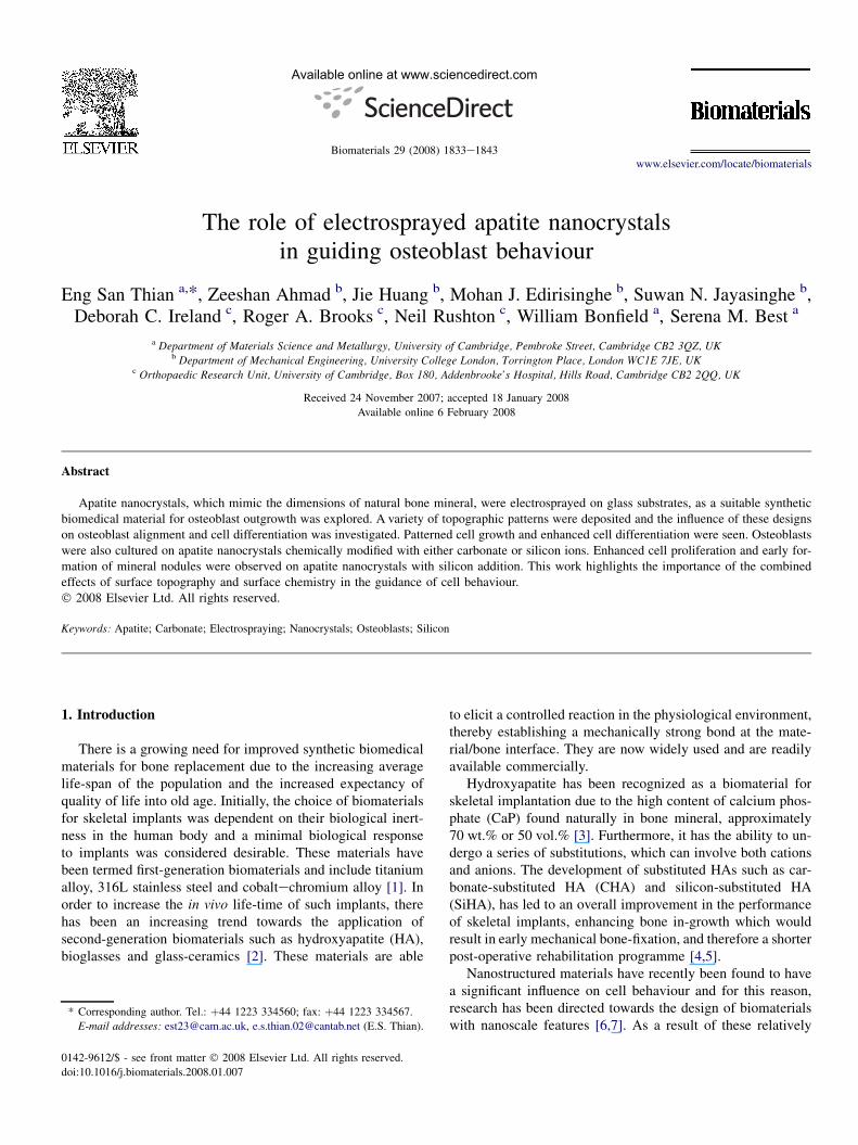
Available online at www.sciencedirect.com
Biomaterials 29 (2008) 1833e1843www.elsevier.com/locate/biomaterials
The role of electrosprayed apatite nanocrystalsin guiding osteoblast behaviour
Eng San Thian a,*, Zeeshan Ahmad b, Jie Huang b, Mohan J. Edirisinghe b, Suwan N. Jayasinghe b,Deborah C. Ireland c, Roger A. Brooks c, Neil Rushton c, William Bonfield a, Serena M. Best a
a Department of Materials Science and Metallurgy, University of Cambridge, Pembroke Street, Cambridge CB2 3QZ, UKb Department of Mechanical Engineering, University College London, Torrington Place, London WC1E 7JE, UK
c Orthopaedic Research Unit, University of Cambridge, Box 180, Addenbrooke’s Hospital, Hills Road, Cambridge CB2 2QQ, UK
Received 24 November 2007; accepted 18 January 2008
Available online 6 February 2008
Abstract
Apatite nanocrystals, which mimic the dimensions of natural bone mineral, were electrosprayed on glass substrates, as a suitable syntheticbiomedical material for osteoblast outgrowth was explored. A variety of topographic patterns were deposited and the influence of these designson osteoblast alignment and cell differentiation was investigated. Patterned cell growth and enhanced cell differentiation were seen. Osteoblastswere also cultured on apatite nanocrystals chemically modified with either carbonate or silicon ions. Enhanced cell proliferation and early for-mation of mineral nodules were observed on apatite nanocrystals with silicon addition. This work highlights the importance of the combinedeffects of surface topography and surface chemistry in the guidance of cell behaviour.� 2008 Elsevier Ltd. All rights reserved.
Keywords: Apatite; Carbonate; Electrospraying; Nanocrystals; Osteoblasts; Silicon
1. Introduction
There is a growing need for improved synthetic biomedicalmaterials for bone replacement due to the increasing averagelife-span of the population and the increased expectancy ofquality of life into old age. Initially, the choice of biomaterialsfor skeletal implants was dependent on their biological inert-ness in the human body and a minimal biological responseto implants was considered desirable. These materials havebeen termed first-generation biomaterials and include titaniumalloy, 316L stainless steel and cobaltechromium alloy [1]. Inorder to increase the in vivo life-time of such implants, therehas been an increasing trend towards the application ofsecond-generation biomaterials such as hydroxyapatite (HA),bioglasses and glass-ceramics [2]. These materials are able
* Corresponding author. Tel.: þ44 1223 334560; fax: þ44 1223 334567.
E-mail addresses: [email protected], [email protected] (E.S. Thian).
0142-9612/$ - see front matter � 2008 Elsevier Ltd. All rights reserved.
doi:10.1016/j.biomaterials.2008.01.007
to elicit a controlled reaction in the physiological environment,thereby establishing a mechanically strong bond at the mate-rial/bone interface. They are now widely used and are readilyavailable commercially.
Hydroxyapatite has been recognized as a biomaterial forskeletal implantation due to the high content of calcium phos-phate (CaP) found naturally in bone mineral, approximately70 wt.% or 50 vol.% [3]. Furthermore, it has the ability to un-dergo a series of substitutions, which can involve both cationsand anions. The development of substituted HAs such as car-bonate-substituted HA (CHA) and silicon-substituted HA(SiHA), has led to an overall improvement in the performanceof skeletal implants, enhancing bone in-growth which wouldresult in early mechanical bone-fixation, and therefore a shorterpost-operative rehabilitation programme [4,5].
Nanostructured materials have recently been found to havea significant influence on cell behaviour and for this reason,research has been directed towards the design of biomaterialswith nanoscale features [6,7]. As a result of these relatively

Table 1
Electrohydrodynamic atomization processing conditions
Islands/coating Pattern
Ground electrode Ring Point
Needle inner diameter (mm) 500 260
Suspension concentration (wt.%) 6.4 6.4
Suspension flow rate (ml min�1) 7.0 2.0
Applied voltage (kV) 5.6e5.8 3.8e4.4
Fig. 1. Characterisation of topographic structures fabricated by electrohydrodynami
(A) dispersed islands, (B) full coating, (C) printed line pattern, (D) printed grid p
topographic structure. (G) Transmission electron microscopy image of electrospra
1834 E. S. Thian et al. / Biomaterials 29 (2008) 1833e1843
recent findings, a new class of biomaterials is being developedwhich combine the excellent biological properties of the sec-ond-generation biomaterials with the unique properties ofnanomaterials, for example nanoscale HA.
Collectively, substituted nanostructured ceramics hold greatpromise for enhancing bonding at the material/bone interface,and thus improving the overall implant efficacy. The potentialof such chemically modified materials could lead to the
c atomization, using nHA suspension. Scanning electron microscopy images of
attern and (E) printed circular pattern. (F) Atomic force microscopy image of
yed apatite nanocrystals.

1835E. S. Thian et al. / Biomaterials 29 (2008) 1833e1843
development of novel coating technology for orthopaedic anddental applications.
The cell/material interaction is also modulated by surfacetopography. Alterations in topography alone can influence cel-lular response ranging from adhesion, orientation and motilityto cell signaling. As such, there is increasing awareness of therole that micrometer- or nanometer-scaled features such asgrooves, ridges and wells, play in controlling cell behaviour[8e11]. Several studies have been conducted, illustrating sur-faces that have been patterned using a range of techniques[12e14].
In this study, electrohydrodynamic atomization (EHDA)was used to fabricate various two-dimensional microstruc-tures, resembling the macrostructure of natural bone. Thesepatterns and islands comprised of apatite nanocrystals. Thesecrystals mimic the nanoscale dimensions and composition ofthe mineral crystals found in natural bone. EHDA is a simpleand low-cost technique capable of producing rapidly surfacefeatures much finer than those attained using conventionalink-jet printing. The effects of the topography and chemistryof these materials on osteoblast outgrowth were investigatedin order to evaluate their potential for bone regeneration.
Fig. 2. Confocal fluorescence microscopy images of nuclei DNA (stained blue), acti
HOB cells as revealed with triple labeling using TOTO-3, FITC-conjugated phalloid
at day 1, (B) underlying glass substrate (acting as a control) at day 1, (C) patterned s
(A, C) were observed on the patterned surface whilst actin bundles were undevelo
2. Materials and methods
2.1. Electrohydrodynamic atomization (EHDA)
Phase-pure apatite nanocrystals prepared in our laboratory namely nHA,
nCHA containing 0.9 wt.% carbonate, and nSiHA containing 0.8 wt.% silicon,
were used to fabricate patterns or islands by electrohydrodynamic atomization,
as described previously [15,16]. Various apatite suspensions (diluted to
6.4 wt.% in ethanol) were delivered by a syringe pump through a stainless steel
needle at a controlled flow rate in a high electric field, resulting in fine apatite
sprays. Under suitable conditions, a stable cone-jet spraying mode was attained,
and samples of the spray were collected on glass microscope slides (Table 1).
The sprayed and printed samples were then examined using a JEOL 6340
field emission gun scanning electron microscope (SEM), operating at an accel-
erating voltage of 5 kV. All samples were sputtered with a thin layer of palla-
dium (Pd) prior to examination. For transmission electron microscopy (TEM)
analysis, samples of the apatite spray were collected on TEM copper grids. A
JEOL 200CX TEM, operating at an accelerating voltage of 200 kV, was then
used to study the morphology of the apatite nanocrystal.
2.2. In vitro cell culture
Human osteoblast (HOB) cells were isolated from hip bone obtained during
total joint replacement surgery, following the guidelines for local ethical com-
mittee approval and informed patient consent. HOB cells were cultured in
n microfilaments (stained green) and vinculin adhesion plaques (stained red) in
in and Texas red-conjugated streptavidin, respectively, on (A) patterned surface
urface at day 3 and (D) control at day 3. Well-defined and aligned actin bundles
ped and random (B, D) on the control surface.
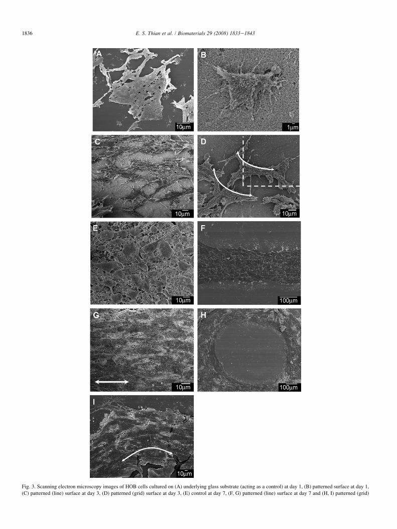
Fig. 3. Scanning electron microscopy images of HOB cells cultured on (A) underlying glass substrate (acting as a control) at day 1, (B) patterned surface at day 1,
(C) patterned (line) surface at day 3, (D) patterned (grid) surface at day 3, (E) control at day 7, (F, G) patterned (line) surface at day 7 and (H, I) patterned (grid)
1836 E. S. Thian et al. / Biomaterials 29 (2008) 1833e1843

Fig. 4. Quantification of protein expression: (A) alkaline phosphatase activity and (B) type 1 collagen production by osteoblasts on non-patterned and patterned
(line or grid) surfaces. *, Significant difference ( p< 0.05 using Student’s t-test of sample size, n¼ 6) compared to non-patterned surface. Note that ALP level was
not detected on all samples at day 1.
1837E. S. Thian et al. / Biomaterials 29 (2008) 1833e1843
McCoy’s 5A medium containing 10% human male AB serum, 1% glutamine
and vitamin C (30 mg ml�1). Patterns were printed using nHA suspension to
form lines, grids and circles. The performance of these patterned samples was
compared with non-printed samples (underlying substrate acting as a control).
In addition, sprayed relics were produced using nHA, nSiHA and nCHA suspen-
sions. Samples of dimensions 10 mm by 10 mm were sterilized using dry heat at
400 �C for 4 h, followed by soaking in ethanol for 48 h before treatment with
ultra-violet radiation for 30 min. Twenty thousand cells in 1 ml of culture me-
dium were then seeded on each sample before incubating under static culture
conditions in a humidified atmosphere containing 5% carbon dioxide at 37 �C.
2.3. Immunocytochemistry
After 1 and 3 days of culture, cells were fixed with 4% paraformaldehyde
in phosphate buffer saline (PBS) solution with 1% sucrose for 15 min, washed
with PBS solution and permeabilised at 4 �C for 5 min. They were then
surface at day 7. An absence of filopodia attachment (A) was noted on the control
with numerous filopodia (B). By day 3, these cells were responding to the contou
randomly organized (E) at day 7, but an oriented cell organization was observed o
incubated with 1% bovine serum albumin (BSA)/PBS solution at 37 �C for
5 min to block the non-specific binding. Anti-vinculin monoclonal antibody
(anti-human raised in mouse hVIN-1, 1:200 in 1% BSA/PBS, Sigma, Poole,
UK) was added and incubated at 37 �C for 1 h. After washing thoroughly
with 0.5% Tween 20/PBS solution for 5 min, a secondary biotin-conjugated
rabbit anti-mouse antibody (1:50 in 1% BSA/PBS, DAKO, UK) was added
and incubated at 37 �C for 1 h. A further wash was conducted before incubat-
ing at 4 �C for 30 min in Texas red-conjugated streptavidin (1:100 in 1% BSA/
PBS, Vector Laboratories, Peterborough, UK). FITC-conjugated phalloidin
(1:100 in 1% BSA/PBS, Sigma, Poole, UK) was later added at 37 �C for
1 h after the samples were washed. After washing for three times with 0.5%
Tween 20/PBS solution for 5 min, TOTO-3 (1:5000 in Tris/EDTA buffer at
pH 8.0, Molecular Probes, Paisley, UK) was added at room temperature for
5 min. The samples were then given a final wash (5 min� 3) before mounting
under Vectashield antifade mountant (Vector Laboratories, UK) and viewed on
a Leica SP2 confocal laser scanning microscope (CLSM).
whilst osteoblasts were spreading and stretching well on the patterned surface,
rs of the patterned surfaces (C, D). On the control surface, HOB cells were
n the patterned surfaces (FeI).
1838 E. S. Thian et al. / Biomaterials 29 (2008) 1833e1843
2.4. Cell morphology
Cultured samples were rinsed with 1% PBS solution after decanting the
medium, freeze-dried, and sputter-coated with a thin layer of Pd before
SEM examination.
2.5. Cell proliferation
Cell number was determined using a CyQUANT� cell proliferation assay
kit (Invitrogen, Paisley, UK), according to manufacturer’s protocol. Briefly,
medium was removed from the sample wells and stored for later quantification
of type 1 collagen. Cultured samples were washed three times with PBS solu-
tion, followed by a freeze-thaw cycle at �80 �C and 24 �C. Next, 300 ml
CyQUANT� buffer was added to each sample. The cell lysate was sonicated
for 5 min and centrifuged for 5 min at 1400 rpm to obtain the supernatant. One
hundred ml of the cell supernatant was added to 100 ml CyQUANT� GR dye.
After 5 min incubation at room temperature, the FLUOstar OPTIMA plate
reader was read at 480/520 nm. The observed fluorescence intensity was
then converted to cell number using a standard curve.
2.6. Alkaline phosphatase (ALP) activity
ALP activity was assessed fluorimetrically at 355/460 nm, following the
mixing 50 ml of the supernatant prepared as above with 50 ml ALP assay buffer
containing 6,8-difluoro-4-methylumbelliferyl phosphate and incubating at
37 �C for 15 min, releasing the fluorescent compound 6,8-difluoro-7-
hydroxy-4-methylcoumarin (DifMU). Alkaline phosphatase activity was deter-
mined using a DifMU standard curve.
2.7. Type 1 collagen (COL1) production
COL1 production was determined by enzyme immunoassay (Metra� CICP
EIA kit, Quidel, Dorking, UK). For analysis, culture medium was first diluted
with assay buffer at a ratio of 1:3. One hundred ml of sample was then added
to each well and incubated at room temperature for 2 h. Wells were washed three
times with 300 ml wash buffer before 100 ml rabbit anti-CICP was added and in-
cubated at room temperature for 50 min. Wells were again washed three times
with wash buffer. Next, 100 ml reconstituted enzyme conjugate was added and
incubated at 24 �C for 50 min. A final wash was performed before 100 ml work-
ing substrate solution was added and incubated at 24 �C for 30 min. Finally,
50 ml of stop solution was added and fluorescence was read at 405 nm. Based
on the standard curve, COL1 expression was determined.
Fig. 5. Quantitative measurement of cell numbers on nHA, nCHA and nSiHA sam
n¼ 6) compared to nCHA. **, Significant difference ( p< 0.05) compared to nHA
2.8. Osteocalcin (OC) expression
The OC level in the culture medium was measured by an enzyme immu-
noassay (Metra� Osteocalcin EIA kit, Quidel, Dorking, UK). Twenty-five ml
of cell medium and 125 ml of anti-osteocalcin antibody were added to each
well before incubating at room temperature for 2 h. Wells were then washed
three times with 300 ml wash buffer. Following that, 150 ml reconstituted
enzyme conjugate was added and incubated at room temperature for 1 h. Wells
were again washed three times before 150 ml working substrate solution was
added and incubated at room temperature for 35 min. Finally, 50 ml of stop
solution was added and fluorescence was read at 405 nm
2.9. Statistical analysis
A two-tailed t-test was used to determine whether any significant differ-
ences existed between the mean values of the experimental groups. Differences
between groups were considered to be significant at p< 0.05.
3. Results
3.1. Surface structures
SEM was used to image the surface structures created byEHDA. By using a ring-shaped ground electrode, microme-ter-sized islands between 10 mm and 220 mm were randomlydistributed across substrate’s surface (Fig. 1A). A full coatingcan be achieved by simply increasing the deposition time(Fig. 1B). This result highlights that EHDA is a potential alter-native technique for coating metallic implants. In contrast, var-ious defined surface patterns in the form of lines, arrays orcircles were printed using a pointed ground electrode(Fig. 1CeE). The width of these printed patterns was approx-imately 100 mm. Atomic force microscopy revealed that thesestructures comprised of densely-packed apatite nanocrystals(Fig. 1F). Transmission electron microscopy further revealedthat these apatite nanocrystals exhibited a rod-like morphol-ogy of dimensions approximately 75 nm in length by 40 nmin width. In addition, selective area electron diffraction imageshowed a spotted pattern, indicating that these apatite nanopar-ticles were crystalline in nature (Fig. 1G).
ples. *, Significant difference ( p< 0.05 using Student’s t-test of sample size,
.
1839E. S. Thian et al. / Biomaterials 29 (2008) 1833e1843
3.2. Surface topography
3.2.1. ImmunocytochemistryImmunofluorescent staining at day 1 revealed distinct dif-
ferences in cell morphology among the sample surfaces.
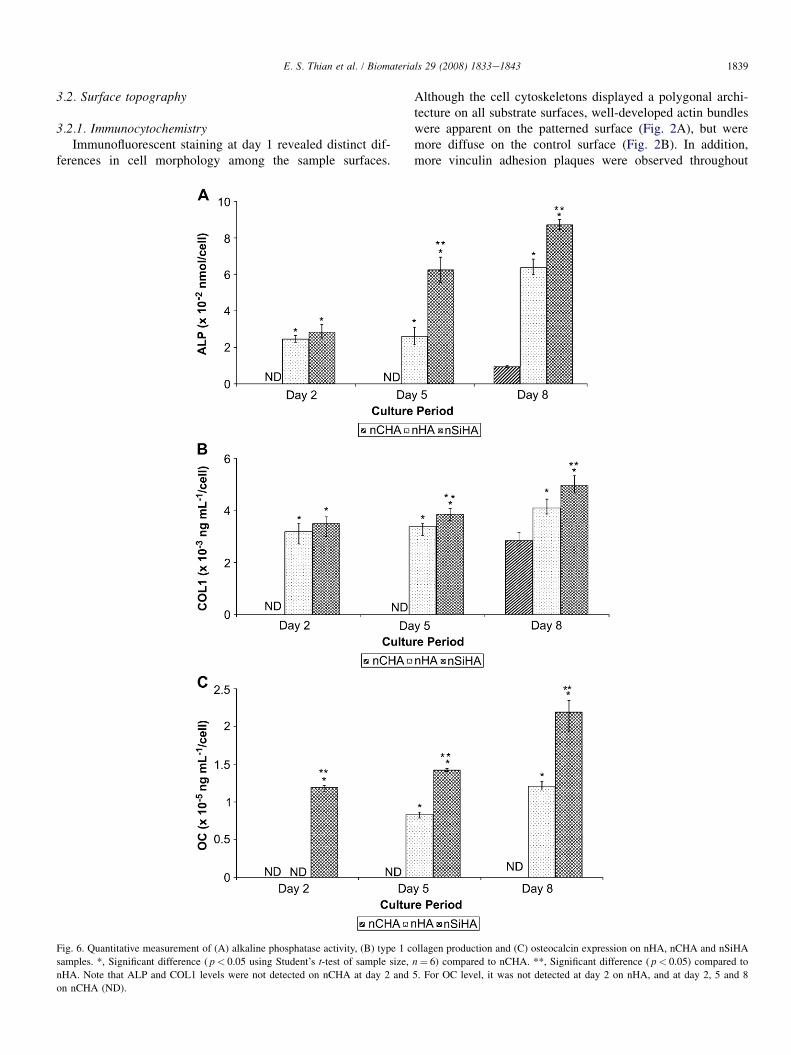
Fig. 6. Quantitative measurement of (A) alkaline phosphatase activity, (B) type 1 c
samples. *, Significant difference ( p< 0.05 using Student’s t-test of sample size,
nHA. Note that ALP and COL1 levels were not detected on nCHA at day 2 and
on nCHA (ND).
Although the cell cytoskeletons displayed a polygonal archi-tecture on all substrate surfaces, well-developed actin bundleswere apparent on the patterned surface (Fig. 2A), but weremore diffuse on the control surface (Fig. 2B). In addition,more vinculin adhesion plaques were observed throughout
ollagen production and (C) osteocalcin expression on nHA, nCHA and nSiHA
n¼ 6) compared to nCHA. **, Significant difference ( p< 0.05) compared to
5. For OC level, it was not detected at day 2 on nHA, and at day 2, 5 and 8

1840 E. S. Thian et al. / Biomaterials 29 (2008) 1833e1843
the osteoblasts that had attached to the patterned surface. Ad-hesion plaques were concentrated only at the periphery of thecell cytoplasm on the control surfaces. Cells were abundant onboth substrates at day 3, demonstrating that the surfaces sup-ported osteoblast outgrowth. Interestingly, HOB cells followedthe guidance of the patterned (line) surface and exhibited littlebranching since the actin bundles were aligned in the directionof the long axis of the printed pattern (Fig. 2C). In contrast,osteoblasts were randomly organized on the non-patterned(control) surface (Fig. 2D).
3.2.2. Cell morphologyAt culture day 1, cells were adhering and spreading on the
control (Fig. 3A) and patterned (Fig. 3B) surfaces. However,numerous filopodia were observed to anchor the cells on thepatterned surface, but this was not the case for the control.Cells seem to respond well towards surface topography byday 3. On the line pattern, osteoblasts were observed to bestretching along one direction (Fig. 3C) whilst on the grid pat-tern, ‘cell bridging’ was seen at sharp corners (Fig. 3D). After7 days of culturing, distinctive differences in cell morphologywere observed. On the control surface (Fig. 3E), HOB cellswere noticeably less flattened and exhibited a random cell or-ganization. On the patterned surfaces (Fig. 3F, H), a confluent
Fig. 7. Fluorescent microscopy images of osteoblasts cultured for 24 h on (A) nH
vinculin adhesion plaques were stained blue, green and red, respectively. HOB c
increase in the number and size of focal adhesion plaques on nSiHA (C) as comp
and oriented cell layer was formed. At high magnification,these osteoblasts appeared elongated and well-stretched, fol-lowing the contours of the printed patterns (Fig. 3G, I).
3.2.3. Cell differentiationALP activity is used as an early biochemical marker for
osteoblast differentiation. The onset of ALP activity for allsamples was seen only at day 3 (Fig. 4A). Generally, the levelof ALP activity of HOB cells cultured on all samples increasedwith culturing time. A significant increase in ALP activity wasobserved for the patterned (line or grid) surfaces at day 3 and 7when compared to the control surface, indicating greater celldifferentiation.
An overall increase in COL1 expression with culture timewas observed on all samples (Fig. 4B). In particular, HOB cellsgrowing on the patterned surface expressed significantly higherCOL1 production at day 3 and 7, compared to the control.
3.3. Surface chemistry
3.3.1. Cell proliferationTo study the effects of surface chemistry on osteoblast
response, micrometer-sized islands of nHA, nCHA andnSiHA were randomly deposited on substrate’s surface. The
A, (B) nCHA and (C) nSiHA surfaces. Cell nuclei, actin microfilaments and
ells exhibited well-defined cytoskeletal organization on all samples. Note the
ared to nHA (A) and nCHA (B).

Fig. 8. Morphology of osteoblasts cultured on (A) nHA at day 2, (B) nCHA at day 2, (C) nSiHA at day 2, (D) nHA at day 5, (E) nCHA at day 5, (F) nSiHA at day 5,
(G) nHA at day 8, (H) nCHA at day 8 and (I) nSiHA at day 8. At day 2, cells were well-flattened and attached on nHA (A) and nSiHA (C), with numerous fi-
lopodia. There was a decrease in filopodia on nCHA (B) even though the cells were spreading well. By day 5, osteoblasts became confluent and were observed to
secrete type 1 collagen on nHA (D) and nSiHA (F). Presence of filopodia attachment was noted on nCHA (E). Biomineralisation occurred on nHA (G) and nSiHA
(I) by day 8, with more CaP nodules being observed on nSiHA, but this was not the case on nCHA (H).
1841E. S. Thian et al. / Biomaterials 29 (2008) 1833e1843

1842 E. S. Thian et al. / Biomaterials 29 (2008) 1833e1843
CyQUANT� assay revealed that the number of HOB cellsgrowing on nHA and nSiHA surfaces increased with culturingtime, but those on nCHA appeared to decrease (Fig. 5). Over-all, osteoblasts tended to multiply significantly on nHA andnSiHA as compared to nCHA. The metabolic activity of thecells growing on nSiHA seemed to slow-down after day 5,suggesting that HOBs were advancing into differentiationstage at early time points.
3.3.2. Cell differentiationThe level of protein expression on these nanoapatites was
also determined. An increase in the ALP activity with culturetime was observed for nHA and nSiHA, but was only detect-able for nCHA at day 8 (Fig. 6A). When comparing all sam-ples, ALP activity on nSiHA was statistically the highest atday 5 and 8, compared to nHA and nCHA.
Generally, a steady increase in COL1 expression was de-tected on nHA and nSiHA at all time points, with significantquantity of COL1 being produced on nSiHA at day 5 and 8(Fig. 6B). In contrast, osteoblasts cultured on nCHA onlystarted to secrete COL1 from day 8 onwards.
For cells attaching on nSiHA, OC was detected from day 2onwards and increased with culturing time (Fig. 6C). Con-versely, the onset of OC expression was observed at day 5for nHA. As for nCHA, no OC protein could be detectedthroughout the culture period.
3.3.3. ImmunocytochemistryFluorescent microscopy of cytoskeletal organization re-
vealed that HOB cells were spreading and stretching on allnanoapatites at day 1, generating well-defined actin bundles(Fig. 7AeC). However, cell adherence was more pronouncedon nSiHA, with more distinct and thick vinculin adhesion pla-ques within cells. This effect is likely to modulate intracellularsignaling pathways that regulate transcriptional activity andgene expression, which in turn can modify and binding to ex-tracellular matrix proteins via integrins.
3.3.4. Cell morphologySEM investigations again revealed that osteoblasts were
spreading and growing on all apatite nanocrystals at day 2,but filopodia were only seen in significant numbers projectingfrom the cell edges for nHA and nSiHA (Fig. 8AeC). This be-haviour indicates that nHA and nSiHA particles were acting asanchors for the cells to attach, thereby improving cell/substrateadhesion. A confluent cell layer was formed on nHA andnSiHA surfaces by day 5, along with the secretion of COL1(Fig. 8DeF). After 8 days of culturing, biomineralisationstarted to occur on both nHA and nSiHA, with more CaP min-eral nodules nucleating on the collagen matrices for nSiHA(Fig. 8G, I) whilst cells became more confluent on nCHA(Fig. 8H).
4. Discussion
Electrohydrodynamic atomization (EHDA), or known aselectrospraying, has been used extensively for the deposition
of calcium phosphate coatings for biomedical applications[17e20]. Now, it is possible to deposit and retain apatite nano-crystals, in their original form, of various surface topographiesand chemistries onto implant surfaces using EHDA. A recentstudy using acellular simulated body fluid has demonstratedthe bioactive potential of these apatite nanocrystals in vitro[15]. For the current work, the effects of contact guidanceand chemical composition of these apatite nanocrystals oncell morphological changes, proliferation and differentiationwere evaluated.
Electrosprayed apatite nanocrystals were able to providecontact guidance to osteoblast cells. Patterns created usingnHA induced an obvious alignment of cells. In contrast,HOB cells cultured on control surfaces adopted a randomly or-ganized morphology. A significant increase of ALP activityand COL1 expression in HOBs cultured on printed patternsat early time points, suggested that cell alignment could pro-mote the differentiation and maturation of osteoblast cells.These results highlight that the creation of surface topographyusing nHA not only promote and direct osteoblast outgrowth,but also that it supports cell attachment, proliferation and spe-cifically cell differentiation processes. If unidirectional proper-ties are required, a printed line pattern will be suffice. On theother hand, a grid pattern is desirable if multidirectional prop-erties are needed for an implant prosthesis. Nevertheless, theprecise mechanism relating the changes in cell morphologyto gene expression remains unknown.
To validate the enhancement of cell maturation by chemicalcues from electrosprayed apatite nanocrystals, proliferationand differentiation assays were performed and analysed. Adecline in cell viability on nCHA with culturing time wasobserved. This effect was due to the highest solubility rateamong the studied apatite nanocrystals, owing to its relativelylow crystallinity, thereby rendering the surface unfavourablefor cell attachment [15]. However, more cells began to attachand proliferate on nCHA at a later culture period (day 8onwards). This phenomenon was attributed to the formationof a newly-grown carbonate-containing apatite layer, and asa result, developing a more biostable surface. Conversely,there was evidence of cells advancing into differentiation stageand developed bone minerals on nSiHA at early culturingtime. The expression of the following proteins namely ALP,COL1 and OC was statistically high throughout the culture pe-riod, indicating the efficacy of nSiHA in driving osteoblastcells towards the maturation stage. In addition, a decline orslow-down in cell proliferation usually signals that cell differ-entiation is occurring [21].
Taken together, these findings demonstrate that the underly-ing material surface chemistry of different apatite nanocrystalscan be used to control cell behaviour. In this case, it is clearthat the incorporation of a minute amount (0.8 wt.%) of Siinto HA, plays an important and significant role in directingcellular response, thereby promoting osteoblast proliferation,differentiation, collagen synthesis as well as the biomineralisa-tion processes. The enhanced bioactivity of nSiHA can beattributed to a combination of effects. nSiHA facilitated earlyprecipitation of a biologically equivalent apatite layer, creating

1843E. S. Thian et al. / Biomaterials 29 (2008) 1833e1843
and conditioning a surface associated with increased adsorp-tion and incorporation of biological moieties that served as at-tachment sites for osteoblasts to mineralize [22e24]. Therelease of Si complexes into the biological media or the pres-ence of Si complexes at the material surface, both in the formof Si(OH)4, triggered genes that encoded proteins involved inosteoblast proliferation, differentiation and biomineralisation[25e27]. However, the exact mechanism by which how Siaffects bone formation needs further investigation.
5. Conclusions
In this study, we have clearly demonstrated that topo-graphic patterns are able to generate patterned cell growthand promote cell differentiation, and the latter can be furtherenhanced by chemical modification of the apatite nanocrystals,especially with silicon substitution. The combination of bioac-tive, with a selective apatite surface pattern could provide boththe physical and chemical cues to enhance and direct osteo-blast outgrowth. Therefore, electrosprayed apatite nanocrys-tals can be used as an alternative biomaterial for coating theorthopaedic and dental implants.
Acknowledgements
This work was funded by grants from Engineering andPhysical Sciences Research Council, United Kingdom (GR/S97873 and GR/S97880).
References
[1] Hench LL. Biomaterials. Science 1980;208:826e31.
[2] Hench LL, Wilson J. Surface-active biomaterials. Science 1984;226:
630e6.
[3] Aoki H. Science and medical applications of hydroxyapatite. Tokyo:
JAAS; 1991.
[4] Porter AE, Patel N, Skepper JN, Best SM, Bonfield W. Comparison of in
vivo dissolution processes in hydroxyapatite and silicon-substituted
hydroxyapatite bioceramics. Biomaterials 2003;24:4609e20.
[5] Doi Y, Shibutani T, Moriwaki Y, Kajimoto T, Iwayama Y. Sintered
carbonate apatites as resorbable bone substitutes. J Biomed Mater Res
1998;39:603e10.
[6] Webster TJ, Ejiofor JU. Increased osteoblast adhesion on nanophase
metals: Ti, Ti6Al4V, and CoCrMo. Biomaterials 2004;25:4731e9.
[7] Zanello LP, Zhao B, Hu H, Haddon RC. Bone cell proliferation on
carbon nanotubes. Nano Lett 2006;6:562e7.
[8] Flemming RG, Murphy CJ, Abrams GA, Goodman SL, Nealy PF. Effects
of synthetic micro- and nano-structured surfaces on cell behaviour.
Biomaterials 1999;20:573e85.
[9] Wilkinson CDW, Riehle M, Wood M, Gallagher J, Curtis ASG. The use
of materials patterned on a nano- and micro-metric scale in cellular
engineering. Mater Sci Eng C 2002;19:263e9.
[10] Dalby MJ, Riehle MO, Johnstone H, Affrosman S, Curtis ASG. In vitroreaction of endothelial cells to polymer demixed nanotopography.
Biomaterials 2002;23:2945e54.
[11] Berry CC, Campbell G, Spadiccino A, Robertson M, Curtis ASG. The
influence of microscale topography on fibroblast attachment and motility.
Biomaterials 2004;25:5781e8.
[12] Bao LR, Cheng X, Huang XD, Guo LJ, Pang SW, Yee AF. Nanoimprint-
ing over topography and multilayer three-dimensional printing. J Vac Sci
Tech B 2002;20:2881e6.
[13] Limpanuphap S, Derby B. Manufacture of biomaterials by a novel
printing process. J Mater Sci Mater Med 2002;13:1163e6.
[14] Harnett CK, Satyalakshmi KM, Craighead HG. Bioactive templates fab-
ricated by low-energy electron beam lithography of self-assembled
monolayers. Langmuir 2001;17:178e82.
[15] Thian ES, Ahamd Z, Huang J, Edirisinghe MJ, Jayasinghe SN,
Ireland DC, et al. Bioactivity of nanoapatite produced by electrohydrody-
namic atomisation. J Bionanosci 2007;1:60e3.
[16] Ahamd Z, Huang J, Edirisinghe MJ, Jayasinghe SN, Best SM,
Bonfield W, et al. Electrohydrodynamic print-patterning of nano-hy-
droxyapatite. J Biomed Nanotechnol 2006;2:201e7.
[17] Leeuwenburgh S, Wolke J, Schoonman J, Jansen J. Electrostatic spray
deposition (ESD) of calcium phosphate coatings. J Biomed Mater Res
2003;66:330e4.
[18] Leeuwenburgh SCG, Heine MC, Wolke JGC, Pratsinis SE, Schoonman J,
Jansen JA. Morphology of calcium phosphate coatings for biomedical
applications deposited using electrostatic spray deposition. Thin Solid
Films 2006;503:69e78.
[19] Huang J, Jayasinghe SN, Best SM, Edirisinghe MJ, Brooks RA,
Bonfield W. Electrospraying of a nano-hydroxyapatite suspension. J
Mater Sci 2004;39:1029e32.
[20] Huang J, Jayasinghe SN, Best SM, Edirisinghe MJ, Brooks RA,
Rushton N, et al. Novel deposition of nano-sized silicon substituted
hydroxyapatite by electrostatic spraying. J Mater Sci Mater Med
2005;16:1137e42.
[21] Aparicio C, Gil FJ, Planell JA, Engel E. Human-osteoblast proliferation
and differentiation on grit-blasted and bioactive titanium for dental
applications. J Mater Sci Mater Med 2002;13:1105e11.
[22] Ducheyne P, Qui Q. Bioactive ceramics: the effect of surface reactivity
on bone formation and bone cell function. Biomaterials 1999;20:
2287e303.
[23] Neo M, Nakamura T, Ohtsuki C, Kokubo T, Yamamuro T. Apatite
formation on three kinds of bioactive materials at early stage in vivo:
a comparative study by transmission electron microscopy. J Biomed
Mater Res 1993;27:999e1006.
[24] Canham LT, Newey JP, Reeves CL, Houlton MR, Loni A, Simons AJ,
et al. The effects of DC electric currents on the in-vitro calcification of
bioactive silicon wafers. Adv Mater 1996;8:847e9.
[25] Xynos ID, Edgar AJ, Buttery LD, Hench LL, Polak JM. Gene expression
profiling of human osteoblasts following treatment with the ionic
products of Bioglass� 45S5 dissolution. J Biomed Mater Res 2001;55:
151e7.
[26] Reffitt DM, Ogston N, Jigdaoshsingh R, Cheung HFJ, Evans BAJ.
Orthosilicic acid stimulates collagen type 1 synthesis and osteoblastic
differentiation in human osteoblast-like cells in vitro. Bone
2003;32:127e35.
[27] Gupta G, Kirakodu S, El-Ghannam A. Dissolution kinetics of a Si-rich
nanocomposite and its effect on osteoblast gene expression. J Biomed
Mater Res 2007;80A:486e96.
Copyright © 2022 FDOKUMEN