Role of nitric oxide- and vasoactive intestinal polypeptide-containing neurones in human gastric...
9
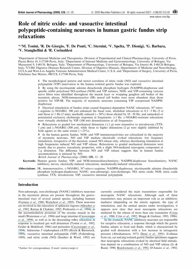
Role of nitric oxide- and vasoactive intestinal polypeptide-containing neurones in human gastric fundus strip relaxations * ,1 M. Tonini, 2 R. De Giorgio, 3 F. De Ponti, 4 C. Sternini, 1 V. Spelta, 5 P. Dionigi, 2 G. Barbara, 2 V. Stanghellini & 2 R. Corinaldesi 1 Department of Internal Medicine and Therapeutics, Division of Experimental and Clinical Pharmacology, University of Pavia, Piazza Botta 10, I-27100 Pavia, Italy; 2 Department of Internal Medicine and Gastroenterology, University of Bologna, Via Massarenti 9, I-40138, Bologna, Italy; 3 Department of Pharmacology, University of Bologna, Via Irnerio 48, I-40126 Bologna, Italy; 4 CURE Digestive Diseases Research Center, Division of Digestive Diseases, Departments of Medicine and Neurobiology, UCLA and West-Los Angeles Veterans Administration Medical Center, U.S.A. and 5 Department of Surgery, University of Pavia, Policlinico San Matteo, IRCCS, I-27100 Pavia, Italy 1 The morphological pattern and motor correlates of nitric oxide (NO) and vasoactive intestinal polypeptide (VIP) innervation in the human isolated gastric fundus was explored. 2 By using the nicotinamide adenine dinucleotide phosphate hydrogen (NADPH)-diaphorase and specific rabbit polyclonal NO-synthase (NOS) and VIP antisera, NOS- and VIP-containing varicose nerve fibres were identified throughout the muscle layer or wrapping ganglion cell bodies of the myenteric plexus. NOS-immunoreactive (IR) neural cell bodies were more abundant than those positive for VIP-IR. The majority of myenteric neurones containing VIP coexpressed NADPH- diaphorase. 3 Electrical stimulation of fundus strips caused frequency-dependent NANC relaxations. N G -nitro- L-arginine (L-NOARG: 300 mM) enhanced the basal tone, abolished relaxations to 0.3 – 3 Hz (5 s) and those to 1 Hz (5 min), markedly reduced (*50%) those elicited by 10 – 50 Hz, and unmasked or potentiated excitatory cholinergic responses at frequencies 51 Hz. L-NOARG-resistant relaxations were virtually abolished by VIP (100 nM) desensitization at all frequencies. 4 Relaxations to graded low mechanical distension (41 g) were insensitive to tetrodotoxin (TTX: 1 mM) and L-NOARG (300 mM), while those to higher distensions (2 g) were slightly inhibited by both agents to the same extent (*25%). 5 In the human gastric fundus, NOS- and VIP immunoreactivities are colocalized in the majority of myenteric neurones. NO and VIP mediate electrically evoked relaxations: low frequency stimulation, irrespective of the duration, caused NO release only, whereas shortlasting stimulation at high frequencies induced NO and VIP release. Relaxations to graded mechanical distension were mostly due to passive viscoelastic properties, with a slight NO-mediated neurogenic component at 2g distension. The dierence between NO and VIP release suggests that in human fundus accommodation is initiated by NO. British Journal of Pharmacology (2000) 129, 12 – 20 Keywords: Human gastric fundus; VIP- and NOS-immunohistochemistry; NADPH-diaphorase histochemistry; NANC inhibitory nerves; electrically-induced relaxations; mechanically-induced relaxations Abbreviations: IR, immunoreactive; L-NOARG, N G -nitro-L-arginine; NADPH-diaphorase, nicotinamide adenine dinucleotide phosphate hydrogen-diaphorase; NANC, non-adrenergic, non-cholinergic; NO, nitric oxide; NOS, nitric oxide synthase; TTX, tetrodotoxin; VIP, vasoactive intestinal polypeptide Introduction Non-adrenergic, non-cholinergic (NANC) inhibitory neurones in the myenteric plexus are present throughout the gastro- intestinal tract of several animal species, including humans (Furness et al., 1988; Wattchow et al., 1995). These neurones are involved in the relaxation of sphincter regions (Allescher et al., 1992; Rattan & Chakder, 1992; Preiksaitis et al., 1994), in the accommodation processes of the circular muscle in the small (Waterman et al., 1994) and large intestine (Ciccocioppo et al., 1994), as well as in the descending reflex relaxation in response to localized gut wall distension (Tonini et al., 1982; Grider & Makhlouf, 1986) and peristalsis (Ciccocioppo et al., 1994). Adenosine 5’-triphosphate (ATP) (Hoyle & Burnstock, 1989), vasoactive intestinal polypeptide (VIP) (Fahrenkrug, 1989), and nitric oxide (NO) (Sanders & Ward, 1992) are currently considered the main transmitters responsible for neurogenic NANC relaxations. Although each of these transmitters may possess an important role as an inhibitory mediator (depending on the enteric segment, the type of stimulation, and the animal species under investigation), it appears now clear that most neurogenic relaxations are mediated by the release of more than one transmitter (Costa et al., 1986; Crist et al., 1992; Maggi & Giuliani, 1993; 1996). In the stomach, NANC inhibitory neurones are responsible for receptive relaxation, a response through which the gastric fundus adapts to food and fluids, which is characterized by graded wall distension with a low increase in intragastric pressure (Abrahamsson, 1973; Desai et al., 1991). Evidence from rat, cat and pig isolated gastric fundus specimens suggests that neurogenic relaxations evoked by electrical field stimula- tion depend on a combination of NO and VIP release (Li & Rand, 1990; Boeckxstaens et al., 1992; D’Amato et al., 1992; *Author for correspondence; E-mail: [email protected] British Journal of Pharmacology (2000) 129, 12 – 20 ª 2000 Macmillan Publishers Ltd All rights reserved 0007 – 1188/00 $15.00 www.nature.com/bjp
-
Upload
independent -
Category
Documents
-
view
0 -
download
0
Transcript of Role of nitric oxide- and vasoactive intestinal polypeptide-containing neurones in human gastric...
Role of nitric oxide- and vasoactive intestinalpolypeptide-containing neurones in human gastric fundus striprelaxations
*,1M. Tonini, 2R. De Giorgio, 3F. De Ponti, 4C. Sternini, 1V. Spelta, 5P. Dionigi, 2G. Barbara,2V. Stanghellini & 2R. Corinaldesi
1Department of Internal Medicine and Therapeutics, Division of Experimental and Clinical Pharmacology, University of Pavia,Piazza Botta 10, I-27100 Pavia, Italy; 2Department of Internal Medicine and Gastroenterology, University of Bologna, ViaMassarenti 9, I-40138, Bologna, Italy; 3Department of Pharmacology, University of Bologna, Via Irnerio 48, I-40126 Bologna,Italy; 4CURE Digestive Diseases Research Center, Division of Digestive Diseases, Departments of Medicine and Neurobiology,UCLA and West-Los Angeles Veterans Administration Medical Center, U.S.A. and 5Department of Surgery, University of Pavia,Policlinico San Matteo, IRCCS, I-27100 Pavia, Italy
1 The morphological pattern and motor correlates of nitric oxide (NO) and vasoactive intestinalpolypeptide (VIP) innervation in the human isolated gastric fundus was explored.
2 By using the nicotinamide adenine dinucleotide phosphate hydrogen (NADPH)-diaphorase andspeci®c rabbit polyclonal NO-synthase (NOS) and VIP antisera, NOS- and VIP-containing varicosenerve ®bres were identi®ed throughout the muscle layer or wrapping ganglion cell bodies of themyenteric plexus. NOS-immunoreactive (IR) neural cell bodies were more abundant than thosepositive for VIP-IR. The majority of myenteric neurones containing VIP coexpressed NADPH-diaphorase.
3 Electrical stimulation of fundus strips caused frequency-dependent NANC relaxations. NG-nitro-L-arginine (L-NOARG: 300 mM) enhanced the basal tone, abolished relaxations to 0.3 ± 3 Hz (5 s)and those to 1 Hz (5 min), markedly reduced (*50%) those elicited by 10 ± 50 Hz, and unmasked orpotentiated excitatory cholinergic responses at frequencies 51 Hz. L-NOARG-resistant relaxationswere virtually abolished by VIP (100 nM) desensitization at all frequencies.
4 Relaxations to graded low mechanical distension (41 g) were insensitive to tetrodotoxin (TTX:1 mM) and L-NOARG (300 mM), while those to higher distensions (2 g) were slightly inhibited byboth agents to the same extent (*25%).
5 In the human gastric fundus, NOS- and VIP immunoreactivities are colocalized in the majorityof myenteric neurones. NO and VIP mediate electrically evoked relaxations: low frequencystimulation, irrespective of the duration, caused NO release only, whereas shortlasting stimulation athigh frequencies induced NO and VIP release. Relaxations to graded mechanical distension weremostly due to passive viscoelastic properties, with a slight NO-mediated neurogenic component at2 g distension. The di�erence between NO and VIP release suggests that in human fundusaccommodation is initiated by NO.British Journal of Pharmacology (2000) 129, 12 ± 20
Keywords: Human gastric fundus; VIP- and NOS-immunohistochemistry; NADPH-diaphorase histochemistry; NANCinhibitory nerves; electrically-induced relaxations; mechanically-induced relaxations
Abbreviations: IR, immunoreactive; L-NOARG, NG-nitro-L-arginine; NADPH-diaphorase, nicotinamide adenine dinucleotidephosphate hydrogen-diaphorase; NANC, non-adrenergic, non-cholinergic; NO, nitric oxide; NOS, nitric oxidesynthase; TTX, tetrodotoxin; VIP, vasoactive intestinal polypeptide
Introduction
Non-adrenergic, non-cholinergic (NANC) inhibitory neuronesin the myenteric plexus are present throughout the gastro-
intestinal tract of several animal species, including humans(Furness et al., 1988; Wattchow et al., 1995). These neuronesare involved in the relaxation of sphincter regions (Allescher et
al., 1992; Rattan & Chakder, 1992; Preiksaitis et al., 1994), inthe accommodation processes of the circular muscle in thesmall (Waterman et al., 1994) and large intestine (Ciccocioppo
et al., 1994), as well as in the descending re¯ex relaxation inresponse to localized gut wall distension (Tonini et al., 1982;Grider & Makhlouf, 1986) and peristalsis (Ciccocioppo et al.,1994). Adenosine 5'-triphosphate (ATP) (Hoyle & Burnstock,
1989), vasoactive intestinal polypeptide (VIP) (Fahrenkrug,1989), and nitric oxide (NO) (Sanders & Ward, 1992) are
currently considered the main transmitters responsible forneurogenic NANC relaxations. Although each of these
transmitters may possess an important role as an inhibitorymediator (depending on the enteric segment, the type ofstimulation, and the animal species under investigation), it
appears now clear that most neurogenic relaxations aremediated by the release of more than one transmitter (Costaet al., 1986; Crist et al., 1992; Maggi & Giuliani, 1993; 1996).
In the stomach, NANC inhibitory neurones are responsiblefor receptive relaxation, a response through which the gastricfundus adapts to food and ¯uids, which is characterized bygraded wall distension with a low increase in intragastric
pressure (Abrahamsson, 1973; Desai et al., 1991). Evidencefrom rat, cat and pig isolated gastric fundus specimens suggeststhat neurogenic relaxations evoked by electrical ®eld stimula-
tion depend on a combination of NO and VIP release (Li &Rand, 1990; Boeckxstaens et al., 1992; D'Amato et al., 1992;*Author for correspondence; E-mail: [email protected]
British Journal of Pharmacology (2000) 129, 12 ± 20 ã 2000 Macmillan Publishers Ltd All rights reserved 0007 ± 1188/00 $15.00
www.nature.com/bjp
Barbier & Lefebvre, 1993; Lefebvre et al., 1995), which may actas co-transmitters. Other ®ndings suggest that NO is thepredominant transmitter mediating accommodation to ¯uids
and relaxation to short and sustained electrical ®eldstimulation in the guinea-pig isolated stomach (Desai et al.,1991; Lefebvre et al., 1992). Furthermore, NO plays a pivotalrole in gastric fundus relaxations to electrical vagal stimulation
in intact dogs (Meulemans et al., 1995), and in isolatedstomach preparations from the guinea-pig (Desai et al., 1994;Meulemans et al., 1995) and mouse (Yano et al., 1995).
In the human isolated gastric fundus, the nature ofneurogenic NANC relaxations elicited either electrically ormechanically has never been explored. This study was
therefore designed to examine the distribution of NOsynthesizing and VIP containing neurones in human gastricfundus specimens, and to establish their role in the control of
fundus strip relaxations.
Methods
Tissue collection and preparation
Fresh specimens of human gastric fundus were obtained fromten male and three female patients (age range, 47 ± 82 years)undergoing total gastrectomy due to cancer. The use of human
tissue was approved by the Ethics Committees of theUniversities of Pavia and Bologna. As soon as the organ wasremoved, a portion of apparently normal gastric fundus was
placed in oxygenated (95% O2 and 5% CO2) modi®ed Krebssolution at 48C and transported to the laboratory within 30 ±45 min. All specimens were used for functional studies; four ofthese specimens were also used for morphological studies. In
this case, gastric fundus specimens were thoroughly rinsed withsaline, stretched slightly and pinned on wax plates. Specimenswere ®xed by immersion-®xation with 4% paraformaldehyde
in 0.1 M phosphate bu�er (PB), pH 7.4, overnight at 48C andsubsequently placed in 25% sucrose in 0.1 M PB forcryoprotection until sectioning. Tissues were cut with a
cryostat at 10 ± 12 mm, mounted onto chrome-alum gelatincoated slides, and stored at 7208C until processing.
To evaluate the general morphology and the quality of thecollected tissues, some sections from each gastric fundus
specimen were stained with haematoxylin-eosin. No obviousabnormalities were detected in the mucosa, submucosa andmuscle layers.
Immunohistochemistry
Tissue sections were processed with the indirect immuno-¯uorescence method (De Giorgio et al., 1992). Brie¯y, sectionswere washed in 0.1 M PB, pretreated for 30 min at room
temperature with 10% normal goat serum, and incubated inrabbit VIP (VIP7913, 1 : 500) (Furness et al., 1981) or neuronalNOS polyclonal antisera (1 : 100) (Schmidt et al., 1992)overnight at 48C in a humid chamber. Sections were then
washed in PB and incubated for 2 h at room temperature ina�nity puri®ed goat anti-rabbit IgG (diluted 1 : 50) conjugatedto either ¯uorescein isothiocyanate or tetramethyl rhodamine
isothiocyanate (Sigma Immunochemicals, St. Louis, MO,U.S.A.). Sections were washed again in 0.1 M PB andcoverslipped with 9 : 1 glycerol/PB. Both primary and
secondary antibodies were diluted in 0.5% Triton X-100 in0.1 M PB. Sections were analysed with a Leitz Dialuxmicroscope using a Ploem epi-illumination system with `I2'or `L2' and `N2' ®lter cubes to visualize ¯uorescein
isothiocyanate and tetramethyl rhodamine isothiocyanate¯uorescence, respectively.
Speci®city studies included the following control experi-
ments: (a) omission of the primary antibodies; (b) substitutionof neuronal NOS and VIP antibodies with commerciallyavailable normal rabbit serum used at a dilution of 1 : 50; and(c) incubation with the primary antibody preadsorbed for 12 ±
16 h at 48C with 10 mM synthetic VIP peptide (Bachem,Torrance, CA, U.S.A.).
NADPH-diaphorase histochemistry
The histochemical technique was performed as previously
described (De Giorgio et al., 1994). Cryostat sections werewashed in 0.1 M PB (three times, 10 min each), preincubated in0.3% Triton X-100 in 0.1 M PB for 20 min at room
temperature, and then incubated in a mixture containing1 mg ml71 reduced b-nicotinamide adenine phosphate dinu-cleotide (NADPH) and 0.25 mg ml71 nitro blue tetrazolium(Sigma, St. Louis, MO, U.S.A.) in 0.1 M PB in the dark at
378C for 1 h. To stop the reaction, sections were thoroughlywashed in 0.1 M PB, dehydrated through graded ethanols, and®nally coverslipped with mounting medium. The results
obtained were evaluated with a Leitz Dialux microscope usingbright ®eld optics.
Control experiments were performed by incubating sections
with solutions in which the b-NADPH reagent was omitted.
Double labelling
A sequential staining procedure was used for colocalizationexperiments (De Giorgio et al., 1994). Sections processed forimmuno¯uorescence were analysed and photographed, and the
coordinates of the photographic images were recorded forsubsequent examination following NADPH-diaphorase stain-ing. After photography, coverslips were removed and sections
were thoroughly washed in 0.1 M PB, and then processed forNADPH-diaphorase histochemistry as described above.Preparations were examined, and the same areas previously
photographed for VIP-immunoreactivity (IR) were identi®edby the coordinates with bright®eld microscope and photo-graphed again for NADPH-diaphorase staining.
Functional studies
Six to nine circular muscular strips (25 mm long, 3 mm
wide) were prepared by removing the serosal and mucosallayers from each surgical segment and were mountedisometrically (tension: 20 mN) in 5 ml organ baths contain-
ing modi®ed Krebs solution maintained at 378C, and gassedwith a mixture of 95% O2 and 5% CO2. Each strip servedfor a distinct pharmacological procedure (see below). A
minimum initial equilibration period of 90 min was allowedbefore the experiments were started, during which time thesolution was changed every 15 min. The tension wasrecorded by means of an isometric transducer connected to
a paper chart recorder (Servocorder SR6221, Graphtec).Electrical ®eld stimulation was applied via two platinumelectrodes placed at the top and the bottom of the chamber,
connected to a MARB ST 87 stimulator. Isometric motorresponses to electrical ®eld stimulation were evoked usingtrains of pulses at 0.3 ± 50 Hz and 5 s in duration, delivered
at 5 min intervals, at 0.5 ms pulse width and 60 V. In someexperiments, to assess the nature of the relaxation duringsustained stimulation, electrical ®eld stimulation was appliedat 1 Hz for 5 min. To investigate the nature of inhibitory
NO, VIP and human gastric fundus 13M. Tonini et al
British Journal of Pharmacology

motor responses the following drugs were used: 1 mMtetrodotoxin (TTX), 1 mM phentolamine and 1 mM propra-nolol (a- and b-adrenoceptor blockers), 300 mM NG-nitro-L-
arginine (L-NOARG: a NO-synthase inhibitor), and 300 mMsuramin (a non selective P2 purinoceptor antagonist). Forthe same purpose, some tissues were desensitized by high(100 nM) VIP concentrations. Under this condition, the
e�ectiveness of a second VIP administration (100 nM) wasmarkedly reduced after the inhibitory e�ect of the primingVIP concentration had declined. TTX (1 mM) and hyoscine
(3 mM) were used to investigate the type of excitatory motorresponses.
In separate experiments, strips were mounted isotonically
(basal load: 0.2 ± 0.4 g) in 20 ml organ baths. Isotonicrelaxations to graded smooth muscle distension were evokedby enhancing strip load (range 0.25 ± 2 g) for 3 min at 10 min
intervals, in the absence and in the presence of TTX (1 mM) orL-NOARG (300 mM).
Data analysis
Electrically-induced motor responses in the absence or in thepresence of drug treatment (10 ± 20 min incubation for
antagonists and ionic channel blockers; 30 min for L-NOARG) were expressed as a percentage of the relaxationobtained at 50 Hz (100% response). To assess the inhibitory
e�ect of hyoscine or TTX, the electrically-induced contractionsin the presence of L-NOARG were expressed as a percentageof the contraction obtained at 50 Hz. Relaxations to graded
distension were expressed as a percentage of the relaxationinduced by 2 g. Data were calculated as means+s.e. mean andn refers to the number of the tissues from di�erent subjects.Statistical signi®cance of mean di�erences was assessed by one
way analysis of variance with Sche�e F test for multiplecomparisons. P values 50.05 were considered signi®cant.
Solutions and drugs
The modi®ed Krebs solution (pH 7.4) had the following
composition (mM): NaCl 120, KCl 4.7, MgSO47H2O 0.6,KH2PO4 1.2, NaHCO3 25, CaCl2 2.0 and glucose 10.Suramin was a kind gift of Dr A. Faggiotto (Bayer S.p.A.,Italy). NG-nitro-L-arginine (L-NOARG) was donated by
Janssen Chimica (Geel, Belgium); tetrodotoxin (TTX) wasobtained from Sankyo (Kyoto, Japan); vasoactive intestinalpolypeptide (VIP), hyoscine hydrobromide, phentolamine
hydrochloride, propranolol hydrochloride, isoprenaline hy-drochloride, acetylcholine chloride were obtained fromSigma Chimica (Milan, Italy).
Results
Immunohistochemistry and NADPH-diaphorasehistochemistry
Speci®city for VIP and NOS immunostaining was con®rmedby the absence of immunoreaction in sections incubated withnormal rabbit serum or with VIP antibody preadsorbed with
the homologous peptide. Speci®city for NADPH-diaphorasestaining was demonstrated by the absence of labelling insections in which NADPH was omitted.
VIP containing ®bres were found, running singly or insmall fascicles (Figure 1a,b), in the muscle layer as well as inmyenteric plexus encircling stained (Figure 1c) or unstainedperikarya. VIP-IR varicose ®bres were occasionally observed
in the submucosa. VIP-IR could not be identi®ed in thesubmucosal plexus. In the mucosa, some VIP positive nerveprocesses were seen in the muscularis mucosae and in
association with the gastric glands.NOS-IR was observed in nerve ®bres (either single beaded
processes or bundles) distributed to the muscle layer (Figure2a) and to the myenteric ganglia where they formed a dense
network surrounding enteric neurones (Figure 2b). NOS-IR
Figure 1 Representative examples of the VIP innervation in thehuman gastric fundus. (a) VIP-IR varicose processes and thinbundles of ®bres (arrows) and (b) thick bundles of processesthroughout the muscle layer. (c) VIP-IR in a ganglion cell body(arrow) and ®bres in the myenteric plexus. Calibration bar=25 mm.
NO, VIP and human gastric fundus14 M. Tonini et al
British Journal of Pharmacology

was also found in the majority of myenteric neurones (Figure2b). In the submucosa, few NOS-IR ®bres were seen inassociation with the vasculature, mostly in paravascular
position. NOS positive ganglion cells were not seen in thesubmucosal plexus. The muscularis mucosae was richlysupplied by NOS-IR nerve ®bres. The mucosa showed
occasional NOS positive processes mainly con®ned at the baseof the gastric glands. The pattern observed with NADPH-diaphorase histochemistry was comparable to that describedwith NOS immunohistochemistry. Overall, the NOS/NADPH
labelled innervation was denser than that displaying VIP-IR.
Figure 2 NOS-IR in the human gastric fundus. Similarly to the VIPinnervation, NOS labelled varicose nerve ®bres (arrows) were denselydistributed to the muscle layer of the gastric fundus (a), as well as inthe myenteric plexus where they surrounded immunostained perikar-ya (arrows) (b). Calibration bar=25 mm.
Figure 3 Representative photomicrographs showing colocalizationof VIP-IR (a) and NADPH-diaphorase histochemistry (b) in neurons(arrows) of the myenteric plexus of the human gastric fundus.Calibration bar=25 mm.
Figure 4 A representative tracing illustrating the motor response of a human isolated gastric fundus strip to electrical stimulationunder control conditions or in the presence of 300 mM L-NOARG. Repetitive trains of electrical pulses at 1 ± 50 Hz and 5 s induration were delivered at 5 min intervals at 0.5 ms pulse width and 60 V. W indicates washing. Note the increase in tone, thereduction of NANC relaxations and the appearance of contractions induced by L-NOARG.
NO, VIP and human gastric fundus 15M. Tonini et al
British Journal of Pharmacology

Double labelling
The majority of the VIP containing myenteric ganglion cell
bodies and nerve processes coexpressed NADPH-diaphorase(Figure 3) con®rming and extending previous ®ndings in themammalian gastrointestinal tract (Costa et al., 1992; Singaramet al., 1994; Lefebvre et al., 1995).
Motor responses to electrical stimulation
During the equilibration period (590 min), a slowlydeveloping increase in the basal tone (range 5 ± 12 mN)was observed in most (87%) of human isolated gastric
fundus strips. The minority of preparations showing notone was not included in this study. Electrical ®eldstimulation produced frequency-dependent (0.3 ± 50 Hz)
relaxations, which were preceeded by a brief smallcontraction at the highest (30, 50 Hz) frequencies (Figure4). These results somewhat di�er from those of Sanger(1985), who found that electrical stimulation primarily
evoked nerve-mediated contractions in the human isolatedstomach. However, he used longitudinal muscle of any partof the stomach and high strength electrical stimulation
(80 ± 120 V cm71).The electrically-induced relaxations were una�ected by a
combination of hyoscine, propranolol and phentolamine
(each at 1 mM, n=4) and abolished by TTX (1 mM, n=4),indicating that they were mediated by NANC inhibitory
nerves. Therefore, hyoscine, propranolol and phentolaminewere not used in the subsequent experiments.
Administration of 300 m ML-NOARG induced the following
e�ects. Firstly, it induced a tonic contraction which was60.6+12.7% (n=3) of that induced by 10 mM acetylcholine,andwas reduced to 41.0+5.8%(n=3) and26.1+6.6%(n=3) inthe presence of hyoscine (1 mM) or TTX (1 mM), respectively.
Hyoscine per se had no e�ect on basal tone, while TTX caused atransient increase in basal tone with superimposed phasiccontractions. Secondly, it abolished the relaxation to short (5 s
up to 3 Hz) (Figures 4 and5) and sustained (5 min at 1 Hz, n=3)trains of stimulation (Figure 6). Thirdly, it signi®cantly reducedrelaxation evoked by 10 ± 50 Hz (Figures 4 and 5), and
concomitantly unmasked or potentiated a contractile response,whose amplitude was directly related to the frequency ofstimulation (Figures 4 and 5). Contractile responses that were
unmasked or potentiated by L-NOARG were markedlyinhibited by hyoscine (3 mM, n=6) or a combination of hyoscineandTTX (1 mM, n=6)by approximately the same extent (Figure7).
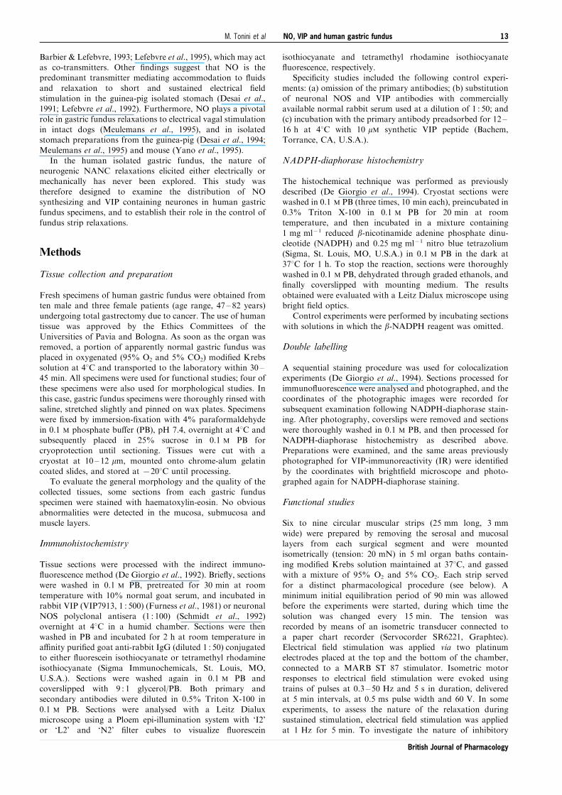
Administration of VIP (100 nM) produced a slowlydeveloping relaxation, which was followed by a slow declinein the response up to recovery of the initial tone (Figure 8).
The peak relaxation was reached after 20.1+1.3 min (n=8) ofVIP exposure, while recovery of the basal tone was obtainedafter 54.0+4.4 min (n=5). VIP-induced relaxations, which
were 52.7+5.4% (n=5) of those induced by 1 mM isoprenaline(peak response: 2.5 min), were una�ected by TTX (1 mM: n=3)
Figure 5 Frequency-dependent relaxant and contractile responses ofgastric fundus strips to electrical stimulation, under controlconditions or in the presence of L-NOARG. Values are expressedas per cent of the absolute value of the control inhibitory responseobtained at 50 Hz and represent the means+s.e.mean of tenpreparations. *P50.05 versus control responses.
Figure 6 Tracing illustrating the e�ect of L-NOARG on electrically-evoked relaxations induced by stimulation at 1 Hz for 5 min. Note thatL-NOARG (300 mM) increased the basal tone and turned the relaxant response into a contractile one.
Figure 7 Frequency-dependent contractile responses of gastricfundus strips to electrical stimulation in the presence of L-NOARGalone, in combination with hyoscine, or in combination with hyoscineplus TTX. Values are expressed as per cent of the maximalcontraction obtained at 50 Hz in the presence of L-NOARG andrepresent the means of six preparations. For clarity, error bars(+s.e.mean) are included in the L-NOARG curve only. *P50.05versus L-NOARG alone.
NO, VIP and human gastric fundus16 M. Tonini et al
British Journal of Pharmacology
and L-NOARG (300 mM: n=3) pretreatment, but reduced to
23.3+5.2% (n=3) of isoprenaline-induced relaxation in thepresence of 300 mM suramin. When a strip was treated with apriming (100 nM) VIP concentration, a second 100 nM VIP
administration (carried out as soon as the e�ect of the primingdose had vanished) was ine�ective in three strips (Figure 8) orcaused a slight response (12% of the original one) in twoadditional preparations, indicating the occurrence of VIP
desensitization. By contrast, relaxations caused by 1 mMisoprenaline were una�ected by VIP desensitization procedure(n=3). In tissues desensitized by 100 nM VIP, the L-NOARG-
resistant component of the electrically-induced relaxations wasvirtually abolished (Figure 9).
Suramin (300 mM) had no e�ect per se on the frequency-
dependent inhibitory curve and, unlike L-NOARG, did notunmask or potentiate contractile responses at any frequency ofstimulation (n=4, data not shown). Conversely, combination of
suramin (300 mM) plus L-NOARG (300 mM) was slightly, butsigni®cantly more e�ective than L-NOARG alone in inhibitingrelaxations to 50 Hz (Figure 9).
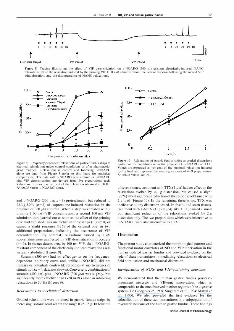
Relaxations to mechanical distension
Graded relaxations were obtained in gastric fundus strips by
increasing isotonic load within the range 0.25 ± 2 g. In four out
of seven tissues, treatment with TTX (1 mM) had no e�ect on the
relaxations evoked by 41 g distension, but caused a slight(20%) albeit signi®cant reduction of the responses obtainedwith2 g load (Figure 10). In the remaining three strips, TTX was
ine�ective at any distension tested. In ®ve out of seven tissues,treatment with L-NOARG (300 mM), like TTX, caused a smallbut signi®cant reduction of the relaxations evoked by 2 g
distension only. The two preparations which were insensitive toL-NOARG were also insensitive to TTX.
Discussion
The present study characterized the morphological pattern and
functional motor correlates of NO and VIP innervation in thehuman isolated gastric fundus and provided evidence on therole of these transmitters in mediating relaxations to electrical
®eld stimulation and mechanical distension.
Identi®cation of NOS- and VIP-containing neurones
We demonstrated that the human gastric fundus possessesprominent nitrergic and VIPergic innervation, which iscomparable to the one observed in other regions of the digestive
system (De Giorgio et al., 1994; Singaram et al., 1994; Matini etal., 1995). We also provided the ®rst evidence for thecolocalization of these two transmitters in a subpopulation of
myenteric neurons of the human gastric fundus. These ®ndings
Figure 8 Tracing illustrating the e�ect of VIP desensitization on L-NOARG (300 mM)-resistant electrically-induced NANCrelaxations. Note the relaxation induced by the priming VIP (100 nM) administration, the lack of response following the second VIPadministration, and the disappearance of NANC relaxations.
Figure 9 Frequency-dependent relaxations of gastric fundus strips toelectrical stimulation under control conditions or after pharmacolo-gical treatment. Relaxations in control and following L-NOARGalone are data from Figure 5 (refer to this ®gure for statisticalcomparisons). The data with L-NOARG plus suramin or L-NOARGplus VIP desensitization are derived from ®ve preparations each.Values are expressed as per cent of the relaxation obtained at 50 Hz.*P50.05 versus L-NOARG alone.
Figure 10 Relaxations of gastric fundus strips to graded distensionsunder control conditions or in the presence of L-NOARG or TTX.Values are expressed as per cent of the maximal relaxation inducedby 2 g load and represent the means+s.e.mean of 4 ± 9 preparations.*P50.05 versus control.
NO, VIP and human gastric fundus 17M. Tonini et al
British Journal of Pharmacology

expand previous observations ofNOandVIP distribution in theenteric nervous system of the mammalian stomach (Belai et al.,1992; Timmermans et al., 1994; Lefebvre et al., 1995), thus
providing a structural basis to interpret the functional responsesobtained by electrical ®eld stimulation and mechanicaldistension.
Characterization of transmitters involved inelectrically-evoked NANC relaxations
The presence of NO and VIP in myenteric neurones and ®bressupplying smooth muscle cells suggests the possibility thatboth inhibitory transmitters participate in neurogenic NANC
relaxations, accounting for the reservoir function of thisstomach region. Nevertheless, the colocalization of di�erenttransmitters within a neural structure does not necessarily
imply their concomitant release at any level of electrical ormechanical stimulation. NO-mediated relaxations are prefer-entially activated by shorter trains or lower frequencies ofelectrical ®eld stimulation in both gastric (Li & Rand, 1990)
and extra-gastric isolated preparations (Maggi & Giuliani,1996). Conversely, VIP is released either by brief stimuli athigh frequency or by sustained stimuli at low frequency
(Lefebvre et al., 1995; CurroÁ & Preziosi, 1998). In the humanisolated gastric fundus strips, our results with L-NOARGindicate that electrically-induced NANC relaxations are
mediated by NO in the frequency range of 0.3 ± 3 Hz withtrains lasting 5 s. Even with longer trains (5 min) at lowfrequency (1 Hz) NO seems the only inhibitory transmitter
involved. In this respect, the characteristics of stimulation forNO release parallel those of other established transmitters,such as acetylcholine or noradrenaline, which require a lowerlevel of stimulation than any other accompanying peptide
transmitter (Bartfai et al., 1988). By contrast, L-NOARG onlysigni®cantly reduced (without suppressing) relaxations ob-tained in the range of 10 ± 50 Hz, suggesting the participation
of other inhibitory transmitter(s) in addition to NO.A VIP desensitization procedure was used to assess whether
VIP participates in the L-NOARG-resistant component of
NANC relaxation. The attenuation of receptor function bymeans of agonist-induced receptor desensitization is considereda reliable alternative to the use of antagonists, especially whenthe latter lack potency and selectivity, such as the C-terminal
fragment of VIP, VIP(10 ± 28), which may (Crist et al., 1992; Jin etal., 1994) or may not antagonize relaxations to applied VIP(Morris & Murphy, 1989; Maggi & Giuliani, 1993). When
desensitization was induced to the relaxant e�ect of VIP, it alsomarkedly reduced the relaxant e�ects of electrical ®eldstimulation (range 10 ± 50 Hz) in the presence of L-NOARG.
Compared to control, the relevant inhibition of NANCrelaxations observed under these conditions (590%) suggeststhat at frequencies510 Hz, relaxations are mostly mediated by
the concomitant release of NO and VIP, which are costored inneurones innervating smooth muscle cells, as observed in ourimmunohistochemical experiments. In our hands, VIP desensi-tization probably involved VIP receptors speci®cally (and not
other receptor types or post-receptor mechanisms), sinceisoprenaline-induced relaxation, which is mediated via cyclicAMP formation like that of VIP (Bitar &Makhlouf, 1982), was
not a�ected by this procedure. Furthermore, the ®nding that therelaxation induced by VIP was insensitive to L-NOARG couldbe taken as evidence that in human gastric fundus strips, like in
pig or canine fundus strips (Lefebvre et al., 1995; Bayguinov etal., 1999) or guinea-pig isolated whole stomach (Desai et al.,1994), VIP does not seem to produce part of its relaxant e�ect byreleasing NO from neurones or directly from isolated smooth
muscle cells (Grider et al., 1992; Murthy et al., 1993; Chakder &Rattan, 1996; Jin et al., 1996). However, since human intestinaland rabbit gastric smooth muscle cells were recently found to
express a constitutive endothelial NOS (Teng et al., 1998),experiments with human dispersed smooth muscle cells fromfundus are required to establish whether VIP is able to promoteNO release under these conditions.
With regard to the L-NOARG-resistant component ofNANC relaxations evoked at the highest frequencies (50 Hz),it was slightly, but signi®cantly reduced by suramin, a non-
selective P2 purinoceptor antagonist (Hoyle et al., 1990).Although this could be taken as evidence for the involvementof ATP as an additional mediator of NANC relaxations, we
believe thatATPplays no role inour experimental conditions forseveral reasons. Firstly, combined L-NOARG and VIPdesensitization virtually abolished NANC relaxations; sec-
ondly, in our hands suramin antagonized VIP-inducedrelaxations, as observed by Briejer et al. (1995) in the guinea-pig proximal colon.Therefore, the suramin-sensitive componentofNANC relaxation could be tentatively ascribed to partial VIP
inhibition. In our hands, however, this minor inhibition bysuramin was detectable only after suppression of the nitrergicfunction, since suramin was ine�ective when the nitrergic
innervation was fully operating.At frequencies 53 Hz, L-NOARG unmasked an early
contractile response that was substantially reduced by TTX and
hyoscine. This suggests that endogenous NO may tonicallyinhibit the release of excitatory transmitters. Prejunctionalmodulation of cholinergic transmission by endogenous NO has
been reported in the guinea-pig and dog intestine and in rabbitgastric corpus (Knudsen & Tùttrup, 1992; Baccari et al., 1993;Hryhorenko et al., 1994), while evidence for a postjunctionalmechanism was obtained in the guinea-pig (Milenov & Kal®n,
1996) and pig gastric fundus (Leclere & Lefebvre, 1998). Inaddition, as observed in isolated intestinal preparations fromseveral mammalian species (Li &Rand, 1990;Meulemans et al.,
1993; Ciccocioppo et al., 1994), L-NOARG enhanced the basaltone of human fundus strips through a mechanism that waspartially sensitive to TTX and hyoscine. This suggests that L-
NOARG acts by removing a tonic nitrergic inhibition both onsmooth muscle cells and excitatory cholinergic neurones.
Relaxations to graded mechanical distension
Electrical ®eld stimulation is generally regarded as a reliableprocedure to evoke transmitter release from enteric nerve
terminals, although its physiological relevance is still debated.In our study, we also resorted to mechanical distension toassess the role of the neural mechanisms underlying NANC
relaxation.Although relaxations of gastric fundus strips to graded
distension were mainly due to passive elongation of smooth
muscle cells, a neurogenic component (*25%) apparentlymediated only by NO was observed at 2 g distension. Based onour results, NO is released upon mild electrical or mechanicalstimulation. Therefore, NO could be viewed as the primary
inhibitory transmitter initiating accommodation in the humangastric fundus, as already observed in other species (Desai etal., 1991) and in other parts of the gut (Waterman et al., 1994;
Ciccocioppo et al., 1994).
Conclusions
In conclusion, NO, VIP and NO/VIP were identi®ed inganglion cells of the myenteric plexus and in nerve ®bressupplying smooth muscle cells of the human gastric fundus.
NO, VIP and human gastric fundus18 M. Tonini et al
British Journal of Pharmacology

NO is responsible for NANC relaxations evoked by lowfrequency electrical stimulation or by mild mechanicaldistension, whereas VIP release occurs at high frequency
stimulation. In addition, NO was found to exert an inhibitoryin¯uence both on cholinergic innervation and basal tone.
This work was supported in part by the Italian Ministry forUniversity and Scienti®c Research (MURST) and FAR 1998 Funds
to M. Tonini, and by NIH grants DK54155 and DK41301(Morphology/Imaging Core) to C. Sternini. The authors wish tothank Ms Helen C. Wong and Dr John H. Walsh of the AntibodyCore (subsection of DK 41301 CURE: Digestive Diseases ResearchCenter Grant) for the rabbit polyclonal VIP7913, and Dr HaraldH.H.W. Schmidt (WuÈ rzburg University, Germany) for the rabbitpolyclonal NOS antiserum. The authors wish also to thank Drs E.Messori, M. Tagliani and S.M. Candura for their critical andhelpful comments on the manuscript.
References
ABRAHAMSSON, H. (1973). Studies on the inhibitory nervouscontrol of gastric motility. Acta Physiol. Scand., 390, 1 ± 38.
ALLESCHER, H.D., TOUGAS, G., VERGARA, P., LU, S. & DANIEL,
E.E. (1992). Nitric oxide as a putative nonadrenergic noncholi-nergic inhibitory transmitter in the canine pylorus in vivo. Am. J.Physiol., 262, G695 ±G702.
BACCARI, M.C., BERTINI, M. & CALAMAI, F. (1993). E�ects of L-NG-nitro arginine on cholinergic transmission in the gastricmuscle of the rabbit. Neuroreport, 4, 1102 ± 1104.
BARBIER, A.J. & LEFEBVRE, R.A. (1993). Involvement of the L-arginine: nitric oxide pathway in nonadrenergic noncholinergicrelaxation of the cat gastric fundus. J. Pharmacol. Exp. Ther.,266, 172 ± 178.
BARTFAI, T., IVERFELDT, K. & FISONE, G. (1988). Regulation of therelease of coexisting neurotransmitters. Ann. Rev. Pharmacol.Toxicol., 28, 285 ± 310.
BAYGUINOV, O., KEEF, K.D., HAGEN, B. & SANDERS, K.M. (1999).Parallel pathways mediate inhibitory e�ects of vasoactiveintestinal polypeptide and nitric oxide in canine fundus. Br. J.Pharmacol., 126, 1543 ± 1552.
BELAI, A., SCHMIDT, H.H.H.W., HOYLE, C.H.V., HASSAL, C.J.S.,
SAFFREY, M.J., MOSS, J., FOÈ RSTERMANN, U., MURAD, F. &
BURNSTOCK, G. (1992). Colocalization of nitric oxide synthaseand NADPH-diaphorase in the myenteric plexus of the rat gut.Neurosci. Lett., 143, 60 ± 64.
BITAR, K.N. & MAKHLOUF, G.M. (1982). Relaxation of isolatedsmooth muscle cells by vasoactive intestinal peptide. Science,216, 531 ± 533.
BOECKXSTAENS, G.E., PELCKMANS, P.A., DE MAN, J.G., BULT, H.,
HERMAN, A.G. & VAN MAERCKE, Y.M. (1992). Evidence for adi�erential release of nitric oxide and vasoactive intestinalpolypeptide by nonadrenergic noncholinergic nerves in the ratgastric fundus. Arch. Int. Pharmacodyn., 318, 107 ± 115.
BRIEJER, M.R., AKKERMANS, L.M.A., MEULEMANS, A.L., LE-
FEBVRE, R.A. & SCHUURKES, J.A.J. (1995). 5-HT-inducedneurogenic relaxations of the guinea-pig proximal colon:investigation into the role of ATP and VIP in addition to nitricoxide. Naunyn-Schmiedeberg's Arch. Pharmacol., 351, 126 ± 135.
CHAKDER, S. & RATTAN, S. (1996). Evidence for VIP-inducedincrease in NO production in myenteric neurons of opossuminternal anal sphincter. Am. J. Physiol., 270, G492 ±G497.
CICCOCIOPPO, R., ONORI, L., MESSORI, E., CANDURA, S.M.,
COCCINI, T. & TONINI, M. (1994). Role of nitric oxide-dependentand -independent mechanisms in peristalsis and accommodationin the rabbit distal colon. J. Pharmacol. Exp. Ther., 270, 929 ±937.
COSTA, M., FURNESS, J.B. & HUMPHREYS, C.M.S. (1986). Apamindistinguishes two types of relaxation mediated by enteric nervesin the guinea-pig gastrointestinal tract. Naunyn-Schmiedeberg'sArch. Pharmacol., 332, 79 ± 88.
COSTA, M., FURNESS, J.B., POMPOLO, S., BROOKES, S.J.H., BORN-
STEIN, J.C., BREDT, D.S. & SNYDER, S.H. (1992). Projections andchemical coding of neurons with immunoreactivity for nitricoxide synthase in the guinea-pig small intestine. Neurosci. Lett.,148, 121 ± 125.
CRIST, J.R., HE, X.D. & GOYAL, R.K. (1992). Both ATP and thepeptide VIP are inhibitory neurotransmitters in the guinea-pigileum circular muscle. J. Physiol., 447, 119 ± 131.
CURRO', D. & PREZIOSI, P. (1998). Non-adrenergic non-cholinergicrelaxation of the rat stomach. Gen. Pharmacol., 31, 697 ± 703.
D'AMATO, M., CURRO', D. & MONTUSCHI, P. (1992). Evidence fordual components in the non-adrenergic non-cholinergic relaxa-tion in the rat gastric fundus: role of endogenous nitric oxide andvasoactive intestine polypeptide. J. Autonom. Nerv. Syst., 37,175 ± 186.
DE GIORGIO, R., PARODI, J.E., BRECHA, N.C., BRUNICARDI, F.C.,
BECKER, J.M., GO, V.L.W. & STERNINI, C. (1994). Nitric oxideproducing neurons in the monkey and human digestive system. J.Comp. Neurol., 342, 619 ± 627.
DE GIORGIO, R., STERNINI, C., ANDERSON, K., BRECHA, N.C. &
GO, V.L.W. (1992). Tissue distribution and innervation pattern ofpeptide immunoreactivities in the rat pancreas. Peptides, 13, 91 ±98.
DESAI, K.M., SESSA, W.C. & VANE, J.R. (1991). Involvement of nitricoxide in the re¯ex relaxation of the stomach to accommodatefood or ¯uid. Nature, 351, 477 ± 479.
DESAI, K.M., WARNER, T.D., BISHOP, A.E., POLAK, J.M. & VANE,
J.R. (1994). Nitric oxide, and not vasoactive intestinal peptide, asthe main neurotransmitter of vagally induced relaxation of theguinea-pig stomach. Br. J. Pharmacol., 113, 1197 ± 1202.
FAHRENKRUG, J. (1989). VIP and autonomic neurotransmission.Pharmacol. Ther., 41, 515 ± 534.
FURNESS, J.B., COSTA, M. & WALSH, J.H. (1981). Evidence for andsigni®cance of the projection of VIP neurons from the myentericplexus to the taenia coli in the guinea pig. Gastroenterology, 80,1557 ± 1561.
FURNESS, J.B., LLEWELLYN-SMITH, I.J., BORNSTEIN, J.C. &
COSTA, M. (1988). Chemical neuroanatomy and the analysis ofneuronal circuitry in the enteric nervous system. In: BjorkLund,A. & HoÈ ktelt T. (eds.). Handbook of Chemical Neuroanatomy.The Peripheral Nervous System. Vol 6, Elsevier: Amsterdam. pp.161 ± 218.
GRIDER, J.R. & MAKHLOUF, G.M. (1986). Colonic peristaltic re¯ex:Identi®cation of vasoactive intestinal polypeptide as mediator ofdescending relaxation. Am. J. Physiol., 251, G40 ±G45.
GRIDER, J.R., MURTHY, K.S., JIN, J.-G. & MAKHLOUF, G.M. (1992).Stimulation of nitric oxide from muscle cells by VIP: prejunc-tional enhancement of VIP release. Am. J. Physiol., 262, G774 ±G778.
HOYLE, C.H.V. & BURNSTOCK, G. (1989). Neuromuscular transmis-sion in the gastrointestinal tract. In: Makhtouf, G.M. (ed.).Handbook of Physiology. The Gastrointestinal System., Vol 1,Sect 6. American Physiological Society: Washington DC. pp.435 ± 464.
HOYLE, C.H.V., KNIGHT, G.E. & BURNSTOCK, G. (1990). Suraminantagonizes responses to P2-purinoceptor agonists and puriner-gic nerve stimulation in the guinea-pig urinary bladder and taeniacoli. Br. J. Pharmacol., 99, 617 ± 621.
HRYHORENKO, L.M., WOSKOWSKA, Z. & FOX-THRELKELD, J.-
A.E.T. (1994). Nitric oxide (NO) inhibits release of acetylcholinefrom nerves of isolated circular muscle of the canine ileum:relationship to motility and release of nitric oxide. J. Pharmacol.Exp. Ther., 271, 918 ± 926.
JIN, J.-G., KATSOULIS, S., SCHMIDT, W.E. & GRIDER, J.R. (1994).Inhibitory transmission in taenia coli mediated by distinct VIPand apamin-sensitive PACAP receptors. J. Pharmacol. Exp.Ther., 270, 433 ± 439.
JIN, J.-G., MURTHY, K.S., GRIDER, J.R. & MAKHLOUF, G.M. (1996).Stoichiometry of neurally induced VIP release, NO formation,and relaxation in rabbit and rat gastric muscle. Am. J. Physiol.,271, G357 ±G369.
KNUDSEN, M.A. & TéTTRUP, A. (1992). A possible role of the L-arginine-nitric oxide pathway in the modulation of cholinergictransmission in the guinea-pig taenia coli. Br. J. Pharmacol., 107,837 ± 841.
LECLERE, P.G. & LEFEBVRE, R.A. (1998). Investigation of theinteraction between cholinergic and nitrergic neurotranmissionin the pig gastric fundus. Br. J. Pharmacol., 125, 1779 ± 1787.
NO, VIP and human gastric fundus 19M. Tonini et al
British Journal of Pharmacology

LEFEBVRE, R.A., BAERT, E. & BARBIER, A.J. (1992). In¯uence ofNG-nitro-L-arginine on non-adrenergic non-cholinergic relaxa-tion in the guinea-pig gastric fundus. Br. J. Pharmacol., 106,173 ± 179.
LEFEBVRE, R.A., SMITS, G.J.M. & TIMMERMANS, J.-P. (1995). Studyof NO and VIP as non-cholinergic neurotransmitters in the piggastric fundus. Br. J. Pharmacol., 116, 2017 ± 2026.
LI, C.G. & RAND, M.J. (1990). Nitric oxide and vasoactive intestinalpolypeptide mediate non-adrenergic, non-cholinergic inhibitorytransmission to smooth muscle of the rat gastric fundus. Eur. J.Pharmacol., 191, 303 ± 309.
MAGGI, C.A. & GIULIANI, S. (1993). Multiple inhibitory mechanismsmediate non-adrenergic non-cholinergic relaxation in the circularmuscle of guinea-pig colon. Naunyn-Schmiedeberg's Arch.Pharmacol., 347, 630 ± 634.
MAGGI, C.A. & GIULIANI, S. (1996). Characterization of the apamin-and L-nitroarginine-resistant NANC inhibitory transmission tothe circular muscle of guinea-pig colon. J. Auton. Pharmacol., 16,131 ± 145.
MATINI, P., FAUSSONE-PELLEGRINI, M.S., CORTESINI, C. &
MAYER, B. (1995). Vasoactive intestinal polypeptide and nitricoxide synthase distribution in the enteric plexuses of the humancolon: an histochemical study and quantitative analysis.Histochemistry, 103, 415 ± 423.
MEULEMANS, A.L., EELEN, J.G. & SCHUURKES, J.A.J. (1995). NOmediates gastric relaxations after brief vagal stimulation inanesthetized dogs. Am. J. Physiol., 269, G255 ±G261.
MEULEMANS, A.L., HELSEN, L.F. & SCHUURKES, J.A.J. (1993).Role of NO in vagally mediated relaxations of guinea-pigstomach. Naunyn-Schmiedeberg's Arch. Pharmacol., 347, 225 ±230.
MILENOV, K. & KALFIN, R. (1996). Cholinergic-nitrergic interac-tions in the guinea-pig gastric fundus. Neuropeptides, 30, 365 ±371.
MORRIS, J.L. & MURPHY, R. (1989). Analogues of VIP contract theguinea-pig uterine artery but do not antagonize VIP-inducedrelaxations. Eur. J. Pharmacol., 162, 375 ± 379.
MURTHY, K.S., ZHANG, K.-M., JIN, J.-G., GRIDER, J.R. & MAKH-
LOUF, G.M. (1993). VIP-mediated G protein-coupled Ca2+ in¯uxactivates a constitutive NOS in dispersed gastric muscle cells.Am. J. Physiol., 265, G660 ±G671.
PREIKSAITIS, H.G., TREMBLAY, L. & DIAMANT, N.E. (1994). Nitricoxide mediates inhibitory nerve e�ects in human esophagus andlower esophageal sphincter. Dig. Dis. Sci., 39, 770 ± 775.
RATTAN, S. & CHAKDER, S. (1992). Role of nitric oxide as amediator of internal anal sphincter relaxation. Am. J. Physiol.,262, 6107 ± 6112.
SANDERS, K.M. & WARD, S.M. (1992). Nitric oxide as mediator ofnonadrenergic noncholinergic neurotransmission. Am. J. Phy-siol., 262, G379 ±G392.
SANGER, G.J. (1985). E�ects of metoclopramide and domperidoneon cholinergically mediated contractions of human isolatedstomach muscle. J. Pharm. Pharmacol., 37, 661 ± 664.
SCHMIDT, H.H.H.W., CAGNE, G.D., NAKANE, M., POLLOCK, J.S.,
MILLER, M.F. & MURAD, F. (1992). Mapping of the nitric oxidesynthase in the rat suggests co-localization with NADPH-diaphorase but not soluble guanylyl cyclase, and novel para-neural functions for nitrinergic signal transduction. J. Histo-chem. Cytochem., 40, 1439 ± 1456.
SINGARAM, C., SENGUPTA, A., SWEET, M.A., SUGARBAKER, D.J. &
GOYAL, R.K. (1994). Nitrinergic and peptidergic innervation ofthe human oesophagus. Gut, 35, 1690 ± 1696.
TENG, B., MURTHY, K.S., KUEMMERLE, J.F., GRIDER, J.R., SASE,
K., MICHEL, T. & MAKHLOUF, G.M. (1998). Expression ofendothelial nitric oxide synthase in human and rabbit gastro-intestinal smooth muscle cells. Am. J. Physiol., 275,G342 ±G351.
TIMMERMANS, J.P., BARBIERS, M., SCHEUERMANN, D.W., BO-
GERS, J.J., ADRIAENSEN, D., FEKETE, E., MAYER, B., VAN
MARCK, E.A. & DE GRODT-LASSEEL, M.H. (1994). Nitric oxidesynthase immunoreactivity in the enteric nervous system of thedeveloping human digestive tract. Cell. Tissue Res., 275, 235 ±245.
TONINI, M., ONORI, L., LECCHINI, S., FRIGO, G.M. & CREMA, A.
(1982). Mode of action of ATP on propulsive activity in rabbitcolon. Eur. J. Pharmacol., 82, 21 ± 28.
WATERMAN, S.A., COSTA, M. & TONINI, M. (1994). Accommodationmediated by enteric inhibitory re¯exes in the isolated guinea-pigsmall intestine. J. Physiol. (Lond.), 474, 539 ± 546.
WATTCHOW, D.A., BROOKES, S.J.H. & COSTA, M. (1995). Themorphology and projections of retrogradely labeled myentericneurons in the human intestine. Gastroenterology, 109, 866 ± 875.
YANO, S., KIYOTA, Y., YAMAMOTO, M. & WATANABE, K. (1995).Pharmacological features of non-adrenergic non-cholinergic(NANC) relaxation induced by electrical vagal stimulation inisolated mouse stomach. Jap. J. Pharmacol., 69, 9 ± 15.
(Received August 8, 1999Revised September 13, 1999Accepted October 1, 1999)
NO, VIP and human gastric fundus20 M. Tonini et al
British Journal of Pharmacology