Interleukin-1 alpha and vasoactive interstinal peptide: Enigmatic regulation of neuronal survival
Upload
independentCategory
view
2download
0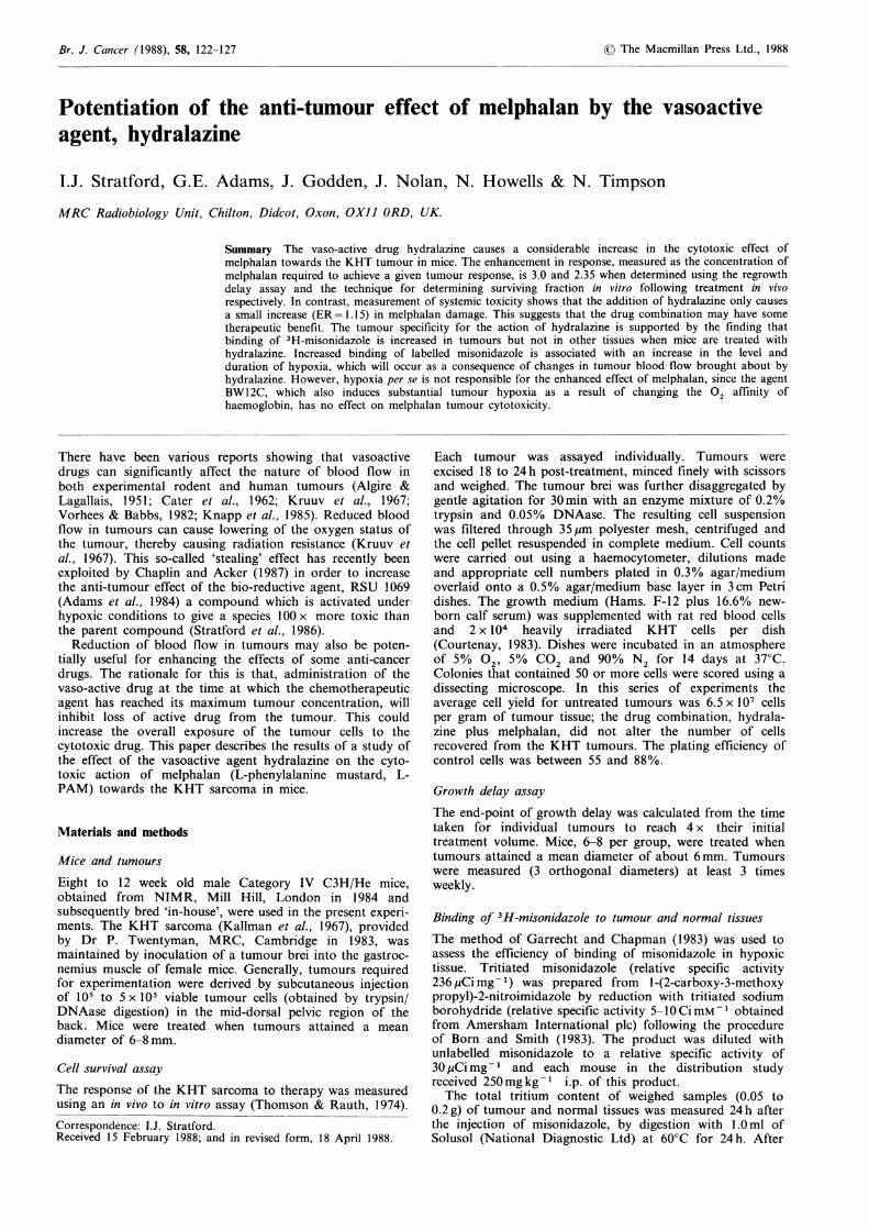
B8 The Macmillan Press Ltd., 1988
Potentiation of the anti-tumour effect of melphalan by the vasoactiveagent, hydralazine
I.J. Stratford, G.E. Adams, J. Godden, J. Nolan, N. Howells & N. Timpson
MRC Radiobiology Unit, Chilton, Didcot, Oxon, OX]ll ORD, UK.
Summary The vaso-active drug hydralazine causes a considerable increase in the cytotoxic effect ofmelphalan towards the KHT tumour in mice. The enhancement in response, measured as the concentration ofmelphalan required to achieve a given tumour response, is 3.0 and 2.35 when determined using the regrowthdelay assay and the technique for determining surviving fraction in vitro following treatment in vivorespectively. In contrast, measurement of systemic toxicity shows that the addition of hydralazine only causesa small increase (ER= 1.15) in melphalan damage. This suggests that the drug combination may have sometherapeutic benefit. The tumour specificity for the action of hydralazine is supported by the finding thatbinding of 3H-misonidazole is increased in tumours but not in other tissues when mice are treated withhydralazine. Increased binding of labelled misonidazole is associated with an increase in the level andduration of hypoxia, which will occur as a consequence of changes in tumour blood flow brought about byhydralazine. However, hypoxia per se is not responsible for the enhanced effect of melphalan, since the agentBW12C, which also induces substantial tumour hypoxia as a result of changing the 02 affinity ofhaemoglobin, has no effect on melphalan tumour cytotoxicity.
There have been various reports showing that vasoactivedrugs can significantly affect the nature of blood flow inboth experimental rodent and human tumours (Algire &Lagallais, 1951; Cater et al., 1962; Kruuv et al., 1967;Vorhees & Babbs, 1982; Knapp et al., 1985). Reduced bloodflow in tumours can cause lowering of the oxygen status ofthe tumour, thereby causing radiation resistance (Kruuv etal., 1967). This so-called 'stealing' effect has recently beenexploited by Chaplin and Acker (1987) in order to increasethe anti-tumour effect of the bio-reductive agent, RSU 1069(Adams et al., 1984) a compound which is activated underhypoxic conditions to give a species 100 x more toxic thanthe parent compound (Stratford et al., 1986).
Reduction of blood flow in tumours may also be poten-tially useful for enhancing the effects of some anti-cancerdrugs. The rationale for this is that, administration of thevaso-active drug at the time at which the chemotherapeuticagent has reached its maximum tumour concentration, willinhibit loss of active drug from the tumour. This couldincrease the overall exposure of the tumour cells to thecytotoxic drug. This paper describes the results of a study ofthe effect of the vasoactive agent hydralazine on the cyto-toxic action of melphalan (L-phenylalanine mustard, L-PAM) towards the KHT sarcoma in mice.
Materials and methods
Mice and tumours
Eight to 12 week old male Category IV C3H/He mice,obtained from NIMR, Mill Hill, London in 1984 andsubsequently bred 'in-house', were used in the present experi-ments. The KHT sarcoma (Kallman et al., 1967), providedby Dr P. Twentyman, MRC, Cambridge in 1983, wasmaintained by inoculation of a tumour brei into the gastroc-nemius muscle of female mice. Generally, tumours requiredfor experimentation were derived by subcutaneous injectionof 105 to 5 x 10⁵ viable tumour cells (obtained by trypsin/DNAase digestion) in the mid-dorsal pelvic region of theback. Mice were treated when tumours attained a meandiameter of 6-8mm.
Cell survival assay
The response of the KHT sarcoma to therapy was measuredusing an in vivo to in vitro assay (Thomson & Rauth, 1974).
Correspondence: I.J. Stratford.Received 15 February 1988; and in revised form, 18 April 1988.
Each tumour was assayed individually. Tumours wereexcised 18 to 24h post-treatment, minced finely with scissorsand weighed. The tumour brei was further disaggregated bygentle agitation for 30min with an enzyme mixture of 0.2%trypsin and 0.05% DNAase. The resulting cell suspensionwas filtered through 35pm polyester mesh, centrifuged andthe cell pellet resuspended in complete medium. Cell countswere carried out using a haemocytometer, dilutions madeand appropriate cell numbers plated in 0.3% agar/mediumoverlaid onto a 0.5% agar/medium base layer in 3cm Petridishes. The growth medium (Hams. F-12 plus 16.6% new-born calf serum) was supplemented with rat red blood cellsand 2 x 10⁴ heavily irradiated KHT cells per dish(Courtenay, 1983). Dishes were incubated in an atmosphereof 5% 02, 5% CO2 and 90% N2 for 14 days at 37°C.Colonies that contained 50 or more cells were scored using adissecting microscope. In this series of experiments theaverage cell yield for untreated tumours was 6.5 x 10⁷ cellsper gram of tumour tissue; the drug combination, hydrala-zine plus melphalan, did not alter the number of cellsrecovered from the KHT tumours. The plating efficiency ofcontrol cells was between 55 and 88%.
Growth delay assay
The end-point of growth delay was calculated from the timetaken for individual tumours to reach 4 x their initialtreatment volume. Mice, 6-8 per group, were treated whentumours attained a mean diameter of about 6mm. Tumourswere measured (3 orthogonal diameters) at least 3 timesweekly.
Binding of 3H-misonidazole to tumour and normal tissues
The method of Garrecht and Chapman (1983) was used toassess the efficiency of binding of misonidazole in hypoxictissue. Tritiated misonidazole (relative specific activity236 ,uCi mg- 1) was prepared from 1-(2-carboxy-3-methoxypropyl)-2-nitroimidazole by reduction with tritiated sodiumborohydride (relative specific activity 5-10 Ci M - obtainedfrom Amersham International plc) following the procedureof Born and Smith (1983). The product was diluted withunlabelled misonidazole to a relative specific activity of30 1Ci mg- 1 and each mouse in the distribution studyreceived 250mg kg- I i.p. of this product.The total tritium content of weighed samples (0.05 to
0.2 g) of tumour and normal tissues was measured 24 h afterthe injection of misonidazole, by digestion with 1.0 ml ofSolusol (National Diagnostic Ltd) at 60°C for 24 h. After
Br. J. Cancer (1988), 58, 122-127
POTENTIATION OF THE ANTI-TUMOUR EFFECT OF MELPHALAN BY HYDRALAZINE 123
digestion, 3.0 ml ethanol and 15.0 ml scintillant (6.5 g 1PPO 0.5 g 1- 1 POPOP in toluene) were added and thesamples were counted on a Packard Tricarb liquid scintilla-tion counter. A set of variably quenched standards were usedfor calibration. These were prepared by adding a knownactivity of tritiated water to a set of 8 samples containingbetween zero and 0.2ml of whole blood digested as above.Any very highly coloured samples were bleached by incuba-tion at 60°C for 2h with benzoyl peroxide (0.4ml, 5% w/vin toluene) before the addition of ethanol and scintillant.
Three groups of 5 male C3H mice bearing the KHTtumour were injected with tritium-labelled misonidazole(250mgkg-1). Group 1 received misonidazole only, group 2received in i.v. injection of hydralazine (5.0mgkg-1) 15minutes after the misonidazole and group 3 recieved 3injections of hydralazine at 90 min intervals starting 15 minafter the misonidazole. All animals were autopsied 24h afterthe injection of misonidazole and tritium analysis carried outimmediately thereafter.
Systemic toxicity
Non-tumour bearing C3H male mice were injected withdifferent concentrations of melphalan 15min prior to treat-ment with hydralazine. The number of mice surviving at day7 and day 90 were used to calculate LD50 values.
Drugs
Melphalan and BW 1 2C were obtained from WellcomeResearch Laboratories, Beckenham, Kent. Melphalan wasdissolved in 2% acid/alcohol, diluted at least 1:10 in PBSand injected i.p. at 0.5ml/25g mouse. BW12C was made upin alkaline saline, adjusted to pH 7.4 by addition of HCI andadministered to mice i.v. at 0.1 ml/20 g mouse. Hydralazine,obtained from Sigma Ltd., Poole, Dorset, was dissolved inphosphate buffered saline and injected i.v. at 0.1 ml/20 gmouse. All drug solutions were prepared immediately beforeuse.
I - Melphalan + hydralazine
10-
io
0
C.)
m) 10-C
10-
6
5 10 15
Melphalan (mg kg-')Figure 1 The effect of hydralazine (5mgkg-'), given i.v. to mice15 min after treatment with varying doses of melphalan, onsurvival of KHT tumour cells. 0, melphalan alone; 0, melpha-lan plus hydralazine. The numbers of tumours used at each doselevel are indicated, together with the standard errors of thepooled survival determinations.
Results
Tumour response
Data given in Figure I show the response of KHT tumourcells taken from mice given various doses of melphalan withor without treatment with 5mg kg-1 hydralazine 15 minlater. The numbers of tumours used to determine each pointare indicated, together with standard errors of the pooledsurvival determinations. It is clear that post treatment ofmice with hydralazine substantially increases the anti-tumoureffect of melphalan. The enhancement ratio (ER) is 2.35 andis equal to the ratio of the slopes derived from regressionlines fitted through a surviving fraction of I at zero melpha-lan dose. At a dose of 5mgkg-1 hydralazine alone has noeffect on survival of KHT tumour cells.
Figure 2 shows the effect of different doses of hydralazineadministered i.v. 15 min after treatment of mice with6mgkg- 1 melphalan. The enhancing effect of hydralazine indecreasing cell survival reaches a maximum value over thedose range 5-15mgkg-1. However, even at the low dose of1 mg kg- 1, hydralazine can cause a significant increase in cellkilling relative to melphalan alone, reducing the survivingfraction from 9.6x10⁻² to 3.7x10⁻².The enhancing effect of hydralazine (5 mg kg 1) on
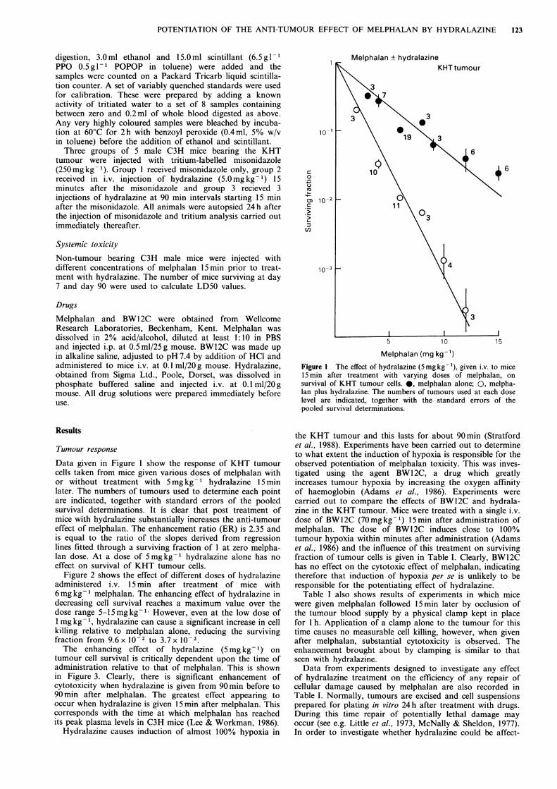
tumour cell survival is critically dependent upon the time ofadministration relative to that of melphalan. This is shownin Figure 3. Clearly, there is significant enhancement ofcytotoxicity when hydralazine is given from 90 min before to90 min after melphalan. The greatest effect appearing tooccur when hydralazine is given 15 min after melphalan. Thiscorresponds with the time at which melphalan has reachedits peak plasma levels in C3H mice (Lee & Workman, 1986).
Hydralazine causes induction of almost 100% hypoxia in
the KHT tumour and this lasts for about 90 min (Stratfordet al., 1988). Experiments have been carried out to determineto what extent the induction of hypoxia is responsible for theobserved potentiation of melphalan toxicity. This was inves-tigated using the agent BW12C, a drug which greatlyincreases tumour hypoxia by increasing the oxygen affinityof haemoglobin (Adams et al., 1986). Experiments werecarried out to compare the effects of BW12C and hydrala-zine in the KHT tumour. Mice were treated with a single i.v.dose of BW12C (70mgkg-1) 15min after administration ofmelphalan. The dose of BW12C induces close to 100%tumour hypoxia within minutes after administration (Adamset al., 1986) and the influence of this treatment on survivingfraction of tumour cells is given in Table I. Clearly, BW12Chas no effect on the cytotoxic effect of melphalan, indicatingtherefore that induction of hypoxia per se is unlikely to beresponsible for the potentiating effect of hydralazine.
Table I also shows results of experiments in which micewere given melphalan followed 15min later by occlusion ofthe tumour blood supply by a physical clamp kept in placefor 1 h. Application of a clamp alone to the tumour for thistime causes no measurable cell killing, however, when givenafter melphalan, substantial cytotoxicity is observed. Theenhancement brought about by clamping is similar to thatseen with hydralazine.Data from experiments designed to investigate any effect
of hydralazine treatment on the efficiency of any repair ofcellular damage caused by melphalan are also recorded inTable I. Normally, tumours are excised and cell suspensionsprepared for plating in vitro 24 h after treatment with drugs.During this time repair of potentially lethal damage mayoccur (see e.g. Little et al., 1973, McNally & Sheldon, 1977).In order to investigate whether hydralazine could be affect-
124 I.J. STRATFORD et al.
AaPlnhalan + hvcdrala7inaKHT tumour
1
c
00
%.n1C
C,)
3
3
03 T 6
10-2
L3
Melphalan + hydralazine: KHT tumour
Melphalan only
I I2 1-__
MelphalanHydralazine betore(hours)
1 2 3
Hydralazine after(hours)
Figure 3 The effect on tumour cell survival of giving hydrala-zine (5 mg kg- 1) i.v. to mice at varying times before or aftermelphalan (6mg kg- '). Symbols + s.e. are surviving fractionsderived from separate determinations on at least three tumours.
5 10
Hydralazine (mg kg-')
Figure 2 The effect of various doses of hydralazine, given i.v. tomice 15min after treatment with 6mgkg-' melphalan, on survi-val of KHT tumour cells. The numbers of tumours used at eachdose level are indicated, together with the standard errors of thepooled survival determinations.
ing such a repair process, tumours from treated mice were
excised 6h after treatment and tumour cell survival com-pared with that obtained when excision was at 24h. Resultsgiven in Table I show that hydralazine induced enhancementof cell killing is no different at 6 h from that at 24 h. Thissuggests that inhibition of repair processes is unlikely tocontribute to the observed effect.
All the experiments so far described were carried out withsubcutaneous tumours. Substantial differences in blood flowmay occur in tumours implanted in alternative sites. There-fore, the effect of hydralazine on the toxicity of melphalanwas evaluated in mice with the KHT tumour implanted intothe gastrocnemius muscle. Table I shows that, for KHTcells implanted i.m., hydralazine also causes an increase incell killing, similar to that observed for the subcutaneoustumour cells.
The effect of prolonged exposure to hydralazine was
investigated. Table II lists values of surviving fraction fromexperiments where mice are adminisfered 4mgkg-1 melpha-lan followed by a single or multiple doses of hydralazinegiven at 90 min intervals. Multiple doses of hydralazinealone have no cytotoxic effect on the KHT tumour. How-ever, when combined with melphalan, there is an evengreater enhancement of cell kill compared to that obtainedfollowing a single dose of hydralazine.Growth delay of the KHT tumour has been used as an
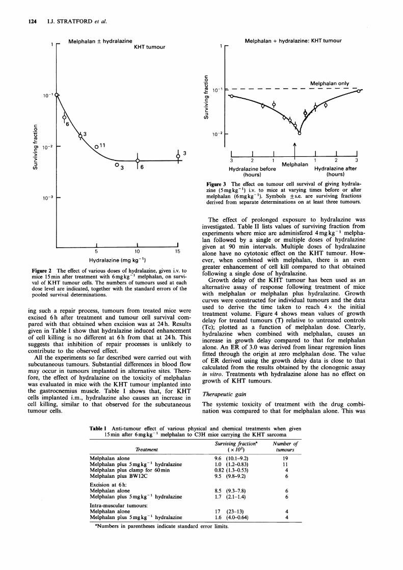
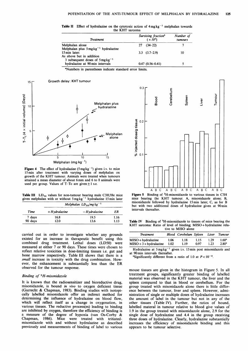
alternative assay of response following treatment of micewith melphalan or melphalan plus hydralazine. Growthcurves were constructed for individual tumours and the dataused to derive the time taken to reach 4 x the initialtreatment volume. Figure 4 shows mean values of growthdelay for treated tumours (T) relative to untreated controls(Tc); plotted as a function of melphalan dose. Clearly,hydralazine when combined with melphalan, causes anincrease in growth delay compared to that for melphalanalone. An ER of 3.0 was derived from linear regression linesfitted through the origin at zero melphalan dose. The valueof ER derived using the growth delay data is close to thatcalculated from the results obtained by the clonogenic assayin vitro. Treatments wth hydralazine alone has no effect on
growth of KHT tumours.
Therapeutic gain
The systemic toxicity of treatment with the drug combi-nation was compared to that for melphalan alone. This was
Table I Anti-tumour effect of various physical and chemical treatments when given15min after 6mgkg-1 melphalan to C3H mice carrying the KHT sarcoma
Surviving Jractiona Number ofTreatment ( x 102) tumours
Melphalan alone 9.6 (10.1-9.2) 19Melphalan plus 5mgkg-1 hydralazine 1.0 (1.2-0.83) 11Melphalan plus clamp for 60min 0.82 (1.3-0.53) 4Melphalan plus BW12C 9.5 (9.8-9.2) 6
Excision at 6h:Melphalan alone 8.5 (9.3-7.8) 6Melphalan plus 5mgkg-1 hydralazine 1.7 (2.1-1.4) 6
Intra-muscular tumours:Melphalan alone 17 (23-13) 4Melphalan plus 5mgkg-1 hydralazine 1.6 (4.0-0.64) 4
aNumbers in parentheses indicate standard error limits.
10-
0.cJ
10C,)
10
1=
kv
POTENTIATION OF THE ANTI-TUMOUR EFFECT OF MELPHALAN BY HYDRALAZINE 125
Table II Effect of hydralazine on the cytotoxic action of 4mgkg- 1 melphalan towardsthe KHT sarcoma
Surviving fractiona Number ofTreatment ( x 102) tumours
Melphalan alone 27 (34-22) 7Melphalan plus 5mg kg- 1 hydralazine15min later: 3.3 (3.7-2.9) 10As above but in addition
3 subsequent doses of 5mgkg-1hydralazine at 90min intervals 0.47 (0.56-0.41) 5
aNumbers in parentheses indicate standard error limits.
15
coa)
E
C:
x 5
0.3
In plusazine
lelphalanalone
5 10
a)
U)._-
0)
0
'a
a)cJC.)
a1)
0-
0.2
0.115
Melphalan (mg kg-')
Figure 4 The effect of hydralazine (5 mgkg-1) given i.v. to mice15min after treatment with varying doses of melphalan ongrowth of the KHT tumour. Animals were treated when tumoursattained a mean diameter of about 6mm and 6 to 8 animals wereused per group. Values of T-Tc are given++ s.e.
Table III LD50 values for non-tumour bearing male C3H/He micegiven melphalan with or without 5mgkg-' hydralazine 15min later
Melphalan LD50/mgkg-1
Time + Hydralazine -Hydralazine ER
7 days 16.8 19.5 1.1690 days 12.0 13.6 1.13
carried out in order to investigate whether any groundsexisted for an increase in therapeutic benefit using thiscombined drug treatment. Lethal doses (LD50) weremeasured at either 7 or 90 days. These times were chosen toreflect relative toxicities in dose-limiting tissues i.e. gut andbone marrow respectively. Table III shows that there is asmall increase in toxicity with the drug combination. How-ever, the enhancement is substantially less than the ERobserved for the tumour response.
Binding of 3H-misonidazole
It is known that the radiosensitizer and bioreductive drug,misonidazole, is bound in vivo to oxygen deficient tissue(Garrecht & Chapman, 1983). Binding studies with isotopi-cally labelled misonidazole offer an indirect method fordetermining the influence of hydralazine on blood flow,which will reflect itself as a change in oxygenation, invarious tissues. The reductive process(es) leading to bindingare inhibited by oxygen, therefore the efficiency of binding isa measure of the degree of hypoxia (van Os-Corby &Chapman, 1986). Mice were treated with tritiated-misonidazole with and without hydralazine as describedpreviously and measurements of binding of label to various
'a0
0Fn
E
.0a)Q
u)
c
a)a)'aC,)
0
E
H3
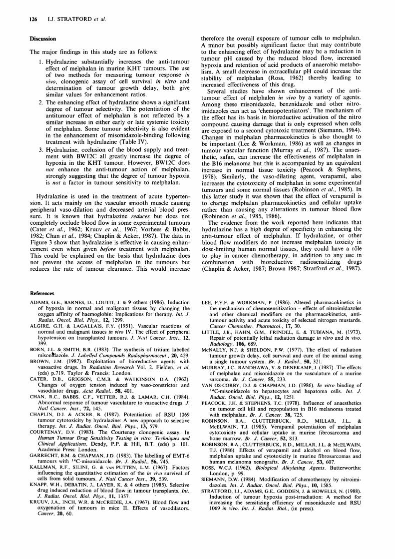
A B C A B C A B C A B C A B CFigure 5 Binding of 3H-misonidazole to various tissues in C3Hmice bearing the KHT tumour. A, misonidazole alone; B,misonidazole followed by hydralazine 15min later; C, as for Bbut with two additional doses of hydralazine given at 90minintervals thereafter.
Table IV Binding of 3H-misonidazole to tissues of mice bearing theKHT sarcoma: Ratio of level of binding; MISO+hydralazine rela-
tive to MISO alone
Treatment Blood Cerebelum Spleen Liver Tumour
MISO+hydralazine 1.06 1.18 1.15 1.29 1.68aMISO + 3 x hydralazine 1.02 1.19 0.97 1.23 2.80a
Hydralazine at 5mgkg-1 given i.v. 15min post misonidazole andat 90min intervals thereafter.
aSignificantly different from a ratio of 1.0 at P< 10-4.
mouse tissues are given in the histogram in Figure 5. In alltreatment groups, significantly greater binding of labelledmaterial was observed in the KHT tumour, the liver and thespleen compared to that in blood or cerebellum. For thegroup treated with misonidazole alone there is little differ-ence between the tumour, liver and spleen. However, admi-nistration of single or multiple doses of hydralazine increasesthe amount of label in the tumour but not in any of theother tissues (Table IV). Further, the ratios of bound,labelled materal in tumour relative to blood give values of1.9 in the group treated with misonidazole alone, 2.9 for thesingle dose of hydralazine and 4.4 in the group receivingthree doses of hydralazine. Clearly, hydralazine substantiallyincreases the efficiency of misonidazole binding and thisappears to be tumour selective.
. . . * . L---l ~- I L-i I I_
.. . I Ile _-f -
_
1-
126 I.J. STRATFORD et al.
Discussion
The major findings in this study are as follows:
1. Hydralazine substantially increases the anti-tumoureffect of melphalan in murine KHT tumours. The useof two methods for measuring tumour response invivo, clonogenic assay of cell survival in vitro anddetermination of tumour growth delay, both givesimilar values for enhancement ratios.
2. The enhancing effect of hydralazine shows a significantdegree of tumour selectivity. The potentiation of theantitumour effect of melphalan is not reflected by asimilar increase in either early or late systemic toxicityof melphalan. Some tumour selectivity is also evidentin the enhancement of misonidazole-binding followingtreatment with hydralazine (Table IV).
3. Hydralazine, occlusion of the blood supply and treat-ment with BW12C all greatly increase the degree ofhypoxia in the KHT tumour. However, BW12C doesnot enhance the anti-tumour action of melphalan,strongly suggesting that the degree of tumour hypoxiais not a factor in tumour sensitivity to melphalan.
Hydralazine is used in the treatment of acute hyperten-sion. It acts mainly on the vascular smooth muscle causingperipheral vaso-dilation and decreased arterial blood pres-sure. It is known that hydralazine reduces but does notcompletely occlude blood flow in some experimental tumours(Cater et al., 1962; Kruuv et al., 1967; Vorhees & Babbs,1982; Chan et al., 1984; Chaplin & Acker, 1987). The data inFigure 3 show that hydralazine is effective in causing enhan-cement even when given before treatment with melphalan.This could be explained on the basis that hydralazine doesnot prevent the access of melphalan in the tumours butreduces the rate of tumour clearance. This would increase
therefore the overall exposure of tumour cells to melphalan.A minor but possibly significant factor that may contributeto the enhancing effect of hydralazine may be a reduction intumour pH caused by the reduced blood flow, increasedhypoxia and retention of acid products of anaerobic metabo-lism. A small decrease in extracellular pH could increase thestability of melphalan (Ross, 1962) thereby leading toincreased effectiveness of this drug.
Several studies have shown enhancement of the anti-tumour effect of melphalen in vivo by a variety of agents.Among these misonidazole, benznidazole and other nitro-imidazoles can act as 'chemopotentiators'. The mechanism ofthe effect has its basis in bioreductive activation of the nitrocompound causing damage that is only expressed when cellsare exposed to a second cytotoxic treatment (Siemann, 1984).Changes in melphalan pharmacokinetics is also thought tobe important (Lee & Workman, 1986) as well as changes intumour vascular function (Murray et al., 1987). The anaes-thetic, safan, can increase the effectiveness of melphalan inthe B16 melanoma but this is accompanied by an equivalentincrease in normal tissue toxicity (Peacock & Stephens,1978). Similarly, the vaso-dilating agent, verapamil, alsoincreases the cytotoxicity of melphalan in some experimentaltumours and some normal tissues (Robinson et al., 1985). Inthis latter study it was shown that the effect of verapamil isto change melphalan pharmacokinetics and cellular uptakerather than causing any alterations in tumour blood flow(Robinson et al., 1985, 1986).The evidence from the work reported here indicates that
hydralazine has a high degree of specificity in enhancing theanti-tumour effect of melphalan. If hydralazine, or otherblood flow modifiers do not increase melphalan toxicity indose-limiting human normal tissues, they could have a r6leto play in cancer chemotherapy, in addition to any use incombination with bioreductive rad;osensitizing drugs(Chaplin & Acker, 1987; Brown 1987; Stratford et al., 1987).
References
ADAMS, G.E., BARNES, D., LOUTIT, J. & 9 others (1986). Inductionof hypoxia in normal and malignant tissues by changing theoxygen affinity of haemoglobin: Implications for therapy. Int. J.Radiat. Oncol. Biol. Phys., 12, 1299.
ALGIRE, G.H. & LAGALLAIS, F.Y. (1951). Vascular reactions ofnormal and malignant tissues in vivo IV. The effect of peripheralhypotension on transplanted tumours. J. Ntli Cancer. Inst., 12,399.
BORN, J.L. & SMITH, B.R. (1983). The synthesis of tritium labelledmisontilazole. J. Labelled Compounds Radiopharmaceut., 20, 429.
BROWN, J.M. (1987). Exploitation of bioreductive agents withvasoactive drugs. In Radiation Research Vol. 2. Fielden, et al.(eds) p.719. Taylor & Francis: London.
CATER, D.B., GRIGSON, C.M.B. & WATKINSON D.A. (1962).Changes of oxygen tension induced by vaso-constrictor andvasodilator drugs. Acta Radiol., 58, 401.
CHAN, R.C., BABBS, C.F., VETTER, R.J. & LAMAR, C.H. (1984).Abnormal response of tumour vasculature to vasoactive drugs. J.Nati Cancer. Inst., 72, 145.
CHAPLIN, D.J. & ACKER, B. (1987). Potentiation of RSU 1069tumour cytotoxicity by hydralazine: A new approach to selectivetherapy. Int. J. Radiat. Oncol. Biol. Phys., 13, 579.
COURTENAY, D.V. (1983). The Courtenay clonogenic assay. InHuman Tumour Drug Sensitivity Testing in vitro: Techniques andClinical Applications, Dendy, P.P. & Hill, B.T. (eds) p. 101.Academic Press: London.
GARRECHT, B.M. & CHAPMAN, J.D. (1983). The labelling of EMT-6tumours with '4C-misonidazole. Br. J. Radiol., 56, 745.
KALLMAN, R.F., SILINI, G. & VAN PUTTEN, L.M. (1967). Factorsinfluencing the quantitative estimation of the in vivo survival ofcells from solid tumours. J. Natl Cancer Inst., 39, 539.
KNAPP, W.H., DEBATIN, J., LAYER, K. & 4 others (1985). Selectivedrug induced reduction of blood flow in tumour transplants. Int.J. Radiat. Oncol. Biol. Phys., 11, 1357.
KRUUV, J.A., INCH, W.R. & McCREDIE, J.A. (1967). Blood flow andoxygenation of tumours in mice II. Effects of vasodilators.Cancer, 20, 60.
LEE, F.Y.F. & WORKMAN, P. (1986). Altered pharmacokinetics inthe mechanism of chemosensitization - effects of nitroimidazolesand other chemical modifiers on the pharmacokinetics, anti-tumour activity and acute toxicity of selected nitrogen mustards.Cancer Chemother. Pharmacol., 17, 30.
LITTLE, J.B., HAHN, G.M., FRINDEL, E. & TUBIANA, M. (1973).Repair of potentially lethal radiation damage in vitro and in vivo.Radiology, 106, 689.
McNALLY, N.J. & SHELDON, P.W. (1977). The effect of radiationtumour growth delay, cell survival and cure of the animal usinga single tumour system. Br. J. Radiol., 50, 321.
MURRAY, J.C., RANDHAWA, V. & DENEKAMP, J. (1987). The effectsof melphalan and misonidazole on the vasculature of a murinesarcoma. Br. J. Cancer, 55, 233.
VAN OS-CORBY, D.J. & CHAPMAN, J.D. (1986). In vitro binding of14C-misonidazole to hepatocytes and hepatoma cells. Int. J.Radiat. Oncol. Biol. Phys., 12, 1251.
PEACOCK, J.H. & STEPHENS, T.C. (1978). Influence of anaestheticson tumour cell kill and repopulation in B16 melanoma treatedwith melphalan. Br. J. Cancer, 38, 725.
ROBINSON, B.A., CLUTTERBUCK, R.D., MILLAR, J.L.. &McELWAIN, T.J. (1985). Verapamil potentiation of melphalancytotoxicity and cellular uptake in murine fibrosarcoma andbone marrow. Br. J. Cancer, 52, 813.
ROBINSON, B.A., CLUTTERBUCK, R.D., MILLAR, J.L. & McELWAIN,T.J. (1986). Effects of verapamil and alcohol on blood flow,melphalan uptake and cytotoxicity in murine fibrosarcomas andhuman melanoma xenografts. Br. J. Cancer, 53, 607.
ROSS, W.C.J. (1962). Biological Alkylating Agents. Butterworths:London, p. 99.
SIEMANN, D.W. (1984). Modification of chemotherapy by nitroimi-dazoles. Int. J. Radiat. Oncol. Biol. Phys., 10, 1585.
STRATFORD, I.J., ADAMS, G.E., GODDEN, J. & HOWELLS, N. (1988).Induction of tumour hypoxia post-irradiation: A method forincreasing the sensitizing efficiency of misonidazole and RSU1069 in vivo. Int. J. Radiat. Biol., (in press).

POTENTIATION OF THE ANTI-TUMOUR EFFECT OF MELPHALAN BY HYDRALAZINE 127
STRATFORD, I.J., GODDEN, J., HOWELLS, N., EMBLING, P. &ADAMS, G.E. (1987). Manipulation of tumour oxygenation byhydralazine increases the potency of bioreductive radiosensitizersand enhances the effect of melphalan in experimental tumours.Radiation Research, Vol. 2, Fielden, et al. (eds) p. 737. Taylor &Francis: London.
STRATFORD, I.J., WALLING, J.M. & SILVER, A.R.J. (1986). Thedifferential cytotoxicity of RSU 1069: Cell survival studiesindicating interaction with DNA as a possible mode of action.Br. J. Cancer, 53, 339.
THOMSON, J.E. & RAUTH, A.M. (1974). An in vitro assay to measurethe viability of KHT tumour cells not previously exposed toculture conditions. Radiat. Res., 58, 262.
VOORHEES, W.D. & BABBS, C.F. (1982). Hydralazine-enhanced selec-tive heating of transmissible venereal tumour implants in dogs.Eur. J. Cancer Clin. Oncol., 19, 1027.
Copyright © 2022 FDOKUMEN