Potentiation of evoked calcitonin gene-related peptide release from oral mucosa: a potential basis...
28
Potentiation of evoked calcitonin gene-related peptide release from oral mucosa: a potential basis for the pro-inflammatory effects of nicotine Gregory O. Dussor 1 , Anthony S. Leong 2 , Nicholas B. Gracia 2 , Sonja Kilo † , Theodore J. Price 1 , Kenneth M. Hargreaves 1,2 , and Christopher M. Flores 1,2,* 1 Department of Pharmacology, The University of Texas Health Science Center, San Antonio, TX, USA 2 Department of Endodontics, The University of Texas Health Science Center, San Antonio, TX, USA Abstract Inflammation of the buccal mucosa, gingiva and periodontal tissues is a significant problem in users of nicotine-containing tobacco products; however, the potential role of nicotine in the development of this inflammation is unclear. In many tissues, nicotine, acting through nicotinic acetylcholine receptors (nAChRs), has been shown to increase the release of the pro-inflammatory mediator calcitonin gene-related peptide (CGRP) thereby potentially contributing to neurogenic inflammation. The purpose of the present studies was to determine the effects of nicotine and other nAChR agonists on capsaicin-evoked immunoreactive CGRP (iCGRP) release from rat buccal mucosa and to identify a potential cellular basis for these effects. Using a previously validated model of in vitro superfusion, we show that the nAChR agonists nicotine (EC 50 557 μM), epibatidine (EC 50 317 pM) and cytisine (EC 50 4.83 nM) potentiated capsaicin-evoked iCGRP release in a concentration-dependent manner by 123, 70 and 76%, respectively. The expression and distribution patterns of the mRNA transcripts encoding the α3, α4 and α6 nAChR subunits and their colocalization with CGRP and the capsaicin receptor VR1 were examined in rat trigeminal ganglion using combined in situ hybridization and immunohistofluorescence. Of all trigeminal neurons counted, mRNA encoding the α3, α4 and α6 subunits was found, respectively, in 14.45, 9.2 and 19.21% of neurons. The cell body diameter of most neurons containing any nAChR subunit was in the 30–40 μm range with slightly fewer in the 20–30 μm range. Co-localization of these α subunit transcripts with either CGRP or VR1 immunoreactivity ranged from approximately 5 to 7% for α4 and over 8% for α3 to 18% for α6. These data support the hypothesis that nicotinic agents, acting at nAChRs contained on primary sensory neurons, are capable of directly modulating the stimulated release of iCGRP In the case of users of nicotine- containing tobacco products, this modulation could contribute to inflammatory processes within the oral cavity. Keywords buccal mucosa; capsaicin; cytisine; epibatidine; nicotinic acetylcholine receptor; sensory neuron Correspondence: Dr Christopher M. Flores, at *present address below. [email protected]. * Present address: Drug Discovery, Johnson and Johnson Pharmaceutical Research and Development, L.L.C., Welsh and McKean Roads, Spring House, PA 19477-0776, USA. † On leave from Institut für Physiologie und Experimentelle Pathophysiologie, Universitätsstrasse 17, D-91054 Erlangen, Germany. NIH Public Access Author Manuscript Eur J Neurosci. Author manuscript; available in PMC 2010 August 3. Published in final edited form as: Eur J Neurosci. 2003 November ; 18(9): 2515–2526. NIH-PA Author Manuscript NIH-PA Author Manuscript NIH-PA Author Manuscript
-
Upload
independent -
Category
Documents
-
view
0 -
download
0
Transcript of Potentiation of evoked calcitonin gene-related peptide release from oral mucosa: a potential basis...

Potentiation of evoked calcitonin gene-related peptide releasefrom oral mucosa: a potential basis for the pro-inflammatoryeffects of nicotine
Gregory O. Dussor1, Anthony S. Leong2, Nicholas B. Gracia2, Sonja Kilo†, Theodore J.Price1, Kenneth M. Hargreaves1,2, and Christopher M. Flores1,2,*1 Department of Pharmacology, The University of Texas Health Science Center, San Antonio, TX,USA2 Department of Endodontics, The University of Texas Health Science Center, San Antonio, TX,USA
AbstractInflammation of the buccal mucosa, gingiva and periodontal tissues is a significant problem inusers of nicotine-containing tobacco products; however, the potential role of nicotine in thedevelopment of this inflammation is unclear. In many tissues, nicotine, acting through nicotinicacetylcholine receptors (nAChRs), has been shown to increase the release of the pro-inflammatorymediator calcitonin gene-related peptide (CGRP) thereby potentially contributing to neurogenicinflammation. The purpose of the present studies was to determine the effects of nicotine and othernAChR agonists on capsaicin-evoked immunoreactive CGRP (iCGRP) release from rat buccalmucosa and to identify a potential cellular basis for these effects. Using a previously validatedmodel of in vitro superfusion, we show that the nAChR agonists nicotine (EC50 557 μM),epibatidine (EC50 317 pM) and cytisine (EC50 4.83 nM) potentiated capsaicin-evoked iCGRPrelease in a concentration-dependent manner by 123, 70 and 76%, respectively. The expressionand distribution patterns of the mRNA transcripts encoding the α3, α4 and α6 nAChR subunits andtheir colocalization with CGRP and the capsaicin receptor VR1 were examined in rat trigeminalganglion using combined in situ hybridization and immunohistofluorescence. Of all trigeminalneurons counted, mRNA encoding the α3, α4 and α6 subunits was found, respectively, in 14.45,9.2 and 19.21% of neurons. The cell body diameter of most neurons containing any nAChRsubunit was in the 30–40 μm range with slightly fewer in the 20–30 μm range. Co-localization ofthese α subunit transcripts with either CGRP or VR1 immunoreactivity ranged fromapproximately 5 to 7% for α4 and over 8% for α3 to 18% for α6. These data support thehypothesis that nicotinic agents, acting at nAChRs contained on primary sensory neurons, arecapable of directly modulating the stimulated release of iCGRP In the case of users of nicotine-containing tobacco products, this modulation could contribute to inflammatory processes withinthe oral cavity.
Keywordsbuccal mucosa; capsaicin; cytisine; epibatidine; nicotinic acetylcholine receptor; sensory neuron
Correspondence: Dr Christopher M. Flores, at *present address below. [email protected].*Present address: Drug Discovery, Johnson and Johnson Pharmaceutical Research and Development, L.L.C., Welsh and McKeanRoads, Spring House, PA 19477-0776, USA.†On leave from Institut für Physiologie und Experimentelle Pathophysiologie, Universitätsstrasse 17, D-91054 Erlangen, Germany.
NIH Public AccessAuthor ManuscriptEur J Neurosci. Author manuscript; available in PMC 2010 August 3.
Published in final edited form as:Eur J Neurosci. 2003 November ; 18(9): 2515–2526.
NIH
-PA Author Manuscript
NIH
-PA Author Manuscript
NIH
-PA Author Manuscript
IntroductionNeurogenic inflammation is a process that occurs in response to the stimulation of certainperipheral primary sensory neurons (Janscó et al., 1967). This process is mediated in part byseveral pro-inflammatory neuropeptides, including calcitonin gene-related peptide (CGRP)and substance P, that are released from the peripheral terminals of these neurons (Gazelius etal., 1987; Cruwys et al., 1992; Karimian & Ferrell, 1994; Brain et al., 1985; Brain &Williams, 1985). In support of the involvement of these neuropeptides in the inflammatoryresponse, it has been shown that CGRP and substance P undergo axonal transport from thenodose and dorsal root ganglia to the periphery along the vagus and sciatic nerves,respectively (Brimijoin et al., 1980; Kashihara et al., 1989), and that their peripheraladministration produces vasodilation (Brain et al., 1985; Gazelius et al., 1987) and plasmaextravasation (Gamse & Saria, 1985). In addition, antidromic electrical stimulation of thetrigeminal ganglion causes vasodilation in facial skin through a process that is dependent onCGRP (Escott et al., 1995). Collectively, these studies strongly implicate CGRP as amediator of neurogenic inflammation and validate its utility as a marker for this process in avariety of experimental settings.
Nicotinic acetylcholine receptors (nAChRs) are members of the ligand-gated ion channelsuperfamily. The pentameric stoichiometry of these receptors comprises two alpha subunitsand three beta subunits (Anand et al., 1991; Cooper et al., 1991) or, in the case of α-bungarotoxin-sensitive nicotinic receptors, five alpha subunits. Activation of these receptorsresults in a conformational change in the receptor complex, allowing the conductance ofNa+, K+ and Ca2+ ions to varying extents depending on the nAChR subtype(s) involved.Subunits known to be expressed in the mammalian nervous system include α2–α7, α9, α10and β2–β4. In the rat trigeminal ganglion, subtypes made up of α4β2 and α3β4 subunitcombinations have been demonstrated as has mRNA encoding the α2, α5–α7, α9 and β3subunits (Wada et al., 1989, 1990; Flores et al., 1996; Liu et al., 1998; Keiger & Walker,2000). However, the functional role of these sensory neuronal nAChRs is not wellunderstood.
Nicotine is capable of modulating the activity of sensory neurons and the transmittersubstances they secrete. Nicotine or other nicotinic agonists applied to sensory neurons hasbeen shown to activate these neurons both in vitro (Steen & Reeh, 1993; Liu & Simon,1993) and in vivo (Tanelian, 1991). Nicotine is known to directly stimulate the release ofimmunoreactive CGRP (iCGRP) in several tissues including heart (Franco-Cereceda et al.,1991, 1992), trachea (Hua et al., 1994; Jinno et al., 1994), pulmonary tissue (Lou et al.,1991, 1992) and cultured dorsal root ganglion neurons (Franco-Cereceda et al., 1992). Inaddition, nicotine enhances the evoked release of iCGRP in dental pulp (Hargreaves et al.,1992) and rat paw skin (Kilo et al., 1995). Taken together, these studies show that nicotineis capable of modulating the release of CGRP from sensory neurons and may therebycontribute to the process of neurogenic inflammation.
Similar to studies on the trunk and limbs, evidence shows that the development of certainoral inflammatory diseases has a neurogenic component (Györfi et al., 1992). The linkbetween the use of nicotine-containing tobacco products and increases in the incidence oforal inflammatory disease is well established. For example, smoking is associated with anincrease in periodontal disease (Haber et al., 1993) and gingivitis (Ismail et al., 1983). Thestudies detailed here sought to provide a potential mechanistic explanation linking nicotineexposure of oral tissue to inflammatory disease. To address this hypothesis, studies wereperformed using nAChR agonists in a previously validated model system for the study ofneurogenic inflammation in oral tissues (Flores et al., 2001). In addition, nAChR subunits
Dussor et al. Page 2
Eur J Neurosci. Author manuscript; available in PMC 2010 August 3.
NIH
-PA Author Manuscript
NIH
-PA Author Manuscript
NIH
-PA Author Manuscript
were localized to CGRP-/VR1-containing neurons using combined in situ hybridization andimmunohistofluorescence.
Materials and methodsAnimals
All experiments were carried out in accordance with protocols approved by the InstitutionalAnimal Care and Use Committee of The University of Texas Health Science Center at SanAntonio as well as with the guidelines for the ethical treatment of animals established by theNational Institutes of Health. All experiments utilized tissues obtained from adult (175–250g), male Sprague-Dawley rats (Charles River, Wilmington, MA, USA).
In vitro superfusionRats were killed by decapitation and the buccal mucosae were dissected from the underlyingbuccinator muscle and placed in a super-fusion chamber (the mucosae from both sides of theoral cavity of one animal per chamber) housed in a 37 °C waterbath (for details see Flores etal., 2001). The tissue was continuously supervised with Krebs buffer containing 1 mMNaH2PO4, 135 mM NaCl, 3.5 mM KCl, 1 mM NaHCO3, 1 mM MgCl2, 2.5 mM CaCl2, 0.1mM ascorbic acid, 16 μM thiorphan (BACHEM, Bubendorf, Switzerland), 3.3 mMdextrose, 10 mM Hepes and 0.1% bovine serum albumin at a flow rate of approximately0.21 mL/min. The buffer (pH 7.4) was oxygenated and immersed in a separate 37 °Cwaterbath. Superfusate was collected in 12 fractions, 10 min each, using an automatedfraction collector (Gilson, Middleton, WI, USA). Tissue was perfused for 70 min, followedby a 10-min application of buffer containing either capsaicin or capsaicin plus drug. Tomaximize the opportunity of observing nicotinic agonist-induced facilitatory or inhibitoryeffects, whichever occurred, in all superfusion experiments, capsaicin was used at asubmaximal approximately EC80 concentration (100 μM) as previously described. Cytisine(Sigma, St. Louis, MO, USA), (–)-nicotine hydrogen tartrate salt (Sigma) and epibatidine(RBI, Nitick, MA, USA) were dissolved in distilled H2O and diluted in Krebs buffer. Thelength of tubing between the reservoir and chamber was designed so that there would be a 3-min lag period between the time the drug was added and the time it would reach thechamber. Thus, drug treatments were applied 3min before the start of the next fraction.Varying concentrations of drug in combination with 100 μM capsaicin (Fluka,Ronkonkoma, NY, USA) were added to individual reservoirs of buffer to determineconcentration–response curves for each drug. Each concentration was applied to one groupof chambers (n = 5–6) and compared with chambers receiving buffer containing capsiacinonly (n = 6). Tissue was stimulated only once and superfusion was completed with a 40-minwashout period using Krebs buffer alone. In antagonist experiments, buffer containingmecamylamine (Sigma) was applied for a total of 30 min, including one fraction before, thefraction during and the fraction following the application of capsaicin plus drug. Superfusionwas completed with a 30-min washout period using Krebs buffer alone. ImmunoreactiveCGRP release was measured over the 120-min period using radioimmunoassayquantification.
RadioimmunoassayFollowing superfusion, samples as well as tubes containing known CGRP concentrations(1–300 fmol) were incubated with a C-terminally directed anti-CGRP antiserum (DrMichael Iadarola, NIDCR, NIH, Bethesda, MD, USA) generated in rabbit. Following a 48-hincubation period at 4°C, 100 μL of [125I]CGRP28–37 (approximately 22 000 cpm) and 50μL of goat antirabbit antibody coupled to magnetic beads (PerSeptive Biosystems,Framingham, MA, USA) were added. After a second 48 h incubation at 4 °C, bound CGRPwas separated from free CGRP through immunomagnetic separation. Radioactive counts
Dussor et al. Page 3
Eur J Neurosci. Author manuscript; available in PMC 2010 August 3.
NIH
-PA Author Manuscript
NIH
-PA Author Manuscript
NIH
-PA Author Manuscript

were measured by a gamma counter. Samples containing known CGRP concentrations wereused to generate standard curves. Unknown concentrations of iCGRP were determined usinga logit-log transformation. The minimum detection limit for this assay is approximately 2–3fmol/tube with 50% displacement occurring at 10–30 fmol/tube. All drugs used in theseexperiments were tested for effects on the standard curves and none were observed.
StatisticsResults for concentration response curves were analysed using nonlinear regression analysisand antagonist experiments were analysed using an unpaired, one-tailed Student’s t-test(GraphPad Prism, San Diego, CA, USA). Statistical significance was accepted when theprobability of a type I error was less than 5% (P < 0.05). Data on graphs represent mean±SEM.
cDNAs and probe synthesisPlasmid constructs containing the cDNA clones encoding the α3 (PCA48E), α4 (pA4.1) andα6 (pSN119) neuronal nicotinic receptor subunits were kindly provided by Dr Jim Boulter(UCLA, Los Angeles, CA, USA). For antisense probe synthesis, 1 μg of plasmid DNA waslinearized with the appropriate restriction endonuclease (Bam HI for α3, BstEII for α4 orNaeI for α6) and riboprobes were synthesized using an in vitro transcription kit (Promega,Madison, WI, USA) in the presence of 250 μCi each of [35S]CTPαS and [35S]UTPαS(Amersham, Arlington Heights, IL, USA) at 37 °C for 2h. Antisense probes corresponded tothe last 504 bases C-terminal for α3, the last 864 bases C-terminal for α4 and the last 449bases C-terminal for α6. Sense probes were also generated as controls. Probes were purifiedusing G-50 spin column chromatography (Ambion, Austin, TX, USA), diluted to aconcentration of 1 × 10⁸ cpm/mL in hybridization buffer containing 50% formamide, 0.3 MNaCl, 10 mM Tris, 1 mM EDTA, 1 × Denhardt’s solution, 10% dextran sulphate, 50 μg/mLyeast tRNA (Roche, Indianapolis, IN, USA) and 10 mM dithiothreitol and stored at −20 °C.
In situ hybridization immunohistochemistryRats were killed by decapitation; trigeminal ganglia were freshly dissected, frozen on dryice, immobilized in mounting medium, cut into 20-μm sections, mounted onto glass slidesand stored at −80 °C. The sections were fixed for 1 h in ice-cold 10% formalin (3.7%formaldehyde w/v), permeabilized with 0.5% Triton X-100, acylated with 0.0025% aceticanhydride in 0.1 M triethanolamine, dehydrated and delipidated in alcohol and chloroform.Sections were then hybridized with in vitro-transcribed [35S]-labelled riboprobes (75 μL 1 ×10⁷cpm/mL per slide) complementary to mRNA encoding the α3, α4 or α6 neuronalnicotinic receptor subunits. Slides were coverslipped with Parafilm and incubated in ahumidified chamber at 55 °C overnight. After RNase treatment and standard washingprocedures, the tissue was blocked in phosphate-buffered saline containing 10% normal goatserum (Gibco, Rockville, MD, USA) and then incubated in rabbit-derived anti-CGRPantiserum (1:750; Peninsula, Belmont, CA, USA) or guinea pig-derived anti-VR1 antiserum(1:3000, Neuromics, Minneapolis, MN, USA) at 4 °C overnight. For singleimmunolabelling, tissue was then incubated for 1 h with Alexa Fluor 488-conjugated goatantirabbit or Alexa Fluor 488-conjugated goat antiguinea pig secondary antibody (1:300;Molecular Probes, Eugene, OR, USA), respectively. For double immunolabelling, tissue wasfirst incubated with anti-CGRP antiserum overnight at 4 °C, followed by secondary antibodydetailed above, then incubated with anti-VR1 antiserum overnight at 4 °C, followed byAlexa Fluor 594-conjugated goat antiguinea pig secondary antibody (1:300; MolecularProbes). Slides were then subjected to emulsion autoradiography using NTB-3 (Kodak,Rochester, NY, USA), developed and coverslipped.
Dussor et al. Page 4
Eur J Neurosci. Author manuscript; available in PMC 2010 August 3.
NIH
-PA Author Manuscript
NIH
-PA Author Manuscript
NIH
-PA Author Manuscript

ImagingImages were acquired using an E600 microscope (Nikon, Melville, NY, USA) equippedwith a Photometries Sensys black and white digital CCD camera and analysed usingMetamorph V4.1 (Universal Image Corporation, Downingtown, PA, USA). Approximately14–16 images at 20× magnification were necessary to cover an entire trigeminal ganglionsection. Cell size–frequency histograms were generated from 20 × images by measuringtotal area (which was later converted to diameter). Only cells that showed visible nucleiwere included in the analysis. To determine total cell number or cells containing visiblenuclei, the contrast of captured images was increased so that all cells, labelled andunlabelled by whichever method, could be seen. Cell counts for in situ-positive cells wereperformed by counting neurons that demonstrated above background silver grain labelling.Similarly, for immunopositive cells, counts were made on cells that were above backgroundlabelling using Metamorph’s built-in scaling feature by establishing an average pixel valuefor background labelling and designating neurons above that pixel value as positive. Imageswere overlayed (dark field in situ hybridization and fluorescent immunohistochemistryimage) to assess the presence of both nAChR mRNA and CGRP/VR1 immunoreactivity. Inthe case of triple labelling, three images were overlayed (dark field in situ and twofluorescent immunohistochemistry images). Dark field in situ and immunofluorescentimages presented were pseudo-coloured using Metamorph. Percentages for eachcolocalization condition represent counts made on one section (14–16 images) from threeseparate animals while percentages for total nAChR mRNA-positive cells represent countsmade on two sections each from three separate animals, wherein counted sections were >100 μm apart.
ResultsModulation of capsaicin-evoked immunoreactive calcitonin gene-related peptide releaseby nicotinic acetylcholine receptor agonists
Figure 1A shows the concentration-dependent potentiation of capsaicin-evoked iCGRPrelease by the prototypic nicotinic receptor agonist, nicotine. Peak effects (Emax 223%) ofnicotine were seen at 10 mM and the concentration that produced a half-maximal effect(EC50) was 556.5 μM. To determine whether this effect was mediated by nAChRs, tissuewas treated with the nAChR antagonist mecamylamine (10 μM) and cotreated with 10 mMnicotine. As seen in Fig. 1B, the effects of nicotine were completely blocked in the presenceof mecamylamine but not by the α7 nAChR antagonist methyllycaconitine (100 nM; notshown). Epibatidine, a high potency neuronal nicotinic receptor agonist, was also tested. Theconcentration–response curve for epibatidine is shown in Fig. 2A. Epibatidine evoked aconcentration-dependent potentiation of capsaicin-evoked iCGRP release with maximaleffects of 170.3% occurring at 10 nM (EC50 317.2 pM). Co-administration ofmecamylamine (10 μM) in perfusion buffer again resulted in complete attenuation of theability of epibatidine to potentiate capsaicin-evoked iCGRP release (Fig. 2B).
The effects of cytisine, a pharmacologically distinct nAChR agonist, on capsaicin-evokediCGRP release are shown in Fig. 3A. Net peak iCGRP release was potentiated in aconcentration-dependent manner with a maximal effect of 176.2% occurring at 1 μM (EC504.8 nM). Figure 3B shows complete attenuation of the potentiation of the capsaicin-evokediCGRP release seen with cytisine when mecamylamine is included. Nicotine, epibatidine orcytisine, in the absence of capsaicin, did not significantly alter CGRP release nor didmecamylamine have any effect on basal or capsaicin-evoked release (data not shown).
Dussor et al. Page 5
Eur J Neurosci. Author manuscript; available in PMC 2010 August 3.
NIH
-PA Author Manuscript
NIH
-PA Author Manuscript
NIH
-PA Author Manuscript
Cell size distribution of nicotinic acetylcholine receptor subunit mRNATo determine the profile of trigeminal ganglion neurons expressing nAChR mRNA, cellsize–frequency histograms were generated for all three subunit mRNAs (Fig. 4A–C). Onlycells whose nucleus was clearly visible were subjected to cell size measurements. For eachsubunit, random images were selected from those taken of trigeminal ganglion sections fromthree separate animals. The mRNA for the α3 nAChR subunit (Fig. 4A) was found mainly inneurons with an average diameter in the 20–30 μm (62 of 143 cells) and 30–40 μm (69 of143 cells) range. Few cells with diameters in either the 10–20 μm (two of 143) or 40–50 μm(10 of 143) range were found and no α3-positive cells were found in cell bodies withdiameters larger than 50 μm. Very similar results were obtained for α4 (Fig. 4B) with the20–30 μm (60 of 141) and 30–40 μm (69 of 141) diameter ranges containing the mostmRNA-positive cells, while the 10–20 μm (two of 141) and 40–50 μm (10 of 141) rangescontained fewer positive cells. Again, no cells larger than 50 μm in diameter contained α4mRNA. The distribution pattern for α6 mRNA (Fig. 4C) differed only slightly from that ofα3 and α4. The 10–20 μm (two of 148) diameter range contained the least positive cells, the20–30 μm (54 of 148) and 30–40 μm (70 of 148) ranges contained the most, while the 40–50μm (22 of 148) range contained an intermediate number of positive cells, being slightlymore than that of α3 or α4. As before, positive cells were not seen in neurons with a cellbody diameter larger than 50 μm. No above background labelling was seen with sensecontrol probes for α3, α4 or α6 (data not shown).
Colocalization of mRNA encoding nicotinic acetylcholine receptor with either calcitoningene-related peptide or VR1 immunoreactivity
Results of combined in situ hybridization and immunohistochemistry on sections of rattrigeminal ganglion are detailed in Figs 5–7. Figure 5 shows the cellular distribution patternof α3 mRNA with respect to CGRP or VR1 immunoreactivity. mRNA encoding the α3, α4and α6 nAChR subunits was found, respectively, in 14, 9 and 19% of neurons, whileimmunoreactivity for CGRP or VR1 was found in approximately 36% of neurons (Table 1).Of those neurons expressing CGRP or VR1 immunoreactivity 5–9% coexpressed mRNAencoding either the α3 or α4 subunits, while almost twice as many (12–19%) coexpressed α6mRNA (Table 2). Conversely, in neurons expressing α3 (Fig. 5) or α4 (Fig. 6) mRNA, 20–26% of these coexpressed CGRP or VR1 immunoreactivity and, in those expressing α6mRNA (Fig. 7), the number coexpressing VR1 immunoreactivity reached 34% (Table 3).No difference was observed in the percentages for nAChR mRNA-positive cells in sectionslabelled using only in situ hybridization versus sections colabelled usingimmunohistochemistry (data not shown).
Co-localization of nicotinic acetylcholine receptor mRNA with both calcitonin gene-relatedpeptide and VR1
To determine more definitively whether nAChR mRNA and CGRP and VR1immunoreactivities are simultaneously coexpressed in the same neuron, triple-labellingexperiments were performed (Figs 8–10). Figure 8 shows triple labelling for α3, Fig. 9 forα4 and Fig. 10 for α6, indicating that, for all three mRNA species, cells can be found thatcontain all three labels (a subunit mRNA, CGRP and VR1). Consistent with the highercolocalization percentages seen for α6 in Table 2, more triple-labelled cells can be seen inFig. 10 than in Figs 8 and 9 although, for illustrative purposes, only one triple-labelled cell ishighlighted in each figure.
DiscussionCapsaicin has been used extensively as an investigational tool for its ability to activate asubpopulation of unmyelinated and thinly myelinated, nociceptive sensory neurons (for
Dussor et al. Page 6
Eur J Neurosci. Author manuscript; available in PMC 2010 August 3.
NIH
-PA Author Manuscript
NIH
-PA Author Manuscript
NIH
-PA Author Manuscript

reviews, see Chapman et al., 1961; Janscó et al., 1967; Bevan & Szolcsanyi, 1990). Morerecently, the activation by capsaicin of a ligand-gated ion channel (VR1; also called TRPV1)expressed predominantly on C-fibre nociceptors has been described (Caterina et al., 1997).Activation of these VR1 channels by capsaicin evokes the release of certain vasoactiveneuropeptides, including CGRP and substance P (for review, see Holzer, 1991), whichproduce vasodilation and increased permeability of the endothelial membrane, therebyleading to erythema and oedema. The recent development of a model system in which tostudy capsaicin-stimulated CGRP release from rat buccal mucosa (Flores et al., 2001) hasenabled a more comprehensive evaluation of the mechanisms governing neuropeptiderelease from oral tissue in response to various stimuli. Consequently, this approach shouldallow for the determination of the potential roles played by these stimuli in the developmentof inflammatory disease within the oral cavity.
According to the World Health Organization, approximately 1.1 billion people smokeworldwide (http://www.who.int) and many more use smokeless tobacco. Not surprisingly,inflammation of the buccal, gingival and periodontal tissues is a major oral health problemamong users of nicotine-containing tobacco products (see Haber et al., 1993). Indeed,nicotine concentrations in the saliva of smokeless tobacco users has been measured to be ashigh as 10 mM (Hoffman & Adams, 1981), providing for the probable activation of multiplenicotinic receptor subtypes contained in oral tissues. The present studies were aimed atdetermining the effects of neuronal nicotinic receptor agonists on capsaicin-evoked CGRPrelease from rat buccal mucosa. In addition, experiments were performed to elucidate thepresence of neuronal nicotinic receptor subunit transcripts in CGRP- or VR1-containingneurons whose cell bodies in the trigeminal ganglion innervate the oral cavity. These latterstudies sought to suggest a possible mechanism for the increases in inflammation in oraltissues of tobacco users. As VR1 is a receptor for capsaicin (Caterina et al., 1997), andstimulation of VR1-expressing neurons by capsaicin results in CGRP release (Holzer, 1991),the expression of functional nicotinic receptors on VR1-/CGRP-immunoreactive neuronswould provide a molecular basis upon which to hypothesize a direct action of nicotinicagonists on capsaicin-sensitive neurons in the modulation of CGRP release. By extension,this line of evidence supports a similar potential mechanism of action to explain the pro-inflammatory effects of nicotine or nicotine-containing tobacco products on the oral mucosa.
Here we provide the initial report on the distribution of α6 mRNA in sensory ganglia at thesingle cell level as well as the colocalization of the α3, α4 or α6 transcripts in CGRP- and/orVR1-immunoreactive neurons. As the origin of cell bodies whose axons innervate the buccalmucosa, the trigeminal ganglion represents a relevant anatomical location in which tovisualize the cellular distribution of nAChR mRNA. Neuronal nicotinic receptor subtypespreviously demonstrated in neurons of the trigeminal ganglion include the heteromeric α3β4and α4β2 subtypes (Flores et al., 1996) as well as α-bungarotoxin-binding sites, probably α7receptors (Schechter et al., 1978). Functional α7-,α3β4- and α4β2-like receptors have alsobeen measured in rat trigeminal (Liu & Simon, 1993; Liu et al., 1998) and dorsal root(Genzen et al., 2001) ganglia using electrophysiological techniques. In addition, reversetranscription–polymerase chain reaction studies have indicated that the trigeminal ganglionexpresses mRNA encoding the α6 and, possibly, α9 subunits (Liu et al., 1998; Keiger &Walker, 2000).
The diameter of most cells expressing nAChR subunit mRNA here was between 20 and 40μm, implying that this expression occurs in unmyelinated or lightly myelinated C-fibre andAδ-fibre sensory neurons (Harper & Lawson, 1985), many of which are known to containCGRP and express VR1 (Lawson et al., 1993; Helliwell et al., 1998; Tominaga et al., 1998).To directly evaluate this possibility, double-labelling experiments were performed. Of cellsthat were positive for CGRP immunoreactivity, the percentage that colocalized α3, α4 or α6
Dussor et al. Page 7
Eur J Neurosci. Author manuscript; available in PMC 2010 August 3.
NIH
-PA Author Manuscript
NIH
-PA Author Manuscript
NIH
-PA Author Manuscript

nAChR subunit mRNA was approximately 7–12%. Colocalization of VR1-positive neuronswith nAChR mRNA was similar but reached over 18% for α6. These observations, however,must take into account the current lack of information regarding the rate and extent to whichCGRP or VR1 protein is trafficked out of the cell body. If transport occurs relatively rapidlyand extensively after synthesis, then our colocalization studies might have underestimatedthe coexpression of these nAChR transcripts with CGRP and/or VR1. Additionally,depending on the sensitivity of the antibodies used to detect CGRP or VR1immunoreactivity, the level of coexpression might have been similarly underestimated. Inany event, the presence of these subunit mRNAs in VR1- or CGRP-expressing neuronsconstitutes supportive anatomical evidence concerning the ability of the nicotinic agoniststested to directly modulate CGRP release evoked by capsaicin. Insofar as the total amount ofCGRP released by capaicin in the presence of any of the nicotinic agonists tested wassubmaximal and a small fraction of the total pool (see Flores et al., 2001), the relativelymodest degree of colocalization between any given nAChR subunit, VR1 and CGRP isapparently sufficient and appropriate (i.e. a higher proportion of colocalizing neurons wouldhave resulted in greater levels of enhanced release).
Although α3β4 and α4β2 receptors have been demonstrated previously in the trigeminalganglion, it is not yet known whether the mRNA encoding the α6 subunit gives rise tofunctional nAChRs in this tissue; however, in combination with α3, β2, β3 or β4 subunit, α6forms functional receptor channels in chick retina or Xenopus oocytes (Gerzanich et al.,1997; Kuryatov et al., 2000; Vailati et al., 1999). In addition, an α6β2 site was recentlyproposed in dopaminergic neurons in the striatum, based on a loss of α-conotoxin MIIbinding in α6 knockout mice (Champtiaux et al., 2002). Whether or not nAChRs thatcontain the α6 subunit are involved in the neurosecretory processes described here,nonetheless their present demonstration serves to increase the reported nAChRheterogeneity and distribution in sensory neurons. Indeed, the cellular localization of the α6subunit suggests that the nAChRs that it contributes in forming may play a role in themodulation of inflammatory processes and/or nociception.
The studies detailed here in trigeminal ganglion neurons examined only the mRNAlocalization of certain nAChR α subunits. However, we may infer their translation andassembly in this structure based on previously cited studies documenting high affinity [3H]-epibatidine-binding sites and nAChR-mediated electrophysiological responses. Takentogether with the present data, demonstrating functional nAChRs in a tissue innervated byneurons originating in the trigeminal ganglion, we hypothesize that receptors formed by thesubunits we have measured at the mRNA level are probably trafficked peripherally to themucosa in the same way that α-bungarotoxin-binding nAChRs are transported peripherallyfrom the dorsal root ganglion within the sciatic nerve (Ninkovic & Hunt, 1983).
However, there are non-neuronal cell types in buccal mucosa that express several receptorsthat are relevant to the present work. For example, human keratinocytes have recently alsobeen shown to express functional VR1 (Inoue et al., 2002; Southall et al., 2003) and areknown to express several nicotinic receptor subunits including α3, α5, α7, β2 and β4(Grando et al., 1995, 1996). However, these epithelial-derived cells do not elaborate CGRPand we are unaware of any other resident cells that do; therefore, these are not the target/source of capsaicin-evoked CGRP release described here. Moreover, there is considerableevidence to indicate that the origin of CGRP in orofacial tissues, including the buccalmucosa, is neuronal as well as capsaicin-sensitive and this has been extensively reviewed inthe original work describing the model used in the present studies (Flores et al., 2001). Thus,the simplest interpretation of the preponderance of experimental data is that theneurosecretory effects of nicotine and its congeners shown here are mediated by a direct
Dussor et al. Page 8
Eur J Neurosci. Author manuscript; available in PMC 2010 August 3.
NIH
-PA Author Manuscript
NIH
-PA Author Manuscript
NIH
-PA Author Manuscript

action on capsaicin-sensitive, CGRP-containing and nAChR-expressing trigeminal nerveterminals in the mucosa.
None of the nicotinic agonists used here possesses sufficient pharmacological selectivity tounequivocally implicate one or another nAChR subtype in mediating the potentiation ofCGRP release. Nevertheless, the present data, taken together with previously publishedinvestigations, allow for reasonable speculation. That nicotine, epibatidine and cytisine eachproduced similar maximal effects (i.e. 70–123% potentiation) is consistent with thehypothesis that these drugs do so by activating the same receptor subtype. The fact thatcytisine (Leutje & Patrick, 1991) and epibatidine (Buisson et al., 2000) have been shown tobe weak to moderate partial agonists at the α4β2 subtype but are thought to be full agonistsat the α3β4 subtype would tend to implicate the latter, while the involvement of α7-containing receptors can be largely ruled out based on the ability of mecamylamine and, atleast in the case of nicotine, not methyllycaconitine to antagonize the actions of all of theagonists used. Although the EC50 of cytisine demonstrated here is inexplicably low (≈5nM),perhaps owing to differential post-translational modifications and/or activation–inactivationkinetics of peripheral sensory neuronal nAChRs, that of nicotine (≈500μM) is virtuallyidentical to published values for activating the α3β4 subtype and is one to three orders ofmagnitude higher than that required to activate α4β2- or α6-containing subtypes (Gerzanichet al., 1995, 1997). Consistent with this view, no potentiation of capsaicin-evoked CGRPrelease was exhibited by the relatively selective α4β2 agonist ABT-594 (Dussor and Flores,unpublished observations). Moreover, approximately 75% of all high affinity [3H]-epibatidine binding in the trigeminal ganglion can be accounted for by receptors nominallycomprised of α3 and β4 subunits (Flores et al., 1996). Finally, colocalization of CGRP and/or VR1 immunoreactivity with mRNA encoding the α3 or α6 subunit was relatively morefrequent compared with the α4 subunit. However, these studies cannot rule out thepossibility of activation of other subtypes not yet characterized in primary sensory neurons.The development and application of additional compounds with greater subtype selectivityor adaptation of these functional assays in tissues derived from nAChR subunit knockoutmice would provide valuable insights toward the resolution of these questions.
With respect to the concentration of capsaicin used in the present studies (i.e. 100 μM), thisis an approximate EC80 (see Flores et al., 2001) designed to provide a two-tailed window forobserving either an inhibitory or an excitatory effect, whichever occurred. As detailed in theDiscussion of the earlier work, we do not believe that this is the actual concentration reachedat the neuronal terminal VR1 receptors in the mucosa, owing to the thickness andpermeability of this tissue and the high lipophilicity of capsaicin. Moreover, theconcentrations required to activate peripheral versus central VR1 are up to three orders ofmagnitude higher and the concentrations used here are consistent with those demonstrated toevoke CGRP release from paw skin (Kilo et al., 2001). The fact that the effectiveconcentrations of nicotinic agonists shown here are more in keeping with their publishedEC50 values probably reflects the fact that these compounds are more hydrophilic thancapsaicin, thereby distributing more predictably in the extracellular fluid.
It is remarkable that, in contrast to other studies (Franco-Cereceda et al., 1992; Hua et al.,1994), none of the nAChR agonists used in this study were able to directly stimulate therelease of iCGRP in the absence of capsaicin. However, these other studies utilized culturedneurons, visceral tissues (e.g. lung) or an alternate species (e.g. guinea-pig). This is not tosay that there are no direct effects of nAChR activation on the trigeminal neuron terminalscharacterized here, as trigeminal or dorsal root ganglion neurons have been shown to befunctionally activated by nAChR agonists. However, the only dependent measure assessedin the present studies was CGRP release. The primary neurotransmitters in the trigeminalganglion have yet to be determined but immunohistochemical studies suggest glutamate as
Dussor et al. Page 9
Eur J Neurosci. Author manuscript; available in PMC 2010 August 3.
NIH
-PA Author Manuscript
NIH
-PA Author Manuscript
NIH
-PA Author Manuscript

at least one strong possibility (Inagaki et al., 1987; Kai-Kai & Howe, 1991; Stoyanova et al.,1998; Bae et al., 2000). In fact, there are important differences with respect to the frequency/intensity of stimulation necessary to cause the release of glutamate, which is contained insmall clear vesicles, and CGRP, which is predominantly found in large, dense core vesicles(for review see Langley & Grant, 1997). Accordingly, activation of nicotinic receptors mayproduce a frequency/intensity of stimulation necessary to release gluamate but not CGRP,although this remains to be determined. In this scenario, only in response to activation of thehighly Ca2+-permeable VR1 receptor by capsaicin and the additional influx of Ca2+
provided through nAChR activation would the release of CGRP be seen. Consistent withthis rationale, nicotinic stimulation of motor neurons has no effect on acetylcholine releasealone. It is only when motor neurons are stimulated electrically at sufficiently highfrequencies that nicotinic agonists are able to modulate acetylcholine release (Bowman etal., 1988; Vizi & Somogyi, 1989; Wessler, 1989).
Another consideration in terms of the net effect of vanilloid and nicotinic stimulation onCGRP release relates to the activation state of the respective receptors. In fact, it is likelythat VR1 and/or the nAChRs being activated in the present studies become desensitized inthe course of the 10-min period during which their agonists are being coapplied, althoughthe resolution of this method does not provide a ready means for determining the relativecontributions of these differentially activatable receptor species. That said, thepreponderance of experimental evidence in this and other systems supports the conclusionthat capsaicin-induced CGRP release arises from VR1 agonism. Similarly, and insofar as theeffects of nicotinic agonists described here are mecamylamine-reversible, it is reasonable toconclude that they arise from nAChR agonism. Lastly, it would be interesting to determinethe extent to which intracellular messengers engaged and mobilized by VR1 and/or nAChRactivation may play a role. In any case, the ability of nAChR agonists, especially nicotine, topotentiate stimulated CGRP release, while having no effect on their own, suggests that theymay predispose oral tissues to an enhanced neurogenic inflammatory response to otherinsults, such as thermal, chemical and mechanical tissue damage or infection.
In summary, we report here the potentiation of capsaicin-evoked CGRP release in buccalmucosa by the nAChR agonists nicotine, epibatidine and cytisine and provide evidencesupporting the existence of several nicotinic receptor subtypes on CGRP-/VR1-containingneurons in trigeminal ganglia, the origin of sensory innervation to the buccal mucosa.Accordingly, these data provide a potential mechanism for the pro-inflammatory actions ofnicotine on the oral tissues of tobacco users. Taken together, the present studies not onlyimplicate sensory neuronal nAChRs in the mechanism by which the nicotine contained intobacco products may contribute to inflammation within the oral cavity but also suggest thatthese receptors may constitute viable targets for the future development of a novel class ofanti-inflammatory drugs.
AcknowledgmentsThis work was supported by NIDA grants DA05982 and DA10510. The authors wish to acknowledge SarahRamsay for technical assistance.
Abbreviations
CGRP calcitonin gene-related peptide
iCGRP immunoreactive CGRP
nAChR nicotinic acetylcholine receptor
Dussor et al. Page 10
Eur J Neurosci. Author manuscript; available in PMC 2010 August 3.
NIH
-PA Author Manuscript
NIH
-PA Author Manuscript
NIH
-PA Author Manuscript
ReferencesAnand R, Conroy WG, Schoepfer R, Whiting P, Lindstrom J. Neuronal nicotinic acetylcholine
receptors expressed in Xenopus oocytes have a pentameric quaternary structure. J Biol Chem1991;266:11 192–11 198.
Bae YC, Ihn HJ, Park MJ, Ottersen OP, Moritani M, Yoshida A, Shigenaga Y. Identification of signalsubstances in synapses made between primary afferents and their associated axon terminals in therat trigeminal sensory nuclei. J Comp Neurol 2000;418:299–309. [PubMed: 10701828]
Bevan S, Szolcsanyi J. Sensory neuron-specific actions of capsaicin: mechanisms and applications.Trends Pharmacol Sci 1990;11:330–333. [PubMed: 2203194]
Bowman WC, Marshall IG, Gibb AJ, Harborne AJ. Feedback control of transmitter release at theneuromuscular junction. Trends Pharmacol Sci 1988;9:16–20. [PubMed: 2907694]
Brain SJ, Morris H, MacIntyre I. Calcitonin gene-related peptide is a potent vasodilator. Nature 1985a;313:54–56. [PubMed: 3917554]
Brain S, Williams T. Inflammatory edema induced by synergism between CGRP and mediators ofincreased vascular permeability. Br J Pharmacol 1985b;86:855–860. [PubMed: 2416378]
Brimijoin S, Lundberg J, Brodin E, Hokfelt T, Nilsson G. Axonal transport of substance P in the vagusand sciatic nerves of the guinea pig. Brain Res 1980;191:443–457. [PubMed: 6155172]
Buisson B, Vallejo YF, Green WN, Bertrand D. The unusual nature of epibatidine responses at theα4β2 nicotinic acetylcholine receptor. Neuropharmacology 2000;39:2561–2569. [PubMed:11044727]
Caterina MJ, Schumacher MA, Tominaga M, Rosen TA, Levine JD, Julius D. The capsaicin receptor:a heat-activated ion channel in the pain pathway. Nature 1997;389:816–824. [PubMed: 9349813]
Champtiaux N, Zhi-Yan H, Bessis A, Rossi FM, Zoli M, Marubio L, McIntosh JM, Changeux JP.Distribution and pharmacology of α6-containing nicotinic acetylcholine receptors analyzed withmutant mice. J Neurosci 2002;22:1208–1217. [PubMed: 11850448]
Chapman LF, Ramos AO, Goddell H, Wolff HG. Neurohumoral features of afferent fibers in man.Arch Neurol 1961;4:617–650. [PubMed: 13692408]
Cooper E, Couturier S, Ballivet M. Pentameric structure and subunit stoichiometry of a neuronalacetylcholine receptor. Nature 1991;350:235–238. [PubMed: 2005979]
Cruwys SC, Kidd BL, Mapp PI, Walsh DA, Blake DR. The effects of calcitonin gene-related peptideon formation of intra-articular oedema by inflammatory mediators. Br J Pharmacol 1992;107:116–119. [PubMed: 1384904]
Escott KJ, Beattie DT, Connor HE, Brain SD. Trigeminal ganglion stimulation increases facial skinblood flow in the rat: a major role for calcitonin gene-related peptide. Brain Res 1995;669:93–99.[PubMed: 7536103]
Flores CM, DeCamp RM, Kilo S, Rogers SW, Hargreaves KM. Neuronal nicotinic receptor expressionin sensory neurons of the rat trigeminal ganglion: demonstration of α3β4, a novel subtype in themammalian nervous system. J Neurosci 1996;16:7892–7901. [PubMed: 8987817]
Flores CM, Leong AS, Dussor GO, Harding-Rose C, Hargreaves KM, Kilo S. Capsaicin-evokedCGRP release from rat buccal mucosa: development of a model system for studying trigeminalmechanisms of neurogenic inflammation. Eur J Neurosci 2001;14:1113–1120. [PubMed:11683903]
Franco-Cereceda A, Lou YP, Lundberg JM. Ruthenium-red inhibits CGRP release by capsaicin andresiniferatoxin but not by ouabain, bradykinin or nicotine in guinea-pig heart: correlation witheffects on cardiac contractility. Br J Pharmacol 1991;104:305–310. [PubMed: 1724624]
Franco-Cereceda A, Ryda M, Dalsgaard D. Nicotine- and capsaicin-, but not potassium-evoked CGRPrelease from cultured guinea pig spinal ganglia is inhibited by Ruthenium red. Neurosci Lett1992;137:72–74. [PubMed: 1378220]
Gamse R, Saria A. Potentiation of tachykinin-induced plasma protein extravasation by CGRP. Eur JPharmacol 1985;114:61–66. [PubMed: 2412851]
Gazelius B, Edwall B, Olgart L, Lundberg J, Hokfelt T, Fischer J. Vasodilatory effects and coexistenceof CGRP and substance P in sensory nerves of cat dental pulp. Acta Physiol Scand 1987;130:33–40. [PubMed: 2438899]
Dussor et al. Page 11
Eur J Neurosci. Author manuscript; available in PMC 2010 August 3.
NIH
-PA Author Manuscript
NIH
-PA Author Manuscript
NIH
-PA Author Manuscript
Genzen JR, Van Cleve W, McGehee DS. Dorsal root ganglion neurons express multiple nicotinicacetylcholine receptor subtypes. J Neurophysiol 2001;86:1773–1782. [PubMed: 11600638]
Gerzanich V, Peng X, Wang F, Wells G, Anand R, Fletcher S, Lindstrom J. Comparitivepharmacology of epibatidine: a potent agonist for neuronal nicotinic acetylcholine receptors. JPharmacol Exp Ther 1995;48:774–782.
Gerzanich V, Kuryatov A, Anand A, Lindstrom J. ‘Orphan’ α6 nicotinic AChR subunit can form afunctional heteromeric acetylcholine receptor. J Pharmacol Exp Ther 1997;51:320–327.
Grando SA, Horton RM, Pereira EFR, George PM, Diethelm-Okita BM. A nocitinic acetylcholinereceptor regulating cell adhesion and motility is expressed in human keratinocytes. J InvestDermatol 1995;105:774–781. [PubMed: 7490471]
Grando SA, Horton RM, Mauro TM, Kist DA, Lee TX, Dahl MV. Activation of keratinocyte nicotiniccholinergic receptors stimulates calcium influx and enhances cell differentiation. J Invest Dermatol1996;107:412–418. [PubMed: 8751979]
Györfi A, Fazekas A, Rosivall L. Neurogenic inflammation and the oral mucosa. J Clin Periodontal1992;19:731–736.
Haber J, Wattles J, Crowby M, Mandel R, Kaunusi J, Kent R. Evidence for smoking as a major riskfactor for periodontitis. J Periodontal 1993;64:16–23.
Hargreaves KM, Bowles WR, Garry M. An in vitro method to evaluate regulation of neuropeptiderelease from dental pulp. J Endodontics 1992;18:597–600.
Harper AA, Lawson SN. Conduction velocity is related to morphological cell type in rat dorsal rootganglion neurones. J Physiol (Lond) 1985;359:31–46. [PubMed: 3999040]
Helliwell RJ, McLatchie LM, Clarke M, Winter J, Bevan S, McIntyre P. Capsaicin sensitivity isassociated with the expression of the vanilloid (capsaicin) receptor (VR1) mRNA in adult ratsensory ganglia. Neurosci Lett 1998;250:177–180. [PubMed: 9708861]
Hoffman D, Adams JD. Carcinogenic tobacco-specific N-nitrosamines in snuff and in the saliva ofsnuff dippers. Cancer Res 1981;41:4305–4308. [PubMed: 7198005]
Holzer, P. Capsaicin as a tool for studying neuron functions. In: Costa, M.; Surrenti, C.; Gorini, S.;Maggi, CA.; Meli, A., editors. Sensory Nerves and Neuropeptides in Gastroenterology. PlenumPress; New York: 1991. p. 3-16.
Hua XY, Jinno S, Back SM, Tam EK, Yaksh TL. Multiple mechanisms for the effects of capsaicin,bradykinin and nicotine on CGRP release from tracheal afferent nerves: Role of prostaglandins,sympathetic nerves and mast cells. Neuropharmacology 1994;33:1147–1154. [PubMed: 7862250]
Inagaki N, Kamisaki Y, Kiyama H, Horio Y, Tohyama M, Wada H. Immunocytochemicallocalizations of cytosolic and mitochondrial glutamic oxaloacetic transaminase isozymes in ratprimary sensory neurons as a marker for the glutamate neuronal system. Brain Res 1987;402:197–200. [PubMed: 2881599]
Inoue K, Koizumi S, Fuziwara S, Denda S, Inoue K, Denda M. Functional vanilloid receptors incultured normal human epidermal keratinocytes. Biochem Biophys Res Comm 2002;291:124–129.[PubMed: 11829471]
Ismail II, Burt BA, Eklund SA. Epidemiologic patterns of smoking and periodontal disease in theUnited States. JAMA 1983;106:617–623.
Janscó N, Janscó-Gabor A, Szolcsányi J. Direct evidence for neurogenic inflammation and itsprevention by denervation and by pretreatment with capsaicin. Br J Pharmacol 1967;31:138–151.
Jinno S, Hua XY, Yaksh TL. Nicotine and acetylcholine induce release of calcitonin gene-relatedpeptide from rat trachea. J Appl Physiol 1994;76:1651–1656. [PubMed: 8045845]
Kai-Kai MA, Howe R. Glutamate-immunoreactivity in the trigeminal and dorsal root ganglia, andintraspinal neurons and fibres in the dorsal horn of the rat. Ristochem J 1991;23:171–179.
Karimian M, Ferrell WR. Plasma protein extravasation into the rat knee joint induced by calcitoningene-related peptide. Neurosci Lett 1994;166:39–42. [PubMed: 8190355]
Kashihara Y, Sakaguchi M, Kuno M. Axonal transport and distribution of endogenous calcitonin gene-related peptide in rat peripheral nerve. J Neurosci 1989;11:3796–3802. [PubMed: 2479725]
Keiger CJ, Walker JC. Individual variation in the expression profiles of nicotinic receptors in theolfactory bulb and trigeminal ganglion and identification of α2, α6, α9 and β3 transcripts. BiochemPharmacol 2000;59:233–240. [PubMed: 10609551]
Dussor et al. Page 12
Eur J Neurosci. Author manuscript; available in PMC 2010 August 3.
NIH
-PA Author Manuscript
NIH
-PA Author Manuscript
NIH
-PA Author Manuscript

Kilo S, Hargreaves KM, Flores CM. Peripheral modulation of capsaicin-induced neuropeptide releaseby nicotine. Abstr Soc Neurosci 1995;21:1414.
Kilo S, Harding-Rose C, Hargreaves KM, Flores CM. Peripheral CGRP release as a marker forneurogenic inflammation: a model system for the study of neuropeptide secretion in rat paw skin.Pain 2001;73:201–207. [PubMed: 9415506]
Kuryatov A, Olale F, Cooper J, Choi C, Lindstrom J. Human alpha6 AChR subtypes: subunitcomposition, assembly and pharmacological responses. Neuropharmacology 2000;39:2570–2590.[PubMed: 11044728]
Langley K, Grant NJ. Are exocytosis mechanisms neurotransmitter specific? Neurochem Int1997;31:739–757. [PubMed: 9413835]
Lawson SN, Perry MJ, Prabhakar E, McCarthy PW. Primary sensory neurones: neurofilament,neuropeptides and conduction velocity. Brain Res Bull 1993;30:239–243. [PubMed: 7681350]
Leutje CW, Patrick J. Both α- and β-subunits contribute to the agonist sensitivity of neuronal nicotinicacetylcholine receptors. J Neurosci 1991;11:837–845. [PubMed: 1705971]
Liu L, Simon S. Responses of cultured rat trigeminal ganglion nerves to chemical stimuli. Abstr SocNeurosci 1993;19:518.
Liu L, Chang GQ, Jiao YQ, Simon SA. Neuronal nicotinic acetylcholine receptors in rat trigeminalganglia. Brain Res 1998;809:238–245. [PubMed: 9853116]
Lou YP, Karlsson A, Franco-Cereceda A, Lundberg J. Selectivity of ruthenium red in inhibitingbronchoconstriction and CGRP release induced by afferent C-fiber activation in the guinea piglung. Acta Physiol Scand 1991;142:191–199. [PubMed: 1715114]
Lou YP, Franco-Cereceda A, Lundberg J. Different ion channel mechanisms between lowconcentration of capsaicin and high concentrations of capsaicin and nicotine regarding peptiderelease from pulmonary afferents. Acta Physiol Scand 1992;146:119–127. [PubMed: 1279940]
Ninkovic M, Hunt SP. α-Bungarotoxin binding sites on sensory neurones and their axonal transport insensory afferents. Brain Res 1983;272:57–69. [PubMed: 6193836]
Schechter N, Handy IC, Pezzeminti L, Schmidt J. Distribution of alpha-bungarotoxin binding sites inthe central nervous system and peripheral organs of the rat. Toxicon 1978;16:245–251. [PubMed:653752]
Southall MD, Li T, Gharibova LS, Pei Y, Nicol GD, Travers JB. Activation of epidermal vanilloidreceptor-1 induces release of priinflammatory mediators in human keratinocytes. J Pharmacol ExpTher 2003;304:217–222. [PubMed: 12490594]
Steen K, Reeh P. Actions of cholinergic agonists and antagonists on sensory nerve endings in rat skinin vitro. J Neurophysiol 1993;70:397–405. [PubMed: 8103089]
Stoyanova I, Dandov A, Lazarov N, Chouchkov C. GABA- and glutamate-immunoreactivity insensory ganglia of cat: a quantitative analysis. Arch Physiol Biochem 1998;106:362–369.[PubMed: 10441058]
Tanelian DL. Cholinergic activation of a population of corneal afferent nerves. Exp Brain Res1991;86:414–420. [PubMed: 1756814]
Tominaga M, Caterina MJ, Malmberg AB, Rosen TA, Gilbert H, Skinner K, Raumann BE, BasbaumAI, Julius D. The cloned capsaicin receptor integrates multiple pain-producing stimuli. Neuron1998;21:531–543. [PubMed: 9768840]
Vailati S, Hanke W, Bejan A, Barabino B, Longhi R, Balestra B, Moretti M, Clementi F, Gotti C.Functional α6-containing nicotinic receptors are present in chick retina. J Pharmacol Exp Ther1999;56:11–19.
Vizi ES, Somogyi GT. Prejunctional modulation of acetylcholine release from the skeletalneuromuscular junction: link between positive (nicotinic) – and negative (muscarinic) – feedbackmodulation. Br J Pharmacol 1989;97:65–70. [PubMed: 2720313]
Wada E, Wada K, Boulter J, Deneris E, Heinemann S, Patrick J, Swanson LW. Distribution of alpha-2,alpha-3, alpha-4 and beta-2 neuronal nicotinic receptor subunit mRNAs in the central nervoussystem: a hybridization histochemical study in the rat. J Comp Neurol 1989;284:314–335.[PubMed: 2754038]
Dussor et al. Page 13
Eur J Neurosci. Author manuscript; available in PMC 2010 August 3.
NIH
-PA Author Manuscript
NIH
-PA Author Manuscript
NIH
-PA Author Manuscript

Wada E, McKinnon D, Heinemann S, Patrick J, Swanson LW. The distribution of mRNA encoded bya new member of the neuronal nicotinic acetylcholine receptor gene family (α5) in the rat centralnervous system. Brain Res 1990;526:45–53. [PubMed: 2078817]
Wessler I. Control of transmitter release from the motor nerve by presynaptic nicotinic and muscarinicautoreceptors. Trends Pharmacol Sci 1989;10:110–114. [PubMed: 2574510]
Dussor et al. Page 14
Eur J Neurosci. Author manuscript; available in PMC 2010 August 3.
NIH
-PA Author Manuscript
NIH
-PA Author Manuscript
NIH
-PA Author Manuscript

Fig. 1.(A) Potentiation of capsaicin-evoked immunoreactive calcitonin gene-related peptide(iCGRP) release by nicotine. Tissue was washed for 70 min with physiologic Krebs buffer(pH 7.4), followed by a 10-min superfusion of separate groups with capsaicin alone (100μM, n = 6) or capsaicin plus nicotine (100 μM–0mM, n = 5–6/group). Data are expressed aspercentage control of net peak iCGRP release, calculated as the arithmetic differencebetween peak and basal release in fmol/sample. Concentration required to produce 50%effect (EC50) was 556.5 μM and maximal effect (Emax) was 123% over control. (B)Reversal of potentiation by nicotine of capsaicin-evoked iCGRP release by mecamylamine.Mecamylamine (10 μM) was given 20 min before, 10 min during and 10 min following 10mM nicotine + capsaicin superfusion. Groups were analysed by an unpaired, one-tailedStudent’s t-test (*P < 0.01) and data are represented as mean ±SEM.
Dussor et al. Page 15
Eur J Neurosci. Author manuscript; available in PMC 2010 August 3.
NIH
-PA Author Manuscript
NIH
-PA Author Manuscript
NIH
-PA Author Manuscript
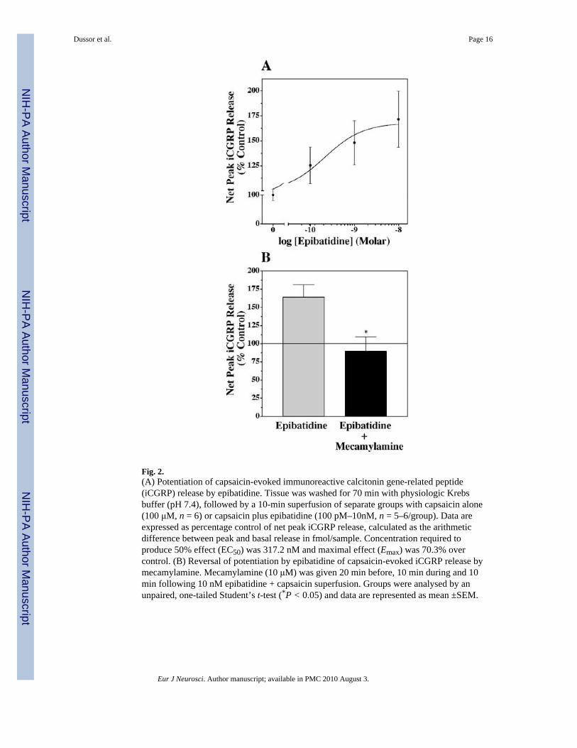
Fig. 2.(A) Potentiation of capsaicin-evoked immunoreactive calcitonin gene-related peptide(iCGRP) release by epibatidine. Tissue was washed for 70 min with physiologic Krebsbuffer (pH 7.4), followed by a 10-min superfusion of separate groups with capsaicin alone(100 μM, n = 6) or capsaicin plus epibatidine (100 pM–10nM, n = 5–6/group). Data areexpressed as percentage control of net peak iCGRP release, calculated as the arithmeticdifference between peak and basal release in fmol/sample. Concentration required toproduce 50% effect (EC50) was 317.2 nM and maximal effect (Emax) was 70.3% overcontrol. (B) Reversal of potentiation by epibatidine of capsaicin-evoked iCGRP release bymecamylamine. Mecamylamine (10 μM) was given 20 min before, 10 min during and 10min following 10 nM epibatidine + capsaicin superfusion. Groups were analysed by anunpaired, one-tailed Student’s t-test (*P < 0.05) and data are represented as mean ±SEM.
Dussor et al. Page 16
Eur J Neurosci. Author manuscript; available in PMC 2010 August 3.
NIH
-PA Author Manuscript
NIH
-PA Author Manuscript
NIH
-PA Author Manuscript
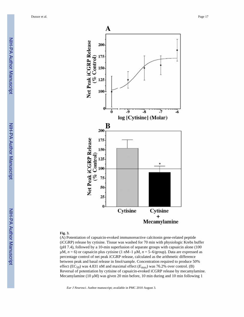
Fig. 3.(A) Potentiation of capsaicin-evoked immunoreactive calcitonin gene-related peptide(iCGRP) release by cytisine. Tissue was washed for 70 min with physiologic Krebs buffer(pH 7.4), followed by a 10-min superfusion of separate groups with capsaicin alone (100μM, n = 6) or capsaicin plus cytisine (1 nM–1 μM, n = 5–6/group). Data are expressed aspercentage control of net peak iCGRP release, calculated as the arithmetic differencebetween peak and basal release in fmol/sample. Concentration required to produce 50%effect (EC50) was 4.831 nM and maximal effect (Emax) was 76.2% over control. (B)Reversal of potentiation by cytisine of capsaicin-evoked iCGRP release by mecamylamine.Mecamylamine (10 μM) was given 20 min before, 10 min during and 10 min following 1
Dussor et al. Page 17
Eur J Neurosci. Author manuscript; available in PMC 2010 August 3.
NIH
-PA Author Manuscript
NIH
-PA Author Manuscript
NIH
-PA Author Manuscript
μM cytisine + capsaicin superfusion. Groups were analysed by an unpaired, one-tailedStudent’s t-test (*P < 0.05) and data are represented as mean ±SEM.
Dussor et al. Page 18
Eur J Neurosci. Author manuscript; available in PMC 2010 August 3.
NIH
-PA Author Manuscript
NIH
-PA Author Manuscript
NIH
-PA Author Manuscript

Fig. 4.Cell size frequency histograms for (A) α3, (B) α4 or (C) α6 nicotinic acetylcholine receptorsubunit mRNA. Only cells with a clearly identifiable nucleus were used for sizing. Cellswere sized using Metamorph imaging software, counted and recorded in bins based on cellbody diameter. Data are expressed as number of cells in each diameter range of the totalcells counted for each subunit (total cells counted, 143 for α3, 141 for α4 and 148 for α6).
Dussor et al. Page 19
Eur J Neurosci. Author manuscript; available in PMC 2010 August 3.
NIH
-PA Author Manuscript
NIH
-PA Author Manuscript
NIH
-PA Author Manuscript

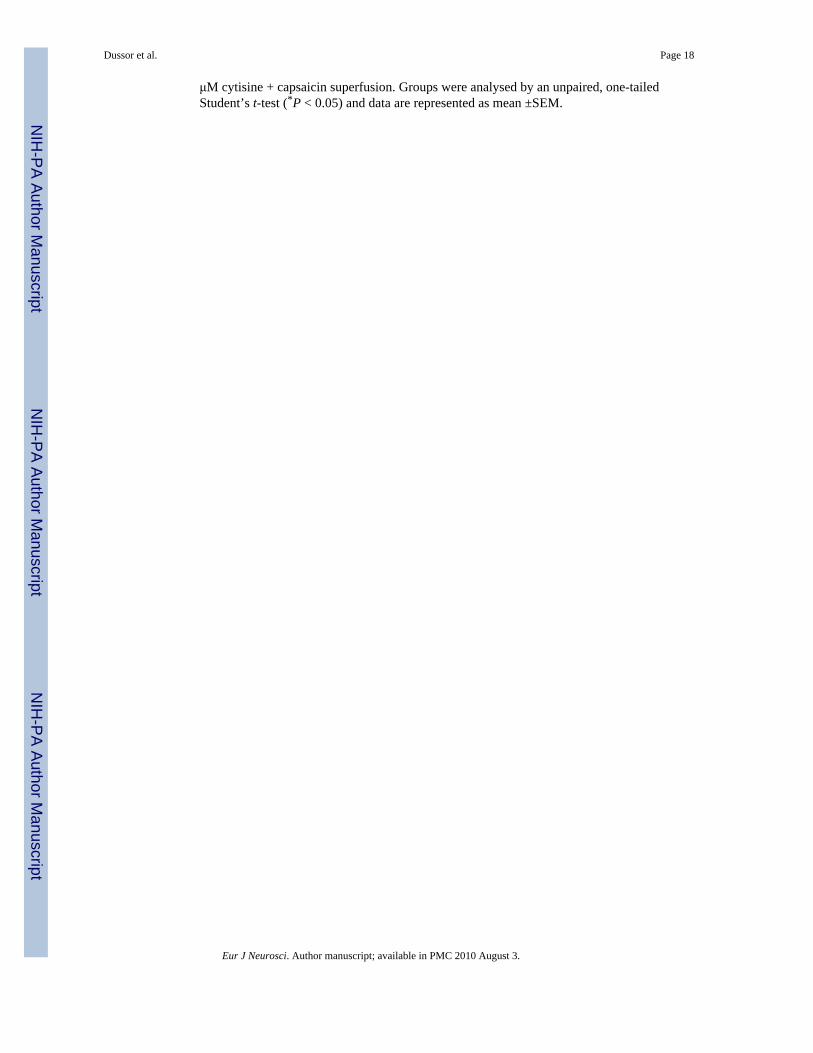
Fig. 5.Colocalization of α3 nicotinic acetylcholine receptor mRNA with calcitonin gene-relatedpeptide (CGRP) or VR1. A–C and D–F are from two separate sections. Large arrowheads,mRNA-positive cells; small arrowheads, immunopositive cells; arrows, double-labelledcells. Scale bars, 100 μm. A and D are dark-field in situ hybridization images for μ3 whichhave been pseudo-coloured red. Immunohistochemistry for CGRP or VR1 is shown in B andE, respectively. (C) Double labelling for α3 mRNA and CGRP; (F) double labelling for α3mRNA and VR1.
Dussor et al. Page 20
Eur J Neurosci. Author manuscript; available in PMC 2010 August 3.
NIH
-PA Author Manuscript
NIH
-PA Author Manuscript
NIH
-PA Author Manuscript
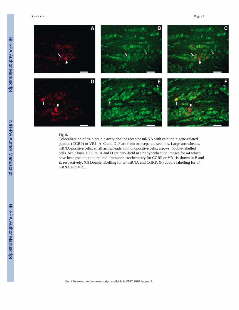
Fig. 6.Colocalization of α4 nicotinic acetylcholine receptor mRNA with calcitonin gene-relatedpeptide (CGRP) or VR1. A–C and D–F are from two separate sections. Large arrowheads,mRNA-positive cells; small arrowheads, immunopositive cells; arrows, double-labelledcells. Scale bars, 100 μm. A and D are dark-field in situ hybridization images for α4 whichhave been pseudo-coloured red. Immunohistochemistry for CGRP or VR1 is shown in B andE, respectively. (C) Double labelling for α4 mRNA and CGRP; (F) double labelling for α4mRNA and VR1.
Dussor et al. Page 21
Eur J Neurosci. Author manuscript; available in PMC 2010 August 3.
NIH
-PA Author Manuscript
NIH
-PA Author Manuscript
NIH
-PA Author Manuscript
Fig. 7.Colocalization of α6 nicotinic acetylcholine receptor mRNA with calcitonin gene-relatedpeptide (CGRP) or VR1. A–C and D–F are from two separate sections. Large arrowheads,mRNA-positive cells; small arrowheads, immunopositive cells; arrows, double-labelledcells. Scale bars, 100 μm. A and D are dark-field in situ hybridization images for α6 whichhave been pseudo-coloured red. Immunohistochemistry for CGRP or VR1 is shown in B andE, respectively. (C) Double labelling for α3 mRNA and CGRP; (F) double labelling for α6mRNA and VR1.
Dussor et al. Page 22
Eur J Neurosci. Author manuscript; available in PMC 2010 August 3.
NIH
-PA Author Manuscript
NIH
-PA Author Manuscript
NIH
-PA Author Manuscript
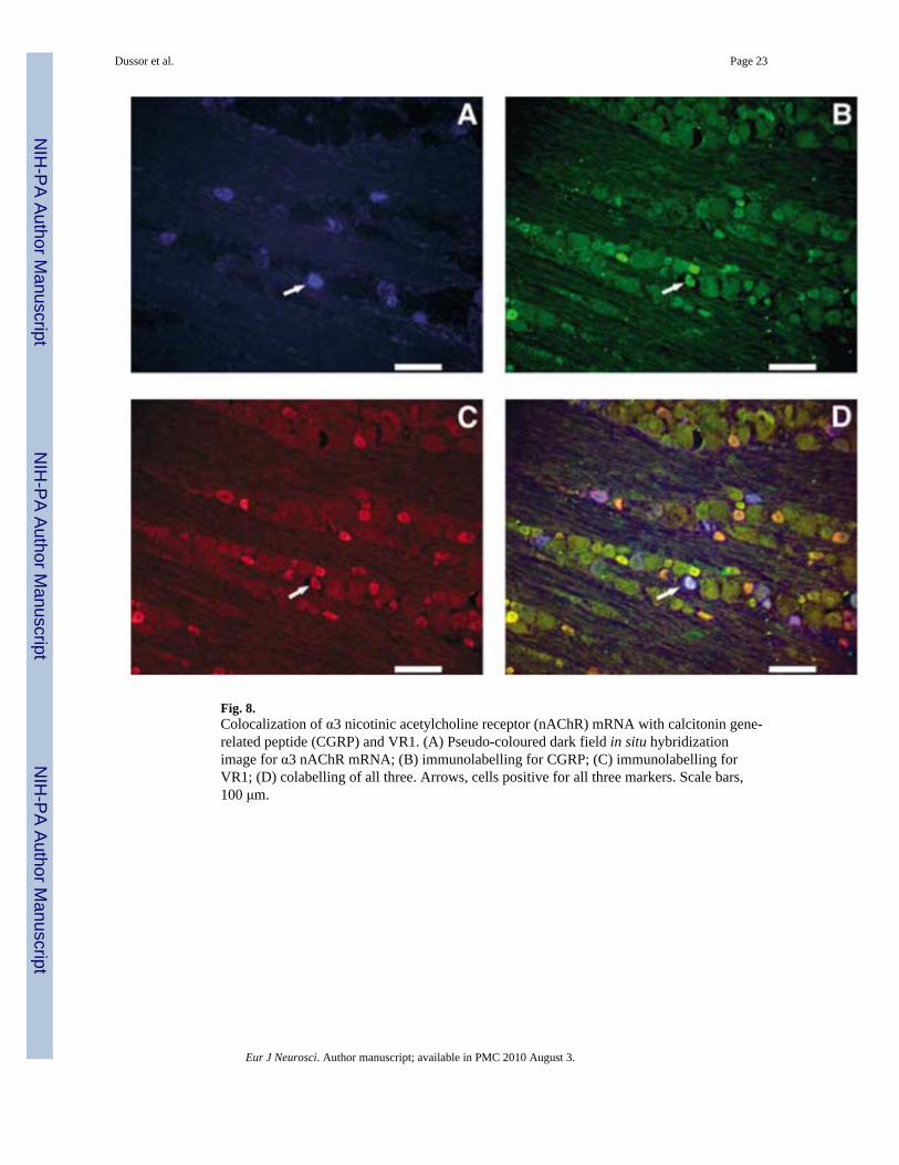
Fig. 8.Colocalization of α3 nicotinic acetylcholine receptor (nAChR) mRNA with calcitonin gene-related peptide (CGRP) and VR1. (A) Pseudo-coloured dark field in situ hybridizationimage for α3 nAChR mRNA; (B) immunolabelling for CGRP; (C) immunolabelling forVR1; (D) colabelling of all three. Arrows, cells positive for all three markers. Scale bars,100 μm.
Dussor et al. Page 23
Eur J Neurosci. Author manuscript; available in PMC 2010 August 3.
NIH
-PA Author Manuscript
NIH
-PA Author Manuscript
NIH
-PA Author Manuscript
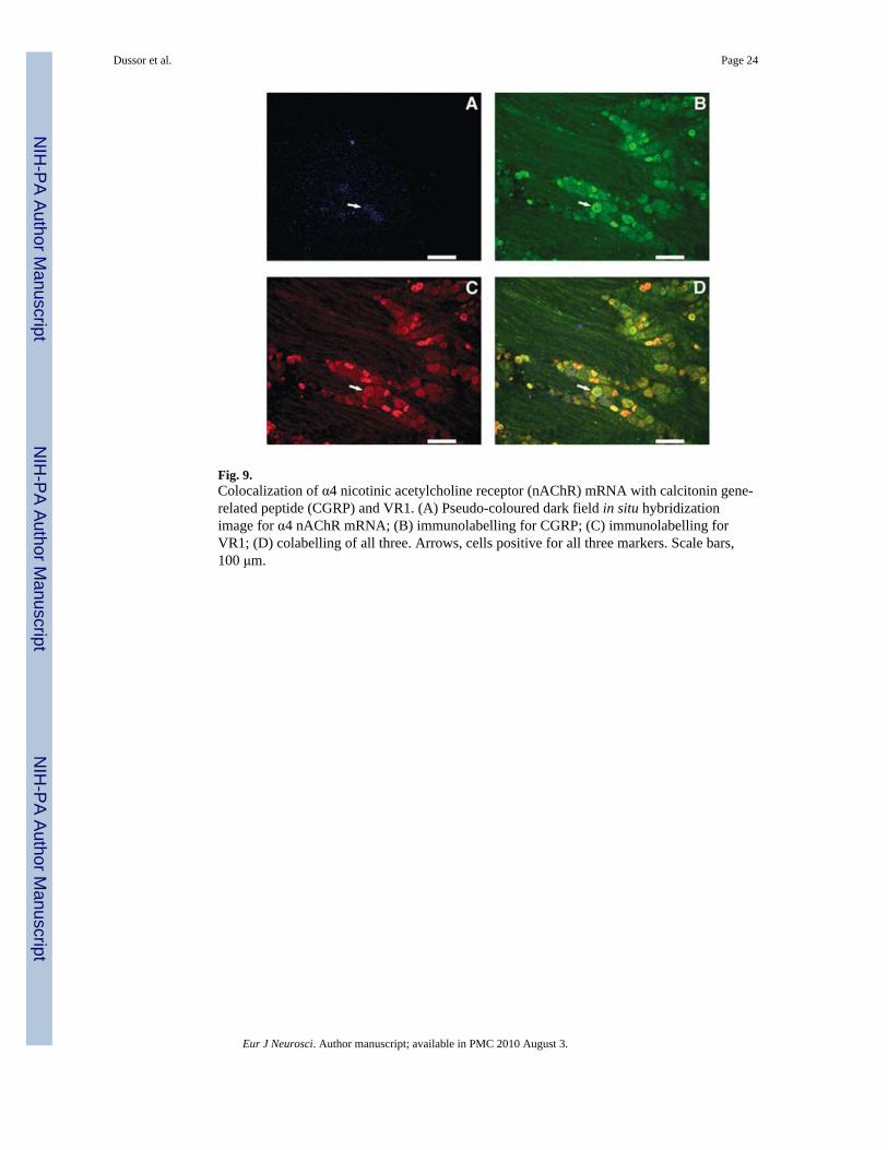
Fig. 9.Colocalization of α4 nicotinic acetylcholine receptor (nAChR) mRNA with calcitonin gene-related peptide (CGRP) and VR1. (A) Pseudo-coloured dark field in situ hybridizationimage for α4 nAChR mRNA; (B) immunolabelling for CGRP; (C) immunolabelling forVR1; (D) colabelling of all three. Arrows, cells positive for all three markers. Scale bars,100 μm.
Dussor et al. Page 24
Eur J Neurosci. Author manuscript; available in PMC 2010 August 3.
NIH
-PA Author Manuscript
NIH
-PA Author Manuscript
NIH
-PA Author Manuscript

Fig. 10.Colocalization of α6 nicotinic acetylcholine receptor (nAChR) mRNA with calcitonin gene-related peptide (CGRP) and VR1. (A) Pseudo-coloured dark field in situ hybridizationimage for α6 nAChR mRNA; (B) immunolabelling for CGRP; (C) immunolabelling forVR1; (D) colabelling of all three. Arrows, cells positive for all three markers. Scale bars,100 μm.
Dussor et al. Page 25
Eur J Neurosci. Author manuscript; available in PMC 2010 August 3.
NIH
-PA Author Manuscript
NIH
-PA Author Manuscript
NIH
-PA Author Manuscript

NIH
-PA Author Manuscript
NIH
-PA Author Manuscript
NIH
-PA Author Manuscript
Dussor et al. Page 26
Table 1
Expression of nicotinic acetylcholine receptor mRNA and calcitonin gene-related peptide (CGRP)/VR1immunoreactivity in trigeminal neurons
Analysis Labelled/total number* (%)‡
α3 mRNA 1080/7421 (14.45 ± 1.54)
α4 mRNA 691/7445 (9.20) ± 1.48)
α6 mRNA 1436/7504 (19.21 ± 2.91)
CGRP IR 4011/11016 (36.37 ± 2.54)
VR1 IR 4145/11354 (36.54 ± 2.58
*Data for mRNA-positive cells represent counts of six entire sections (two from each of three animals). Data for immunoreactive (IR) cells
represent counts of nine entire sections (three from each of three animals).
‡Data are expressed as mean ±SD.
Eur J Neurosci. Author manuscript; available in PMC 2010 August 3.
NIH
-PA Author Manuscript
NIH
-PA Author Manuscript
NIH
-PA Author Manuscript
Dussor et al. Page 27
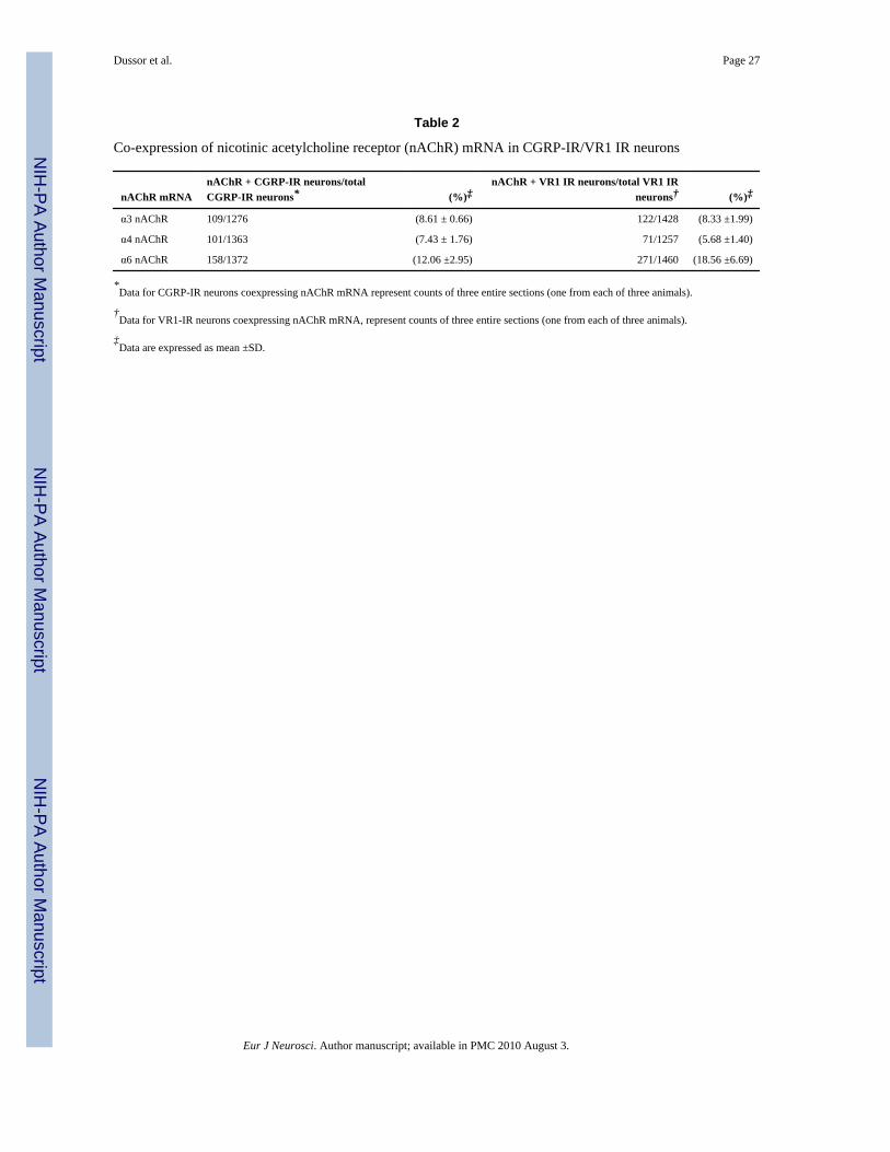
Table 2
Co-expression of nicotinic acetylcholine receptor (nAChR) mRNA in CGRP-IR/VR1 IR neurons
nAChR mRNAnAChR + CGRP-IR neurons/totalCGRP-IR neurons* (%)‡
nAChR + VR1 IR neurons/total VR1 IRneurons† (%)‡
α3 nAChR 109/1276 (8.61 ± 0.66) 122/1428 (8.33 ±1.99)
α4 nAChR 101/1363 (7.43 ± 1.76) 71/1257 (5.68 ±1.40)
α6 nAChR 158/1372 (12.06 ±2.95) 271/1460 (18.56 ±6.69)
*Data for CGRP-IR neurons coexpressing nAChR mRNA represent counts of three entire sections (one from each of three animals).
†Data for VR1-IR neurons coexpressing nAChR mRNA, represent counts of three entire sections (one from each of three animals).
‡Data are expressed as mean ±SD.
Eur J Neurosci. Author manuscript; available in PMC 2010 August 3.
NIH
-PA Author Manuscript
NIH
-PA Author Manuscript
NIH
-PA Author Manuscript
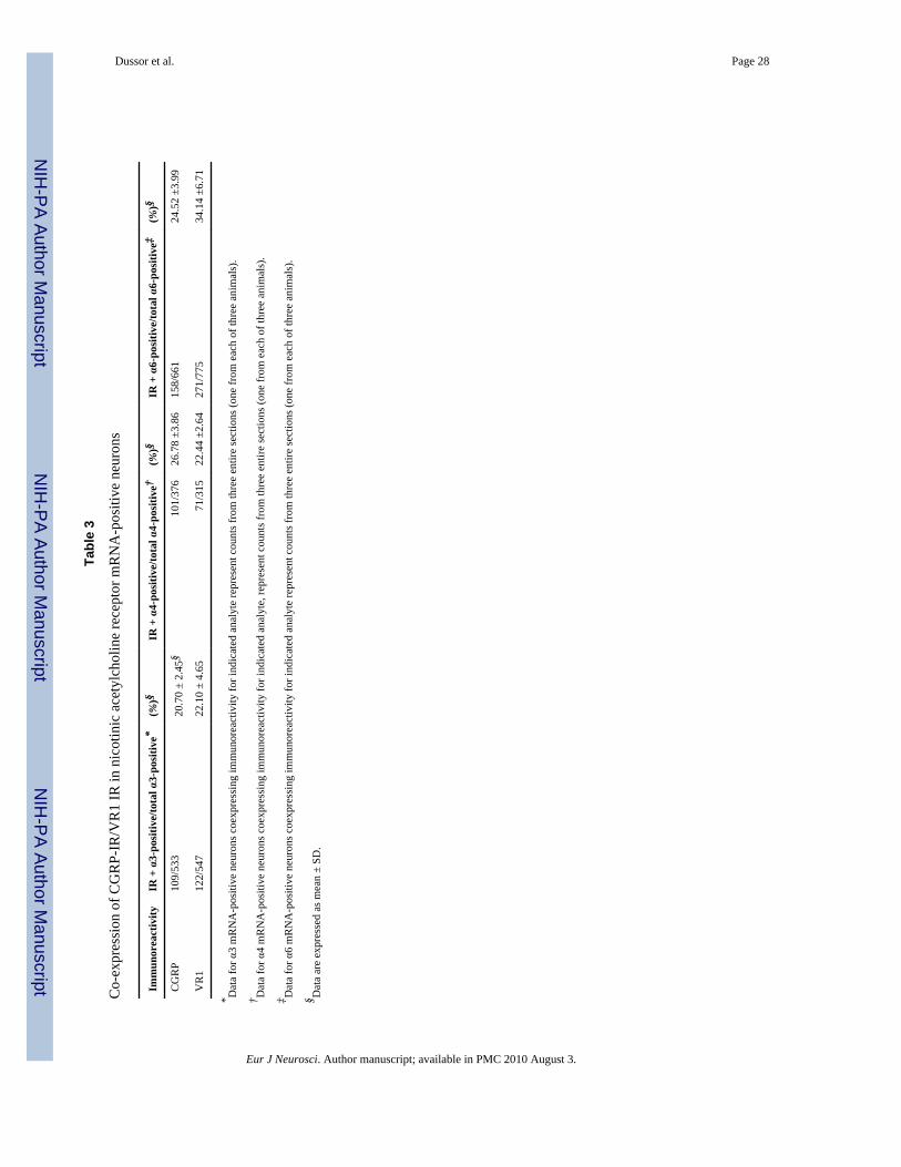
Dussor et al. Page 28
Tabl
e 3
Co-
expr
essi
on o
f CG
RP-
IR/V
R1
IR in
nic
otin
ic a
cety
lcho
line
rece
ptor
mR
NA
-pos
itive
neu
rons
Imm
unor
eact
ivity
IR +
α3-
posi
tive/
tota
l α3-
posi
tive*
(%)§
IR +
α4-
posi
tive/
tota
l α4-
posi
tive†
(%)§
IR +
α6-
posi
tive/
tota
l α6-
posi
tive‡
(%)§
CG
RP
109/
533
20.7
0 ±
2.45
§10
1/37
626
.78
±3.8
615
8/66
124
.52
±3.9
9
VR
112
2/54
722
.10
± 4.
6571
/315
22.4
4 ±2
.64
271/
775
34.1
4 ±6
.71
* Dat
a fo
r α3
mR
NA
-pos
itive
neu
rons
coe
xpre
ssin
g im
mun
orea
ctiv
ity fo
r ind
icat
ed a
naly
te re
pres
ent c
ount
s fro
m th
ree
entir
e se
ctio
ns (o
ne fr
om e
ach
of th
ree
anim
als)
.
† Dat
a fo
r α4
mR
NA
-pos
itive
neu
rons
coe
xpre
ssin
g im
mun
orea
ctiv
ity fo
r ind
icat
ed a
naly
te, r
epre
sent
cou
nts f
rom
thre
e en
tire
sect
ions
(one
from
eac
h of
thre
e an
imal
s).
‡ Dat
a fo
r α6
mR
NA
-pos
itive
neu
rons
coe
xpre
ssin
g im
mun
orea
ctiv
ity fo
r ind
icat
ed a
naly
te re
pres
ent c
ount
s fro
m th
ree
entir
e se
ctio
ns (o
ne fr
om e
ach
of th
ree
anim
als)
.
§ Dat
a ar
e ex
pres
sed
as m
ean
± SD
.
Eur J Neurosci. Author manuscript; available in PMC 2010 August 3.