Vasoactive intestinal peptide-induced neurite remodeling in human neuroblastoma SH-SY5Y cells...
14
Vasoactive intestinal peptide-induced neurite remodeling in human neuroblastoma SH-SY5Y cells implicates the Cdc42 GTPase and is independent of Ras-ERK pathway Ce ´line Alleaume a , Alain Eyche `ne b , Thomas Harnois c , Nicolas Bourmeyster c , Bruno Constantin d , Evelyne Caigneaux a , Jean-Marc Muller a , Michel Philippe a, * a Equipe Neuropeptides, Institut de Physiologie et Biologie Cellulaires, CNRS UMR 6187, Universite ´ de Poitiers-Po ˆ le Biologie Sante ´, 86022 Poitiers Cedex, France b CNRS UMR 146, Institut Curie-Recherche, Centre Universitaire, 91405 Orsay Cedex, France c Equipe Ge ´ne ´tique Mole ´culaire de l’Adressage et de la Signalisation, Institut de Physiologie et Biologie Cellulaires, CNRS UMR 6187, Universite ´ de Poitiers-Po ˆle Biologie Sante ´, 86022 Poitiers Cedex, France d Equipe Physiologie et Physiopathologie Musculaire, Institut de Physiologie et Biologie Cellulaires, CNRS UMR 6187, Universite ´ de Poitiers-Po ˆle Biologie Sante ´, 86022 Poitiers Cedex, France Received 5 April 2004, revised version received 15 June 2004 Available online 25 July 2004 Abstract Vasoactive intestinal peptide (VIP) is known to regulate proliferation or differentiation in normal and tumoral cells. SH-SY5Y is a differentiated cell subclone derived from the SK-N-SH human neuroblastoma cell line and possess all the components for an autocrine action of VIP. In the present study, we investigated the morphological changes and intracellular signaling pathways occurring upon VIP treatment of SH-SY5Y cells. VIP induced an early remodeling of cell projections: a branched neurite network spread out and prominent varicosities developed along neurites. Although activated by VIP, the Ras/ERK pathway was not required for the remodeling process. In contrast, pull- down experiments revealed a strong Cdc42 activation by VIP while expression of a dominant-negative Cdc42 prevented the VIP-induced neurite changes, suggesting an important role for this small GTPase in the process. These data provide the first evidence for a regulation of the activity of Rho family GTPases by VIP and bring new insights in the signaling pathways implicated in neurite remodeling process induced by VIP in neuroblastoma cells. D 2004 Elsevier Inc. All rights reserved. Keywords: VIP; SH-SY5Y neuroblastoma cells; Neurite remodeling; Cdc42; ERK; Ras Introduction The vasoactive intestinal peptide (VIP) belongs to the so- called secretin family of bioactive peptides, which also includes glucagon, a peptide having carboxy-terminal isoleucine/methionine (PHI/PHM) and pituitary adenylate cyclase activating polypeptide (PACAP). These neuropep- tides are widely distributed in peripheral and central nervous systems where they exert a large array of biological effects. Besides its action on exocrine secretions, smooth muscle relaxation and metabolism [1,2], and its trophic effect [3,4], VIP was also shown to regulate proliferation or differ- entiation in numerous cell lines, such as neuroblastoma, glioblastoma, pheochromocytoma, and a variety of epithe- lial tumors [5–13]. VIP acts through G-protein-coupled seven-transmembrane-domain receptors [14,15] and exerts many of its effects through activation of adenylate cyclase [14]. However, alternative signaling pathways have been 0014-4827/$ - see front matter D 2004 Elsevier Inc. All rights reserved. doi:10.1016/j.yexcr.2004.06.016 * Corresponding author. Equipe Neuropeptides, Institut de Physiologie et Biologie Cellulaires, CNRS UMR 6187, Universite ´ de Poitiers-Po ˆle Biologie Sante ´, 40 Avenue du Recteur Pineau, 86022 Poitiers Cedex, France. Fax: +33 5 49 45 39 76. E-mail address: [email protected] (M. Philippe). Experimental Cell Research 299 (2004) 511 – 524 www.elsevier.com/locate/yexcr
-
Upload
independent -
Category
Documents
-
view
0 -
download
0
Transcript of Vasoactive intestinal peptide-induced neurite remodeling in human neuroblastoma SH-SY5Y cells...
www.elsevier.com/locate/yexcr
Experimental Cell Researc
Vasoactive intestinal peptide-induced neurite remodeling in human
neuroblastoma SH-SY5Y cells implicates the Cdc42 GTPase and is
independent of Ras-ERK pathway
Celine Alleaumea, Alain Eycheneb, Thomas Harnoisc, Nicolas Bourmeysterc,
Bruno Constantind, Evelyne Caigneauxa, Jean-Marc Mullera, Michel Philippea,*
aEquipe Neuropeptides, Institut de Physiologie et Biologie Cellulaires, CNRS UMR 6187, Universite de Poitiers-Pole Biologie Sante,
86022 Poitiers Cedex, FrancebCNRS UMR 146, Institut Curie-Recherche, Centre Universitaire, 91405 Orsay Cedex, France
cEquipe Genetique Moleculaire de l’Adressage et de la Signalisation, Institut de Physiologie et Biologie Cellulaires, CNRS UMR 6187,
Universite de Poitiers-Pole Biologie Sante, 86022 Poitiers Cedex, FrancedEquipe Physiologie et Physiopathologie Musculaire, Institut de Physiologie et Biologie Cellulaires, CNRS UMR 6187,
Universite de Poitiers-Pole Biologie Sante, 86022 Poitiers Cedex, France
Received 5 April 2004, revised version received 15 June 2004
Available online 25 July 2004
Abstract
Vasoactive intestinal peptide (VIP) is known to regulate proliferation or differentiation in normal and tumoral cells. SH-SY5Y is a
differentiated cell subclone derived from the SK-N-SH human neuroblastoma cell line and possess all the components for an autocrine action
of VIP. In the present study, we investigated the morphological changes and intracellular signaling pathways occurring upon VIP treatment of
SH-SY5Y cells. VIP induced an early remodeling of cell projections: a branched neurite network spread out and prominent varicosities
developed along neurites. Although activated by VIP, the Ras/ERK pathway was not required for the remodeling process. In contrast, pull-
down experiments revealed a strong Cdc42 activation by VIP while expression of a dominant-negative Cdc42 prevented the VIP-induced
neurite changes, suggesting an important role for this small GTPase in the process. These data provide the first evidence for a regulation of
the activity of Rho family GTPases by VIP and bring new insights in the signaling pathways implicated in neurite remodeling process
induced by VIP in neuroblastoma cells.
D 2004 Elsevier Inc. All rights reserved.
Keywords: VIP; SH-SY5Y neuroblastoma cells; Neurite remodeling; Cdc42; ERK; Ras
Introduction
The vasoactive intestinal peptide (VIP) belongs to the so-
called secretin family of bioactive peptides, which also
includes glucagon, a peptide having carboxy-terminal
isoleucine/methionine (PHI/PHM) and pituitary adenylate
0014-4827/$ - see front matter D 2004 Elsevier Inc. All rights reserved.
doi:10.1016/j.yexcr.2004.06.016
* Corresponding author. Equipe Neuropeptides, Institut de Physiologie
et Biologie Cellulaires, CNRS UMR 6187, Universite de Poitiers-Pole
Biologie Sante, 40 Avenue du Recteur Pineau, 86022 Poitiers Cedex,
France. Fax: +33 5 49 45 39 76.
E-mail address: [email protected] (M. Philippe).
cyclase activating polypeptide (PACAP). These neuropep-
tides are widely distributed in peripheral and central nervous
systems where they exert a large array of biological effects.
Besides its action on exocrine secretions, smooth muscle
relaxation and metabolism [1,2], and its trophic effect [3,4],
VIP was also shown to regulate proliferation or differ-
entiation in numerous cell lines, such as neuroblastoma,
glioblastoma, pheochromocytoma, and a variety of epithe-
lial tumors [5–13]. VIP acts through G-protein-coupled
seven-transmembrane-domain receptors [14,15] and exerts
many of its effects through activation of adenylate cyclase
[14]. However, alternative signaling pathways have been
h 299 (2004) 511–524

C. Alleaume et al. / Experimental Cell Research 299 (2004) 511–524512
proposed depending on cell type (Ref. [15] for review),
including stimulation of phospholipase C, calcium mobi-
lization, and stimulation of nitric oxide synthase.
Distinct subclones were derived from the human neuro-
blastoma cell line SK-N-SH originally isolated from bone
marrow metastasis. We previously demonstrated that these
subclones possess all the components for an autocrine action
of VIP [16]. The SH-SY5Y subclone exhibits a neuroblastic
phenotype [17] and VIP treatment for 48 h was recently
shown to induce a stable neurite-like outgrowth process in
these cells [18]. In the present study, we examined the
morphological effects of a short-term VIP treatment on SH-
SY5Y cells and investigated the signaling pathways
involved.
The mitogen-activated protein kinase (MAPK) or ERK
pathway is frequently activated in differentiating cells.
However, it is now well known that this signaling pathway
is not the only one to be able to induce neurite outgrowth
and neuronal differentiation. Thus in the rat pheochromo-
cytoma cell line PC12, another well-characterized cell
model used to study neuronal differentiation pathways, both
VIP and PACAP induce neurite outgrowth [13,19–21].
However, only PACAP-induced neurite outgrowth has been
linked to ERK activation [20,21]. VIP also induces ERK1/2
activation in PC12 cells but no correlation has been reported
between ERK activation and the differentiation processes
induced by this neuropeptide [13].
It has been demonstrated that an increase in intracellular
cAMP content could activate the MAPK/ERK cascade in
particular cell types [19,22–27] and that SH-SY5Y cells
respond to cAMP by developing neurite-like extensions
[28]. In addition, we previously reported that VIP triggered
an increase in cAMP intracellular content in these cells [11].
In the present study, we therefore investigated whether VIP
could induce neurite morphological changes in SH-SY5Y
cells and, if so, whether ERK activation was required for the
process. Changes in the actin cytoskeleton are essential
determinants of neurite genesis and remodeling, and Rho
family GTPases have been assigned important roles in these
processes [29,30]. Therefore, we also investigated a possible
regulation of Rho GTPase activities by VIP.
We found that VIP induces neurite remodeling charac-
terized by development of varicosities along the neurites
and extension of the neurite network. Data suggest a key
role for Cdc42 GTPase in neurite remodeling promoted by
VIP in SH-SY5Y neuroblastoma cells, while the Ras/ERK
pathway is not required for this process.
Materials and methods
Materials
Reagents
Aprotinin, bestatin, antipaRn, pepstatin, leupeptin, N-
octylglucopyranoside, actinomycin D, and sodium orthova-
nadate were purchased from Sigma. The MEK inhibitor
U0126 was from Calbiochem. ECL+ detection system and
Glutathione SepharoseR were from Amersham Biosciences.
Immobilon-P membranes were from Millipore Corp.
Antibodies
The polyclonal anti-Cdc42 and anti-ERK1 antibodies
and the monoclonal anti-phosphorylated forms of ERKs,
anti-RhoA, and anti-cK-Ras antibodies were from Santa
Cruz. The monoclonal anti-h-tubulin antibody was from
Sigma. The monoclonal anti-Rac1 antibody was from
Transduction Laboratories and the monoclonal anti-HA1
antibody was from Bapco. The anti-mouse and anti-rabbit
peroxidase-conjugated antibodies were purchased from
Calbiochem. Goat anti-mouse antibody conjugated with
Cyanine dye Cy3 was from Caltag.
Plasmids
The plasmid encoding the GST-B-Raf-RBD protein was
described previously [26]. This construct encodes the Ras-
binding domain (amino acids 146–227) of quail B-Raf [31]
fused to the GST. GST-PAK-CD containing the Rac and
Cdc42 binding domain from Human PAK1B was produced
as described by Sander et al. [32]. GST-C21 fusion protein
containing the Rho binding region from the Rho effector
protein Rhotekin was produced according to Sander et al.
[33]. The plasmids encoding the Cdc42 wt, Cdc42 N17,
Cdc42 L61, Rac1 wt, Rac1 N17, and Rac1 L61-GFP fusion
proteins were obtained by subcloning each mutant into the
pEGFP C1 vector (Clontech) and were a kind gift from Dr
Philippe FORT (CNRS FRE 2593, CRBM, Montpellier).
The HA-Ras V12 and HA-Ras N17 plasmids were
described previously [26].
Cell culture, specific cell treatments, and transfection
SH-SY5Y neuroblastoma cells and NIH3T3 fibroblasts
were cultured in Dulbecco’s modified Eagle’s medium
supplemented with 10% fetal calf serum (FCS) and 100
UI/ml penicillin and 100 Ag/ml streptomycin (Life Tech-
nology) in a humidified atmosphere containing 5% CO2 in
air at 378C.In all experiments and before treatment, SH-SY5Y cells
were grown near confluence and then depleted of growth
factors for at least 72 h. NIH3T3 cells were depleted of
growth factors for at least 3 h. Cells were treated with VIP (at
indicated concentration), 1 AM bradykinin, 0.2 UI/ml
insulin, or 10% serum for the indicated times. Cells were
incubated with 1 AM of the MAPK/ERK kinase (MEK)
inhibitor U0126 solubilized in DMSO for at least 30 min
before addition of neuropeptide. DMSO was used as control.
To study the effects of Cdc42 and Rac1 mutants on cell
morphology, SH-SY5Y cells were seeded in 30-mm glass
dishes. After 24 h, cells were transfected in Optimem I
medium (Life Technology) with 3 Ag of DNA and 15 AlLipofectin (Invitrogen) for 8 h. Cells were then starved for

C. Alleaume et al. / Experimental Cell Research 299 (2004) 511–524 513
24 h, treated or not with 10 nM VIP for 4 h, and then
observed by confocal microscopy. To study the effect of
constitutively active Ras (HA-Ras V12) or dominant-
negative Ras (HA-Ras N17) expression on neurite remod-
eling, cells were seeded at density of 2.2 � 106 cells on
glass coverslips coated with Matrigel basement membrane
matrix (BD Biosciences). Cells were transfected and
stimulated with VIP as described above and coverslips
were then treated for observation of HA-positive cells by
immunofluorescence.
Analysis of cell morphology
To study the effect of VIP on cell morphology, SH-SY5Y
cells were seeded on glass coverslips and grown for 24 h in
complete culture medium. They were then depleted of FCS
for 24 h before treatment with 10 nM VIP and with or
without 1 AM U0126 pretreatment. Cells were fixed with
4% para-formaldehyde in PBS for 20 min at 4 8C and
observed by phase-contrast microscopy using a Zeiss
Axioplan microscope.
Indirect immunofluorescence
For immunostaining of HA1-tagged Ras-transfected
cells, cells cultured on coverslips were fixed with 4%
para-formaldehyde in TBS for 5 min at room temperature,
washed three times in TBS, and then permeabilized for 3
min in TBS containing 0.05% Triton X-100. Cells were
washed three times in TBS, blocked in TBS containing 2%
BSA for 30 min at room temperature, and exposed to
monoclonal anti-HA1 antibody diluted 1/500 in TBS
containing 1% BSA for 2 h at room temperature. A goat
anti-mouse conjugated with Cyanine dye Cy3 was used as
secondary antibody.
Confocal microscopy and data analysis
EGFP-labeled cells and HA1-Ras-transfected cells were
analyzed by laser scanning confocal microscopy (LSCM)
using a BioRad MRC 1024 ES (BioRad, Hemel Hempstead,
UK). The confocal unit was connected to an inverted
microscope (Olympus IX70, Tokyo, Japan). Fluorescence
signal collection, image construction, and scaling were
performed through the control software (Lasersharp 3.2;
BioRad).
ERK activation assay
Cells were grown in 25-cm2 flasks, serum starved for
72 h, and treated with agents as indicated. Following
treatments, cells were lysed in electrophoresis sample buffer
containing 0.06 M Tris (pH 6.8), 2% SDS, 4.5 M urea, and
5% h-Mercaptoethanol.
Proteins (100 Ag) were separated on 12% SDS-poly-
acrylamide gels and transferred to nylon membranes
(Immobilon-P). ERK activation was analyzed by immuno-
blotting using an activation-specific antibody recognizing
the dual phosphorylated forms of both p42 ERK2 and p44
ERK1. Total protein levels were assessed by Western
blotting using either a monoclonal anti-h-tubulin antibody
or a polyclonal anti-ERK antibody that recognizes both
ERK1 and ERK2.
Pull-down assay for detection of activated Ras
Cells were grown in 100-mm dishes, serum starved for
72 h, and treated with agents as indicated. Cells were then
washed with ice-cold PBS and lysed for 20 min on ice in
buffer containing 50 mM Tris pH 7.5, 15 mM NaCl, 20 mM
MgCl2, 5 mM EGTA, 1% Triton X-100, 1% N-octylgluco-
pyranoside, 1 Ag/ml bestatin, 1 Ag/ml antipain, 1 Ag/ml
pepstatin, 5 Ag/ml leupeptin, 1 mM 4-(2-aminoethyl)-
benzene-sulfonyl fluoride (AEBSF). Lysates were clarified
by centrifugation at 13,000 rpm for 20 min at 48C. Proteinextracts (700 Ag) were incubated with 30 Al GST-B-Raf-RBD fusion protein precoupled to glutathione-Sepharose
beads for 2 h at 48C. GST-B-Raf-RBD fusion protein used
to trap active Ras (GTP-bound form) was produced and
immobilized on glutathione-Sepharose beads as described
previously [26]. After incubation, beads were washed three
times in lysis buffer and resuspended in Laemmli sample
buffer. Samples were analyzed by electrophoresis on SDS-
polyacrylamide gel (15%) followed by transfer to nylon
membranes (Immobilon-P). Affinity-purified activated Ras
was detected by immunoblotting using a monoclonal anti-
K-Ras antibody. Total Ras level was evaluated by Western
blotting of whole cell extracts (100-Ag proteins) using the
same antibody.
Pull-down assays for detection of activated Rac1, Cdc42,
and RhoA
Following treatments, cells were lysed for 20 min on ice
in buffer containing 50 mM Tris pH 7.4, 150 mM NaCl, 5
mM MgCl2, 0.05% NP-40, 0.25% sodium deoxycholate,
0.25% Triton X-100, 0.025% SDS, 10 Ag/ml aprotinin, 10
Ag/ml leupeptin, 1 Ag/ml bestatin, 1 Ag/ml antipain, 1 Ag/ml
pepstatin, 1 mM AEBSF. Lysates were clarified by
centrifugation at 13,000 rpm for 5 min at 48C. Proteins(700 Ag) were incubated with 30 Al GST fusion protein
precoupled to glutathione-Sepharose beads overnight at
48C. GST-PAK-CD fusion protein was used to trap Rac1
and Cdc42 in their GTP-bound active form whereas GST-
C21 fusion protein was used to trap active RhoA. After
incubation, beads were washed three times in buffer
containing 50 mM Tris pH 7.4, 150 mM NaCl, 5 mM
MgCl2, and 0.05% NP-40, and resuspended in Laemmli
sample buffer. Samples were analyzed by electrophoresis on
SDS-polyacrylamide gel (15%) followed by transfer to
nylon membranes. Affinity-purified activated Rac1, Cdc42,
and RhoA were detected by immunoblotting using mono-

C. Alleaume et al. / Experimental Cell Research 299 (2004) 511–524514
clonal anti-Rac1, polyclonal anti-Cdc42, and monoclonal
anti-RhoA antibodies. Total Rac1, Cdc42, and RhoA levels
were evaluated by Western blotting of whole cell extracts
(50- or 100-Ag proteins) using the same antibodies.
Results
VIP induces early neurite morphological changes in
SH-SY5Y neuroblastoma cells
To investigate the effect of VIP, serum-starved SH-SY5Y
cells were treated with 10 nM VIP for 4 h and morphological
changes were observed by phase-contrast microscopy. Con-
trol cells already presented extended slender and straight
neurites with few branches (Fig. 1). After 4 h of treatment,
VIP induced an extension of the neuritic network between
adjacent cells and branching appeared more pronounced (Fig.
1). In addition, numerous prominent varicosities formed
Fig. 1. VIP induces neurite morphological changes in SH-SY5Y neuro-
blastoma cells. Serum-starved SH-SY5Y cells were unstimulated (CTRL)
or treated with 10 nM VIP for 4 h (VIP). The developing morphology was
observed by phase contrast microscopy. Black and open arrows indicate
varicosities and neurite network, respectively. Scale bars represent 50 Am.
Micrographs are representative of four independent experiments.
along the neurites. The varicosities were varying in size with
largest ones evoking neurite swelling. These effects were
rapid, occurring in less than 3 h of treatment and remained
visible after 6 h. However, they were transient and reversible
because no difference remained observable between control
and VIP-stimulated cells after 24 h of VIP treatment.
VIP activates the small G protein Ras
To determine whether a Ras-mediated signaling pathway
may be triggered by VIP treatment, we investigated the
potential activation of this small GTPase by the neuropeptide.
To this aim, a Ras pull-down assay was performed using the
Ras binding domain of B-Raf fused to the GST protein (GST-
B-Raf-RBD) to trap Ras in its GTP-bound active form. The
pool of active Ras was then detected by Western blotting
using an anti-K-Ras antibody (Fig. 2). The amount of active
Ras increased as early as 5 min after the onset of the 10-nM
VIP treatment. Activation was sustained because it remained
detectable after 60 min of neuropeptide treatment.
VIP induces a time- and concentration-dependent ERKs
activation
We next examined the ability of VIP to induce ERK
activation in SH-SY5Y cells. Starved cells were treated with
10 nM VIP for 5 min and up to 24 h (Fig. 3A). The activation
of MAP kinases was estimated using a specific antibody
recognizing the phosphorylated thus activated forms of ERKs
(Fig. 3A, upper panel). This kinetic study revealed that the
two MAP kinases, ERK1 and ERK2, were simultaneously
activated by VIP. Activation was first observed after 15 min
of treatment and appeared to be maximal after 60min. Longer
stimulation led to a decrease of the activation intensity, but
MAP kinase phosphorylation was still detectable after 24 h.
The dose-dependent effect of VIP on ERK activation was
assessed using concentrations of neuropeptide ranging from
0.01 to 100 nM for 60 min (Fig. 3B, upper panel). ERK
activation was reached using at least 10 nM VIP, lower
concentrations tested being inefficient.
To analyze the requirement for MEK in ERK activation, a
specific MEK inhibitor was used in the VIP-induced ERK
activation assay. Cells were pretreated for 30 min with 1 AMU0126 (Fig. 3C) before addition or in absence of 10 nM VIP
for 60 min. VIP induced an increase in ERK activation as
compared to control cells (Fig. 3C, upper panel) and U0126
completely inhibited VIP-induced MAP kinases activation.
Neurite remodeling process induced by VIP does not require
the Ras/ERK pathway
Because both Ras and ERK were activated by VIP, we
looked for the requirement of this pathway in the morpho-
logical changes induced by the neuropeptide. To study the
role of Ras on neurite remodeling, SH-SY5Y cells were
transfected with EGFP alone or with either constitutively

Fig. 2. VIP activates the small G protein Ras. SH-SY5Y cells were treated
with 10 nM VIP for the indicated times. Ctrl: unstimulated cells. Pull-down
experiments were performed on cell lysates using the GST-B-Raf RBD
fusion protein. Activated Ras (Ras-GTP) was detected by Western blotting
using anti K-Ras antibody (upper panel). Ras activation was quantified by
comparison with the control. Total Ras content was estimated by blotting
equal quantity of cell lysates with the same antibody (lower panel). Data
shown are representative of three independent experiments.
C. Alleaume et al. / Experimental Cell Research 299 (2004) 511–524 515
active Ras V12 or dominant-negative Ras N17 fused to the
HA1 epitope. Cells were then treated or not with 10 nM VIP
for 4 h. Immunofluorescence was performed using an anti-
Fig. 3. VIP induces a time- and concentration-dependent ERK activation that requi
nM VIP (A) or with different concentrations of neuropeptide for 60 min (B). Fe
activation. Cells were also pretreated with 1 AM of the MEK inhibitor U0126 for 30
(C). Ctrl: unstimulated cells. Cells were then lysed in electrophoresis sample buffer
anti phospho-ERKs antibody (A, B, and C: upper panels). Anti-h-tubulin antibodythe same blot to verify protein amounts in each sample. Data presented are repre
HA1 antibody and HA1-Ras-transfected cells were analyzed
by LSCM (Fig. 4A). Control cells transfected with EGFP
extended straight unbranched neurites (panel a). VIP-treated
cells developed a winding and branched neuritic tree with
short lateral ramifications and several varicosities along the
neurites (panel b). No effect on morphology was observed in
Ras N17-transfected cells (panel c) when compared to cells
transfected with EGFP. In addition, expression of the
dominant-negative mutant of Ras did not impair VIP-induced
neurite remodeling (panel d), indicating that Ras was not
involved in this process. Transfection of constitutively active
Ras (Ras V12) significantly altered cell morphology (panel
e), inducing neurite elongation and increasing growth cone
number. VIP stimulation of Ras V12-transfected cells
amplified the neuritic tree complexity especially in growth
cones that extended a profusion of growth processes
displaying numerous varicosities (panel f). Higher magnifi-
cation revealed that VIP also induced short and thin lateral
outgrowth processes along the neurites as well as numerous
filopodial-like protrusions emerging from the growth cones
(panel g) and around the cell body (panel h). To investigate
res MEK activity. SH-SY5Y cells were stimulated for various times with 10
tal calf serum (10% FCS for 5 min) was used as positive control of ERK
min before addition of 10 nM VIP for 1 h or in the absence of neuropeptide
. ERK phosphorylation/activation was assessed by Western blotting using an
(A and B: lower panels) or anti-ERK antibody (C: lower panel) was used on
sentative of three independent experiments.

C. Alleaume et al. / Experimental Cell Research 299 (2004) 511–524516
the role of ERK, serum-free cell cultures were pretreated with
1 AMof the MEK inhibitor U0126 for 30 min before addition
of 10 nMVIP or in absence of the neuropeptide (Fig. 4B). The
MEK inhibitor effect was examined after 4 h of VIP
treatment. U0126 alone had no effect on cell morphology
because treated cells presented extended long neurites with
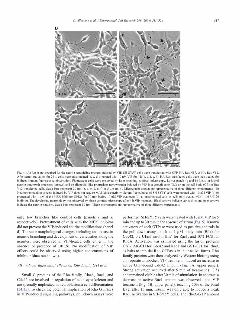
Fig. 4. (A) Ras is not required for the neurite remodeling process induced by VIP. SH-SY5Y cells were transfected with GFP, HA-Ras N17, or HA-Ras V12.
After serum starvation for 24 h, cells were unstimulated (a, c, e) or treated with 10 nM VIP for 4 h (b, d, f, g, h). HA-Ras-transfected cells were then treated for
indirect immunofluorescence observation. Fluorescent cells were observed by laser scanning confocal microscopy. Lower panels (g and h) focus on lateral
neurite outgrowth processes (arrows) and on filopodial-like protrusions (arrowheads) induced by VIP in a growth cone (GC) or on the cell body (CB) of Ras
V12-transfected cells. Scale bars represent 20 Am (a, b, c, d, e, f) or 5 Am (g, h). Micrographs shown are representative of three different experiments. (B)
Neurite remodeling process induced by VIP does not require MAP kinase activity. Serum-free cultures of SH-SY5Y cells were treated with 10 nM VIP (b) or
pretreated with 1 AM of the MEK inhibitor U0126 for 30 min before 10 nM VIP treatment (d). a: unstimulated cells. c: cells only treated with 1 AM U0126
inhibitor. The developing morphology was observed by phase contrast microscopy after 4 h VIP treatment. Black arrows indicate varicosities and open arrows
indicate the neurite network. Scale bars represent 50 Am. These micrographs are representative of three different experiments.
C. Alleaume et al. / Experimental Cell Research 299 (2004) 511–524 517
only few branches like control cells (panels c and a,
respectively). Pretreatment of cells with the MEK inhibitor
did not prevent the VIP-induced neurite modifications (panel
d). The samemorphological changes, including an increase in
neuritic branching and development of varicosities along the
neurites, were observed in VIP-treated cells either in the
absence or presence of U0126. No modification of VIP
effects could be observed using higher concentrations of
inhibitor (data not shown).
VIP induces differential effects on Rho family GTPases
Small G proteins of the Rho family, RhoA, Rac1, and
Cdc42 are involved in regulation of actin cytoskeleton and
are specially implicated in neuroblastoma cell differentiation
[34,35]. To check the potential implication of Rho GTPases
in VIP-induced signaling pathways, pull-down assays were
performed. SH-SY5Y cells were treated with 10 nMVIP for 5
min and up to 30min in the absence of serum (Fig. 5). Known
activators of each GTPase were used as positive controls in
the pull-down assays, such as 1 AM bradykinin (Bdk) for
Cdc42, 0.2 UI/ml insulin (Ins) for Rac1, and 10% FCS for
RhoA. Activation was estimated using the fusion proteins
GST-PAK-CD for Cdc42 and Rac1 and GST-C21 for RhoA
as baits to trap the Rho GTPases in their active forms. Rho
family proteins were then analyzed byWestern blotting using
appropriate antibodies. VIP treatment induced an increase in
active GTP-bound Cdc42 amount (Fig. 5A, upper panel).
Strong activation occurred after 5 min of treatment (�3.5)
and remained visible after 30min of stimulation. In contrast, a
decrease in active Rac1 amount was observed upon VIP
treatment (Fig. 5B, upper panel), reaching 50% of the basal
level after 15 min. Insulin was only able to induce a weak
Rac1 activation in SH-SY5Y cells. The RhoA-GTP amount

Fig. 5. VIP induces differential effects on Rho family GTPases. SH-SY5Y cells were treated with 10 nM VIP for the indicated times, 1 AM bradykinin (Bdk)
for 5 min, 0.2 UI/ml insulin (Ins) for 5 min. NIH3T3 cells used as control were treated with 10% fetal calf serum (FCS) for 10 min. Ctrl: unstimulated cells.
Cell lysates were subjected to pull-down experiments using the GST-PAK-CD fusion protein (A and B) or GST-C21 fusion protein (C). Activated Cdc42
(Cdc42-GTP), Rac1 (Rac1-GTP), and RhoA (RhoA-GTP) were detected by Western blotting using specific Cdc42, Rac1, or RhoA antibodies, respectively (A,
B, and C: upper panels). Activation or inhibition was quantified by comparison with the control. Total Cdc42, Rac1, and RhoA contents were estimated by
blotting equal quantity of cell lysates with the same antibodies (A, B, and C: lower panels). Data presented are representative of three independent experiments.
C. Alleaume et al. / Experimental Cell Research 299 (2004) 511–524518
first increased after 5 min of treatment and then returned to its
basal level after 15 min of VIP treatment (Fig. 5C, upper
panel). NIH3T3 cells were used as positive control of RhoA
activation by FCS because serum was not able to induce
activation of this GTPase in SH-SY5Y cells.
Dominant-negative or constitutively active mutants of
Cdc42 and Rac1 block the neurite morphological
modifications induced by VIP
To investigate the potential role of Cdc42 and Rac1
GTPases in the morphological processes induced by VIP,
Fig. 6. Dominant-negative or constitutively active mutants of Cdc42 and Rac1 bl
cells. SH-SY5Y cells were transfected with GFP, GFP-Cdc42 wt, GFP-Cdc42 N17
L61 (B). After serum starvation for 24 h, cells were treated with 10 nM VIP for 4
shown are representative of three independent experiments and illustrate the pred
SH-SY5Y cells were transfected with positive and negative
mutants of these GTPases fused to EGFP. Cells were then
treated or not with 10 nM VIP for 4 h and analyzed by
LSCM (Figs. 6A and B). Two major phenotypes could be
distinguished: bipolar cells with axon-like process and
growth cone or non-polarized cells characterized by neurites
all around their cell body. Control cells transfected with
pEGFP exhibited some long and slender neurites with few
branches. VIP treatment (GFP + VIP) induced an increase in
the complexity of the neuritic network, mostly characterized
by short lateral ramifications growing, an increase of the
occurrence of growth cones (see Fig. 6A), and the develop-
ock the neurite morphological modifications induced by VIP in SH-SY5Y
, or GFP-Cdc42 L61 (A), GFP, GFP-Rac1 wt, GFP-Rac1 N17, or GFP-Rac1
h and then observed by laser scanning confocal microscopy. Micrographs
ominant phenotypes observed in cell cultures. Scale bars represent 20 Am.

C. Alleaume et al. / Experimental Cell Research 299 (2004) 511–524 519

Fig. 6 (continued ).
C. Alleaume et al. / Experimental Cell Research 299 (2004) 511–524520

C. Alleaume et al. / Experimental Cell Research 299 (2004) 511–524 521
ment of dendritic tree-like structures (see Fig. 6B). Depend-
ing on the original phenotype, VIP-induced remodeling was
varying: bipolar cells were engaged in growth cone
formation and short lateral branching, while nonpolarized
cells developed a widespread arborization with numerous
varicosities. GFP-Cdc42 and GFP-Rac1 wild-type-express-
ing cells (Cdc42 wt and Rac1 wt) displayed a morphology
quite similar to that of pEGFP-transfected cells. Following
VIP stimulation, these transfected cells (Cdc42 wt + VIP
and Rac1 wt + VIP) exhibited enhanced branching as
mentioned above. New phenotypes were observed when
SH-SY5Y cells were transfected with dominant-negative or
constitutively active mutants of Cdc42 and Rac1. Both
GFP-Cdc42 N17- and GFP-Rac1 N17-expressing cells
showed similar phenotypes characterized by the presence
of several long and threadlike processes with few branching
all around the cell body. Interestingly, no additional
morphological effect was observed after VIP stimulation
(Cdc42 N17 + VIP, Rac1 N17 + VIP). In contrast, GFP-
Cdc42 L61- and GFP-Rac1 L61-transfected cells developed
large lamellipodia. It should be noticed that only few cells
harbored lamellipodia and neurites simultaneously. VIP
treatment (Cdc42 L61 + VIP, Rac1 L61 + VIP) did not
counteract lamellipodia formation and classical VIP-induced
remodeling was not observed.
Discussion
The functions of the ubiquitous neuropeptide VIP as a
regulator of proliferation or differentiation in normal and
tumoral cells are well known. However, the intracellular
signaling pathways induced by this neuropeptide are not yet
clearly elucidated [5–11,36,37]. In the present work, we
used the human neuroblastoma cell line SH-SY5Y, a well-
characterized model of neuronal differentiation, to examine
the early morphological effects of VIP and to investigate the
signaling pathways triggered by neuropeptide stimulation.
In this way we expected to gain new information about the
fine regulation of neuronal plasticity by VIP neuropeptide.
Within 4 h, a remodeling of SH-SY5Y cell projections
was induced by 10 nM VIP, namely an extension of the
neurite network between adjacent cells and development of
varicosities varying in size along the neurites. EGFP
expression coupled to laser scanning confocal microscopy
analysis allowed a fine resolution of these modifications,
especially the formation of new neurite branching, which
markedly increased the complexity of the dendritic tree.
These early induced processes were transient because they
were no longer observed after 24 h of neuropeptide
stimulation. Indeed, this remodeling should not be consid-
ered as a differentiation process per se but only as
morphological changes underlying neuronal plasticity. In
this way, it differed from the stable neuritogenesis produced
by VIP in these cells, which needed both serum starvation
for 6–7 days and a 48-h treatment with the neuropeptide
[18]. A similar neurite remodeling could be produced in the
presence of 10 AM forskolin, an activator of adenylate
cyclase (data not shown). Since we previously reported that
VIP triggered an increase in cAMP intracellular content in
SH-SY5Y cells [11], it is postulated that the VIP-induced
morphological changes may be mediated by a cAMP
signaling pathway. Interestingly, it should be noted that a
cAMP-activated pathway regulating neuronal differentiation
has been recently identified in SH-SY5Y cells [28].
In the pheochromocytoma cell line PC12, which derives
from neural crest-like SH-SY5Y cells, both VIP and
PACAP have been previously shown to induce neurito-
genesis [13,19–21] and to activate MAP kinases [20,21,38].
Therefore, we first investigated whether Ras and ERKs
could be potential targets of VIP’s effect in SH-SY5Y
neuroblastoma cells. Using pull-down experiments, we
found that Ras was rapidly but weakly activated. Concom-
itantly, VIP also triggered sustained ERK activation. We
then investigated whether Ras or MAPK could be required
for the VIP-mediated neurite remodeling process. We found
that expression of Ras N17-dominant negative mutant had
no effect on neurite morphology in SH-SY5Y cells and did
not prevent the VIP effect, strongly suggesting that Ras
activity was not required for neurite remodeling. In contrast,
the constitutively active Ras V12 mutant induced specific
changes, namely neurite elongation and an increase in the
growth cone number. In addition, the overall neuritic-tree
complexity was even more pronounced when Ras V12-
transfected cells were stimulated with VIP. Therefore, these
findings further confirm that the VIP-mediated neurite
remodeling is likely not dependent on Ras activity.
Similarly, the MEK inhibitor U0126 completely inhibited
the VIP-induced ERK activation but did not modify the
morphological changes induced by the neuropeptide.
Indeed, extension of the neurite network and formation of
varicosities along the neurites induced by VIP treatment
were observed both in the absence and presence of the
inhibitor. This result suggests that VIP-induced neurite
remodeling does not require ERK activity. Interestingly,
Olsson and Nanberg [39] have investigated the role of ERK
and Ras during neuronal differentiation of SH-SY5Y cells
after phorbol ester treatment. As observed in the present
study, they reported a sustained activation of ERK and
pointed out that expression of constitutively active Ras V12
induced a pronounced formation of neurites with growth
cones. However, they demonstrated that ERK activation was
only involved in the gene induction process but was not
required for neurite outgrowth and that the mechanism by
which phorbol ester stimulated neurite formation was
distinct from that of Ras-induced outgrowth. In this way,
our results represent another example of neurite morpho-
logical remodeling that proved to be independent of both
Ras and ERK activities.
Reorganization of both the actin cytoskeleton and
microtubules is an essential phenomenon accompanying
neuronal changes such as neurite development or collapse.

C. Alleaume et al. / Experimental Cell Research 299 (2004) 511–524522
Rho family GTPases including RhoA, Rac1, and Cdc42
play key roles in the regulation of these processes
[29,30,34,40]. In neuroblastoma and PC12 cell lines,
Cdc42 and Rac1 control neurite extension [41–45], and
conversely, RhoA competes with Cdc42 and Rac1 in this
process [42,43,46]. Cdc42 and Rac1 also promoted for-
mation of filopodia and lamellipodia influencing growth
cone morphology and guidance [42]. Induction of neurite
remodeling by VIP in SH-SY5Y cells led us to look for a
potential implication of Rho family GTPases. Using pull-
down assays, we demonstrate that VIP induced a rapid and
strong activation of Cdc42. In contrast, the neuropeptide
appeared to inhibit the small GTPase Rac1. Considering the
major Cdc42 activation observed, we hypothesized that this
GTPase could be responsible for the remodeling triggered
by VIP stimulation and therefore we used constitutively
active or dominant-negative mutants of Cdc42 and Rac1 to
test this hypothesis. Overexpression of wild-type GTPases
did not significantly modify the phenotype observed in
GFP-transfected cells both in the absence and presence of
VIP stimulation. Considering the increase in the expression
level of GTPases in transfected cells, these observations
would suggest the existence of a strong intrinsic regulation
of Cdc42 and Rac1 activities in SH-SY5Y neuroblastoma
cells. In contrast, overexpression of dominant-negative or
constitutively active Cdc42 or Rac1 proteins resulted in two
peculiar phenotypes. Dominant-negative Cdc42/Rac1-trans-
fected cells displayed long and threadlike neurites all around
the cell body, whereas constitutively active Cdc42/Rac1-
transfected cells acquired lamellipodia structures. Surpris-
ingly, overexpression of constitutively active Cdc42 induced
a lamellipodial phenotype, evoking a typical Rac-dependent
effect and suggesting a sequential activation of this GTPase
downstream of Cdc42. This finding was not in accordance
with VIP effect because VIP stimulation of SH-SY5Y cells
resulted in Cdc42 activation only, but rather seemed to
inhibit Rac1. According to the well-known signaling
hierarchies that exist between the activation state of the
Rho family GTPases [47], our findings strongly suggested
that the lamellipodium formation observed in constitutively
active Cdc42-transfected cells would result from Cdc42-
triggered activation of Rac1.
A major finding was that transfection of SH-SY5Y cells
with dominant-negative N17 mutant of Cdc42 completely
blocked VIP effects, suggesting that Cdc42 is indeed
required for VIP-induced neurite remodeling. Consistently,
we observed a substantial activation of Cdc42 in cells
stimulated with VIP as well as VIP-induced growth of
typical Cdc42-dependent filopodial protrusions, further
suggesting a fundamental role for this GTPase.
Although VIP did not induce Rac1 activation, the
dominant-negative mutant of Rac1 also seemed to prevent
the neuropeptide effect on neurites. One possibility would
be that a permissive and reduced Rac1 activity is yet needed
to enable the VIP-mediated neurite changes. Such a
requirement for low level of active Rac1 has been already
demonstrated for Ras-transformed cells to proliferate [48].
In this hypothesis, further activation of Rac1 by VIP would
not be required for neurite remodeling, which is consistent
with the Rac1 pull-down assay data. Another hypothesis
would be that a turnover of the two GTPases is needed to
accomplish the remodeling program of VIP. In fact,
constitutive activation or inhibition of Rac1 and Cdc42
induces particular phenotypes that prevent any further VIP
effect on cellular morphology.
Taken together, our findings bring new insights in the
VIP signaling pathways and the VIP-associated remodeling
process in neuroblastoma cells. VIP was able to induce Ras
and ERK activation in SH-SY5Y cells, but neither Ras nor
ERK was required for VIP-induced morphological changes.
In addition, we showed that VIP induces a strong activation
of Cdc42. To our knowledge, this is the first report showing
a regulatory effect of a neuropeptide of the VIP family on
the activity of Rho GTPases and demonstrating that Cdc42
may play a key role in the cytoskeleton reorganization
accompanying VIP-induced neurite remodeling.
Acknowledgments
We would like to thank Dr R. Busca and C. Peyssonnaux
for their help with pull-down assays, A. Cantereau for
technical assistance with confocal microscopy, and J.
Habrioux for desktop publishing. We are grateful to Dr. J.
Collard for GST-PAK-CD and GST-C21 plasmids and Dr. P.
Fort for pEGFP C1 expression vectors. This work was
supported by CNRS and C. Alleaume was the recipient of
PhD fellowship from the bRegion Poitou-CharentesQ.
References
[1] S.I. Said, Vasoactive intestinal peptide, J. Endocrinol. Invest. 9 (1986)
191–200.
[2] S.I. Said, Vasoactive intestinal polypeptide, biologic role in health and
disease, Trends Endocrinol. Metab. 2 (1991) 107–112.
[3] P. Gressens, J.M. Hill, I. Gozes, M. Fridkin, D.E. Brenneman, Growth
factor function of vasoactive intestinal peptide in whole cultured
mouse embryos, Nature 362 (1993) 155–158.
[4] J.A. Waschek, Vasoactive intestinal peptide: an important trophic
factor and developmental regulator? Dev. Neurosci. 17 (1995) 1–7.
[5] M.S. O’Dorisio, D.J. Fleshman, S.J. Qualman, T.M. O’Dorisio,
Vasoactive intestinal peptide: autocrine growth factor in neuro-
blastoma, Regul. Pept. 37 (1992) 213–226.
[6] J.C. Pence, N.A. Shorter, The autocrine function of vasoactive
intestinal peptide on human neuroblastoma cell growth and differ-
entiation, Arch. Surg. 128 (1993) 591–595.
[7] Y. Wollman, G. Lilling, M.N. Goldstein, M. Fridkin, I. Gozes,
Vasoactive intestinal peptide: a growth promoter in neuroblastoma
cells, Brain Res. 624 (1993) 339–341.
[8] V. Lelievre, L. Becq-Giraudon, A.C. Meunier, J.M. Muller, Switches
in the expression and function of PACAP and VIP receptors during
phenotypic interconversion in human neuroblastoma cells, Neuro-
peptides 30 (1996) 313–322.
[9] M. Hoshino, M. Li, L.Q. Zheng, M. Suzuki, T. Mochizuki, N.
Yanaihara, Pituitary adenylate cyclase activating peptide and vaso-

C. Alleaume et al. / Experimental Cell Research 299 (2004) 511–524 523
active intestinal polypeptide: differentiation effects on human neuro-
blastoma NB-OK-1 cells, Neurosci. Lett. 159 (1993) 35–38.
[10] P. Vertongen, I. Camby, F. Darro, R. Kiss, P. Robberecht, VIP and
pituitary adenylate cyclase-activating polypeptide (PACAP) have an
antiproliferative effect on the T98G human glioblastoma cell line
through interaction with VIP 2 receptor, Neuropeptides 30 (1996)
491–496.
[11] J.M. Muller, V. Lelievre, L. Becq-Giraudon, A.C. Meunier, VIP as a
cell-growth and differentiation neuromodulator role in neurodevelop-
ment, Mol. Neurobiol. 10 (1995) 115–134.
[12] J.C. Pence, N.A. Shorter, In vitro differentiation of human neuro-
blastoma cells caused by vasoactive intestinal peptide, Cancer Res. 50
(1990) 5177–5183.
[13] R.A. Colbert, D. Balbi, A. Johnson, J.A. Bailey, J.M. Allen,
Vasoactive intestinal peptide stimulates neuropeptide Y gene expres-
sion and causes neurite extension in PC12 cells through independent
mechanisms, J. Neurosci. 14 (1994) 7141–7147.
[14] A.J. Harmar, A. Arimura, I. Gozes, L. Journot, M. Laburthe, J.R.
Pisegna, S.R. Rawlings, P. Robberecht, S.I. Said, S.P. Sreedharan,
S.A. Wank, J.A. Waschek, International Union of Pharmacology.
XVIII. Nomenclature of receptors for vasoactive intestinal peptide and
pituitary adenylate cyclase-activating polypeptide, Pharmacol. Rev. 50
(1998) 265–270.
[15] M. Laburthe, A. Couvineau, Molecular pharmacology and structure
of VPAC receptors for VIP and PACAP, Regul. Pept. 108 (2002)
165–173.
[16] J.M. Muller, S.J. Lolait, V.C. Yu, W. Sadee, J.A. Waschek, Functional
vasoactive intestinal polypeptide (VIP) receptors in human neuro-
blastoma subclones that contain VIP precursor mRNA and release
VIP-like substances, J. Biol. Chem. 264 (1989) 3647–3650.
[17] J.L. Biedler, L. Helson, B.A. Spengler, Morphology and growth,
tumorigenicity, and cytogenetics of human neuroblastoma cells in
continuous culture, Cancer Res. 33 (1973) 2643–2652.
[18] C. Heraud, S. Hilairet, J.M. Muller, J.F. Leterrier, C. Chadeneau,
Neuritogenesis induced by vasoactive intestinal peptide, pituitary
adenylate cyclase-activating polypeptide, and peptide histidine
methionine in SH-SY5Y cells is associated with regulated expression
of cytoskeleton mRNAs and proteins, J. Neurosci. Res. 75 (2004)
320–329.
[19] M. Frfdin, P. Peraldi, E. Van Obberghen, Cyclic AMP activates the
mitogen-activated protein kinase cascade in PC12 cells, J. Biol. Chem.
269 (1994) 6207–6214.
[20] A.P. Barrie, A.M. Clohessy, C.S. Buensuceso,M.V. Rogers, J.M. Allen,
Pituitary adenylyl cyclase-activating peptide stimulates extracellular
signal-regulated kinase 1 or 2 (ERK1/2) activity in a Ras-independent,
mitogen-activated protein Kinase/ERK kinase 1 or 2-dependentmanner
in PC12 cells, J. Biol. Chem. 272 (1997) 19666–19671.
[21] P. Lazarovici, H. Jiang, D. Fink Jr., The 38-amino-acid form of
pituitary adenylate cyclase-activating polypeptide induces neurite
outgrowth in PC12 cells that is dependent on protein kinase C and
extracellular signal-regulated kinase but not on protein kinase A
nerve growth factor receptor tyrosine kinase, p21ras G protein, and
pp60c-src cytoplasmic tyrosine kinase, Mol. Pharmacol. 54 (1998)
547–558.
[22] M.G. Seidel, M. Klinger, M. Freissmuth, C. Holler, Activation of
mitogen-activated protein kinase by the A2A-adenosine receptor via a
rap1-dependent and via a p21ras dependent pathway, J. Biol. Chem.
274 (1999) 25833–25841.
[23] S.W. Young, M. Dickens, J.M. Tavare, Differentiation of PC12 cells in
response to a cAMP analogue is accompanied by sustained activation
of mitogen-activated protein kinase. Comparison with the effects of
insulin, growth factors and phorbol esters, FEBS Lett. 338 (1994)
212–216.
[24] W. Englaro, R. Rezzonico, M. Durand-Clement, D. Lallemand, J.P.
Ortonne, R. Ballotti, Mitogen-activated protein kinase pathway and
AP-1 are activated during cAMP-induced melanogenesis in B-16
melanoma cells, J. Biol. Chem. 270 (1995) 24315–24320.
[25] M.R. Vossler, H. Yao, R.D. York, M.G. Pan, C.S. Rim, P.J.S. Stork,
cAMP activates MAP kinase and Elk-1 through a B-Raf and Rap1-
dependent pathway, Cell 89 (1997) 73–82.
[26] R. Busca, P. Abbe, F. Mantoux, E. Aberdam, C. Peyssonnaux, A.
Eychene, J.P. Ortonne, R. Ballotti, Ras mediates the cAMP-dependent
activation of extracellular signal-regulated kinases (ERKs) in mela-
nocytes, EMBO J. 19 (2000) 2900–2910.
[27] C. Peyssonnaux, A. Eychene, The Raf/MEK/ERK pathway: new
concepts of activation, Biol. Cell. 93 (2001) 53–62.
[28] S. Sanchez, C. Jimenez, A.C. Carrera, J. Diaz-Nido, J. Avila, F.
Wandosell, A cAMP-activated pathway, including PKA and PI3K,
regulates neuronal differentiation, Neurochem. Int. 44 (2004)
231–242.
[29] D.J. Mackay, C.D. Nobes, A. Hall, The Rho’s progress: a potential
role during neuritogenesis for the Rho family of GTPases, Trends
Neurosci. 18 (1995) 496–501.
[30] E. Tanaka, J. Sabry, Making the connection: cytoskeletal rearrange-
ments during growth cone guidance, Cell 83 (1995) 171–176.
[31] C. Papin, A. Denouel-Galy, D. Laugier, G. Calothy, A. Eychene,
Modulation of kinase activity and oncogenic properties by alternative
splicing reveals a novel regulatory mechanism for B-Raf, J. Biol.
Chem. 273 (1998) 24939–24947.
[32] E.E. Sander, S. van Delft, J.P. ten Klooster, T. Reid, R.A. van der
Kammen, F. Michiels, J.G. Collard, Matrix-dependent Tiam1/Rac
signaling in epithelial cells promotes either cell-cell adhesion or cell
migration and is regulated by phosphatidylinositol 3-kinase, J. Cell
Biol. 143 (1998) 1385–1398.
[33] E.E. Sander, J.P. ten Klooster, S. van Delft, T. Reid, R.A. van der
Kammen, J.G. Collard, Rac downregulates Rho activity: reciprocal
balance between both GTPases determines cellular morphology and
migratory behavior, J. Cell Biol. 147 (1999) 1009–1021.
[34] A. Hall, Rho GTPases and the actin cytoskeleton, Science 279 (1998)
509–514.
[35] M. Nikolic, The role of Rho GTPases and associated kinases in
regulating neurite outgrowth, Int. J. Biochem. Cell Biol. 34 (2002)
731–745.
[36] P.J. Deutsch, V.C. Schadlow, N. Barzilai, 38-amino acid form of
pituitary adenylate cyclase activating peptide induces process out-
growth in human neuroblastoma cells, J. Neurosci. Res. 35 (1993)
312–320.
[37] P.J. Deutsch, Y. Sun, The 38-amino acid form of pituitary adenylate
cyclase-activating polypeptide stimulates dual signaling cascades in
PC12 cells and promotes neurite outgrowth, J. Biol. Chem. 267 (1992)
5108–5113.
[38] N. Okumara, Y. Miyatake, T. Takao, T. Tamaru, K. Nagai, M. Okada,
H. Nakagawa, Vasoactive intestinal peptide induces differentiation
and MAP kinase activation in PC12h cells, J. Biochem. 115 (1994)
304–308.
[39] A.K. Olsson, E. Nanberg, A functional role for Erk in gene induction,
but not in neurite outgrowth in differentiating neuroblastoma cells,
Exp. Cell Res. 265 (2001) 21–30.
[40] J.W. Erickson, R.A. Cerione, Multiples roles for Cdc42 in cell
regulation, Curr. Opin. Cell Biol. 13 (2001) 153–157.
[41] P. Lamoureux, Z.F. Altun-Gultekin, C. Lin, J.A. Wagner, S.R.
Heidemann, Rac is required for growth cone function but not neurite
assembly, J. Cell Sci. 110 (1997) 635–641.
[42] R. Kozma, S. Sarner, S. Ahmed, L. Lim, Rho family GTPases and
neuronal growth cone remodelling: relationship between increased
complexity induced by Cdc42Hs, Rac1, and acetylcholine and
collapse induced by RhoA and lysophosphatidic acid, Mol. Cell.
Biol. 17 (1997) 1201–1211.
[43] S. Sarner, R. Kozma, S. Ahmed, L. Lim, Phosphatidylinositol 3-
kinase, Cdc42 and Rac1 act downstream of Ras in integrin-dependent
neurite outgrowth in N1E-115 neuroblastoma cells, Mol. Cell. Biol.
20 (2000) 158–172.
[44] Y. Kita, K.D. Kimura, M. Kobayashi, S. Ihara, K. Kaibuchi, S.
Kuroda, M. Ui, H. Iba, H. Konishi, U. Kikkawa, S. Nagata, Y. Fukui,

C. Alleaume et al. / Experimental Cell Research 299 (2004) 511–524524
Microinjection of activated phosphatidylinositol-3 kinase induces
process outgrowth in rat PC12 cells through the Rac-JNK signal
transduction pathway, J. Cell Sci. 111 (1998) 907–915.
[45] R.H. Daniels, P.S. Hall, G.M. Bokoch, Membrane targeting of p21-
activated kinase 1 (PAK1) induces neurite outgrowth from PC12 cells,
EMBO J. 17 (1998) 754–764.
[46] F.N. Leeuwen, H.E. Kain, R.A. Kammen, F. Michiels, O.W.
Kranenburg, J.G. Collard, The guanine nucleotide exchange factor
Tiam1 affects neuronal morphology; opposing roles for the small
GTPases Rac and Rho, J. Cell Biol. 139 (1997) 797–807.
[47] C.D. Nobes, A. Hall, Rho, rac and cdc42 GTPases regulate the
assembly of multimolecular focal complexes associated with actin
stress fibers, lamellipodia and filopodia, Cell 81 (1995) 53–62.
[48] E. Sahai, M.F. Olson, C.J. Marshall, Cross-talk between Ras and Rho
signalling pathways in transformation favours proliferation and
increased motility, EMBO J. 20 (2001) 755–766.

![Arf nucleotide binding site opener [ARNO] promotes sequential activation of Arf6, Cdc42 and Rac1 and insulin secretion in INS 832/13 β-cells and rat islets](https://static.fdokumen.com/doc/165x107/6316194efc260b7102104d00/arf-nucleotide-binding-site-opener-arno-promotes-sequential-activation-of-arf6.jpg)