Ambisonics Directional Room Impulse Response as a ... - BINCI
Upload
independentCategory
view
0download
0
Cell, Vol. 114, 215–227, July 25, 2003, Copyright 2003 by Cell Press
Directional Sensing Requires G��-Mediated PAK1and PIX�-Dependent Activation of Cdc42
kocytes at sites of injury and inflammation. Most chemo-attractants bind to serpentine cell surface receptors thatactivate the Gi family of G proteins to elicit a range of
Zhong Li,1,7 Michael Hannigan,1,7
Zhicheng Mo,1 Bo Liu,1
Wei Lu,2 Yue Wu,3
responses in leukocytes including chemotaxis (Murphy,Alan V. Smrcka,4 Guanqing Wu,5
1994). Chemotaxis is an intriguing biological process inLin Li,6 Mingyao Liu,2
which cells interpret gradients of chemoattractants toChi-Kuang Huang,3 and Dianqing Wu1,*move toward higher concentrations of chemoattractants.1Department of Genetics andDuring chemotaxis, chemoattractants elicit a number ofDevelopmental Biologychanges in cells. These include morphological changesUniversity of Connecticut Health Centercharacterized by cell elongation and formation of lamel-Farmington, Connecticut 06030lae at the leading edge. There are also biochemical2 Department of Medical Biochemistry and Geneticschanges characterized by the polarized distribution ofAlkek Institute of Biosciences and Technologysome intracellular proteins. Many of these processesTexas A&M University System Healthdepend on cytoskeleton reorganization (Parent andScience CenterDevreotes, 1999; Rickert et al., 2000; Chung et al., 2001;Houston, Texas 77030Bourne and Weiner, 2002; Iijima et al., 2002). The Rho3 Department of PathologyGTPase family of small G proteins has been demon-University of Connecticut Health Centerstrated to play a key role in mediating extracellularFarmington, Connecticut 06030stimulus-induced cytoskeleton reorganization, often by4 Department of Pharmacology and Physiologystimulating the formation of various polymerized actinUniversity of Rochesterstructures. In fibroblasts, Rho proteins promote stressRochester, New York 14642fiber formation, Rac proteins promote lamellipodia for-5 Department of Medicine/Genetic Medicinemation, and Cdc42 induces extension of filopodia (Bo-Vanderbilt Universitykoch, 2000; Etienne-Manneville and Hall, 2002). In HL-Nashville, Tennessee 3723260 cells, the Rho family of GTPases, probably Rac, are6 Institute of Biochemistry and Cell Biologybelieved to have a positive feedback role in formationChinese Academy of Sciencesof the leading edge (Wang et al., 2002; Weiner et al.,200031 Shanghai2002). In addition, Rac is required for F-actin formationChina(Roberts et al., 1999; Srinivasan et al., 2003), whereasCdc42 may be involved in consolidation of the leadingedge (Srinivasan et al., 2003).Summary
Cdc42 has been shown to interact with and/or activateproteins known to be involved in cytoskeleton reorgani-Efficient chemotaxis requires directional sensing andzation, including the PAK kinases and Wiscott Aldrichcell polarization. We describe a signaling mechanismsyndrome protein (WASP) (Schmidt and Hall, 1998; Bo-involving G��, PAK-associated guanine nucleotide ex-koch, 2000). However, the precise mechanisms by whichchange factor (PIX�), Cdc42, and p21-activated kinaseCdc42 regulates cytoskeleton reorganization are not(PAK) 1. This pathway is utilized by chemoattractantsclear. There are three highly homologous PAK isoformsto regulate directional sensing and directional migra-that contain a C-terminal kinase domain, a PIX bindingtion of myeloid cells. Our results suggest that G��domain, several proline-rich domains, and an autoregu-binds PAK1 and, via PAK-associated PIX�, activateslatory segment. The autoregulatory segment contains a
Cdc42, which in turn activates PAK1. Thus, in this path-CRIB (Cdc42/Rac interaction/binding) domain, dimer-
way, PAK1 is not only an effector for Cdc42, but it ization sequences, an inhibitory switch domain, and aalso functions as a scaffold protein required for Cdc42 kinase inhibition sequence (Lim et al., 1996; Bagrodiaactivation. This G��-PAK1/PIX�/Cdc42 pathway is es- and Cerione, 1999; Daniels and Bokoch, 1999). The PIXsential for the localization of F-actin formation to the domain binds with a very high affinity to PIX guanineleading edge, the exclusion of PTEN from the leading nucleotide exchange factors (GEFs), consisting of PIX�edge, directional sensing, and the persistent direc- and PIX� (also called Cool1 and Cool2, respectively).tional migration of chemotactic leukocytes. Although PAK and PIX proteins are readily coimmunoprecipitatedligand-induced production of PIP3 is not required for from unstimulated cells (Manser et al., 1998), suggestingactivation of this pathway, PIP3 appears to localize the that these two proteins are normally associated. PIX�activation of Cdc42 by the pathway. has been shown to stimulate nucleotide exchange activ-
ity for Cdc42 and Rac in vivo and in vitro (Manser et al.,Introduction 1998; Daniels et al., 1999; Feng et al., 2002).
PAK1 protein forms a homodimer (Lei et al., 2000;Chemoattractants play a central role in regulation of Parrini et al., 2002). The binding of GTP bound Cdc42inflammatory reactions by attracting and activating leu- or Rac dissociates the PAK dimer, leading to PAK activa-
tion. In addition, translocation of PAK proteins to mem-branes, such as mediated by binding to Nck, moderately*Correspondence: [email protected]
7These authors contributed equally to this work. activates PAK kinase activity (Galisteo et al., 1996; Lu
Cell216
et al., 1997). One effect of PAK1 activation is the regula- kinase dead PAK1 mutant PAKkd showed little activity(Figure 1C, lanes 5 and 6). In contrast, the activatedtion of actin cytoskeletal organization and dynamics
(Sells et al., 1997), which may underlie PAK’s role in regu- PAKac resisted further activation by G�� (Figure 1C,lanes 3 and 4), suggesting that phosphorylation of Thr-lating cell migration (Chung and Firtel, 1999; Kiosses et
al., 1999; Sells et al., 1999) and axon guidance (Hing et 423 may be an important step in G��-mediated PAKactivation.al., 1999).
Chemoattractants have been shown to activate Rac,Cdc42, and PAK kinases in leukocytes (Knaus et al., G�� Binds to PAK11995; Huang et al., 1998; Benard et al., 1999). The recent We used immunoprecipitation to determine if G�� inter-discovery of P-Rex1, a G�� and phosphatidylinositol acts with PAK1, Cdc42, or PIX�. Cos-7 cells were co-(3,4,5) triphosphate (PIP3)-dependent GEF for Rac, sug- transfected with G�1�2 and HA-tagged PAK1. Cell ly-gests that chemoattractants may regulate Rac via sates were precipitated with an anti-G�1 antibody, andP-Rex1 (Welch et al., 2002); however, the mechanisms coprecipitated HA-PAK1 was readily detected (Figureby which chemoattractants regulate Cdc42 remain elu- 2A, lane 7), suggesting that G�� interacts with PAK1.sive. In this report, we have identified a signaling mecha- On the other hand, we could not detect PIX� (Figurenism by which G�� directly interacts with PAK1 and 2A, lane 6) or Cdc42 (data not shown) in the anti-G�activates Cdc42 through PAK1-associated PIX�. Acti- precipitates without overexpression of PAK1. Coexpres-vated Cdc42 in turn activates PAK1. Utilizing short inter- sion of Cdc42 and PIX� with G�1�2 and PAK1 enhancedference RNA (siRNA)-mediated knockdown and mouse the interaction between PAK1 and G�� (Figure 2A, lanegene-targeting approaches, we demonstrate that che- 3). This enhancement, which requires the concomitantmoattractants use this pathway to specifically activate presence of Cdc42 and PIX� (Supplemental Figure S2ACdc42, but not Rac, in myeloid cells and reveal an essen- and Figure 2A), activated Cdc42 (Supplemental Figuretial role for this signaling pathway in establishing cell S2B), or PIX�-PAK interaction (Supplemental Figuredirectionality during chemotaxis. S2C), may function as a positive feedback.
We next delineated the amino acid sequences of PAK1required for the interaction with G��. We generated aResultsPAK mutant (PAKN) that lacks the C-terminal 26 aminoacids (Figure 2B). This sequence was previously impli-G��-Induced PAK1 Activation Requires PIX�cated in binding to G�� based on the interaction be-and Cdc42tween yeast G�� and the yeast PAK homolog Ste20The chemoattractant C5a can induce rapid activation(Leeuw et al., 1998). However, PAKN still coimmuno-of PAK1 and Cdc42 in primary mouse neutrophils inde-precipitated with G�� (Figure 2C). To further delineatependently of ligand-stimulated production of PIP3 (seethe interaction sequence and confirm the direct interac-Supplemental Figure S1 online at http://www.cell.com/tion of G�� with PAK1, we purified recombinant GST-cgi/content/full/114/2/215/DC1), which has been be-PAK1 and PAK1 fragments (Figure 2B) from E. coli andlieved to be an upstream regulator of Cdc42 and PAK.tested their interactions with G�1�2 that was synthe-To elucidate the mechanisms for PAK regulation by che-sized in vitro (Figure 2D) or purified from SF9 cells in-moattractants, we examined the effect of G�� and anfected with the G�1 and G�2 expression baculovirusactivated Gi� protein on PAK kinase activity in Cos-7(data not shown). While the C-terminal 26 amino acidscells. While expression of Gi�2QL, an activated form of(CD) of PAK1 or PBD could not bind to G�� (Figure 2D,Gi�2, showed no effect on PAK1 activity (data not shown),lanes 3 and 5), two PAK fragments, N70 and KD (Figureexpression of G�� showed slight stimulation of PAK12D, lanes 2 and 4), showed strong binding to G��.kinase activity (Figure 1A, lanes 1 and 2). More impor-
The direct interaction between G�� and PAK1 raised atantly, a marked increase in PAK kinase activity waspossibility that G�� activates PAK1 simply by recruitingobserved when Cdc42 and PIX� were coexpressed withPAK1 to the plasma membrane, where G�� is normallyPAK1 and G�� (Figure 1A, lane 3), and this increase islocalized. Localization of PAK to the membrane canclearly dependent on G�� (Figure 1A, lane 4). Expressionactivate PAK1 (Galisteo et al., 1996; Lu and Mayer, 1999).of various combinations of PIX�, Cdc42, and G�� to-As expected, when a membrane-associated myristoy-gether with PAK1 revealed that, although many of thelated PAK1 was expressed, more than a 2-fold increasecombinations showed some activation of PAK1, a signif-in PAK1 activity was detected (Figure 2E, lanes 1 andicant higher activity was seen only when all four proteins2). However, coexpression of G��, PIX�, and Cdc42were coexpressed (Figure 1B, lane 11). Thus, PIX�, andcould further activate this membrane-associated PAK1Cdc42 are required for optimal activation of PAK1 byby 8-fold, compared to myristylated-PAK1 (Figure 2E,G��. In addition, a mutant PIX� lacking the GEF activitylanes 2 and 6). Thus, G�� does not merely function asfailed to stimulate PAK1 (Figure 1B, lane 12), suggestinga membrane anchor for PAK1.that PIX� functions as a GEF in this pathway.
The effect of coexpression of G��, Cdc42, and PIX�on two PAK mutants—PAKac and PAKkd—was examined. PAK1 Is Required for Cdc42 Activation by G��
and C5aPAKac has a substitution of Glu for Thr-423 at the activa-tion loop autophosphorylation site, and PAKkd has a sub- The requirement of PIX�, a GEF, for PAK1 activation by
G�� and Cdc42 suggests that Cdc42 may be activatedstitution of Arg for the ATP binding residue Lys-299(Sells et al., 1997). The Glu residue in PAKac mimics by G�� and PIX�. Coexpression of G�1�2 with PIX� and
Cdc42 led to a 1- to 1.5-fold increase in Cdc42 activityphosphorylation at this site, which leads to an increasein its kinase activity. As expected, cells expressing the compared to expression of Cdc42 alone (Figure 3A,
Cdc42 Activation in Directional Sensing217
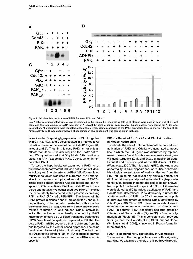
Figure 1. G��-Mediated Activation of PAK1 Requires PIX� and Cdc42
Cos-7 cells were transfected with cDNAs as indicated in the figures. For each cDNA, 0.2 �g of plasmid were used in each well of a 6-wellplate, and the total amount of cDNA was kept at 1 �g/well by using a control LacZ plasmid. Kinase assays were carried out 1 day aftertransfection. All experiments were repeated at least three times. Western analysis of the PAK1 expression level is shown in the top of (B).Kinase activity in (B) was quantified by a phosphoimager. This experiment was carried out in triplicate.
lanes 2 and 4). Surprisingly, expression of PAK1 together PIX� Is Required for Cdc42 and PAK1 Activationin Mouse Neutrophilswith G�1�2, PIX�, and Cdc42 resulted in a marked (over
6-fold) increase in the level of active Cdc42 (Figure 3A, To validate the role of PIX� in chemoattractant-inducedactivation of PAK1 and Cdc42, we generated a mouselanes 2 and 5). Thus, in this case PAK1 is not only an
effector for Cdc42, it is also required for Cdc42 activa- line in which the PIX� gene was disrupted by replace-ment of exons 8 and 9 with a neomycin-resistant genetion. We hypothesized that G�� binds PAK1 and acti-
vates, via PAK1-associated PIX�, Cdc42, which in turn via gene targeting (Z.M. and D.W., unpublished data).Exons 8 and 9 encode part of the DH domain of PIX�activates PAK1.
To test the hypothesis, we examined if PAK1 is re- (Wang et al., 2001). The mice lacking PIX� show no grossabnormality in size, appearance, or routine behaviors.quired for chemoattractant-induced activation of Cdc42
in leukocytes. Short interference RNA (siRNA)-mediated Histological examination of various tissues from thePIX� null mice did not reveal any obvious defect, normRNA knockdown was used to suppress PAK1 expres-
sion in a mouse macrophage-like cell line, RAW274. did flow cytometry analysis of various leukocyte popula-tions reveal defects in hematopoiesis (data not shown).These cells contain intrinsic C5a receptors and can re-Neutrophils from the wild-type and PIX� null littermatesspond to C5a to activate PAK1 and Cdc42 and to un-were isolated, and C5a-induced activation of PAK1 anddergo chemotaxis. We established two RAW274 clonesCdc42 was determined. PIX�-deficiency blunted thethat were stably transfected with a plasmid expressingacute activation of PAK1 by C5a in mouse neutrophilsPAK1 siRNA (PAK1pSUPER-GFP). The levels of the(Figure 3C) and almost abolished Cdc42 activation byPAK1 protein in clones 7 and 11 are about 20% and 30%,C5a (Figure 3D). Thus, PIX� plays an important role inrespectively, of that in cells transfected with a controlchemoattractant-induced activation of Cdc42 andplasmid (Figure 3B, top). Cells from both clones showedPAK1. In contrast, PIX� deficiency had little effect onmarked reduction in C5a-induced Cdc42 activation,C5a-induced Rac activation (Figure 3D) or F-actin poly-while Rac activation was hardly affected by PAK1merization (Figure 3E). This is consistent with previousknockdown (Figure 3B). We also transiently transfectedfindings that Rac (Roberts et al., 1999), but not Cdc42RAW274 cells with a synthetic duplexed siRNA that tar-(Srinivasan et al., 2003), is involved in F-actin formationgets a PAK1 mRNA sequence that is different from thein neutrophils.one targeted by the vector-based approach. The same
result was observed (data not shown). The fact thatsiRNAs targeting different Pak1 mRNA sequences showed PAK1 Is Required for Directionality in Chemotaxis
To understand the biological functions of this signalingthe same result demonstrates that the siRNA effect isspecific. pathway, we examined the role of this pathway in regula-
Cell218
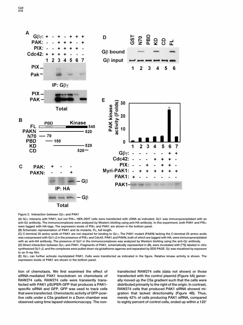
Figure 2. Interaction between G�� and PAK1
(A) G�� interacts with PAK1, but not PIX�. HEK-293T cells were transfected with cDNA as indicated. G�1 was immunoprecipitated with ananti-G� antibody. The immunocomplexes were analyzed by Western blotting using anti-HA antibody. In this experiment, both PAK1 and PIX�
were tagged with HA-tags. The expression levels of PIX� and PAK1 are shown in the bottom panel.(B) Schematic representation of PAK1 and its mutants. FL, full length.(C) C-terminal 25 amino acids of PAK1 are not required for binding to G��. The PAK1 mutant (PAKN) lacking the C-terminal 25 amino acidswas coexpressed with G�1�2 in the presence of PIX� and Cdc42. PAK1 and PAKN, both of which are tagged with HA, were immunoprecipitatedwith an anti-HA antibody. The presence of G�1 in the immunocomplexes was analyzed by Western blotting using the anti-G� antibody.(D) Direct interaction between G�� and PAK1. Fragments of PAK1, schematically represented in (B), were incubated with [35S] labeled in vitrosynthesized G�1�2, and the complexes were pulled down via glutathione-agarose and separated by SDS PAGE. G� was visualized by exposureto an X-ray film.(E) G�� can further activate myristylated PAK1. Cells were transfected as indicated in the figure. Relative kinase activity is shown. Theexpression levels of PAK1 are shown in the bottom panel.
tion of chemotaxis. We first examined the effect of transfected RAW274 cells (data not shown) or thosetransfected with the control plasmid (Figure 4A) gener-siRNA-mediated PAK1 knockdown on chemotaxis of
RAW274 cells. RAW274 cells were transiently trans- ally moved up the C5a gradient such that the cells weredistributed primarily to the right of the origin. In contrast,fected with PAK1 pSUPER-GFP that produces a PAK1-
specific siRNA and GFP. GFP was used to track cells RAW274 cells that produced PAK1 siRNA showed mi-gration that lacked directionality (Figure 4B). Thus,that were transfected. Chemotactic activity of GFP-posi-
tive cells under a C5a gradient in a Dunn chamber was merely 42% of cells producing PAK1 siRNA, comparedto eighty percent of control cells, ended up within a 120�observed using time lapsed videomicroscopy. The non-

Cdc42 Activation in Directional Sensing219
Figure 3. Regulation of Cdc42, Rac, and PAK Activity in Myeloid Cells
(A) G�� activates Cdc42 only in the presence of PIX� and PAK1. Cos-7 cells were transfected with cDNAs as indicated in the figure. PBDpull down was performed followed by Western analysis with an anti-Cdc42 antibody. The levels of active Cdc42 pulled down by PBD andtotal Cdc42 expressed in the cells are shown. The relative active Cdc42 levels were quantified by densitometry, normalized against totalCdc42 levels, and are shown at the bottom of the figure.(B) PAK1 is required for C5a-induced activation of Cdc42 in Raw 274 cells. The levels of active Cdc42 and Rac1/2 in two RAW274 clonesthat were stably transfected with a plasmid expressing PAK1 siRNA were quantified by densitometry and normalized against the total Cdc42levels. The effects of siRNA treatments on PAK1 expression are shown in the top panel. The expression levels of G� were used as an internalcontrol.(C) PIX� is required for PAK activation in mouse neutrophils. Neutrophils isolated from the wild-type (Wt) and PIX� null (pix) mice were treatedwith 100 �M C5a for the indicated durations. Kinase activity was determined after immunoprecipitation using an anti-PAK1 antibody andquantified by a phosphoimager.(D) PIX� is required for Cdc42, but not Rac, activation in mouse neutrophils. Neutrophils from the wild-type and PIX� null mice were stimulatedwith 100 �M C5a for 10 s (2 and 4). Relative active Cdc42 and Rac1/2 levels were determined and analyzed as in (B).(E) PIX� is not required for F-actin formation in mouse neutrophils. Neutrophils were stimulated with 100 �M C5a for indicated durations,permeabilized, stained with FITC-labeled phalloidin, and analyzed by a flowcytometer. AU, arbitrary unit.
Cell220
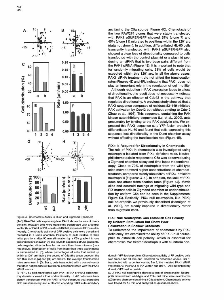
arc facing the C5a source (Figure 4C). Chemotaxis ofthe two RAW274 clones that were stably transfectedwith PAK1 pSUPER-GFP showed 39% (clone 7) and45% (clone 11) migrated to positions within the 120� arc(data not shown). In addition, differentiated HL-60 cellstransiently transfected with PAK1 pSUPER-GFP alsoshowed a clear loss of directionality compared to cellstransfected with the control plasmid or a plasmid pro-ducing an siRNA that is two base pairs different fromthe PAK1 siRNA (Figure 4E). It is important to note thatfor randomly migrating cells, 33% of cells would beexpected within this 120� arc. In all the above cases,PAK1 siRNA treatment did not affect the translocationrates (Figures 4D and 4F), indicating that PAK1 does notplay an important role in the regulation of cell motility.
Although reduction in PAK expression leads to a lossof directionality, this result does not necessarily indicatethat PAK is an effector of Cdc42 in the pathway thatregulates directionality. A previous study showed that aPAK1 sequence composed of residues 83–149 inhibitedPAK activation by Cdc42 but without binding to Cdc42(Zhao et al., 1998). This sequence, containing the PAKkinase autoinhibitory sequences (Lei et al., 2000), actspresumably by binding to the PAK catalytic site. We ex-pressed this PAK1 sequence as a YFP-fusion protein indifferentiated HL-60 and found that cells expressing thissequence lost directionality in the Dunn chamber assaywithout affecting the translocation rate (Figure 4E).
PIX� Is Required for Directionality in ChemotaxisThe role of PIX� in chemotaxis was investigated usingneutrophils isolated from PIX�-deficient mice. Neutro-phil chemotaxis in response to C5a was observed usinga Zigmond chamber assay and time lapse videomicros-copy. Close to 70% of neutrophils from the wild-typemice moved toward higher concentrations of chemoat-tractants, compared to only about 35% of PIX�-deficientneutrophils (Figures4G–4I). In addition, the lack of PIX�does not affect translocation rates (Figure 4J). Movieclips and centroid tracings of migrating wild-type andPIX mutant cells in Zigmond chamber or under stimula-tion by uniform C5a can be seen in the SupplementalFigure S3. Basically, PIX� null neutrophils, like PI3K�null neutrophils we previously described (Hannigan etal., 2002), are clearly impaired in directionality ratherthan migration itself.
Figure 4. Chemotaxis Assay in Dunn and Zigmond Chambers PIX� Null Neutrophils Can Establish Cell Polarity(A–D) RAW274 cells expressing less PAK1 showed a loss of direc- by Uniform Stimulation but Show Poortionality. RAW274 cells were transiently transfected with a control Polarization in Shallow Gradientsvector (A) or PAK1 siRNA construct (B) that expresses GFP simulta-
To understand the impairment of chemotaxis by PIX�neously. Chemotactic activity of GFP positive cells were traced anddeficiency, we examined the ability of PIX-� null neutro-recorded in a Dunn chamber. Positions of cells relative to theirphils to establish cell polarity, which is essential forinitial positions after 90 min stimulation by a C5a gradient in one
experiment are shown in (A) and (B). In the absence of C5a gradients, chemotaxis. We treated neutrophils with a uniform con-cells migrated directionless for no more than three microns (datanot shown). Distribution of cells from more than three experimentsis summarized in (C), where percentages of cells that ended up
domain-YFP fusion protein. Chemotactic activity of FP positive cellswithin a 120� arc facing the source of C5a (the areas between thewas traced for 60 min and recorded as described above. Bar 1,two thin lines in [A] and [B]) are shown. The average translocationtransfected with a control vector; Bar 2, the mutated PAK1 siRNArates are shown in (D). Bar a, cells transfected with a control vectorvector; Bar 3, the PAK1 siRNA vector; and Bar 4, PAk1 autoinhibitorythat does not produce siRNA; Bar b, cells transfected with the PAK1domain-YFP fusion protein.siRNA vector.(G–J) PIX� null neutrophils showed a loss of directionality. Neutro-(E–F) HL-60 cells transfected with PAK1 siRNA or PAK1 autoinhibi-phils isolated from wild-type and PIX� null mice were examined intory domain showed a loss of directionality. HL-60 cells were tran-a Zigmond chamber containing a C5a gradient. Chemotactic activitysiently transfected with the PAK1 siRNA construct that expresseswas traced for 15 min and analyzed as described above.GFP simultaneously and a plasmid encoding PAk1 auto-inhibitory
Cdc42 Activation in Directional Sensing221
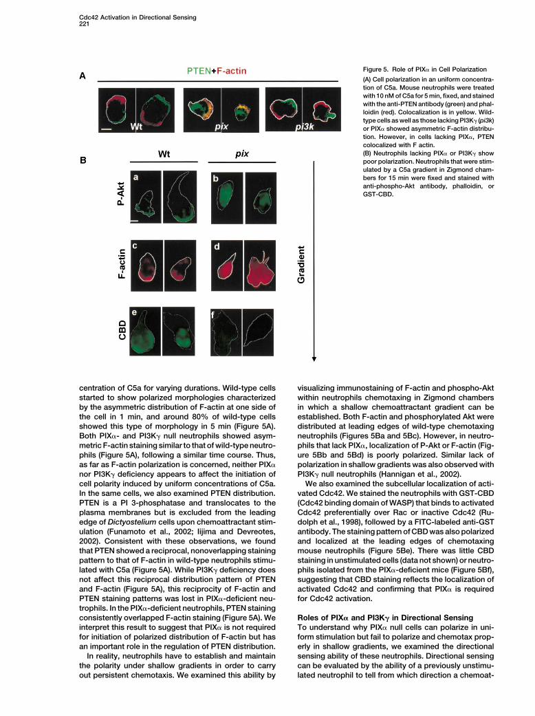
Figure 5. Role of PIX� in Cell Polarization
(A) Cell polarization in an uniform concentra-tion of C5a. Mouse neutrophils were treatedwith 10 nM of C5a for 5 min, fixed, and stainedwith the anti-PTEN antibody (green) and phal-loidin (red). Colocalization is in yellow. Wild-type cells as well as those lacking PI3K� (pi3k)or PIX� showed asymmetric F-actin distribu-tion. However, in cells lacking PIX�, PTENcolocalized with F actin.(B) Neutrophils lacking PIX� or PI3K� showpoor polarization. Neutrophils that were stim-ulated by a C5a gradient in Zigmond cham-bers for 15 min were fixed and stained withanti-phospho-Akt antibody, phalloidin, orGST-CBD.
centration of C5a for varying durations. Wild-type cells visualizing immunostaining of F-actin and phospho-Aktwithin neutrophils chemotaxing in Zigmond chambersstarted to show polarized morphologies characterized
by the asymmetric distribution of F-actin at one side of in which a shallow chemoattractant gradient can beestablished. Both F-actin and phosphorylated Akt werethe cell in 1 min, and around 80% of wild-type cells
showed this type of morphology in 5 min (Figure 5A). distributed at leading edges of wild-type chemotaxingneutrophils (Figures 5Ba and 5Bc). However, in neutro-Both PIX�- and PI3K� null neutrophils showed asym-
metric F-actin staining similar to that of wild-type neutro- phils that lack PIX�, localization of P-Akt or F-actin (Fig-ure 5Bb and 5Bd) is poorly polarized. Similar lack ofphils (Figure 5A), following a similar time course. Thus,
as far as F-actin polarization is concerned, neither PIX� polarization in shallow gradients was also observed withPI3K� null neutrophils (Hannigan et al., 2002).nor PI3K� deficiency appears to affect the initiation of
cell polarity induced by uniform concentrations of C5a. We also examined the subcellular localization of acti-vated Cdc42. We stained the neutrophils with GST-CBDIn the same cells, we also examined PTEN distribution.
PTEN is a PI 3-phosphatase and translocates to the (Cdc42 binding domain of WASP) that binds to activatedCdc42 preferentially over Rac or inactive Cdc42 (Ru-plasma membranes but is excluded from the leading
edge of Dictyostelium cells upon chemoattractant stim- dolph et al., 1998), followed by a FITC-labeled anti-GSTantibody. The staining pattern of CBD was also polarizedulation (Funamoto et al., 2002; Iijima and Devreotes,
2002). Consistent with these observations, we found and localized at the leading edges of chemotaxingmouse neutrophils (Figure 5Be). There was little CBDthat PTEN showed a reciprocal, nonoverlapping staining
pattern to that of F-actin in wild-type neutrophils stimu- staining in unstimulated cells (data not shown) or neutro-phils isolated from the PIX�-deficient mice (Figure 5Bf),lated with C5a (Figure 5A). While PI3K� deficiency does
not affect this reciprocal distribution pattern of PTEN suggesting that CBD staining reflects the localization ofactivated Cdc42 and confirming that PIX� is requiredand F-actin (Figure 5A), this reciprocity of F-actin and
PTEN staining patterns was lost in PIX�-deficient neu- for Cdc42 activation.trophils. In the PIX�-deficient neutrophils, PTEN stainingconsistently overlapped F-actin staining (Figure 5A). We Roles of PIX� and PI3K� in Directional Sensing
To understand why PIX� null cells can polarize in uni-interpret this result to suggest that PIX� is not requiredfor initiation of polarized distribution of F-actin but has form stimulation but fail to polarize and chemotax prop-
erly in shallow gradients, we examined the directionalan important role in the regulation of PTEN distribution.In reality, neutrophils have to establish and maintain sensing ability of these neutrophils. Directional sensing
can be evaluated by the ability of a previously unstimu-the polarity under shallow gradients in order to carryout persistent chemotaxis. We examined this ability by lated neutrophil to tell from which direction a chemoat-
Cell222
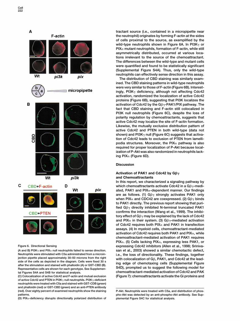
tractant source (i.e., contained in a micropipette nearthe neutrophil) originates by forming F-actin at the sidesof cells proximal to the source, as exemplified by thewild-type neutrophils shown in Figure 6A. In PI3K� orPIX� mutant neutrophils, formation of F-actin, while stillasymmetrically distributed, occurred at various loca-tions irrelevant to the source of the chemoattractant.The differences between the wild-type and mutant cellswere quantified and found to be statistically significant(Supplemental Figure S4A). Thus, only the wild-typeneutrophils can effectively sense direction in this assay.
The distribution of CBD staining was similarly exam-ined. The CBD staining patterns in wild-type neutrophilswere very similar to those of F-actin (Figure 6B). Interest-ingly, PI3K� deficiency, although not affecting Cdc42activation, randomized the localization of active Cdc42proteins (Figure 6B), suggesting that PI3K localizes theactivation of Cdc42 by the G��-PAK1/PIX pathway. Thefact that CBD staining and F-actin still colocalized inPI3K null neutrophils (Figure 6C), despite the loss ofpolarity regulation by chemoattractants, suggests thatactive Cdc42 may localize the site of F-actin formation.Likewise, the mutually exclusive distribution pattern ofactive Cdc42 and PTEN in both wild-type (data notshown) and PI3K� null (Figure 6C) suggests that activa-tion of Cdc42 leads to exclusion of PTEN from lamelli-podia structures. Moreover, the PIX� pathway is alsorequired for proper localization of P-Akt because local-ization of P-Akt was also randomized in neutrophils lack-ing PIX� (Figure 6D).
Discussion
Activation of PAK1 and Cdc42 by G��and ChemoattractantsIn this report, we characterized a signaling pathway bywhich chemoattractants activate Cdc42 in a G��-medi-ated, PAK1 and PIX�-dependent manner. Our findingsare as follows. (1) G�� strongly activates PAK1 onlywhen PIX� and CDC42 are coexpressed. (2) G�� bindsto PAK1 directly. The previous report showing that puri-fied G�� directly inhibited N-terminal truncated PAK1confirms the interaction (Wang et al., 1999). The inhibi-tory effect of G�� may be explained by the lack of Cdc42and PIX� in their system. (3) G��-mediated activationof Cdc42 requires both PIX� and PAK1 in transfectionassays. (4) In myeloid cells, chemoattractant-mediatedactivation of Cdc42 requires both PAK1 and PIX�, whilechemoattractant-mediated activation of PAK1 requiresPIX�. (5) Cells lacking PIX�, expressing less PAK1, or
Figure 6. Directional Sensing expressing Cdc42 inhibitors (Allen et al., 1998; Sriniva-(A and B) PI3K� and PIX� null neutrophils failed to sense direction. san et al., 2003) showed a similar chemotactic defect,Neutrophils were stimulated with C5a administrated from a microin- i.e., the loss of directionality. These findings, togetherjection pipette placed approximately 30–50 microns from the right with colocalization of G�, PAK1, and Cdc42 at the lead-side of the cells as depicted in the diagram. Cells were fixed 30 s ing edge of chemotaxing cells (Supplemental Figureafter the stimulation and stained with phalloidin (A) or GST-CBD (B).
S4D), prompted us to suggest the following model forRepresentative cells are shown for each genotype. See Supplemen-chemoattractant-mediated activation of Cdc42 and PAKtal Figures S4A and S4B for statistical analysis.
(C) Colocalization of active Cdc42 and F-actin and mutual exclusion (Figure 7): chemoattractants activate the Gi proteins andof active Cdc42 and PTEN in PI3K� null neutrophils. PI3K�-deficientneutrophils were treated with C5a and stained with GST-CDB (green)and phalloidin (red) or GST-CBD (green) and an anti-PTEN antibody(red). Over eighty percent of examined neutrophils show the staining P-Akt. Neutrophils were treated with C5a, and distribution of phos-patterns. pho-Akt was detected by an anti-phospho-Akt antibody. See Sup-(D) PIX�-deficiency disrupts directionally polarized distribution of plemental Figure S4C for statistical analysis.
Cdc42 Activation in Directional Sensing223
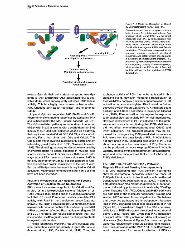
Figure 7. A Model for Regulation of Cdc42by Chemoattractant via G�� and PIX�
Chemoattractant bound receptors activateheterotrimeric G proteins and release G��
proteins, which recruit PAK1 via the directinteraction and PIX� by its association withPAK1 to activate Cdc42. Active Cdc42 acti-vates PAK1. Activated PAK1and/or otherCdc42 effectors regulate PTEN and F-actinlocalization. This pathway is required for di-rectional sensing, persistent directionalmovement, and establishment of cell polarityin a shallow chemoattractant gradient. PIP3
produced by PI3K� is required for localizationof this signaling pathway to the leading edge,while localization of PIP3 is also influencedby this pathway via its regulation of PTENdistribution.
release G�� via their cell surface receptors, free G�� exchange activity of PIX� has to be activated in thissignaling event. However, membrane translocation ofbinds to PAK1 and brings PAK1-associated PIX� to acti-
vate Cdc42, which subsequently activates PAK1 kinase the PAK1/PIX� complex does not appear to result in PIXactivation because myristylated PAK1 could be furtheractivity. This is a highly unusual mechanism in which
PAK functions both as an “activator” and effector for activated by G�� (Figure 2E). Since PI3K inhibitors couldpartially inhibit Cdc42 activation in leukocytes (BenardCdc42.
In yeast, G�� also regulates PAK (Ste20) and Cdc42. et al., 1999), PIX� activation may require the exposureto phospholipids, particularly PIP3 on cell membranes.Pheromone elicits mating responses by activating PAK
and subsequently the MAP kinase cascade via G��. However, involvement of PIP3 in activation of this path-way would contradict our finding that PI3K� deficiencyThis G��-mediated pathway requires direct interaction
of G�� with Ste20 as well as with a scaffold protein Ste5 did not affect chemoattractant-induced Cdc42 andPAK1 activation. This apparent paradox may be ex-(Leeuw et al., 1998). G�� activates Cdc42 via a pathway
that requires at least a Cdc42 GEF, Cdc24, and a scaffold plained by distinguishing PI3K�-mediated increases inPIP3 levels from the basal levels of PIP3. PI3K inhibitorsprotein, Far1p that binds both G�� and Cdc24. This
Cdc42 pathway is involved in cell polarity establishment not only inhibit PI3K�-mediated production of PIP3, butshould also reduce the basal levels of PIP3. The latterin budding yeast (Butty et al., 1998; Nern and Arkowitz,
1999). The signaling pathway we describe here used by can be produced by house keeping PI3Ks or PI3K acti-vated by crosstalk with chemoattractant-activated path-chemoattractants to sense direction in myeloid cells
shares some remarkable similarities with the yeast path- ways and other mechanisms that are not inhibited byPI3K� deficiency.ways except PAK1 seems to have a dual role. PAK1 is
not only an effector for Cdc42, but also appears to func-tion as a scaffold protein reminiscent of the role of yeast The PAK1/PIX�/Cdc42 and PI3K� Pathways
Regulate Directional Sensing InterdependentlyFar1p and Ste5 proteins because it is required for Cdc42activation. Mammalian homologs to either Far1p or Ste5 It is very interesting that PIX�-deficient neutrophils
showed chemotactic behaviors similar to those ofhave not been identified.PI3K�-deficient; a lack of directionality with unaffectedtranslocation rates (Hannigan et al., 2002). In addition,PIX� Is a Physiological GEF Required for Specific
Activation of Cdc42 in Mouse Neutrophils both deficiencies also disrupted directional F-actin for-mation induced by point source stimulation by C5a (Fig-PIX� can act as an exchange factor for Cdc42 and Rac
in vitro or in overexpression systems (Manser et al., ure 6). Thus, the PAK1/PIX�/Cdc42 and PI3K� pathwaysare both part of the molecular “compass” (Bourne and1998; Daniels et al., 1999; Feng et al., 2002). Despite the
fact that G�� and PIX� could weakly stimulate PAK Weiner, 2002) required for sensing direction. We believethat these two pathways are interdependent becauseactivity with Rac1 in the transfection assay (data not
shown), PIX� is not a physiological GEF for Rac in mouse loss of PIX� disrupted directional localization of P-Akt(Figure 6D), a downstream target of the PI3K� pathway,myeloid cells because neither PIX�-deficiency nor PAK1
siRNA expression affected Rac1 activation (Figures 3B while loss of PI3K� disrupted directional localization ofactive Cdc42 (Figure 6B). Given that PIX� deficiencyand 3D). Therefore, our results demonstrate that PIX�
is a specific Cdc42 regulator used by chemoattractants does not affect PI3K� activation (data not shown) orvice versa (Supplemental Figure S1), these two path-in myeloid cells in vivo.
The full-length wild-type PIX� has very low basal gua- ways may regulate each other via a convergent localiza-tion. Thus, activation of the PAK1/PIX�/Cdc42 pathwaynine nucleotide exchange activity (Figure 3A, lane 4)
(Manser et al., 1998; Daniels et al., 1999). Thus, the would be required for proper localization of PI3K�’s
Cell224
product PIP3, perhaps through exclusion of PTEN (see distribution of PTEN remain to be determined. Bothbelow). And proper localization of PIP3 would be re- Cdc42 and PAK1 have the capability of activating down-quired for localizing the G��-PAK1/PIX�/Cdc42 signal- stream effectors. Ample evidence indicates that PAK1ing complex, possibly via PIX�’s PH domain. can regulate cytoskeleton reorganization, cell morpho-
logical changes, and cell migration. In human neutro-The PIX� Pathway Regulates Directionality phils, PAK1 colocalized with cortical actin to membraneby Regulating Localization of F-Actin ruffles and lamellipodia at the leading edge upon stimu-Formation and PTEN lation by chemoattractants as we found in mouse neu-Although PIX�- and PI3K�-deficiencies impaired direc- trophils, and this localization could be blocked by a PI3Ktional localization of F-actin toward chemoattractant inhibitor, wortmannin (Dharmawardhane et al., 1999). Onsources (Figure 6A), localization of active Cdc42 always the other hand, Cdc42 can regulate actin dynamics andoverlaps that of F-actin in PI3K� null neutrophils (Figure reorganization via regulation of other independent ef-6C). Therefore, activated Cdc42 appears to determine fectors such as WASP (Erickson and Cerione, 2001).where F-actin is polymerized in a cell, although it does While PAK1 kinase activity is required for directionality,not stimulate F-actin formation itself (Figure 3E) (Sriniva- we do not know what roles, if any, other Cdc42 effectorssan et al., 2003). play in the regulation of chemotaxis.
In addition to localizing the F-actin formation, thisPIX�/Cdc42 pathway also has an essential role in PTENdistribution. In both wild-type and PI3K�-deficient neu- Concluding Remarkstrophils stimulated by C5a, PTEN and F-actin staining As summarized in Figure 7, we have characterized aalways showed mutual exclusion; however, PTEN re- signaling mechanism by which chemoattractants acti-mained together with F-actin in cells lacking PIX� (Figure vate Cdc42 and PAK1. We believe that that this signaling5A). This suggests that the function supplied by PIX� is pathway, together with the PI3K�-pathway, is an essen-required for repelling PTEN from the leading edge. The tial part of the molecular compass that is required forfact that CBD and PTEN staining always show a recipro- sensing direction. This new signaling pathway is in-cal pattern in PI3K� null cells (Figure 6C) where localiza- volved in the establishment of directionality by repellingtion of activated Cdc42 is not regulated (Figure 6B) con- PTEN from the leading edge, while localizing sites offirms that PTEN is actively excluded from locations F-actin formation to the leading edge. This latter processwhere the Cdc42 is activated. Exclusion of PTEN from also requires the PI3K�-linked pathway. Further workthe leading edge should be important for directional is needed for understanding many of the mechanisticsensing, because PTEN, as a PI-3 phosphatase, influ- details.ences the local level of PIP3. This conclusion is consis- PIX� was also implicated as a candidate gene fortent with what was observed in chemotactic Dictyoste- X linked mental retardation based on disruption, bylium cells (Funamoto et al., 2002; Iijima and Devreotes, chromosome rearrangement, of the PIX� gene of a sin-2002). In addition, given that PIX� deficiency does not gle patient (Kutsche et al., 2000). Although a number ofaffect phosphorylation of Akt (data not shown), this PIX/ other X lined metal retardation patients from a singleCdc42 pathway may not regulate the activity of PTEN kindred contain a mutation that apparently causesbut, merely, PTEN’s localization. Because PI3K� defi- exon 2 skipping, leading to production of a PIX� proteinciency only regulates where the Cdc42 is activated by lacking 28 amino acids at the N-terminal CH domain,G�� rather than regulating the activity of Cdc42, it is the consequence of this mutation to the activity of PIXreasonable that PI3K� deficiency does not affect the mutant is not clear. It is unlikely that the exon-skippingreciprocal distribution patterns of CBD and PTEN (Fig- mutation causes loss of function. On the contrary, theure 5A). mutation may lead to gain of function, because a PIX
mutant lacking the entire CH domain showed enhancedDirection Sensing Is Required for Polarization activation of Cdc42 and PAK1 (Manser et al., 1998). Thusin Shallow Gradients far, we have not observed any gross phenotype fromNeither PIX� or PI3K� deficinecy affects asymmetric the PIX�-deficient mice. Systemic neurological charac-distribution of F-actin in response to point source or terization of this mouse line is currently underway. Theuniform stimulation by C5a, suggesting these mutant lack of a broad range of obvious phenotypes with thiscells still possess the ability to polarize stochastically. mutant mouse line, on the other hand, suggests thatThus, we think that neutrophils and probably other cells PIX� has a highly specialized physiological significance.have intrinsic ability of stochastic polarization, which
Barring new adverse phenotypes, PIX� may be a poten-is a related, but independent, process from directional
tial anti-inflammation target as the neutrophils lackingsensing. However, most of the PIX� or PI3K� null cells
this protein show significant impairment in chemotaxis.failed to show well-polarized morphologies as mostwild-type neutrophils did when stimulated by a shallowgradient of C5a (Figure 5A) (Hannigan et al., 2002). Thus, Experimental Proceduresdirectional sensing is important for promoting and stabi-
Cell Culture, Transfection, and Constructionlizing polarization under this condition.of Expression PlasmidsMonkey kidney Cos-7 cells and human embryonic kidney cells
Downstream Effectors A293T cells were maintained as previously described (Li et al., 1999).for the G��-PAK/PIX�/Cdc42 Pathway For kinase and immunoprecipitation assays, Cos-7 cells in 6-wellThe mechanisms by which this G��-PAK/PIX�/Cdc42 plates were seeded at 2 � 105 cells/well and transfected with 1 �g
DNA/well using Lipofectamine Plus (Invitrogen, CA), as suggestedpathway regulates actin cytoskeleton remodeling and
Cdc42 Activation in Directional Sensing225
by the manufacturer. Cell extracts were collected 24 hr after trans- chemotaxis assays. For uniform treatment, neutrophils, cells (0.1 mlat 1 � 105 cells/ml) were stimulated with C5a in a suspension offection.
The wild-type and mutant forms of human PAK1, PIX�, Cdc42, Hanks buffer. These cells were allowed to settle on a coverslip, andfixed with 100 �l of ice cold paraformaldehyde (4%). Coverslipsbovine G�1, and G�2 were generated by PCR using the high-fidelity
thermostable DNA polymerase Pfu (Stratagene, CA). PIXDH con- were incubated on ice for 10 min and stained as described below.tains Leu residues (383–384) to Ala substitution mutations in the DHdomain. Epitope tags were introduced into some of molecules as Chemotaxis Chamber Assaysindicated in the figures and text. The expression of these molecules For analysis of neutrophil chemotaxis in Zigmond chambers, thewas driven by a CMV promoter. All constructs were verified by DNA coverslips, prepared as described above, were placed on a Zigmondsequencing. chamber (Neuroprobes, Cabin John, MD). Aliquots (0.1 ml) of a
solution (Hanks buffer containing a 1:10 dilution of 10% gelatin inH20) were added to one side of the chamber and the same solutionImmunoprecipitation, PAK Kinase Assay, and PBDcontaining C5a (10�5 M) was added to the other side (Zigmond,Pull-Down Assay1988). Analysis of RAW274 and HL-60 chemotaxis were carried outCells were lysed in the lysis buffer (50 mM HEPES [pH 7.4], 150 mMin Dunn chambers. Cells were also allowed to settle and adhere toNaCl, 15 mM NaF, 2 mM EDTA, 1 mM EGTA, 10% glycerol, 1%coverslips, which were rinsed and placed on a Dunn chamber. TheTriton X-100, 20 mM �-glycerophosphate, 1 mM orthovanadate,inner well contained Hanks’ buffer (0.1 ml), while the outer well1 mM Sodium pyrophosphate, 10 ug/ml aprotinin, and10 ug/ml leu-contained C5a (10�5 M) in Hanks buffer.peptin). The cell lysates were subject to high-speed centrifugation.
Time-lapsed videomicroscopy was used to examine cell move-The supernatants were then incubated with an antibody and 20 �lments in chemotactic chambers. Cells in either chamber were re-of protein A/G-Sepharose beads (Santa Cruz Biotech, CA) for 2 hrcorded crawling in the absence or presence of C5a gradients. Theat 4�C. The immunocomplexes were pelleted and washed threemicroscope was equipped with differential interference contrast op-times with cold lysis buffer in the absence of protease and phospha-tics and a 20� objective. Images were captured at 5 s intervals withtase inhibitors. The proteins were released from beads by boiling ina PXL-EEV37 CCD camera (Photometrics, Waterloo, ON Canada)SDS-sample buffer. The samples were analyzed by Western blotting.and Metamorph imaging software (West Chester, PA). Time-lapsedThe results were visualized using a Raytest imaging system with avideo images were used to calculate the final position of a cellcooled CCD camera. The PBD pull-down assay was carried out asrelative to its starting position, and the data were graphed. Time-described previously (Benard et al., 1999).lapsed videomicroscopy data were also analyzed with the Meta-For kinase assays, PAK proteins were immunoprecipitated asmorph software to determine migration parameters as describeddescribed above, and the immunocomplexes were washed threepreviously (Hannigan, 2002).times with cold lysis buffer and twice with cold kinase buffer (50
mM HEPES [pH7.4], 10 mM MgCl2, and 2 mM MnCl2). The kinaseImmunofluorescence Staining and Microscopyreactions were performed for 30 min at 30�C in the presence ofFor examination of cells from Zigmond chambers, cells were fixed1 �Ci [�-32P]ATP, 30 �M ATP, and 10 �g of histone H4 peptidein chambers after being exposed to C5a gradients for 15 min. Che-(Huang and Laramee, 1991; Knaus et al., 1995). The reactions weremotaxis buffers were carefully removed from the chamber wells andterminated by addition of 4� SDS-sample buffer. The samples wereimmediately replaced with 2% paraformaldehyde in PBS for 15 minboiled and loaded on 20% SDS-PAGE gels. The results were visual-at 37�C. For point source treatment of neutrophils, cells were treatedized and quantified using a Phosphoimager (Packard, IL).with C5a gradients administered by a microinjection pipette placed30–40 microns from the cell for 30 s. Then, 4% paraformaldehydeIn Vitro Assay for the Interaction between G�� and PAK1in PBS was added for fixation for 15 min on ice.Full-length PAK1 or fragments of PAK1 (Figure 2B) were prepared
For immunostaining, coverslips containing fixed cells were incu-as GST fusion proteins from E. coli. G�1�2 was prepared by in vitrobated with a solution of 0.1% Tween-20 in PBS containing andtranslation using a Promega in vitro protein synthesis kit containingprimary antibodies (1:50 dilution). Coverslips were then incubated[35S] Met and Cys or purified from SF9 cells infected with baculovirusovernight (4�C). The next morning, the cells were washed and incu-expressing G�1 and G�2. GST-PAK was incubated with G�1�2,bated with 100 �l of a solution (Tween-20 containing appropriateand the complexes were pulled down via glutathione-agarose andsecondary antibodies [1:100 dilution]). Following this incubation,separated by SDS-PAGE. G� was visualized by exposure to an X-raycells were washed and coverslips mounted with a drop of Slowfadefilm or Western analysis using anti-G�1 antibody.reagent. These samples were then examined using a Zeiss 510confocal imaging system. Excitation wavelengths of 488 and 568siRNA Design and Treatmentnm and emission filters were used to detect FITC (515-520 nm) and/A duplexed siRNA targeting the PAK1 mRNA sequence (AAGGTTor rhodamine (590 nm).GACATCTGGTCCCTG) was synthesized by Dharmacon (Lafayette,
CO). pSUPER-GFP vector was constructed by inserting into pGFP-AcknowledgmentsN1 (BD Clontech, Palo Alto, CA) a human H1 promoter followed by
HindIII and BglII sites for cloning the hairpin sequence for synthesisWe thank Alan Hall, Jonathan Chernoff, Silvio Gutkind, and Bruceof siRNA in vivo. PAK1 pSUPER-GFP targets the PAK1 mRNA se-Mayer for cDNAs and Henry Bourne, Bruce Mayer, and Peter Mayequence (AAGATTGGACAAGGTGCTTCA). The mutated control PAK1for reading and commenting on the manuscript. This work is sup-pSUPER-GFP contains an effective targeting sequence (AAGATTGported by grants from NIH (to D.W., M.L., A.V.S, G.W.) and 973GAGAACGTGCTTCA). Mutations are underlined.program (2002CB513000 to L.L.). D.W. is an established InvestigatorRAW274 cells were transiently transfected with chemically syn-of AHA.thesized siRNA oligo duplex using EPEI oligo delivery reagent from
GeneTool (Philomath, OR) as instructed. Three days later, cells wereReceived: April 2, 2003analyzed for PAK levels and Cdc42 and Rac activity. RAW274 cellsRevised: July 8, 2003were also transfected with PAK1 pSUPER-GFP and selected withAccepted: July 8, 2003G418. Two stable clones were established.Published: July 24, 2003
Neutrophil Preparation and StimulationReferences
Murine bone marrow neutrophils were prepared by centrifugationthrough Percoll gradients. Purified neutrophils were suspended in
Allen, W.E., Zicha, D., Ridley, A.J., and Jones, G.E. (1998). A roleHanks’ buffer (0.14 M NaCl, 5.4 mM KCl, 1 mM Tris, 1.1 mM CaCl2, for Cdc42 in macrophage chemotaxis. J. Cell Biol. 141, 1147–1157.0.4 mM MgSO4 , and 1 mM HEPES [pH 7.2]) containing 5 mg/ml
Bagrodia, S., and Cerione, R.A. (1999). Pak to the future. TrendsBSA. For chemotaxis chamber assays, isolated neutrophils wereCell Biol. 9, 350–355.allowed to adhere to glass coverslips for 5 min at 37�C. The cov-
erslips were then rinsed and placed on the appropriate chamber for Benard, V., Bohl, B.P., and Bokoch, G.M. (1999). Characterization
Cell226
of rac and cdc42 activation in chemoattractant-stimulated human Kutsche, K., Yntema, H., Brandt, A., Jantke, I., Nothwang, H.G., Orth,U., Boavida, M.G., David, D., Chelly, J., Fryns, J.P., et al. (2000).neutrophils using a novel assay for active GTPases. J. Biol. Chem.
274, 13198–13204. Mutations in ARHGEF6, encoding a guanine nucleotide exchangefactor for Rho GTPases, in patients with X-linked mental retardation.Bokoch, G.M. (2000). Regulation of cell function by Rho familyNat. Genet. 26, 247–250.GTPases. Immunol. Res. 21, 139–148.Leeuw, T., Wu, C., Schrag, J.D., Whiteway, M., Thomas, D.Y., andBourne, H.R., and Weiner, O. (2002). A chemical compass. NatureLeberer, E. (1998). Interaction of a G-protein beta-subunit with a419, 21.conserved sequence in Ste20/PAK family protein kinases. Nature
Butty, A.C., Pryciak, P.M., Huang, L.S., Herskowitz, I., and Peter, M. 391, 191–195.(1998). The role of Far1p in linking the heterotrimeric G protein to
Lei, M., Lu, W., Meng, W., Parrini, M.C., Eck, M.J., Mayer, B.J.,polarity establishment proteins during yeast mating. Science 282,and Harrison, S.C. (2000). Structure of PAK1 in an autoinhibited1511–1516.conformation reveals a multistage activation switch. Cell 102,
Chung, C.Y., and Firtel, R.A. (1999). PAKa, a putative PAK family 387–397.member, is required for cytokinesis and the regulation of the cy-
Li, L., Yuan, H., Xie, W., Mao, J., McMahon, E., Sussman, D., andtoskeleton in Dictyostelium discoideum cells during chemotaxis. J.Wu, D. (1999). Dishevelled proteins lead to two different signalingCell Biol. 147, 559–576.pathways; regulation of the JNK and b-catenin pathways. J. Biol.
Chung, C.Y., Funamoto, S., and Firtel, R.A. (2001). Signaling path- Chem. 274, 129–134.ways controlling cell polarity and chemotaxis. Trends Biochem. Sci.
Lim, L., Manser, E., Leung, T., and Hall, C. (1996). Regulation of26, 557–566.phosphorylation pathways by p21 GTPases. The p21 Ras-related
Daniels, R.H., and Bokoch, G.M. (1999). p21-activated protein ki- Rho subfamily and its role in phosphorylation signalling pathways.nase: a crucial component of morphological signaling? Trends Bio- Eur. J. Biochem. 242, 171–185.chem. Sci. 24, 350–355.
Lu, W., and Mayer, B.J. (1999). Mechanism of activation of Pak1Daniels, R.H., Zenke, F.T., and Bokoch, G.M. (1999). alphaPix stimu- kinase by membrane localization. Oncogene 18, 797–806.lates p21-activated kinase activity through exchange factor-depen-
Lu, W., Katz, S., Gupta, R., and Mayer, B.J. (1997). Activation of Pakdent and -independent mechanisms. J. Biol. Chem. 274, 6047–6050.
by membrane localization mediated by an SH3 domain from theDharmawardhane, S., Brownson, D., Lennartz, M., and Bokoch, G.M. adaptor protein Nck. Curr. Biol. 7, 85–94.(1999). Localization of p21-activated kinase 1 (PAK1) to pseudo-
Manser, E., Loo, T.H., Koh, C.G., Zhao, Z.S., Chen, X.Q., Tan, L., Tan,podia, membrane ruffles, and phagocytic cups in activated human
I., Leung, T., and Lim, L. (1998). PAK kinases are directly coupled toneutrophils. J. Leukoc. Biol. 66, 521–527.
the PIX family of nucleotide exchange factors. Mol. Cell 1, 183–192.Erickson, J.W., and Cerione, R.A. (2001). Multiple roles for Cdc42
Murphy, P.M. (1994). The molecular biology of leukocyte chemoat-in cell regulation. Curr. Opin. Cell Biol. 13, 153–157.
tractant receptors. Annu. Rev. Immunol. 12, 593–633.Etienne-Manneville, S., and Hall, A. (2002). Rho GTPases in cell Nern, A., and Arkowitz, R.A. (1999). A Cdc24p-Far1p-Gbetagammabiology. Nature 420, 629–635. protein complex required for yeast orientation during mating. J. CellFeng, Q., Albeck, J.G., Cerione, R.A., and Yang, W. (2002). Regula- Biol. 144, 1187–1202.tion of the Cool/Pix proteins: key binding partners of the Cdc42/Rac Parent, C.A., and Devreotes, P.N. (1999). A cell’s sense of direction.targets, the p21-activated kinases. J. Biol. Chem. 277, 5644–5650. Science 284, 765–770.Funamoto, S., Meili, R., Lee, S., Parry, L., and Firtel, R.A. (2002). Parrini, M.C., Lei, M., Harrison, S.C., and Mayer, B.J. (2002). Pak1Spatial and temporal regulation of 3-phosphoinositides by PI kinase homodimers are autoinhibited in trans and dissociated upon3-kinase and PTEN mediates chemotaxis. Cell 109, 611–623. activation by Cdc42 and Rac1. Mol. Cell 9, 73–83.Galisteo, M.L., Chernoff, J., Su, Y.C., Skolnik, E.Y., and Schlessinger, Rickert, P., Weiner, O.D., Wang, F., Bourne, H.R., and Servant, G.J. (1996). The adaptor protein Nck links receptor tyrosine kinases (2000). Leukocytes navigate by compass: roles of PI3Kgamma andwith the serine-threonine kinase Pak1. J. Biol. Chem. 271, 20997– its lipid products. Trends Cell Biol. 10, 466–473.21000.
Roberts, A.W., Kim, C., Zhen, L., Lowe, J.B., Kapur, R., Petryniak,Hannigan, M., Zhan, L., Li, Z., Ai, Y., Wu, D., and Huang, C.K. (2002). B., Spaetti, A., Pollock, J.D., Borneo, J.B., Bradford, G.B., et al.Neutrophils lacking phosphoinositide 3-kinase gamma show loss (1999). Deficiency of the hematopoietic cell-specific Rho familyof directionality during N-formyl-Met-Leu-Phe-induced chemotaxis. GTPase Rac2 is characterized by abnormalities in neutrophil func-Proc. Natl. Acad. Sci. USA 99, 3603–3608. tion and host defense. Immunity 10, 183–196.Hing, H., Xiao, J., Harden, N., Lim, L., and Zipursky, S.L. (1999). Rudolph, M.G., Bayer, P., Abo, A., Kuhlmann, J., Vetter, I.R., andPak functions downstream of Dock to regulate photoreceptor axon Wittinghofer, A. (1998). The Cdc42/Rac interactive binding regionguidance in Drosophila. Cell 97, 853–863. motif of the Wiskott Aldrich syndrome protein (WASP) is necessary
but not sufficient for tight binding to Cdc42 and structure formation.Huang, C.K., and Laramee, G.R. (1991). Stimulation of a histone H4J. Biol. Chem. 273, 18067–18076.protein kinase in Triton X-100 lysates of rabbit peritoneal neutrophils
pretreated with chemotactic factors: effect of leukotriene B4 and Schmidt, A., and Hall, M.N. (1998). Signaling to the actin cytoskele-cytochalasin B. J. Leukoc. Biol. 49, 158–162. ton. Annu. Rev. Cell Dev. Biol. 14, 305–338.Huang, R., Lian, J.P., Robinson, D., and Badwey, J.A. (1998). Neutro- Sells, M.A., Knaus, U.G., Bagrodia, S., Ambrose, D.M., Bokoch, G.M.,phils stimulated with a variety of chemoattractants exhibit rapid and Chernoff, J. (1997). Human p21-activated kinase (Pak1) regu-activation of p21-activated kinases (Paks): separate signals are re- lates actin organization in mammalian cells. Curr. Biol. 7, 202–210.quired for activation and inactivation of paks. Mol. Cell. Biol. 18, Sells, M.A., Boyd, J.T., and Chernoff, J. (1999). p21-activated kinase7130–7138. 1 (Pak1) regulates cell motility in mammalian fibroblasts. J. Cell Biol.Iijima, M., and Devreotes, P. (2002). Tumor suppressor PTEN medi- 145, 837–849.ates sensing of chemoattractant gradients. Cell 109, 599–610. Srinivasan, S., Wang, F., Glavas, S., Ott, A., Hofmann, F., Aktories,Iijima, M., Huang, Y.E., and Devreotes, P. (2002). Temporal and K., Kalman, D., and Bourne, H.R. (2003). Rac and Cdc42 play distinctspatial regulation of chemotaxis. Dev. Cell 3, 469–478. roles in regulating PI(3,4,5)P3 and polarity during neutrophil chemo-
taxis. J. Cell Biol. 160, 375–385.Kiosses, W.B., Daniels, R.H., Otey, C., Bokoch, G.M., and Schwartz,M.A. (1999). A role for p21-activated kinase in endothelial cell migra- Wang, F., Herzmark, P., Weiner, O.D., Srinivasan, S., Servant, G.,tion. J. Cell Biol. 147, 831–843. and Bourne, H.R. (2002). Lipid products of PI(3)Ks maintain persis-
tent cell polarity and directed motility in neutrophils. Nat. Cell Biol.Knaus, U.G., Morris, S., Dong, H.J., Chernoff, J., and Bokoch, G.M.4, 513–518.(1995). Regulation of human leukocyte p21-activated kinases
through G protein–coupled receptors. Science 269, 221–223. Wang, J., Frost, J.A., Cobb, M.H., and Ross, E.M. (1999). Reciprocal

Cdc42 Activation in Directional Sensing227
signaling between heterotrimeric G proteins and the p21-stimulatedprotein kinase. J. Biol. Chem. 274, 31641–31647.
Wang, Y.J., Oba, S.M., Yoshii, S., Song, J.P., Wang, Y., Kanamori,M., Ota, S., Tanaka, M., and Sugimura, H. (2001). Genomic structureof human alpha-pix, and variable deletions in a poly (T) tract ingastric cancer tissue. Cancer Lett. 164, 69–75.
Weiner, O.D., Neilsen, P.O., Prestwich, G.D., Kirschner, M.W., Cant-ley, L.C., and Bourne, H.R. (2002). A PtdInsP(3)- and Rho GTPase-mediated positive feedback loop regulates neutrophil polarity. Nat.Cell Biol. 4, 509–513.
Welch, H.C., Coadwell, W.J., Ellson, C.D., Ferguson, G.J., Andrews,S.R., Erdjument-Bromage, H., Tempst, P., Hawkins, P.T., and Ste-phens, L.R. (2002). P-Rex1, a PtdIns(3,4,5)P3- and Gbetagamma-regulated guanine-nucleotide exchange factor for Rac. Cell 108,809–821.
Zhao, Z.S., Manser, E., Chen, X.Q., Chong, C., Leung, T., and Lim,L. (1998). A conserved negative regulatory region in alphaPAK: inhi-bition of PAK kinases reveals their morphological roles downstreamof Cdc42 and Rac1. Mol. Cell. Biol. 18, 2153–2163.
Zigmond, S.H. (1988). Orientation chamber in chemotaxis. MethodsEnzymol. 162, 65–72.
Copyright © 2022 FDOKUMEN