PAK1 Kinase Is Required for CXCL1-Induced Chemotaxis †
17
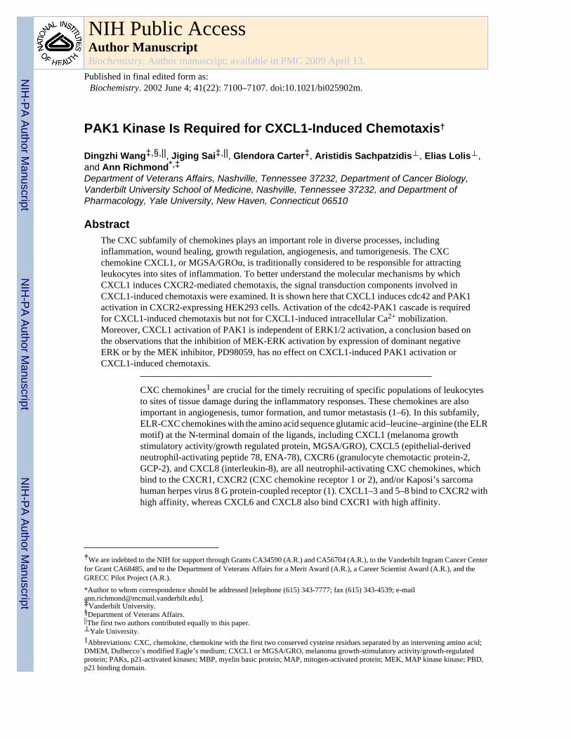
PAK1 Kinase Is Required for CXCL1-Induced Chemotaxis † Dingzhi Wang ‡,§,|| , Jiging Sai ‡,|| , Glendora Carter ‡ , Aristidis Sachpatzidis ⊥ , Elias Lolis ⊥ , and Ann Richmond *,‡ Department of Veterans Affairs, Nashville, Tennessee 37232, Department of Cancer Biology, Vanderbilt University School of Medicine, Nashville, Tennessee 37232, and Department of Pharmacology, Yale University, New Haven, Connecticut 06510 Abstract The CXC subfamily of chemokines plays an important role in diverse processes, including inflammation, wound healing, growth regulation, angiogenesis, and tumorigenesis. The CXC chemokine CXCL1, or MGSA/GROα, is traditionally considered to be responsible for attracting leukocytes into sites of inflammation. To better understand the molecular mechanisms by which CXCL1 induces CXCR2-mediated chemotaxis, the signal transduction components involved in CXCL1-induced chemotaxis were examined. It is shown here that CXCL1 induces cdc42 and PAK1 activation in CXCR2-expressing HEK293 cells. Activation of the cdc42-PAK1 cascade is required for CXCL1-induced chemotaxis but not for CXCL1-induced intracellular Ca 2+ mobilization. Moreover, CXCL1 activation of PAK1 is independent of ERK1/2 activation, a conclusion based on the observations that the inhibition of MEK-ERK activation by expression of dominant negative ERK or by the MEK inhibitor, PD98059, has no effect on CXCL1-induced PAK1 activation or CXCL1-induced chemotaxis. CXC chemokines 1 are crucial for the timely recruiting of specific populations of leukocytes to sites of tissue damage during the inflammatory responses. These chemokines are also important in angiogenesis, tumor formation, and tumor metastasis (1–6). In this subfamily, ELR-CXC chemokines with the amino acid sequence glutamic acid–leucine–arginine (the ELR motif) at the N-terminal domain of the ligands, including CXCL1 (melanoma growth stimulatory activity/growth regulated protein, MGSA/GRO), CXCL5 (epithelial-derived neutrophil-activating peptide 78, ENA-78), CXCR6 (granulocyte chemotactic protein-2, GCP-2), and CXCL8 (interleukin-8), are all neutrophil-activating CXC chemokines, which bind to the CXCR1, CXCR2 (CXC chemokine receptor 1 or 2), and/or Kaposi’s sarcoma human herpes virus 8 G protein-coupled receptor (1). CXCL1–3 and 5–8 bind to CXCR2 with high affinity, whereas CXCL6 and CXCL8 also bind CXCR1 with high affinity. † We are indebted to the NIH for support through Grants CA34590 (A.R.) and CA56704 (A.R.), to the Vanderbilt Ingram Cancer Center for Grant CA68485, and to the Department of Veterans Affairs for a Merit Award (A.R.), a Career Scientist Award (A.R.), and the GRECC Pilot Project (A.R.). *Author to whom correspondence should be addressed [telephone (615) 343-7777; fax (615) 343-4539; e-mail [email protected]]. ‡ Vanderbilt University. § Department of Veterans Affairs. || The first two authors contributed equally to this paper. ⊥ Yale University. 1 Abbreviations: CXC, chemokine, chemokine with the first two conserved cysteine residues separated by an intervening amino acid; DMEM, Dulbecco’s modified Eagle’s medium; CXCL1 or MGSA/GRO, melanoma growth-stimulatory activity/growth-regulated protein; PAKs, p21-activated kinases; MBP, myelin basic protein; MAP, mitogen-activated protein; MEK, MAP kinase kinase; PBD, p21 binding domain. NIH Public Access Author Manuscript Biochemistry. Author manuscript; available in PMC 2009 April 13. Published in final edited form as: Biochemistry. 2002 June 4; 41(22): 7100–7107. doi:10.1021/bi025902m. NIH-PA Author Manuscript NIH-PA Author Manuscript NIH-PA Author Manuscript
-
Upload
vanderbilt -
Category
Documents
-
view
1 -
download
0
Transcript of PAK1 Kinase Is Required for CXCL1-Induced Chemotaxis †
PAK1 Kinase Is Required for CXCL1-Induced Chemotaxis†
Dingzhi Wang‡,§,||, Jiging Sai‡,||, Glendora Carter‡, Aristidis Sachpatzidis⊥, Elias Lolis⊥,and Ann Richmond*,‡Department of Veterans Affairs, Nashville, Tennessee 37232, Department of Cancer Biology,Vanderbilt University School of Medicine, Nashville, Tennessee 37232, and Department ofPharmacology, Yale University, New Haven, Connecticut 06510
AbstractThe CXC subfamily of chemokines plays an important role in diverse processes, includinginflammation, wound healing, growth regulation, angiogenesis, and tumorigenesis. The CXCchemokine CXCL1, or MGSA/GROα, is traditionally considered to be responsible for attractingleukocytes into sites of inflammation. To better understand the molecular mechanisms by whichCXCL1 induces CXCR2-mediated chemotaxis, the signal transduction components involved inCXCL1-induced chemotaxis were examined. It is shown here that CXCL1 induces cdc42 and PAK1activation in CXCR2-expressing HEK293 cells. Activation of the cdc42-PAK1 cascade is requiredfor CXCL1-induced chemotaxis but not for CXCL1-induced intracellular Ca2+ mobilization.Moreover, CXCL1 activation of PAK1 is independent of ERK1/2 activation, a conclusion based onthe observations that the inhibition of MEK-ERK activation by expression of dominant negativeERK or by the MEK inhibitor, PD98059, has no effect on CXCL1-induced PAK1 activation orCXCL1-induced chemotaxis.
CXC chemokines1 are crucial for the timely recruiting of specific populations of leukocytesto sites of tissue damage during the inflammatory responses. These chemokines are alsoimportant in angiogenesis, tumor formation, and tumor metastasis (1–6). In this subfamily,ELR-CXC chemokines with the amino acid sequence glutamic acid–leucine–arginine (the ELRmotif) at the N-terminal domain of the ligands, including CXCL1 (melanoma growthstimulatory activity/growth regulated protein, MGSA/GRO), CXCL5 (epithelial-derivedneutrophil-activating peptide 78, ENA-78), CXCR6 (granulocyte chemotactic protein-2,GCP-2), and CXCL8 (interleukin-8), are all neutrophil-activating CXC chemokines, whichbind to the CXCR1, CXCR2 (CXC chemokine receptor 1 or 2), and/or Kaposi’s sarcomahuman herpes virus 8 G protein-coupled receptor (1). CXCL1–3 and 5–8 bind to CXCR2 withhigh affinity, whereas CXCL6 and CXCL8 also bind CXCR1 with high affinity.
†We are indebted to the NIH for support through Grants CA34590 (A.R.) and CA56704 (A.R.), to the Vanderbilt Ingram Cancer Centerfor Grant CA68485, and to the Department of Veterans Affairs for a Merit Award (A.R.), a Career Scientist Award (A.R.), and theGRECC Pilot Project (A.R.).*Author to whom correspondence should be addressed [telephone (615) 343-7777; fax (615) 343-4539; [email protected]].‡Vanderbilt University.§Department of Veterans Affairs.||The first two authors contributed equally to this paper.⊥Yale University.1Abbreviations: CXC, chemokine, chemokine with the first two conserved cysteine residues separated by an intervening amino acid;DMEM, Dulbecco’s modified Eagle’s medium; CXCL1 or MGSA/GRO, melanoma growth-stimulatory activity/growth-regulatedprotein; PAKs, p21-activated kinases; MBP, myelin basic protein; MAP, mitogen-activated protein; MEK, MAP kinase kinase; PBD,p21 binding domain.
NIH Public AccessAuthor ManuscriptBiochemistry. Author manuscript; available in PMC 2009 April 13.
Published in final edited form as:Biochemistry. 2002 June 4; 41(22): 7100–7107. doi:10.1021/bi025902m.
NIH
-PA Author Manuscript
NIH
-PA Author Manuscript
NIH
-PA Author Manuscript

Our earlier studies demonstrated that CXCL1 induces activation of the transcription factor NF-κB through a Ras-MEKK1-MEK4/6-p38 MAP kinase cascade in melanocytes (7). Thispathway is involved in CXCL1-induced melanocyte transformation (6). Activation of thephospholipase C–β/PKC/IP3 cascade is required for the CXC chemokine-induced intracellularcalcium mobilization in neutrophils (8). Although the chemotactic response to CXCL1 andCXCL8 is well characterized, the signal transduction pathways for the chemotactic responseshave not been fully elucidated.
The activated GTPases interact with specific targets that serve as effectors to regulatedownstream signaling cascades. The Rho GTPase subfamily, including RhoA, RhoB, RhoC,Rac, and cdc42, has been implicated in the regulation of diverse cellular functions, includingactin cytoskeletal dynamics, oxidant generation, transformation, membrane trafficking,apoptosis, transcription, and cell cycle control (9–12). Rac and cdc42 appear to be criticaldownstream components for the classic chemoattractant fMet-Leu-Phe (13–14). SignificantRac/cdc42 targets are the p21-activated kinases (PAKs).
PAKs play an important role in diverse cellular processes, including cytoskeletalrearrangements (15–19), growth, and apoptosis (20–22). PAKs are Ser/Thr protein kinases,which contain a p21 binding domain (PDB). PAK1 undergoes autophosphorylation andactivation upon interacting with the active forms of the small GTPase (p21) Rac or Cdc42(23). PAK activation is regulated by a variety of external stimuli that act through cell surfacereceptors, including G protein-coupled receptors (24), growth factor receptor tyrosine kinases(25), proinflammatory cytokine receptors (26), Fc receptors (27), and integrins (28–29).Moreover, a variety of chemoattractants induce rapid activation of PAKs (30). However, therole of PAK1 in chemokine gradient-directed cell movement (chemotaxis) has not been clearlydelineated.
Mitogen-activated protein (MAP) kinases represent a point of convergence for cell surfacesignals regulating cell growth and division. MAP kinases are serine/threonine protein kinases.One member of the MAP kinase family is extra-cellular signal-related protein kinase (ERK).ERK is phosphorylated and activated by MAP kinase kinase (MEK1) (31), which in turn isphosphorylated and activated by the Raf (32). CXCL8 has also been demonstrated to activatethe PI3-kinase/Ras/Raf cascade in neutrophils (33). Similarly, CXCL1 induces the activationof ERK through Ras/Raf1 dependent or independent pathways (34). However, it remainscontroversial whether ERK activation is required for the CXC ligand-induced chemotaxis(33,35). Van Lint et al. reported that ERK activation is involved in IL-8-induced chemotaxisin neutrophils (35). However, Knall et al. reported that the regulation of cell migration by IL-8is independent of ERK kinase and ERK activation because the ERK kinase inhibitor PD098059had no effect on IL-8-induced cell migration of human neutrophils (33).
To determine what signal transducers are involved in CXCL1-induced chemotaxis, we usedthe HEK293 and RBL systems, which provide cellular models to characterize the signalingmechanisms of CXCR2, as such studies are notoriously difficult to perform in primaryneutrophils, which express multiple chemokine receptors. Our findings demonstrate thatCXCL1 induces PAK1 activation through cdc42. This cdc42–PAK1 cascade is required forCXCL1-induced chemotaxis. In contrast, we demonstrate that the CXCL1 induction of MEK-ERK1/2 is not involved in the CXCL1-induced chemotaxis. Moreover, cdc42–PAK1 and ERKare not required for the intracellular Ca2+ mobilization induced by CXCL1.
Wang et al. Page 2
Biochemistry. Author manuscript; available in PMC 2009 April 13.
NIH
-PA Author Manuscript
NIH
-PA Author Manuscript
NIH
-PA Author Manuscript

EXPERIMENTAL PROCEDURESCell Culture
Human embryonic kidney 293 cells (HEK293) were cultured in DMEM supplemented with50 units/mL penicillin, 50 μg/mL streptomycin, 3 mM glutamine, and 5% heat-inactivated fetalbovine serum (GIBCO BRL, Rockville, MD). The CXCR2-expressing HEK293 polyclonalcells were cultured in the same medium supplemented with 800 μg/mL G418 (Sigma, St. Louis,MO) as previously described (36). The expression level of CXCR2 receptor in the HEK293cells has been previously verified (36). RBL-2H3 cells and CXCR2-expressing RBL stableclone cells were gifts from Dr. Ricardo Richardson. RBL-2H3 cells were cultured in DMEMsupplemented with 50 units/mL penicillin, 50 μg/mL streptomycin, 3 mM glutamine, 15%heat-inactivated fetal bovine serum (GIBCO BRL, Rockville, MD). CXCR2-expressing RBLwere cultured in the same medium supplemented with 1000 μg/mL G418 (Sigma) as previouslydescribed. The expression level of CXCR2 receptor in the RBL-2H3 cells has been previouslyverified (37). Purified recombinant human CXCL1 (a kind gift of Repligen Corp., Needham,MA) was used at 50 ng/mL. MEK kinase inhibitor, PD98059 (Calbiochem, La Jolla, CA), wasadded at the indicated concentration overnight prior to stimulation with CXCL1.
TransfectionsCXCR2-expressing HEK293 cells cultured to 80% confluence were transiently transfectedwith either the empty expression vector, the dominant negative PAK1 (232 K/A) plasmid (agift from Dr. Jeffrey Frost) (38), dominant negative cdc42, or the dominant negative ERKplasmid (a gift from Dr. Melanie Cobb), using the Lipo-fectAMINE PLUS reagent (GIBCOBRL) according to the manufacturer’s protocol. RBL cells (107 cells) were transientlycotransfected with CXCR2 receptor (20 μg) and either the empty expression vector (20 μg) orthe dominant negative PAK1 (232 K/A) plasmid (20 μg), using electroporation (37). Weroutinely achieved a transfection efficiency of ~80% with these procedures.
Whole Cell Extracts and Western BlotWhole cell extracts were prepared from CXCR2-expressing HEK293 treated with CXCL1 forthe indicated time after serum starvation for 14 h. Western blots were performed followingprotocols provided by Santa Cruz Biotechnology Inc. (Santa Cruz, CA). The cells were washedat 4 °C with 1× PBS and lysed in 0.6 mL of RIPA buffer (1× PBS, 1% NP-40, 0.5% sodiumdeoxycholate, and 0.1% SDS) with protease inhibitor cocktail tablets (Boehringer MannheimCorp., Indianapolis, IN) and 0.2 mM sodium orthovanadate. Fifty micrograms of solubleprotein was boiled, subjected to electrophoresis on a 10% SDS–PAGE reducing gel, and thenelectrophoretically transferred to a 0.45-μm nitrocellulose membrane (Bio-Rad, Hercules, CA).The membrane was blocked with 5% dry milk in TBS-T buffer (10 mM Tris-HCl, pH 8.0; 150mM NaCl; and 0.05% Tween-20) for 1 h and then incubated for 12–16 h at 4 °C in a 1:1000dilution (0.2 μg/mL) of the anti-Cdc42 antibody (Santa Cruz Biotechnology, Inc.) and anti-ERK1/2 or the anti-phospho-ERK1/2 (Santa Cruz Biotechnology, Inc.) in TBS-T buffercontaining 5% dry milk. After three washings with TBS-T buffer, the membrane was incubatedin a 1:3000 dilution of the appropriate anti-mouse or anti-rabbit immunoglobulin conjugatedwith horseradish peroxidase (Boehringer Mannheim Corp.) in TBS-T buffer with 5% dry milkfor 1 h at room temperature. After three washings with TBS-T buffer, the protein bands weredetected with the ECL Western blotting detection reagents (Amersham Pharmacia Biotech,Piscataway, NJ) according to the manufacturer’s instructions. The blots were stripped andreprobed with anti-ERK2.
Wang et al. Page 3
Biochemistry. Author manuscript; available in PMC 2009 April 13.
NIH
-PA Author Manuscript
NIH
-PA Author Manuscript
NIH
-PA Author Manuscript

Immune Complex Kinase AssaysWhole cell extracts were prepared from CXCR2-expressing HEK293 or CXCR2-expressingRBL-2H3 cells treated with CXCL1 after overnight serum starvation. PAK1 kinase assayswere performed as described in the manufacturer’s protocol (Upstate Biotechnology, LakePlacid, NY). Four hundred micrograms of protein of each whole cell extract wasimmunoprecipitated with 1 μg of PAK1 antibody. Immunoprecipitated PAK1 activity wasassayed using the PAK1 substrate myelin basic protein (MBP) (Sigma). Kinase reactions wereinitiated by addition of 2 μg of MBP and kinase buffer containing 500 μM cold ATP and 10μCi of [γ-32P]ATP. Reactions were incubated for 30 min at 30 °C and terminated by the additionof an equal volume of 2 × SDS loading buffer followed by boiling for 5 min. Phosphorylatedproteins were resolved on a 10% SDS–PAGE reducing gel and transferred to a 0.45-μmnitrocellulose membrane (Bio-Rad). The phosphorylated bands were visualized byautoradiography. The blot was probed with PAK1 antibody to monitor equal loading of PAK1.
Chemotaxis AssayChemotaxis assays were performed on the HEK293 or the RBL-2H3 cells transientlycotransfected with CXCR2 and other constructs as described previously (36,37). Briefly, a 96-well chemotaxis chamber (Neuroprobe Inc., Gaithersburg, MD) was used, and the lowercompartment of the chamber was loaded with 450 mL of chemotaxis buffer (1 mg/mLovalbumin/DMEM) containing CXCL1 diluted at the indicated concentration in thechemotaxis buffer. Polycarbonate membranes (10- or 8-μm pore size) were coated on bothsides with 20 μg/mL human collagen type IV (Sigma) and incubated for 2 h at 37 °C. The cellswere removed from the plate by trypsinization and incubated in 5% FBS/DMEM for 2 h at 37°C to allow restoration of receptor expression. The cells were washed with chemotaxis bufferand then loaded into the upper chamber in 250 μL of chemotaxis buffer at 5 × 10⁶ cells/mL.The chamber was incubated for 4 h at 37 °C with 5% CO2, and then the membrane was removed,washed, fixed, and stained with a Diff-Quik kit. Cell chemotaxis was quantifiedmicroscopically by counting cells in five high-power fields (× 40). The relative chemotacticindex represented the mean number of cells migrating in response to ligand stimulation ascompared to that without ligand stimulation.
Cdc42 Activity AssayPBD (p21 binding domain)-based assays of CDC42 were performed as described by Benardet al. (13). Briefly, CXCR2 expressing HEK293 cells were stimulated with 50 ng/mL CXCL1for the indicated time, and cells were immediately lysed by sonication in RIPA buffercontaining cocktail protease inhibitor. Four hundred micrograms of protein of each whole cellextract was incubated with purified GST–PBD (GST-conjugated p21 binding domain) beadsfor 30 min at 4 °C. The bound GTP–Cdc42 and total level of Cdc42 were detected by Westernblotting using a cdc42 polyclonal antibody (SC-87) (Santa Cruz Biotechnology).
Intracellular Ca2+ MobilizationChemokine-induced intracellular Ca2+ mobilization was measured as described by Wang etal. (39). Briefly, subconfluent CXCR2-expressing HEK293 cells transfected with vector,dominant negative PAK1, dominant negative cdc42, or dominant negative ERK1/2 were platedon glass-bottom microwells and grown overnight. Prior to the experiment, the cells wereincubated in serum-free media for 3–4 h. The cells were then rinsed with wash buffer (10 mMHepes, pH 7.4; 140 mM NaCl; 5mM KCl; 1 mM MgCl2; and 0.55 mM glucose) and loadedwith 1 μM Fluo-3 AM for 30 min at room temperature. After a wash with wash buffer, 1 mLof wash buffer containing 1mM CaCl2 was added to the cells. The microwell was then placedon a Zeiss Axiovert 135 confocal microscope, and the cells were stimulated with CXCL1 (100ng/mL) at room temperature. The emitted fluorescence at a wavelength of 488 nm was
Wang et al. Page 4
Biochemistry. Author manuscript; available in PMC 2009 April 13.
NIH
-PA Author Manuscript
NIH
-PA Author Manuscript
NIH
-PA Author Manuscript

recorded. All images from the scanning were processed to analyze the change of relativefluorescence intensity at the single-cell level using the NIH Image program. The relativefluorescence intensity of each sample in the figures represents the mean of the relativefluorescence intensity of six randomly chosen fields (10 cells were counted in each field).
RESULTSCXCL1 Induces PAK1 Activation
To determine whether CXCL1 induces PAK1 activation through activation of CXCR2, PAK1kinase assays were performed to evaluate endogenous PAK1 kinase activity in the CXCR2-expressing HEK293 cells stimulated with CXCL1 for the indicated times. The results of theseassays showed that CXCL1 stimulation of CXCR2-expressing HEK293 cells with CXCL1increased the ability of PAK1 to phosphorylate myelin basic protein (MBP), which is asubstrate of PAK1 (Figure 1A, top panel). The PAK1 activation started at 5 min, reached themaximum at 30 min, and was almost back to the basal level at 120 min. The expression levelof PAK1 in the samples from the various time points was equivalent (Figure 1A, lower panel).In contrast, CXCL1 failed to induce PAK1 activation in parental HEK293 cells (data notshown). These data demonstrate that CXCL1 induces PAK1 activation through CXCR2.
PAK1 Mediates CXCL1-Induced ChemotaxisLigand-stimulated CXCR2-mediated chemotaxis is a direct and effective functional test toaccess the chemokine receptor signal transduction. Because PAK1 activation is involved inthe regulation of cytoskeletal organization, it was of interest to determine whether PAK1activation was required for CXCL1-induced chemotaxis. A dominant negative PAK1(pCMV5M/PAK1 232 K/A) (38) was transfected into HEK293 cells stably expressing CXCR2to determine whether loss of PAK1 activation could abolish the CXCR2-mediated chemotaxisin a modified Boyden chamber assay. This dominant negative form of PAK1 (232 K/A) hasonly a catalytic domain of PAK1 (amino acids 232–544) containing a point mutation thatrenders it inactive (K298A). Because it lacks the N-terminal regulatory domain, it cannot bindto Rac1 or Cdc42 (38). We observed a CXCL1 concentration-dependent chemotactic responsein the control cells (CXCR2-expressing HEK293 cells transfected with empty expressionvector of PAK1) with a peak migration occurring at a concentration of ~25 ng/mL CXCL1.Chemotaxis was inhibited at higher concentrations of CXCL1 (Figure 1B, empty bar), asreported earlier (36). In contrast, the expression of dominant negative PAK1 (232 K/A) resultedin a marked attenuation of CXCR2-mediated chemotaxis (Figure 1B, solid bar). In addition,the expression of a dominant negative PAK1 (R298), which lacks only kinase activity but canstill bind Rac and cdc42, also blocked CXCL1-induced chemotaxis (data not shown). BecauseCXCL1 failed to induce a chemotactic response in the parental HEK293 cells (data not shown),these data demonstrate that PAK1 is required for CXCL1-stimulated CXCR2-mediatedchemotaxis.
PAK1 Is a Downstream Target of Cdc42Recent studies showed that PDK1 and Akt mediators activate PAK1 independent of activationof cdc42 and Rac (40,41). Because activation of another chemoattractant receptor, the fMLPreceptor, activates cdc42 (13), we examined whether CXCL1 activation of CXCR2 would alsoenhance cdc42 activation. Cdc42 activation assays were performed to evaluate endogenouscdc42 activity in the CXCR2-expressing HEK293 cells stimulated with 50 ng/mL of CXCL1for the indicated times. The stimulation of CXCL1 increased the amount of endogenous GTP-bound cdc42 (active form of cdc42) (Figure 2A, upper panel). The levels of total cdc42 (GTP-cdc42 + GDP-cdc42) from the different samples were equivalent (Figure 2A, lower panel).The profile of cdc42 activation is consistent with that of PAK1 activation. To determinewhether PAK1 is a substrate of cdc42 in CXCR2-expressing HEK293 cells, we tested whether
Wang et al. Page 5
Biochemistry. Author manuscript; available in PMC 2009 April 13.
NIH
-PA Author Manuscript
NIH
-PA Author Manuscript
NIH
-PA Author Manuscript

the inhibition of cdc42 activation by expression of the dominant negative cdc42 would blockCXCL1-induced PAK1 activation. Figure 2B shows that the dominant negative cdc42 inhibitedCXCL1-induced PAK1 activation. This experiment demonstrates that CXCL1-induced PAK1activation is dependent on cdc42 activation. To further test whether cdc42 is involved inCXCL1-induced PAK1-mediated chemotaxis, modified Boyden chamber assays wereperformed. Figure 2C shows that a CXCL1 concentration-dependent chemotactic response wasobserved in the CXCR2-expressing HEK293 cells transfected with the empty vector control(Figure 2C, white bar), but not in the same cells transfected with the dominant negative cdc42expression plasmid (Figure 2C, black bar). This experiment demonstrates that cdc42 is requiredfor CXCL1-induced chemotaxis. Taken together, these experiments demonstrate that a cdc42–PAK1 cascade is involved in CXCL1-induced chemotaxis mediated through CXCR2.
ERK Is Not a Downstream Target of PAK1Previous studies demonstrated that CXCL1 activated ERK1/2 in CXCR2 stably expressingHEK293 cells, but not in the parental HEK293 cells (36). Because PAK was shown to facilitateERK kinase activation by phosphorylating MEK (42), we examined whether ERK is adownstream target of PAK1 in response to CXCL1. Expression of dominant negative PAK1(232 K/A) in the CXCR2-expressing HEK293 cells did not block CXCL1-induced ERKactivation (Figure 3A). The data demonstrate that in CXCR2-expressing HEK293 cells, ERKsare not downstream targets of CXCL1-induced PAK1. However, we could not exclude thepossibility that ERK activation is involved in chemotaxis from these data. Therefore, toevaluate whether ERK activation is involved in CXCL1-induced chemotaxis, we examined theeffects of expression of dominant negative ERK on CXCR2-mediated chemotaxis. Theexpression of dominant negative ERK failed to block CXCL1-induced chemotaxis, ascompared to the vector control (Figure 3B). These data demonstrate that ERK activation is notrequired for CXCL1-stimulated CXCR2-mediated chemotaxis in HEK 293 cells.
CXCL1 Triggers Two Independent Signal Pathways To Activate PAK1 and ERK1/2,Respectively
To further determine whether CXCL1-induced PAK1 is independent of the MEK1–ERKkinase pathway, the MEK1/2 inhibitor, PD98059, was used to inhibit CXCL1-induced ERKactivation. PD98059 is an effective and specific inhibitor of ERK-mediated signaling (43).Figure 4A confirms that 25 μM PD98059 abrogated the CXCL1-induced ERK activation.However, inhibition of the MEK–ERK pathway with 25 μM PD98059 had essentially no effecton CXCL1-induced PAK1 activation (Figure 4B). Similarly, PD98059 (10–50 μM) did notblock CXCL1-induced chemotaxis (Figure 4C). This result is consistent with the results foundwith the expression of dominant negative ERK. Taken together, these data demonstrate thatCXCL1-induced PAK1 activation is independent of the MEK-ERK cascade.
Cdc42–PAK1 and ERK1/2 Are Not Required for CXCL1-Induced Intracellular Ca2+
MobilizationCXCL1 induces intracellular Ca2+ mobilization through CXCR2 in CXCR2-expressingHEK293 cells (36). Because the CXCL1 also induces cdc42-PAK1 and ERK1/2 activation,we examined whether PAK1, ERK1/2, or cdc42 is involved in the CXCL1-inducedintracellular Ca2+ mobilization. We performed calcium mobilization assays using fluo-3 Amloaded HEK293 cells stably expressing CXCR2. Cells were stimulated with CXCL1, and freeintracellular calcium localization was examined and quantified by confocal microscopy asdescribed under Experimental Procedures (39). The results are presented in Figure 5. Theexpression of either dominant negative PAK1, ERK1/2, or cdc42 did not block CXCL1-induced intracellular Ca2+ mobilization, as compared to the vector control. These experiments
Wang et al. Page 6
Biochemistry. Author manuscript; available in PMC 2009 April 13.
NIH
-PA Author Manuscript
NIH
-PA Author Manuscript
NIH
-PA Author Manuscript

demonstrate that the cdc42–PAK1 cascade and ERK are not involved in CXCL1-inducedintracellular Ca2+ mobilization.
RBL-2H3 CellsTo test whether the biological functions of PAK1 in HEK293 cells can be observed in RBL-2H3cells, we examined whether PAK1 activation is required for CXCL1-induced chemotaxis inRBL-2H3 cells. The CXCR2-expressing RBL stable clone cells were stimulated with CXCL1for the indicated times. As shown in Figure 6A, CXCL1 also can increase PAK1 kinase activityin RBL-2H3 cells. For chemotaxis assays, the RBL-2H3 cells were transiently transfected withCXCR2 receptor and either dominant negative PAK1 (232 K/A) or empty vector control forPAK1. Figure 6B shows that the expression of dominant negative PAK1 (232 K/A) inhibitedCXCL1-induced chemotaxis (sold bars). In addition, the expression of another dominantnegative PAK1 (R298) also blocked CXCL1-induced chemotaxis (data not shown). BecauseCXCL1 failed to induce a PAK1 activation and a chemotactic response in the parentalRBL-2H3 cells, these results demonstrated that PAK1 is required for CXCL1-stimulatedCXCR2-mediated chemotaxis.
DISCUSSIONLigand-bound receptors activate G proteins by catalyzing the exchange of GDP bound to theα subunit with GTP, leading to dissociation of α-GTP from the βγ subunit. Several majorintracellular signaling pathways are regulated by both α and βγ subunits. These include thecAMP/PKA pathway, the MAP kinase pathway, and the phosphatidylinositol/calciumpathway. CXCL8 activation of the PI3-kinase pathway is required for human neutrophilmigration. The overall mechanism(s) responsible for the CXCL1 activation of the PI3-kinasepathway is (are) likely to be the same for CXCL8 activation. PI3-kinase can regulate PAKactivation through Rac/cdc42 (44). The activation of cdc42 in response to CXCL1 in CXCR2-expressing HEK293 cells is more delayed, peaking at 5–10 min, compared to the activation ofcdc42 in response to fMLP in human neutrophils, where the peak activation occurs at 0.5–1min. The time course for Rac activation in response to CXCL1 is similar to the cdc42 inCXCR2-expressing HEK293 cells (data not shown). These differences in time course of Racand cdc42 activation might be due to (1) the differences between classic chemoattractantsversus CXC chemokines, (2) fMLP receptor versus CXCR2 receptor; and/or (3) cell typedifferences.
To date, four PAKs have been cloned, PAK1–4 (45–48). PAK1, 2, and 4 participate in theregulation of cytoskeletal organization (15–19,45,47,48). PAK2 is involved in the regulationof apoptosis (45,48,49). It has been reported that PAK1 is required for endothelial and fibroblastcell motility induced by an immobilized fibronectin (50,51). Here, we demonstrate that PAK1is required for chemokine gradient-directed cell movement (chemotaxis) by using dominantnegative PAK1. The expression of a dominant negative PAK1 (R299), which is defective onlyin kinase activity, blocked CXCL-induced chemotaxis (data not shown). However, this mutantmay be inhibiting chemotaxis by sequestering cdc42 because it can still bind to Rac and cdc42.Dr. Melanie Cobb’s group developed a novel dominant negative PAK1 mutant (232 K/A),which lacks kinase activity and fails to bind Rac and cdc42. So this PAK1 mutant (232 K/A)blocks only endogenous PAK1 activity but does not sequester the endogenous cdc42 and Racand inhibit their interactions with other effectors (38). In CXCR2-expressing HEK293 cells,this PAK1 mutant (232 K/A) also blocked the endogenous PAK1 activation induced by CXCL1(data not shown). We used this dominant negative PAK1 (232 K/A) to test whether PAK1activation is required for a chemokine gradient directional cell movement. Our datademonstrated that PAK1 is required for CXCL1-induced chemotaxis in both HEK293 andRBL-2H3 cells.
Wang et al. Page 7
Biochemistry. Author manuscript; available in PMC 2009 April 13.
NIH
-PA Author Manuscript
NIH
-PA Author Manuscript
NIH
-PA Author Manuscript

PAKs have been shown to regulate the MAP kinases ERK, JNK, and/or p38 in response tostimuli from cytokines, chemoattractants, and various stresses in certain types of the cell (41,45). In CXCR2-expressing HEK293 cells, ERK is not a downstream target of PAK1. Recently,published data indicated that PAKs phosphorylate key signaling components such as paxillin(52), myosin light chain kinase (19), and LIM kinase (18), all of which are involved inregulation of the cytoskeletal organization. We have not, however, determined the exactdownstream targets for PAK in CXCR2-expressing HEK293 cells. Future studies will addressthese unsolved issues.
In general, G-protein coupled receptors activate ERK1/2 via a Gβγ subunit complex. Thesignals for ERK1/2 activation are independent of receptor-mediated effects onphosphatidylinositol hydrolysis, calcium flux, or inhibition of adenyl cyclase (53,54). Ourearlier data showed that CXCL1 activates the Ras–MEKK cascade, which is an upstream signaltransduction pathway for MEK–ERK activation (7). Here, we show that ERK1/2 are notdownstream targets of PAK1. However, it has been reported that ERK activation down-regulates p38 MAP kinase activity (55). It is possible that the ERKs may be indirectly involvedin CXCL1-induced chemotaxis by altering downstream signaling of PAK1. Our datademonstrate that ERK activation is not involved in CXCL1-induced chemotaxis in CXCR2-expressing HEK293 cells.
For the first time, we demonstrate here that the cdc42–PAK1 cascade is required for CXCL1-induced chemotaxis in the CXCR2-expressing HEK293 and RBL cells. The activation ofcdc42–PAK1 by CXCL1 is insensitive to inhibition of MEK1/2–ERK. ERK activation is alsonot required for CXCL1-induced chemotaxis. Moreover, CXCL1-induced intracellular Ca2+
mobilization is independent of both the cdc42–PAK1 and MEK–ERK cascades. Thisconclusion is consistent with the previous observation that CXC-chemokine-induced calciummobilization is mediated by a phospholipase C-β, protein kinase C, and the IP3 cascade (8).Taken together, our findings further define the signal transduction pathways for diversebiologic functions of CXCL1. Advances in the relationship between ligand biologic functionand signal transduction pathways should lead to development of specific inhibitors, which canbe useful for pharmacological targets.
AcknowledgementsWe also are indebted to Dr. Gary Bokoch for providing GST-PBD/hPCR construct, Dr. Melanie Cobb for providingthe mutant PAK1 (232 K/A) construct, and Xuejie Wang for assistance with calcium mobilization assays.
References1. Rossi D, Zlotnik A. Annu Rev Immunol 2000;18:217–242. [PubMed: 10837058]2. Clark-Lewis I, Schumacher C, Baggiolini M, Moser B. J Biol Chem 1991;266:23128–23134. [PubMed:
1744111]3. Strieter RM, Polverini PJ, Kunkel SL, Arenberg DA, Burdick MD, Kasper J, Dzuiba J, Van Damme
J, Walz A, Marriott D, Chan SY, Roczniak S, Shanafelt AB. J Biol Chem 1995;270:348–357.4. Arenberg DA, Polverini PJ, Kunkel S, Shanafelt A, Hesselgesser JR, Strieter M. J Leukocyte Biol
1997;62:554–562. [PubMed: 9365108]5. Luan J, Shattuck-Brandt R, Haghnegahdar H, Owen JD, Strieter R, Burdick M, Nirodi C, Beauchamp
D, Johnson KN, Richmond A. J Leukocyte Biol 1997;62:588–597. [PubMed: 9365113]6. Wang D, Yang W, Du J, Devalaraja MN, Liang P, Matsumoto K, Tsubakimoto K, Endo T, Richmond
A. Oncogene 2000;19:4647–4659. [PubMed: 11030154]7. Wang D, Richmond A. J Biol Chem 2001;276:3650–3659. [PubMed: 11062239]8. Spivak-Kroizman T, Lemmon MA, Dikic I, Ladbury JE, Pinchasi D, Huang J, Jaye M, Crumley G,
Schlessinger J, Lax I. Cell 1994;79:1015–1024. [PubMed: 7528103]
Wang et al. Page 8
Biochemistry. Author manuscript; available in PMC 2009 April 13.
NIH
-PA Author Manuscript
NIH
-PA Author Manuscript
NIH
-PA Author Manuscript

9. Hall A. Science 1998;279:509–514. [PubMed: 9438836]10. Bokoch GM. Trends Cell Biol 1995;5:109–113. [PubMed: 14732165]11. Zigmond SH. Curr Opin Cell Biol 1996;8:66–73. [PubMed: 8791404]12. Van Aelst L, D’Souza-Schorey C. Genes Dev 1997;11:2295–2322. [PubMed: 9308960]13. Benard V, Bohl BP, Bokoch GM. J Biol Chem 1999;274:13198–13204. [PubMed: 10224076]14. Schraufstatter IU, Chung J, Burger M. Am J Physiol Lung Cell Mol Physiol 2000;280:L1094–L1103.
[PubMed: 11350788]15. Sells MA, Knaus UG, Bagrodia S, Ambrose DM, Bokoch GM, Chernoff J. Curr Biol 1997;7:202–
210. [PubMed: 9395435]16. Manser E, Huang HY, Loo TH, Chen XQ, Dong JM, Leung T, Lim L. Mol Cell Biol 1997;17:1129–
1143. [PubMed: 9032240]17. Sells MA, Boyd TJ, Chernoff J. J Cell Biol 1999;145:837–849. [PubMed: 10330410]18. Edwards CD, Sanders CL, Gill NG, Bokoch MG. Nat Cell Biol 1999;1:253–259. [PubMed:
10559936]19. Sanders CL, Matsumura F, Bokoch MG, de Lanerolle P. Science 1999;283:2083–2085. [PubMed:
10092231]20. Qu J, Cammarano MS, Shi Q, Ha KC, de Lanerolle P, Minden A. Mol Cell Biol 2001;21:3523–3533.
[PubMed: 11313478]21. Gnesutta N, Qu J, Minden A. J Biol Chem 2001;276:14414–12219. [PubMed: 11278822]22. Jakobi R, Moertl E, Koeppel MA. J Biol Chem 2001;276:16624–16634. [PubMed: 11278362]23. Manser E, Leung T, Salihuddin H, Zhao ZS, Lim L. Nature 1994;367:40–46. [PubMed: 8107774]24. Knaus UG, Morris S, Dong HJ, Chernoff J, Bockoch GM. Science 1995;269:221–223. [PubMed:
7618083]25. Dharmawardhane S, Sanders LC, Martin SS, Daniels RH, Bokoch GM. J Cell Biol 1997;138:1265–
1278. [PubMed: 9298982]26. Zhang S, Han J, Sells MA, Cheeroff J, Knaus UG, Ulevitch RJ, Bokoch GM. J Biol Chem
1995;270:23934–23936. [PubMed: 7592586]27. Jones SL, Knaus UG, Bokoch GM, Brown EJ. J Biol Chem 1998;273:10556–10566. [PubMed:
9553116]28. Price LS, Leng J, Schwartz MA, Bokoch GM. Mol Cell Biol 1998;9:1863–1871.29. Kiosses WB, Daniels RH, Otey C, Bokoch GM, Schwartz MA. J Cell Biol 1999;147:831–844.
[PubMed: 10562284]30. Huang R, Lian PJ, Robinson D, Badwey AJ. Mol Cell Biol 1998;18:7130–7238. [PubMed: 9819399]31. Robinson MJ, Cobb MH. Curr Opin Cell Biol 1997;9:180–186. [PubMed: 9069255]32. Moodies SA, Willumsen BM, Weber MJ, Wolfman A. Science 1993;260:1658–1661. [PubMed:
8503013]33. Knall C, Worthen GS, Johnson GL. Proc Natl Acad Sci USA 1997;94:3052–3057. [PubMed:
9096344]34. Shyamala V, Khoja H. Biochemistry 1998;37:15918–15924. [PubMed: 9843397]35. Van Lint J, Van Damme J, Billiau A, Merlevede W, Vandenheede JR. Mol Cell Biochem
1993;128:171–17712. [PubMed: 7523847]36. Yang W, Schraw PW, Mueller GS, Richmond A. Biochemistry 1997;36:15193–15200. [PubMed:
9398246]37. Richardson RM, Pridgen BC, Haribabu B, Snyderman R. J Biol Chem 2000;275:9201–9208.
[PubMed: 10734056]38. Frost JA, Swantek JL, Stippec S, Yin MJ, Gaynor R, Cobb MH. J Biol Chem 2000;275:19693–19699.
[PubMed: 10779525]39. Wang XJ, Liao HJ, Chattopadhyay A, Carpenter G. Exp Cell Res 2001;267:28–36. [PubMed:
11412035]40. King CC, Gardiner EM, Zenke FT, Bohl BP, Newton AC, Hemmings BA, Bokoch GM. J Biol Chem
2000;275:41201–41209. [PubMed: 10995762]41. Chung CY, Potikyan G, Firtel RA. Mol Cell 2001;7:937–947. [PubMed: 11389841]
Wang et al. Page 9
Biochemistry. Author manuscript; available in PMC 2009 April 13.
NIH
-PA Author Manuscript
NIH
-PA Author Manuscript
NIH
-PA Author Manuscript

42. Frost JA, Steen H, Shapiro P, Lewis T, Ahn N, Shaw PE, Cobb MH. EMBO J 1997;16:6426–6438.[PubMed: 9351825]
43. Zhang C, Baumgartner RA, Yamada K, Beaven MA. J Biol Chem 1997;272:13397–13402. [PubMed:9148963]
44. Krasilnikov MA. Biochemistry (Moscow) 2000;65:59–67. [PubMed: 10702641]45. Lim L, Manser E, Leung T, Hall C. Eur J Biochem 1996;242:171–185. [PubMed: 8973630]46. Abo A, Qu J, Cammarano MS, Dan C, Fritsch A, Baud V, Belisle B, Minden. EMBO J 1998;17:6527–
6540. [PubMed: 9822598]47. Zhao ZS, Manser E, Chen Q, Chong C, Leung T, Lim L. Mol Cell Biol 1998;18:2153–2163. [PubMed:
9528787]48. Lee LF, Li G, Templeton DJ, Ting JPY. J Biol Chem 1998;273:28253–28260. [PubMed: 9774447]49. Knaus UG, Bokoch GM. Int J Biochem Cell Biol 1998;30:857–862. [PubMed: 9744077]50. Sells MA, Boyd JT, Chernoff J. J Cell Biol 1999;145:837–849. [PubMed: 10330410]51. Kiosses WB, Daniels RH, Otey C, Bokoch GM, Schwartz MA. J Cell Biol 1999;147:831–843.
[PubMed: 10562284]52. Hashimoto S, Tsubouchi A, Mazaki Y, Sabe H. J Biol Chem 2001;276:6037–6045. [PubMed:
11096073]53. Alblas J, van Corven EJ, Hordijk PL, Milligan G, Moolenaar WH. J Biol Chem 1993;268:22235–
22238. [PubMed: 8226727]54. van Corven EJ, Groenink A, Jalink K, Eichholtz T, Moolenaar WH. Cell 1989;59:45–54. [PubMed:
2551506]55. Carter AB, Hunninghake GW. J Biol Chem 2000;275:27858–27864. [PubMed: 10878013]
Wang et al. Page 10
Biochemistry. Author manuscript; available in PMC 2009 April 13.
NIH
-PA Author Manuscript
NIH
-PA Author Manuscript
NIH
-PA Author Manuscript

Figure 1.CXCL1 induces PAK1 activity, which is required for chemotaxis in CXCR2-expressingHEK293 cells: (A) PAK1 kinase activity. CXCR2-expressing HEK293 cells were eitheruntreated or treated with 50 ng/mL CXCL1 for the indicated times after serum starvation for14 h. Endogenous PAK1 was immunoprecipitated by PAK1 antibody from whole cell extracts.PAK1 activity was determined by an immunocomplex kinase assay using MBP as a substrateas described under Experimental Procedures. Phosphorylated proteins were resolved on a 10%SDS–PAGE reducing gel and transferred to a nitrocellulose membrane. The phosphorylatedMBP bands were visualized by autoradiography (upper panel). The blot was probed with PAK1antibody to monitor equal loading of PAK1 (lower panel). This figure is representative of threedifferent experiments with similar results. (B) Effect of the dominant negative PAK1 onCXCL1-stimulated CXCR2-mediated chemotaxis in CXCR2-expressing HEK293 cells.CXCR2-expressing HEK293 cells were transiently transfected with either the emptyexpression vector (white bars) or dominant negative PAK1 plasmid (black bars). Two daysafter transfection, cells were compared for chemotactic response to CXCL1 stimulation asdescribed under Experimental Procedures. Values represent the means ± SE of threeindependent experiments. The data were analyzed using Student’s paired t test (p < 0.05).
Wang et al. Page 11
Biochemistry. Author manuscript; available in PMC 2009 April 13.
NIH
-PA Author Manuscript
NIH
-PA Author Manuscript
NIH
-PA Author Manuscript

Figure 2.CXCL1 induces cdc42 activation, which is required for chemotaxis: (A) Cdc42 activity.CXCR2-expressing HEK293 cells were either untreated or treated with 50 ng/mLCXCL1 forthe indicated times after serum starvation for 14 h. Endogenous GTP-bound cdc42 wasprecipitated from whole cell extracts by GST-PBD and immunoblotted with cdc42 antibody.The lower panel represents the total cdc42 expression level from whole cell extracts as theloading control. This figure is representative of three different experiments with similar results.(B) Effect of dominant negative cdc42 on the CXCL1-induced PAK1 activation. CXCR2-expressing HEK293 cells were transfected with either vector or dominant negative cdc42plasmid as described in Figure 1B. After 2 days, cells were either untreated or treated with 50ng/mL CXCL1 for 10 min. The PAK1 activation was detected by in vitro PAK1 kinase assaysas described in Figure 1A. The lower panel was reprobed with PAK1 antibody to monitor equalloading of PAK1. These figures are representative of three different experiments with similarresults. (C) Effect of the dominant negative cdc42 on CXCL1-stimulated CXCR2-mediatedchemotaxis in CXCR2-expressing HEK293 cells. CXCR2-expressing HEK293 cells weretransiently transfected with either the empty expression vector (white bars) or dominantnegative cdc42 plasmid (black bars). Two days after transfection, cells were compared forchemotactic response to CXCL1 stimulation as described under Experimental Procedures.Values represent the means ± SE of three independent experiments. The data were analyzedusing Student’s paired t test (p < 0.05).
Wang et al. Page 12
Biochemistry. Author manuscript; available in PMC 2009 April 13.
NIH
-PA Author Manuscript
NIH
-PA Author Manuscript
NIH
-PA Author Manuscript

Figure 3.ERK activation is not required for CXCL1-induced chemotaxis: (A) ERKs are not downstreamtargets of PAK1. CXCR2-expressing HEK293 cells were transfected with either vector ordominant negative PAK1 plasmid as described in Figure 1B. After 2 days, cells were eitheruntreated or treated with 50 ng/mL CXCL1 for 10 min. The ERK1/2 activation was detectedby Western blot as described in Figure 2A. The blot was reprobed with ERK antibody tomonitor equal loading of ERK1/2 (lower right panel). These figures are representative of threedifferent experiments. (B) Effect of the dominant negative ERK on CXCL1-stimulatedCXCR2-mediated chemotaxis in HEK293 cells. CXCR2-expressing HEK293 cells weretransiently transfected with either the empty expression vector (white bars) or dominant
Wang et al. Page 13
Biochemistry. Author manuscript; available in PMC 2009 April 13.
NIH
-PA Author Manuscript
NIH
-PA Author Manuscript
NIH
-PA Author Manuscript

negative ERK plasmid (black bars). Two days after transfection, cells were compared forchemotactic response to CXCL1 stimulation as described under Experimental Procedures.Values represent the mean ± SE of three independent experiments performed in duplicate. Thedata were analyzed using Student’s paired t test (p < 0.05).
Wang et al. Page 14
Biochemistry. Author manuscript; available in PMC 2009 April 13.
NIH
-PA Author Manuscript
NIH
-PA Author Manuscript
NIH
-PA Author Manuscript

Figure 4.MEK inhibitor fails to block PAK1 activation and chemotaxis: (A) MEK inhibitor, PD98059,blocks CXCL1-induced ERK activation. The CXCR2 stably expressing HEK293 cells weretreated with DMSO or different concentrations of PD98059 overnight before stimulation withCXCL1. ERK activation assays were performed as described in Figure 3A. (B) PD98059 failedto inhibit CXCL1-induced PAK1 activation. CXCR2 stably expressing HEK293 cells weretreated with DMSO or PD98059 as described above. PAK1 kinase assays were performed asdescribed in Figure 1. (C) PD98059 did not inhibit CXCL1-induced chemotaxis. CXCR2-expressing HEK293 were treated with DMSO or PD98059 as described above. Chemotaxisassays were performed as described in Figure 1B. Values represent the means ± SE of threeindependent experiments. The data were analyzed using Student’s paired t test (p < 0.05).
Wang et al. Page 15
Biochemistry. Author manuscript; available in PMC 2009 April 13.
NIH
-PA Author Manuscript
NIH
-PA Author Manuscript
NIH
-PA Author Manuscript
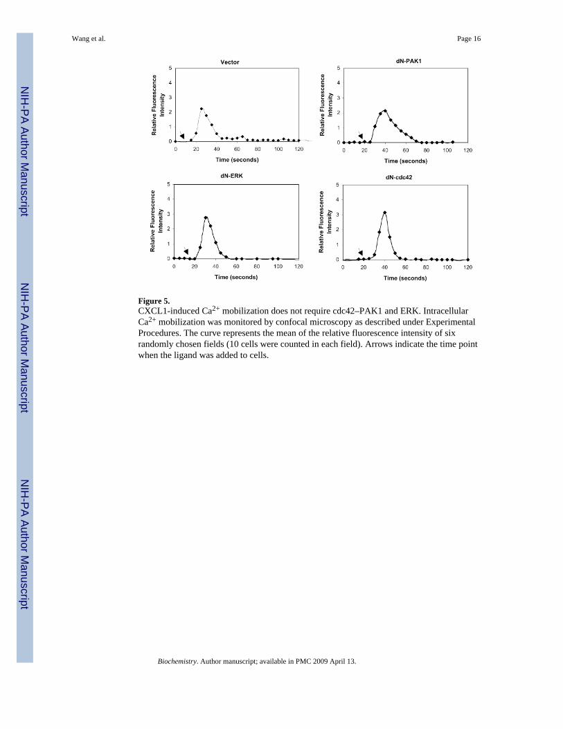
Figure 5.CXCL1-induced Ca2+ mobilization does not require cdc42–PAK1 and ERK. IntracellularCa2+ mobilization was monitored by confocal microscopy as described under ExperimentalProcedures. The curve represents the mean of the relative fluorescence intensity of sixrandomly chosen fields (10 cells were counted in each field). Arrows indicate the time pointwhen the ligand was added to cells.
Wang et al. Page 16
Biochemistry. Author manuscript; available in PMC 2009 April 13.
NIH
-PA Author Manuscript
NIH
-PA Author Manuscript
NIH
-PA Author Manuscript

Figure 6.PAK1 is required for CXCL1-induced chemotaxis in RBL-2H3 cells: (A) PAK1 kinaseactivity. The PAK1 activation in CXCR2-expressing RBL-2H3 cells was detected by in vitroPAK1 kinase assays as described in Figure 1A. The lower panel was reprobed with PAK1antibody to monitor equal loading of PAK1. These figures are representative of three differentexperiments with similar results. (B) Effect of dominant negative PAK1 (232 K/A) on CXCL1-induced PAK1 activation in RBL-2H3 cells. RBL-2H3 cells were transiently cotransfectedwith CXCR2 and either vector or dominant negative PAK1 plasmid, and chemotaxis assayswere performed as described in Figure 1B. Values represent the means ± SE of threeindependent experiments. The data were analyzed using Student’s paired t test (p < 0.05).
Wang et al. Page 17
Biochemistry. Author manuscript; available in PMC 2009 April 13.
NIH
-PA Author Manuscript
NIH
-PA Author Manuscript
NIH
-PA Author Manuscript