Flood Grouting for Infiltration Reduction on Private Side Sewers
Upload
khangminh22Category
view
0download
0
cancers
Review
Enhancing T Cell Chemotaxis and Infiltration in Glioblastoma
Kirit Singh * , Kelly M. Hotchkiss, Kisha K. Patel , Daniel S. Wilkinson, Aditya A. Mohan, Sarah L. Cookand John H. Sampson *
�����������������
Citation: Singh, K.; Hotchkiss, K.M.;
Patel, K.K.; Wilkinson, D.S.;
Mohan, A.A.; Cook, S.L.;
Sampson, J.H. Enhancing T Cell
Chemotaxis and Infiltration in
Glioblastoma. Cancers 2021, 13, 5367.
https://doi.org/10.3390/
cancers13215367
Academic Editor: Edward Pan
Received: 27 September 2021
Accepted: 25 October 2021
Published: 26 October 2021
Publisher’s Note: MDPI stays neutral
with regard to jurisdictional claims in
published maps and institutional affil-
iations.
Copyright: © 2021 by the authors.
Licensee MDPI, Basel, Switzerland.
This article is an open access article
distributed under the terms and
conditions of the Creative Commons
Attribution (CC BY) license (https://
creativecommons.org/licenses/by/
4.0/).
Duke Brain Tumor Immunotherapy Program, Department of Neurosurgery, Duke University Medical Center,Durham, NC 27710, USA; [email protected] (K.M.H.); [email protected] (K.K.P.);[email protected] (D.S.W.); [email protected] (A.A.M.); [email protected] (S.L.C.)* Correspondence: [email protected] (K.S.); [email protected] (J.H.S.)
Simple Summary: Immunotherapy in glioblastoma has so far failed to yield a survival benefit. Thisfailure can be attributed to a paucity of immune cells at the tumor site which can be reinvigoratedto kill tumor cells. Therefore, driving effector immune cells such as cytotoxic T lymphocytes to thetumor is a necessary pre-requisite of any effective immunotherapy approach. In this review, we willdiscuss therapeutic approaches possible for trafficking T cells from the periphery to travel throughthe blood–brain barrier and tissue of the brain to reach the tumor.
Abstract: Glioblastoma is an immunologically ‘cold’ tumor, which are characterized by absent orminimal numbers of tumor-infiltrating lymphocytes (TILs). For those tumors that have been invadedby lymphocytes, they are profoundly exhausted and ineffective. While many immunotherapyapproaches seek to reinvigorate immune cells at the tumor, this requires TILs to be present. Therefore,to unleash the full potential of immunotherapy in glioblastoma, the trafficking of lymphocytes tothe tumor is highly desirable. However, the process of T cell recruitment into the central nervoussystem (CNS) is tightly regulated. Naïve T cells may undergo an initial licensing process to enter themigratory phenotype necessary to enter the CNS. T cells then must express appropriate integrinsand selectin ligands to interact with transmembrane proteins at the blood–brain barrier (BBB).Finally, they must interact with antigen-presenting cells and undergo further licensing to enterthe parenchyma. These T cells must then navigate the tumor microenvironment, which is rich inimmunosuppressive factors. Altered tumoral metabolism also interferes with T cell motility. Inthis review, we will describe these processes and their mediators, along with potential therapeuticapproaches to enhance trafficking. We also discuss safety considerations for such approaches as wellas potential counteragents.
Keywords: immunotherapy; glioblastoma; blood–brain barrier; central nervous system; T cells;T lymphocytes
1. Introduction
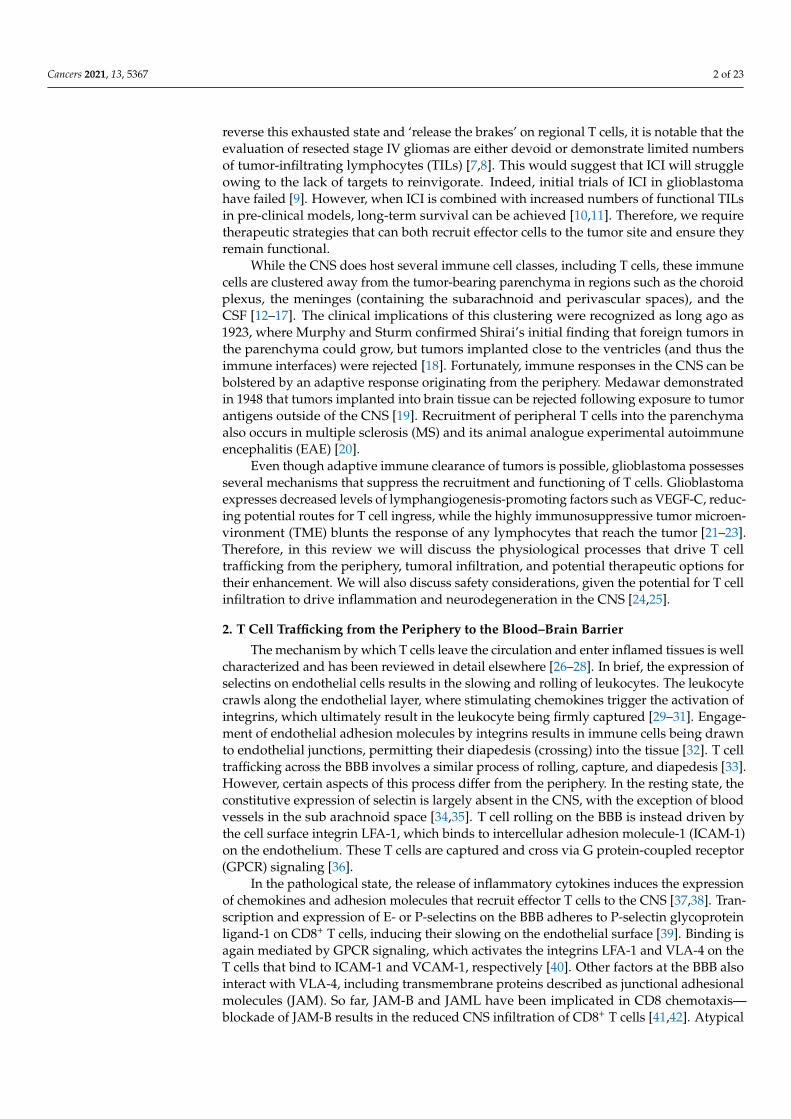
Immune surveillance of the central nervous system (CNS) is essential for environ-mental homeostasis and pathogen clearance. Without immune surveillance, opportunisticinfections in the CNS commonly develop [1]. However, the entry of immune cells into theCNS is tightly controlled by the blood–brain barrier (BBB) and the blood–cerebrospinalfluid (BCSF) barrier. These formidable barriers lack fenestrations, exhibit a low degree ofpinocytosis, and are sealed together by a network of intracellular junctions [2,3]. Whilethis close control is desirable in health to avoid runaway immune responses in the CNS,restricted immune cell entry severely hampers the effectiveness of immunotherapy inglioblastoma [4]. This is further complicated by the immunosuppressive tumor microenvi-ronment (TME), which consists of endothelial cells, pericytes, fibroblasts, and regulatoryimmune cells [5]. The TME drives effector immune cell exhaustion, thereby shielding solidmalignancies from immune attack [6]. While immune checkpoint inhibition (ICI) seeks to
Cancers 2021, 13, 5367. https://doi.org/10.3390/cancers13215367 https://www.mdpi.com/journal/cancers
Cancers 2021, 13, 5367 2 of 23
reverse this exhausted state and ‘release the brakes’ on regional T cells, it is notable that theevaluation of resected stage IV gliomas are either devoid or demonstrate limited numbersof tumor-infiltrating lymphocytes (TILs) [7,8]. This would suggest that ICI will struggleowing to the lack of targets to reinvigorate. Indeed, initial trials of ICI in glioblastomahave failed [9]. However, when ICI is combined with increased numbers of functional TILsin pre-clinical models, long-term survival can be achieved [10,11]. Therefore, we requiretherapeutic strategies that can both recruit effector cells to the tumor site and ensure theyremain functional.
While the CNS does host several immune cell classes, including T cells, these immunecells are clustered away from the tumor-bearing parenchyma in regions such as the choroidplexus, the meninges (containing the subarachnoid and perivascular spaces), and theCSF [12–17]. The clinical implications of this clustering were recognized as long ago as1923, where Murphy and Sturm confirmed Shirai’s initial finding that foreign tumors inthe parenchyma could grow, but tumors implanted close to the ventricles (and thus theimmune interfaces) were rejected [18]. Fortunately, immune responses in the CNS can bebolstered by an adaptive response originating from the periphery. Medawar demonstratedin 1948 that tumors implanted into brain tissue can be rejected following exposure to tumorantigens outside of the CNS [19]. Recruitment of peripheral T cells into the parenchymaalso occurs in multiple sclerosis (MS) and its animal analogue experimental autoimmuneencephalitis (EAE) [20].
Even though adaptive immune clearance of tumors is possible, glioblastoma possessesseveral mechanisms that suppress the recruitment and functioning of T cells. Glioblastomaexpresses decreased levels of lymphangiogenesis-promoting factors such as VEGF-C, reduc-ing potential routes for T cell ingress, while the highly immunosuppressive tumor microen-vironment (TME) blunts the response of any lymphocytes that reach the tumor [21–23].Therefore, in this review we will discuss the physiological processes that drive T celltrafficking from the periphery, tumoral infiltration, and potential therapeutic options fortheir enhancement. We will also discuss safety considerations, given the potential for T cellinfiltration to drive inflammation and neurodegeneration in the CNS [24,25].
2. T Cell Trafficking from the Periphery to the Blood–Brain Barrier
The mechanism by which T cells leave the circulation and enter inflamed tissues is wellcharacterized and has been reviewed in detail elsewhere [26–28]. In brief, the expression ofselectins on endothelial cells results in the slowing and rolling of leukocytes. The leukocytecrawls along the endothelial layer, where stimulating chemokines trigger the activation ofintegrins, which ultimately result in the leukocyte being firmly captured [29–31]. Engage-ment of endothelial adhesion molecules by integrins results in immune cells being drawnto endothelial junctions, permitting their diapedesis (crossing) into the tissue [32]. T celltrafficking across the BBB involves a similar process of rolling, capture, and diapedesis [33].However, certain aspects of this process differ from the periphery. In the resting state, theconstitutive expression of selectin is largely absent in the CNS, with the exception of bloodvessels in the sub arachnoid space [34,35]. T cell rolling on the BBB is instead driven bythe cell surface integrin LFA-1, which binds to intercellular adhesion molecule-1 (ICAM-1)on the endothelium. These T cells are captured and cross via G protein-coupled receptor(GPCR) signaling [36].
In the pathological state, the release of inflammatory cytokines induces the expressionof chemokines and adhesion molecules that recruit effector T cells to the CNS [37,38]. Tran-scription and expression of E- or P-selectins on the BBB adheres to P-selectin glycoproteinligand-1 on CD8+ T cells, inducing their slowing on the endothelial surface [39]. Binding isagain mediated by GPCR signaling, which activates the integrins LFA-1 and VLA-4 on theT cells that bind to ICAM-1 and VCAM-1, respectively [40]. Other factors at the BBB alsointeract with VLA-4, including transmembrane proteins described as junctional adhesionalmolecules (JAM). So far, JAM-B and JAML have been implicated in CD8 chemotaxis—blockade of JAM-B results in the reduced CNS infiltration of CD8+ T cells [41,42]. Atypical
Cancers 2021, 13, 5367 3 of 23
chemokine receptor-1 (ACKR1) also mediates trafficking in the inflammatory state, trans-porting pro-infiltrative chemokines to the luminal aspect of the BBB [43]. Interleukin (IL)-1signaling in BBB endothelial cells is associated with upregulated expression of VCAM-1,ICAM-1, and ACKR1 and therefore may offer a potential strategy for enhancing T cellcapture if delivered intra-tumorally [44]. Frewert et al. reported that intra-tumoral infusionof IL-1β or interferon-γ via convection enhanced delivery enhanced the number of CD4+
and CD8+ TILs in a rat glioma model [45]. This may therefore be a rational combinatorialapproach alongside ICI.
It should be noted that the expression of these adhesion molecules is also influencedby perivascular stromal cells such as regional pericytes [46,47]. In health, these cells co-ordinate with endothelial cells to control both the development and permeability of thevasculature [48]. Pericytes also inhibit endocytosis by endothelial cells, limiting transcel-lular routes of migration [49,50]. Indeed, mice deficient in pericytes display significantlyincreased expression of VCAM-1 and ICAM-1 on the BBB endothelium, resulting in amass influx of leukocytes [51]. In the tumor setting, overgrowth of pericytes derived fromglioma stem cells (GSCs) results in the blockage of the entrance of therapeutic drugs suchas temozolomide (TMZ) [52]. Pericyte coverage is inversely correlated with survival inpatients with glioblastoma following chemotherapy [53]. Interestingly, selective targetingof these cells using ibrutinib was shown to enhance delivery of TMZ in orthotopic modelsof glioma by disrupting the blood–tumor barrier [53]. This may also have a double effectof interrupting the pericyte secretion of CCL5, which acts on CCR5 on glioblastoma cells,inducing resistance to TMZ by promoting DNA damage repair mechanisms [54]. However,pericytes can re-organize themselves to cover areas of deficient coverage and their functionmay be compensated for by other local cells such as astrocytes [55].
The CCL5–CCR5 axis is also associated with enhanced regulatory T cell recruit-ment [56]. CCR5 antagonists such as maraviroc (licensed for HIV-1) have been foundto deplete regulatory T cells, which express CCR5/CXCR4 ratios differently to T effec-tor cells [57,58]. Blockade of CCR5 has also been demonstrated to reduce the growth oforthotopically injected colon cancer cells by limiting cancer-associated fibroblast accumu-lation. Maraviroc has also been shown to reverse CCL5 resistance to TMZ and is alsoBBB penetrable with a favorable safety profile, making this an agent of significant interestas part of future combinatorial approaches [54,59,60]. CCR5 also binds CCL3 and CCL4and the interaction between CCR5 and its ligands appears to have location-specific proor anti-tumor effects [61]. For example, CCL4 can help to recruit cytolytic CCR5+ T cellsin esophageal squamous cell carcinoma, but the CCL4–CCR5 interaction can enhance theinvasion ability of glioblastoma in vitro [62,63].
To determine how best to drive T cells into the CNS, we need to identify the optimalpro-infiltrative phenotype of lymphocytes. A high expression of integrins and chemokinereceptors as seen in autoimmune disease is likely beneficial for enhancing T cell chemotaxisin glioblastoma. When reviewing T cell phenotypes that predominate in autoimmunediseases, CNS-infiltrative lymphocytes are predictably dominated by effector memoryT cells (CD62LLo and CCR7Lo) [64–69]. While sensitizing T cells in the periphery would beideal, the high degree of heterogeneity in glioblastoma makes it impossible to identify auniversal target [70]. A more optimal approach would be trafficking antigen-naïve T cellsthat can interact with antigen-presenting cells (APCs) that endogenously present tumorantigen. This strategy benefits from the fact that T cells do not require target antigenrecognition before they are able to cross the BBB [71]. In glioblastoma, T cells primed intumor-draining cervical lymph nodes strongly upregulated VLA-4, leading to preferentialinfiltration of the CNS [72]. Therefore, activating tumor-antigen naïve T cells to expressVLA-4 will help to achieve this objective. Administration of IL-12 to mice bearing multipletumor types appeared to enhance the induction of LFA-1 and VLA-4 and subsequently T cellmigration, resulting in tumor regression [73]. However, this response differed across tumortypes, with IL-12 resulting in largely CD4 migration in fibrosarcoma but pre-dominantly
Cancers 2021, 13, 5367 4 of 23
CD8 migration in ovarian cancer, and further work is required to determine its impactin glioblastoma [73].
While antigen specificity is not a pre-requisite of migration into the CNS, it is interest-ing to note that adoptively transferred tumor-naïve T cells appear to undergo a period ofresidence in the lungs, where their gene expression profile switches to a migratory phe-notype [74]. Determining mediators of this ‘licensing’ process may therefore yield usefultherapeutic targets of interest. Before entering the lungs, adoptively transferred T cellspredominantly migrate towards homeostatic chemokines CCL19 and CCL21 (expressed inbronchus-associated lymphoid tissues). After transiting through the lungs, T cell homingshifts towards chemokine gradients associated with inflammation such as CXCL11 andCCL5 [74]. CXCL11 binds the chemokine receptor CXCR3 (expressed on effector T cells),and this binding can promote T cell infiltration into tumors [75]. Conversely, inhibition ofCXCR3 binding results in reduced invasion of effector T cells [76].
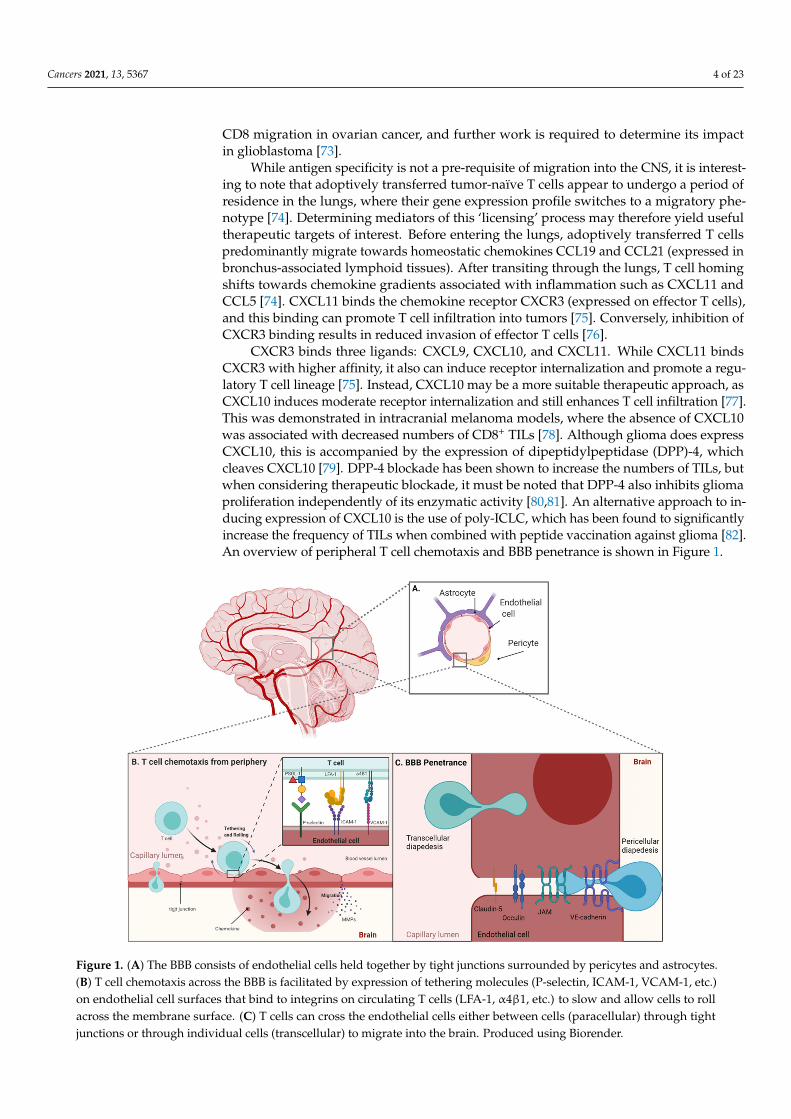
CXCR3 binds three ligands: CXCL9, CXCL10, and CXCL11. While CXCL11 bindsCXCR3 with higher affinity, it also can induce receptor internalization and promote a regu-latory T cell lineage [75]. Instead, CXCL10 may be a more suitable therapeutic approach, asCXCL10 induces moderate receptor internalization and still enhances T cell infiltration [77].This was demonstrated in intracranial melanoma models, where the absence of CXCL10was associated with decreased numbers of CD8+ TILs [78]. Although glioma does expressCXCL10, this is accompanied by the expression of dipeptidylpeptidase (DPP)-4, whichcleaves CXCL10 [79]. DPP-4 blockade has been shown to increase the numbers of TILs, butwhen considering therapeutic blockade, it must be noted that DPP-4 also inhibits gliomaproliferation independently of its enzymatic activity [80,81]. An alternative approach to in-ducing expression of CXCL10 is the use of poly-ICLC, which has been found to significantlyincrease the frequency of TILs when combined with peptide vaccination against glioma [82].An overview of peripheral T cell chemotaxis and BBB penetrance is shown in Figure 1.
Figure 1. (A) The BBB consists of endothelial cells held together by tight junctions surrounded by pericytes and astrocytes.(B) T cell chemotaxis across the BBB is facilitated by expression of tethering molecules (P-selectin, ICAM-1, VCAM-1, etc.)on endothelial cell surfaces that bind to integrins on circulating T cells (LFA-1, α4β1, etc.) to slow and allow cells to rollacross the membrane surface. (C) T cells can cross the endothelial cells either between cells (paracellular) through tightjunctions or through individual cells (transcellular) to migrate into the brain. Produced using Biorender.
Cancers 2021, 13, 5367 5 of 23
3. Blood–Brain Barrier Specific Targets
Following the shift towards a pro-infiltrative phenotype, T cells must cross the BBB.Although glioblastoma is a disease state in which the BBB is disrupted, regions of thetumor are likely surrounded by intact portions of barrier [83]. These privileged regionsmay act as the site of regrowth, shielded from immunotherapeutic attack [8,83]. Suchprivileged regions correspond with the non-contrast enhancing infiltrating edge, whichcan form the site of recurrence following core resection [84]. Infiltrating glioma cells atthe leading edge demonstrate upregulated fibroblast growth factor-mediated signalingthat promotes tumorigenesis [85]. The changes in cellular phenotype at the leading edgeare driven by histone deacetylase signaling from the tumor core [86]. This results in apermanent alteration at the border to a pro-infiltrative milieu of glioma-initiating cells,which does not reverse following resection of the core [86]. Immune cell populations differat this interface zone also. Spatial single-cell RNA-Seq analysis performed by Darmaniset al. revealed that tumor associated macrophages (TAMs) dominated the core whilebrain-derived microglia dominated the peritumoral zone [87]. These both have key rolesin T cell activity at the tumor site. Macrophages can express the cytokine TGF-β, whichenhances glioblastoma cell growth, migration, and invasion and downregulates antitumorimmunity [88,89]. Microglia in the peritumoral zone show an increased expression ofligands for T cell exhaustion-associated receptors such as PD1 and CTLA4 [87]. Microgliaalso express CCL2 and CCL5 which, as mentioned previously, enhances regulatory T cellrecruitment and myeloid-derived suppressor cells [56,90]. Notably, CCL5 also acts as anauto-stimulatory signal for GBM cells by binding to the non-conventional receptor CD44,resulting in increased cell survival, invasion, and proliferation [91]. Therefore, targeting thiszone of recurrence and immune exhaustion protected by intact barrier is key to enhancingthe efficacy of immunotherapy. However, achieving this requires CTLs to traffic throughthe BBB.
The BBB is a highly regulated physical and metabolic barrier which extends fromthe CNS microvasculature to the endothelial cells of postcapillary venules [92]. Duringneuro-inflammation the permeability of the endothelial cells changes to allow for theentrance of lymphocytes into the CNS. This is achieved by changes in BBB junctionalmorphology that allow lymphocytes access either by squeezing between endothelial cells(paracellular diapedesis) or crossing through pores in the endothelial cell membrane(transcellular diapedesis) [93]. Recent single-cell RNA sequencing of the neuro-vasculaturealso shows enhanced endothelial cell expression of MHC class II genes in the disease state.The endothelial cell signature also changes from CNS specific to mirroring the periphery,thereby promoting immune trafficking from the blood (preprint [94]). The endothelialcells of the BBB are sealed by adherens junctions, a continuous series of complex tightjunctions, and recently discovered tricellular junctions [95]. In the inflammatory state,these tricellular junctions have been suggested to be the primary site of cellular migration,through the downregulation of proteins (tricellulin and angulins) which normally maintaintheir morphology [95,96]. Interestingly, recombinant CCL2 and CCL5 administration wasdemonstrated in vitro to enhance T cell diapedesis through tricellular junctions. This maytherefore offer a therapeutic strategy specifically to enhance paracellular crossing at theBBB, although their effect on recruiting regulatory T cells must also be considered [95,96].
Tight junctions can also be targeted to allow for entry of therapeutic agents [97,98].These junctions are maintained on the basolateral side by the transmembrane adhesion pro-teins VE-cadherin and platelet endothelial cell adhesion molecule (PECAM)-1 [99,100]. Theapical side of endothelial tight junctions is secured by occludin and claudin-1/3/5/12 [101].Together, these proteins seal the tight junctions together by binding with each other onopposite endothelial cells, reducing the intercellular distance [101]. Claudin-5 is the mostcommonly expressed protein in tight junctions [101], and can be targeted with recombinantprotein inhibitors such as the non-toxic C-terminal domain of the Clostridium Perfringensenterotoxin [102]. This can reversibly open endothelial tight junctions and allow ingressof therapeutic agents. Targeting of claudin-5 in vitro results in reduced paracellular di-
Cancers 2021, 13, 5367 6 of 23
apedesis of lymphocytes while increasing transcellular diapedesis [103]. Other studieshave also shown that knockout of adhesion molecules such as PECAM-1 does not resultin enhanced paracellular movement, but instead increases migration via cell membranechannels [104]. Taken together, it becomes apparent that functional tight junction regulatingproteins are required for paracellular diapedesis, and that disruption of these proteins mayshift trafficking towards endocytic lymphocyte migration patterns similar to those foundin neuroinflammatory CNS states [93].
The process of transcellular diapedesis is mediated by endocytosis at the endothelialcell membrane. This endocytosis occurs through vesicles containing caveolin (Cav)-1,which are increased in number during disease states such as EAE [105,106]. Regions of theBBB rich in Cav1 upregulate expression of adhesion receptors such as ICAM-1, capturingT cells at regions of the BBB where endocytic vesicles are present [107]. Interestingly, ininflammatory conditions such as EAE, ICAM-1 is highly expressed on the endothelium,and this over-expression promotes transcellular diapedesis. This contrasts with the restingstate where low/intermediate expression of ICAM-1 favors paracellular diapedesis [108].Therefore, promoting the expression of LFA-1 on T cells which can bind to over-expressedICAM-1 may enhance T cell trafficking (therapeutic approaches described in the previoussection). However, whether this effect also extends to CD8+ T cells in the context ofglioblastoma is unclear.
Differential trafficking of T cell subsets was also demonstrated by experiments usingCav1−/− mice which induced almost total loss of Th1 transcellular migration but did notimpair migration of Th17 cells [109]. In EAE, Th17 T cells have been demonstrated touse CCR6 to bind CCL20 produced by the choroid plexus epithelial cells to gain accessto the ventricular CSF [110,111]. When considering the CCR6–CCL20 axis for therapeutictargeting in glioblastoma, CD8+CCR6+ T cells also migrate towards CCL20 and blockadeof CCL20 or CCR6 has also been demonstrated to reduce neuroinflammation in murinemodels of subarachnoid hemorrhage [112,113]. However, over-expression of CCL20 bytumors also correlates with tumor progression in multiple cancer types, as well as decreasedsurvival [114]. Importantly though, the tumor-promoting effects of CCR6 signaling appearto rely on CCR6+ stromal cells but not CCR6+ immune cells [114]. Upregulation of CCR6 onimmune cells may therefore be the more prudent therapeutic approach for enhancing T cellinfiltration while maintaining tumor control. Transforming growth factor (TGF)-β has beenshown to promote CCR6 expression on human CD4 T cells but is also implicated in thepromotion of regulatory FOXP3 expression [115]. However, TGF-β priming also generatesa fractional population of CCR6+FOXP3− cells [116]. Further selection of this populationwould therefore be desirable to achieve a pro-infiltrative, effector T cell phenotype. Modelsof EAE have also found that increased expression of CCL19 and CCL21 from mononuclearinflammatory cells binds CCR7+ T cells in the CSF [117]. CCL19 has been shown toenhance the frequency of antigen responsive IFN-γ+ CD8+ T cells in viral infection andCCR7 chemotaxis may be stimulated in vitro using by-products of coagulation factor XIIa(high-molecular-weight kininogen domain 5) [118,119]. However, CCL19 may also promotethe migration of regulatory T cells (CD4+CD25+FoxP3+) and therefore its usefulness inglioblastoma is unclear [120].
While these mechanisms are of interest therapeutically to allow T cells to cross theBBB from the periphery, this is only the initial step in accessing the parenchyma. Inter-action with professional antigen-presenting cells in the perivascular spaces is a key stepbefore penetration of the glia limitans, which lines the blood vessels and the surface ofthe brain [121].
4. The Glia Limitans—Accessing the Parenchyma
Between the outer BBB and the parenchyma lies the glia limitans. The glia lim-itans is formed by astrocyte foot processes associating with the basal lamina of theparenchyma [110]. It is divided into two membranes: the glia limitans perivascularis(surrounding blood vessels) and the glia limitans superficialis (covering the surface of the
Cancers 2021, 13, 5367 7 of 23
brain) [122]. In much of the brain, these two membranes lie so closely together that they areindistinguishable, but beyond the capillaries at the venules, inflammation can cause thesetwo membranes to separate, forming a perivascular space. This space communicates withthe CSF and allows for APCs to present antigens to entering T cells [123]. This interactionis critical in allowing T cells to access the parenchyma—indeed, the effects of T cells inEAE only begin once immune cells have crossed the glia limitans [124]. The APC–T cellinteraction drives the production of further pro-inflammatory cytokines which triggersthe recruitment of more immune cells [111,125]. Interestingly, while the initial T cellsthat enter these perivascular spaces tend to have increased expression of CCR6, furtherrecruitment occurs in a CCR6-independent manner [110,111]. This would suggest thatCCR6+ T cells form part of an initial ‘licensing’ step and that their interaction with APCs inthe perivascular spaces facilitates further entry of T cells in a non-CCR6-specific manner.
In normal physiology, T cell crossing at the glia limitans is mediated by the expressionof laminins [126]. For example, the parenchymal membrane of the glia limitans containsα1 and α2 laminins [127], which CD4+ T cells are unable to bind in the non-inflammatorystate. However, in EAE, CD4+ T cells can bypass this control mechanism by using matrixmetalloproteinases (MMPs) which disrupt the astrocytic foot processes, breaking downbarrier integrity and allowing for T cell ingress [124]. While this might suggest that MMPagonism may be an attractive prospect for opening the glia limitans, MMPs are involved inthe angiogenesis and invasion of glioma [128]. Inhibition of MMP was even trialed using abroad-spectrum MMP inhibitor, but this resulted in widespread reports of musculoskeletaltoxicity due to on-target, off-tumor effects [129,130]. Given these experiences, it is unlikelythat MMP agonism in glioblastoma will be a desirable therapeutic target.
Another mediator of T cell entry into the parenchyma is CXCL12. In murine models,T cells have been noted to be held in perivascular spaces due to expression of CXCL12 [131].This ‘hold’ is released in inflammatory conditions, as increased levels of IL-17 drive theexpression of CXCR7 on endothelial cells, resulting in the internalization of CXCL12 [132].This leads to increased CXCR4 expression on T cells and subsequent T cell entry into theparenchyma [131,132]. However, when considering the downregulation of CXCL12 asa therapeutic strategy, it is worth noting that recent studies evaluating T cell responsesto viral infection in vitro have found that CXCL12 at the BBB endothelium can promoteCD8+ migration across the BCSF interface, suggestive of a location-dependent role [133].A summary of these selected targets and therapeutic considerations is shown in Table 1.
Table 1. A summary of selected factors that may enhance trafficking and infiltration of T cells across the BBB.
Interactor Behavior Therapeutic Considerations References
T cell processes
LFA-1T cell integrin which binds ICAM-1. PromotesT cell capture and rolling in inflammatory and
non-inflammatory state.
IL-12 induces LFA-1 expression and can enhanceT cell migration in several murine malignancies. [36,73]
VLA-4(α4β1)
Integrin on T cell which binds VCAM-1 in theinflammatory state and interacts with other
transmembrane proteins (JAM-B, JAML, etc.).
IL-12 induces LFA-1 and VLA-4 expression andenhances T cell migration in several murine
malignancies. Effect may bemalignancy dependent.
[41,42,73]
CXCL9: Polarizes T cells to aTh1/Th17 phenotype.
Mediated lymphocyte infiltration and suppressestumor growth in cutaneous fibrosarcoma. [134]
CXCR3(3 ligands)
CXCL10: Only moderately induces CXCR3internalization and enhances T cell infiltration.
DPP-4 blockade increases TILs but is alsotumorigenic (independent of enzymatic
function). Combinatorial poly-ICLC enhancesCXCL10 expression.
[81–83]
CXCL11: Binds CXCR3 strongly and inducesreceptor internalization. Promotes lineage of regulatory T cells. [75]
Cancers 2021, 13, 5367 8 of 23
Table 1. Cont.
Interactor Behavior Therapeutic Considerations References
CCR4 CCL2, CCL22 (and others): Overexpressed onglioma cells, recruits regulatory T cells.
CCR4-CCL22 signaling recruits regulatory Tcells. Blockade of CCR4 in vitro can reduceregulatory T cell migration. TMZ can also
mitigate production of CCL2.
[135,136]
CCR5Binds CCL3, CCL4, and CCL5. May help torecruit cytolytic T cells but also regulatory
T cells.
CCL4 can help recruit cytolytic CCR5+ T cells inesophageal squamous cell carcinoma butCCL4–CCR5 interaction can enhance theinvasion ability of glioblastoma in vitro.
CCL5 is also associated with enhanced T celldiapedesis at tricellular junctions. However,CCL5 also binds CD44 on GBM cells to driveproliferation and survival and is produced by
perivascular stromal cells such as pericytes.Blockade of CCR5 (maraviroc) may limit
cancer-associated fibroblast accumulation.
[54,62,63,91,95]
CCR6 Binds CCL20 expressed at the choroid plexus.CD8+ T cells migrate to CCL20 in murine SAH.
TGF- β promotes CCR6 expression but also isimplicated in the promotion of FOXP3+ cells.
However, a fraction of the population isCCR6+FOXP3−. CCR6 T cells may also be
involved with licensing further recruitment toperivascular spaces.
[115,116]
CCR7 Present on activated CD8 T cells (and centralmemory T cells).
Interacts with CCL19 and may mediate integrinactivation on immune cells or diapedesis.
Chemotaxis may be enhanced by a peptidederived from the byproduct of coagulation factor
XIIa cleavage. May also promote regulatoryT cells.
[117,119,120]
Blood–brain barrier processes
E/P-Selectin
Expressed in inflammatory state only. BindsPSGL-1+ CD8 T cells, slowing them on
BBB endothelium.
Expression enhanced in response toinflammatory cytokines (e.g., IL-1 or TNF α).
IL-1 has been delivered via CED in rat modelsof glioma.
[45,137]
Claudin-5,PECAM-1
Commonly expressed proteins involved insealing tight junctions at BBB.
Modified Clostridium perfringens enterotoxincan reversibly open tight junctions. May drive
T cells to transcellular migration.[93,103,104]
ACKR1 Trafficking of pro-infiltrative chemokines fromabluminal to luminal surface of BBB.
IL-1 signaling associated with upregulatedexpression ACKR1 (along with VCAM-1,
ICAM-1). Trialed using CED in rat glioma.[45]
Caveolin-1 Expressed in endocytic vesicles at BBB and actsas a mediator of transcellular diapedesis.
Regions of BBB rich in CAV-1 are also rich inICAM-1. Enhancing ICAM-1 on BBB (e.g., via
IL-1) may capture more T cells that can undergopara and transcellular diapedesis.
[108]
CXCL12Acts as a T cell, holding factor cells in
perivascular spaces. Expression of CXCR7 onendothelial cells internalizes CXCL12.
IL-17 drives expression of CXCR7 on endothelialcells and CXCR4 on T cells which licenses theirentry into the parenchyma. However, CXCL12
may promote CD8+ migration across BCSFbarrier—may be a location-specific role.
[131–133]
This table only provides selected examples and is not exhaustive.
Cancers 2021, 13, 5367 9 of 23
5. T Cell Trafficking through the Parenchyma
Once past the glia limitans, effector T cells must reach and infiltrate the tumor toexert their cytotoxic effect. As discussed in the introduction, glioblastoma can restrictT cell trafficking due to the downregulated expression of VEGF-C, resulting in restrictedlymphangiogenesis [22]. Notably, in patients treated with neoadjuvant anti-PD-1, VEGF-Cexpression was highly correlated with increased infiltration of T cells [138]. Thus, restoringlevels of lymphangiogenesis-promoting factors such as VEGF-C could also enhance T cellhoming and infiltration to the tumor. This is supported by the findings of Song et al.,who demonstrated that intra-cisterna magna injections of an adeno-associated viral vectorcoding for VEGF-C could remodel meningeal lymphatic vessels in murine models ofglioma [22]. Further enhanced expression of VEGF-C in lymphatic endothelial cells couldpotentiate the effect of checkpoint blockade due to enhanced T cell infiltration [22].
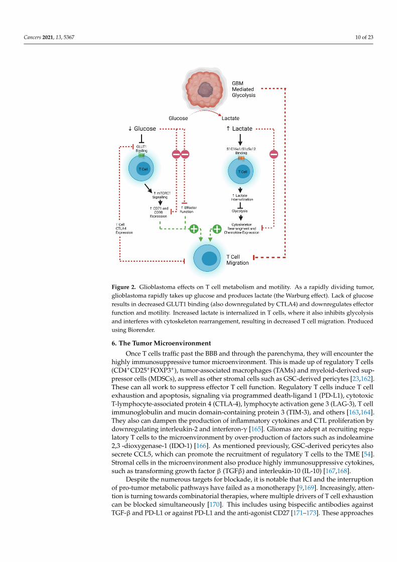
T cell motility is also dependent on metabolic pathways that are often usurped byrapidly proliferating tumors. Tumor cells demonstrate increased glucose uptake and lac-tose production, even in the presence of oxygen and functioning mitochondria (known asthe Warburg effect) [139,140]. This affords the tumor and other rapidly proliferating cellsessential anabolic precursors for cell proliferation [140]. The increased glucose demandby tumor cells therefore decreases the amount available for circulating T cells to maintaineffector and migratory function [141]. Aerobic glycolysis is the main source of ATP pro-duction in leukocytes, which is required for the energetic demands of migration [142,143].Inhibition of the T cell glycolytic pathway through administration of 2-DG and rapamycincauses a decrease in naïve T cell motility, demonstrating the importance of glucose in T cellhoming [144,145]. The associated build-up of lactate caused by the Warburg effect alsoresults in decreased migration of CD4+ T cells and a loss of cytolytic function of CD8+
T cells by interfering with T cell glycolysis [145–148]. However, this effect can be reversed,as demonstrated in an animal model of peritonitis where antibody-mediated blockade oflactate transporters on T cells allowed them to maintain their migratory potential [149].Expression of CTLA-4 decreases the expression of the glucose transporter GLUT-1 onT cells, and further decreases effector function, implying that combinatorial approachesusing checkpoint blockade may aid with T cell trafficking as well as reinvigoration offunction [142,150]. However, recent work suggests that exhausted human CD8+ T cellsmay actually become more mobile [151]. CTLA-4 signaling can lead to a RAP1-mediatedincrease in LFA-1 binding, which can induce migration [152]. This has potential impli-cations for considering which form of ICI would best work with a tumor where T celltrafficking poses a significant challenge. An overview of the metabolic pathways limitingT cell efficacy in glioblastoma is shown in Figure 2.
Another mediator of T cell glycolysis is the PI3K/AKT/mTOR pathway, whoseactivation can also downregulate the expression of adhesion and migration moleculesCD62L, CCR7, and S1P1 in CD8+ T cells [142,153]. Loss of S1P1 has been shown tomediate T cell sequestration in bone marrow in glioblastoma, while S1P1+ cells are resistantto sequestration and can return into the circulation [142,154–156]. Therefore, reversingsequestration will be critical for future immunotherapy efficacy and is currently the subjectof ongoing therapeutic investigation [157]. While one approach may be to inhibit thePI3K/AKT/mTOR pathway, this inhibition must be selective, as AKT possesses threeisoforms which have varying pro- and anti-tumor effects. AKT signaling also plays animportant role for the development of effector-like memory CD8+ T cells necessary fortumor immune surveillance [158]. Interestingly, recent work has described small-moleculeinhibitors that may be capable of targeting pathogenic AKT isoforms only (AKT1 andAKT2) while leaving the tumor-suppressive functionality of AKT3 intact [159,160]. Indeed,specific AKT1 and 2 inhibition has been associated with enhanced central memory CD8+
T cell proliferation with prolonged cytokine and Granzyme B production, making this apotential future therapeutic strategy [158–161].
Cancers 2021, 13, 5367 10 of 23
Figure 2. Glioblastoma effects on T cell metabolism and motility. As a rapidly dividing tumor,glioblastoma rapidly takes up glucose and produces lactate (the Warburg effect). Lack of glucoseresults in decreased GLUT1 binding (also downregulated by CTLA4) and downregulates effectorfunction and motility. Increased lactate is internalized in T cells, where it also inhibits glycolysisand interferes with cytoskeleton rearrangement, resulting in decreased T cell migration. Producedusing Biorender.
6. The Tumor Microenvironment
Once T cells traffic past the BBB and through the parenchyma, they will encounter thehighly immunosuppressive tumor microenvironment. This is made up of regulatory T cells(CD4+CD25+FOXP3+), tumor-associated macrophages (TAMs) and myeloid-derived sup-pressor cells (MDSCs), as well as other stromal cells such as GSC-derived pericytes [23,162].These can all work to suppress effector T cell function. Regulatory T cells induce T cellexhaustion and apoptosis, signaling via programmed death-ligand 1 (PD-L1), cytotoxicT-lymphocyte-associated protein 4 (CTLA-4), lymphocyte activation gene 3 (LAG-3), T cellimmunoglobulin and mucin domain-containing protein 3 (TIM-3), and others [163,164].They also can dampen the production of inflammatory cytokines and CTL proliferation bydownregulating interleukin-2 and interferon-γ [165]. Gliomas are adept at recruiting regu-latory T cells to the microenvironment by over-production of factors such as indoleamine2,3 -dioxygenase-1 (IDO-1) [166]. As mentioned previously, GSC-derived pericytes alsosecrete CCL5, which can promote the recruitment of regulatory T cells to the TME [54].Stromal cells in the microenvironment also produce highly immunosuppressive cytokines,such as transforming growth factor β (TGFβ) and interleukin-10 (IL-10) [167,168].
Despite the numerous targets for blockade, it is notable that ICI and the interruptionof pro-tumor metabolic pathways have failed as a monotherapy [9,169]. Increasingly, atten-tion is turning towards combinatorial therapies, where multiple drivers of T cell exhaustioncan be blocked simultaneously [170]. This includes using bispecific antibodies againstTGF-β and PD-L1 or against PD-L1 and the anti-agonist CD27 [171–173]. These approaches
Cancers 2021, 13, 5367 11 of 23
are currently being evaluated in Phase I trials in advanced solid tumors (NCT04429542,NCT04440943). Cytokine modulation approaches are also a potential avenue for enhancingT cell activity in the TME, as seen in ‘armored’ CAR-T constructs. The addition of IL-12,IL-15, or IL-18 along with antigen specificity to T cells appears to result in greater CTLactivity and anti-tumor efficacy [174–176]. A high percentage of regulatory T cells in theperipheral blood of GBM patients express CCR4 compared to controls (74 vs. 43%) [135].CCL4 binds CCL22 (and others), which has been shown to be overexpressed in freshlyresected human glioma cells, and blockade of CCR4 in vitro can significantly reduceregulatory T cell migration [135]. Targeting fibroblast activation proteins or introduc-ing heparinase-expressing agents may also help to disrupt immunosuppressive stromalelements [146,177,178]. Intratumoral APCs are also necessary to stimulate and retain infil-trating lymphocytes at the tumor site, as well as carrying antigens to draining lymph nodesand cross priming peripheral CD8 T cells [179–181]. The administration of intratumoralFMS-like tyrosine kinase 3 ligand (Flt3L) and poly I:C has been shown to expand and ma-ture dendritic cell precursors, resulting in greater antitumor efficacy when combined withimmunotherapies such as PD-L1 blockade or oncolytic herpes simplex viruses [179,182].
Standard-of-care therapies also can help drive a more potent immune response. Temo-zolomide (TMZ) is an alkylating chemotherapy whose main function is to induce DNAdouble-stranded breaks, resulting in tumor cell death [183]. Interestingly, TMZ can alsohelp to reduce the numbers of peripheral regulatory T cells, as well as interrupting theirmigration [136,184]. In disease states such as glioblastoma, tumor cells and platelet-derivedgrowth factor receptor beta (PDGFRβ)-expressing cells of the neurovascular sub-units(such as pericytes and perivascular fibroblast-like cells) produce CCL2 to recruit regulatoryT cells and dampen the effector response [185]. TMZ interrupts the CCL2–CCR4 axis,thereby reducing this effect [136,184]. Combining immunotherapy with radiotherapy alsocan help to polarize the T cell response to a cytotoxic phenotype by inducing greaterT cell receptor diversity and expanding the numbers of tumor-infiltrating lymphocytesand effector memory T cells [186]. In pre-clinical murine models of glioma, radiotherapycombined with antibodies against markers of exhaustion such as TIM-3 and PD-1 was ableto produce long-term survival [11].
7. Modeling the BBB
Animal and in vitro models have contributed greatly to our knowledge regardingthe cell and protein interactions required to cross the BBB. Rudimentary animal modelsfrom the 1980s first established how BBB permeability could change in response to sys-temic compounds by tracking the CNS uptake of Evan’s Blue dye following intravenousinfusion [187–190]. These models established protocols to visualize membrane cellularcomponents and tissue hierarchy through fluorescent microscopy and histology, allowingfor the elucidation of fundamental mechanisms behind membrane permeability. Suchmodels included mice with proteins essential for T cell chemotaxis across the BBB knockedout, including tight junction proteins claudin 5 [100] and occludin [191]. Unfortunately, fullknockout of these proteins results in non-viable pups or other dysfunctional phenotypes,suggesting the importance of tight junction proteins in development. Additional knockoutmice focusing on proteins involved in T cell rolling, p-selectin, and its ligand PSGL-1 [192]were developed and confirmed less BBB breakdown and leukocyte trafficking into theCNS. Similarly, the use of antibody natalizumab to block the α4 subunit on T cells has beensuccessful in preventing BBB chemotaxis in MS [193]. Genetic models targeting pericyteand astrocyte function have also been generated to establish how these cell types supportBBB formation in development and regulate tight junctions in injury and disease. The useof two-photon microscopy has allowed for imaging at depths up to 1mm, but real-timehigh-resolution imaging and cell tracking capabilities are limited. Animal models closelymimic BBB features by including all cell types within the vascular interface, fluid flowand biochemical concentrations. However, there are still challenges translating in vivofinding to clinical significance. Genetic, molecular, and immunological differences between
Cancers 2021, 13, 5367 12 of 23
humans and rodents, as well as high cost and ethical concerns with animal testing, havegenerated a need for robust in vitro models.
In vitro models of the BBB range from simple endothelial cell monolayers to complexthree-dimensional systems with fluid flow and ionic gradients [194,195]. These modelshave the advantage of using human cells as well as being cost effective and allowing forhigh-throughput screening of a variety of different conditions or molecules. Transwell,hydrogels, and microfluidic devices with three or four different cell types have beencreated in attempts to best mimic native BBB function. Simplified in vitro models allowfor researchers to specifically modify or track elements of the BBB. Cell types used inthese models have traditionally been primary brain endothelial cells or immortalized celllines. Immune factors affecting BBB permeability have been most studied with BECs dueto their accurate expression of chemokine and cytokine receptors. Interestingly, thesemodels found CCL2 to cause redistribution of tight junction proteins, such as claudin-5 andoccluding, under physiological and pathological conditions [196,197], which gives insightinto the mechanism of increased T cell chemotaxis during inflammation and elevatedCCL2. Brain-cancer-specific models have focused on integrating vasculature and tumorcells to test the toxicity of therapeutics prior to animal studies [198,199]. These modelsrecapitulated the three-dimensional structure of a brain tumor but used lung fibroblasts,HUVECs, and gelatin, which may not accurately represent the blood–brain barrier andbrain microenvironment. The development of induced pluripotent stem cells (iPSCs)has allowed for genetically identical personalized in vitro models to test drug and cellinteractions with BBB of specific individuals and disease conditions [200]. Overall, bothin vivo and in vitro models of the BBB have limitations but can provide valuable insight toimprove T cell chemotaxis in GBM.
8. Safety
While this review has largely focused on strategies by which T cells can be recruitedand restored to a cytotoxic effector status, it must be noted that rapid increases in acti-vated T cells in the circulation can potentially lead to cytokine release syndrome (CRS),mediated by the release of pro-inflammatory cytokines such as IL-6 [201]. Therefore, whenconsidering therapies that will increase circulating activated T cells and subsequent CNST cell infiltration, careful consideration must be given to the safety of any such approach.Such therapies may lead to systemic and neurological complications, even when not usedspecifically to treat CNS malignancies.
This is demonstrated by the example of clinically used therapeutics such as ipili-mumab, which re-invigorates T cells by blockade of CTLA-4 [202]. Ipilimumab has beenassociated with pituitary inflammation (hypophysitis), occurring in up to 17% of patientsreceiving ipilimumab treatment [203,204]. Similar syndromes are also observed whenusing anti-PD-1 and anti-PD-L1 therapies, albeit at a lower frequency compared to anti-CTLA-4 [205]. The mechanism of how ipilimumab causes hypophysitis remains unclear,but it is speculated that ipilimumab can release the brakes on T cells that target and destroypituitary cells, or that expression of CTLA-4 on pituitary cells leads to complement fixationmediated by ipilimumab, resulting in the destruction of pituitary cells [206,207]. In rarercircumstances (less than 0.2% of patients), ICI, especially ipilimumab or combination ipili-mumab/nivolumab (anti-PD-1), has caused aseptic meningitis and encephalitis [208–212].Like with hypophysitis, the exact mechanism is unclear. However, in the case of encephali-tis, there is evidence that the effect is autoimmune in origin, as some patients treated withICI exhibit autoantibodies to the NMDA receptor, a characteristic of other autoimmuneencephalopathies [213,214]. ICI has also reportedly induced new CNS demyelination andexacerbated existing CNS demyelination in MS patients [215,216]. These rare, but seriousneurological deficits resulting from systemic ICI emphasize the need for careful monitoringof patients receiving therapies that enhance T cell trafficking and function.
Cancers 2021, 13, 5367 13 of 23
The experience of treatments using adoptively transferred chimeric antigen receptor(CAR) T cells in extracranial and intracranial malignancies can also be illustrative forpotential systemic and neurological toxicities. The most common CAR-related toxicity iscytokine release syndrome (CRS), occurring in up to 37–93% of patients with lymphomaor leukemia receiving CD19 CARs [217]. As described previously, CRS is caused by rapidactivation of CAR T cells upon administration and subsequent release of pro-inflammatorycytokines, such as IL-6 [201]. High levels of serum IL-6 were found to correlate with severeCRS, which led to the FDA approval of tocilizumab, an anti-IL-6 receptor antagonist [218].Strategies to reduce CRS include administering lower doses of CAR T cells over multipleinfusions as opposed to one single bolus [219,220].
Neurological-specific toxicities after CAR administration are also possible. Immuneeffector cell-associated neurotoxicity syndrome (ICANS) can develop in around 50% ofpatients following systemic CAR infusion [221]. ICANS manifests with minor symptomssuch as lethargy and confusion but can also cause seizures and coma. The pathophysiol-ogy of ICANS remains unclear, but evidence suggests that release of pro-inflammatorycytokines, such as IL-6 and IL-1β, by CAR T cells can disrupt the BBB, resulting in the ac-cumulation of CAR T cells and pro-inflammatory cytokines in the CNS [222]. Klinger et al.recently described a mechanism whereby CD19 bi-specific T cell engagers (blinatumomab)can induce T cell adhesion to endothelial cells of the BBB followed by T cell migrationinto the perivascular space in a CD19-independent manner. Once past the BBB, theymay encounter rare target CD19 cells in the CNS and release pro-inflammatory cytokines,triggering ICANS-like symptoms [223]. However, unlike CRS, ICANS does not respondto tocilizumab treatment, and symptoms are typically managed with corticosteroids orcessation of therapy [224]. Klinger et al. also reported that the non-specific entry of CD19T cells into the CNS could be abrogated by the administration of anti-adhesion agents(anti-VLA4, natalizumab), offering another potential therapeutic if toxicity occurs [223]. Insummary, while enhanced T cell chemotaxis and infiltration of glioblastoma are necessaryfor effective immune-mediated treatment of tumors, this must be carefully balanced withthe risks described above.
9. Conclusions
For immunotherapy in glioblastoma to be successful, sustained recruitment of effectorlymphocytes from the periphery to the tumor is necessary. However, achieving this in aunique immune environment such as the CNS must overcome both physical and chemicalbarriers. In this review, we have described the process by which effector T cells can berecruited from the periphery and what modifications may result in a pro-infiltrative pheno-type. We have described both T cell and BBB factors that would be desirable therapeutictargets and set out strategies by which this may be achieved. Adhesional factors on the BBBendothelium such as ICAM-1, VCAM-1, or ACKR1 may be upregulated by IL-1β or IFN-γ,which can be delivered via convection-enhanced delivery (CED) directly to the tumor site.Delivery of these cytokines and other inflammatory factors can have profound effects onincreasing BBB penetration and the migration potential of T cells. Induced expression ofCXCL10 by using poly-ICLC can also interact with CXCR3 on effector T cells, promptingtheir infiltration into tumor. Co-culture with IL-12 may help drive the expression of keyintegrins such as LFA-1 on the surface of T cells in preparation for adoptive transfer tofurther enhance their adhesive capabilities. CCL2 and CCL5 may promote paracellulardiapedesis through tricellular junctions in the BBB endothelium, while TGF-β primingof T cells can increase their CCR6 expression, which can promote transcellular crossing.However, CCL2 and CCL5 may also mediate regulatory T cell recruitment, perhaps ne-cessitating co-administration with checkpoint blockade. Subsequent navigation throughthe glia limitans may be aided by IL-17-mediated downregulation of CXCL12, althoughthis may be a location-specific effect. Once inside the parenchyma, lymphangiogenesis-promoting factors such as VEGF-C may further enhance trafficking of T cells to the tumor.Metabolic mediators such as the PI3K/AKT/mTOR pathway may also be therapeutically
Cancers 2021, 13, 5367 14 of 23
targeted using small-molecule inhibitors of the AKT1 and AKT2 isoforms. Combinatorialapproaches to stimulate T cells and block checkpoint inhibition will likely be necessary toovercome the microenvironment. This may be achieved using novel bispecific constructsor co-administration with immune stimulatory cytokines such as IL-12, IL-15, or IL-18.Standard-of-care therapies such as TMZ and radiotherapy may also help to blockade regu-latory T cell recruitment and drive a more diverse and potent T cell response. Importantly,however, any approach that enhances T cell infiltration into the CNS must consider safety,and although there are therapeutic options for adverse events, future trial designs usingpro-infiltrative therapies should err on the side of caution. Nevertheless, enhanced T celltrafficking and infiltration of glioblastoma is essential for immunotherapeutic efficacy.While ICI seeks to ‘release the brakes’ on T cell activity, in the case of glioblastoma, wemust first drive T cells to the tumor.
Author Contributions: K.S., writing—original draft preparation, review and editing, visualization,project administration, revision. K.M.H., writing—original draft preparation, review and editing,visualization, revision. K.K.P., writing—original draft preparation, D.S.W., writing—original draftpreparation. A.A.M., visualization. S.L.C., writing—review and editing. J.H.S., supervision. Allauthors have read and agreed to the published version of the manuscript.
Funding: This work was supported by grants from the National Institutes of Health (NIH) (U01NS090284(JHS) and P50CA190991(JHS)).
Conflicts of Interest: K.S., K.M.H., K.K.P., D.S.W., A.A.M. and S.L.C. report no conflict of interest.J.H.S. has an equity interest in Istari Oncology, which has licensed intellectual property from Dukerelated to the use of poliovirus and D2C7 in the treatment of glioblastoma. J.H.S. is an inventoron patents related to the PEP-CMV DC vaccine with tetanus, as well as the poliovirus vaccine andD2C7 in the treatment of glioblastoma. J.H.S. has an equity interest in Annias Immunotherapeutics,which has licensed intellectual property from Duke related to the use of the pepCMV vaccine in thetreatment of glioblastoma.
References1. Klein, R.S.; Hunter, C.A. Protective and Pathological Immunity during Central Nervous System Infections. Immunity 2017,
46, 891–909. [CrossRef]2. Daneman, R.; Prat, A. The blood-brain barrier. Cold Spring Harb. Perspect. Biol. 2015, 7, a020412. [CrossRef] [PubMed]3. Engelhardt, B.; Sorokin, L. The blood-brain and the blood-cerebrospinal fluid barriers: Function and dysfunction. Semin. Im-
munopathol. 2009, 31, 497–511. [CrossRef] [PubMed]4. Khasraw, M.; Reardon, D.A.; Weller, M.; Sampson, J.H. PD-1 Inhibitors: Do they have a Future in the Treatment of Glioblastoma?
Clin. Cancer Res. Off. J. Am. Assoc. Cancer Res. 2020, 26, 5287–5296. [CrossRef]5. Quail, D.F.; Joyce, J.A. Microenvironmental regulation of tumor progression and metastasis. Nat. Med. 2013, 19, 1423–1437. [CrossRef]6. Quail, D.F.; Joyce, J.A. The Microenvironmental Landscape of Brain Tumors. Cancer Cell 2017, 31, 326–341. [CrossRef] [PubMed]7. Yeung, J.T.; Hamilton, R.L.; Ohnishi, K.; Ikeura, M.; Potter, D.M.; Nikiforova, M.N.; Ferrone, S.; Jakacki, R.I.; Pollack, I.F.;
Okada, H. LOH in the HLA Class I Region at 6p21 Is Associated with Shorter Survival in Newly Diagnosed Adult Glioblastoma.Clin. Cancer Res. 2013, 19, 1816–1826. [CrossRef] [PubMed]
8. Johanns, T.M.; Bowman-Kirigin, J.A.; Liu, C.; Dunn, G.P. Targeting Neoantigens in Glioblastoma: An Overview of CancerImmunogenomics and Translational Implications. Neurosurgery 2017, 64, 165–176. [CrossRef]
9. Reardon, D.A.; Brandes, A.A.; Omuro, A.; Mulholland, P.; Lim, M.; Wick, A.; Baehring, J.; Ahluwalia, M.S.; Roth, P.; Bähr, O.; et al.Effect of Nivolumab vs Bevacizumab in Patients With Recurrent Glioblastoma: The CheckMate 143 Phase 3 Randomized ClinicalTrial. JAMA Oncol. 2020, 6, 1003–1010. [CrossRef] [PubMed]
10. Garzon-Muvdi, T.; Theodros, D.; Luksik, A.S.; Maxwell, R.; Kim, E.; Jackson, C.M.; Belcaid, Z.; Ganguly, S.; Tyler, B.; Brem, H.; et al.Dendritic cell activation enhances anti-PD-1 mediated immunotherapy against glioblastoma. Oncotarget 2018, 9, 20681–20697.[CrossRef] [PubMed]
11. Kim, J.E.; Patel, M.A.; Mangraviti, A.; Kim, E.S.; Theodros, D.; Velarde, E.; Liu, A.; Sankey, E.W.; Tam, A.; Xu, H.; et al. CombinationTherapy with Anti-PD-1, Anti-TIM-3, and Focal Radiation Results in Regression of Murine Gliomas. Clin. Cancer Res. 2017,23, 124–136. [CrossRef] [PubMed]
12. Ellwardt, E.; Walsh, J.T.; Kipnis, J.; Zipp, F. Understanding the Role of T Cells in CNS Homeostasis. Trends Immunol. 2016,37, 154–165. [CrossRef] [PubMed]
13. Ziv, Y.; Ron, N.; Butovsky, O.; Landa, G.; Sudai, E.; Greenberg, N.; Cohen, H.; Kipnis, J.; Schwartz, M. Immune cells contribute tothe maintenance of neurogenesis and spatial learning abilities in adulthood. Nat. Neurosci. 2006, 9, 268–275. [CrossRef]
Cancers 2021, 13, 5367 15 of 23
14. Kipnis, J.; Cohen, H.; Cardon, M.; Ziv, Y.; Schwartz, M. T cell deficiency leads to cognitive dysfunction: Implications fortherapeutic vaccination for schizophrenia and other psychiatric conditions. Proc. Natl. Acad. Sci. USA 2004, 101, 8180–8185.[CrossRef] [PubMed]
15. Engelhardt, B.; Vajkoczy, P.; Weller, R.O. The movers and shapers in immune privilege of the CNS. Nat. Immunol. 2017, 18, 123–131.[CrossRef] [PubMed]
16. Baruch, K.; Schwartz, M. CNS-specific T cells shape brain function via the choroid plexus. Brain Behav. Immun. 2013, 34, 11–16.[CrossRef] [PubMed]
17. Korin, B.; Ben-Shaanan, T.L.; Schiller, M.; Dubovik, T.; Azulay-Debby, H.; Boshnak, N.T.; Koren, T.; Rolls, A. High-dimensional,single-cell characterization of the brain’s immune compartment. Nat. Neurosci. 2017, 20, 1300–1309. [CrossRef] [PubMed]
18. Murphy, J.B.; Sturm, E. Conditions determining the transplantability of tissues in the brain. J. Exp. Med. 1923, 38, 183–197.[CrossRef] [PubMed]
19. Medawar, P.B. Immunity to homologous grafted skin; the fate of skin homografts transplanted to the brain, to subcutaneoustissue, and to the anterior chamber of the eye. Br. J. Exp. Pathol. 1948, 29, 58–69. [PubMed]
20. Schläger, C.; Körner, H.; Krueger, M.; Vidoli, S.; Haberl, M.; Mielke, D.; Brylla, E.; Issekutz, T.; Cabañas, C.; Nelson, P.J.; et al.Effector T-cell trafficking between the leptomeninges and the cerebrospinal fluid. Nature 2016, 530, 349–353. [CrossRef] [PubMed]
21. Hu, X.; Deng, Q.; Ma, L.; Li, Q.; Chen, Y.; Liao, Y.; Zhou, F.; Zhang, C.; Shao, L.; Feng, J.; et al. Meningeal lymphatic vesselsregulate brain tumor drainage and immunity. Cell Res. 2020, 30, 229–243. [CrossRef] [PubMed]
22. Song, E.; Mao, T.; Dong, H.; Boisserand, L.S.B.; Antila, S.; Bosenberg, M.; Alitalo, K.; Thomas, J.-L.; Iwasaki, A. VEGF-C-drivenlymphatic drainage enables immunosurveillance of brain tumours. Nature 2020, 577, 689–694. [CrossRef] [PubMed]
23. Tomaszewski, W.; Sanchez-Perez, L.; Gajewski, T.F.; Sampson, J.H. Brain Tumor Microenvironment and Host State: Implicationsfor Immunotherapy. Clin. Cancer Res. 2019, 25, 4202–4210. [CrossRef] [PubMed]
24. González, H.; Pacheco, R. T-cell-mediated regulation of neuroinflammation involved in neurodegenerative diseases. J. Neuroin-flamm. 2014, 11, 201. [CrossRef] [PubMed]
25. Sommer, A.; Winner, B.; Prots, I. The Trojan horse—Neuroinflammatory impact of T cells in neurodegenerative diseases.Mol. Neurodegener. 2017, 12, 78. [CrossRef] [PubMed]
26. Muller, W.A. Getting leukocytes to the site of inflammation. Vet. Pathol. 2013, 50, 7–22. [CrossRef] [PubMed]27. Lawrence, M.B.; Springer, T.A. Leukocytes roll on a selectin at physiologic flow rates: Distinction from and prerequisite for
adhesion through integrins. Cell 1991, 65, 859–873. [CrossRef]28. Von Andrian, U.H.; Chambers, J.D.; McEvoy, L.M.; Bargatze, R.F.; Arfors, K.E.; Butcher, E.C. Two-step model of leukocyte-
endothelial cell interaction in inflammation: Distinct roles for LECAM-1 and the leukocyte beta 2 integrins in vivo. Proc. Natl.Acad. Sci. USA 1991, 88, 7538–7542. [CrossRef] [PubMed]
29. Ley, K.; Laudanna, C.; Cybulsky, M.I.; Nourshargh, S. Getting to the site of inflammation: The leukocyte adhesion cascadeupdated. Nat. Rev. Immunol. 2007, 7, 678–689. [CrossRef] [PubMed]
30. Nourshargh, S.; Alon, R. Leukocyte Migration into Inflamed Tissues. Immunity 2014, 41, 694–707. [CrossRef]31. Montresor, A.; Toffali, L.; Constantin, G.; Laudanna, C. Chemokines and the signaling modules regulating integrin affinity.
Front. Immunol. 2012, 3, 127. [CrossRef] [PubMed]32. Kim, S.H.J.; Hammer, D.A. Integrin cross-talk modulates stiffness-independent motility of CD4+ T lymphocytes. Mol. Biol. Cell
2021, 32, 1749–1757. [CrossRef]33. Marchetti, L.; Engelhardt, B. Immune cell trafficking across the blood-brain barrier in the absence and presence of neuroinflam-
mation. Vasc. Biol. 2020, 2, H1–H18. [CrossRef]34. Nourshargh, S.; Hordijk, P.L.; Sixt, M. Breaching multiple barriers: Leukocyte motility through venular walls and the interstitium.
Nat. Rev. Mol. Cell Biol. 2010, 11, 366–378. [CrossRef] [PubMed]35. Engelhardt, B.; Ransohoff, R.M. Capture, crawl, cross: The T cell code to breach the blood–brain barriers. Trends Immunol. 2012,
33, 579–589. [CrossRef] [PubMed]36. Vajkoczy, P.; Laschinger, M.; Engelhardt, B. α4-integrin-VCAM-1 binding mediates G protein–independent capture of encephali-
togenic T cell blasts to CNS white matter microvessels. J. Clin. Investig. 2001, 108, 557–565. [CrossRef] [PubMed]37. Sporici, R.; Issekutz, T.B. CXCR3 blockade inhibits T-cell migration into the CNS during EAE and prevents development of
adoptively transferred, but not actively induced, disease. Eur. J. Immunol. 2010, 40, 2751–2761. [CrossRef]38. Murphy, C.A.; Hoek, R.M.; Wiekowski, M.T.; Lira, S.A.; Sedgwick, J.D. Interactions between hemopoietically derived TNF and
central nervous system-resident glial chemokines underlie initiation of autoimmune inflammation in the brain. J. Immunol. 2002,169, 7054–7062. [CrossRef] [PubMed]
39. Battistini, L.; Piccio, L.; Rossi, B.; Bach, S.; Galgani, S.; Gasperini, C.; Ottoboni, L.; Ciabini, D.; Caramia, M.D.; Bernardi, G.; et al.CD8+ T cells from patients with acute multiple sclerosis display selective increase of adhesiveness in brain venules: A critical rolefor P-selectin glycoprotein ligand-1. Blood 2003, 101, 4775–4782. [CrossRef] [PubMed]
40. Steiner, O.; Coisne, C.; Cecchelli, R.; Boscacci, R.; Deutsch, U.; Engelhardt, B.; Lyck, R. Differential roles for endothelial ICAM-1,ICAM-2, and VCAM-1 in shear-resistant T cell arrest, polarization, and directed crawling on blood-brain barrier endothelium.J. Immunol. 2010, 185, 4846–4855. [CrossRef] [PubMed]
Cancers 2021, 13, 5367 16 of 23
41. Martin-Blondel, G.; Pignolet, B.; Tietz, S.; Yshii, L.; Gebauer, C.; Perinat, T.; Van Weddingen, I.; Blatti, C.; Engelhardt, B.; Liblau, R.Migration of encephalitogenic CD8 T cells into the central nervous system is dependent on the α4β1-integrin. Eur. J. Immunol.2015, 45, 3302–3312. [CrossRef]
42. Alvarez, J.I.; Kébir, H.; Cheslow, L.; Charabati, M.; Chabarati, M.; Larochelle, C.; Prat, A. JAML mediates monocyte and CD8 Tcell migration across the brain endothelium. Ann. Clin. Transl. Neurol. 2015, 2, 1032–1037. [CrossRef]
43. Minten, C.; Alt, C.; Gentner, M.; Frei, E.; Deutsch, U.; Lyck, R.; Schaeren-Wiemers, N.; Rot, A.; Engelhardt, B. DARC shuttlesinflammatory chemokines across the blood-brain barrier during autoimmune central nervous system inflammation. Brain 2014,137, 1454–1469. [CrossRef] [PubMed]
44. Hauptmann, J.; Johann, L.; Marini, F.; Kitic, M.; Colombo, E.; Mufazalov, I.A.; Krueger, M.; Karram, K.; Moos, S.; Wanke, F.; et al.Interleukin-1 promotes autoimmune neuroinflammation by suppressing endothelial heme oxygenase-1 at the blood-brain barrier.Acta Neuropathol. 2020, 140, 549–567. [CrossRef]
45. Frewert, S.; Stockhammer, F.; Warschewske, G.; Zenclussen, A.C.; Rupprecht, S.; Volk, H.D.; Woiciechowsky, C. Intratumoralinfusion of interleukin-1beta and interferon-gamma induces tumor invasion with macrophages and lymphocytes in a rat gliomamodel. Neurosci. Lett. 2004, 364, 145–148. [CrossRef] [PubMed]
46. Armulik, A.; Genové, G.; Mäe, M.; Nisancioglu, M.H.; Wallgard, E.; Niaudet, C.; He, L.; Norlin, J.; Lindblom, P.;Strittmatter, K.; et al. Pericytes regulate the blood-brain barrier. Nature 2010, 468, 557–561. [CrossRef]
47. Daneman, R.; Zhou, L.; Kebede, A.A.; Barres, B.A. Pericytes are required for blood-brain barrier integrity during embryogenesis.Nature 2010, 468, 562–566. [CrossRef] [PubMed]
48. Nikolakopoulou, A.M.; Montagne, A.; Kisler, K.; Dai, Z.; Wang, Y.; Huuskonen, M.T.; Sagare, A.P.; Lazic, D.; Sweeney, M.D.;Kong, P.; et al. Pericyte loss leads to circulatory failure and pleiotrophin depletion causing neuron loss. Nat. Neurosci. 2019,22, 1089–1098. [CrossRef]
49. Ben-Zvi, A.; Lacoste, B.; Kur, E.; Andreone, B.J.; Mayshar, Y.; Yan, H.; Gu, C. Mfsd2a is critical for the formation and function ofthe blood–brain barrier. Nature 2014, 509, 507–511. [CrossRef]
50. Shulman, Z.; Cohen, S.J.; Roediger, B.; Kalchenko, V.; Jain, R.; Grabovsky, V.; Klein, E.; Shinder, V.; Stoler-Barak, L.;Feigelson, S.W.; et al. Transendothelial migration of lymphocytes mediated by intraendothelial vesicle stores rather than byextracellular chemokine depots. Nat. Immunol. 2012, 13, 67–76. [CrossRef]
51. Török, O.; Schreiner, B.; Schaffenrath, J.; Tsai, H.-C.; Maheshwari, U.; Stifter, S.A.; Welsh, C.; Amorim, A.; Sridhar, S.; Utz, S.G.; et al.Pericytes regulate vascular immune homeostasis in the CNS. Proc. Natl. Acad. Sci. USA 2021, 118, e2016587118. [CrossRef]
52. Park, J.S.; Kim, I.K.; Han, S.; Park, I.; Kim, C.; Bae, J.; Oh, S.J.; Lee, S.; Kim, J.H.; Woo, D.C.; et al. Normalization of Tumor Vesselsby Tie2 Activation and Ang2 Inhibition Enhances Drug Delivery and Produces a Favorable Tumor Microenvironment. Cancer Cell2016, 30, 953–967. [CrossRef]
53. Zhou, W.; Chen, C.; Shi, Y.; Wu, Q.; Gimple, R.C.; Fang, X.; Huang, Z.; Zhai, K.; Ke, S.Q.; Ping, Y.F.; et al. Targeting GliomaStem Cell-Derived Pericytes Disrupts the Blood-Tumor Barrier and Improves Chemotherapeutic Efficacy. Cell Stem Cell 2017,21, 591–603.e594. [CrossRef]
54. Zhang, X.-N.; Yang, K.-D.; Chen, C.; He, Z.-C.; Wang, Q.-H.; Feng, H.; Lv, S.-Q.; Wang, Y.; Mao, M.; Liu, Q.; et al. Pericytesaugment glioblastoma cell resistance to temozolomide through CCL5-CCR5 paracrine signaling. Cell Res. 2021, 31, 1072–1087.[CrossRef] [PubMed]
55. Berthiaume, A.A.; Grant, R.I.; McDowell, K.P.; Underly, R.G.; Hartmann, D.A.; Levy, M.; Bhat, N.R.; Shih, A.Y. Dynamic Remodeling ofPericytes In Vivo Maintains Capillary Coverage in the Adult Mouse Brain. Cell Rep. 2018, 22, 8–16. [CrossRef] [PubMed]
56. Schlecker, E.; Stojanovic, A.; Eisen, C.; Quack, C.; Falk, C.S.; Umansky, V.; Cerwenka, A. Tumor-infiltrating monocytic myeloid-derived suppressor cells mediate CCR5-dependent recruitment of regulatory T cells favoring tumor growth. J. Immunol. 2012,189, 5602–5611. [CrossRef]
57. Pozo-Balado, M.M.; Martínez-Bonet, M.; Rosado, I.; Ruiz-Mateos, E.; Méndez-Lagares, G.; Rodríguez-Méndez, M.M.; Vidal, F.;Muñoz-Fernández, M.A.; Pacheco, Y.M.; Leal, M. Maraviroc Reduces the Regulatory T-Cell Frequency in Antiretroviral-NaiveHIV-Infected Subjects. J. Infect. Dis. 2014, 210, 890–898. [CrossRef] [PubMed]
58. Moreno-Fernandez, M.E.; Zapata, W.; Blackard, J.T.; Franchini, G.; Chougnet, C.A. Human regulatory T cells are targets forhuman immunodeficiency Virus (HIV) infection, and their susceptibility differs depending on the HIV type 1 strain. J. Virol. 2009,83, 12925–12933. [CrossRef]
59. Halama, N.; Zoernig, I.; Berthel, A.; Kahlert, C.; Klupp, F.; Suarez-Carmona, M.; Suetterlin, T.; Brand, K.; Krauss, J.;Lasitschka, F.; et al. Tumoral Immune Cell Exploitation in Colorectal Cancer Metastases Can Be Targeted Effectively byAnti-CCR5 Therapy in Cancer Patients. Cancer Cell 2016, 29, 587–601. [CrossRef]
60. Joy, M.T.; Ben Assayag, E.; Shabashov-Stone, D.; Liraz-Zaltsman, S.; Mazzitelli, J.; Arenas, M.; Abduljawad, N.; Kliper, E.;Korczyn, A.D.; Thareja, N.S.; et al. CCR5 Is a Therapeutic Target for Recovery after Stroke and Traumatic Brain Injury. Cell 2019,176, 1143–1157.e1113. [CrossRef] [PubMed]
61. Struyf, S.; Menten, P.; Lenaerts, J.P.; Put, W.; D’Haese, A.; De Clercq, E.; Schols, D.; Proost, P.; Van Damme, J. Diverging bindingcapacities of natural LD78beta isoforms of macrophage inflammatory protein-1alpha to the CC chemokine receptors 1, 3 and 5 affecttheir anti-HIV-1 activity and chemotactic potencies for neutrophils and eosinophils. Eur. J. Immunol. 2001, 31, 2170–2178. [CrossRef]
Cancers 2021, 13, 5367 17 of 23
62. Liu, J.Y.; Li, F.; Wang, L.P.; Chen, X.F.; Wang, D.; Cao, L.; Ping, Y.; Zhao, S.; Li, B.; Thorne, S.H.; et al. CTL- vs Treg lymphocyte-attracting chemokines, CCL4 and CCL20, are strong reciprocal predictive markers for survival of patients with oesophagealsquamous cell carcinoma. Br. J. Cancer 2015, 113, 747–755. [CrossRef] [PubMed]
63. Wang, Y.; Liu, T.; Yang, N.; Xu, S.; Li, X.; Wang, D. Hypoxia and macrophages promote glioblastoma invasion by the CCL4-CCR5axis. Oncol. Rep. 2016, 36, 3522–3528. [CrossRef] [PubMed]
64. Sallusto, F.; Lenig, D.; Förster, R.; Lipp, M.; Lanzavecchia, A. Two subsets of memory T lymphocytes with distinct homingpotentials and effector functions. Nature 1999, 401, 708–712. [CrossRef]
65. Joshi, N.S.; Cui, W.; Chandele, A.; Lee, H.K.; Urso, D.R.; Hagman, J.; Gapin, L.; Kaech, S.M. Inflammation directs memoryprecursor and short-lived effector CD8(+) T cell fates via the graded expression of T-bet transcription factor. Immunity 2007,27, 281–295. [CrossRef]
66. Intlekofer, A.M.; Takemoto, N.; Wherry, E.J.; Longworth, S.A.; Northrup, J.T.; Palanivel, V.R.; Mullen, A.C.; Gasink, C.R.; Kaech,S.M.; Miller, J.D.; et al. Effector and memory CD8+ T cell fate coupled by T-bet and eomesodermin. Nat. Immunol. 2005,6, 1236–1244. [CrossRef]
67. Rutishauser, R.L.; Martins, G.A.; Kalachikov, S.; Chandele, A.; Parish, I.A.; Meffre, E.; Jacob, J.; Calame, K.; Kaech, S.M.Transcriptional repressor Blimp-1 promotes CD8(+) T cell terminal differentiation and represses the acquisition of central memoryT cell properties. Immunity 2009, 31, 296–308. [CrossRef]
68. Cannarile, M.A.; Lind, N.A.; Rivera, R.; Sheridan, A.D.; Camfield, K.A.; Wu, B.B.; Cheung, K.P.; Ding, Z.; Goldrath, A.W.Transcriptional regulator Id2 mediates CD8+ T cell immunity. Nat. Immunol. 2006, 7, 1317–1325. [CrossRef] [PubMed]
69. Yang, C.Y.; Best, J.A.; Knell, J.; Yang, E.; Sheridan, A.D.; Jesionek, A.K.; Li, H.S.; Rivera, R.R.; Lind, K.C.; D’Cruz, L.M.; et al.The transcriptional regulators Id2 and Id3 control the formation of distinct memory CD8+ T cell subsets. Nat. Immunol. 2011,12, 1221–1229. [CrossRef] [PubMed]
70. Soeda, A.; Hara, A.; Kunisada, T.; Yoshimura, S.-i.; Iwama, T.; Park, D.M. The Evidence of Glioblastoma Heterogeneity. Sci. Rep.2015, 5, 1–7. [CrossRef]
71. Hickey, W.F.; Hsu, B.L.; Kimura, H. T-lymphocyte entry into the central nervous system. J. Neurosci. Res. 1991, 28, 254–260.[CrossRef] [PubMed]
72. Calzascia, T.; Masson, F.; Di Berardino-Besson, W.; Contassot, E.; Wilmotte, R.; Aurrand-Lions, M.; Rüegg, C.; Dietrich, P.Y.;Walker, P.R. Homing phenotypes of tumor-specific CD8 T cells are predetermined at the tumor site by crosspresenting APCs.Immunity 2005, 22, 175–184. [CrossRef]
73. Ogawa, M.; Tsutsui, T.; Zou, J.P.; Mu, J.; Wijesuriya, R.; Yu, W.G.; Herrmann, S.; Kubo, T.; Fujiwara, H.; Hamaoka, T. Enhancedinduction of very late antigen 4/lymphocyte function-associated antigen 1-dependent T-cell migration to tumor sites followingadministration of interleukin 12. Cancer Res. 1997, 57, 2216–2222. [PubMed]
74. Odoardi, F.; Sie, C.; Streyl, K.; Ulaganathan, V.K.; Schläger, C.; Lodygin, D.; Heckelsmiller, K.; Nietfeld, W.; Ellwart, J.; Klinkert, W.E.; et al.T cells become licensed in the lung to enter the central nervous system. Nature 2012, 488, 675–679. [CrossRef] [PubMed]
75. Karin, N. CXCR3 Ligands in Cancer and Autoimmunity, Chemoattraction of Effector T Cells, and Beyond. Front. Immunol. 2020,11, 976. [CrossRef] [PubMed]
76. Korn, T.; Kallies, A. T cell responses in the central nervous system. Nat. Rev. Immunol. 2017, 17, 179–194. [CrossRef]77. Smith, J.S.; Alagesan, P.; Desai, N.K.; Pack, T.F.; Wu, J.-H.; Inoue, A.; Freedman, N.J.; Rajagopal, S. CXC motif chemokine receptor 3 splice
variants differentially activate beta-arrestins to regulate downstream signaling pathways. Mol. Pharmacol. 2017, 92, 136–150. [CrossRef]78. Nishimura, F.; Dusak, J.E.; Eguchi, J.; Zhu, X.; Gambotto, A.; Storkus, W.J.; Okada, H. Adoptive transfer of type 1 CTL mediates effective
anti–central nervous system tumor response: Critical roles of IFN-inducible protein-10. Cancer Res. 2006, 66, 4478–4487. [CrossRef]79. Maru, S.V.; Holloway, K.A.; Flynn, G.; Lancashire, C.L.; Loughlin, A.J.; Male, D.K.; Romero, I.A. Chemokine production and
chemokine receptor expression by human glioma cells: Role of CXCL10 in tumour cell proliferation. J. Neuroimmunol. 2008,199, 35–45. [CrossRef]
80. Barreira da Silva, R.; Laird, M.E.; Yatim, N.; Fiette, L.; Ingersoll, M.A.; Albert, M.L. Dipeptidylpeptidase 4 inhibition enhanceslymphocyte trafficking, improving both naturally occurring tumor immunity and immunotherapy. Nat. Immunol. 2015, 16,850–858. [CrossRef] [PubMed]
81. Busek, P.; Stremenova, J.; Sromova, L.; Hilser, M.; Balaziova, E.; Kosek, D.; Trylcova, J.; Strnad, H.; Krepela, E.; Sedo, A. Dipeptidylpeptidase-IV inhibits glioma cell growth independent of its enzymatic activity. Int. J. Biochem. Cell Biol. 2012, 44, 738–747. [CrossRef]
82. Zhu, X.; Fallert-Junecko, B.A.; Fujita, M.; Ueda, R.; Kohanbash, G.; Kastenhuber, E.R.; McDonald, H.A.; Liu, Y.; Kalinski, P.;Reinhart, T.A.; et al. Poly-ICLC promotes the infiltration of effector T cells into intracranial gliomas via induction of CXCL10 inIFN-alpha and IFN-gamma dependent manners. Cancer Immunol. Immunother. 2010, 59, 1401–1409. [CrossRef]
83. Sarkaria, J.N.; Hu, L.S.; Parney, I.F.; Pafundi, D.H.; Brinkmann, D.H.; Laack, N.N.; Giannini, C.; Burns, T.C.; Kizilbash, S.H.;Laramy, J.K.; et al. Is the blood-brain barrier really disrupted in all glioblastomas? A critical assessment of existing clinical data.Neuro-Oncology 2018, 20, 184–191. [CrossRef] [PubMed]
84. Kotrotsou, A.; Elakkad, A.; Sun, J.; Thomas, G.A.; Yang, D.; Abrol, S.; Wei, W.; Weinberg, J.S.; Bakhtiari, A.S.; Kircher, M.F.; et al.Multi-center study finds postoperative residual non-enhancing component of glioblastoma as a new determinant of patientoutcome. J. Neuro-Oncol. 2018, 139, 125–133. [CrossRef] [PubMed]
85. Turner, N.; Grose, R. Fibroblast growth factor signalling: From development to cancer. Nat. Rev. Cancer 2010, 10, 116–129.[CrossRef] [PubMed]
Cancers 2021, 13, 5367 18 of 23
86. Bastola, S.; Pavlyukov, M.S.; Yamashita, D.; Ghosh, S.; Cho, H.; Kagaya, N.; Zhang, Z.; Minata, M.; Lee, Y.; Sadahiro, H.; et al.Glioma-initiating cells at tumor edge gain signals from tumor core cells to promote their malignancy. Nat. Commun. 2020, 11, 4660.[CrossRef] [PubMed]
87. Darmanis, S.; Sloan, S.A.; Croote, D.; Mignardi, M.; Chernikova, S.; Samghababi, P.; Zhang, Y.; Neff, N.; Kowarsky, M.;Caneda, C.; et al. Single-Cell RNA-Seq Analysis of Infiltrating Neoplastic Cells at the Migrating Front of Human Glioblastoma.Cell Rep. 2017, 21, 1399–1410. [CrossRef]
88. Platten, M.; Wick, W.; Weller, M. Malignant glioma biology: Role for TGF-β in growth, motility, angiogenesis, and immune escape.Microsc. Res. Tech. 2001, 52, 401–410. [CrossRef]
89. Li, C.; Jiang, P.; Wei, S.; Xu, X.; Wang, J. Regulatory T cells in tumor microenvironment: New mechanisms, potential therapeuticstrategies and future prospects. Mol. Cancer 2020, 19, 116. [CrossRef] [PubMed]
90. Chang, A.L.; Miska, J.; Wainwright, D.A.; Dey, M.; Rivetta, C.V.; Yu, D.; Kanojia, D.; Pituch, K.C.; Qiao, J.; Pytel, P.; et al.CCL2 Produced by the Glioma Microenvironment Is Essential for the Recruitment of Regulatory T Cells and Myeloid-DerivedSuppressor Cells. Cancer Res. 2016, 76, 5671–5682. [CrossRef] [PubMed]
91. Pan, Y.; Smithson, L.J.; Ma, Y.; Hambardzumyan, D.; Gutmann, D.H. Ccl5 establishes an autocrine high-grade glioma growthregulatory circuit critical for mesenchymal glioblastoma survival. Oncotarget 2017, 8, 32977–32989. [CrossRef]
92. Vanlandewijck, M.; He, L.; Mäe, M.A.; Andrae, J.; Ando, K.; Del Gaudio, F.; Nahar, K.; Lebouvier, T.; Laviña, B.; Gouveia, L.; et al.A molecular atlas of cell types and zonation in the brain vasculature. Nature 2018, 554, 475–480. [CrossRef] [PubMed]
93. Wolburg, H.; Wolburg-Buchholz, K.; Engelhardt, B. Diapedesis of mononuclear cells across cerebral venules during experimentalautoimmune encephalomyelitis leaves tight junctions intact. Acta Neuropathol. 2005, 109, 181–190. [CrossRef]
94. Wälchli, T.; Ghobrial, M.; Schwab, M.; Takada, S.; Zhong, H.; Suntharalingham, S.; Vetiska, S.; Rodrigues Rodrigues, D.;Rehrauer, H.; Wu, R.; et al. Molecular atlas of the human brain vasculature at the single-cell level. bioRxiv 2021. [CrossRef]
95. Castro Dias, M.; Odriozola Quesada, A.; Soldati, S.; Bösch, F.; Gruber, I.; Hildbrand, T.; Sönmez, D.; Khire, T.; Witz, G.; McGrath,J.L.; et al. Brain endothelial tricellular junctions as novel sites for T cell diapedesis across the blood-brain barrier. J. Cell Sci. 2021,134, jcs253880. [CrossRef] [PubMed]
96. Ikenouchi, J.; Furuse, M.; Furuse, K.; Sasaki, H.; Tsukita, S.; Tsukita, S. Tricellulin constitutes a novel barrier at tricellular contactsof epithelial cells. J. Cell Biol. 2005, 171, 939–945. [CrossRef]
97. Fujita, K.; Katahira, J.; Horiguchi, Y.; Sonoda, N.; Furuse, M.; Tsukita, S. Clostridium Perfringens enterotoxin binds to the secondextracellular loop of claudin-3, a tight junction integral membrane protein. FEBS Lett. 2000, 476, 258–261. [CrossRef]
98. Veshnyakova, A.; Piontek, J.; Protze, J.; Waziri, N.; Heise, I.; Krause, G. Mechanism of Clostridium Perfringens enterotoxininteraction with claudin-3/-4 protein suggests structural modifications of the toxin to target specific claudins. J. Biol. Chem. 2012,287, 1698–1708. [CrossRef] [PubMed]
99. Taddei, A.; Giampietro, C.; Conti, A.; Orsenigo, F.; Breviario, F.; Pirazzoli, V.; Potente, M.; Daly, C.; Dimmeler, S.; Dejana, E.Endothelial adherens junctions control tight junctions by VE-cadherin-mediated upregulation of claudin-5. Nat. Cell Biol. 2008,10, 923–934. [CrossRef]
100. Nitta, T.; Hata, M.; Gotoh, S.; Seo, Y.; Sasaki, H.; Hashimoto, N.; Furuse, M.; Tsukita, S. Size-selective loosening of the blood-brainbarrier in claudin-5–deficient mice. J. Cell Biol. 2003, 161, 653–660. [CrossRef] [PubMed]
101. Greene, C.; Hanley, N.; Campbell, M. Claudin-5: Gatekeeper of neurological function. Fluids Barriers CNS 2019, 16, 3. [CrossRef]102. Neuhaus, W.; Piontek, A.; Protze, J.; Eichner, M.; Mahringer, A.; Subileau, E.A.; Lee, I.M.; Schulzke, J.D.; Krause, G.; Piontek, J.
Reversible opening of the blood-brain barrier by claudin-5-binding variants of Clostridium Perfringens enterotoxin’s claudin-binding domain. Biomaterials 2018, 161, 129–143. [CrossRef]
103. Paul, D.; Baena, V.; Ge, S.; Jiang, X.; Jellison, E.R.; Kiprono, T.; Agalliu, D.; Pachter, J.S. Appearance of claudin-5(+) leukocytes inthe central nervous system during neuroinflammation: A novel role for endothelial-derived extracellular vesicles. J. Neuroinflamm.2016, 13, 292. [CrossRef] [PubMed]
104. Wimmer, I.; Tietz, S.; Nishihara, H.; Deutsch, U.; Sallusto, F.; Gosselet, F.; Lyck, R.; Muller, W.A.; Lassmann, H.; Engelhardt, B.PECAM-1 Stabilizes Blood-Brain Barrier Integrity and Favors Paracellular T-Cell Diapedesis Across the Blood-Brain BarrierDuring Neuroinflammation. Front. Immunol. 2019, 10, 711. [CrossRef]
105. Nag, S. Ultracytochemical studies of the compromised blood-brain barrier. Methods Mol. Med. 2003, 89, 145–160. [CrossRef] [PubMed]106. Claudio, L.; Brosnan, C.F. Effects of prazosin on the blood-brain barrier during experimental autoimmune encephalomyelitis.
Brain Res. 1992, 594, 233–243. [CrossRef]107. Millán, J.; Hewlett, L.; Glyn, M.; Toomre, D.; Clark, P.; Ridley, A.J. Lymphocyte transcellular migration occurs through recruitment
of endothelial ICAM-1 to caveola- and F-actin-rich domains. Nat. Cell Biol. 2006, 8, 113–123. [CrossRef]108. Abadier, M.; Haghayegh Jahromi, N.; Cardoso Alves, L.; Boscacci, R.; Vestweber, D.; Barnum, S.; Deutsch, U.; Engelhardt, B.;
Lyck, R. Cell surface levels of endothelial ICAM-1 influence the transcellular or paracellular T-cell diapedesis across the blood–brain barrier. Eur. J. Immunol. 2015, 45, 1043–1058. [CrossRef] [PubMed]
109. Lutz, S.E.; Smith, J.R.; Kim, D.H.; Olson, C.V.L.; Ellefsen, K.; Bates, J.M.; Gandhi, S.P.; Agalliu, D. Caveolin1 Is Required for Th1Cell Infiltration, but Not Tight Junction Remodeling, at the Blood-Brain Barrier in Autoimmune Neuroinflammation. Cell Rep.2017, 21, 2104–2117. [CrossRef]
110. Engelhardt, B.; Carare, R.O.; Bechmann, I.; Flügel, A.; Laman, J.D.; Weller, R.O. Vascular, glial, and lymphatic immune gatewaysof the central nervous system. Acta Neuropathol. 2016, 132, 317–338. [CrossRef]
Cancers 2021, 13, 5367 19 of 23
111. Reboldi, A.; Coisne, C.; Baumjohann, D.; Benvenuto, F.; Bottinelli, D.; Lira, S.; Uccelli, A.; Lanzavecchia, A.; Engelhardt, B.;Sallusto, F. C-C chemokine receptor 6-regulated entry of TH-17 cells into the CNS through the choroid plexus is required for theinitiation of EAE. Nat. Immunol. 2009, 10, 514–523. [CrossRef] [PubMed]
112. Kondo, T.; Takata, H.; Takiguchi, M. Functional expression of chemokine receptor CCR6 on human effector memory CD8+ T cells.Eur. J. Immunol. 2007, 37, 54–65. [CrossRef] [PubMed]
113. Liao, L.-S.; Zhang, M.-W.; Gu, Y.-J.; Sun, X.-C. Targeting CCL20 inhibits subarachnoid hemorrhage-related neuroinflammation inmice. Aging 2020, 12, 14849–14862. [CrossRef] [PubMed]
114. Hippe, A.; Braun, S.A.; Oláh, P.; Gerber, P.A.; Schorr, A.; Seeliger, S.; Holtz, S.; Jannasch, K.; Pivarcsi, A.; Buhren, B.; et al.EGFR/Ras-induced CCL20 production modulates the tumour microenvironment. Br. J. Cancer 2020, 123, 942–954. [CrossRef]
115. Fantini, M.C.; Becker, C.; Monteleone, G.; Pallone, F.; Galle, P.R.; Neurath, M.F. Cutting edge: TGF-beta induces a regulatory phenotypein CD4+CD25− T cells through Foxp3 induction and down-regulation of Smad7. J. Immunol. 2004, 172, 5149–5153. [CrossRef]
116. Rivino, L.; Gruarin, P.; Häringer, B.; Steinfelder, S.; Lozza, L.; Steckel, B.; Weick, A.; Sugliano, E.; Jarrossay, D.; Kühl, A.A.; et al.CCR6 is expressed on an IL-10–producing, autoreactive memory T cell population with context-dependent regulatory function.J. Exp. Med. 2010, 207, 565–577. [CrossRef]
117. Krumbholz, M.; Theil, D.; Steinmeyer, F.; Cepok, S.; Hemmer, B.; Hofbauer, M.; Farina, C.; Derfuss, T.; Junker, A.;Arzberger, T.; et al. CCL19 is constitutively expressed in the CNS, up-regulated in neuroinflammation, active and also inactivemultiple sclerosis lesions. J. Neuroimmunol. 2007, 190, 72–79. [CrossRef]
118. Yan, Y.; Zhao, W.; Liu, W.; Li, Y.; Wang, X.; Xun, J.; Davgadorj, C. CCL19 enhances CD8(+) T-cell responses and accelerates HBVclearance. J. Gastroenterol. 2021, 56, 769–785. [CrossRef]
119. Ponda, M.P.; Breslow, J.L. Serum stimulation of CCR7 chemotaxis due to coagulation factor XIIa-dependent production ofhigh-molecular-weight kininogen domain 5. Proc. Natl. Acad. Sci. USA 2016, 113, E7059–E7068. [CrossRef]
120. Schneider, M.A.; Meingassner, J.G.; Lipp, M.; Moore, H.D.; Rot, A. CCR7 is required for the in vivo function of CD4+ CD25+
regulatory T cells. J. Exp. Med. 2007, 204, 735–745. [CrossRef]121. Greter, M.; Heppner, F.L.; Lemos, M.P.; Odermatt, B.M.; Goebels, N.; Laufer, T.; Noelle, R.J.; Becher, B. Dendritic cells permit
immune invasion of the CNS in an animal model of multiple sclerosis. Nat. Med. 2005, 11, 328–334. [CrossRef]122. Owens, T.; Bechmann, I.; Engelhardt, B. Perivascular Spaces and the Two Steps to Neuroinflammation. J. Neuropathol. Exp. Neurol.
2008, 67, 1113–1121. [CrossRef]123. Mundt, S.; Mrdjen, D.; Utz, S.G.; Greter, M.; Schreiner, B.; Becher, B. Conventional DCs sample and present myelin antigens in the
healthy CNS and allow parenchymal T cell entry to initiate neuroinflammation. Sci. Immunol. 2019, 4. [CrossRef]124. Agrawal, S.; Anderson, P.; Durbeej, M.; van Rooijen, N.; Ivars, F.; Opdenakker, G.; Sorokin, L.M. Dystroglycan is selectively cleaved
at the parenchymal basement membrane at sites of leukocyte extravasation in experimental autoimmune encephalomyelitis.J. Exp. Med. 2006, 203, 1007–1019. [CrossRef]
125. Tzartos, J.S.; Friese, M.A.; Craner, M.J.; Palace, J.; Newcombe, J.; Esiri, M.M.; Fugger, L. Interleukin-17 production in centralnervous system-infiltrating T cells and glial cells is associated with active disease in multiple sclerosis. Am. J. Pathol. 2008,172, 146–155. [CrossRef]
126. Zhang, X.; Wang, Y.; Song, J.; Gerwien, H.; Chuquisana, O.; Chashchina, A.; Denz, C.; Sorokin, L. The endothelial basementmembrane acts as a checkpoint for entry of pathogenic T cells into the brain. J. Exp. Med. 2020, 217, e20191339. [CrossRef]
127. Sixt, M.; Engelhardt, B.; Pausch, F.; Hallmann, R.; Wendler, O.; Sorokin, L.M. Endothelial cell laminin isoforms, laminins 8 and 10,play decisive roles in T cell recruitment across the blood-brain barrier in experimental autoimmune encephalomyelitis. J. Cell Biol.2001, 153, 933–946. [CrossRef] [PubMed]
128. Pullen, N.A.; Anand, M.; Cooper, P.S.; Fillmore, H.L. Matrix metalloproteinase-1 expression enhances tumorigenicity as well astumor-related angiogenesis and is inversely associated with TIMP-4 expression in a model of glioblastoma. J. Neuro-Oncol. 2012,106, 461–471. [CrossRef]
129. Steward, W.P.; Thomas, A.L. Marimastat: The clinical development of a matrix metalloproteinase inhibitor. Expert Opin.Investig. Drugs 2000, 9, 2913–2922. [CrossRef] [PubMed]
130. Butler, G.S.; Overall, C.M. Updated biological roles for matrix metalloproteinases and new “intracellular” substrates revealed bydegradomics. Biochemistry 2009, 48, 10830–10845. [CrossRef]
131. McCandless, E.E.; Wang, Q.; Woerner, B.M.; Harper, J.M.; Klein, R.S. CXCL12 limits inflammation by localizing mononuclearinfiltrates to the perivascular space during experimental autoimmune encephalomyelitis. J. Immunol. 2006, 177, 8053–8064. [CrossRef]
132. Cruz-Orengo, L.; Holman, D.W.; Dorsey, D.; Zhou, L.; Zhang, P.; Wright, M.; McCandless, E.E.; Patel, J.R.; Luker, G.D.; Littman,D.R.; et al. CXCR7 influences leukocyte entry into the CNS parenchyma by controlling abluminal CXCL12 abundance duringautoimmunity. J. Exp. Med. 2011, 208, 327–339. [CrossRef]
133. Wiatr, M.; Stump-Guthier, C.; Latorre, D.; Uhlig, S.; Weiss, C.; Ilonen, J.; Engelhardt, B.; Ishikawa, H.; Schwerk, C.;Schroten, H.; et al. Distinct migratory pattern of naive and effector T cells through the blood-CSF barrier following Echovirus 30infection. J. Neuroinflamm. 2019, 16, 232. [CrossRef] [PubMed]
134. Zohar, Y.; Wildbaum, G.; Novak, R.; Salzman, A.L.; Thelen, M.; Alon, R.; Barsheshet, Y.; Karp, C.L.; Karin, N. CXCL11-dependent inductionof FOXP3-negative regulatory T cells suppresses autoimmune encephalomyelitis. J. Clin. Investig. 2014, 124, 2009–2022. [CrossRef]
Cancers 2021, 13, 5367 20 of 23
135. Haynes, A.B.; Weiser, T.G.; Berry, W.R.; Lipsitz, S.R.; Breizat, A.H.; Dellinger, E.P.; Herbosa, T.; Joseph, S.; Kibatala, P.L.;Lapitan, M.C.; et al. A surgical safety checklist to reduce morbidity and mortality in a global population. New Engl. J. Med. 2009,360, 491–499. [CrossRef]
136. Jordan, J.T.; Sun, W.; Hussain, S.F.; DeAngulo, G.; Prabhu, S.S.; Heimberger, A.B. Preferential migration of regulatory T cellsmediated by glioma-secreted chemokines can be blocked with chemotherapy. Cancer Immunol. Immunother. 2008, 57, 123–131.[CrossRef] [PubMed]
137. Gotsch, U.; Jäger, U.; Dominis, M.; Vestweber, D. Expression of P-selectin on endothelial cells is upregulated by LPS andTNF-alpha in vivo. Cell Adhes. Commun. 1994, 2, 7–14. [CrossRef] [PubMed]
138. Cloughesy, T.F.; Mochizuki, A.Y.; Orpilla, J.R.; Hugo, W.; Lee, A.H.; Davidson, T.B.; Wang, A.C.; Ellingson, B.M.; Rytlewski, J.A.;Sanders, C.M.; et al. Neoadjuvant anti-PD-1 immunotherapy promotes a survival benefit with intratumoral and systemic immuneresponses in recurrent glioblastoma. Nat. Med. 2019, 25, 477–486. [CrossRef] [PubMed]
139. Strickland, M.; Stoll, E.A. Metabolic Reprogramming in Glioma. Front. Cell Dev. Biol. 2017, 5, 43. [CrossRef] [PubMed]140. Poff, A.; Koutnik, A.P.; Egan, K.M.; Sahebjam, S.; D’Agostino, D.; Kumar, N.B. Targeting the Warburg effect for cancer treatment:
Ketogenic diets for management of glioma. Semin. Cancer Biol. 2019, 56, 135–148. [CrossRef]141. Macintyre, A.N.; Gerriets, V.A.; Nichols, A.G.; Michalek, R.D.; Rudolph, M.C.; Deoliveira, D.; Anderson, S.M.; Abel, E.D.;
Chen, B.J.; Hale, L.P.; et al. The glucose transporter Glut1 is selectively essential for CD4 T cell activation and effector function.Cell Metab. 2014, 20, 61–72. [CrossRef] [PubMed]
142. Mauro, C.; Fu, H.; Marelli-Berg, F.M. T cell trafficking and metabolism: Novel mechanisms and targets for immunomodulation.Curr. Opin. Pharm. 2012, 12, 452–457. [CrossRef] [PubMed]
143. Marelli-Berg, F.M.; Jangani, M. Metabolic regulation of leukocyte motility and migration. J. Leukoc. Biol. 2018, 104, 285–293. [CrossRef]144. Chapman, N.M.; Boothby, M.R.; Chi, H. Metabolic coordination of T cell quiescence and activation. Nat. Rev. Immunol. 2020,
20, 55–70. [CrossRef] [PubMed]145. Lim, A.R.; Rathmell, W.K.; Rathmell, J.C. The tumor microenvironment as a metabolic barrier to effector T cells and immunother-
apy. Elife 2020, 9, e55185. [CrossRef] [PubMed]146. Haas, R.; Smith, J.; Rocher-Ros, V.; Nadkarni, S.; Montero-Melendez, T.; D’Acquisto, F.; Bland, E.J.; Bombardieri, M.; Pitzalis, C.;
Perretti, M.; et al. Lactate Regulates Metabolic and Pro-inflammatory Circuits in Control of T Cell Migration and EffectorFunctions. PLoS Biol. 2015, 13, e1002202. [CrossRef]
147. Fischer, K.; Hoffmann, P.; Voelkl, S.; Meidenbauer, N.; Ammer, J.; Edinger, M.; Gottfried, E.; Schwarz, S.; Rothe, G.; Hoves, S.; et al.Inhibitory effect of tumor cell-derived lactic acid on human T cells. Blood 2007, 109, 3812–3819. [CrossRef] [PubMed]
148. Hu, J.; Mao, Y.; Li, M.; Lu, Y. The profile of Th17 subset in glioma. Int. Immunopharmacol. 2011, 11, 1173–1179. [CrossRef] [PubMed]149. Kramer, P.A.; Chacko, B.K.; George, D.J.; Zhi, D.; Wei, C.C.; Dell’Italia, L.J.; Melby, S.J.; George, J.F.; Darley-Usmar, V.M. Decreased
Bioenergetic Health Index in monocytes isolated from the pericardial fluid and blood of post-operative cardiac surgery patients.Biosci. Rep. 2015, 35, e00237. [CrossRef]
150. Oelkrug, C.; Ramage, J.M. Enhancement of T cell recruitment and infiltration into tumours. Clin. Exp. Immunol. 2014, 178, 1–8. [CrossRef]151. You, R.; Artichoker, J.; Fries, A.; Edwards, A.W.; Combes, A.J.; Reeder, G.C.; Samad, B.; Krummel, M.F. Active surveillance
characterizes human intratumoral T cell exhaustion. J. Clin. Investig. 2021, 131. [CrossRef]152. Kloog, Y.; Mor, A. Cytotoxic-T-lymphocyte antigen 4 receptor signaling for lymphocyte adhesion is mediated by C3G and Rap1.
Mol. Cell. Biol. 2014, 34, 978–988. [CrossRef]153. Finlay, D.; Cantrell, D.A. Metabolism, migration and memory in cytotoxic T cells. Nat. Rev. Immunol. 2011, 11, 109–117. [CrossRef]154. Garris, C.S.; Blaho, V.A.; Hla, T.; Han, M.H. Sphingosine-1-phosphate receptor 1 signalling in T cells: Trafficking and beyond.
Immunology 2014, 142, 347–353. [CrossRef] [PubMed]155. Chongsathidkiet, P.; Jackson, C.; Koyama, S.; Loebel, F.; Cui, X.; Farber, S.H.; Woroniecka, K.; Elsamadicy, A.A.; Dechant, C.A.;
Kemeny, H.R.; et al. Sequestration of T cells in bone marrow in the setting of glioblastoma and other intracranial tumors. Nat. Med.2018, 24, 1459–1468. [CrossRef]
156. Wilkinson, D.S.; Chongsathidkiet, P.; Dechant, C.; Fecci, P. Sphingosine-1-phosphate receptor 1 (S1P1) loss mediates T cellsequestration in bone marrow in the setting of intracranial tumors: A novel mode of cancer-induced immunosuppression.J. Immunol. 2019, 202, 138.135.
157. Wilkinson, D.S.; Champion, C.; Chongsathidkiet, P.; Wang, H.; Laskowitz, D.; Fecci, P. Bone marrow T cell sequestration as anovel mode of CNS immune privilege. J. Immunol. 2020, 204, 78.14.
158. Rogel, A.; Willoughby, J.E.; Buchan, S.L.; Leonard, H.J.; Thirdborough, S.M.; Al-Shamkhani, A. Akt signaling is critical for memoryCD8(+) T-cell development and tumor immune surveillance. Proc. Natl. Acad. Sci. USA 2017, 114, E1178–E1187. [CrossRef] [PubMed]
159. Joy, A.; Kapoor, M.; Georges, J.; Butler, L.; Chang, Y.; Li, C.; Crouch, A.; Smirnov, I.; Nakada, M.; Hepler, J.; et al. The role of AKTisoforms in glioblastoma: AKT3 delays tumor progression. J. Neuro-Oncol. 2016, 130, 43–52. [CrossRef]
160. Quambusch, L.; Depta, L.; Landel, I.; Lubeck, M.; Kirschner, T.; Nabert, J.; Uhlenbrock, N.; Weisner, J.; Kostka, M.; Levy, L.M.; et al.Cellular model system to dissect the isoform-selectivity of Akt inhibitors. Nat. Commun. 2021, 12, 5297. [CrossRef]
161. Abu Eid, R.; Friedman, K.M.; Mkrtichyan, M.; Walens, A.; King, W.; Janik, J.; Khleif, S.N. Akt1 and -2 inhibition diminishes terminaldifferentiation and enhances central memory CD8(+) T-cell proliferation and survival. Oncoimmunology 2015, 4, e1005448. [CrossRef]
162. Guedan, S.; Ruella, M.; June, C.H. Emerging Cellular Therapies for Cancer. Annu. Rev. Immunol. 2019, 37, 145–171. [CrossRef]
Cancers 2021, 13, 5367 21 of 23
163. Kmiecik, J.; Poli, A.; Brons, N.H.; Waha, A.; Eide, G.E.; Enger, P.; Zimmer, J.; Chekenya, M. Elevated CD3+ and CD8+ tumor-infiltrating immune cells correlate with prolonged survival in glioblastoma patients despite integrated immunosuppressivemechanisms in the tumor microenvironment and at the systemic level. J. Neuroimmunol. 2013, 264, 71–83. [CrossRef]
164. El Andaloussi, A.; Lesniak, M.S. An increase in CD4+CD25+FOXP3+ regulatory T cells in tumor-infiltrating lymphocytes ofhuman glioblastoma multiforme. Neuro-Oncology 2006, 8, 234–243. [CrossRef]
165. Thornton, A.M.; Shevach, E.M. CD4+CD25+ immunoregulatory T cells suppress polyclonal T cell activation in vitro by inhibitinginterleukin 2 production. J. Exp. Med. 1998, 188, 287–296. [CrossRef] [PubMed]
166. Uyttenhove, C.; Pilotte, L.; Théate, I.; Stroobant, V.; Colau, D.; Parmentier, N.; Boon, T.; Van den Eynde, B.J. Evidence for a tumoral immuneresistance mechanism based on tryptophan degradation by indoleamine 2,3-dioxygenase. Nat. Med. 2003, 9, 1269–1274. [CrossRef]
167. Vitkovic, L.; Maeda, S.; Sternberg, E. Anti-inflammatory cytokines: Expression and action in the brain. Neuroimmunomodulation2001, 9, 295–312. [CrossRef]
168. Gong, D.; Shi, W.; Yi, S.J.; Chen, H.; Groffen, J.; Heisterkamp, N. TGFβ signaling plays a critical role in promoting alternativemacrophage activation. BMC Immunol. 2012, 13, 31. [CrossRef] [PubMed]
169. Long, G.V.; Dummer, R.; Hamid, O.; Gajewski, T.F.; Caglevic, C.; Dalle, S.; Arance, A.; Carlino, M.S.; Grob, J.-J.; Kim, T.M.; et al.Epacadostat plus pembrolizumab versus placebo plus pembrolizumab in patients with unresectable or metastatic melanoma(ECHO-301/KEYNOTE-252): A phase 3, randomised, double-blind study. Lancet Oncol. 2019, 20, 1083–1097. [CrossRef]
170. Singh, K.; Batich, K.A.; Wen, P.Y.; Tan, A.C.; Bagley, S.J.; Lim, M.; Platten, M.; Colman, H.; Ashley, D.M.; Chang, S.M.; et al. DesigningClinical Trials for Combination Immunotherapy: A Framework for Glioblastoma. Clin. Cancer Res. 2021. [CrossRef] [PubMed]
171. Study of Safety and Tolerability of BCA101 Alone and in Combination with Pembrolizumab in Patients With EGFR-drivenAdvanced Solid Tumors—Full Text View—ClinicalTrials.gov. Available online: https://clinicaltrials.gov/ct2/show/NCT04429542 (accessed on 30 December 2020).
172. Tesselaar, K.; Gravestein, L.A.; van Schijndel, G.M.; Borst, J.; van Lier, R.A. Characterization of murine CD70, the ligand of theTNF receptor family member CD27. J. Immunol. 1997, 159, 4959–4965. [PubMed]
173. Hintzen, R.Q.; Lens, S.M.; Lammers, K.; Kuiper, H.; Beckmann, M.P.; van Lier, R.A. Engagement of CD27 with its ligand CD70provides a second signal for T cell activation. J. Immunol. 1995, 154, 2612–2623. [PubMed]
174. Slaney, C.Y.; Wang, P.; Darcy, P.K.; Kershaw, M.H. CARs versus BiTEs: A Comparison between T Cell-Redirection Strategies forCancer Treatment. Cancer Discov. 2018, 8, 924–934. [CrossRef]
175. Hurton, L.V.; Singh, H.; Najjar, A.M.; Switzer, K.C.; Mi, T.; Maiti, S.; Olivares, S.; Rabinovich, B.; Huls, H.; Forget, M.A.; et al.Tethered IL-15 augments antitumor activity and promotes a stem-cell memory subset in tumor-specific T cells. Proc. Natl. Acad.Sci. USA 2016, 113, E7788–E7797. [CrossRef]
176. Avanzi, M.P.; Yeku, O.; Li, X.; Wijewarnasuriya, D.P.; van Leeuwen, D.G.; Cheung, K.; Park, H.; Purdon, T.J.; Daniyan, A.F.;Spitzer, M.H.; et al. Engineered Tumor-Targeted T Cells Mediate Enhanced Anti-Tumor Efficacy Both Directly and throughActivation of the Endogenous Immune System. Cell Rep. 2018, 23, 2130–2141. [CrossRef] [PubMed]
177. Caruana, I.; Savoldo, B.; Hoyos, V.; Weber, G.; Liu, H.; Kim, E.S.; Ittmann, M.M.; Marchetti, D.; Dotti, G. Heparanase promotestumor infiltration and antitumor activity of CAR-redirected T lymphocytes. Nat. Med. 2015, 21, 524–529. [CrossRef] [PubMed]
178. Wang, L.C.; Lo, A.; Scholler, J.; Sun, J.; Majumdar, R.S.; Kapoor, V.; Antzis, M.; Cotner, C.E.; Johnson, L.A.; Durham, A.C.; et al.Targeting fibroblast activation protein in tumor stroma with chimeric antigen receptor T cells can inhibit tumor growth andaugment host immunity without severe toxicity. Cancer Immunol. Res. 2014, 2, 154–166. [CrossRef] [PubMed]
179. Salmon, H.; Idoyaga, J.; Rahman, A.; Leboeuf, M.; Remark, R.; Jordan, S.; Casanova-Acebes, M.; Khudoynazarova, M.; Agudo, J.;Tung, N.; et al. Expansion and Activation of CD103(+) Dendritic Cell Progenitors at the Tumor Site Enhances Tumor Responses toTherapeutic PD-L1 and BRAF Inhibition. Immunity 2016, 44, 924–938. [CrossRef]
180. Pellegatta, S.; Poliani, P.L.; Stucchi, E.; Corno, D.; Colombo, C.A.; Orzan, F.; Ravanini, M.; Finocchiaro, G. Intra-tumoral dendritic cellsincrease efficacy of peripheral vaccination by modulation of glioma microenvironment. Neuro-Oncology 2010, 12, 377–388. [CrossRef]
181. Calzascia, T.; Di Berardino-Besson, W.; Wilmotte, R.; Masson, F.; de Tribolet, N.; Dietrich, P.Y.; Walker, P.R. Cutting edge: Cross-presentation as a mechanism for efficient recruitment of tumor-specific CTL to the brain. J. Immunol. 2003, 171, 2187–2191. [CrossRef]
182. Barnard, Z.; Wakimoto, H.; Zaupa, C.; Patel, A.P.; Klehm, J.; Martuza, R.L.; Rabkin, S.D.; Curry, W.T., Jr. Expression of FMS-liketyrosine kinase 3 ligand by oncolytic herpes simplex virus type I prolongs survival in mice bearing established syngeneicintracranial malignant glioma. Neurosurgery 2012, 71, 741–748. [CrossRef] [PubMed]
183. Wick, A.; Felsberg, J.; Steinbach, J.P.; Herrlinger, U.; Platten, M.; Blaschke, B.; Meyermann, R.; Reifenberger, G.; Weller, M.; Wick, W.Efficacy and tolerability of temozolomide in an alternating weekly regimen in patients with recurrent glioma. J. Clin. Oncol. 2007,25, 3357–3361. [CrossRef]
184. Heimberger, A.B.; Sun, W.; Hussain, S.F.; Dey, M.; Crutcher, L.; Aldape, K.; Gilbert, M.; Hassenbusch, S.J.; Sawaya, R.; Schmittling,B.; et al. Immunological responses in a patient with glioblastoma multiforme treated with sequential courses of temozolomideand immunotherapy: Case study. Neuro-Oncology 2008, 10, 98–103. [CrossRef] [PubMed]
185. Duan, L.; Zhang, X.D.; Miao, W.Y.; Sun, Y.J.; Xiong, G.; Wu, Q.; Li, G.; Yang, P.; Yu, H.; Li, H.; et al. PDGFRβ Cells Rapidly RelayInflammatory Signal from the Circulatory System to Neurons via Chemokine CCL2. Neuron 2018, 100, 183–200.e8. [CrossRef] [PubMed]
186. Twyman-Saint Victor, C.; Rech, A.J.; Maity, A.; Rengan, R.; Pauken, K.E.; Stelekati, E.; Benci, J.L.; Xu, B.; Dada, H.;Odorizzi, P.M.; et al. Radiation and dual checkpoint blockade activate non-redundant immune mechanisms in cancer. Nature2015, 520, 373–377. [CrossRef]

Cancers 2021, 13, 5367 22 of 23
187. Wiranowska, M.; Wilson, T.C.; Bencze, K.S.; Prockop, L.D. A mouse model for the study of blood-brain barrier permeability.J. Neurosci. Methods 1988, 26, 105–109. [CrossRef]
188. Wiranowska, M. Blood-brain barrier and treatment of central nervous system tumors. J. Fla. Med. Assoc. 1992, 79, 707–710.189. Cook, P.R.; Myers, D.; Barker, M.; Jones, J.G. Blood-brain barrier to pertechnetate following drug-induced hypotension.
Br. J. Anaesth. 1989, 62, 402–408. [CrossRef]190. Wiranowska, M.; Gonzalvo, A.A.; Saporta, S.; Gonzalez, O.R.; Prockop, L.D. Evaluation of blood-brain barrier permeability and
the effect of interferon in mouse glioma model. J. Neurooncol. 1992, 14, 225–236. [CrossRef]191. Saitou, M.; Furuse, M.; Sasaki, H.; Schulzke, J.D.; Fromm, M.; Takano, H.; Noda, T.; Tsukita, S. Complex phenotype of mice
lacking occludin, a component of tight junction strands. Mol. Biol. Cell 2000, 11, 4131–4142. [CrossRef]192. Jin, A.Y.; Tuor, U.I.; Rushforth, D.; Kaur, J.; Muller, R.N.; Petterson, J.L.; Boutry, S.; Barber, P.A. Reduced blood brain barrier
breakdown in P-selectin deficient mice following transient ischemic stroke: A future therapeutic target for treatment of stroke.BMC Neurosci. 2010, 11, 12. [CrossRef] [PubMed]
193. Hutchinson, M. Natalizumab: A new treatment for relapsing remitting multiple sclerosis. Clin. Risk Manag. 2007, 3, 259–268.[CrossRef] [PubMed]
194. Cakir, B.; Xiang, Y.; Tanaka, Y.; Kural, M.H.; Parent, M.; Kang, Y.J.; Chapeton, K.; Patterson, B.; Yuan, Y.; He, C.S.; et al. Engineeringof human brain organoids with a functional vascular-like system. Nat. Methods 2019, 16, 1169–1175. [CrossRef] [PubMed]
195. Pham, M.T.; Pollock, K.M.; Rose, M.D.; Cary, W.A.; Stewart, H.R.; Zhou, P.; Nolta, J.A.; Waldau, B. Generation of humanvascularized brain organoids. Neuroreport 2018, 29, 588–593. [CrossRef] [PubMed]
196. Stamatovic, S.M.; Keep, R.F.; Wang, M.M.; Jankovic, I.; Andjelkovic, A.V. Caveolae-mediated internalization of occludin and claudin-5during CCL2-induced tight junction remodeling in brain endothelial cells. J. Biol. Chem. 2009, 284, 19053–19066. [CrossRef]
197. Stamatovic, S.M.; Dimitrijevic, O.B.; Keep, R.F.; Andjelkovic, A.V. Protein kinase Calpha-RhoA cross-talk in CCL2-inducedalterations in brain endothelial permeability. J. Biol. Chem. 2006, 281, 8379–8388. [CrossRef]
198. Ngo, M.T.; Harley, B.A.C. Perivascular signals alter global gene expression profile of glioblastoma and response to temozolomidein a gelatin hydrogel. Biomaterials 2019, 198, 122–134. [CrossRef]
199. Ozturk, M.S.; Lee, V.K.; Zou, H.; Friedel, R.H.; Intes, X.; Dai, G. High-resolution tomographic analysis of in vitro 3D glioblastomatumor model under long-term drug treatment. Sci. Adv. 2020, 6, eaay7513. [CrossRef] [PubMed]
200. Plummer, S.; Wallace, S.; Ball, G.; Lloyd, R.; Schiapparelli, P.; Quinones-Hinojosa, A.; Hartung, T.; Pamies, D. A Human iPSC-derived 3D platform using primary brain cancer cells to study drug development and personalized medicine. Sci. Rep. 2019,9, 1407. [CrossRef] [PubMed]
201. Lee, D.W.; Gardner, R.; Porter, D.L.; Louis, C.U.; Ahmed, N.; Jensen, M.; Grupp, S.A.; Mackall, C.L. Current concepts in thediagnosis and management of cytokine release syndrome. Blood 2014, 124, 188–195. [CrossRef] [PubMed]
202. Leach, D.R.; Krummel, M.F.; Allison, J.P. Enhancement of antitumor immunity by CTLA-4 blockade. Science 1996, 271, 1734–1736. [CrossRef]203. Dillard, T.; Yedinak, C.G.; Alumkal, J.; Fleseriu, M. Anti-CTLA-4 antibody therapy associated autoimmune hypophysitis: Serious
immune related adverse events across a spectrum of cancer subtypes. Pituitary 2010, 13, 29–38. [CrossRef] [PubMed]204. Chodakiewitz, Y.; Brown, S.; Boxerman, J.L.; Brody, J.M.; Rogg, J.M. Ipilimumab treatment associated pituitary hypophysitis:
Clinical presentation and imaging diagnosis. Clin. Neurol. Neurosurg. 2014, 125, 125–130. [CrossRef]205. Lupi, I.; Brancatella, A.; Cosottini, M.; Viola, N.; Lanzolla, G.; Sgro, D.; Dalmazi, G.D.; Latrofa, F.; Caturegli, P.; Marcocci, C.
Clinical heterogeneity of hypophysitis secondary to PD-1/PD-L1 blockade: Insights from four cases. Endocrinol. Diabetes Metab.Case Rep. 2019, 2019. [CrossRef] [PubMed]
206. Mahzari, M.; Liu, D.; Arnaout, A.; Lochnan, H. Immune checkpoint inhibitor therapy associated hypophysitis. Clin. Med. InsightsEndocrinol. Diabetes 2015, 8, 21–28. [CrossRef]
207. Iwama, S.; De Remigis, A.; Callahan, M.K.; Slovin, S.F.; Wolchok, J.D.; Caturegli, P. Pituitary expression of CTLA-4 mediateshypophysitis secondary to administration of CTLA-4 blocking antibody. Sci. Transl. Med. 2014, 6, 230ra245. [CrossRef] [PubMed]
208. Bossart, S.; Thurneysen, S.; Rushing, E.; Frontzek, K.; Leske, H.; Mihic-Probst, D.; Nagel, H.W.; Mangana, J.; Goldinger, S.M.;Dummer, R. Case Report: Encephalitis, with Brainstem Involvement, Following Checkpoint Inhibitor Therapy in MetastaticMelanoma. Oncologist 2017, 22, 749–753. [CrossRef]
209. Robert, L.; Langner-Lemercier, S.; Angibaud, A.; Sale, A.; Thepault, F.; Corre, R.; Lena, H.; Ricordel, C. Immune-related Encephalitis inTwo Patients Treated With Immune Checkpoint Inhibitor. Clin. Lung Cancer 2020, 21, e474–e477. [CrossRef] [PubMed]
210. Cabral, G.; Ladeira, F.; Gil, N. Nivolumab-induced seronegative encephalitis. J. Neuroimmunol. 2020, 347, 577350. [CrossRef]211. Astaras, C.; de Micheli, R.; Moura, B.; Hundsberger, T.; Hottinger, A.F. Neurological Adverse Events Associated with Immune
Checkpoint Inhibitors: Diagnosis and Management. Curr. Neurol. Neurosci. Rep. 2018, 18, 3. [CrossRef]212. Dalakas, M.C. Neurological complications of immune checkpoint inhibitors: What happens when you ‘take the brakes off’ the
immune system. Adv. Neurol. Disord. 2018, 11, 1756286418799864. [CrossRef]213. Feng, S.; Coward, J.; McCaffrey, E.; Coucher, J.; Kalokerinos, P.; O’Byrne, K. Pembrolizumab-Induced Encephalopathy: A Review
of Neurological Toxicities with Immune Checkpoint Inhibitors. J. Thorac. Oncol. 2017, 12, 1626–1635. [CrossRef]214. Dalmau, J.; Bataller, L. Limbic encephalitis: The new cell membrane antigens and a proposal of clinical-immunological classifica-
tion with therapeutic implications. Neurologia 2007, 22, 526–537. [PubMed]
Cancers 2021, 13, 5367 23 of 23
215. Pillonel, V.; Dunet, V.; Hottinger, A.F.; Berthod, G.; Schiappacasse, L.; Peters, S.; Michielin, O.; Aedo-Lopez, V. Multiple nivolumab-induced CNS demyelination with spontaneous resolution in an asymptomatic metastatic melanoma patient. J. Immunother. Cancer2019, 7, 336. [CrossRef] [PubMed]
216. Gettings, E.J.; Hackett, C.T.; Scott, T.F. Severe relapse in a multiple sclerosis patient associated with ipilimumab treatment ofmelanoma. Mult. Scler. 2015, 21, 670. [CrossRef]
217. Santomasso, B.; Bachier, C.; Westin, J.; Rezvani, K.; Shpall, E.J. The Other Side of CAR T-Cell Therapy: Cytokine Release Syndrome,Neurologic Toxicity, and Financial Burden. Am. Soc. Clin. Oncol. Educ. Book 2019, 39, 433–444. [CrossRef] [PubMed]
218. Norelli, M.; Camisa, B.; Barbiera, G.; Falcone, L.; Purevdorj, A.; Genua, M.; Sanvito, F.; Ponzoni, M.; Doglioni, C.;Cristofori, P.; et al. Monocyte-derived IL-1 and IL-6 are differentially required for cytokine-release syndrome and neurotoxicitydue to CAR T cells. Nat. Med. 2018, 24, 739–748. [CrossRef] [PubMed]
219. Brown, C.E.; Alizadeh, D.; Starr, R.; Weng, L.; Wagner, J.R.; Naranjo, A.; Ostberg, J.R.; Blanchard, M.S.; Kilpatrick, J.; Simpson, J.; et al.Regression of Glioblastoma after Chimeric Antigen Receptor T-Cell Therapy. New Engl. J. Med. 2016, 375, 2561–2569. [CrossRef]
220. Akhavan, D.; Alizadeh, D.; Wang, D.; Weist, M.R.; Shepphird, J.K.; Brown, C.E. CAR T cells for brain tumors: Lessons learnedand road ahead. Immunol. Rev. 2019, 290, 60–84. [CrossRef]
221. Pennisi, M.; Jain, T.; Santomasso, B.D.; Mead, E.; Wudhikarn, K.; Silverberg, M.L.; Batlevi, Y.; Shouval, R.; Devlin, S.M.; Batlevi,C.; et al. Comparing CAR T-cell toxicity grading systems: Application of the ASTCT grading system and implications formanagement. Blood Adv. 2020, 4, 676–686. [CrossRef] [PubMed]
222. Gust, J.; Hay, K.A.; Hanafi, L.A.; Li, D.; Myerson, D.; Gonzalez-Cuyar, L.F.; Yeung, C.; Liles, W.C.; Wurfel, M.; Lopez, J.A.; et al.Endothelial Activation and Blood-Brain Barrier Disruption in Neurotoxicity after Adoptive Immunotherapy with CD19 CAR-TCells. Cancer Discov. 2017, 7, 1404–1419. [CrossRef] [PubMed]
223. Klinger, M.; Zugmaier, G.; Nagele, V.; Goebeler, M.E.; Brandl, C.; Stelljes, M.; Lassmann, H.; von Stackelberg, A.; Bargou, R.C.;Kufer, P. Adhesion of T Cells to Endothelial Cells Facilitates Blinatumomab-Associated Neurologic Adverse Events. Cancer Res.2020, 80, 91–101. [CrossRef] [PubMed]
224. Neelapu, S.S.; Tummala, S.; Kebriaei, P.; Wierda, W.; Gutierrez, C.; Locke, F.L.; Komanduri, K.V.; Lin, Y.; Jain, N.; Daver, N.; et al.Chimeric antigen receptor T-cell therapy—assessment and management of toxicities. Nat. Rev. Clin. Oncol. 2018, 15, 47–62.[CrossRef] [PubMed]
Copyright © 2022 FDOKUMEN