Infiltration and Extravasation
9
ABSTRACT Infiltration and extravasation are risks of intra- venous administration therapy involving unintend- ed leakage of solution into the surrounding tissue. Consequences range from local irritation to amputation. While immediate action using appro- priate measures (ie, dilution, extraction, antidotes, and supportive treatments) can decrease the need for surgical intervention, many injuries may be prevented by following established policy and procedures. However, timely surgical intervention, when necessary, can prevent more serious adverse outcomes. Clinicians should be prepared to act promptly when an event occurs. Thorough incident documentation helps determine whether infusion care meets the standard of practice and is a keystone to medicolegal defense. Author Affiliations: Vascular Access Specialist and Neonatal/Pediatric PICC Nurse, Cincinnati Children’s Hospital, Cincinnati, Ohio (Ms Doellman); President, Lynn Hadaway Associates, Inc, Milner, Georgia (Ms Hadaway); Vascular Access Specialist, University of Louisville Hospital, Louisville, Kentucky (Ms Bowe-Geddes); CPHQ Director, Clinical Services Group, Hospital Corporation of America, Nashville, Tennessee (Ms Franklin); General Surgeon, Greater Baltimore Medical Center, Baltimore, Maryland (Dr LeDonne); Clinical Nurse Manager, Vascular Access Service, University of Michigan Health Systems, Ann Arbor (Ms Papke-O’Donnell); Vascular Access Specialist, Neonatal Nurse Practitioner, Doctors Medical Center, Modesto, California (Ms Pettit); Oncology Consultant, River Ridge, Louisiana (Ms Schulmeister); and Vice President, Operations East, Critical Homecare Solutions, Conshohocken, Pennsylvania (Dr Stranz). Editorial support was provided by Barbara Joan Goldman, RPh, of Advogent, and funded by Baxter Healthcare Corporation. Corresponding Author: Darcy Doellman, BSN, RN, CRNI ® , Cincinnati Children’s Hospital, 3333 Burnet Ave, Cincinnati, OH 45229 ([email protected]). I nfiltration—the inadvertent leakage of a nonvesi- cant solution into surrounding tissue—and extravasation—the inadvertent leakage of a vesicant solution into surrounding tissue 1 —are both known risks of intravenous (IV) therapy. 2 While the injury is usually minor and resolves spontaneously, 3 some cases result in serious complica- tions, including full-thickness skin loss and muscle and tendon necrosis requiring reconstructive surgery or even amputation, leading to longer hospital stays, increased morbidity, 4 and increased costs. 5,6 However, management of infiltration and extravasation lacks evidence-based standardization, and many institutions do not have adequate policies and procedures in place. Furthermore, because infiltration and extravasation occur infrequently and ethical concerns prohibit controlled research, most treatments are empirical and are based on small uncontrolled trials, case reports, or animal studies. 7 Additional barriers to optimal man- agement include failure to identify the problem in a timely fashion; failure to disseminate or update management information; inadequate staffing; high staff turnover; lack of knowledge about effective treatments due to research limitations, as described above; and cost. To better understand infiltration and extravasation, a panel of clinicians with expertise in nursing, vascular access, general surgery, and pharmacy convened in September 2007 in Phoenix, Arizona. Objectives of the advisory roundtable were to present current clinical practice, review the pertinent literature, and summarize current management options. The discussion was cochaired by Darcy Doellman, BSN, RN, CRNI ® , and Lynn Hadaway, MEd, RN,BC, CRNI ® , and made possible by an educational grant from Baxter International, Inc. While the panel discussion covered VOLUME 32 | NUMBER 4 | JULY/AUGUST 2009 203 Infiltration and Extravasation Update on Prevention and Management The Art and Science of Infusion Nursing The Art and Science of Infusion Nursing Darcy Doellman, BSN, RN, CRNI ® ; Lynn Hadaway, MEd, RN,BC, CRNI ® , Leigh Ann Bowe-Geddes, BS, RN, CRNI ® , Michelle Franklin, BSN, RN, MBA, CRNI ® , Jack LeDonne, MD; Lorelei Papke-O’Donnell, MSN, RN, CRNI ® , Janet Pettit, MSN, RNC, NNP, CNS, Lisa Schulmeister, MN, RN, APRN-BC, OCN ® , FAAN; Marc Stranz, PharmD NAN200044_203-211 7/2/09 4:53 PM Page 203
-
Upload
independent -
Category
Documents
-
view
0 -
download
0
Transcript of Infiltration and Extravasation

ABSTRACTInfiltration and extravasation are risks of intra-venous administration therapy involving unintend-ed leakage of solution into the surrounding tissue.Consequences range from local irritation to amputation. While immediate action using appro-priate measures (ie, dilution, extraction, antidotes,and supportive treatments) can decrease theneed for surgical intervention, many injuries maybe prevented by following established policy andprocedures. However, timely surgical intervention,when necessary, can prevent more seriousadverse outcomes. Clinicians should be preparedto act promptly when an event occurs. Thoroughincident documentation helps determine whetherinfusion care meets the standard of practice andis a keystone to medicolegal defense.
Author Affiliations: Vascular Access Specialist andNeonatal/Pediatric PICC Nurse, Cincinnati Children’s Hospital,Cincinnati, Ohio (Ms Doellman); President, Lynn HadawayAssociates, Inc, Milner, Georgia (Ms Hadaway); Vascular AccessSpecialist, University of Louisville Hospital, Louisville, Kentucky(Ms Bowe-Geddes); CPHQ Director, Clinical Services Group,Hospital Corporation of America, Nashville, Tennessee (Ms Franklin); General Surgeon, Greater Baltimore Medical Center,Baltimore, Maryland (Dr LeDonne); Clinical Nurse Manager,Vascular Access Service, University of Michigan Health Systems,Ann Arbor (Ms Papke-O’Donnell); Vascular Access Specialist,Neonatal Nurse Practitioner, Doctors Medical Center, Modesto,California (Ms Pettit); Oncology Consultant, River Ridge, Louisiana(Ms Schulmeister); and Vice President, Operations East, CriticalHomecare Solutions, Conshohocken, Pennsylvania (Dr Stranz).
Editorial support was provided by Barbara Joan Goldman, RPh, ofAdvogent, and funded by Baxter Healthcare Corporation.
Corresponding Author: Darcy Doellman, BSN, RN, CRNI®,Cincinnati Children’s Hospital, 3333 Burnet Ave, Cincinnati, OH45229 ([email protected]).
Infiltration—the inadvertent leakage of a nonvesi-cant solution into surrounding tissue—andextravasation—the inadvertent leakage of avesicant solution into surrounding tissue1—areboth known risks of intravenous (IV) therapy.2
While the injury is usually minor and resolvesspontaneously,3 some cases result in serious complica-tions, including full-thickness skin loss and muscle andtendon necrosis requiring reconstructive surgery oreven amputation, leading to longer hospital stays,increased morbidity,4 and increased costs.5,6 However,management of infiltration and extravasation lacksevidence-based standardization, and many institutionsdo not have adequate policies and procedures in place.Furthermore, because infiltration and extravasationoccur infrequently and ethical concerns prohibitcontrolled research, most treatments are empirical andare based on small uncontrolled trials, case reports, oranimal studies.7 Additional barriers to optimal man-agement include failure to identify the problem in atimely fashion; failure to disseminate or updatemanagement information; inadequate staffing; highstaff turnover; lack of knowledge about effective treatments due to research limitations, as describedabove; and cost.
To better understand infiltration and extravasation, apanel of clinicians with expertise in nursing, vascularaccess, general surgery, and pharmacy convened inSeptember 2007 in Phoenix, Arizona. Objectives of theadvisory roundtable were to present current clinicalpractice, review the pertinent literature, and summarizecurrent management options. The discussion wascochaired by Darcy Doellman, BSN, RN, CRNI®, andLynn Hadaway, MEd, RN,BC, CRNI®, and madepossible by an educational grant from BaxterInternational, Inc. While the panel discussion covered
VOLUME 32 | NUMBER 4 | JULY/AUGUST 2009 203
Infiltration and ExtravasationUpdate on Prevention and Management
The Art and Science of Infusion NursingThe Art and Science of Infusion NursingDarcy Doellman, BSN, RN, CRNI®; Lynn Hadaway, MEd, RN,BC, CRNI®,
Leigh Ann Bowe-Geddes, BS, RN, CRNI®,
Michelle Franklin, BSN, RN, MBA, CRNI®,
Jack LeDonne, MD; Lorelei Papke-O’Donnell, MSN, RN, CRNI®,
Janet Pettit, MSN, RNC, NNP, CNS,
Lisa Schulmeister, MN, RN, APRN-BC, OCN®, FAAN; Marc Stranz, PharmD
NAN200044_203-211 7/2/09 4:53 PM Page 203

4 key areas (prevention, recognition, management, anddocumentation), this article focuses primarily on themost current management options for infiltration andextravasation.
MECHANISMS OF TISSUE INJURY IN INFILTRATION AND EXTRAVASATION INJURIES
Infiltration and extravasation can be caused by mechani-cal, physiologic, or pharmacologic factors (Table 1).Mechanical factors, occurring either during initial catheterinsertion or while the catheter is in place,2,8 and physiolog-ic factors relating to preexisting or emerging veinproblems8,11,12 can be contributing factors.9,13 Regardlessof the mechanism, specific management of infiltration andextravasation is usually determined by the pharmacologiccharacteristics of the offending infusion.
Pharmacologic Factors
While infiltration of nonvesicant agents generally doesnot cause tissue necrosis,8,10 the agents can sometimesresult in long-term disability due to local inflammatoryreactions caused by their irritant properties2,8,10,14 or bycompression of surrounding tissues by a large volume ofinfiltrate, known as acute limb compartment syndrome(ALCS).15,16 In contrast, vesicant extravasation producesprogressive tissue damage, the full extent of which maybecome evident only days or weeks after exposure.7,10
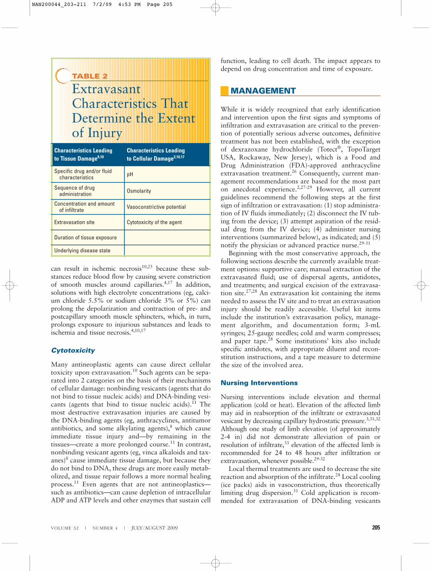
Variables that determine the extent of tissue damagecaused by extravasation are summarized in Table 2.
pH and Osmolarity
Solutions that irritate the venous endothelium andvessel wall ultimately raise the risk of venous rupture,allowing the solutions to escape into the surroundingtissue.18 To minimize venous irritation, infused solu-tions should be close to physiologic pH (7.35-7.40)19
and osmolarity (281-282 mOsm/L).20,21 Extreme pH(both alkaline and acidic) can reduce peripheral veintolerance22 by damaging cell proteins and eventuallycausing cell death, leading to venous endothelialdamage and making it susceptible to rupture.21 The pHof infusion solutions should generally be between 5 and 9.21
For solutions that escape the vein, osmolarity canalso influence the degree of tissue injury. For example,extravasation of hypertonic fluids such as 10% dextroseor parenteral nutrition solutions can cause skin necrosisand serious tissue damage.18,23 Furthermore, dextrosesolutions are rarely administered alone: other productsare often added, which can increase osmolarity evenmore. Extravasation of hypertonic solutions (eg,
osmolarity � 350 mOsm/L) causes a fluid shift frominside cells to the interstitial space,2 which disrupts cellfunction.18,24 The shift from subcutaneous cells to inter-stitial tissues leads to swelling and increased local pres-sure, which can cause ALCS and other injuries.18 Whilehyperosmolar solutions can shrink cells and collapse theinternal structures, hypoosmolar solutions can swellcells and lead to cell rupture.
Vasoconstriction
Extravasation of vasoactive substances (eg, dobutamine,dopamine, epinephrine, norepinephrine, and vasopressin)
204 Journal of Infusion Nursing
TABLE 1
Factors Contributingto the Risk forInfiltration andExtravasation
Mechanical factors
Small size and poor condition of veins8
Larger catheter size relative to vein size2
Choice of site (eg, areas of joint flexion, dominant hand)2
Unstable catheter2
Poor securing of implanted port access needle9
Patient activity2
Multiple venipuncture sites (eg, a second puncture above the first)2
Use of an infusion pump or power injector during the infiltration or extravasation event2
Catheter port separation or catheter fracture10
Physiologic factors
Clot formation above the cannulation site2
Thrombus or fibrin sheath at the catheter tip8
Lymphedema8
Pharmacologic factors
pH
Osmolarity
Vasoconstrictive potential
Cytotoxicity
NAN200044_203-211 7/2/09 4:53 PM Page 204
can result in ischemic necrosis10,25 because these sub-stances reduce blood flow by causing severe constrictionof smooth muscles around capillaries.4,17 In addition,solutions with high electrolyte concentrations (eg, calci-um chloride 5.5% or sodium chloride 3% or 5%) canprolong the depolarization and contraction of pre- andpostcapillary smooth muscle sphincters, which, in turn,prolongs exposure to injurious substances and leads toischemia and tissue necrosis.4,10,17
Cytotoxicity
Many antineoplastic agents can cause direct cellulartoxicity upon extravasation.10 Such agents can be sepa-rated into 2 categories on the basis of their mechanismsof cellular damage: nonbinding vesicants (agents that donot bind to tissue nucleic acids) and DNA-binding vesi-cants (agents that bind to tissue nucleic acids).11 Themost destructive extravasation injuries are caused bythe DNA-binding agents (eg, anthracyclines, antitumorantibiotics, and some alkylating agents),8 which causeimmediate tissue injury and—by remaining in thetissues—create a more prolonged course.11 In contrast,nonbinding vesicant agents (eg, vinca alkaloids and tax-anes)8 cause immediate tissue damage, but because theydo not bind to DNA, these drugs are more easily metab-olized, and tissue repair follows a more normal healingprocess.11 Even agents that are not antineoplastics—such as antibiotics—can cause depletion of intracellularADP and ATP levels and other enzymes that sustain cell
function, leading to cell death. The impact appears todepend on drug concentration and time of exposure.
MANAGEMENT
While it is widely recognized that early identificationand intervention upon the first signs and symptoms ofinfiltration and extravasation are critical to the preven-tion of potentially serious adverse outcomes, definitivetreatment has not been established, with the exceptionof dexrazoxane hydrochloride (Totect®, TopoTargetUSA, Rockaway, New Jersey), which is a Food andDrug Administration (FDA)-approved anthracyclineextravasation treatment.26 Consequently, current man-agement recommendations are based for the most parton anecdotal experience.2,27-29 However, all currentguidelines recommend the following steps at the firstsign of infiltration or extravasation: (1) stop administra-tion of IV fluids immediately; (2) disconnect the IV tub-ing from the device; (3) attempt aspiration of the resid-ual drug from the IV device; (4) administer nursinginterventions (summarized below), as indicated; and (5)notify the physician or advanced practice nurse.29-31
Beginning with the most conservative approach, thefollowing sections describe the currently available treat-ment options: supportive care; manual extraction of theextravasated fluid; use of dispersal agents, antidotes,and treatments; and surgical excision of the extravasa-tion site.27,28 An extravasation kit containing the itemsneeded to assess the IV site and to treat an extravasationinjury should be readily accessible. Useful kit itemsinclude the institution’s extravasation policy, manage-ment algorithm, and documentation form; 3-mLsyringes; 25-gauge needles; cold and warm compresses;and paper tape.28 Some institutions’ kits also includespecific antidotes, with appropriate diluent and recon-stitution instructions, and a tape measure to determinethe size of the involved area.
Nursing Interventions
Nursing interventions include elevation and thermalapplication (cold or heat). Elevation of the affected limbmay aid in reabsorption of the infiltrate or extravasatedvesicant by decreasing capillary hydrostatic pressure.3,31,32
Although one study of limb elevation (of approximately2-4 in) did not demonstrate alleviation of pain orresolution of infiltrate,33 elevation of the affected limb isrecommended for 24 to 48 hours after infiltration orextravasation, whenever possible.29-32
Local thermal treatments are used to decrease the sitereaction and absorption of the infiltrate.28 Local cooling(ice packs) aids in vasoconstriction, thus theoreticallylimiting drug dispersion.31 Cold application is recom-mended for extravasation of DNA-binding vesicants
VOLUME 32 | NUMBER 4 | JULY/AUGUST 2009 205
TABLE 2
ExtravasantCharacteristics ThatDetermine the Extentof Injury
Characteristics Leading to Tissue Damage8,10
Characteristics Leading to Cellular Damage2,10,17
Specific drug and/or fluid characteristics pH
Sequence of drug administration Osmolarity
Concentration and amount of infiltrate Vasoconstrictive potential
Extravasation site Cytotoxicity of the agent
Duration of tissue exposure
Underlying disease state
NAN200044_203-211 7/2/09 4:53 PM Page 205
(with the exception of mechlorethamine [nitrogen mus-tard]),30 contrast media,3,34 and hyperosmolar fluids.2,35
The use of local warming therapy (dry heat) is based onthe theory that it enhances vasodilation, thus enhancingdispersion of the vesicant agent and decreasing drugaccumulation in the local tissue.31 The use of localwarming is recommended for extravasation ofnon–DNA-binding vesicants.30 Although clear benefithas not been demonstrated with thermal applica-tions,17,35,36 it remains standard supportive care, and therecommended application schedule for both warm andcold applications is 15 to 20 minutes, every 4 hours, for24 to 48 hours.30 It should be noted that heat and coldapplications are not well supported in neonates andyoung infants.
Aspiration and Extraction
As soon as infiltration or extravasation has been identi-fied, the infusion should be stopped, the IV tubingshould be disconnected (leaving the catheter in place),and then an attempt should be made to aspirate theresidual drug from the IV device using a small (1- to 3-mL) syringe.30
Documented experience with methods of manuallyextracting infiltrated or extravasated agents is lacking,so these techniques are not routinely recommended, andshould be performed only by a physician or other qual-ified personnel. The “squeeze maneuver” was describedin a report of 8 patients with more than 50 mL ofcontrast-media extravasation that had resulted in vascu-lar compromise of the fingers. After stopping the infu-sion, the IV catheter was removed and an 18-gaugeneedle was used to create 5 to 8 holes near the insertionsite (avoiding vessels, tendons, and muscles). Thepatients’ tissue was then “milked” from the edges of thedistended area toward the needle holes, squeezing outthe extravasated contrast media until distal circulationhad been restored.37 The advantages of this procedureare that it can be performed immediately and it does notrequire anesthesia37; however, experience with its use islimited.
Other reported manual extraction methods includepercutaneous needle aspiration, liposuction, and surgi-cal fenestration and irrigation.27 Early washout usingsurgical fenestration and irrigation has been describedas a simple, practical procedure,23,38 which immediatelyreduces the amount of extravasated agent at the sitewith good outcomes. In an early report, 44 pediatricpatients with extravasations of calcium, potassium,sodium bicarbonate, or 10% dextrose were treatedwithin 24 hours of extravasation injury with stab inci-sions and 500 mL of normal saline flush, with drainsleft in place for 24 hours. In these patients, 86% healedwithout soft tissue loss.23 In a later report, 8 patientswith suspected vinca alkaloid or anthracycline
chemotherapy extravasations were treated with severalincisions within 6 hours of the extravasation, followedby infiltration with 300 to 500 mL of normal saline viaa large catheter.38 All 8 patients experienced normalhealing without functional impairment. Although theseearly reports of saline washout were positive, they havelimited clinical utility and have not been incorporatedinto infusion guidelines.30
Dispersal With Saline
The theory behind the use of dispersal is that tissueinjury will be decreased secondary to the dilution of thevesicant across a larger area of tissue.29 Clysis withsaline has been described as the simplest method todilute the concentration of a vesicant extravasation inorder to prevent tissue injury.27 In a 1994 report on 40patients with suspected extravasations of vinca alka-loids or doxorubicin, conservative treatment with 20 to90 mL of saline solution, injected at the extravasationsite 3 to 6 times, was administered over a course of sev-eral days to all patients except 3 with deep lesions.39
Pain and erythema were resolved in 4 days or less andsuperficial ulcerations in 10 to 14 days in all treatedpatients; surgery was required in the 3 patients withdeep ulcers. Variations of the saline wash procedureinclude the saline flush-out technique described in thepreceding section.
Pharmacologic Antidotes
Although a number of pharmacologic antidotes have been investigated for management of vesicantextravasation,12,40 their use remains controversial.2
While several antidotes described in this section havebeen shown to limit tissue damage caused by extravasa-tion of specific vesicants and are recommended inempirical guidelines,8,28,29,32,41 the most recentOncology Nursing Society guidelines do not recom-mend antidotes for extravasation of chemotherapeuticand biotherapeutic agents, with the exception of sodiumthiosulfate.30 Therefore, the package insert or otherreference material should be consulted before consideringthe use of an antidote for management of extravasationof specific agents.7
Hyaluronidase
Hyaluronidase enzymatically increases tissue permeabil-ity, which facilitates systemic absorption of infiltratedvesicant agents.29 Hyaluronidase rapidly (within 10 min-utes) results in diffusion of extravasated fluid over anarea 3 to 5 times larger than an area left untreated, andtissue permeability is restored within 24 to 48 hours.42
In both animal studies and human reports, good out-comes have been observed with hyaluronidase treatment
206 Journal of Infusion Nursing
NAN200044_203-211 7/2/09 4:53 PM Page 206
VOLUME 32 | NUMBER 4 | JULY/AUGUST 2009 207
of suspected extravasations of DNA-binding drugs (eg,doxorubicin),43,44 non–DNA-binding drugs (eg, vincaalkaloids),45,46 irritant drugs (eg, nafcillin),42 hyperos-molar solutions (eg, dextrose 10%, parenteral nutri-tion),42 and many other agents. In a recent case report onparenteral nutrition extravasation in a premature infant,early subcutaneous hyaluronidase followed by salineflushing resulted in a dramatic response with almost nosign of injury after 5 days.47 Hyaluronidase is not recom-mended for use with dopamine or �-agonist drugs.48
Clinicians should consider patients with allergies andwith religious/cultural proscriptions when choosingbetween animal-derived forms of hyaluronidase and thenew recombinant human form (rHuPH20).48
Sodium Thiosulfate
Sodium thiosulfate has long been recognized as an effec-tive antidote to mechlorethamine (nitrogen mustard).40
Furthermore, a study of 63 patients who had injuriesinduced by extravasation of a variety of vesicant agents(eg, doxorubicin, epirubicin, vinblastine, and mito-mycin C) showed that sodium thiosulfate combinedwith conservative treatment significantly improvedhealing time compared with conservative treatmentalone.49 The recommended administration of sodiumthiosulfate is through immediate subcutaneous injectionof 2 mL of 0.17M solution.50
Dimethyl Sulfoxide
Dimethyl sulfoxide (DMSO) is a topically applied sol-vent that may improve systemic absorption ofextravasated vesicants. It also acts as a free radical scav-enger, thus preventing DNA damage from oxygen freeradicals that might be produced by cytotoxic agents.40,41
Topical administration of DMSO after vesicant drugextravasation has been shown to prevent tissue necrosisin several animal studies51,52; however, in another study,DMSO failed to reduce ulceration after anthracyclineextravasation53; and a third study suggested that DMSOmight potentiate, rather than inhibit, the toxicity of cyto-toxic agents.54 Although good results have been reportedin clinical trials in patients with suspected extravasationsof cytotoxic agents using 90% to 99% DMSO,55-57
medical-grade DMSO at concentrations higher than50% is not available in the United States.31 Because ofconflicting efficacy data, limited drug availability, andthe advent of new treatments such as Totect®, the use ofDMSO as an antidote is not recommended in theOncology Nursing Society guidelines.30
Phentolamine
Extravasation of vasopressor agents (eg, dopamine,epinephrine, and norepinephrine) causes local vasocon-
striction and ischemia, which can result in tissue necro-sis and ulceration.58 Phentolamine competitively blocks�-adrenergic receptors, reversing these effects, therebymitigating the tissue injury. As early as 1957, phento-lamine was reported to reduce skin loss caused byextravasation of vasoconstrictor agents in animal mod-els and in case studies.59 While one early reportdescribed a vasopressor extravasation injury that didnot respond to phentolamine administered 48 hoursafter extravasation,60 a later animal study demonstratedthat early administration of phentolamine was criticalto its beneficial effects on vasopressor extravasationinjuries.61 Results of these animal studies are supportedby the findings of several case reports,62,63 and adminis-tration of phentolamine no more than 12 hours afterextravasation is supported in empirical guidelines forthe management of vasopressor extravasation in bothchildren32,58 and adults.17
Dexrazoxane Hydrochloride (Totect®)
Totect® is a new FDA-approved treatment for IVanthracycline extravasation.64 The mechanism by whichit reduces tissue damage is unknown.64 Efficacy in treat-ing anthracycline extravasations has been demonstratedin animal studies,65-67 case reports,68-71 and 2 multicen-ter, prospective clinical trials.72 In the 2 clinical trials, 54patients with biopsy-confirmed doxorubicin and epiru-bicin extravasations received 3 days of IV dexrazoxane(1000, 1000, and 500 mg/m2) treatment beginning nolater than 6 hours after the event.72 Skin and tissueintegrity remained intact in 53 of 54 patients (98.2%efficacy); only 1 of the 54 patients (2.8%) required sur-gical resection of necrosis. Sequelae were reported asmild among those not requiring surgery, and 74% of thepatients were able to continue anthracycline chemother-apy on schedule.
Totect® is packaged as an emergency treatment kitfor single patient use and contains a complete course oftreatment. The drug is administered for 3 consecutivedays as a 1- to 2-hour IV infusion in a large-caliber veinin an extremity other than the one affected by extrava-sation. The recommended dose is the same as thatadministered in the clinical trials64: 1000 mg/m2 on day1; 1000 mg/m2 on day 2, and 500 mg/m2 on day 3.
Surgical Intervention
The majority of suspected extravasation injuries arebelieved to heal without surgical intervention, so a con-servative approach is advisable.3,27,29,73,74 However, ithas been estimated that surgery is required for up toone-third of cases.29,40,75 Therefore, timely surgical con-sultation is important to minimize adverse outcomeswhen extravasation of nonanthracycline vesicantsoccurs (anthracycline extravasations are immediately
NAN200044_203-211 7/2/09 4:53 PM Page 207
treated with Totect®).2,28,39 Continuing pain afteradministration of conservative local treatment has beencited as an indication for surgical consultation.11,29,40,76
When surgery is indicated, early debridement of necrot-ic tissues and entrapped drug can minimize the risk forsubsequent damage to deeper tissues; and when healingis delayed, wide excision and skin grafting are the mostcommon procedures.40,76 More aggressive surgery, suchas mastectomy, may be required in cases of extensivecentral venous catheter extravasation injury.9
In contrast, the primary treatment of ALCS isdecompression with fasciotomy.16 Because outcomesare optimal when decompression occurs less than12 hours after the onset of ALCS,77,78 surgical treat-ment should be initiated as soon as possible to mini-mize complications.16
Experimental Treatments
Hyperbaric Oxygen Therapy
Hyperbaric oxygen therapy involves intermittentinhalation of 100% oxygen in a treatment chamberwith pressure greater than 1 atm.79,80 The rationale isthat intermittently increasing tissue oxygen tensionoptimizes fibroblast proliferation and collagen synthe-sis, thereby enhancing the oxidative killing capacity ofwhite blood cells during hyperoxia and stimulatingangiogenesis during hypoxia.80 Enhancement of healingin selected problem wounds is one of the 13 indicationsapproved for hyperbaric oxygen therapy by theHyperbaric Oxygen Committee of the Underwater andHyperbaric Medical Society.81
Studies of hyperbaric oxygen therapy in animal mod-els of doxorubicin extravasation have reported mixedresults. In an early study, ulcers in rats treated withhyperbaric oxygen therapy were significantly larger at2, 3, and 5 weeks as compared with those in untreatedanimals, suggesting that cytotoxic effects of doxoru-bicin were potentiated by this treatment modality.80 Incontrast, a more recent study using a similar modelreported no difference in lesion size in hyperbaric-oxygen-treated animals and in control animals on days7 or 14, but a significant reduction in the treatedanimals on days 21 and 28, with complete wound healingby day 40 in 37% of the treated animals versus no woundhealing in the control group.79 These findings arepreliminary, and currently there are no reports of hyper-baric oxygen therapy in humans for the treatment oftissue necrosis caused by vesicant extravasation.Moreover, the use of hyperbaric oxygen therapy is notdiscussed in the current guidelines for the practice ofchemotherapy and biotherapy.30
The theoretical mechanism of hyperbaric oxygentherapy for the treatment of ALCS is that hyperoxy-genation increases the supply of oxygen and causes
vasoconstriction, resulting in decreased capillaryblood pressure and transudation.16 Hyperbaric oxygentherapy has been shown to reduce edema and tissuenecrosis in experimental models of ALCS82,83 andpromote wound healing in patients with ALCS result-ing from traumatic injury. In one study, 17 of 18patients who were given hyperbaric treatment follow-ing surgical fasciotomy experienced complete woundhealing as compared with 10 of 18 patients whoreceived fasciotomy alone.84 Hyperbaric therapy iscurrently recommended as an adjunct to fasciotomy orin those cases in which immediate surgical treatment isnot possible.16
Special Issues in Neonatal and Pediatric Patients
Total parenteral nutrition, electrolyte therapy, antibi-otics, vasoactive medications, and other drug thera-pies requiring peripheral IV therapy are often neededin low-birth-weight infants and in children with com-plex illnesses.58 However, their small, fragile veinsand inability to verbally communicate their pain anddiscomfort leave them particularly prone to infiltra-tion and extravasation injuries.29,85 Although woundand skin care is emphasized in newborns andinfants,18 treatment for children, like that for adults,is based on the anecdotal literature.32 The value of hotor cold compresses is debatable; there is little evidenceof efficacy and some risk of skin maceration withmoist heat.
IMPORTANCE OF PREVENTIONAND RECOGNITION
In view of the current controversy regarding the indi-cations for some treatments and the limitations ofothers, prevention has been correctly stated to bepreferable to cure.86 Careful adherence to properprocedures and timely identification of signs andsymptoms are critical to avoiding potentially life-alteringcomplications.2,28 Although the primary responsibilityfor the prevention and early recognition of injury due to infiltration and extravasation rests with thenursing staff,2,8 physicians and pharmacists should beknowledgeable about the most up-to-date treatmentoptions and institutional protocols and should beprepared to act promptly in order to prevent seriouscomplications.
DOCUMENTATION
Because errors associated with IV administration canresult in fatal or life-threatening outcomes, administration
208 Journal of Infusion Nursing
NAN200044_203-211 7/2/09 4:53 PM Page 208
of IV fluids and medications can be high-risk, withadverse outcomes potentially leading to malpracticeclaims.5 While nurses are named as defendants in suchlawsuits in increasing numbers, physicians also can benamed if it appears that intervention was not properor timely. For example, the Closed Claims Project
database, which summarizes data from professionalliability carriers, showed that 2.1% of injury claimsfrom 1970 to 2001 were related to peripheralcatheters.6 Among these claims, 28% were related toskin slough or necrosis; 17% were related to swelling,inflammation, or infection; 17% were related to nervedamage (with 22% of these caused by ALCS); 16%were related to fasciotomy scars resulting from ALCS;and 3% were related to heat compresses used to treatIV infiltrations.6 Approximately 54% of peripheralcatheter claims resulted in successful litigation for theplaintiffs, with compensation ranging from $275 to$10,050,000.6
Evidence-based management and complete andaccurate documentation are the keys to an effectivelegal defense in the event of a medicolegal claim.5 TheJoint Commission defines a sentinel event as “an unex-pected occurrence involving death or serious physicalor psychological injury, or the risk thereof. Seriousinjury specifically includes loss of limb or function.”87
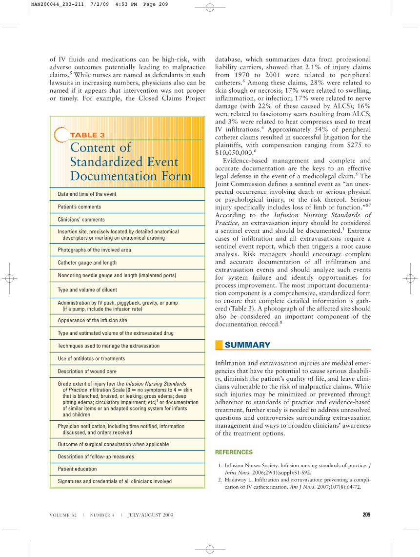
According to the Infusion Nursing Standards ofPractice, an extravasation injury should be considereda sentinel event and should be documented.1 Extremecases of infiltration and all extravasations require asentinel event report, which then triggers a root causeanalysis. Risk managers should encourage completeand accurate documentation of all infiltration andextravasation events and should analyze such eventsfor system failure and identify opportunities forprocess improvement. The most important documenta-tion component is a comprehensive, standardized formto ensure that complete detailed information is gath-ered (Table 3). A photograph of the affected site shouldalso be considered an important component of thedocumentation record.8
SUMMARY
Infiltration and extravasation injuries are medical emer-gencies that have the potential to cause serious disabili-ty, diminish the patient’s quality of life, and leave clini-cians vulnerable to the risk of malpractice claims. Whilesuch injuries may be minimized or prevented throughadherence to standards of practice and evidence-basedtreatment, further study is needed to address unresolvedquestions and controversies surrounding extravasationmanagement and ways to broaden clinicians’ awarenessof the treatment options.
REFERENCES
1. Infusion Nurses Society. Infusion nursing standards of practice. JInfus Nurs. 2006;29(1)(suppl):S1-S92.
2. Hadaway L. Infiltration and extravasation: preventing a compli-cation of IV catheterization. Am J Nurs. 2007;107(8):64-72.
VOLUME 32 | NUMBER 4 | JULY/AUGUST 2009 209
TABLE 3
Content ofStandardized EventDocumentation Form
Date and time of the event
Patient’s comments
Clinicians’ comments
Insertion site, precisely located by detailed anatomical descriptors or marking an anatomical drawing
Photographs of the involved area
Catheter gauge and length
Noncoring needle gauge and length (implanted ports)
Type and volume of diluent
Administration by IV push, piggyback, gravity, or pump (if a pump, include the infusion rate)
Appearance of the infusion site
Type and estimated volume of the extravasated drug
Techniques used to manage the extravasation
Use of antidotes or treatments
Description of wound care
Grade extent of injury (per the Infusion Nursing Standards of Practice Infiltration Scale [0 � no symptoms to 4 � skin that is blanched, bruised, or leaking; gross edema; deep pitting edema; circulatory impairment; etc]1 or documentation of similar items or an adapted scoring system for infants and children
Physician notification, including time notified, information discussed, and orders received
Outcome of surgical consultation when applicable
Description of follow-up measures
Patient education
Signatures and credentials of all clinicians involved
NAN200044_203-211 7/2/09 4:53 PM Page 209

3. Bellin MF, Jakobsen JA, Tomassin I, et al. Contrast mediumextravasation injury: guidelines for prevention and management.Eur Radiol. 2002;12(11):2807-2812.
4. Upton J, Mulliken JB, Murray JE. Major intravenous extravasa-tion injuries. Am J Surg. 1979;137(4):497-506.
5. Diehl-Svrjcek BC, Dawson B, Duncan LL. Infusion nursing:aspects of practice liability. J Infus Nurs. 2007;30(5):274-279.
6. Liau DW. Injuries and liability related to peripheral catheters: aClosed Claims analysis. Newsl Am Soc Anesthesiol. 2006;70(6).http://www.asahq.org/Newsletters/2006/06-06/liau06_06.html.Accessed June 4, 2008.
7. Ener RA, Meglathery SB, Styler M. Extravasation of systemichemato-oncological therapies. Ann Oncol. 2004;15(6):858-862.
8. Goolsby TV, Lombardo FA. Extravasation of chemotherapeuticagents: prevention and treatment. Semin Oncol. 2006;33(1):139-143.
9. Schulmeister L, Camp-Sorrell D. Chemotherapy extravasationfrom implanted ports. Oncol Nurs Forum. 2000;27(3):531-538.
10. Sauerland C, Engelking C, Wickham R, Corbi D. Vesicantextravasation, I: mechanisms, pathogenesis, and nursing care toreduce risk. Oncol Nurs Forum. 2006;33(6):1134-1141.
11. Rudolph R, Larson DL. Etiology and treatment of chemothera-peutic agent extravasation injuries: a review. J Clin Oncol.1987;5(7):1116-1126.
12. Ignoffo RJ, Friedman MA. Therapy of local toxicities caused byextravasation of cancer chemotherapeutic drugs. Cancer TreatRev. 1980;7(1):17-27.
13. Haynes S. Infusion phlebitis and extravasation. Prof Nurse.1989;5(3):160-161.
14. Luke E. Mitoxantrone-induced extravasation. Oncol NursForum. 2005;32(1):27-29.
15. Mubarak SJ, Hargens AR. Acute compartment syndromes. SurgClin North Am. 1983;63(3):539-565.
16. Tiwari A, Haq AI, Myint F, Hamilton G. Acute compartment syn-dromes. Br J Surg. 2002;89(4):397-412.
17. Schummer W, Schummer C, Bayer O, Muller A, Bredle D, KarzaiW. Extravasation injury in the perioperative setting. AnesthAnalg. 2005;100(3):722-727.
18. Pettit J, Hughes K. Intravenous extravasation: mechanisms, man-agement, and prevention. J Perinat Neonatal Nurs. 1993;6(4):69-78.
19. Guyton AC, Hall JE. Regulation of acid-base balance. In: GuytonAC, Hall JE, eds. Textbook of Medical Physiology. 10th ed.Philadelphia, PA: WB Saunders; 2000:346-363.
20. Guyton AC, Hall JE. The body fluid compartments: extracellularand intracellular fluids: interstitial fluid and edema. In: GuytonAC, Hall JE, eds. Textbook of Medical Physiology. 10th ed.Philadelphia, PA: WB Saunders; 2000:264-278.
21. Vesely TM, Stranz M, Masoorli S, Hadaway LC. The diverse andconflicting standards and practices in infusion therapy. J VascAccess Devices. 2002;7(3):9-25.
22. Kuwahara T, Asanami S, Kubo S. Experimental infusionphlebitis: tolerance osmolality of peripheral venous endothelialcells. Nutrition. 1998;14(6):496-501.
23. Gault DT. Extravasation injuries. Br J Plast Surg. 1993;46(2):91-96.24. Robijns BJ, de Wit WM, Bosma NJ, van Vloten WA. Localized
bullous eruptions caused by extravasation of commonly usedintravenous infusion fluids. Dermatologica. 1991;182(1):39-42.
25. Gaze NR. Tissue necrosis caused by commonly used intravenousinfusions. Lancet. 1978;2(8086):417-419.
26. Totect® [package insert]. Rockaway, NJ: TopoTarget USA Inc;2007.
27. Langstein HN, Duman H, Seelig D, Butler CE, Evans GR.Retrospective study of the management of chemotherapeuticextravasation injury. Ann Plast Surg. 2002;49(4):369-374.
28. Wickham R, Engelking C, Sauerland C, Corbi D. Vesicantextravasation, II: evidence-based management and continuingcontroversies. Oncol Nurs Forum. 2006;33(6):1143-1150.
29. Kassner E. Evaluation and treatment of chemotherapy extravasa-tion injuries. J Pediatr Oncol Nurs. 2000;17(3):135-148.
30. Polovich M, White JM, Kelleher LO. Immediate complications ofcytotoxic therapy. In: Polovich M, White JM, Kelleher LO, eds.Chemotherapy and Biotherapy Guidelines and Recommendationsfor Practice. Pittsburgh, PA: Oncology Nursing Society; 2005:78-88.
31. Schulmeister L. Extravasation management. Semin Oncol Nurs.2007;23(3):184-190.
32. Montgomery LA, Hanrahan K, Kottman K, Otto A, Barrett T,Hermiston B. Guideline for IV infiltrations in pediatric patients.Pediatr Nurs. 1999;25(2):167-180.
33. Yucha CB, Hastings-Tolsma M, Szeverenyi NM. Effect of eleva-tion on intravenous extravasations. J Intraven Nurs. 1994;17(5):231-234.
34. Elam EA, Dorr RT, Lagel KE, Pond GD. Cutaneous ulcerationdue to contrast extravasation: experimental assessment of injuryand potential antidotes. Invest Radiol. 1991;26(1):13-16.
35. Hastings-Tolsma MT, Yucha CB, Tompkins J, Robson L,Szeverenyi N. Effect of warm and cold applications on the reso-lution of IV infiltrations. Res Nurs Health. 1993;16(3):171-178.
36. Hastings-Tolsma M, Yucha CB. IV infiltration no clear signs, noclear treatment? RN. 1994;57(12):34-38.
37. Tsai YS, Cheng SM, Ng SP, et al. Squeeze maneuver: an easy wayto manage radiological contrast-medium extravasation. ActaRadiol. 2007;48(6):605-607.
38. Giunta R. Early subcutaneous wash-out in acute extravasations.Ann Oncol. 2004;15(7):1146.
39. Scuderi N, Onesti MG. Antitumor agents: extravasation, man-agement, and surgical treatment. Ann Plast Surg. 1994;32(1):39-44.
40. Dorr RT. Antidotes to vesicant chemotherapy extravasations.Blood Rev. 1990;4(1):41-60.
41. Bertelli G. Prevention and management of extravasation of cyto-toxic drugs. Drug Saf. 1995;12(4):245-255.
42. Zenk KE, Dungy CI, Greene GR. Nafcillin extravasation injury:use of hyaluronidase as an antidote. Am J Dis Child. 1981;135(12):1113-1114.
43. Disa JJ, Chang RR, Mucci SJ, Goldberg NH. Prevention of adri-amycin-induced full-thickness skin loss using hyaluronidase infil-tration. Plast Reconstr Surg. 1998;101(2):370-374.
44. Heckler FR. Current thoughts on extravasation injuries. ClinPlast Surg. 1989;16(3):557-563.
45. Dorr RT, Alberts DS. Vinca alkaloid skin toxicity: antidote anddrug disposition studies in the mouse. J Natl Cancer Inst. 1985;74(1):113-120.
46. Bertelli G, Dini D, Forno GB, et al. Hyaluronidase as an antidoteto extravasation of Vinca alkaloids: clinical results. J Cancer ResClin Oncol. 1994;120(8):505-506.
47. Siu SLY, Kwong KL, Poon SST, So KT. The use of hyaluronidasefor treatment of extravasations in a premature infant. Hong KongJ Paediatr. 2007;12:130-132.
48. Hylenex® [package insert]. Deerfield, IL: Baxter HealthcareCorporation; 2008.
49. Tsavaris NB, Komitsopoulou P, Karagiaouris P, et al. Preventionof tissue necrosis due to accidental extravasation of cytostatic
210 Journal of Infusion Nursing
NAN200044_203-211 7/2/09 4:53 PM Page 210

drugs by a conservative approach. Cancer Chemother Pharmacol.1992;30(4):330-333.
50. Schrijvers DL. Extravasation: a dreaded complication ofchemotherapy. Ann Oncol. 2003;14(suppl 3):iii26-iii30.
51. Dorr RT, Soble MJ, Liddil JD, Keller JH. Mitomycin C skin tox-icity studies in mice: reduced ulceration and altered pharmacoki-netics with topical dimethyl sulfoxide. J Clin Oncol.1986;4(9):1399-1404.
52. Lebredo L, Barrie R, Woltering EA. DMSO protects against adri-amycin-induced tissue necrosis. J Surg Res. 1992;53(1):62-65.
53. Dorr RT, Alberts DS. Failure of DMSO and vitamin E to preventdoxorubicin skin ulceration in the mouse. Cancer Treat Rep.1983;67(5):499-501.
54. Pommier RF, Woltering EA, Fletcher WS. Synergistic cytotoxicityof DMSO-antineoplastic combinations against P388 leukemia inCD-F1 mice. Surg Forum. 1988;39:471-473.
55. Ludwig CU, Stoll HR, Obrist R, Obrecht JP. Prevention of cyto-toxic drug induced skin ulcers with dimethyl sulfoxide (DMSO)and alpha-tocopherole. Eur J Cancer Clin Oncol. 1987;23(3):327-329.
56. Bertelli G, Gozza A, Forno GB, et al. Topical dimethyl sulfoxidefor the prevention of soft tissue injury after extravasation of vesi-cant cytotoxic drugs: a prospective clinical study. J Clin Oncol.1995;13(11):2851-2855.
57. Olver IN, Aisner J, Hament A, Buchanan L, Bishop JF, KaplanRS. A prospective study of topical dimethyl sulfoxide for treatinganthracycline extravasation. J Clin Oncol. 1988;6(11):1732-1735.
58. Flemmer L, Chan JS. A pediatric protocol for management ofextravasation injuries. Pediatr Nurs. 1993;19(4):355-358, 424.
59. Zucker G. Use of phentolamine to prevent necrosis due tolevarterenol. JAMA. 1957;163(16):1477-1479.
60. Boltax RS, Dineen JP, Scarpa FJ. Gangrene resulting from infil-trated dopamine solution. N Engl J Med. 1977;296(14):823.
61. Aycock BG, Hawtof DB, Moody SB. Treatment of peripheralischemia secondary to lidocaine containing epinephrine. AnnPlast Surg. 1989;23(1):27-30.
62. Alexander CS, Sako Y, Mikulic E. Pedal gangrene associated withthe use of dopamine [medical intelligence]. N Engl J Med. 1975;293(12):591.
63. Greenlaw CW, Null LW. Dopamine-induced ischaemia [letter].Lancet. 1977;2(8037):555.
64. Siwy BK, Sadove AM. Acute management of dopamine infiltra-tion injury with Regitine. Plast Reconstr Surg. 1987;80(4):610-612.
65. Langer SW, Sehested M, Jensen PB. Treatment of anthracyclineextravasation with dexrazoxane. Clin Cancer Res. 2000;6(9):3680-3686.
66. Langer SW, Sehested M, Jensen PB. Dexrazoxane is a potent andspecific inhibitor of anthracycline induced subcutaneous lesionsin mice. Ann Oncol. 2001;12(3):405-410.
67. Langer SW, Thougaard AV, Sehested M, Jensen PB. Treatment ofanthracycline extravasation in mice with dexrazoxane with orwithout DMSO and hydrocortisone. Cancer ChemotherPharmacol. 2006;57(1):125-128.
68. Bos AM, van der Graaf WT, Willemse PH. A new conservativeapproach to extravasation of anthracyclines with dimethylsulfox-ide and dexrazoxane. Acta Oncol. 2001;40(4):541-542.
69. El Saghir N, Otrock Z, Mufarrij A, et al. Dexrazoxane foranthracycline extravasation and GM-CSF for skin ulceration andwound healing. Lancet Oncol. 2004;5(5):320-321.
70. Jensen JN, Lock-Andersen J, Langer SW, Mejer J. Dexrazoxane—a promising antidote in the treatment of accidental extravasationof anthracyclines. Scand J Plast Reconstr Surg Hand Surg. 2003;37(3):174-175.
71. Langer SW, Sehested M, Jensen PB, Buter J, Giaccone G.Dexrazoxane in anthracycline extravasation [correspondence]. J Clin Oncol. 2000;18(16):3064.
72. Mouridsen HT, Langer SW, Buter J, et al. Treatment of anthracy-cline extravasation with Savene (dexrazoxane): results from twoprospective clinical multicentre studies. Ann Oncol. 2007;18(3):546-550.
73. Bozkurt AK, Uzel B, Akman C, Ozguroglu M, Molinas MN.Intrathoracic extravasation of antineoplastic agents: case reportand systematic review. Am J Clin Oncol. 2003;26(2):121-123.
74. Cohan RH, Dunnick NR, Leder RA, Baker ME. Extravasation ofnonionic radiologic contrast media: efficacy of conservative treat-ment. Radiology. 1990;176(1):65-67.
75. Larson DL. Treatment of tissue extravasation by antitumoragents. Cancer. 1982;49(9):1796-1799.
76. Boyle DM, Engelking C. Vesicant extravasation: myths and real-ities. Oncol Nurs Forum. 1995;22(1):57-67.
77. Sheridan GW, Matsen FA III. Fasciotomy in the treatment of theacute compartment syndrome. J Bone Joint Surg Am. 1976;58(1):112-115.
78. Mullett H, Al-Abed K, Prasad CV, O’Sullivan M. Outcome ofcompartment syndrome following intramedullary nailing of tibialdiaphyseal fractures. Injury. 2001;32(5):411-413.
79. Aktas S, Toklu AS, Olgac V. Hyperbaric oxygen therapy in adri-amycin extravasation: an experimental animal study. Ann PlastSurg. 2000;45(2):167-171.
80. Monstrey SJ, Mullick P, Narayanan K, Ramasastry SS.Hyperbaric oxygen therapy and free radical production: anexperimental study in doxorubicin (Adriamycin) extravasationinjuries. Ann Plast Surg. 1997;38(2):163-168.
81. Undersea & Hyperbaric Medical Society. Indications for hyper-baric oxygen therapy. http://www.uhms.org/ResourceLibrary/Indications/tabid/270/Default.aspx. Accessed June 4, 2008.
82. Strauss MB, Hargens AR, Gershuni DH, et al. Reduction of skele-tal muscle necrosis using intermittent hyperbaric oxygen in amodel compartment syndrome. J Bone Joint Surg Am. 1983;65(5):656-662.
83. Skyhar MJ, Hargens AR, Strauss MB, Gershuni DH, Hart GB,Akeson WH. Hyperbaric oxygen reduces edema and necrosis ofskeletal muscle in compartment syndromes associated with hemor-rhagic hypotension. J Bone Joint Surg Am. 1986;68(8):1218-1224.
84. Bouachour G, Cronier P, Gouello JP, Toulemonde JL, Talha A,Alquier P. Hyperbaric oxygen therapy in the management ofcrush injuries: a randomized double-blind placebo-controlledclinical trial. J Trauma. 1996;41(2):333-339.
85. Phelps SJ, Helms RA. Risk factors affecting infiltration of periph-eral venous lines in infants. J Pediatr. 1987;111(3):384-389.
86. Burd DAR, Santis G, Milward TM. Severe extravasation injury:an avoidable iatrogenic disaster? BMJ. 1985;290:1579-1580.
87. The Joint Commission. Sentinel event. http://www.jointcommission.org/SentinelEvents/. Accessed June 4, 2008.
VOLUME 32 | NUMBER 4 | JULY/AUGUST 2009 211
NAN200044_203-211 7/2/09 4:53 PM Page 211