Nucleation dynamics in two-dimensional cylindrical Ising models and chemotaxis

Polarized signaling endosomes coordinate BDNF-inducedchemotaxis of cerebellar precursors
Pengcheng Zhou2,1, Marimelia Porcionatto2,1,3, Mariecel Pilapil2, Yicheng Chen2, YoojinChoi2, Kimberley F. Tolias4,5, Jay B. Bikoff4, Elizabeth J. Hong4, Michael E. Greenberg4, andRosalind A. Segal2,6
2 Department of Pediatric Oncology, Dana-Farber Cancer Institute and Department of Neurobiology,Harvard Medical School
4 Division of Neuroscience, Children’s Hospital Boston and Department of Neurobiology, Harvard MedicalSchool
SummaryDuring development, neural precursors migrate in response to positional cues such as growth factorgradients. However, the mechanisms that enable precursors to sense and respond to such gradientsare poorly understood. Here we show that cerebellar granule cell precursors (GCPs) migrate alonga gradient of brain-derived neurotrophic factor (BDNF), and we demonstrate that vesicle traffickingis critical for this chemotactic process. Activation of TrkB, the BDNF receptor, stimulates GCPs tosecrete BDNF, thereby amplifying the ambient gradient. The BDNF gradient stimulates endocytosisof TrkB and associated signaling molecules, causing asymmetric accumulation of signalingendosomes at the subcellular location where BDNF concentration is maximal. Thus regulated BDNFexocytosis and TrkB endocytosis enable precursors to polarize and migrate in a directed fashionalong a shallow BDNF gradient.
IntroductionChemotaxis is an oriented cell migration wherein a cell becomes polarized with a defined frontand back, enabling the cell to move in a forward direction toward an attractive agent (Ridleyet al., 2003; Vicente-Manzanares et al., 2005; Webb et al., 2005). Genetic and mechanisticstudies have identified many of the molecules required for the complex interactions amongsubstrate, membrane and cytoskeleton that allow movement (Affolter and Weijer, 2005;Dujardin et al., 2003; Fishell and Hatten, 1991; Myers et al., 2005; Nagano et al., 2004; Naganoet al., 2002; Qin et al., 2000; Snapper et al., 2005). However, the mechanisms that lead topolarization and directional migration in response to extracellular cues are not well understood.
Chemotaxis is critical for neural development, as neural precursors travel long distances fromproliferative zones to reach the correct positions for mature function. At particular stages indevelopment, precursors travel in predictable directions and along defined axes. While early
6 To whom correspondence should be addressed: Rosalind A. Segal, Department of Pediatric Oncology, Dana-Farber Cancer Institute,44 Binney Street, Boston, MA 02115, 617-632-4737, 617-632-2085, [email protected] first two authors contributed equally.3Current address: Department of Biochemistry, Federal University of São Paulo, São Paolo, Brazil.5Current address: Department of Neuroscience, Baylor College of MedicinePublisher's Disclaimer: This is a PDF file of an unedited manuscript that has been accepted for publication. As a service to our customerswe are providing this early version of the manuscript. The manuscript will undergo copyediting, typesetting, and review of the resultingproof before it is published in its final citable form. Please note that during the production process errors may be discovered which couldaffect the content, and all legal disclaimers that apply to the journal pertain.
NIH Public AccessAuthor ManuscriptNeuron. Author manuscript; available in PMC 2009 March 27.
Published in final edited form as:Neuron. 2007 July 5; 55(1): 53–68. doi:10.1016/j.neuron.2007.05.030.
NIH
-PA Author Manuscript
NIH
-PA Author Manuscript
NIH
-PA Author Manuscript

precursors migrate tangentially along a rostral-caudal or circumferential path, both cerebraland cerebellar precursors migrate radially at defined developmental stages. Neurons of thecerebral cortex travel outward from the ventricular zone to the cortical layers, while cerebellargranule cell precursors (GCPs) travel radially inward from a proliferative zone in the externalgranule cell layer (EGL) and traverse the molecular layer to reach the internal granule cell layer(IGL) (Hatten, 1999, 2002; Komuro and Yacubova, 2003; Komuro et al., 2001; Nadarajah etal., 2002). During radial migration, precursors often migrate along glial “tracks” that restrictmigration decisions to a simple choice: the cells can remain stationary, they can move forwardor move backward (Nadarajah et al., 2001; Rakic, 1971; Rivas and Hatten, 1995). Precursorsmove relatively slowly during radial migration, at rates of approximately 10–30 μm/hour, andso migration of GCPs from the EGL to the IGL occurs over the course of several days invivo (Bellion et al., 2005; Edmondson and Hatten, 1987; Fishell and Hatten, 1991; Komuro etal., 2001; Nadarajah and Parnavelas, 2002). Once GCPs reach the appropriate location in theIGL, migration ceases. Together these attributes make it possible to examine the mechanismsthat regulate radial migration of GCPs in vitro, in vivo or in organotypic slice cultures.
The cytoskeletal changes required for radial migration include remodeling of the microtubulenetwork that surrounds the nucleus and extends forward to a centrosome positioned in front ofthe nucleus. In addition, changes of the actin cytoskeleton and associated myosin motors enablethe leading process to move forward and the trailing process to retract as the cell migrates.These cytoskeletal changes are regulated by intracellular molecular components thatorchestrate cell polarity (Bielas and Gleeson, 2004; Hatten, 2002; Tsai and Gleeson, 2005).
In order for cells to move in the appropriate direction in vivo, extracellular cues must regulatethese polarity molecules. Among the possible attractive cues are neurotrophins (Borghesani etal., 2002; Jones et al., 1994; Polleux et al., 2002; Yamauchi et al., 2003; Yoshizawa et al.,2005), chemokines (Klein et al., 2001; Zhu et al., 2002), netrins (Bloch-Gallego et al., 1999;Hamasaki et al., 2001; Yee et al., 1999), semaphorins (Bagnard et al., 2001; Gammill et al.,2006; Kerjan et al., 2005) and ephrins (Conover et al., 2000; Lu et al., 2001; Santiago andErickson, 2002), while repulsive cues include slit (Brose and Tessier-Lavigne, 2000; Krameret al., 2001; Piper and Little, 2003; Wu et al., 1999), and netrin (Hamasaki et al., 2001). Brain-derived neurotrophic factor (BDNF) is an extracellular factor that regulates neural precursormigration. In vivo, neural migration is abnormal in the cerebral and cerebellar cortex of mutantmice that lack or overexpress BDNF, and BDNF can stimulate migration of precursors in tissueculture system (Borghesani et al., 2002; Jones et al., 1994; Polleux et al., 2002; Ringstedt etal., 1998). However, it is not yet known whether this ligand is involved in movement per se,and/or in the directionality of the response. Here we show that a gradient of BDNF exists inthe developing cerebellum, and GCPs migrate along the BDNF gradient that can both orientand stimulate migration. Correct orientation of migration depends on TrkB-dependentsecretion of BDNF by GCPs and subsequent accumulation of activated TrkB receptors inendosomes located in or adjacent to the leading process. We identify Tiam1/Rac and PI (3, 4,5) P3 (PIP3) phospholipids as critical components in the chemotactic response to BDNF anddemonstrate that these components colocalize with TrkB in signaling endosomes at the leadingprocess. Thus, TrkB-regulated BDNF release and localized endocytosis of BDNF/TrkBconstitute mechanisms that enable GCPs to establish an intracellular gradient of signalingcomponents steeper than the shallow extracellular BDNF gradient, so that GCPs migratedirectionally.
ResultsBDNF promotes directed radial migration of granule cell precursors
In bdnf −/− mice, migration of GCPs from the EGL to IGL is impaired (Borghesani et al.,2002). An extracellular factor such as BDNF might stimulate migration with or without
Zhou et al. Page 2
Neuron. Author manuscript; available in PMC 2009 March 27.
NIH
-PA Author Manuscript
NIH
-PA Author Manuscript
NIH
-PA Author Manuscript

providing a directional cue. As an in vivo gradient is needed for a factor to provide a directionalcue, we investigated the distribution of BDNF protein in the developing cerebellum. Weexamined BDNF concentrations in lysates of EGL and IGL microdissected at postnatal day 7(P7), a stage when the GCPs are actively migrating. As shown in Supplemental Figure 1A, theBDNF concentration as determined by ELISA is two-fold higher in IGL than in EGL. Takentogether with previous studies of BDNF expression by in situ hybridization andimmunostaining (Borghesani et al., 2002; Rocamora et al., 1993; Wetmore et al., 1990), theseresults indicate that regulated expression of BDNF in vivo generates a gradient that increasesalong the migratory path from the EGL to the IGL.
To investigate the role of BDNF as a chemotactic factor for neural precursors, we establishedan in vitro real-time migration assay (Supplemental Figure 1B), wherein GCPs purified fromP6 mice are exposed to an exogenous BDNF gradient and observed by time-lapse microscopy.As shown in Supplemental Figure 1B, BDNF loaded into the agarose plug diffuses into themedium, creating a sharp gradient of BDNF as validated by ELISA (Supplemental Figure 1C).Using this experimental system, we traced the migratory paths of individual GCPs during twohours of data acquisition under control condition, when exposed to a BDNF gradient, or whenexposed to a uniform distribution of BDNF. For each condition, we superimposed the paths ofeach GCP from four experiments on a common origin to generate the composites shown inFigure 1A. As shown, while both uniform BDNF and a BDNF gradient increased thepercentage of cells that migrate compared to control condition, the gradient had a greater effect(Figure 1B). Furthermore, we find that cells migrate in a random direction in control or uniformBDNF condition, but preferentially migrate toward the BDNF source in the gradient condition(Figure 1C and Supplemental movie). Thus, BDNF promotes migration most effectively whenpresented as a gradient, and it provides a directional cue for movement.
Mathematical modeling of chemotactic responses has indicated that basal migration can bemodeled by a random walk; chemotactic responses can often be modeled as a biased randomwalk with high coefficient of non-randomness (μ) (Shreiber et al., 2003). As shown in Figure1D, the BDNF gradient significantly increased μ compared with control or uniform BDNFconditions, without significantly altering other attributes of migration, such as velocity(Supplemental Figure 2), or duration (data not shown). Thus BDNF functions as a chemotacticfactor, converting the stationary state or random walk of precursors under basal conditions intoa directed random walk that is oriented towards the BDNF source.
BDNF activation and redistribution of TrkB mediates directional migrationBDNF has two potential receptors: TrkB and p75NTR, both of which are expressed by GCPsand could be involved in BDNF-induced chemotaxis (Carter et al., 2003; Klein et al., 1990;Segal et al., 1995). To distinguish the receptor pathway(s) required for BDNF induced directedmigration of GCPs, we used both pharmacologic and genetic approaches. We find that K252a,an agent that prevents Trk kinase activity (Berg et al., 1992; Tapley et al., 1992), markedlyinhibits directed migration of GCPs (Figure 1E). Treatment with K252a not only reduces thepercentage of migratory cells, but also reduces the directional movement of GCPs toward theBDNF source (Figure 1F). We find that K252a also prevents GCP migration in an organotypicslice culture (Figure 1G), demonstrating that this Trk inhibitor can prevent migration inresponse to endogenous neurotrophin. We have validated that K252a, at the dose used, preventsBDNF-induced activation of TrkB (inserted blot of Figure 1E) and its downstream signalingcomponents (data not shown). Because GCPs express p75NTR as well as TrkB, and p75NTRhas been implicated in Schwann cell migration during development (Bentley and Lee, 2000),we tested the migration of GCPs prepared from p75NTR exon III mutant mice (Lee et al.,1992). BDNF increased both migration and directed movement of p75 mutant cells as observedfor wild type cells (Figure 1H). Taken together with previous in vivo analyses of various TrkB
Zhou et al. Page 3
Neuron. Author manuscript; available in PMC 2009 March 27.
NIH
-PA Author Manuscript
NIH
-PA Author Manuscript
NIH
-PA Author Manuscript
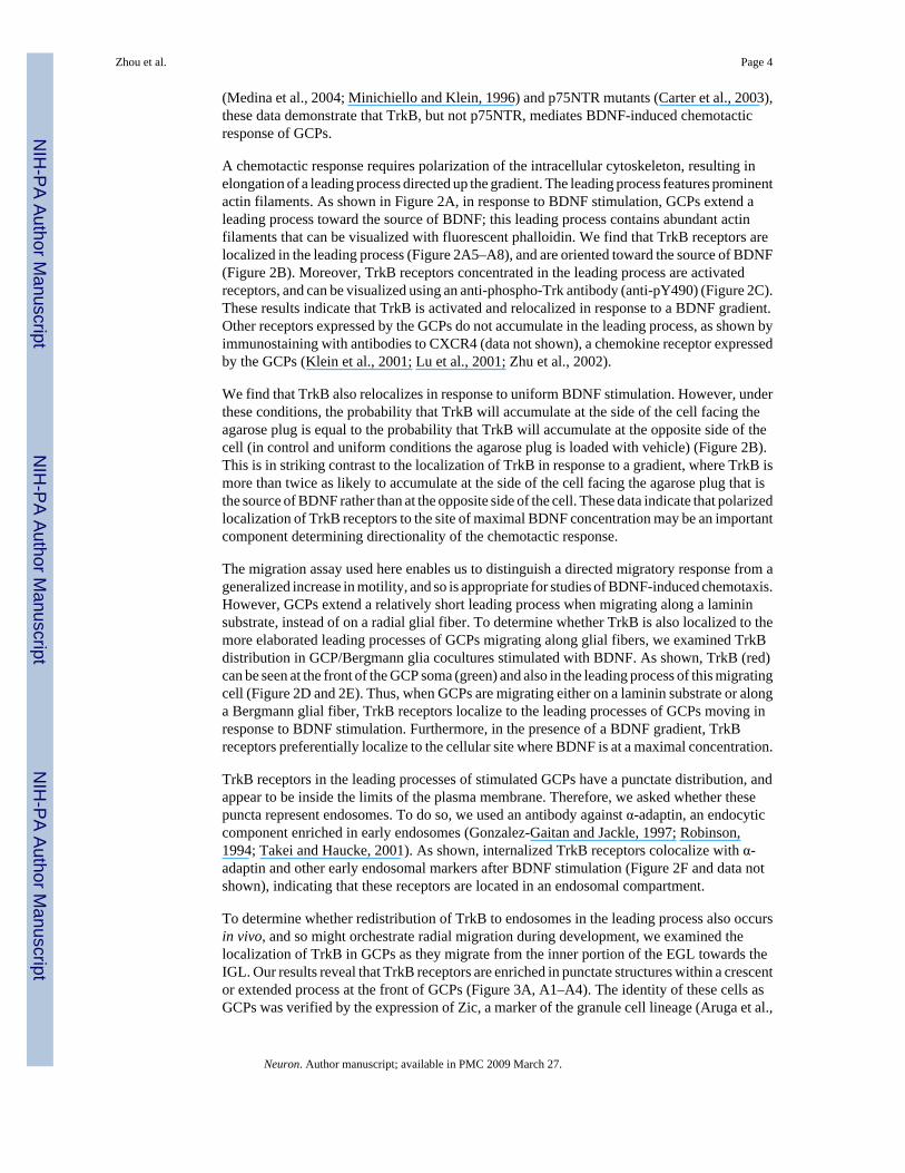
(Medina et al., 2004; Minichiello and Klein, 1996) and p75NTR mutants (Carter et al., 2003),these data demonstrate that TrkB, but not p75NTR, mediates BDNF-induced chemotacticresponse of GCPs.
A chemotactic response requires polarization of the intracellular cytoskeleton, resulting inelongation of a leading process directed up the gradient. The leading process features prominentactin filaments. As shown in Figure 2A, in response to BDNF stimulation, GCPs extend aleading process toward the source of BDNF; this leading process contains abundant actinfilaments that can be visualized with fluorescent phalloidin. We find that TrkB receptors arelocalized in the leading process (Figure 2A5–A8), and are oriented toward the source of BDNF(Figure 2B). Moreover, TrkB receptors concentrated in the leading process are activatedreceptors, and can be visualized using an anti-phospho-Trk antibody (anti-pY490) (Figure 2C).These results indicate that TrkB is activated and relocalized in response to a BDNF gradient.Other receptors expressed by the GCPs do not accumulate in the leading process, as shown byimmunostaining with antibodies to CXCR4 (data not shown), a chemokine receptor expressedby the GCPs (Klein et al., 2001; Lu et al., 2001; Zhu et al., 2002).
We find that TrkB also relocalizes in response to uniform BDNF stimulation. However, underthese conditions, the probability that TrkB will accumulate at the side of the cell facing theagarose plug is equal to the probability that TrkB will accumulate at the opposite side of thecell (in control and uniform conditions the agarose plug is loaded with vehicle) (Figure 2B).This is in striking contrast to the localization of TrkB in response to a gradient, where TrkB ismore than twice as likely to accumulate at the side of the cell facing the agarose plug that isthe source of BDNF rather than at the opposite side of the cell. These data indicate that polarizedlocalization of TrkB receptors to the site of maximal BDNF concentration may be an importantcomponent determining directionality of the chemotactic response.
The migration assay used here enables us to distinguish a directed migratory response from ageneralized increase in motility, and so is appropriate for studies of BDNF-induced chemotaxis.However, GCPs extend a relatively short leading process when migrating along a lamininsubstrate, instead of on a radial glial fiber. To determine whether TrkB is also localized to themore elaborated leading processes of GCPs migrating along glial fibers, we examined TrkBdistribution in GCP/Bergmann glia cocultures stimulated with BDNF. As shown, TrkB (red)can be seen at the front of the GCP soma (green) and also in the leading process of this migratingcell (Figure 2D and 2E). Thus, when GCPs are migrating either on a laminin substrate or alonga Bergmann glial fiber, TrkB receptors localize to the leading processes of GCPs moving inresponse to BDNF stimulation. Furthermore, in the presence of a BDNF gradient, TrkBreceptors preferentially localize to the cellular site where BDNF is at a maximal concentration.
TrkB receptors in the leading processes of stimulated GCPs have a punctate distribution, andappear to be inside the limits of the plasma membrane. Therefore, we asked whether thesepuncta represent endosomes. To do so, we used an antibody against α-adaptin, an endocyticcomponent enriched in early endosomes (Gonzalez-Gaitan and Jackle, 1997; Robinson,1994; Takei and Haucke, 2001). As shown, internalized TrkB receptors colocalize with α-adaptin and other early endosomal markers after BDNF stimulation (Figure 2F and data notshown), indicating that these receptors are located in an endosomal compartment.
To determine whether redistribution of TrkB to endosomes in the leading process also occursin vivo, and so might orchestrate radial migration during development, we examined thelocalization of TrkB in GCPs as they migrate from the inner portion of the EGL towards theIGL. Our results reveal that TrkB receptors are enriched in punctate structures within a crescentor extended process at the front of GCPs (Figure 3A, A1–A4). The identity of these cells asGCPs was verified by the expression of Zic, a marker of the granule cell lineage (Aruga et al.,
Zhou et al. Page 4
Neuron. Author manuscript; available in PMC 2009 March 27.
NIH
-PA Author Manuscript
NIH
-PA Author Manuscript
NIH
-PA Author Manuscript

1994) (Figure 3A). To determine whether localization of TrkB receptors to the leading processin vivo requires a BDNF gradient, we investigated TrkB localization in bdnf −/− mice. Incontrast to the polarized localization of TrkB puncta seen in wild type animals, TrkBimmunostaining in bdnf −/− mice exhibits a random distribution pattern (Figure 3A5–A8; 3Cand 3D), similar to that seen in unstimulated GCPs in vitro (Figure 2A). While TrkB clustersin GCPs are directed toward the IGL in wild type mice, TrkB clusters in GCPs from bdnf −/−mice are more often directed toward or parallel to the pia, and thus point in the wrong directionfor radial migration. Consistent with our studies in vitro, TrkB receptors located in the leadingprocesses of wild type GCPs in vivo are predominantly in endosomes that can beimmunostained with antibodies to α-adaptin (Figure 3B, B1–B4) (65% and 54% of TrkB pixelscolocalize with α-adaptin in wild type and bdnf −/ −mice, respectively. N=5, p < 0.05), thusthe polarization of TrkB signaling endosomes in vivo depends on the presence of a BDNFgradient. Together these data indicate that a chemotactic response to BDNF involvesrelocalization of activated TrkB receptors to endosomes at the cellular site where BDNF ismaximal, and these endosomes mark the direction of movement.
TrkB endocytosis is required for directed cell migrationThe data above suggest that trafficking of vesicular TrkB receptors might coordinate directedmigration of GCPs. While receptor endocytosis was previously viewed as a mechanism forreceptor downregulation and degradation (Burke et al., 2001; Di Fiore and De Camilli,2001), accumulating evidence indicates that intracellular trafficking of signaling endosomescan be important in executing a ligand-initiated response (Beattie et al., 1996; Heerssen et al.,2004; Howe, 2005; Howe and Mobley, 2004; Jekely et al., 2005). This is particularly true forthe Trk receptors, which remain activated for prolonged periods of time following neurotrophinstimulation (Ginty and Segal, 2002). To address the timing and extent of receptor endocytosisin GCPs, we used a biochemical approach in which we analyze surface receptors bybiotinylation. We find that the proportion of TrkB located at the cell surface of GCPs declineswith increased time of BDNF stimulation (Figure 4A and B). The time course of internalizationis fairly slow, and there is a significant lag between internalization and degradation of thereceptor (Figure 4B and C).
To determine whether this slow endocytosis of TrkB receptors might be required for directedmigration, we used monodansyl cadaverine (MDC), a pharmacologic agent that inhibitsinternalization of surface membrane molecules (Davies et al., 1980; Heerssen et al., 2004).Treatment of GCPs with MDC prevents BDNF-induced TrkB receptor endocytosis (lowerpanel, Figure 4E), while there is no effect on TrkB signaling (Figure 4D). We find that MDCsignificantly reduces the percentage of cells that migrate in response to a BDNF gradient(Figure 4E). Further analysis of the paths taken by motile cells revealed that MDC treatmentcompletely abrogates the directionality of GCP migration (Figure 4F). In contrast, MDCactually increases the percentage of cells that migrate in the absence of BDNF (Figure 4F),indicating that this treatment does not interfere with the mechanics of movement. The inhibitoryeffect of MDC on directed migration of GCPs was recapitulated in organotypic slice culture(Figure 4G–I), indicating that endocytosis is required for migration of GCPs stimulated byendogenous BDNF. These data suggest that endocytosis of TrkB receptors is required for achemotactic response to BDNF.
We next used a molecular approach to verify that endocytosis of Trk receptors is needed forGCP migration. We used a dominant-negative form of dynamin (K44A mutant), whichinterferes with both clathrin-dependent and clathrin-independent endocytosis (Damke et al.,1994; Robinson, 1994; van der Bliek et al., 1993). Previous studies indicate that dynaminfunction is needed for endocytosis of activated Trk receptors and other membrane proteins(Damke et al., 1994; Grimes et al., 1996; Robinson, 1994; Valdez et al., 2005; van der Bliek
Zhou et al. Page 5
Neuron. Author manuscript; available in PMC 2009 March 27.
NIH
-PA Author Manuscript
NIH
-PA Author Manuscript
NIH
-PA Author Manuscript

et al., 1993; Zhang et al., 2000). As shown, when we infect organotypic cerebellar slices witha lentivirus expressing dynamin K44A and GFP, most GFP-positive GCPs are unable tomigrate out of the EGL, and remain there three days later (Figure 4K). In contrast, when a sliceculture is infected with control lentivirus expressing wild type dynamin and GFP, virtually allthe GFP-positive GCPs migrate out of the EGL within three days (Figure 4J and L). Thus,expression of dominant-negative dynamin K44A, like treatment with MDC, markedly inhibitsmigration of GCPs in slice cultures responding to endogenous neurotrophins. Together thesefindings indicate that endocytosis of TrkB receptors is critical for BDNF-stimulated cellpolarization and chemotaxis.
BDNF functions as an autocrine ligand to amplify the local gradientIn previous studies we found that migration of GCPs out of the EGL is impaired in bdnf −/−mice in vivo, and that GCPs of these animals exhibit decreased migration in tissue cultureassays (Borghesani et al., 2002). As shown in Figure 5A, both gradient and uniform BDNFincrease migration of bdnf −/− GCPs. However, in contrast to results observed with wild typecells, uniform BDNF stimulates migration of mutant GCPs to the same extent as the BDNFgradient. Furthermore, unlike wild type cells, GCPs of bdnf −/− mice migrate randomly inresponse to a BDNF gradient (Figure 5B and 5C). Thus an exogenous BDNF gradient canstimulate migration of mutant cells, but the cells cannot recognize or respond to the gradientin a directionally appropriate manner.
These findings provide us with a system that dissociates BDNF-induced motility from BDNF-induced directionality, and so can be used to determine the mechanisms whereby a cellpolarizes correctly with a leading process at the location of maximal BDNF concentration. Toascertain whether polarized accumulation of TrkB in the leading process is needed to orientmigration up the BDNF gradient, we visualized TrkB-containing signaling endosomes in GCPsof bdnf −/− mice. As shown in Figure 5D and E, TrkB does redistribute to endosomes thataccumulate at one side of the mutant cells in response to a BDNF gradient. However, in bdnf−/− GCPs, the location where signaling endosomes accumulate is random relative to the BDNFgradient (Figure 5D). These studies suggest that bdnf −/− GCPs migrate randomly in vitrobecause signaling endosomes are not correctly polarized. Taken together, we conclude that aBDNF gradient induces polarized accumulation of signaling endosomes in leading processesof wild type GCPs both in vivo and in vitro, and that the location of the signaling endosomesdictates the direction of migration.
The specific deficit in chemotaxis of bdnf −/− cells could reflect a cell autonomous requirementfor BDNF in order to initiate directed migration. Alternatively, precursors obtained frombdnf −/− mice might acquire additional deficits that preclude a chemotactic response. Todistinguish these possibilities, we asked whether acute reduction in BDNF recapitulates themutant phenotype. Using lentivirus to express siRNA for BDNF, we successfully reduced by80% the level of BDNF protein in GCPs (Figure 6A). As shown in Figures 6B and C, GCPsexpressing bdnf RNAi exhibited impaired chemotaxis in response to a BDNF gradient. Incontrast, GCPs treated in parallel with a control virus remained capable of directed chemotaxis.The data with acute BDNF knock-down are similar to the results obtained using GCPs frombdnf −/− mice (Compare figures 6B and 5A, and figure 6CD and 5C). Thus, BDNF expressionand secretion by GCPs are required for a chemotactic response to an exogenous ligand gradient.
These findings suggest the intriguing possibility that autocrine BDNF might amplify the BDNFgradient and so enhance the directed migration. Previous studies have shown that neurotrophinsthemselves can induce neurotrophin secretion in transfected PC12 cells (Canossa et al.,1997; Kruttgen et al., 1998), in astrocytes (Lambert et al., 2004) and in neurons (Canossa etal., 1997). We therefore asked whether exposing GCPs to a BDNF gradient might induce BDNFrelease and thereby amplify the local gradient. To address this question, we stimulated GCPs
Zhou et al. Page 6
Neuron. Author manuscript; available in PMC 2009 March 27.
NIH
-PA Author Manuscript
NIH
-PA Author Manuscript
NIH
-PA Author Manuscript

with Neurotrophin 4 (NT4). While NT4 is able to stimulate TrkB activation, endocytosis andsignaling (Minichiello et al., 1998; Yuen and Mobley, 1999), the ligand is antigenically distinctfrom BDNF, and so we can monitor release of endogenous BDNF in response to NT4. Asshown in Figure 6E, stimulation of GCPs with NT4 causes BDNF secretion. Based on thetiming and the amount of BDNF released per cell (approximately 1.2 x 10⁻⁷ femptomole/cellreleased within 6 hours), stimulated secretion of BDNF could amplify the local concentrationgradient and so facilitate a chemotactic response.
Given that BDNF is released by GCPs in response to TrkB activation, we asked whether BDNFfunctions in an autocrine manner to stimulate directed migration of GCPs. To do so we analyzedmigration of GCPs in organotypic slice cultures following infection with lentivirus expressingbdnf siRNA together with GFP. We monitor migration by pulse labeling GCPs with BrdU,then allowing 36 hours for BrdU-positive cells to migrate from the EGL to the IGL. As shownin Figure 6F and G, the GFP-positive GCPs expressing bdnf siRNA exhibit reduced migrationcompared with GCPs infected with a control virus. However, migration of the surroundingnon-infected GCPs is not affected by the bdnf siRNA construct. Thus, BDNF is required in acell autonomous fashion for GCP chemotaxis in the slice cultures. Together these data suggesta model wherein the perceived intracellular gradient is amplified in two ways: first by BDNF-stimulated BDNF release, and second by localized accumulation of BDNF/TrkB-containingsignaling endosomes at the site where BDNF concentration is maximal.
PI3 kinase and Tiam1/Rac mediate BDNF-induced chemotaxisTo determine the mechanism by which localized, endocytosed TrkB receptors orchestratedirected migration of GCPs, we asked which of the many signaling pathways activated by TrkBare required for directed migration (Huang and Reichardt, 2003). In non-neural systems boththe phosphatidylinositol kinase (PI3K) and mitogen-activated protein kinase (MAPK)pathways have been implicated in chemotaxis, and these pathways are activated by TrkBsignaling (Funamoto et al., 2002; Huang et al., 2004). Pharmacologic reagents allow us toinhibit the endogenous signaling molecules beginning at the time that we initiate the assay,and so enable us to identify pathways directly involved in BDNF-induced migration.Pretreatment of cerebellar GCPs with LY294002, a PI3K inhibitor, completely blocks BDNF-induced GCP migration (Figure 7A) and the directional bias induced by a BDNF gradient(Figure 7B). We verify that this brief treatment selectively interferes with PI3K signaling inGCPs (Figure 7C) but does not interfere with basal migration or affect survival (data notshown). Thus, the PI3K pathway has a critical role both in the increased motility and directionalresponse induced by a BDNF gradient.
In contrast, pretreatment of GCPs with U0126, an inhibitor of the extracellular signal-regulatedkinase (Erk) pathway, does not alter BDNF-induced initiation or directionality of migration,even though U0126 treatment clearly interferes with Erk1/2 activation (Supplemental Figure3). While previous studies indicated that both PI3K and MAPK can contribute to chemotaxisin response to nerve growth factor (NGF) or other extracellular factors (Huang et al., 2004;Iijima et al., 2002; Merlot and Firtel, 2003; Qi et al., 2001; Sawada et al., 2000), only the PI3kinase pathway is essential for BDNF-induced migration of GCPs.
The products of PI3 kinase, PIP3 phospholipids, can serve as intermediaries that both localizeand stimulate activation of Rho-family G-proteins, and might thereby promote cell motility(Affolter and Weijer, 2005; Bokoch et al., 1996; Hawkins et al., 1995; Raftopoulou and Hall,2004; Sander et al., 1998; Servant et al., 2000). To determine whether PI3 kinase promotesBDNF-induced GCP migration by stimulating Rho family G-proteins, we asked whether thisgroup of proteins is involved in BDNF-induced chemotaxis. Toxin B, which acts quickly toinhibit diverse Rho family GTPases, prevents BDNF-induced migration (Figure 8A). We nextasked which family members are activated by BDNF in cerebellar GCPs. We find that BDNF
Zhou et al. Page 7
Neuron. Author manuscript; available in PMC 2009 March 27.
NIH
-PA Author Manuscript
NIH
-PA Author Manuscript
NIH
-PA Author Manuscript

induces activation of endogenous Rac and cdc42 in GCPs (Figure 8B–D), and that activationof Rac can be abrogated by Toxin B (Figure 8E). We did not detect activation of Rho in responseto BDNF (data not shown). These data suggest that BDNF-induced activation of Rac and / orcdc42 is critical for GCP chemotaxis.
During chemotaxis, PIP3 and Rac often activate one another and thereby amplify a subcellulargradient. Surprisingly, in GCPs Rac activation in response to BDNF does not depend on PIP3phospholipids. As shown, inhibition of BDNF-induced activation of PI3 kinase usingLY294002 has no effect on Rac activation (Figure 8F). Activated Rac itself can stimulate PI3kinase activity under some circumstances (Keely et al., 1997; Tolias et al., 1995; Zheng et al.,1994); however, inhibition of BDNF-induced Rac/cdc42 by toxin B does not prevent PI3 kinaseactivation (Figure 8G). Thus, BDNF-induced migration requires two independent pathwaysactivated simultaneously, one involving Rac/cdc42 and one involving PI3 kinase. Althoughthese pathways are not dependent on one another, both are indeed downstream of TrkB, aspretreatment of GCPs with K252a abolishes BDNF-induced activation of Rac (Figure 8H) andof PI3 kinase activity (Figure 7C). Taken together, these results demonstrate that activationand internalization of TrkB receptors, as well as PI3 kinase and Rac activation are all essentialfor BDNF-induced directed migration.
Since Rac activation is not initiated by PIP3 phospholipids in this system, we asked how TrkBstimulates Rac activity. In the developing cerebellum, a Rac-specific guanine nucleotideexchange factor (GEF), Tiam1, is highly expressed in GCPs (Ehler et al., 1997). While Tiam1is often activated by lipid products of PI3 kinase (Mertens et al., 2003; Miyamoto et al.,2006; Sander et al., 1998), Tiam1 can also be recruited to a signaling complex in a PI3 kinase-independent manner (Lambert et al., 2002). Consistent with recent studies (Miyamoto et al.,2006), we find that in GCPs, endogenous TrkB co-precipitates with endogenous Tiam1 in aBDNF-stimulated manner (Figure 9A), suggesting that this might be a pathway for activationof Rac by internalized TrkB receptors.
To determine whether this pathway is required for directed migration, we used a lentivirussiRNA to reduce expression of Tiam1 (Figure 9B). We find that Tiam1 siRNA, but not controllentivirus, inhibits BDNF-induced Rac activation (Fig. 9B and C). The lentivirus expressingTiam1 siRNA also blocks BDNF-induced directional migration of GCPs in our real-timemigration assay (Figure 9D) and in organotypic slice cultures (Figure 9E–G). Collectively,these results indicate that BDNF activation of TrkB stimulates two parallel pathways, Tiam1/Rac and PI3 kinase, and that both pathways are needed for chemotaxis. We asked whetherTiam1 and phospholipids colocalize with TrkB-containing signaling endosomes. FollowingBDNF gradient stimulation, the PIP3 phospholipid products of PI3 kinase pathway (Figure10A) and Tiam1 (Figure 10B) both colocalize with TrkB in endosomes in the leading process,suggesting that these two pathways are spatially coordinated.
Large signaling complexes containing TrkB and downstream signaling molecules also formin vivo. As shown, Tiam1 colocalizes with TrkB in GCPs of wild type mice (Figure 10C), andthis complex is seen in the leading processes of the cells located at the innermost portion ofthe EGL. TrkB significantly colocalizes with Tiam1 in wild type mice (67% of TrkB pixelscolocalized with Tiam1 in wild type mice while 50% of TrkB pixels colocalized with Tiam1in bdnf −/− mice, n=6, p < 0.001), indicating that signaling endosomes containing both TrkBand Tiam1 are selectively localized to the side of the cell with a maximal concentration ofBDNF. Thus, both in vitro and in vivo, TrkB and associated signaling molecules localize toendosomes located at the cellular site facing the BDNF source with maximal concentration.Collectively, these data indicate that endocytosis and localization of complex signalingendosomes to the front of neural precursors are critical elements of the neurotrophin-inducedchemotactic response.
Zhou et al. Page 8
Neuron. Author manuscript; available in PMC 2009 March 27.
NIH
-PA Author Manuscript
NIH
-PA Author Manuscript
NIH
-PA Author Manuscript

DiscussionDuring development, neural precursors of the cerebral and cerebellar cortex migrate radiallyfrom a proliferative zone, traverse the developing cortex, and then cease migrating once theyreach their final destination (Hatten, 1999, 2002; Nadarajah and Parnavelas, 2002). Cues thatsimultaneously increase migration and provide directionality are optimal for regulating thisbehavior. We find that this is exactly the effect of BDNF. Our results demonstrate that a BDNFgradient not only stimulates motility, but also determines the direction of migration. While anumber of other soluble factors and cell-cell interactions undoubtedly contribute to theregulation of GCP migration and so limit the severity of the migration defect in bdnf −/− mice(Kerjan et al., 2005; Klein et al., 2001; Komuro and Rakic, 1993; Lu et al., 2001; Rio et al.,1997; Tomoda et al., 2004; Zhu et al., 2002), our data indicate that BDNF functions as achemotactic factor both in vivo and in vitro.
A major question in chemotaxis is how cells perceive a subtle gradient and become correctlypolarized. We calculate that the difference in BDNF concentration between the front and theback of the granule cell soma is approximately 6% of the BDNF concentration in vivo,approximately 0.1 to 0.3 femptomolar in both our in vivo and in vitro systems. We haveidentified two mechanisms whereby GCPs amplify the perceived gradient. One simplemechanism for amplifying a gradient is to augment the local concentration of ligand. Consistentwith previous studies in other cell types (Saarelainen et al., 2001; Yang et al., 2006), we findthat activation of TrkB receptors stimulates release of BDNF from wild type GCPs. Thisregulated release of ligand cannot occur in GCPs that lack BDNF due to germline mutationsor due to acute expression of bdnf siRNA. As a result, GCPs that lack BDNF are unable tochemotax properly in response to a BDNF gradient. The timing and extent of BDNF releasein response to TrkB activation suggest that regulated release could play an important role inthe ability of GCPs to exhibit a chemotactic response to BDNF. If the BDNF released by eachcell, approximately 1.2 x 10⁻⁷ femptomole, spreads through a volume of 5 microliters, thesteepness of the gradient would increase by 8%. This increase would have a significant effecton the gradient itself and hence on migration. The protein CAPS2, which promotes localizedand regulated release of BDNF (Sadakata et al., 2004), is likely to be a critical component ofthis autocrine release of ligand. Indeed, a recent study demonstrated that mice lacking CAPS2exhibit impaired GCP migration (Sadakata et al., 2007).
A second mechanism that amplifies the gradient is the localized accumulation of TrkB-containing signaling endosomes at the side of the cell where BDNF concentration is maximal.An accumulation of signaling endosomes containing activated TrkB receptors marks theleading processes of wild type GCPs within the inner EGL that are poised to begin migrating.This localized accumulation is not observed in mice that lack BDNF. To determine whetherlocalized accumulation of TrkB-containing signaling endosomes is required for directedmigration we used both a dissociated cell migration assay and organotypic slice cultures.Pharmacologic interventions that prevent endocytosis of TrkB receptors inhibit BDNF-inducedchemotaxis in a dissociated culture system. In contrast, these pharmacologic interventions donot inhibit basal migration of GCPs, indicating that endocytosis is required for the chemotacticresponse to BDNF rather than for the mechanics of motility. Similarly, both pharmacologicand genetic inhibitors of Trk endocytosis interrupt migration of GCPs in a slice culture whereinGCPs respond to endogenous chemotactic cues. These findings reveal a crucial role of TrkBreceptor endocytosis in the chemotactic response to BDNF.
Although endocytosis has long been regarded as a mechanism of receptor signal attenuation,it is becoming appreciated that the function of endocytosis is not limited to signaldownregulation and receptor removal (Le Roy and Wrana, 2005). In neuronal cells receptorendocytosis is involved in long distance signaling (Ginty and Segal, 2002), and is needed to
Zhou et al. Page 9
Neuron. Author manuscript; available in PMC 2009 March 27.
NIH
-PA Author Manuscript
NIH
-PA Author Manuscript
NIH
-PA Author Manuscript

redistribute signaling molecules during cell fate determination (Hutterer and Knoblich, 2005;Kramer, 2000; Le Borgne, 2006; Parks et al., 2000). In yeast, endocytosis and plasmamembrane recycling contribute to cell polarization (Valdez-Taubas and Pelham, 2003). Vesicleendocytosis and trafficking may also play a role in migration (Rosse et al., 2006; Sheen et al.,2004). Real-time imaging detected endocytic markers (eg. clathrin and dynamin) that arepolarized toward and localized at the leading edge of migrating cells (Rappoport and Simon,2003). In mammary carcinoma cells, an epidermal growth factor (EGF) gradient leads to theaccumulation of endocytosed EGF receptors on the side of the cell facing the EGF source. Inmigrating neural precursors, vesicles accumulate in the leading process (Schaar andMcConnell, 2005). Genetic studies in Drosophila indicate that mutation of the ubiquitin-ligaseD-cbl leads to impaired migration of border cells during oocytogenesis. This mutationattenuates EGF-receptor ubiquitination and so alters receptor endocytosis and signaling (Jekelyet al., 2005). We now show that BDNF-induced initiation and directionality of migration areboth abrogated by endocytosis inhibitors, but these agents do not affect GCP migration in theabsence of BDNF. Thus, endocytosis of TrkB receptors is needed for BDNF-inducedchemotaxis.
Our data indicate that correct localization as well as formation of TrkB-containing signalingendosomes are required for BDNF-induced chemotaxis. Vesicles containing TrkBpreferentially accumulate at the front of wild type GCPs when they are stimulated to chemotaxby a gradient of BDNF. In contrast, when wild type GCPs are stimulated with uniformlydistributed BDNF, TrkB-containing vesicles are oriented in any direction, and the cells migratewith arbitrary directionality. Strikingly, in GCPs prepared from bdnf−/− mice and stimulatedeither by a BDNF gradient or by uniformly distributed BDNF, TrkB containing vesicles arealso oriented in multiple directions. In this genetic model, arbitrary localization of receptors isassociated with random direction of migration. We conclude that both formation andlocalization of signaling endosomes are important for correct cell polarization and directionalmigration of GCPs in response to a BDNF gradient, and that this provides a second mechanismallowing amplification of the chemotactic gradient.
We find that PI3 kinase activation and Tiam1-mediated activation of Rac are additional criticalcomponents of the migratory response to BDNF. Localization of activated TrkB, PIP3phospholipids, and Tiam1 to signaling endosomes ensures that the polarity of cytoskeletalchanges mediated by Rac and by phospholipids are coordinated, so that both designate thesame directionality. As proposed in the local coupling model for directed chemotaxis(Arrieumerlou and Meyer, 2005), we suggest that these cytoskeletal rearrangementscumulatively differentiate the front from the back of the cell. Because activated TrkB receptorsand downstream molecules are in signaling endosomes directed toward the source of thegradient, the cell reliably moves forward toward the BDNF source. In vivo, this insures directedmovement of GCPs from the EGL toward the IGL.
Together, the findings here suggest the following model for BDNF regulation of directedmigration of GCPs. As GCPs mature, they increase expression of BDNF (Maisonpierre et al.,1990). Therefore the mature granule cells that have reached the IGL produce BDNF thatdisperses through the cerebellar cortex, forming an extracellular gradient from the IGL towardthe EGL. This BDNF gradient stimulates TrkB receptors of GCPs that are poised to migrate.In turn, activated TrkB receptors of these precursors initiate autocrine release of BDNF,amplifying the ligand gradient and also causing further TrkB activation and endocytosis.Localized endocytosis of activated TrkB results in accumulation of signaling endosomes at thecellular location where BDNF is maximal, polarizing the cell through the actions of Rac andPIP3 phospholipids. Thus, autocrine release of BDNF and localized accumulation of signalingendosomes amplify the chemotactic gradient and correctly polarize the cell for directedmigration.
Zhou et al. Page 10
Neuron. Author manuscript; available in PMC 2009 March 27.
NIH
-PA Author Manuscript
NIH
-PA Author Manuscript
NIH
-PA Author Manuscript

Experimental ProceduresAnimals
Breeding pairs of brain-derived neurotrophic factor bdnf +/− mice (Ernfors et al., 1994),C57BL/6-Tg (ACTB-EGFP)1Osb/J mice expressing EGFP (Okabe et al., 1997) and p75NTR+/− (exon III) mice (Lee et al., 1992) were purchased from the Jackson Laboratory (Bar Harbor,ME). BALB/c mouse breeding pairs were purchased from Charles River Laboratories(Wilmington, MA). All experimental procedures were performed in accordance with theNational Institutes of Health guidelines and were approved by the Dana-Farber CancerInstitutional Animal Care and Use Committee.
Antibodies and Pharmacological ReagentsThe antibodies and pharmacological reagents used were described in the Supplemental Data.
DNA Constructs and Lentivirus ProductionThe lentivirus construct expressing Tiam1 siRNA was described previously (Tolias et al.,2005). The lentivirus constructs expressing bdnf siRNA, wild type and K44A mutant dynaminare described in the Supplemental Data. Lentivirus was generated as described previously(Rubinson et al., 2003). Viral titers were determined by infection of HEK 293T cells (ATCC).
Primary Granule Cell and Glia Cell CulturePrimary cultures of granule cell precursors (GCPs) from P4 or P6 mouse cerebella wereprepared as described previously (Choi et al., 2005; Klein et al., 2001). See the SupplementalData for details.
Lentiviral Infection of Primary Cerebellar Granule CellsDissociated GCPs from P4 mice were directly resuspended in virus-containing N2 mediumwith 0.4 μg/ml poly-brene (Sigma), plated at high density (2–4 × 10⁶ cell/ml) in an uncoated6-well tissue culture plate overnight and then changed to fresh N2 medium supplemented with2 ug/ml SHH. Forty eight hours later, aggregate cultures were dissociated with papaindissociation kit (Worthington Biochemical Co., Lakewood, New Jersey) according to themanufacturer’s instructions, then used for biochemical assays or real-time migration assay.Infection efficiency of GCPs with lentivirus is 80% on average, determined by the percentageof GFP-positive cells.
Organotypic Slice Culture, BrdU Labeling, Viral Infection and ImmunostainingOrganotyptic cerebellar slices were prepared as described previously (Choi et al., 2005;Stoppini et al., 1991). See the Supplemental Data for details on slice culture, BrdU labeling,viral infection and immunostaing.
Immunoprecipitations and Western Blot AnalysisSee the Supplemental Data for details on immunoprecipitations and Western blotting.
Determination of BDNF ConcentrationBDNF concentrations were determined by ELISA using BDNF Emax
® ImmunoAssay system(Promega). See the Supplemental Data for details.
Zhou et al. Page 11
Neuron. Author manuscript; available in PMC 2009 March 27.
NIH
-PA Author Manuscript
NIH
-PA Author Manuscript
NIH
-PA Author Manuscript
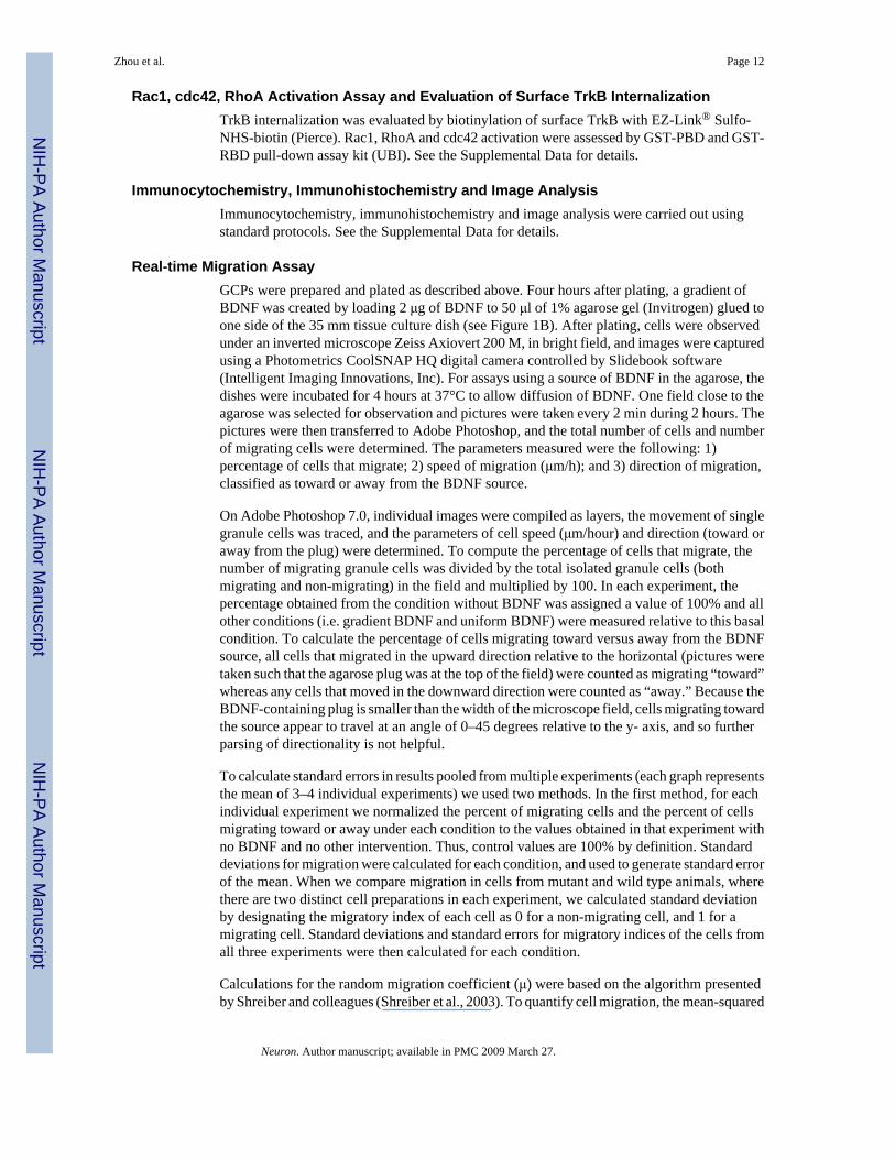
Rac1, cdc42, RhoA Activation Assay and Evaluation of Surface TrkB InternalizationTrkB internalization was evaluated by biotinylation of surface TrkB with EZ-Link® Sulfo-NHS-biotin (Pierce). Rac1, RhoA and cdc42 activation were assessed by GST-PBD and GST-RBD pull-down assay kit (UBI). See the Supplemental Data for details.
Immunocytochemistry, Immunohistochemistry and Image AnalysisImmunocytochemistry, immunohistochemistry and image analysis were carried out usingstandard protocols. See the Supplemental Data for details.
Real-time Migration AssayGCPs were prepared and plated as described above. Four hours after plating, a gradient ofBDNF was created by loading 2 μg of BDNF to 50 μl of 1% agarose gel (Invitrogen) glued toone side of the 35 mm tissue culture dish (see Figure 1B). After plating, cells were observedunder an inverted microscope Zeiss Axiovert 200 M, in bright field, and images were capturedusing a Photometrics CoolSNAP HQ digital camera controlled by Slidebook software(Intelligent Imaging Innovations, Inc). For assays using a source of BDNF in the agarose, thedishes were incubated for 4 hours at 37°C to allow diffusion of BDNF. One field close to theagarose was selected for observation and pictures were taken every 2 min during 2 hours. Thepictures were then transferred to Adobe Photoshop, and the total number of cells and numberof migrating cells were determined. The parameters measured were the following: 1)percentage of cells that migrate; 2) speed of migration (μm/h); and 3) direction of migration,classified as toward or away from the BDNF source.
On Adobe Photoshop 7.0, individual images were compiled as layers, the movement of singlegranule cells was traced, and the parameters of cell speed (μm/hour) and direction (toward oraway from the plug) were determined. To compute the percentage of cells that migrate, thenumber of migrating granule cells was divided by the total isolated granule cells (bothmigrating and non-migrating) in the field and multiplied by 100. In each experiment, thepercentage obtained from the condition without BDNF was assigned a value of 100% and allother conditions (i.e. gradient BDNF and uniform BDNF) were measured relative to this basalcondition. To calculate the percentage of cells migrating toward versus away from the BDNFsource, all cells that migrated in the upward direction relative to the horizontal (pictures weretaken such that the agarose plug was at the top of the field) were counted as migrating “toward”whereas any cells that moved in the downward direction were counted as “away.” Because theBDNF-containing plug is smaller than the width of the microscope field, cells migrating towardthe source appear to travel at an angle of 0–45 degrees relative to the y- axis, and so furtherparsing of directionality is not helpful.
To calculate standard errors in results pooled from multiple experiments (each graph representsthe mean of 3–4 individual experiments) we used two methods. In the first method, for eachindividual experiment we normalized the percent of migrating cells and the percent of cellsmigrating toward or away under each condition to the values obtained in that experiment withno BDNF and no other intervention. Thus, control values are 100% by definition. Standarddeviations for migration were calculated for each condition, and used to generate standard errorof the mean. When we compare migration in cells from mutant and wild type animals, wherethere are two distinct cell preparations in each experiment, we calculated standard deviationby designating the migratory index of each cell as 0 for a non-migrating cell, and 1 for amigrating cell. Standard deviations and standard errors for migratory indices of the cells fromall three experiments were then calculated for each condition.
Calculations for the random migration coefficient (μ) were based on the algorithm presentedby Shreiber and colleagues (Shreiber et al., 2003). To quantify cell migration, the mean-squared
Zhou et al. Page 12
Neuron. Author manuscript; available in PMC 2009 March 27.
NIH
-PA Author Manuscript
NIH
-PA Author Manuscript
NIH
-PA Author Manuscript

displacement <d2(t)> is calculated from position data over all experimental time, ultimatelyyielding a single value for the random cell migration coefficient μ, that describes the averagemigration of the population of cells over the duration of the experiment. Then, <d2(t)> is fit toa persistent random walk model with a GLSR algorithm. Any cell that moved faster than 150μm/hour was excluded because it was presumed to be a dead or floating cell.
Supplementary MaterialRefer to Web version on PubMed Central for supplementary material.
AcknowledgementsWe would like to thank Erin Berry for technical assistance, Louis F. Reichardt for the generous gift of TrkB antibody.We thank Joshua Rubin, Aymeric Hans, Jennifer Chan, Kellie Nazemi, Stephanie Courchesne, Rochelle Witt andCharles Stiles for helpful suggestions. This work was supported by grants from the NIH (NS377057 to RAS andNS045500 to MEG), the Lefler Foundation (to MP and RAS), the Trudy Bettiker/American Brain Tumor AssociationFellowship (to PZ) and the Children’s Hospital Boston Mental Retardation and Developmental Disabilities ResearchCenter, P01 HD18655.
ReferencesAffolter M, Weijer CJ. Signaling to cytoskeletal dynamics during chemotaxis. Dev Cell 2005;9:19–34.
[PubMed: 15992538]Arrieumerlou C, Meyer T. A local coupling model and compass parameter for eukaryotic chemotaxis.
Dev Cell 2005;8:215–227. [PubMed: 15691763]Aruga J, Yokota N, Hashimoto M, Furuichi T, Fukuda M, Mikoshiba K. A novel zinc finger protein, zic,
is involved in neurogenesis, especially in the cell lineage of cerebellar granule cells. J Neurochem1994;63:1880–1890. [PubMed: 7931345]
Bagnard D, Vaillant C, Khuth ST, Dufay N, Lohrum M, Puschel AW, Belin MF, Bolz J, Thomasset N.Semaphorin 3A-vascular endothelial growth factor-165 balance mediates migration and apoptosis ofneural progenitor cells by the recruitment of shared receptor. J Neurosci 2001;21:3332–3341.[PubMed: 11331362]
Beattie EC, Zhou J, Grimes ML, Bunnett NW, Howe CL, Mobley WC. A signaling endosome hypothesisto explain NGF actions: potential implications for neurodegeneration. Cold Spring Harb Symp QuantBiol 1996;61:389–406. [PubMed: 9246468]
Bellion A, Baudoin JP, Alvarez C, Bornens M, Metin C. Nucleokinesis in tangentially migrating neuronscomprises two alternating phases: forward migration of the Golgi/centrosome associated withcentrosome splitting and myosin contraction at the rear. J Neurosci 2005;25:5691–5699. [PubMed:15958735]
Bentley CA, Lee KF. p75 is important for axon growth and schwann cell migration during development.J Neurosci 2000;20:7706–7715. [PubMed: 11027232]
Bielas SL, Gleeson JG. Cytoskeletal-associated proteins in the migration of cortical neurons. J Neurobiol2004;58:149–159. [PubMed: 14598377]
Bloch-Gallego E, Ezan F, Tessier-Lavigne M, Sotelo C. Floor plate and netrin-1 are involved in themigration and survival of inferior olivary neurons. J Neurosci 1999;19:4407–4420. [PubMed:10341242]
Bokoch GM, Vlahos CJ, Wang Y, Knaus UG, Traynor-Kaplan AE. Rac GTPase interacts specificallywith phosphatidylinositol 3-kinase. Biochem J 1996;315(Pt 3):775–779. [PubMed: 8645157]
Borghesani PR, Peyrin JM, Klein R, Rubin J, Carter AR, Schwartz PM, Luster A, Corfas G, Segal RA.BDNF stimulates migration of cerebellar granule cells. Development 2002;129:1435–1442.[PubMed: 11880352]
Brose K, Tessier-Lavigne M. Slit proteins: key regulators of axon guidance, axonal branching, and cellmigration. Curr Opin Neurobiol 2000;10:95–102. [PubMed: 10679444]
Burke P, Schooler K, Wiley HS. Regulation of epidermal growth factor receptor signaling by endocytosisand intracellular trafficking. Mol Biol Cell 2001;12:1897–1910. [PubMed: 11408594]
Zhou et al. Page 13
Neuron. Author manuscript; available in PMC 2009 March 27.
NIH
-PA Author Manuscript
NIH
-PA Author Manuscript
NIH
-PA Author Manuscript

Canossa M, Griesbeck O, Berninger B, Campana G, Kolbeck R, Thoenen H. Neurotrophin release byneurotrophins: implications for activity-dependent neuronal plasticity. Proc Natl Acad Sci U S A1997;94:13279–13286. [PubMed: 9371837]
Carter AR, Berry EM, Segal RA. Regional expression of p75NTR contributes to neurotrophin regulationof cerebellar patterning. Mol Cell Neurosci 2003;22:1–13. [PubMed: 12595234]
Choi Y, Borghesani PR, Chan JA, Segal RA. Migration from a mitogenic niche promotes cell-cycle exit.J Neurosci 2005;25:10437–10445. [PubMed: 16280582]
Conover JC, Doetsch F, Garcia-Verdugo JM, Gale NW, Yancopoulos GD, Alvarez-Buylla A. Disruptionof Eph/ephrin signaling affects migration and proliferation in the adult subventricular zone. NatNeurosci 2000;3:1091–1097. [PubMed: 11036265]
Damke H, Baba T, Warnock DE, Schmid SL. Induction of mutant dynamin specifically blocks endocyticcoated vesicle formation. J Cell Biol 1994;127:915–934. [PubMed: 7962076]
Davies PJ, Davies DR, Levitzki A, Maxfield FR, Milhaud P, Willingham MC, Pastan IH.Transglutaminase is essential in receptor-mediated endocytosis of alpha 2-macroglobulin andpolypeptide hormones. Nature 1980;283:162–167. [PubMed: 6153122]
Di Fiore PP, De Camilli P. Endocytosis and signaling. an inseparable partnership. Cell 2001;106:1–4.[PubMed: 11461694]
Dujardin DL, Barnhart LE, Stehman SA, Gomes ER, Gundersen GG, Vallee RB. A role for cytoplasmicdynein and LIS1 in directed cell movement. J Cell Biol 2003;163:1205–1211. [PubMed: 14691133]
Edmondson JC, Hatten ME. Glial-guided granule neuron migration in vitro: a high-resolution time-lapsevideo microscopic study. J Neurosci 1987;7:1928–1934. [PubMed: 3598656]
Ehler E, van Leeuwen F, Collard JG, Salinas PC. Expression of Tiam-1 in the developing brain suggestsa role for the Tiam-1-Rac signaling pathway in cell migration and neurite outgrowth. Mol CellNeurosci 1997;9:1–12. [PubMed: 9204476]
Ernfors P, Lee KF, Jaenisch R. Mice lacking brain-derived neurotrophic factor develop with sensorydeficits. Nature 1994;368:147–150. [PubMed: 8139657]
Fishell G, Hatten ME. Astrotactin provides a receptor system for CNS neuronal migration. Development1991;113:755–765. [PubMed: 1821847]
Funamoto S, Meili R, Lee S, Parry L, Firtel RA. Spatial and temporal regulation of 3-phosphoinositidesby PI 3-kinase and PTEN mediates chemotaxis. Cell 2002;109:611–623. [PubMed: 12062104]
Gammill LS, Gonzalez C, Gu C, Bronner-Fraser M. Guidance of trunk neural crest migration requiresneuropilin 2/semaphorin 3F signaling. Development 2006;133:99–106. [PubMed: 16319111]
Ginty DD, Segal RA. Retrograde neurotrophin signaling: Trk-ing along the axon. Curr Opin Neurobiol2002;12:268–274. [PubMed: 12049932]
Gonzalez-Gaitan M, Jackle H. Role of Drosophila alpha-adaptin in presynaptic vesicle recycling. Cell1997;88:767–776. [PubMed: 9118220]
Grimes ML, Zhou J, Beattie EC, Yuen EC, Hall DE, Valletta JS, Topp KS, LaVail JH, Bunnett NW,Mobley WC. Endocytosis of activated TrkA: evidence that nerve growth factor induces formationof signaling endosomes. J Neurosci 1996;16:7950–7964. [PubMed: 8987823]
Hamasaki T, Goto S, Nishikawa S, Ushio Y. A role of netrin-1 in the formation of the subcortical structurestriatum: repulsive action on the migration of late-born striatal neurons. J Neurosci 2001;21:4272–4280. [PubMed: 11404412]
Hatten ME. Central nervous system neuronal migration. Annu Rev Neurosci 1999;22:511–539.[PubMed: 10202547]
Hatten ME. New directions in neuronal migration. Science 2002;297:1660–1663. [PubMed: 12215636]Hawkins PT, Eguinoa A, Qiu RG, Stokoe D, Cooke FT, Walters R, Wennstrom S, Claesson-Welsh L,
Evans T, Symons M, et al. PDGF stimulates an increase in GTP-Rac via activation ofphosphoinositide 3-kinase. Curr Biol 1995;5:393–403. [PubMed: 7627555]
Heerssen HM, Pazyra MF, Segal RA. Dynein motors transport activated Trks to promote survival oftarget-dependent neurons. Nat Neurosci 2004;7:596–604. [PubMed: 15122257]
Howe CL. Modeling the signaling endosome hypothesis: why a drive to the nucleus is better than a(random) walk. Theor Biol Med Model 2005;2:43. [PubMed: 16236165]
Zhou et al. Page 14
Neuron. Author manuscript; available in PMC 2009 March 27.
NIH
-PA Author Manuscript
NIH
-PA Author Manuscript
NIH
-PA Author Manuscript

Howe CL, Mobley WC. Signaling endosome hypothesis: A cellular mechanism for long distancecommunication. J Neurobiol 2004;58:207–216. [PubMed: 14704953]
Huang C, Jacobson K, Schaller MD. MAP kinases and cell migration. J Cell Sci 2004;117:4619–4628.[PubMed: 15371522]
Huang EJ, Reichardt LF. Trk receptors: roles in neuronal signal transduction. Annu Rev Biochem2003;72:609–642. [PubMed: 12676795]
Hutterer A, Knoblich JA. Numb and alpha-Adaptin regulate Sanpodo endocytosis to specify cell fate inDrosophila external sensory organs. EMBO Rep 2005;6:836–842. [PubMed: 16113648]
Iijima M, Huang YE, Devreotes P. Temporal and spatial regulation of chemotaxis. Dev Cell 2002;3:469–478. [PubMed: 12408799]
Jekely G, Sung HH, Luque CM, Rorth P. Regulators of endocytosis maintain localized receptor tyrosinekinase signaling in guided migration. Dev Cell 2005;9:197–207. [PubMed: 16054027]
Jones KR, Farinas I, Backus C, Reichardt LF. Targeted disruption of the BDNF gene perturbs brain andsensory neuron development but not motor neuron development. Cell 1994;76:989–999. [PubMed:8137432]
Keely PJ, Westwick JK, Whitehead IP, Der CJ, Parise LV. Cdc42 and Rac1 induce integrin-mediatedcell motility and invasiveness through PI(3)K. Nature 1997;390:632–636. [PubMed: 9403696]
Kerjan G, Dolan J, Haumaitre C, Schneider-Maunoury S, Fujisawa H, Mitchell KJ, Chedotal A. Thetransmembrane semaphorin Sema6A controls cerebellar granule cell migration. Nat Neurosci2005;8:1516–1524. [PubMed: 16205717]
Klein R, Martin-Zanca D, Barbacid M, Parada LF. Expression of the tyrosine kinase receptor gene trkBis confined to the murine embryonic and adult nervous system. Development 1990;109:845–850.[PubMed: 2171894]
Klein RS, Rubin JB, Gibson HD, DeHaan EN, Alvarez-Hernandez X, Segal RA, Luster AD. SDF-1 alphainduces chemotaxis and enhances Sonic hedgehog-induced proliferation of cerebellar granule cells.Development 2001;128:1971–1981. [PubMed: 11493520]
Komuro H, Rakic P. Modulation of neuronal migration by NMDA receptors. Science 1993;260:95–97.[PubMed: 8096653]
Komuro H, Yacubova E. Recent advances in cerebellar granule cell migration. Cell Mol Life Sci2003;60:1084–1098. [PubMed: 12861377]
Komuro H, Yacubova E, Rakic P. Mode and tempo of tangential cell migration in the cerebellar externalgranular layer. J Neurosci 2001;21:527–540. [PubMed: 11160432]
Kramer H. RIPping notch apart: a new role for endocytosis in signal transduction? Sci STKE2000;2000:PE1. [PubMed: 11752592]
Kramer SG, Kidd T, Simpson JH, Goodman CS. Switching repulsion to attraction: changing responsesto slit during transition in mesoderm migration. Science 2001;292:737–740. [PubMed: 11326102]
Kruttgen A, Moller JC, Heymach JV Jr, Shooter EM. Neurotrophins induce release of neurotrophins bythe regulated secretory pathway. Proc Natl Acad Sci U S A 1998;95:9614–9619. [PubMed: 9689129]
Lambert JM, Lambert QT, Reuther GW, Malliri A, Siderovski DP, Sondek J, Collard JG, Der CJ. Tiam1mediates Ras activation of Rac by a PI(3)K-independent mechanism. Nat Cell Biol 2002;4:621–625.[PubMed: 12134164]
Lambert WS, Clark AF, Wordinger RJ. Effect of exogenous neurotrophins on Trk receptorphosphorylation, cell proliferation, and neurotrophin secretion by cells isolated from the humanlamina cribrosa. Mol Vis 2004;10:289–296. [PubMed: 15105791]
Le Borgne R. Regulation of Notch signalling by endocytosis and endosomal sorting. Curr Opin Cell Biol2006;18:213–222. [PubMed: 16488590]
Le Roy C, Wrana JL. Signaling and endocytosis: a team effort for cell migration. Dev Cell 2005;9:167–168. [PubMed: 16054022]
Lee KF, Li E, Huber LJ, Landis SC, Sharpe AH, Chao MV, Jaenisch R. Targeted mutation of the geneencoding the low affinity NGF receptor p75 leads to deficits in the peripheral sensory nervous system.Cell 1992;69:737–749. [PubMed: 1317267]
Zhou et al. Page 15
Neuron. Author manuscript; available in PMC 2009 March 27.
NIH
-PA Author Manuscript
NIH
-PA Author Manuscript
NIH
-PA Author Manuscript

Lu Q, Sun EE, Klein RS, Flanagan JG. Ephrin-B reverse signaling is mediated by a novel PDZ-RGSprotein and selectively inhibits G protein-coupled chemoattraction. Cell 2001;105:69–79. [PubMed:11301003]
Maisonpierre PC, Belluscio L, Friedman B, Alderson RF, Wiegand SJ, Furth ME, Lindsay RM,Yancopoulos GD. NT-3, BDNF, and NGF in the developing rat nervous system: parallel as well asreciprocal patterns of expression. Neuron 1990;5:501–509. [PubMed: 1688327]
Medina DL, Sciarretta C, Calella AM, Von Bohlen Und Halbach O, Unsicker K, Minichiello L. TrkBregulates neocortex formation through the Shc/PLCgamma-mediated control of neuronal migration.Embo J 2004;23:3803–3814. [PubMed: 15372074]
Merlot S, Firtel RA. Leading the way: Directional sensing through phosphatidylinositol 3-kinase andother signaling pathways. J Cell Sci 2003;116:3471–3478. [PubMed: 12893811]
Mertens AE, Roovers RC, Collard JG. Regulation of Tiam1-Rac signalling. FEBS Lett 2003;546:11–16.[PubMed: 12829230]
Minichiello L, Casagranda F, Tatche RS, Stucky CL, Postigo A, Lewin GR, Davies AM, Klein R. Pointmutation in trkB causes loss of NT4-dependent neurons without major effects on diverse BDNFresponses. Neuron 1998;21:335–345. [PubMed: 9728915]
Minichiello L, Klein R. TrkB and TrkC neurotrophin receptors cooperate in promoting survival ofhippocampal and cerebellar granule neurons. Genes Dev 1996;10:2849–2858. [PubMed: 8918886]
Miyamoto Y, Yamauchi J, Tanoue A, Wu C, Mobley WC. TrkB binds and tyrosine-phosphorylatesTiam1, leading to activation of Rac1 and induction of changes in cellular morphology. Proc NatlAcad Sci U S A 2006;103:10444–10449. [PubMed: 16801538]
Myers SA, Han JW, Lee Y, Firtel RA, Chung CY. A Dictyostelium homologue of WASP is required forpolarized F-actin assembly during chemotaxis. Mol Biol Cell 2005;16:2191–2206. [PubMed:15728724]
Nadarajah B, Alifragis P, Wong RO, Parnavelas JG. Ventricle-directed migration in the developingcerebral cortex. Nat Neurosci 2002;5:218–224. [PubMed: 11850632]
Nadarajah B, Brunstrom JE, Grutzendler J, Wong RO, Pearlman AL. Two modes of radial migration inearly development of the cerebral cortex. Nat Neurosci 2001;4:143–150. [PubMed: 11175874]
Nadarajah B, Parnavelas JG. Modes of neuronal migration in the developing cerebral cortex. Nat RevNeurosci 2002;3:423–432. [PubMed: 12042877]
Nagano T, Morikubo S, Sato M. Filamin A and FILIP (Filamin A-Interacting Protein) regulate cellpolarity and motility in neocortical subventricular and intermediate zones during radial migration. JNeurosci 2004;24:9648–9657. [PubMed: 15509752]
Nagano T, Yoneda T, Hatanaka Y, Kubota C, Murakami F, Sato M. Filamin A-interacting protein (FILIP)regulates cortical cell migration out of the ventricular zone. Nat Cell Biol 2002;4:495–501. [PubMed:12055638]
Okabe M, Ikawa M, Kominami K, Nakanishi T, Nishimune Y. 'Green mice' as a source of ubiquitousgreen cells. FEBS Lett 1997;407:313–319. [PubMed: 9175875]
Parks AL, Klueg KM, Stout JR, Muskavitch MA. Ligand endocytosis drives receptor dissociation andactivation in the Notch pathway. Development 2000;127:1373–1385. [PubMed: 10704384]
Piper M, Little M. Movement through Slits: cellular migration via the Slit family. Bioessays 2003;25:32–38. [PubMed: 12508280]
Polleux F, Whitford KL, Dijkhuizen PA, Vitalis T, Ghosh A. Control of cortical interneuron migrationby neurotrophins and PI3-kinase signaling. Development 2002;129:3147–3160. [PubMed:12070090]
Qi M, Ikematsu S, Maeda N, Ichihara-Tanaka K, Sakuma S, Noda M, Muramatsu T, Kadomatsu K.Haptotactic migration induced by midkine. Involvement of protein-tyrosine phosphatase zeta.Mitogen-activated protein kinase, and phosphatidylinositol 3-kinase. J Biol Chem 2001;276:15868–15875. [PubMed: 11340082]
Qin J, Mizuguchi M, Itoh M, Takashima S. A novel migration-related gene product, doublecortin, inneuronal migration disorder of fetuses and infants with Zellweger syndrome. Acta Neuropathol (Berl)2000;100:168–173. [PubMed: 10963364]
Raftopoulou M, Hall A. Cell migration: Rho GTPases lead the way. Dev Biol 2004;265:23–32. [PubMed:14697350]
Zhou et al. Page 16
Neuron. Author manuscript; available in PMC 2009 March 27.
NIH
-PA Author Manuscript
NIH
-PA Author Manuscript
NIH
-PA Author Manuscript

Rakic P. Neuron-glia relationship during granule cell migration in developing cerebellar cortex. A Golgiand electronmicroscopic study in Macacus Rhesus. J Comp Neurol 1971;141:283–312. [PubMed:4101340]
Rappoport JZ, Simon SM. Real-time analysis of clathrin-mediated endocytosis during cell migration. JCell Sci 2003;116:847–855. [PubMed: 12571282]
Ridley AJ, Schwartz MA, Burridge K, Firtel RA, Ginsberg MH, Borisy G, Parsons JT, Horwitz AR. Cellmigration: integrating signals from front to back. Science 2003;302:1704–1709. [PubMed:14657486]
Ringstedt T, Linnarsson S, Wagner J, Lendahl U, Kokaia Z, Arenas E, Ernfors P, Ibanez CF. BDNFregulates reelin expression and Cajal-Retzius cell development in the cerebral cortex. Neuron1998;21:305–315. [PubMed: 9728912]
Rio C, Rieff HI, Qi P, Khurana TS, Corfas G. Neuregulin and erbB receptors play a critical role in neuronalmigration. Neuron 1997;19:39–50. [PubMed: 9247262]
Rivas RJ, Hatten ME. Motility and cytoskeletal organization of migrating cerebellar granule neurons. JNeurosci 1995;15:981–989. [PubMed: 7869123]
Robinson MS. The role of clathrin, adaptors and dynamin in endocytosis. Curr Opin Cell Biol1994;6:538–544. [PubMed: 7986531]
Rocamora N, Garcia-Ladona FJ, Palacios JM, Mengod G. Differential expression of brain-derivedneurotrophic factor, neurotrophin-3, and low-affinity nerve growth factor receptor during thepostnatal development of the rat cerebellar system. Brain Res Mol Brain Res 1993;17:1–8. [PubMed:8381892]
Rosse C, Hatzoglou A, Parrini MC, White MA, Chavrier P, Camonis J. RalB mobilizes the exocyst todrive cell migration. Mol Cell Biol 2006;26:727–734. [PubMed: 16382162]
Rubinson DA, Dillon CP, Kwiatkowski AV, Sievers C, Yang L, Kopinja J, Rooney DL, Ihrig MM,McManus MT, Gertler FB, et al. A lentivirus-based system to functionally silence genes in primarymammalian cells, stem cells and transgenic mice by RNA interference. Nat Genet 2003;33:401–406.[PubMed: 12590264]
Saarelainen T, Vaittinen S, Castren E. trkB-receptor activation contributes to the kainate-induced increasein BDNF mRNA synthesis. Cellular and molecular neurobiology 2001;21:429–435. [PubMed:11775072]
Sadakata T, Kakegawa W, Mizoguchi A, Washida M, Katoh-Semba R, Shutoh F, Okamoto T, NakashimaH, Kimura K, Tanaka M, et al. Impaired cerebellar development and function in mice lacking CAPS2,a protein involved in neurotrophin release. J Neurosci 2007;27:2472–2482. [PubMed: 17344385]
Sadakata T, Mizoguchi A, Sato Y, Katoh-Semba R, Fukuda M, Mikoshiba K, Furuichi T. The secretorygranule-associated protein CAPS2 regulates neurotrophin release and cell survival. J Neurosci2004;24:43–52. [PubMed: 14715936]
Sander EE, van Delft S, ten Klooster JP, Reid T, van der Kammen RA, Michiels F, Collard JG. Matrix-dependent Tiam1/Rac signaling in epithelial cells promotes either cell-cell adhesion or cell migrationand is regulated by phosphatidylinositol 3-kinase. J Cell Biol 1998;143:1385–1398. [PubMed:9832565]
Santiago A, Erickson CA. Ephrin-B ligands play a dual role in the control of neural crest cell migration.Development 2002;129:3621–3632. [PubMed: 12117812]
Sawada J, Itakura A, Tanaka A, Furusaka T, Matsuda H. Nerve growth factor functions as achemoattractant for mast cells through both mitogen-activated protein kinase andphosphatidylinositol 3-kinase signaling pathways. Blood 2000;95:2052–2058. [PubMed: 10706874]
Schaar BT, McConnell SK. Cytoskeletal coordination during neuronal migration. Proc Natl Acad Sci US A 2005;102:13652–13657. [PubMed: 16174753]
Segal RA, Pomeroy SL, Stiles CD. Axonal growth and fasciculation linked to differential expression ofBDNF and NT3 receptors in developing cerebellar granule cells. J Neurosci 1995;15:4970–4981.[PubMed: 7623126]
Servant G, Weiner OD, Herzmark P, Balla T, Sedat JW, Bourne HR. Polarization of chemoattractantreceptor signaling during neutrophil chemotaxis. Science 2000;287:1037–1040. [PubMed:10669415]
Zhou et al. Page 17
Neuron. Author manuscript; available in PMC 2009 March 27.
NIH
-PA Author Manuscript
NIH
-PA Author Manuscript
NIH
-PA Author Manuscript

Sheen VL, Ganesh VS, Topcu M, Sebire G, Bodell A, Hill RS, Grant PE, Shugart YY, Imitola J, KhourySJ, et al. Mutations in ARFGEF2 implicate vesicle trafficking in neural progenitor proliferation andmigration in the human cerebral cortex. Nat Genet 2004;36:69–76. [PubMed: 14647276]
Shreiber DI, Barocas VH, Tranquillo RT. Temporal variations in cell migration and traction duringfibroblast-mediated gel compaction. Biophys J 2003;84:4102–4114. [PubMed: 12770913]
Snapper SB, Meelu P, Nguyen D, Stockton BM, Bozza P, Alt FW, Rosen FS, von Andrian UH, Klein C.WASP deficiency leads to global defects of directed leukocyte migration in vitro and in vivo. JLeukoc Biol 2005;77:993–998. [PubMed: 15774550]
Stoppini L, Buchs PA, Muller D. A simple method for organotypic cultures of nervous tissue. J NeurosciMethods 1991;37:173–182. [PubMed: 1715499]
Takei K, Haucke V. Clathrin-mediated endocytosis: membrane factors pull the trigger. Trends Cell Biol2001;11:385–391. [PubMed: 11514193]
Tolias KF, Bikoff JB, Burette A, Paradis S, Harrar D, Tavazoie S, Weinberg RJ, Greenberg ME. TheRac1-GEF Tiam1 couples the NMDA receptor to the activity-dependent development of dendriticarbors and spines. Neuron 2005;45:525–538. [PubMed: 15721239]
Tolias KF, Cantley LC, Carpenter CL. Rho family GTPases bind to phosphoinositide kinases. J BiolChem 1995;270:17656–17659. [PubMed: 7629060]
Tomoda T, Kim JH, Zhan C, Hatten ME. Role of Unc51.1 and its binding partners in CNS axon outgrowth.Genes Dev 2004;18:541–558. [PubMed: 15014045]
Tsai LH, Gleeson JG. Nucleokinesis in neuronal migration. Neuron 2005;46:383–388. [PubMed:15882636]
Valdez G, Akmentin W, Philippidou P, Kuruvilla R, Ginty DD, Halegoua S. Pincher-mediatedmacroendocytosis underlies retrograde signaling by neurotrophin receptors. J Neurosci2005;25:5236–5247. [PubMed: 15917464]
Valdez-Taubas J, Pelham HR. Slow diffusion of proteins in the yeast plasma membrane allows polarityto be maintained by endocytic cycling. Curr Biol 2003;13:1636–1640. [PubMed: 13678596]
van der Bliek AM, Redelmeier TE, Damke H, Tisdale EJ, Meyerowitz EM, Schmid SL. Mutations inhuman dynamin block an intermediate stage in coated vesicle formation. J Cell Biol 1993;122:553–563. [PubMed: 8101525]
Vicente-Manzanares M, Webb DJ, Horwitz AR. Cell migration at a glance. J Cell Sci 2005;118:4917–4919. [PubMed: 16254237]
Webb DJ, Zhang H, Horwitz AF. Cell migration: an overview. Methods Mol Biol 2005;294:3–11.[PubMed: 15576900]
Wetmore C, Ernfors P, Persson H, Olson L. Localization of brain-derived neurotrophic factor mRNA toneurons in the brain by in situ hybridization. Exp Neurol 1990;109:141–152. [PubMed: 2379553]
Wu W, Wong K, Chen J, Jiang Z, Dupuis S, Wu JY, Rao Y. Directional guidance of neuronal migrationin the olfactory system by the protein Slit. Nature 1999;400:331–336. [PubMed: 10432110]
Yamauchi J, Chan JR, Shooter EM. Neurotrophin 3 activation of TrkC induces Schwann cell migrationthrough the c-Jun N-terminal kinase pathway. Proc Natl Acad Sci U S A 2003;100:14421–14426.[PubMed: 14614136]
Yang ZF, Ho DW, Lau CK, Tam KH, Lam CT, Poon RT, Fan ST. Platelet activation during tumordevelopment, the potential role of BDNF-TrkB autocrine loop. Biochemical and biophysicalresearch communications 2006;346:981–985. [PubMed: 16781670]
Yee KT, Simon HH, Tessier-Lavigne M, O'Leary DM. Extension of long leading processes and neuronalmigration in the mammalian brain directed by the chemoattractant netrin-1. Neuron 1999;24:607–622. [PubMed: 10595513]
Yoshizawa M, Kawauchi T, Sone M, Nishimura YV, Terao M, Chihama K, Nabeshima Y, Hoshino M.Involvement of a Rac activator, P-Rex1, in neurotrophin-derived signaling and neuronal migration.J Neurosci 2005;25:4406–4419. [PubMed: 15858067]
Yuen EC, Mobley WC. Early BDNF, NT-3, and NT-4 signaling events. Exp Neurol 1999;159:297–308.[PubMed: 10486198]
Zhang Y, Moheban DB, Conway BR, Bhattacharyya A, Segal RA. Cell surface Trk receptors mediateNGF-induced survival while internalized receptors regulate NGF-induced differentiation. JNeurosci 2000;20:5671–5678. [PubMed: 10908605]
Zhou et al. Page 18
Neuron. Author manuscript; available in PMC 2009 March 27.
NIH
-PA Author Manuscript
NIH
-PA Author Manuscript
NIH
-PA Author Manuscript

Zheng Y, Bagrodia S, Cerione RA. Activation of phosphoinositide 3-kinase activity by Cdc42Hs bindingto p85. J Biol Chem 1994;269:18727–18730. [PubMed: 8034624]
Zhu Y, Yu T, Zhang XC, Nagasawa T, Wu JY, Rao Y. Role of the chemokine SDF-1 as the meningealattractant for embryonic cerebellar neurons. Nat Neurosci 2002;5:719–720. [PubMed: 12080344]
Zhou et al. Page 19
Neuron. Author manuscript; available in PMC 2009 March 27.
NIH
-PA Author Manuscript
NIH
-PA Author Manuscript
NIH
-PA Author Manuscript

Figure 1. A Gradient of BDNF Promotes TrkB –dependent Chemotaxis of GCPs(A) Pathway traces of individual migrating GCPs from four independent experiments for threeconditions (no BDNF, gradient BDNF and uniform BDNF, respectively) were superimposedon a common origin. Because the BDNF-containing plug is smaller than the width of themicroscope field, cells migrating toward the source travel at angles of 0–45 degrees relativeto the y-axis, and fewer cells are precisely at 0 degrees.(B) Quantitative analysis of GCP migration with no BDNF, gradient or uniform BDNF. Datarepresent mean of four independent experiments and were evaluated by z test (*, p < 0.001)and two-tailed Student’s t test (**, p< 0.05), respectively.(C) Quantitative analysis of directionality of GCP migration with no BDNF, gradient oruniform BDNF. Data shown represent means of four independent experiments ± SEM (n=4).
Zhou et al. Page 20
Neuron. Author manuscript; available in PMC 2009 March 27.
NIH
-PA Author Manuscript
NIH
-PA Author Manuscript
NIH
-PA Author Manuscript
(D) Quantitative analysis of coefficient of non-randomness (μ) from the same experiments asA, B and C. Data were evaluated by two-tailed Student’s t-test (*, p< 0.001).(E) K252a, a Trk receptor inhibitor, blocks BDNF-induced GCP migration. Dissociated GCPswere exposed to a BDNF gradient in the absence or presence of 200 nM K252a. Data wereevaluated by z test (*, p < 0.001) and two-tailed Student’s t-test (**, p< 0.001), respectively.(F) Quantitative analysis of directionality of GCP migration for experiments in 1E.(G) K252a (200 nM) inhibits GCP migration in cultured organotypic cerebellar slices.Cerebellar slices (250μm) from P7 wild type mice were cultured in inserts and treated withK252a or vehicle control for 1 hour prior to BrdU pulse-labeling. After BrdU labeling, sliceswere cultured for additional for 72 hours, fixed with 4% paraformaldehyde and immunostainedwith anti-BrdU. The picture represents one sample of three independent experiments. Arrowsindicate BrdU-positive GCPs in IGL. Scale bar: 100 μm.(H) p75NTR is not required for BDNF-induced migration. Migration assay of dissociatedGCPs from p75NTR wild type or exon III mutant littermates was carried out as describedabove. Data shown represent means ± SEM (n=3) and were evaluated by z test (*, p < 0.05)and by two-tailed Student’s t-test (p < 0.05), respectively.
Zhou et al. Page 21
Neuron. Author manuscript; available in PMC 2009 March 27.
NIH
-PA Author Manuscript
NIH
-PA Author Manuscript
NIH
-PA Author Manuscript

Figure 2. BDNF-dependent Polarization of TrkB Receptor in Cerebellar GCPsGCPs grown on coverslips were or were not exposed to gradient BDNF for 12 hours, fixedand immunostained as indicated. In all cases, BDNF source is at the top. DAPI stainingvisualizes nuclei (blue). Scale bars: 5 μm.(A) BDNF induces TrkB receptor and cytoskeletal polarization. F-actin cytoskeletonrearrangement was visualized by staining with Alexa®-568-phalloidin (red). Localization ofTrkB receptor was visualized by immunofluorescent staining with anti-TrkB (green).(B) Quantitative analysis of TrkB localization. GCPs were exposed to uniform or gradientBDNF for 12 hours, fixed and immunostained with TrkB antibody. TrkB localization in GCPswas assessed with fluorescent microscopy. In each of four experiments, the percentage of cells
Zhou et al. Page 22
Neuron. Author manuscript; available in PMC 2009 March 27.
NIH
-PA Author Manuscript
NIH
-PA Author Manuscript
NIH
-PA Author Manuscript

with TrkB polarized toward the agarose plug (front) or away from the plug (back) wasnormalized to the percentage of cells with TrkB polarized to the front under uniform stimulationcondition. Data shown represent means ± SEM (n=4) and were evaluated by two-tailedStudent’s t-test (*, p< 0.01).(C) Polarized TrkB receptors at the front of the cell are activated, visualized byimmunofluorescent staining with phospho-specific Trk antibody (red).(D) BDNF-dependent polarization of TrkB receptors in GCPs migrating along glia fibers.Bergmann glia and GCPs were co-cultured for one day, treated with 10 ng/ml BDNF for 1hour, then fixed and immunostained with anti-TrkB (red). A projection of three-dimensionalreconstruction is shown. Broken line represents glia fiber. Arrow indicates the GCP soma(green) and arrowheads show polarized TrkB receptors at the front of soma (left) and in theleading process of GCP (right). Scal bar: 5 μm.(E) Schematic of TrkB polarization in GCPs migrating along glia fiber in 2D. (F) InternalizedTrkB receptors localize in early endosomal compartment. GCPs were exposed to gradientBDNF for 12 hours, then fixed and stained with anti-α-adaptin (red) and anti-TrkB (green).Arrow indicates polarized GCPs. BDNF source is on the top of the images. Scale bar: 5 μm.
Zhou et al. Page 23
Neuron. Author manuscript; available in PMC 2009 March 27.
NIH
-PA Author Manuscript
NIH
-PA Author Manuscript
NIH
-PA Author Manuscript

Figure 3. BDNF-dependent Formation and Polarization of TrkB Signaling Endosome in vivoCerebellar sections from P6 bdnf +/+ or −/− mice were used for immunostaining as indicated(A and B). DAPI staining visualizes nuclei (blue). Scale bars: 5 μm.(A) BDNF-dependent polarization of TrkB receptors in vivo. TrkB receptors at the front ofGCPs in EGLb are seen within a crescent or extended leading process in bdnf +/+ mice (A1–A4), but are more randomly distributed within the cells of bdnf −/− mice (A5–A8), as visualizedby immunostaining with anti-TrkB (red) and anti-zic (green). A4 and A8 are magnified viewof boxed region in A3 and A7, respectively.(B) BDNF-dependent formation and polarization of TrkB signaling endosomes in vivo. TrkBreceptors colocalize with α-adaptin at leading edge of GCPs from bdnf +/+ mice (B1–B4), but
Zhou et al. Page 24
Neuron. Author manuscript; available in PMC 2009 March 27.
NIH
-PA Author Manuscript
NIH
-PA Author Manuscript
NIH
-PA Author Manuscript

are distributed in random patterns in bdnf −/− GCPs (B5–B8). Colocalization and polarizationof TrkB and α-adaptin was visualized by immunostaining with anti-TrkB (red) and anti-α-adaptin (green), respectively. B4 and B8 are magnified view of boxed region of B3 and B7,respectively.(C) Schematic of TrkB immunostaining pattern in the inner EGL (EGLb) of sagittal sectionsof developing cerebellum. Cells marked “+” would be scored as correctly polarized. TrkB (red)and Zic (blue).(D) Quantitative analysis of TrkB polarization toward IGL in EGLb in bdnf +/+ and −/− mice.Data shown represent the means ± SEM and were evaluated by two-tailed Student’s t-test (*,p < 0.001; n=6).
Zhou et al. Page 25
Neuron. Author manuscript; available in PMC 2009 March 27.
NIH
-PA Author Manuscript
NIH
-PA Author Manuscript
NIH
-PA Author Manuscript

Figure 4. TrkB Receptor Endocytosis Is Required for BDNF-induced GCP Chemotaxis(A) Time course of BDNF –induced TrkB endocytosis in GCPs. P6 GCPs were treated withBDNF for indicated time and cell surface molecules were labeled with NHS-SS-biotin. Celllysates were precipitated with streptavidin sepharose. Surface TrkB receptors were visualizedby Western blot analysis of the precipitated proteins with anti-TrkB. Total TrkB in lysates wasvisualized as a control (lower panel).(B) Densitometry analysis of surface TrkB and total TrkB at various time course of BDNFtreatment in 4A. Densitometry of TrkB bands was carried out as described in ExperimentalProcedures. Data shown represent means ± SEM (n=3).
Zhou et al. Page 26
Neuron. Author manuscript; available in PMC 2009 March 27.
NIH
-PA Author Manuscript
NIH
-PA Author Manuscript
NIH
-PA Author Manuscript

(C) Calculation of internalized TrkB in 4A. Relative amount of internalized TrkBs werecalculated by subtracting the percentage of surface TrkBs from the percentage of total TrkBat each time point. Data shown represent means ± SEM (n=3).(D) The internalization inhibitor MDC does not block BDNF-triggered TrkB signaling. GCPswere pretreated with vehicle or MDC for 30 min, then stimulated with BDNF for 10 min. Celllysates were subject to Western blot analysis with indicated antibodies.(E) MDC inhibits BDNF-induced TrkB internalization and GCP chemotaxis. GCPs werepretreated with vehicle or MDC for 30 min and exposed to BDNF gradient. GCP migrationwas measured by real-time migration assay (top panel). Data were evaluated by z test (*, p <0.001) and two-tailed Student’s t-test (**, p< 0.001), respectively. MDC inhibition of BDNF-induced TrkB internalization in GCPs was assayed as described in 4A (lower panel).(F) Superimposed path traces of migrating GCPs in experiments of 4E (n=3).(G) – (I) MDC inhibits directed migration of GCPs from the EGL to the IGL in P7 organotypicslice cultures. (G) and (H), One representative picture derived from three independentexperiments. Scale bar: 100μm. (I), Quantitative analysis of inhibitory effect of MDC treatmenton GCP migration in organotypic slice cultures, data shown represent the means ± SEM fromthree independent experiments (*, p < 0.001 by two-tailed Student’s t-test).(J) – (L) Expression of dominant-negative dynamin K44A inhibits directed migration of GCPsfrom the EGL to the IGL in P7 cerebellar slice cultures. Wild-type (wt) dynamin (J) or dynaminK44A (K) expression is driven by the ubiquitin promoter followed by an IRES-eGFPexpression cassette. GFP-positive cells (green) reflect the GCPs infected by virus.Representative picture derived from three independent experiments. Scar bar: 50 μm (J andK). (L), Quantitative analysis of Dyn-K44A effect on GCP migration in organotypic slicecultures, data shown represent the means ± SEM from three independent experiments (*, p <0.001 by two-tailed Student’s t-test).
Zhou et al. Page 27
Neuron. Author manuscript; available in PMC 2009 March 27.
NIH
-PA Author Manuscript
NIH
-PA Author Manuscript
NIH
-PA Author Manuscript

Figure 5. A BDNF Gradient Promotes Motility in GCPs from bdnf −/− Mice, but Does Not Lead toa Directional Response(A) Quantitative analysis of the percentage of GCP migration of wild type and mutant micefrom four independent experiments under three conditions (no BDNF, gradient and uniformBDNF, respectively). Data shown represent means ± SEM (n=4) and were evaluated by two-tailed Student’s t test (*, differs from wild type migration both without BDNF and in uniformBDNF, p < 0.001; **, differ from bdnf −/− migration without BDNF, p < 0.05).(B) The pathway traces of individual migrating GCPs of mutant mice from four independentexperiments in 5A for two conditions (gradient BDNF and uniform BDNF, respectively) weresuperimposed on a common origin.(C) Quantitative analysis of migration directionality of GCP migration from wild type andmutant mice when exposed to BDNF gradient from 5A. Data shown represent means ± SEM(n=4) and were evaluated by two-tailed Student’s t-test. (*, p < 0.005).(D) Quantitative analysis of TrkB localization at the front or the back of cell. GCPs wereexposed to uniform or gradient BDNF for 12 hours, fixed and immunostained with anti-TrkB.TrkB localization was assessed by counting the percentage of cells with TrkB polarized toward
Zhou et al. Page 28
Neuron. Author manuscript; available in PMC 2009 March 27.
NIH
-PA Author Manuscript
NIH
-PA Author Manuscript
NIH
-PA Author Manuscript

the agarose plug (front) or away (back). For each experiment, the percentage of cells with TrkBpolarized to the front under uniform stimulation condition was set as 1. Data shown representmeans ± SEM (n=4).(E) A representative image of the experiments in 5D showing random localization of TrkBsignaling endosomes in bdnf −/− GCPs in response to BDNF gradient. GCPs from P6 bdnf −/− mice were exposed to BDNF gradient for 12 hours, then fixed and stained with anti-α-adaptin(red) and anti-TrkB (green). DAPI staining visualizes nuclei (blue). Arrow indicates the GCPswith polarized TrkB signaling endosomes. BDNF source is on the top of images. Scal bar: 5μm.
Zhou et al. Page 29
Neuron. Author manuscript; available in PMC 2009 March 27.
NIH
-PA Author Manuscript
NIH
-PA Author Manuscript
NIH
-PA Author Manuscript

Figure 6. Autocrine BDNF Promotes GCP Chemotaxis(A) Efficient blockade of BDNF expression by bdnf siRNA in GCPs. GCPs from P6 wild typemice were infected with lentivirus expressing bdnf siRNA or scrambled RNA and cultured for48 hours. GCPs were lysed with RIPA buffer and cleared lysates were subject to Western blotanalysis with anti-BDNF and anti-actin as a loading control.(B) Acute knockdown of BDNF decreases GCP migration in an exogenous BDNF gradient.Wild type GCPs infected with bdnf siRNA or scrambled RNA lentivirus were subject to real-time migration assay in response to a BDNF gradient. Data shown represent mean ± SEM (n=3)and evaluated by z test (*, p < 0.01) and two-tailed Student’s t-test (**, p< 0.05), respectively.
Zhou et al. Page 30
Neuron. Author manuscript; available in PMC 2009 March 27.
NIH
-PA Author Manuscript
NIH
-PA Author Manuscript
NIH
-PA Author Manuscript

(C) Acute knockdown of BDNF impairs directional migration of GCPs. The directionalmigration of GCPs toward a BDNF gradient was analysed for experiments in 6B. Data wereevaluated by z test (*, p < 0.001) and two-tailed Student’s t-test (**, p< 0.05), respectively.(D) Quantitative analysis of migration directionality of GCPs infected with BDNF siRNAlentivirus and control virus when exposed to BDNF gradient from 6B. Data shown representmeans ± SEM (n=3) and were evaluated by two-tailed Student’s t-test. (*, p < 0.05).(E) NT4 stimulation of GCPs induces BDNF secretion. Dissociated GCPs were exposed toNT4 (50 ng/ml) or vehicle for indicated times, the media were collected, concentrated 10 foldand then analyzed for BDNF concentration released by GCPs by ELISA. Data shown representmeans ± SEM (n=3).(F) Acute knockdown of BDNF inhibits directional migration of GCPs in cerebellar slicecultures. Cerebellar slices (250 μm) were infected with lentivirus expressing bdnf siRNA orscrambled RNA for 24 hours, pulse-labeled with BrdU for 6 hours and then cultured foradditional 36 hours before fixation. Slices were immunostained with anti-BrdU and anti-GFP.Data shown are from three independent experiments. The percentage of BrdU- or BrdU andGFP- positive cells in ML and IGL were determined. Data shown represent mean ± SEM (n=3)and were evaluated by two-tailed Student’s t-test (*, p < 0.001).(G) Representative images of the experiments in 6F. G1–G3, scrambled siRNA; G4–G6,bdnf siRNA. Scale bar: 25 μm. Arrow indicates BrdU (red) and GFP (white) double-positiveGCPs.
Zhou et al. Page 31
Neuron. Author manuscript; available in PMC 2009 March 27.
NIH
-PA Author Manuscript
NIH
-PA Author Manuscript
NIH
-PA Author Manuscript

Figure 7. PI3 Kinase Pathway Is Essential for BDNF-induced GCP Chemotaxis(A) PI3 kinase inhibitor LY294002 blocks BDNF-induced GCP migration. GCP real-timemigration was analyzed from three independent experiments in the absence or presence ofLY294002 (10 μM). Data were evaluated by z test (*, p < 0.005) and two-tailed Student’s t-test (**, p< 0.001), respectively.(B) The effect of LY294002 treatment on directionality of GCP migration in response to aBDNF gradient. LY294002 pre-treatment blocks directional migration of GCPs toward theBDNF source. Data shown represent quantitative analysis of the experiments in 7A.(C) Western blot analyses show that LY294002 treatment efficiently inhibits BDNF-inducedactivation of PI3 kinase pathway while there is no effect on BDNF activation of TrkB and Erkin GCPs. Dissociated GCPs were pre-treated with vehicle or inhibitor for 30 min prior to BDNFaddition.
Zhou et al. Page 32
Neuron. Author manuscript; available in PMC 2009 March 27.
NIH
-PA Author Manuscript
NIH
-PA Author Manuscript
NIH
-PA Author Manuscript

Figure 8. Activation of Rho Family GTPases Is Essential for BDNF-induced GCP Chemotaxis(A) Real-time migration assays show that Toxin B, an inhibitor of Rho family GTPases,prevents BDNF-induced migration of GCPs. Data were evaluated by z test (*, p< 0.005) andtwo-tailed Student’s t-test (**, p< 0.001), respectively.(B) Time course of Rac and cdc42 activation in response to BDNF by Western blot analysis.Dissociated GCPs were treated with BDNF for the indicated time, and activation of Rac andcdc42 was measured using GST-PBD to pull down activated proteins that are visualized byWestern blot analysis with anti-Rac or anti-cdc42 antibody. Activation of TrkB and Akt aswell as total levels of each protein in cell lysates are shown.
Zhou et al. Page 33
Neuron. Author manuscript; available in PMC 2009 March 27.
NIH
-PA Author Manuscript
NIH
-PA Author Manuscript
NIH
-PA Author Manuscript

(C) Quantitative analysis of Rac activation induced by BDNF in 8B was carried out bydensitometry as described in Experimental Procedures. Data shown represent means ± SEM(n=3) and were evaluated by z test (*, p< 0.001).(D) Quantitative analysis of cdc42 activation induced by BDNF in 8B was carried out bydensitometry as described in Experimental Procedures. Data shown represent means ± SEM(n=3) and were evaluated by z test (*, p< 0.05).(E) Rho family GTPases inhibitor Toxin B abolishes BDNF-induced Rac activation.Dissociated GCPs were pre-treated with Toxin B for 30 min prior to BDNF treatment. GTP-bound, active Rac and total Rac were measured by pull-down assay.(F) PI3 kinase pathway inhibitor does not block BDNF-induced Rac activation. DissociatedGCPs were pre-treated with PI3 kinase inhibitor LY294002 (10 μM) for 30 min and then treatedwith BDNF for 10 min. Rac activation was measured as described above. Activation of Aktand TrkB were assessed with phospho-specific antibodies.(G) Inhibition of Rac activation fails to abrogate BDNF-induced activation of PI3 kinasepathway. GCPs were pretreated with 40 ng/ml Toxin B for 30 min prior to BDNF addition.(H) TrkB kinase activity is needed for BDNF-induced Rac activation in GCPs. DissociatedGCPs were pretreated with 200 nM K252a for 30 min and then stimulated with BDNF for 10min. Total levels of each protein are also shown.
Zhou et al. Page 34
Neuron. Author manuscript; available in PMC 2009 March 27.
NIH
-PA Author Manuscript
NIH
-PA Author Manuscript
NIH
-PA Author Manuscript

Figure 9. Tiam1 Interacts with TrkB, and Mediates BDNF-induced Rac Activation and Chemotaxis(A) Tiam1 interacts with TrkB in GCPs. Dissociated GCPs were stimulated with or withoutBDNF for 10 min and cell lysates were immunoprecipitated with anti-Tiam1 followed byWestern blot analysis with anti-TrkB. Membrane was stripped and reprobed with anti-Tiam1.(B) Tiam1 mediates BDNF-induced Rac activation. GCPs were infected with Tiam1 siRNAlentivirus or control lentivirus expressing luciferase siRNA (Sh6L3). After 48 hours ofinfection, GCPs were treated with or without 50 ng/ml BDNF for 10 min. Rac activation wasmeasured by GST-PBD pull-down assay. Total levels of Rac and Tiam1 in the lysates are alsoshown.(C) Relative Rac activation was quantitatively analyzed by densitometry of three independentexperiments. Data were evaluated by z test (*, p < 0.001) and two-tailed Student’s t-test (**,p < 0.005), respectively.(D) Attenuated expression of Tiam1 inhibits BDNF-induced migration of GCPs. DissociatedGCPs were infected with Tiam1 siRNA lentivirus or control lentivirus expressing luciferasesiRNA. After 48 hours of infection, BDNF-induced migration of virus-infected GCPs wasmeasured by real-time migration assay. Data were evaluated by z test (*, p < 0.001) and two-tailed Student’s t-test (**, p < 0.005), respectively.(E) Attenuated expression of Tiam1 blocks GCP directed migration from EGL to IGL inorganotypic slices. Data shown represent means ± SEM (n=3) and were evaluated by two-tailed Student’s t-test (*, p < 0.001).(F) and (G) Representative pictures from the experiments in 9E.
Zhou et al. Page 35
Neuron. Author manuscript; available in PMC 2009 March 27.
NIH
-PA Author Manuscript
NIH
-PA Author Manuscript
NIH
-PA Author Manuscript

Figure 10. BDNF-dependent Polarization of Signaling Molecules at the Leading Edge of GCPs invitro and in vivoGCPs were or were not exposed to uniform or gradient BDNF for 12 hours, fixed andimmunostained as indicated. The BDNF source is at the top (panel A and B). Cerebellar sectionsfrom P6 bdnf +/+ or −/− mice were used for immunostaining as indicated (panel C). DAPIstaining visualizes nuclei (blue). Scale bars: 5 μm.(A) TrkB receptors colocalize with PI (3,4,5) P3 phospholipids at the leading edge of GCPsas shown by immunofluorescent staining with anti-TrkB (red) and anti-PI (3,4,5) P3phospholipids (green).(B) TrkB receptors colocalize with Tiam1 at the leading edge of GCPs as shown byimmunofluorescent staining with anti-TrkB (red) and anti-Tiam1 (green).(C) Polarized TrkB(s) colocalize with Tiam1 within punctate structures at the leading edge ofmigrating GCPs from bdnf +/+ mice (C1–C4) at the deeper EGL (EGLb), whereas thesemolecules are distributed randomly in GCPs from bdnf −/− animals (C5–C8). Thecolocalization of TrkB and Tiam1 within GCPs was visualized by immunostaining with anti-TrkB (red) and anti-Tiam1 (green). C4 and C8 are magnified view of boxed region of C3 andC7, respectively.
Zhou et al. Page 36
Neuron. Author manuscript; available in PMC 2009 March 27.
NIH
-PA Author Manuscript
NIH
-PA Author Manuscript
NIH
-PA Author Manuscript
Copyright © 2022 FDOKUMEN