The effect of catecholamines, substance P and vasoactive intestinal polypeptide on blood flow to the...
15
J. exp. Biol. 168, 161-175 (1992) 161 Printed in Great Britain © The Company of Biologists Limited 1992 THE EFFECT OF CATECHOLAMINES, SUBSTANCE P AND VASOACTIVE INTESTINAL POLYPEPTIDE ON BLOOD FLOW TO THE GUT IN THE DOGFISH SQUALUS ACANTHIAS BY SUSANNE HOLMGREN*, MICHAEL AXELSSON AND ANTHONY P. FARRELL Department of Biological Sciences, Simon Fraser University, Burnaby, BC, Canada Accepted 24 March 1992 Summary The effects of adrenaline, noradrenaline, substance P and vasoactive intestinal polypeptide (VIP) on dorsal aortic blood pressure, coeliac artery blood flow and heart rate were studied in unrestrained dogfish, Squalus acanthias. Changes in the coeliac vascular bed were calculated from these variables. Perfused tail prep- arations were used to study the effects of the various drugs on the somatic vascular bed. Corrosion casts were made to study the gross architecture of the coeliac vascular bed. Adrenaline and noradrenaline increased dorsal aortic pressure and reduced the coeliac artery blood flow. Adrenaline caused a small increase in heart rate while noradrenaline caused a small decrease. Both drugs increased the resistance in the coeliac vascular bed. VIP increased dorsal aortic pressure, heart rate and resistance in the coeliac vascular bed. Substance P caused an increase in heart rate, cardiac output and, in particular, in coeliac artery blood flow; dorsal aortic pressure was simultaneously reduced. Voluntary swimming or fright immediately caused a pronounced reduction in blood flow in the coeliac artery, even though cardiac output and dorsal aortic pressure increased. It is concluded that adrenergic mechanisms and possibly VIP are involved in reducing the blood flow to the gut, while substance P increases the flow to the gut. Introduction Redistribution of blood to or from the gut normally occurs after feeding (prostprandially) and during exercise, respectively (Fara, 1984; Rushmer etal. 1961; Axelsson etal. 1989; Axelsson and Fritsche, 1991). A number of intrinsic and extrinsic factors, such as nervous and humoral tone, smooth muscle metabolism, venous pressure, oxygenation of the blood and extravascular com- * Present address: Comparative Neuroscience Unit, Department of Zoophysiology, Univer- sity of Goteborg, Medicinaregatan 18, S-413 90 Goteborg, Sweden. Key words: cardiovascular physiology, elasmobranch, neuropeptides, adrenaline, noradrena- line, dogfish, Squalus acanthias.
-
Upload
independent -
Category
Documents
-
view
4 -
download
0
Transcript of The effect of catecholamines, substance P and vasoactive intestinal polypeptide on blood flow to the...
J. exp. Biol. 168, 161-175 (1992) 1 6 1Printed in Great Britain © The Company of Biologists Limited 1992
THE EFFECT OF CATECHOLAMINES, SUBSTANCE P ANDVASOACTIVE INTESTINAL POLYPEPTIDE ON BLOOD FLOW
TO THE GUT IN THE DOGFISH SQUALUS ACANTHIAS
BY SUSANNE HOLMGREN*, MICHAEL AXELSSONAND ANTHONY P. FARRELL
Department of Biological Sciences, Simon Fraser University, Burnaby, BC,Canada
Accepted 24 March 1992
Summary
The effects of adrenaline, noradrenaline, substance P and vasoactive intestinalpolypeptide (VIP) on dorsal aortic blood pressure, coeliac artery blood flow andheart rate were studied in unrestrained dogfish, Squalus acanthias. Changes in thecoeliac vascular bed were calculated from these variables. Perfused tail prep-arations were used to study the effects of the various drugs on the somatic vascularbed. Corrosion casts were made to study the gross architecture of the coeliacvascular bed.
Adrenaline and noradrenaline increased dorsal aortic pressure and reduced thecoeliac artery blood flow. Adrenaline caused a small increase in heart rate whilenoradrenaline caused a small decrease. Both drugs increased the resistance in thecoeliac vascular bed. VIP increased dorsal aortic pressure, heart rate andresistance in the coeliac vascular bed. Substance P caused an increase in heartrate, cardiac output and, in particular, in coeliac artery blood flow; dorsal aorticpressure was simultaneously reduced. Voluntary swimming or fright immediatelycaused a pronounced reduction in blood flow in the coeliac artery, even thoughcardiac output and dorsal aortic pressure increased.
It is concluded that adrenergic mechanisms and possibly VIP are involved inreducing the blood flow to the gut, while substance P increases the flow to the gut.
Introduction
Redistribution of blood to or from the gut normally occurs after feeding(prostprandially) and during exercise, respectively (Fara, 1984; Rushmer etal.1961; Axelsson etal. 1989; Axelsson and Fritsche, 1991). A number of intrinsicand extrinsic factors, such as nervous and humoral tone, smooth musclemetabolism, venous pressure, oxygenation of the blood and extravascular com-
* Present address: Comparative Neuroscience Unit, Department of Zoophysiology, Univer-sity of Goteborg, Medicinaregatan 18, S-413 90 Goteborg, Sweden.
Key words: cardiovascular physiology, elasmobranch, neuropeptides, adrenaline, noradrena-line, dogfish, Squalus acanthias.
162 S. HOLMGREN, M. AXELSSON AND A. P. FARRELL
pression, determine the resulting blood flow to the gut (Fara, 1984). The release oftransmitters from perivascular neurones permits a rapid control of local vascularbeds, while hormonal control essentially gives a more general, diffuse effect,which is commonly more long-lasting. The bulk of studies in mammals show that,in addition to adrenergic and cholinergic mechanisms, there are several neuropep-tides involved in the nervous control of the blood flow to the gut (Dahlstrom et al.1988). Amongst these, vasoactive intestinal polypeptide (VIP) and substance P(tachykinins) are well established as vasodilators in the gut (Owman, 1988;Edvinsson and Uddman, 1988; Dahlstrom etal. 1988; Mione et al. 1990).
In fish, little is known of the mechanisms behind the control of blood flow to thegut. A persistent increase of flow after feeding has been observed in the sea ravenHemitripterus americanus and the cod Gadus morhua (Axelsson etal. 1989;Axelsson and Fritsche, 1991), while exercise caused a reduction of blood flow tothe gut in the cod. The reduced blood flow during the exercise period was notcompletely explained by an cr-adrenoceptor-mediated vasoconstriction and couldpossibly involve peptidergic or cholinergic mechanisms (Axelsson and Fritsche,1991). In cod, as in mammals, the neuropeptides/gut hormones substance P andVIP increase the flow to the gut. This is achieved, in part, by a decrease in vascularresistance in the coeliac and mesenteric vascular beds (Jensen etal. 1991).Previous histochemical studies have demonstrated the presence of catecholaminesand VIP-like immunoreactive material in perivascular nerves of the coeliac andmesenteric arteries of the spiny dogfish (Holmgren and Nilsson, 1983), whilesubstance-P-like immunoreactivity mainly occurs in endocrine cells and non-vascular gut neurones (Holmgren, 1985).
The aim of the present study was to investigate the effects on coeliac arteryblood flow in vivo of catecholamines, substance P and VIP in an elasmobranch, thespiny dogfish Squalus acanthias.
Materials and methods
Fish
Fifteen spiny dogfish, weighing 500-1500 g, were used. The fish were caught(line and hook) in English Bay outside West Vancouver, BC, Canada, and werekept in aerated, running sea water from the same area. The water temperatureduring the experiments was 8-10°C. The animals were fed once a week. Theexperiments were performed in March-April.
Surgical procedure for in vivo studies
The fish were anaesthetized in MS 222 (tricaine methane sulphonate 100 mgP 1 ,Sigma) until breathing movements ceased. They were transferred to an operatingtable, and the gills were then continuously irrigated with aerated sea watercontaining MS222 (50mgl"~L) throughout the operation. A polyethylene cannula(PE50) filled with heparinized (lOOi.u.ml"1) 1.3% NaCl was inserted non-occlusively in the dorsal aorta 10 cm anterior to the tail end of the fish and was

Gut blood flow in dogfish 163
pushed forward approximately 10 cm. This cannula was used for measurement ofdorsal aortic pressure and heart rate and for injection of drugs.
Cuff-type Doppler flow probes (single crystal, P. Pohl International Inc.) werefitted on the ventral aorta and coeliac artery, essentially as described for theAtlantic cod (Axelsson and Fritsche, 1991). A flow probe was placed around theventral aorta (3-5 mm i.d.) between the third and fourth/fifth afferent branchialarteries. The coeliac artery branches from the dorsal aorta. It is a large vessel thatruns freely in the abdominal cavity for 5-10 cm before giving off branches to thestomach and liver region (see Fig. 1). The second flow probe (2-3 mm i.d.) wasfitted on the vessel about 2 cm from the point where it branched off the dorsalaorta.
The two flow probes were connected to a Doppler flow meter (Iowa University).In this study no attempt was made to calibrate the flow probes and only relativechanges in flow are reported. Since it is practically impossible to place a flow probearound the ventral aorta posterior to the fourth/fifth afferent gill arteries, therecorded flow in this study represents partial cardiac output (<2part)-
The dorsal aortic cannula was attached to a Micron pressure transducer. Thepressure was calibrated against a static column of water. The pressure and flowprobe signals were suitably amplified and displayed on a Grass Polygraph recordersystem model 7D. The signals were also simultaneously sampled by a dataacquisition software program (Labtech Notebook) using an IBM-compatiblecomputer. Sampling frequency was set to 2 samples s"1, and on-line mean valuecalculation over 5 s periods was performed.
After surgery, the animals were transferred to the experimental chambers andleft to recover for between 20 and 70 h before injections of any drugs, to allow theeffects of anaesthesia and handling to wear off (see Smith et al. 1985).
Experimental protocol for in vivo studies
Drugs were injected into the fish in boluses of 0.1 ml kg"1 body mass. Theinjection catheter was flushed with 0.4 ml of saline after each injection, and againafter lOmin or when the peak effect of the drug had been obtained. The drugswere given in random order and not until the variables recorded had returned topre-injection values. Each experiment normally lasted 2 days.
Perfused tail preparation
To study the effects of the various drugs on the somatic vascular bed, five tailperfusion experiments were performed. Each animal was killed by a sharp blow tothe head and rapidly injected with approximately 1000i.u. of heparin intracar-dially. After approximately 5min, the tail was cut off and two polyethylenecannulae were implanted, one in the dorsal aorta and one in the vein. Bothcannulae were secured and the dorsal aortic cannula was connected to a constant-flow peristaltic pump. An elasmobranch Ringer's solution (Nilsson et al. 1975)bubbled continuously with a mixture of O2/CO2 (97%/3%, pH7.7) was used toperfuse the preparation. The preparation was immersed in saline in an organ bath
164 S. HOLMGREN, M. AXELSSON AND A. P. FARRELL
at the same temperature as was used in the rest of the experiments. The drugs wereinjected in bolus doses while the input pressure was continuously recorded.
Drugs used
The following drugs were used: L-adrenaline bitartrate, atropine sulphate, DL-noradrenaline hydrochloride, synthetic substance P, synthetic porcine vasoactiveintestinal polypeptide (VIP). All drugs were purchased from Sigma. Peptides(substance P and VIP) were dissolved in distilled water and subsequently dilutedin 1.3% NaCl containing lmgml"1 of bovine serum albumin. The other drugswere dissolved and diluted in 1.3% NaCl.
Calculations and statistics
Heart rate (/k) was derived from the phasic blood pressure signals by thesoftware program, and expressed as beats min"1.
Vascular resistance was calculated by the software program at each samplingpoint as the pressure drop (see below) over the vascular bed divided by the bloodflow in the same vascular bed. The pre-injection value was set to 100%, andchanges were related to this value. Three assumptions were made for thecalculations of vascular resistance: (1) the blood pressure in the coeliac arteryequals that in the dorsal aorta, (2) central venous pressure equals zero without anymajor changes during the different drug injections, and (3) blood viscosity remainsthe same during the experiment (Kiceniuk and Jones, 1977; Greenway, 1982).Capra and Satchell (1977) have shown that adrenaline and noradrenaline affect thecentral venous pressure in the dogfish; adrenaline produces an increase andnoradrenaline a decrease. The effects are very small compared to the recordedchanges in dorsal aortic pressure and blood flow in the coeliac artery in the presentstudy. The error in the calculation of coeliac vascular resistance caused byassuming a zero pressure in the central venous system is, therefore, going to benegligible, and will not affect the conclusions.
Data are presented in graphs as means±s.E.M., where each point represents amean value over 30 s. Wilcoxon's signed-ranks test for paired samples (two-tailed)was used to determine the statistical significance of observed effects of the drugs.The statistical tests on coeliac artery blood flow, cardiac output and systemic andcoeliac vascular resistance were performed before the data were transformed topercentage changes. The level of significance was set to P^0.05. In the case ofrepeated testing, a sequentially rejective Bonferroni test (Holm, 1979) was used toeliminate, as far as possible, any type I error.
Vascular casts
Corrosion casts of the vasculature were made in two animals. They were killedand heparinized in the same way as described for the tail perfusion experiments, aPE100 cannula was secured in the conus and the vascular system was then flushedwith saline for 15 min. 40 ml of the casting material (Mercox, Ladd Research Ind.

Gut blood flow in dogfish 165
Inc.) was injected slowly and allowed to polymerize. The tissue was then digestedin 30% KOHover48h.
Results
Anatomy of the gut circulation
The corrosion casts showed that the coeliac artery branches off the dorsal aortaas a distinct vessel approximately 1 cm posterior to the last efferent gill arteries.The mesenteric and splanchnic arteries each branch off as major separate vessels.Smaller segmental arteries branch off the dorsal aorta along its length (Fig. 1).The coeliac artery carries blood to the liver and stomach regions.
Resting values and effects of voluntary movements and fright
Resting values for dorsal aortic pressure (PDA; 2.02+0.2 kPa; 7V=8) and heartrate (fa; 18.8±1.3beatsmin~1; N=8) compare well with previously recorded
1
Fig. L. A vascular cast from the spiny dogfish Squalus acanthias. Note the three largearteries leaving the dorsal aorta as distinct vessels well separated from the efferent gillarteries. EBA, efferent branchial arteries; DA, dorsal aorta; CoA, coeliac artery;LGA, lienogastric artery; MeA, mesenteric artery.
166 S. HOLMGREN, M. AXELSSON AND A. P. FARRELL
4-1
£
NI
X
•O)
0-1
8-,
(H1 nun
Spontaneous
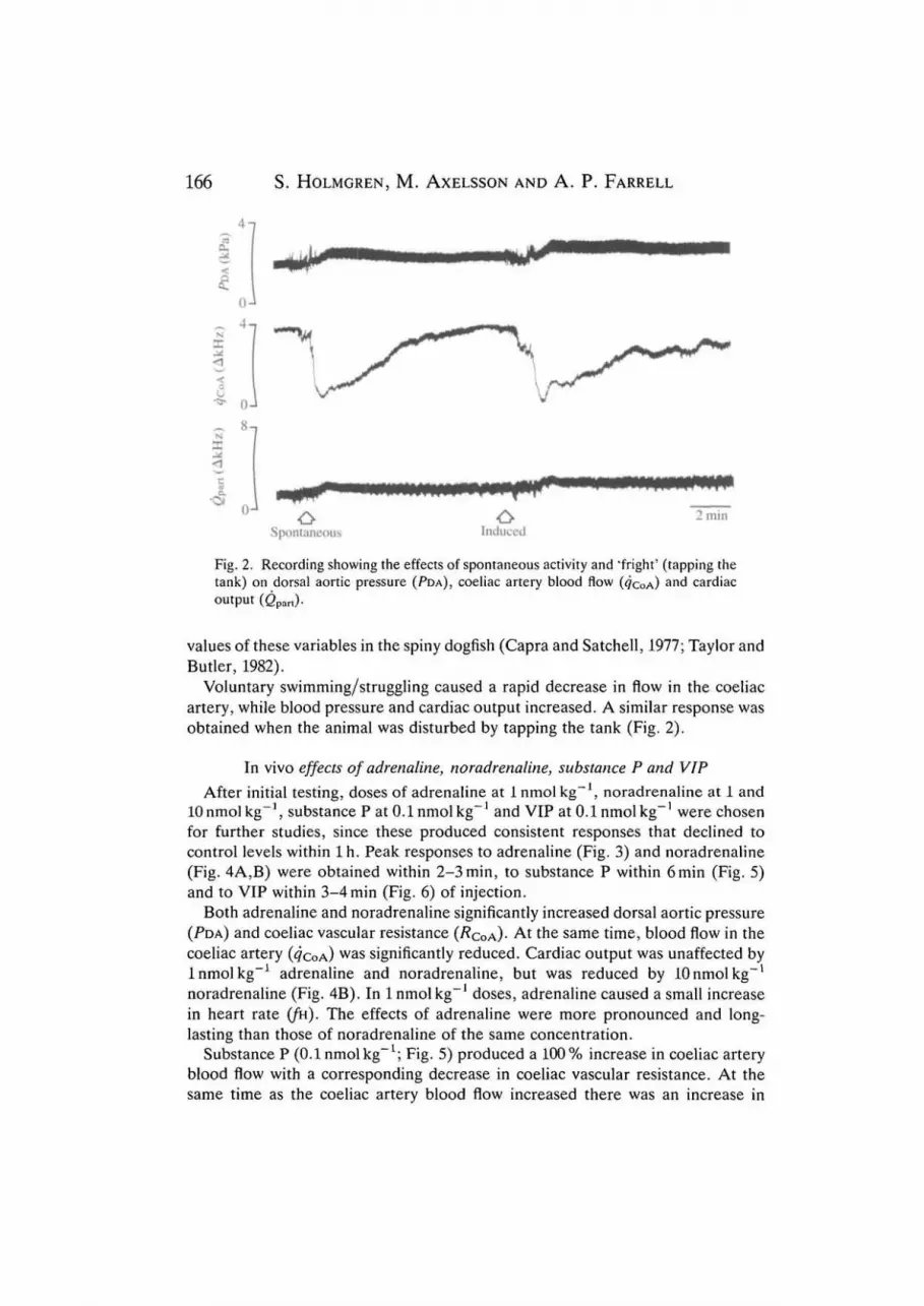
Fig. 2. Recording showing the effects of spontaneous activity and "fright' (tapping thetank) on dorsal aortic pressure (PDA), coeliac artery blood flow (4COA)
a nd cardiacoutput (Qpart).
values of these variables in the spiny dogfish (Capra and Satchell, 1977; Taylor andButler, 1982).
Voluntary swimming/struggling caused a rapid decrease in flow in the coeliacartery, while blood pressure and cardiac output increased. A similar response wasobtained when the animal was disturbed by tapping the tank (Fig. 2).
In vivo effects of adrenaline, noradrenaline, substance P and VIP- IAfter initial testing, doses of adrenaline at 1 nmol kg , noradrenaline at 1 and
lOnmolkg , substance P at 0.1 nmol kg and VIP at 0.1 nmol kg were chosenfor further studies, since these produced consistent responses that declined tocontrol levels within lh . Peak responses to adrenaline (Fig. 3) and noradrenaline(Fig. 4A,B) were obtained within 2-3 min, to substance P within 6min (Fig. 5)and to VIP within 3-4 min (Fig. 6) of injection.
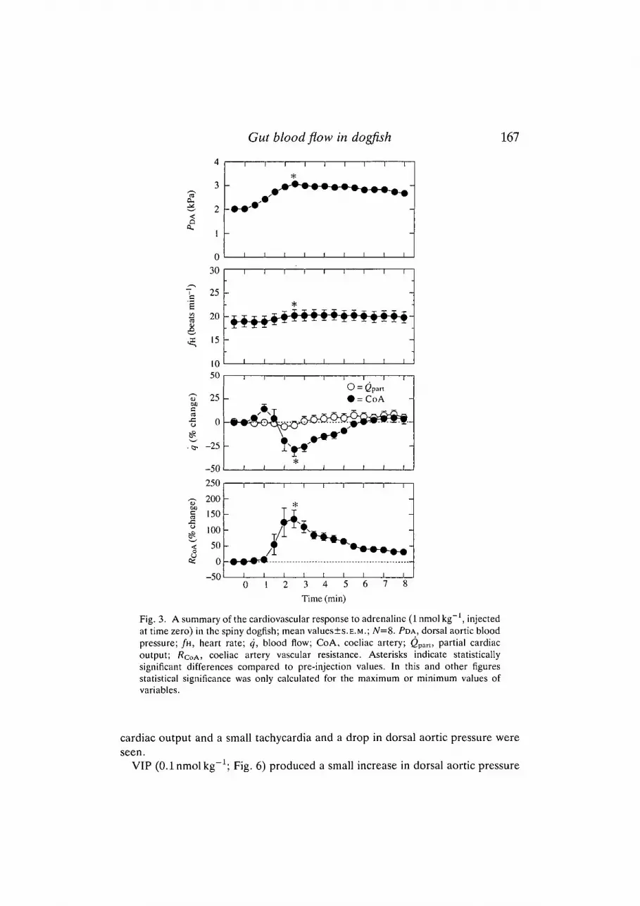
Both adrenaline and noradrenaline significantly increased dorsal aortic pressure(PDA) and coeliac vascular resistance {RQOA)- At the same time, blood flow in thecoeliac artery (<?COA) was significantly reduced. Cardiac output was unaffected by1 nmol kg"1 adrenaline and noradrenaline, but was reduced by lOnmolkg"1
noradrenaline (Fig. 4B). In 1 nmol kg"1 doses, adrenaline caused a small increasein heart rate (/k). The effects of adrenaline were more pronounced and long-lasting than those of noradrenaline of the same concentration.
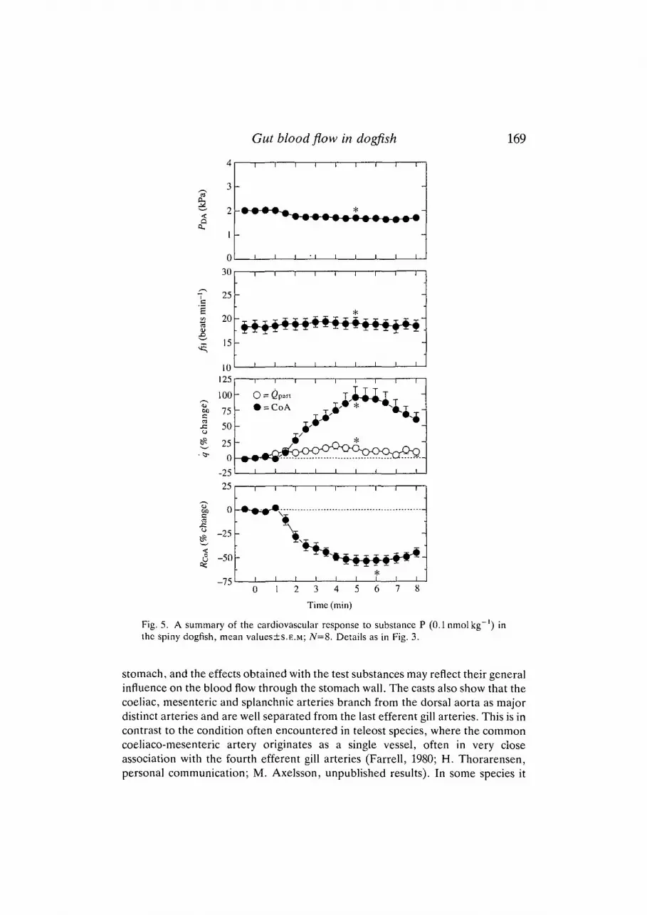
Substance P (0.1 nmol kg"1; Fig. 5) produced a 100% increase in coeliac arteryblood flow with a corresponding decrease in coeliac vascular resistance. At thesame time as the coeliac artery blood flow increased there was an increase in
Gut blood flow in dogfish 167
Q
a.
00
s:o
4
3
2
I h
030
7s 25
3 20eJ: 15
1
1
(
• • *
1
1
*
1
1
1
1
1
1
1
1
i
i
•"•^
i
i
i
I I I I I I I I I
3 4 5Time (min)
Fig. 3. A summary of the cardiovascular response to adrenaline (1 nmol kg"1, injectedat time zero) in the spiny dogfish; mean values±s.E.M.; N=8. PDA, dorsal aortic bloodpressure; /H, heart rate; q, blood flow; CoA, coeliac artery; <2part, partial cardiacoutput; /?COA> coeliac artery vascular resistance. Asterisks indicate statisticallysignificant differences compared to pre-injection values. In this and other figuresstatistical significance was only calculated for the maximum or minimum values ofvariables.
cardiac output and a small tachycardia and a drop in dorsal aortic pressure wereseen.
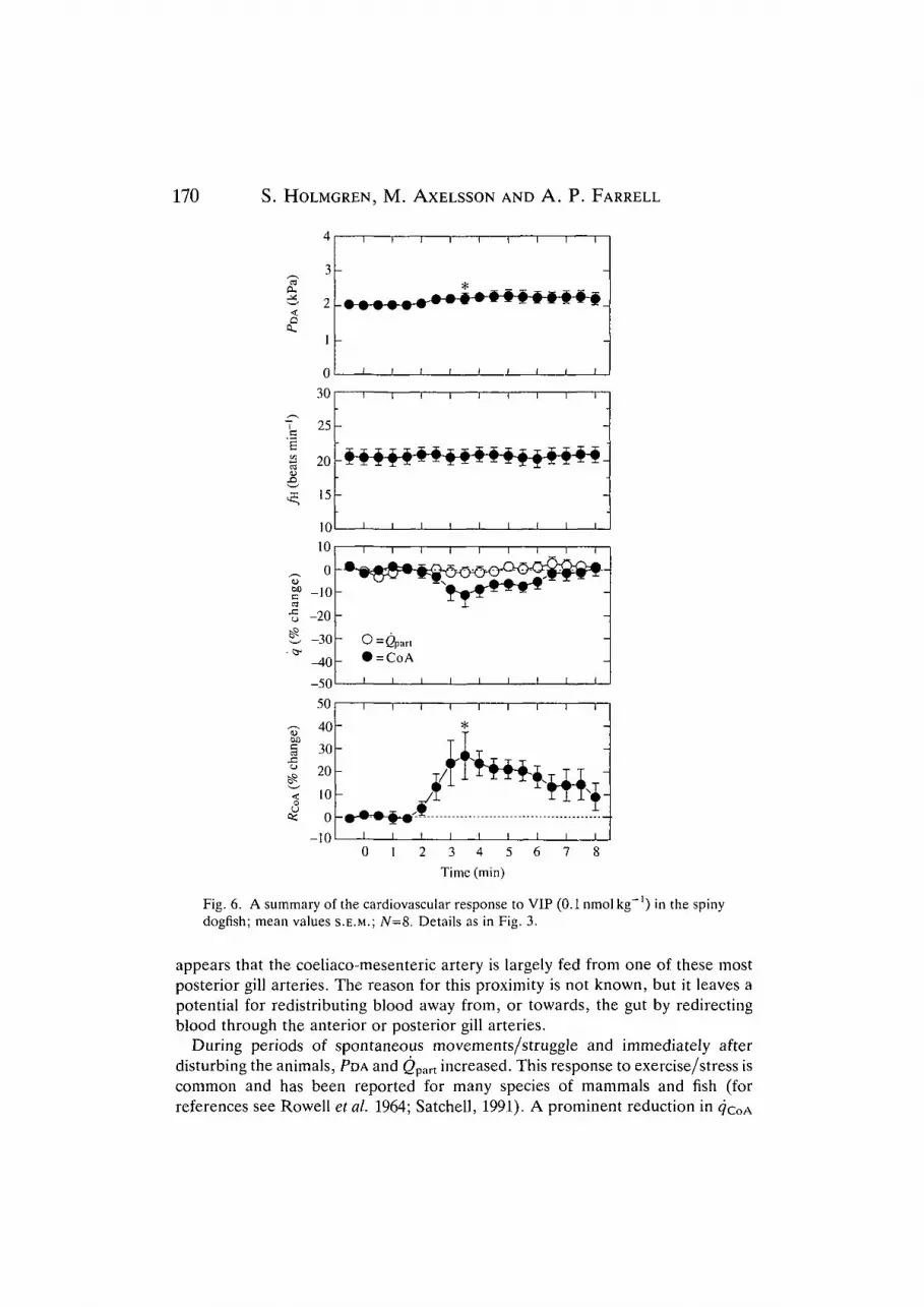
VIP (0.1 nmol kg"1; Fig. 6) produced a small increase in dorsal aortic pressure

168 S. HOLMGREN, M. AXELSSON AND A. P. FARRELL
£Q
0,
C25 -
B 20 -
15 -
10
-
1 1 1 1
i ft i
i i
i
i
-
e«
60C
50
25
0
-25
-50
-75250200150100500
-50
-••So
O = Q part• =CoA
| | | I
i i I i
B
|
1 1
1 1
*
1
1
• ^
t ! ' '
i
-
M.MMMM.MMM
I I I I I I I I I
Time (min)
Fig. 4. A summary of the cardiovascular response to two doses of noradrenaline(A, lnmolkg"1; B, lOnmolkg"1) in the spiny dogfish; mean values±s.E.M.; N=8.Details as in Fig. 3.
and an increase in coeliac vascular resistance. Cardiac output and heart rate wereunaffected. In five out of eight animals, the coeliac flow was reduced (by 8-41 %) .
In the perfused tail preparations, both adrenaline (Fig. 7) and noradrenalineproduced an increase in perfusion pressure, indicating a vasoconstriction of thesomatic vasculature. Substance P caused a small decrease in perfusion pressure,indicating a vasodilatation of the somatic vasculature. VIP had no significant effecton the somatic vasculature in any of the preparations tested.
Discussion
The corrosion casts show that the coeliac artery supplies most parts of the
Gut blood flow in dogfish 169
aa .
/—̂
lUI
bea
:han
ge
'%ch
ange
)
<
4
3
2
1
0
30
25
20
15
10125
100
75
50
25
25
0
-25
50
1
-
• • <
-
1
1
-
"~ x X -
-
I
i
- o =
A m 4
i
i
_
i
0
1 1
i i
1 i
r T X X
1 1
1 1
= Gpart
= CoA
rJI i
1 |
\
i i
1 2
i
• i
—1
1
I
* * *
D-Cr^
i
i
i x*+#i
3
Time
i
i
1
I T
1
1
-CVv
1
1
i
4
(min)
i
*
i
i—
*
i
i
. <
n
I
1
i
5
1
i
- i —
i
•
I
1
• T 1
*
i
6
1
i
— i —
1
1
i
r^i
7
1
-
i
— 1 —
-
-
* •
1
1
i
-
i
8
Fig. 5. A summary of the cardiovascular response to substance P (0.1nmolkg~') inthe spiny dogfish, mean values±s.E.M; /V=8. Details as in Fig. 3.
stomach, and the effects obtained with the test substances may reflect their generalinfluence on the blood flow through the stomach wall. The casts also show that thecoeliac, mesenteric and splanchnic arteries branch from the dorsal aorta as majordistinct arteries and are well separated from the last efferent gill arteries. This is incontrast to the condition often encountered in teleost species, where the commoncoeliaco-mesenteric artery originates as a single vessel, often in very closeassociation with the fourth efferent gill arteries (Farrell, 1980; H. Thorarensen,personal communication; M. Axelsson, unpublished results). In some species it
170 S. HOLMGREN, M. AXELSSON AND A. P. FARRELL
na.
c£
1
ange
o
3̂"
wc
j =
4
3
2
1
0
30
25
20
15
1010
0
-10
-20
-30
-40
-50
50
40
30
20
10
0
10
i
_
-
i
-
fft-A-
-i
1
-
-
- o- •
1
1
-
-
-
-
1
0
• • •
1
1
*
I
1
•#•
= Qpart
= CoA
1
I
• • • #
1
1
1
1
T-
1
|
1
.t.1
2
i t i i
* - -r
»-*-#-+-$~#-#-#-4
i i i i
i i t i
1 i i i
1 1 1 1
i 1 1 1
i i i i
*
r'Km.x i T
i i i
3 4 5 6
Time (min)
1
! II
i
i i
1
1
1
-IJHni
7
1
_
-
i
-
-
!
1
-
-
-
-
i
-
-
- -
—
i
8
Fig. 6. A summary of the cardiovascular response to VIP (0.1 nmol kg"') in the spinydogfish; mean values S.E.M.; N=8. Details as in Fig. 3.
appears that the coeliaco-mesenteric artery is largely fed from one of these mostposterior gill arteries. The reason for this proximity is not known, but it leaves apotential for redistributing blood away from, or towards, the gut by redirectingblood through the anterior or posterior gill arteries.
During periods of spontaneous movements/struggle and immediately afterdisturbing the animals, PDA and <2part increased. This response to exercise/stress iscommon and has been reported for many species of mammals and fish (forreferences see Rowell et al. 1964; Satchell, 1991). A prominent reduction in
Gut blood flow in dogfish 171
Adrenaline
0 -
6
Substance P
0 -VIP
. mm
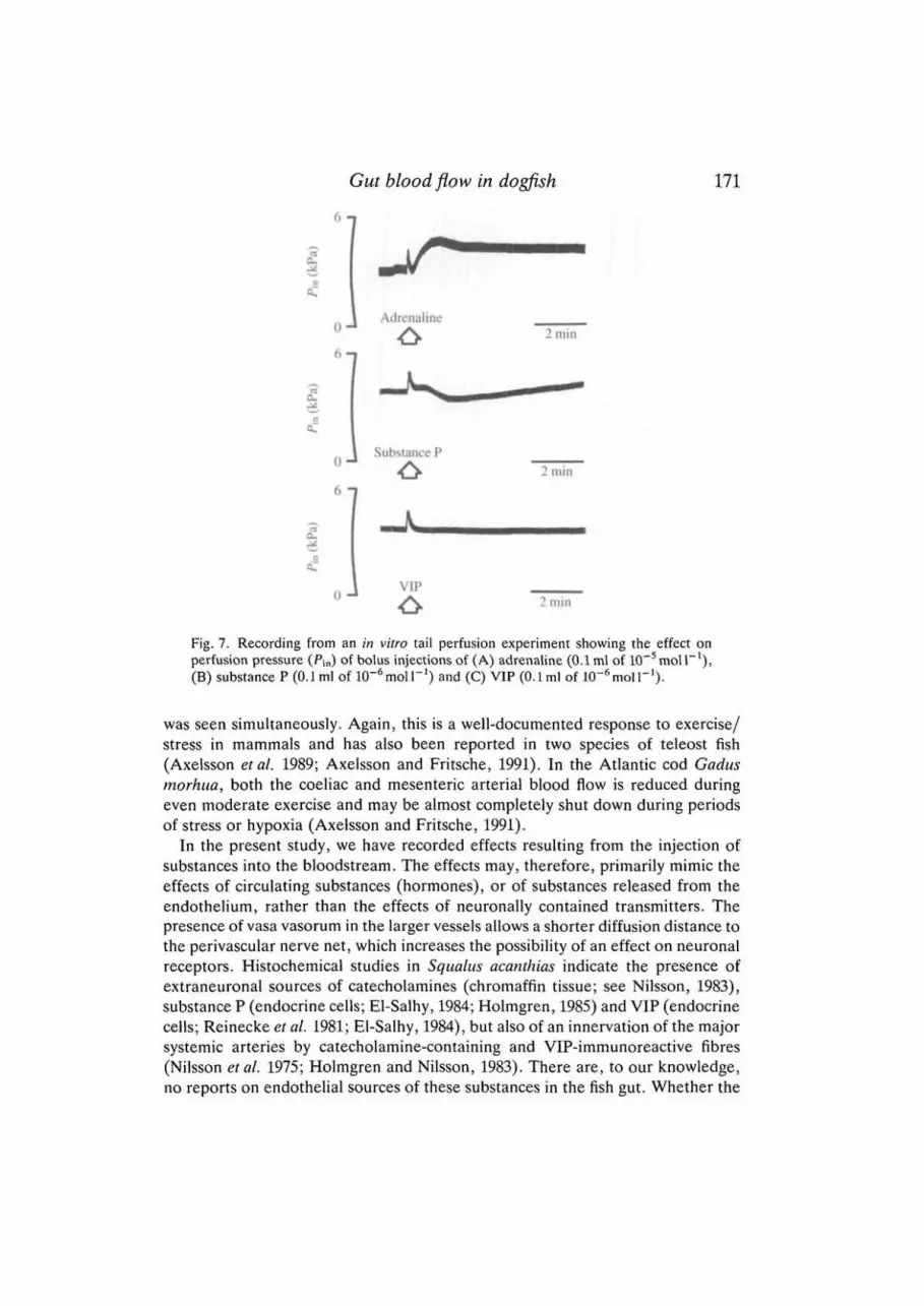
Fig. 7. Recording from an in vitro tail perfusion experiment showing the effect onperfusion pressure (Pm) of bolus injections of (A) adrenaline (0.1 ml of 10~5 mol I"1),(B) substance P (0.J ml of lO^moir 1 ) and (C) VIP (O.Lml of lO^moir 1 ) .
was seen simultaneously. Again, this is a well-documented response to exercise/stress in mammals and has also been reported in two species of teleost fish(Axelsson et al. 1989; Axelsson and Fritsche, 1991). In the Atlantic cod Gadusmorhua, both the coeliac and mesenteric arterial blood flow is reduced duringeven moderate exercise and may be almost completely shut down during periodsof stress or hypoxia (Axelsson and Fritsche, 1991).
In the present study, we have recorded effects resulting from the injection ofsubstances into the bloodstream. The effects may, therefore, primarily mimic theeffects of circulating substances (hormones), or of substances released from theendothelium, rather than the effects of neuronally contained transmitters. Thepresence of vasa vasorum in the larger vessels allows a shorter diffusion distance tothe perivascular nerve net, which increases the possibility of an effect on neuronalreceptors. Histochemical studies in Squalus acanthias indicate the presence ofextraneuronal sources of catecholamines (chromaffin tissue; see Nilsson, 1983),substance P (endocrine cells; El-Salhy, 1984; Holmgren, 1985) and VIP (endocrinecells; Reinecke et al. 1981; El-Salhy, 1984), but also of an innervation of the majorsystemic arteries by catecholamine-containing and VIP-immunoreactive fibres(Nilsson et al. 1975; Holmgren and Nilsson, 1983). There are, to our knowledge,no reports on endothelial sources of these substances in the fish gut. Whether the
172 S. HOLMGREN, M. AXELSSON AND A. P. FARRELL
injected substances reach innervated receptors (junctional receptors) needsfurther testing involving nerve stimulation, the use of specific antagonists andreceptor binding studies, which is beyond the scope of the present study. Furtherstudies are also needed to determine whether the injected substances act directlyon the vascular muscle or via endothelial factors (see Burnstock, 1990).
The responses to adrenaline and noradrenaline are very similar, except thatadrenaline causes a small tachycardia, which agrees with the positive chronotropiceffect reported by Capra and Satchell (1977). Although the effect of adrenalineand noradrenaline on the reduction of flow is the same (indicating a similarvasoactive effect), there is a slight difference in the response of the coeliac vascularresistance to these catecholamines: adrenaline (at lnmolkg"1) produces a largerincrease in resistance, and this may be correlated with the more pronouncedincrease in dorsal aortic blood pressure following the adrenaline injection.
Substance P produces a 'classical' response reducing the coeliac artery vascularresistance in the spiny dogfish. A small vasodilatation occurs in the somaticvascular bed, in addition to the more pronounced effect on the coeliac artery.Numerous mammalian studies report a vasodilator action of substance P andrelated tachykinins (Mione et al. 1990), and in the Atlantic cod, in vivo, substanceP increases the flow in both the coeliac and the mesenteric arteries (Jensen et al.1991). Also, although less pronounced than that seen in mammals, the tachycardiaobserved in dogfish is similar to the effect of substance P on the mammalian heart.In contrast, heart rate is unaffected by substance P in the cod (Jensen et al. 1991).
It has been argued, in mammals, that the vasodilation of arteries produced bytachykinins is not correlated to the density of perivascular nerves, but is dependenton an intact endothelium, and may thus be a humoral or local endothelial effectrather than a neuronal effect (Furchgott, 1983; D'Orleans-Juste et al. 1985;Edvinsson et al. 1985). This may also be the case in Squalus acanthias, where, sofar, no perivascular nerve fibres immunoreactive to substance P have been foundin the gut (Holmgren, 1985, and unpublished results). It is also possible thattachykinins are not endogenous vasodilators in Squalus acanthias.
The increase in resistance of the coeliac artery vascular bed after injection ofVIP is an unusual observation. In mammals, VIP is established as a dilator of mostvascular beds, including that of the gut (see, for example, Fahrenkrug, 1991). Thevasoconstrictor effect may be specific to the gut vascular bed in the dogfish, sincethe perfused somatic bed (tail) did not show the same response, and in the rectalgland of Squalus acanthias VIP causes the same effect as has been reported fromstudies of mammalian exocrine glands: an increase in perfusion flow in combi-nation with an increased secretion (Solomon et al. 1984; Thorndyke etal. 1989).Few studies have been performed on the effect of VIP on the gut of fish or othernon-mammalian vertebrates, but one study in the catfish Ictalurus melas, where anintestinal loop was perfused with porcine VIP as well as with VIP extracts fromcatfish and trout, demonstrated a dilatory effect of VIP (Holder et al. 1983). In thecod Gadus morhua, vascular perfusion of the gas gland and swimbladder with VIPproduces a long-lasting decrease in vascular resistance, probably caused by a
Gut blood flow in dogfish 173
vasodilation of the mesenteric and swimbladder arteries (Lundin and Holmgren,1984). Similarly, in the cod in vivo, injection of VIP causes a reduced resistance inthe vascular bed of the coeliac artery, while in these experiments the increase inflow in the mesenteric artery was due to the increase in cardiac output only (Jensenetal. 1991).
Other details are similar in the teleosts and elasmobranchs investigated; in boththe cod and the spiny dogfish, VIP-immunoreactive perivascular fibres have beendemonstrated in the vessels leading to the gut (Holmgren and Nilsson, 1983,1991;Lundin and Holmgren, 1984). VIP peptides from the dogfish Scyliorhinus caniculaand from the cod have been sequenced and differ from each other at just twopositions; also, both peptides differ from porcine VIP in only five positions(Dimaline and Thorndyke, 1986; Dimaline etal. 1987; Thwaites etal. 1989). It is,therefore, conceivable that the VIP-induced vasoconstriction in the gut of Squalusacanthias indicates a true species difference in the function of VIP. It may,however, be possible that, by acting via the endothelium rather than directly onthe smooth muscle, VIP produces the opposite effect, although there are noreports of this in any other species.
This work was supported by the NSERC Canada, BC, the Yukon HeartFoundation, the Swedish Forestry and Agricultural Science Research Council andthe Swedish Natural Science Research Council. The authors wish to express theirthanks to Mr Philip Bruecker and Dr Mark Graham at the Vancouver aquariumfor help with capturing the animals.
ReferencesAXELSSON, M., DRIEDZIC, W. R., FARRELL, A. P. AND NILSSON, S. (1989). Regulation of cardiac
output and gut blood flow in the sea raven, Hemitripterus americanus. Fish Physiol. Biochem.6, 315-326.
AXELSSON, M. AND FRITSCHE, R. (1991). Effects of exercise, hypoxia and feeding on thegastrointestinal blood flow in the Atlantic cod Gadus morhua. J. exp. Biol. 158, 181-191.
BURNSTOCK, G. (1990). Local mechanisms of blood flow control by perivascular nerves andendothelium. J. Hypertension 8, S95-S106.
CAPRA, M. F. AND SATCHELL, G. H. (1977). The differential haemodynamic responses of theelasmobranch, Squalus acanthias, to the naturally occurring catecholamines, adrenaline andnoradrenaline. Comp. Biochem. Physiol. 58C, 41-47.
DAHLSTROM, A., NILSSON, O., LUNDGREN, O. AND AHLMAN, H. (1988). Nonadrenergic, non-cholinergic inncrvation of gastrointestinal vessels: morphological and physiological aspects.In Nonadrenergic lnnervation of Blood Vessels, vol. II, Regional Innervation (ed.G. Burnstock and S. G. Griffith), pp. 143-172. Boca Raton, Florida: CRC Press, Inc.
DIMALINE, R. AND THORNDYKE, M. C. (1986). Purification and characterisation of VIP from twospecies of dogfish. Peptides 7, 21-25.
DIMALINE, R., YOUNG, J., THWAITES, D. T., LEE, C. M., SHUTTLEWORTH, T. J. AND THORNDYKE,
M. C. (1987). A novel vasoactive intestinal peptide (VIP) from elasmobranch intestine hasfull affinity for mammalian pancreatic VIP receptors. Biochim. biophys. Acta 930, 97-100.
D'ORLEANS-JUSTE, P., DION, S., MIZRAHI, J. AND REGOLI, D. (1985). Effects of peptides andnon-pcptides on isolated arterial smooth muscle: role of endothelium. Eur. J. Pharmac. 114,9-21.
EDVINSSON, L., FREDHOLM, B. B., HAMEL, E., JANSEN, I. AND VERRECHIA, C. (1985).Perivascular peptides relax cerebral arteries concomitant with stimulation of cyclic adenosine
174 S. HOLMGREN, M. AXELSSON AND A. P. FARRELL
monophosphate accumulation or release of an endothelium-derived relaxing factor in the cat.Neurosci. Lett. 58, 213-217.
EDVINSSON, L. AND UDDMAN, R. (1988). Vasoactive Intestinal Polypeptide (VIP): a putativeneurotransmitter in the cardiovascular system. In Nonadrenergic Innervation of BloodVessels, vol. I, Putative Neurotransmitters (ed. G. Burnstock and S. G. Griffith), pp. 101-125.Boca Raton, Florida: CRC Press, Inc.
EL-SALHY, M. (1984). Immunocytochemical investigation of the gastro-entero-pancreatic(GEP) neurohormonal peptides in the pancreas and gastrointestinal tract of the dogfishSqualus acanthias. Histochemistry 80, 193-205.
FAHRENKRUG, J. (1991). Vasoactive intestinal peptide (VIP) and autonomic neurotransmission.In Novel Peripheral Neurotransmitters, Section 135 of International Encyclopedia ofPharmacology and Therapeutics (ed. C. Bell), pp. 113-133. New York: Pergamon Press.
FARA, J. W. (1984). Postprandial mesenteric hyperemia. In Physiology of the IntestinalCirculation (ed. A. Shepherd and D. N. Granger), pp. 99-119. New York: Raven Press.
FARRELL, A. P. (1980). Vascular pathways in the gill of the lingcod, Ophiodon elongatus. Can. J.Zool. 58, 796-806.
FURCHGOTT, R. F. (1983). Role of endothelium responses of vascular smooth muscle. CirculationRes. 35, 557-573.
GREENWAY, C. V. (1982). Mechanisms and quantitative assessment of drug effects on cardiacoutput with a new model of the circulation. Pharmac. Rev. 33, 213-251.
HOLDER, F. C , VINCENT, B., RISTORI, M. T. AND LAURENT, P. (1983). Vascular perfusion of anintestinal loop in the catfish Ictalurus melas. Demonstration of the vasoactive effects of themammalian vasoactive intestinal peptide and gastro-intestinal extracts from teleost fish. C. R.hebd. Seanc. Acad. Sci. Paris, Ser. Ill Sci. Vie 296, 783-788.
HOLM, S. (1979). A simple sequentially rejective multiple test procedure. Scand. J. Statist. 6,65-70.
HOLMGREN, S. (1985). Substance P in the gastrointestinal tract of Squalus acanthias. Molec.Physiol. 8, 119-130.
HOLMGREN, S. AND NILSSON, S. (1983). Bombesin-, gastrin/CCK-, 5-hydoxytryptamine-,neurotensin-, somatostatin-, and VIP-like immunoreactivity and catecholamine fluorescencein the gut of the elasmobranch, Squalus acanthias. Cell Tissue Res. 234, 595-618.
HOLMGREN, S. AND NILSSON, S. (1991). Novel neurotransmitters in the autonomic nervoussystems of non-mammalian vertebrates. In Novel Peripheral Neurotransmitters. Section 135 ofInternational Encyclopedia of Pharmacology and Therapeutics (ed. C. Bell), pp. 293-328.New York: Pergamon Press.
JENSEN, J., AXELSSON, M. AND HOLMGREN, S. (1991). Effects of substance P and vasoactiveintestinal polypeptide on gastrointestinal blood flow in the Atlantic cod Gadus morhua.J. exp. Biol. 156, 361-373.
KICENIUK, J. W. AND JONES, D. R. (1977). The oxygen transport system in trout (Salmogairdneri) during sustained exercise. J. exp. Biol. 69, 247-260.
LUNDIN, K. AND HOLMGREN, S. (1984). Vasointestinal polypeptide-like immunoreactivity andeffects of VIP in the swimbladder of the cod, Gadus morhua. J. comp. Physiol. 154B,627-633.
MIONE, M. C , RALEVIC, V. AND BURNSTOCK, G. (1990). Peptides and vasomotor mechanisms.Pharmac. Ther. 46, 429-668.
NILSSON, S. (1983). Autonomic Nerve Function in the Vertebrates. Berlin, Heidelberg, NewYork: Springer Verlag.
NILSSON, S., HOLMGREN, S. AND GROVE, D. J. (1975). Effects of drugs and nerve stimulation onthe spleen and arteries of two species of dogfish, Scyliorhinus canicula and Squalus acanthias.Ada physiol. scand. 95, 219-230.
OWMAN, C. (1988). Role of neural substance P and coexisting calcitonin gene-related peptide(CGRP) in cardiovascular function. In Nonadrenergic Innervation of Blood Vessels, vol. I,Putative Neurotransmitters (ed. G. Burnstock and S. G. Griffith), pp. 77-100. Boca Raton,Florida: CRC Press, Inc.
REINECKE, M., SCHLUTER, P., YANAIHARA, N. AND FORSSMANN, W. G. (1981). VIPimmunoreactivity in enteric nerves and endocrine cells of the vertebrate gut. Peptides 2,149-156.
Gut blood flow in dogfish 175
ROWELL, L. B., BLACKMON, J. R. AND BRUCE, R. A. (1964). Indocyanine green clearance andestimated hepatic blood flow during mild to maximal exercise in upright man. J. din. Invest.43, 1677-1690.
RUSHMER, R. F., FRANKLIN, D. L., VAN OTTERS, R. L. AND SMITH, O. A. (1961). Changes in
peripheral blood flow distribution in healthy dogs. Circulation Res. 9, 675-687.SATCHELL, G. H. (1991). Physiology and Form of Fish Circulation. Cambridge, New York, Port
Chester, Melbourne, Sydney: Cambridge University Press.SMITH, D. G., WAHLQVIST, I., NILSSON, S. AND ERIKSSON, B.-M. (1985). Nervous control of the
blood pressure in the Atlantic cod, Gadus morhua. J. exp. Biol. 117, 335-347.SOLOMON, R., TAYLOR, M., STOFF, J. S., SILVA, P. AND EPSTEIN, F. H. (1984). In vivo effect of
volume expansion on rectal gland function. I. Humoral factors. Am. J. Physiol. 246,R63-R66.
TAYLOR, E. W. AND BUTLER, P. J. (1982). Nervous control of heart rate: activity in the cardiacvagus of the dogfish. / . appl. Physiol. 53, R1330-R1335.
THORNDYKE, M. C , RIDDELL, J. H., THWAITES, D. T. AND DIMALINE, R. (1989). Vasoactive
intestinal polypeptide and its relatives - biochemistry, distribution, and functions. Biol. Bull.mar. biol. Lab., Woods Hole 177, 183-186.
THWAITES, D. T., YOUNG, J., THORNDYKE, M. C. AND DIMALINE, R. (1989). The isolation and
chemical characterization of a novel vasoactive intestinal peptide-related peptide from ateleost fish, the cod, Gadus morhua. Biochim. biophys. Acta 999, 217-220.