Gastric de novo Muc13 expression and spasmolytic polypeptide-expressing metaplasia during...
14
Published Ahead of Print 27 May 2014. 2014, 82(8):3227. DOI: 10.1128/IAI.01867-14. Infect. Immun. Haesebrouck Flahou, Richard Ducatelle, Sara Linden and Freddy Cheng Liu, Annemieke Smet, Caroline Blaecher, Bram Infection Metaplasia during Helicobacter heilmannii Spasmolytic Polypeptide-Expressing Muc13 Expression and De Novo Gastric http://iai.asm.org/content/82/8/3227 Updated information and services can be found at: These include: SUPPLEMENTAL MATERIAL Supplemental material REFERENCES http://iai.asm.org/content/82/8/3227#ref-list-1 at: This article cites 35 articles, 11 of which can be accessed free CONTENT ALERTS more» articles cite this article), Receive: RSS Feeds, eTOCs, free email alerts (when new http://journals.asm.org/site/misc/reprints.xhtml Information about commercial reprint orders: http://journals.asm.org/site/subscriptions/ To subscribe to to another ASM Journal go to: on July 10, 2014 by UNIVERSITEIT GENT/UZGENT http://iai.asm.org/ Downloaded from on July 10, 2014 by UNIVERSITEIT GENT/UZGENT http://iai.asm.org/ Downloaded from
Transcript of Gastric de novo Muc13 expression and spasmolytic polypeptide-expressing metaplasia during...

Published Ahead of Print 27 May 2014. 2014, 82(8):3227. DOI: 10.1128/IAI.01867-14. Infect. Immun.
HaesebrouckFlahou, Richard Ducatelle, Sara Linden and Freddy Cheng Liu, Annemieke Smet, Caroline Blaecher, Bram InfectionMetaplasia during Helicobacter heilmanniiSpasmolytic Polypeptide-Expressing
Muc13 Expression andDe NovoGastric
http://iai.asm.org/content/82/8/3227Updated information and services can be found at:
These include:
SUPPLEMENTAL MATERIAL Supplemental material
REFERENCEShttp://iai.asm.org/content/82/8/3227#ref-list-1at:
This article cites 35 articles, 11 of which can be accessed free
CONTENT ALERTS more»articles cite this article),
Receive: RSS Feeds, eTOCs, free email alerts (when new
http://journals.asm.org/site/misc/reprints.xhtmlInformation about commercial reprint orders: http://journals.asm.org/site/subscriptions/To subscribe to to another ASM Journal go to:
on July 10, 2014 by UN
IVE
RS
ITE
IT G
EN
T/U
ZG
EN
Thttp://iai.asm
.org/D
ownloaded from
on July 10, 2014 by U
NIV
ER
SIT
EIT
GE
NT
/UZ
GE
NT
http://iai.asm.org/
Dow
nloaded from

Gastric De Novo Muc13 Expression and Spasmolytic Polypeptide-Expressing Metaplasia during Helicobacter heilmannii Infection
Cheng Liu,a Annemieke Smet,a Caroline Blaecher,a Bram Flahou,a Richard Ducatelle,a Sara Linden,b Freddy Haesebroucka
Department of Pathology, Bacteriology and Avian Diseases, Faculty of Veterinary Medicine, Ghent University, Merelbeke, Belgiuma; Mucosal Immunobiology and VaccineCenter, Sahlgrenska Academy, Gothenburg University, Gothenburg, Swedenb
Helicobacter heilmannii is a zoonotic bacterium that has been associated with gastric disease in humans. In this study, themRNA expression of mucins in the stomach of BALB/c mice was analyzed at several time points during a 1-year infection withthis bacterium, during which gastric disease progressed in severity. Markers for acid production by parietal cells and mucousmetaplasia were also examined. In the first 9 weeks postinfection, the mRNA expression of Muc6 was clearly upregulated in boththe antrum and fundus of the stomach of H. heilmannii-infected mice. Interestingly, Muc13 was upregulated already at 1 daypostinfection in the fundus of the stomach. Its expression level remained high in the stomach over the course of the infection.This mucin is, however, not expressed in a healthy stomach, and high expression of this mucin has so far only been described ingastric cancer. In the later stages of infection, mRNA expression of H�/K�-ATPase �/� and KCNQ1 decreased, whereas the ex-pression of Muc4, Tff2, Dmbt1, and polymeric immunoglobulin receptor (pIgR) increased starting at 16 weeks postinfectiononwards, suggesting the existence of spasmolytic polypeptide-expressing metaplasia in the fundus of the stomach. Mucous meta-plasia present in the mucosa surrounding low-grade mucosa-associated lymphoid tissue (MALT) lymphoma-like lesions wasalso histologically confirmed. Our findings indicate that H. heilmannii infection causes severe gastric pathologies and altera-tions in the expression pattern of gastric mucins, such as Muc6 and Muc13, as well as disrupting gastric homeostasis by inducingthe loss of parietal cells, resulting in the development of mucous metaplasia.
Helicobacter pylori is the major predisposing factor for the de-velopment of chronic active gastritis, peptic ulcers, and gas-
tric adenocarcinomas in humans, and approximately 15% of in-fected individuals are estimated to develop such symptoms. Thispathogen can be detected attached to gastric epithelial cells but isfound mainly within the mucus layer and is able to bind to highlyglycosylated mucins. MUC1, MUC5AC, and MUC6 are the majormucins covering the healthy gastric mucosa. The membrane-as-sociated MUC1 and secreted MUC5AC are expressed at the surfaceepithelium, whereas MUC6 is secreted by glandular cells (1–4).
H. pylori infection causes alterations in the expression pattern,glycosylation, and distribution of gastric mucins, as well as dis-rupting the gastric homeostasis by inducing the loss of parietalcells (5, 6). The loss of parietal cells can lead to two distinct types ofmucous metaplasia: intestinal metaplasia and spasmolytic poly-peptide-expressing metaplasia (SPEM). It has been suggested thatintestinal metaplasia develops in the presence of pre-existingSPEM, supporting the role of SPEM as a neoplastic precursor inthe carcinogenesis cascade (6). The intestinal mucins MUC2,MUC4, and MUC13 are not expressed in the healthy gastric mu-cosa but have been detected in gastric adenocarcinomas and dur-ing stages of mucous metaplasia (7–9).
Besides H. pylori, other spiral-shaped non-H. pylori Helicobac-ter spp. (NHPH), such as H. suis in pigs and H. heilmannii (sensustricto), H. felis, H. bizzozeronii, and H. salomonis in cats and dogs,have been associated with gastric disease in humans (10, 11). H.heilmannii is highly prevalent in healthy cats and dogs, as well as inanimals with chronic gastritis (10). In humans, this Helicobacterspecies has been associated with gastritis, gastric and duodenalulcers, and low-grade mucosa-associated lymphoid tissue(MALT) lymphoma. It has been detected in 8 to 19% of gastricbiopsy specimens with histological evidence of NHPH infection(10–12). Living in close contact with cats and dogs has been iden-
tified as a significant risk factor for these infections in humans(10). Since this species has only recently been isolated and cul-tured in vitro, information on how H. heilmannii interacts with thehuman stomach and causes disease still remains poor. Compara-tive genomic analyses showed that although the H. heilmanniigenome contains several genes encoding homologues of known H.pylori virulence factors, it lacks a Cag pathogenicity island (Cag-PAI), as well as genes encoding the vacuolating cytotoxin VacAand several outer membrane proteins involved in the binding ofH. pylori to the gastric mucosa, such as BabA/-B, SabA, AlpA/-B,OipA, HopZ, HopQ, and HomB (13). Thus, factors that contrib-ute to the colonization properties of H. heilmannii, particularlyadhesion, remain to be identified. A recent infection study in aMongolian gerbil model for human Helicobacter-induced pathol-ogy showed variations in colonization capacity and virulence be-tween different H. heilmannii strains isolated from the gastric mu-cosa of cats. These findings are most probably also relevant forinfection with this bacterium in humans (14). Unlike H. pylori,which is mainly observed at the surface epithelium and close toMUC1- and MUC5AC-producing cells (1, 3), H. heilmannii ismostly found in the gastric pits, as has also been described for
Received 2 April 2014 Accepted 12 May 2014
Published ahead of print 27 May 2014
Editor: S. R. Blanke
Address correspondence to Annemieke Smet, [email protected].
C.L. and A.S. share first authorship. S.L. and F.H. share senior authorship.
Supplemental material for this article may be found at http://dx.doi.org/10.1128/IAI.01867-14.
Copyright © 2014, American Society for Microbiology. All Rights Reserved.
doi:10.1128/IAI.01867-14
August 2014 Volume 82 Number 8 Infection and Immunity p. 3227–3239 iai.asm.org 3227
on July 10, 2014 by UN
IVE
RS
ITE
IT G
EN
T/U
ZG
EN
Thttp://iai.asm
.org/D
ownloaded from

Liu et al.
3228 iai.asm.org Infection and Immunity
on July 10, 2014 by UN
IVE
RS
ITE
IT G
EN
T/U
ZG
EN
Thttp://iai.asm
.org/D
ownloaded from

other NHPH (14, 15). This bacterium can be found in close asso-ciation with parietal cells but is also able to bind to human mucus-secreting epithelial cells, as well as to mucin samples containinghighly glycosylated MUC5AC and MUC6 (unpublished data).Whether an H. heilmannii infection has an impact on the distri-bution and expression of the gastric MUC1, MUC5AC, andMUC6 mucins is currently unknown.
Specific-pathogen-free (SPF) inbred C57BL/6 and BALB/cmice have been shown to be useful models for the study of Heli-cobacter-related human gastric disease (15). C57BL/6 mice havebeen described genetically as predominant Th1 responders, whileBALB/c mice are mainly Th2 responders (15). It has been shownthat infection with H. suis induces a predominant Th17/Th2 im-mune response in BALB/c mice and even in C57BL/6 mice in theabsence of a Th1 response, but with a more pronounced inflam-mation in BALB/c mice (16). More recently, it has been suggestedthat infection with H. heilmannii also elicits a Th2 immune re-sponse (14). These results are in contrast to the predominantTh17/Th1 response mostly seen during H. pylori infection in mice(16). Therefore, in the present study, we used Th2-prone BALB/cmice to investigate the expression levels of Muc1, Muc5ac, andMuc6 in the stomach at several time points during a 1-year H.heilmannii infection during which gastric disease progressed fromgastritis to MALT lymphoma-like lesions and mucous metaplasia.Since H. heilmannii has been found close to parietal cells in thegastric pits, markers for acid production by parietal cells wereexamined. Markers for mucous metaplasia (in particular theMuc2, Muc4, and Muc13 intestinal mucins) as a result of parietalcell loss were included as well. Infection with the mouse-adaptedH. pylori SS1, a strain that elicits a Th2 response, was included forcomparison (16).
MATERIALS AND METHODSAnimals. Six-week-old female SPF BALB/c mice were purchased from Har-lan NL (Horst, The Netherlands). The animals were housed in individualfilter-top cages, had free access to water and food (an autoclaved commercialdiet, Teklad 2018S, containing 18% protein; Harlan) throughout the experi-ment, and were monitored daily.
The in vivo experimental protocol was approved by the Ethical Com-mittee of the Faculty of Veterinary Medicine, Ghent University, Belgium(EC 2011-155, 27 October 2011).
Cultivation of H. heilmannii and H. pylori strains used for infec-tion. The highly virulent H. heilmannii strain ASB1.4, isolated from thestomach of a kitten with gastritis, was cultivated as described previously(11, 14). After incubation under microaerobic conditions (11), the bacte-ria were harvested, and the final concentration was adjusted to 7 � 108
viable bacteria/ml.The mouse-adapted H. pylori SS1 strain (17) was grown for 3 days on
blood agar plates (Oxoid) and further cultured overnight in brucella broth
(Oxoid) under microaerobic conditions. The optical density was thenadjusted to 1.5, corresponding to approximately 1 � 109 viable bacteria/ml.
Experimental procedure. For each time point tested, 6 animals wereintragastrically inoculated 3 times at 2-day intervals with 300 �l of anASB1.4 or SS1 bacterial suspension and 3 animals were inoculated withbrucella broth (pH 5) as a negative control. Inoculation was performedunder brief isoflurane anesthesia (2.5%), using a feeding needle. At 1 day,4 days, and 1, 2, 3, 4, 9, 12, 16, 20, 24, 34, and 52 weeks after the firstinoculation, the animals were euthanized by cervical dislocation underdeep isoflurane anesthesia (5%). The stomach and the duodenum of eachmouse were resected, and samples were taken for histopathological exam-ination and quantitative real-time (RT)-PCR analysis.
Histopathology and immunohistochemistry. A longitudinal section,starting from the end of the forestomach and comprising the antrum andthe fundus of the stomach and part of the duodenum, was fixed in 10%phosphate-buffered formalin and embedded in paraffin for light micros-copy. From each animal, several consecutive paraffin slides of 5 �m werecut, deparaffinized, and dehydrated. Heat-induced antigen retrieval (100°Cfor 20 min) was then performed in citrate buffer (pH 6), and endogenousperoxidase activity and nonspecific reactions were blocked by incubatingthe slides with 3% H2O2 in methanol (5 min) and 30% goat serum (30min), respectively. A hematoxylin and eosin (H&E) staining was per-formed on a first slide to score the intensity of the gastritis according to theupdated Sydney system, as described previously (15) but with some mod-ifications, as described in the legend to Fig. 1. On a second slide, B lym-phocytes were visualized by immunohistochemical staining using a poly-clonal rabbit anti-CD20 antibody (1/100; Thermo Scientific, Fremont,CA). On a third slide, parietal cells were identified by immunohistochem-ical staining using a mouse monoclonal antibody against the hydrogenpotassium ATPase (1/200; Abcam Ltd., Cambridge, United Kingdom).
Muc13 expression was evaluated by immunohistochemical staining(one slide for each staining) using an anti-Muc13 antibody. Incubationwith primary antibodies directed against CD20 and Muc13 was followedby incubation with a biotinylated goat anti-rabbit IgG antibody (1/500;DakoCytomation, Heverlee, Belgium). Incubation with primary antibodyagainst the hydrogen potassium ATPase was followed by incubation witha biotinylated goat anti-mouse IgG antibody (1/200; DakoCytomation).After rinsing, the sections were incubated with a streptavidin-biotin-horseradish peroxidase complex (DakoCytomation), and the color wasdeveloped with diaminobenzidine tetrahydrochloride (DAB) and H2O2.
To highlight lymphoepithelial lesions, paraffin slides were stained witha monoclonal mouse anticytokeratin antibody (1/50; DakoCytomation)and further processed using an EnVision� system for use with mouseprimary antibodies (DakoCytomation).
Finally, periodic acid-Schiff stain (PAS)-Alcian blue staining was per-formed for the differential staining of glycoproteins.
DNA extraction and quantification of colonizing Helicobacter spp.in the stomach and duodenum. Samples from the fundus and the antrumof the stomach and from the duodenum of each animal were harvestedinto 1 ml RNAlater (Ambion, Austin, TX, USA) and stored at �70°C untilRNA and DNA extraction. The tissue samples were then homogenized(MagNALyser; Roche, Mannheim, Germany), and RNA and DNA were
FIG 1 Gastric inflammation in H. heilmannii- and H. pylori-infected BALB/c mice. (A) Fundic inflammation was scored on a scale of 0 to 4, as follows: 0, noinfiltration with mononuclear and/or polymorphonuclear cells; 1, mild diffuse infiltration with mononuclear and/or polymorphonuclear cells or the presence ofone small (50 to 200 cells) aggregate of inflammatory cells; 2, moderate diffuse infiltration with mononuclear and/or polymorphonuclear cells and/or thepresence of 2 to 4 inflammatory aggregates; 3, marked diffuse infiltration with mononuclear and/or polymorphonuclear cells and/or the presence of at least fiveinflammatory aggregates; 4, diffuse infiltration of large regions with large aggregates of mononuclear and/or polymorphonuclear cells. Results for individualanimals are depicted by symbols around the means (lines). (B to E) H&E staining of the fundus, antrum, and duodenum of a Helicobacter-infected BALB/cmouse. (B and C) A large infiltrate of mononuclear cells (arrow) at the forestomach/stomach transition zone of a mouse infected with H. heilmannii ASB1.4 (B)and H. pylori SS1 (C) at 52 weeks postinfection. Bar � 30 �m. (D and E) A mild lymphocytic infiltration of the lamina muscularis mucosae in the antrum (D)and duodenum (E) of a mouse infected with H. heilmannii ASB1.4 for 52 weeks (arrows). Bar � 30 �m. (F and G) CD20 staining of the forestomach/stomachtransition zone of a mouse infected with H. heilmannii ASB1.4 (F) and H. pylori SS1 (G) at 52 weeks postinfection, showing B lymphocytes (brown) in germinalcenters of lymphoid follicles (arrows). Bar � 30 �m. (H and I) Cytokeratin staining of the forestomach/stomach transition zone of a mouse infected with H.heilmannii ASB1.4 (bar � 30 �m) (H) and H. pylori SS1 (bar � 10 �m) (I) at 52 weeks postinfection showing numerous lymphoepithelial lesions (arrows).
Muc13 and SPEM Induced by H. heilmannii Sensu Stricto
August 2014 Volume 82 Number 8 iai.asm.org 3229
on July 10, 2014 by UN
IVE
RS
ITE
IT G
EN
T/U
ZG
EN
Thttp://iai.asm
.org/D
ownloaded from

separated (14). The numbers of colonizing H. heilmannii and H. pyloribacteria per mg of gastric tissue were determined in the DNA samplesusing quantitative RT-PCRs specific for the detection of H. heilmannii andH. pylori (14, 16). Helicobacter standards were generated as described pre-viously (18). One microliter of DNA template was suspended in a 10-�lreaction mixture consisting of 3.5 �l high-performance liquid chroma-tography (HPLC)-grade water, 5 �l SensiMix SYBR No-ROX (BiolineReagents Ltd., United Kingdom), and 0.25 �l both primers (see Table S1in the supplemental material) (Integrated DNA Technologies). Standardsand DNA samples were run in duplicate on a CFX96 RT-PCR system witha C1000 thermal cycler (Bio-Rad, Hercules, CA, USA). For quantificationof H. heilmannii and H. pylori DNA in the tissue samples, the Bio-RadCFX Manager (version 1.6) software was used.
RT-PCR for gene expression. The total RNA concentration in eachsample was utilized for first-strand cDNA synthesis using the iScriptcDNA synthesis kit (Bio-Rad). Quantitative RT-PCR was carried out formeasuring the gene expression levels of murine gastric mucins (Muc1,Muc2, Muc4, Muc5AC, Muc5B, Muc6, and Muc13), trefoil factors (Tff1and Tff2), and Dmbt1, polymeric immunoglobulin receptor (pIgR), H�/K�-ATPase, Kcnq1, and Ckb. The housekeeping genes PPIa, H2afz, andHPRT were included as reference genes. The primer sequences are shownin Table S1 in the supplemental material. Reactions were performed in10-�l volumes containing 1 �l cDNA, 0.05 �l both primers (see Table S1),3.9 �l HPLC-grade water, and 5 �l SensiMix SYBR No-ROX. The exper-imental protocol for PCR (40 cycles) was performed on a CFX96 RT-PCRsystem with a C1000 thermal cycler (Bio-Rad). Control reactions withoutthe reverse transcriptase step were implemented to exclude DNA contam-ination of the RNA samples. No-template control reaction mixtures wereincluded, and all samples were run in duplicate. The results are shown asfold changes of mRNA expression in infected animals relative to themRNA expression levels in control animals. Fold changes were calculatedusing the cycle threshold (��CT) method (19), with the mean CT valuesfrom three uninfected mice as a control. Fold changes of �4 were acceptedas upregulation and of �0.25 as downregulation.
Statistical analysis. Statistical analysis was performed using the SPSSStatistics 21 software package (IBM). Gastritis scores were analyzed usingthe nonparametric Mann-Whitney U test to compare groups. Gene ex-pression was compared between different infected groups and controlsusing a Bonferroni post hoc test (analysis of variance [ANOVA]). Forcorrelation between different variables, Spearman’s rho correlation coef-ficients were calculated. Differences were considered statistically signifi-cant at a P value of �0.05.
RESULTSInduction of inflammation by and colonization capacity of H.heilmannii ASB1 and H. pylori SS1. Gastric lymphoid lesionswere not present in any of the noninfected control animals. For allof these animals, the histomorphology was considered to be nor-mal, with only minor inflammatory cell infiltration in the gastricmucosa. Inflammation in H. heilmannii ASB1.4- and H. pyloriSS1-infected mice was characterized by mononuclear and poly-morphonuclear cell infiltration in the lamina propria mucosae,the tunica submucosa, or both, depending on the individual ani-mal. At all time points, inflammation was observed mainly in thefundus. The fundic inflammation scores of each individual animalare shown in Fig. 1A. No statistically significant difference be-tween inflammation scores for mice inoculated with ASB1.4 orSS1 at a certain time point was demonstrated. From 24 weekspostinfection onwards, large lymphoid aggregates of mononu-clear and/or polymorphonuclear cells were mainly seen in a nar-row zone in the fundus near the forestomach/stomach transitionzone (Fig. 1B and C) of both H. heilmannii- and H. pylori-infectedmice. In mice infected with ASB1.4 and SS1 for at least 34 weeks,B-cell-containing germinal centers were seen in those large lym-
phoid aggregates (Fig. 1F and G). In several mice infected withASB1.4 and SS1 for 52 weeks, numerous lymphoepithelial MALTlymphoma-like lesions could be detected in the gastric mucosa(Fig. 1H and I). These were most abundant in a narrow zone in thefundus near the forestomach/stomach transition zone. In all He-licobacter-infected mice, mild signs of inflammation were detectedin the antrum of the stomach and the duodenum at 52 weekspostinfection (Fig. 1D and E). However, inflammation could alsobe noted in the junction between antrum and fundus of miceinfected with H. heilmannii for 52 weeks (data not shown).
Throughout the experiment, all control animals were negativefor Helicobacter DNA in quantitative RT-PCR assays. At all timepoints, Helicobacter DNA was found in both the antrum and fun-dus of the stomach from all infected animals but with a largeramount in the antrum. H. pylori and H. heilmannii DNA wasfound in the duodenum from 3 and 12 weeks postinfection on-wards, respectively (Fig. 2A, B, and C). In general, ASB1.4 colo-nized the stomach of mice at a significantly higher level than SS1(P � 0.002 for antrum at 4, 20, 24, and 34 weeks postinfection; P �0.004 for antrum at 12, 16, and 52 weeks postinfection; P � 0.026for antrum at 9 weeks postinfection; P � 0.009 for fundus at 12weeks postinfection; and P � 0.002 for fundus at 16, 20, 24, and 34weeks postinfection). The amounts of ASB1.4 and SS1 DNA weremuch lower in the duodenum than in the stomach, and a signifi-cant difference between H. heilmannii and H. pylori was only seenat 3 weeks postinfection (P � 0.015) (Fig. 2C).
Changes in Muc1, Muc5AC, Muc5B, and Muc6 expressionduring H. heilmannii colonization. No change in mRNA expres-sion of Muc1 and Muc5AC was seen in the stomach during thewhole experiment (data not shown). In the first 9 weeks postin-fection, quantitative RT-PCR showed clear upregulation in themRNA expression of Muc6 in both the antrum (fold change forASB1.4, 7.43 � 2.08, and for SS1, 6.39 � 2.5) and fundus (foldchange for ASB1.4, 5.88 � 2.66, and for SS1, 6.86 � 3.01) ofHelicobacter-infected mice compared to the expression in the con-trol group (Fig. 3A and B; see also Fig. S1A and B in the supple-mental material). In addition, a significant positive correlationwas observed between Muc6 expression and Helicobacter coloni-zation in the antrum of ASB1.4-infected mice (Fig. 3C).
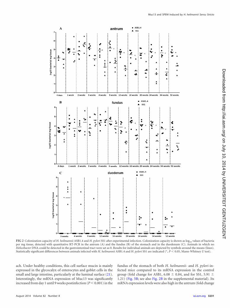
Also in this early stage of infection, Muc5B was abnormallyexpressed in the stomach of mice infected with both species (Fig.4A and B; see also Fig. S1C and D in the supplemental material).This mucin is normally not expressed in a healthy stomach (20).Compared to the results for the control animals, whose mRNAexpression levels were set to 1, the fold differences in Muc5B andMuc5AC expression in the antrum were 6.02 � 2.27 for ASB1.4-and 5.68 � 2.81 for SS1-infected mice. The fold differences inMuc5B and Muc5AC expression in the fundus were 2.88 � 1.63and 3.84 � 1.53 for ASB1.4- and SS1-infected animals, respec-tively.
Alterations in mucin mRNA expression were also evaluated inthe duodenum. The mRNA expression levels of Muc1 were signif-icantly increased at 4 weeks postinfection in the duodenum of H.heilmannii-infected mice (P � 0.036) and H. pylori-infected mice(P � 0.047) (see Fig. S1E in the supplemental material). Signifi-cantly increased expression of Muc5AC was seen at 4 (P � 0.032)and 16 (P � 0.026) weeks postinfection in the duodenum of H.heilmannii-infected mice (see Fig. S1F).
H. heilmannii infection stimulates Muc13 expression. Thetransmembrane mucin Muc13 is not expressed in a healthy stom-
Liu et al.
3230 iai.asm.org Infection and Immunity
on July 10, 2014 by UN
IVE
RS
ITE
IT G
EN
T/U
ZG
EN
Thttp://iai.asm
.org/D
ownloaded from
ach. Under healthy conditions, this cell surface mucin is mainlyexpressed in the glycocalyx of enterocytes and goblet cells in thesmall and large intestine, particularly at the luminal surface (21).Interestingly, the mRNA expression of Muc13 was significantlyincreased from day 1 until 9 weeks postinfection (P 0.001) in the
fundus of the stomach of both H. heilmannii- and H. pylori-in-fected mice compared to its mRNA expression in the controlgroup (fold change for ASB1, 6.08 � 0.84, and for SS1, 5.91 �1.21) (Fig. 5B; see also Fig. 2B in the supplemental material). ItsmRNA expression levels were also high in the antrum (fold change
FIG 2 Colonization capacity of H. heilmannii ASB1.4 and H. pylori SS1 after experimental infection. Colonization capacity is shown as log10 values of bacteriaper mg tissue, detected with quantitative RT-PCR in the antrum (A) and the fundus (B) of the stomach and in the duodenum (C). Animals in which noHelicobacter DNA could be detected in the gastrointestinal tract were set as 0. Results for individual animals are depicted by symbols around the means (lines).Statistically significant differences between animals infected with H. heilmannii ASB1.4 and H. pylori SS1 are indicated (*, P 0.05, Mann-Whitney U test).
Muc13 and SPEM Induced by H. heilmannii Sensu Stricto
August 2014 Volume 82 Number 8 iai.asm.org 3231
on July 10, 2014 by UN
IVE
RS
ITE
IT G
EN
T/U
ZG
EN
Thttp://iai.asm
.org/D
ownloaded from

FIG 3 Muc6 expression in the stomach of Helicobacter-infected and control mice. Expression levels of Muc6 in the antrum (A) and fundus (B) of the stomachof ASB1.4- and SS1-infected BALB/c mice are shown. Data are presented as fold changes in gene expression normalized to 3 reference genes and relative to theresults for the negative-control group, which are set as 1. Data obtained from the time points 1 day, 4 days, and 1, 2, 3, 4, and 9 weeks are pooled and designated“early stage of infection.” Data obtained from the time points 12, 16, 20, and 24 weeks are pooled and designated “mid-stage of infection.” Data obtained fromthe time points 34 and 52 weeks are pooled and designated “late stage of infection.” Data are shown as means � standard deviations. Significant differences in
Liu et al.
3232 iai.asm.org Infection and Immunity
on July 10, 2014 by UN
IVE
RS
ITE
IT G
EN
T/U
ZG
EN
Thttp://iai.asm
.org/D
ownloaded from
for ASB1.4, 9.61 � 4.08, and for SS1, 10.09 � 4.38) and fundus(fold change for ASB1.4, 5.77 � 4.33, and for SS1, 7.17 � 3.67) ofthe stomach in the late stage of infection (Fig. 5A and B; see Fig.S2A and B). A significant positive correlation was observed betweenMuc13 expression and Helicobacter colonization in the fundus ofH. heilmannii- and H. pylori-infected mice in the first 9 weekspostinfection (Fig. 5C and D). Although H. heilmannii DNA couldnot be found in the duodenum during the first weeks of infection,the Muc13 expression levels were significantly increased here also
at 1 (fold change of 8.67 � 1.89; P 0.001) and 2 (fold change of6.97 � 3.09; P 0.001) weeks postinfection (see Fig. S2C).
Immunohistochemical staining showed apical membrane andcytoplasmic Muc13 staining (brown) in mucus-secreting epithe-lial cells of the stomach of Helicobacter-infected mice but not inthe noninfected controls (Fig. 5E and F).
H. heilmannii induces reduced expression of markers forgastric acid secretion by parietal cells in the fundus of the stom-ach. To determine whether Helicobacter colonization has an im-pact on gastric acid secretion, the expression of different markerswas analyzed (22). Quantitative RT-PCR analysis showed cleardownregulation in the expression of H�/K�-ATPase and � pro-ton pump subunits in the fundus of H. heilmannii- and H. pylori-infected mice at 52 weeks postinfection (Fig. 6A and B). Com-pared to the control animals, whose mRNA expression levels wereset to 1, the mean relative expression levels of H�/K�-ATPase were 0.24 � 0.14 and 0.2 � 0.07 for ASB1.4- and SS1-infectedanimals, respectively. The H�/K�-ATPase � relative mean ex-pression levels were 0.23 � 0.15 and 0.22 � 0.14 for ASB1.4- andSS1-infected animals, respectively. The KCNQ1 potassium channel,which colocalizes with the proton pump at the apical membrane, hasbeen proposed to be responsible for K� conductance associated withacid secretion (23). Its expression levels were normal until 34 weekspostinfection but were significantly downregulated (fold change forASB1.4, 0.2 � 0.11, and for SS1, 0.23 � 0.07) at 52 weeks postinfec-tion in the fundus of Helicobacter-infected mice compared to itsexpression levels in the control group (Fig. 6C). The reductions in thefundic expression of H�/K�-ATPase and � proton pump subunitsand the KCNQ1 potassium channel suggest reduced gastric acid se-cretion by parietal cells. A loss of parietal cells could be clearly visual-ized by immunohistochemical staining in the fundus, close to theforestomach/stomach transition zone of the stomach of ASB1.4- andSS1-infected mice (Fig. 6D, E, and F). Quantification of parietal cellsin 5 randomly chosen high-power fields in the fundus of the stomachof animals infected for 52 weeks also showed a clear reduction ofparietal cells in ASB1.4- and SS1-infected mice compared to resultsfor the controls (Fig. 6G).
Markers for metaplastic progression are upregulated in thefundus of the stomach in response to chronic infection with H.heilmannii. The loss of parietal cells can lead to mucous metapla-sia. To investigate whether metaplastic lineages were found in thestomach, the mRNA expression of different markers for metaplas-tic progression was analyzed (6). From 16 weeks postinfectiononwards, Muc4 expression was upregulated in the fundus of thestomach of Helicobacter-infected animals compared to its expres-sion in the controls, and its expression remained high until the endof the study at 52 weeks postinfection (Fig. 7A). The mRNA ex-pression level of Dmbt1, which has a function in epithelial celldifferentiation and gastric mucosal protection (24), was highlyincreased between 20 and 52 weeks postinfection in the fundus ofH. heilmannii- and H. pylori-infected mice (Fig. 7B). Similar re-sults were found for the polymeric immunoglobulin receptor(pIgR), which is involved in the transport of immunoglobulin Aacross mucosal membranes, from the basolateral aspect of epithe-
expression levels between the infected groups and the negative-control group in a certain time frame (ANOVA) are indicated (*, P 0.05; ***, P 0.001). (C)Correlation analysis between Muc6 mRNA expression and the number of colonizing Helicobacter bacteria in the stomach of BALB/c mice. Time points 4 days,1 week, 2, 4 and 9 weeks were taken into account. Correlation was measured by Spearman’s Rho (�).
FIG 4 Muc5B expression in the stomach of Helicobacter-infected and controlmice. Expression of Muc5B in the antrum (A) and fundus (B) of the stomachof ASB1.4- and SS1-infected BALB/c mice is shown. Data are presented as foldchanges in gene expression normalized to 3 reference genes and relative to theresults for the negative-control group, which are set as 1. Data obtained fromthe time points 1 day, 4 days, and 1, 2, 3, 4, and 9 weeks are pooled anddesignated “early stage of infection.” Data obtained from the time points 12,16, 20, and 24 weeks are pooled and designated “mid-stage of infection.” Dataobtained from the time points 34 and 52 weeks are pooled and designated “latestage of infection.” Data are shown as means � standard deviations. Signifi-cant differences in expression levels between the infected groups and the neg-ative-control group in a certain time frame (ANOVA) are indicated (*, P 0.05; ***, P 0.001). Significant differences in expression levels betweengroups inoculated with H. heilmannii ASB1 or H. pylori SS1 in different timeframes (ANOVA) are indicated (#, P 0.05; ###, P 0.001).
Muc13 and SPEM Induced by H. heilmannii Sensu Stricto
August 2014 Volume 82 Number 8 iai.asm.org 3233
on July 10, 2014 by UN
IVE
RS
ITE
IT G
EN
T/U
ZG
EN
Thttp://iai.asm
.org/D
ownloaded from

lial cells to the luminal surface. pIgR is highly expressed in intes-tinal epithelial cells but is not present in the normal gastric mucosa(25). During this in vivo experiment, its expression level washighly upregulated from 20 weeks postinfection onwards in the
fundus of mice infected with both species (Fig. 7C). The expres-sion of Tff2 was significantly increased at 52 weeks postinfectionin the fundus (fold change for ASB1.4, 6.49 � 3.32, and for SS1,3.44 � 1.49) (see Fig. S3 in the supplemental material). Muc4,
FIG 5 Muc13 expression in the stomach of Helicobacter-infected and control mice. Expression of Muc13 in the antrum (A) and fundus (B) of the stomach ofASB1.4- and SS1-infected BALB/c mice is shown. Data are presented as fold changes in gene expression normalized to 3 reference genes and relative to the resultsfor the negative-control group, which are set as 1. Data obtained from the time points 1 day, 4 days, and 1, 2, 3, 4, and 9 weeks are pooled and designated “earlystage of infection.” Data obtained from the time points 12, 16, 20, and 24 weeks are pooled and designated “mid-stage of infection.” Data obtained from the timepoints 34 and 52 weeks are pooled and designated “late stage of infection.” Data are shown as means � standard deviations. Significant differences in expressionlevels between the infected groups and the negative-control group in a certain time frame (ANOVA) are indicated (*, P 0.05; ***, P 0.001). Significantdifferences in expression levels between groups inoculated with H. heilmannii ASB1 or H. pylori SS1 in different time frames (ANOVA) are indicated (#, P 0.05;###, P 0.001). (C and D) Correlation analysis between Muc13 mRNA expression and the number of colonizing Helicobacter bacteria in the stomach of BALB/cmice. The time points 4 days and 1, 2, 4, and 9 weeks were taken into account. Correlation was measured by Spearman’s Rho (�). (E and F) Immunohistochemicalanalysis of Muc13 expression (brown) in the fundus of the stomach of a control mouse (bar � 30 �m) (E) and a mouse infected with H. heilmannii ASB1.4 for4 weeks (bar � 30 �m) (F).
Liu et al.
3234 iai.asm.org Infection and Immunity
on July 10, 2014 by UN
IVE
RS
ITE
IT G
EN
T/U
ZG
EN
Thttp://iai.asm
.org/D
ownloaded from

FIG 6 Analysis of parietal cells in the fundus of the stomach of Helicobacter-infected and control mice. (A to C) Expression levels of Atp4a (A), Atp4b (B), andKcnq1 (C) in the fundus of the stomach of H. heilmannii ASB1.4- and H. pylori SS1-infected mice are shown. Data are presented as fold changes in gene expressionnormalized to 3 reference genes and relative to the results for the negative-control group, which are set as 1. Significant differences in expression levels betweenthe infected groups and the negative-control group at a certain time point (ANOVA) are indicated (*, P 0.05; ***, P 0.001). (D to F) Immunohistochemicalstaining for the hydrogen potassium ATPase. (D) ATPase staining of the fundus of a sham-inoculated mouse. (E and F) Loss of parietal cells (arrows) was seenin the fundus of the stomach of a mouse infected with H. heilmannii ASB1.4 for 52 weeks (bar � 30 �m). (G) The mean numbers of parietal cells in the fundusof the stomach from mice infected with Helicobacter for 52 weeks and control mice are shown. The number of parietal cells in each stomach was determined bycounting ATPase-positive cells in 5 randomly chosen high-power fields at the level of the gastric pits. Significant differences between Helicobacter-infected andcontrol animals (ANOVA) are indicated (***, P 0.001).
Muc13 and SPEM Induced by H. heilmannii Sensu Stricto
August 2014 Volume 82 Number 8 iai.asm.org 3235
on July 10, 2014 by UN
IVE
RS
ITE
IT G
EN
T/U
ZG
EN
Thttp://iai.asm
.org/D
ownloaded from
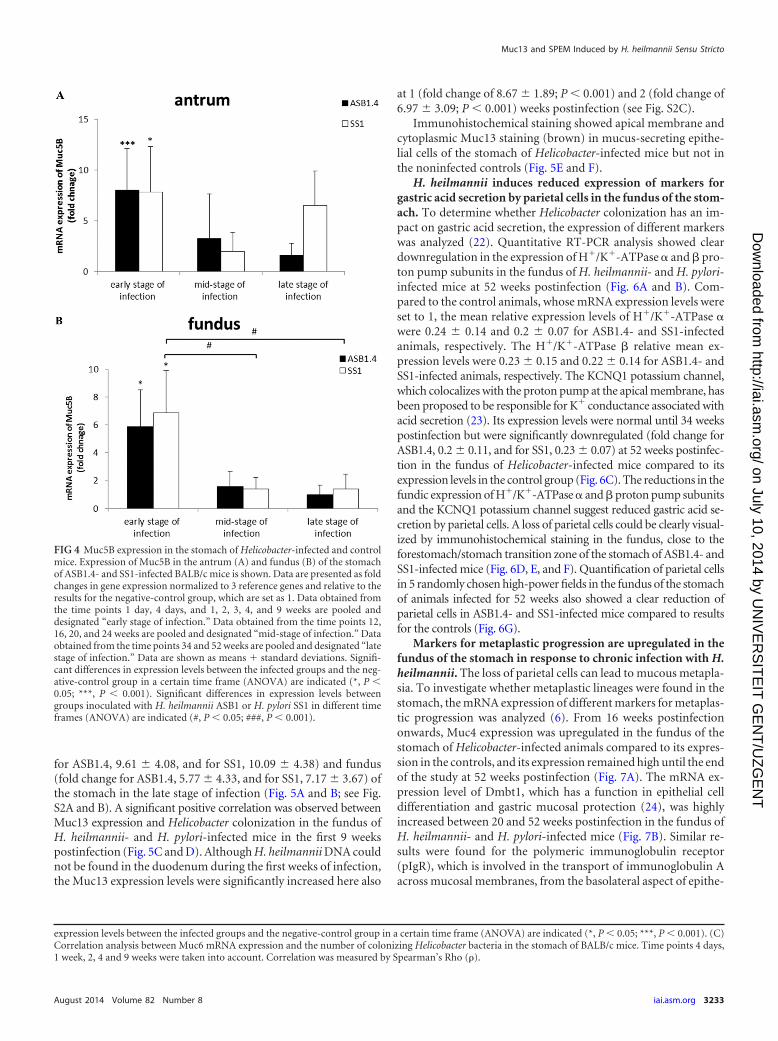
FIG 7 Determination of mucous metaplasia in the fundus of the stomach of Helicobacter-infected mice. (A to C) mRNA expression levels of Muc4 (A), Dmbt1 (B),and pIgR (C) in the fundus of the stomach of H. heilmannii ASB1.4- and H. pylori SS1-infected mice are shown. Data are presented as fold changes in geneexpression normalized to 3 reference genes and relative to the results for the negative-control group, which are set as 1. Data are shown as means � standarddeviations. Significant differences in expression level between the infected groups and the negative-control group at a certain time point (ANOVA) are indicated(*, P 0.05; ***, P 0.001). (D) PAS-Alcian blue staining of the forestomach/stomach transition zone of a sham-inoculated mouse (bar � 30 �m). (E and F)PAS-Alcian blue staining of the antrum (E) and the fundus (F) of the stomach of a mouse infected with H. heilmannii ASB1 for 24 weeks (bars � 10 �m). (G)PAS-Alcian blue staining of the forestomach/stomach transition zone of a mouse infected with H. heilmannii ASB1 for 52 weeks (bar � 30 �m). (E to G)Metaplastic columnar cells, mainly initiating from the transition zone junction between the forestomach and glandular epithelium along the lesser curvature(G) and, to a lesser extent, in the antrum (E) and fundus (F) of the stomach of Helicobacter-infected mice are indicated in blue. (H) The mean numbers of
Liu et al.
3236 iai.asm.org Infection and Immunity
on July 10, 2014 by UN
IVE
RS
ITE
IT G
EN
T/U
ZG
EN
Thttp://iai.asm
.org/D
ownloaded from

Dmbt1, pIgR, and Tff2 have been described to be related to SPEMwith chronic inflammation (6, 26). PAS-Alcian blue staining at 34and 52 weeks postinfection showed evidence for mucous metapla-sia, with metaplastic columnar glands mainly initiating from thetransition zone junction between the forestomach and glandularepithelium along the lesser curvature (Fig. 7G) and, to a lesserextent, in the antrum (Fig. 7E) and fundus (Fig. 7F) of the stomachof Helicobacter-infected mice. Quantification of blue-stainedmetaplastic cells in 5 randomly chosen high-power fields in theforestomach/stomach transition zone also highlighted the pres-ence of mucous metaplasia in the stomach of animals infected for52 weeks with ASB1.4 and SS1 but not in the noninfected controls(Fig. 7H).
DISCUSSION
In BALB/c mice infected with H. heilmannii ASB1.4 and H. pyloriSS1 for 52 weeks, MALT lymphoma-like lesions were observed ina narrow zone in the fundus near the forestomach/stomach tran-sition zone. These pathological lesions might eventually lead togastric MALT lymphoma (12). The risk of developing MALT lym-phoma has been suggested to be higher in humans suffering froman NHPH gastritis than in those infected with H. pylori (15). Gas-tric MALT lymphoma is characterized by a strong proliferation ofB-lymphocytes, which may be dependent on Th2-type cytokines(15, 16). Experimental NHPH infections have indeed been shownto evoke a Th2-polarized response (14, 16), suggesting that Th2-prone BALB/c mice (27) infected with NHPH can be seen as acritical model for the development of MALT lymphoma inducedby NHPH.
It has been demonstrated that H. pylori strains mainly stimu-late Th1 responses both in humans and in mouse models (28).However, as an exception, the H. pylori strain SS1 does not cause asignificant upregulation of gamma interferon (IFN- ), a signatureTh1 marker, in either BALB/c or C57BL/6 mice. Nevertheless, incommon with other NHPH, it elicits a Th2 response in mice (17, 29).This may explain the development of MALT lymphoma-like lesionsin the stomach seen in this and other studies (29). Typical for H.pylori strains inducing MALT lymphoma is that they lack genesencoding major virulence factors, such as a functional CagPAI,Bab, and Sab adhesins (30). H. pylori SS1 indeed lacks a functionalCagPAI (17). This strain also does not bind to the glycan struc-tures Leb and sLex that are expressed by human gastric mucins (1,3; also unpublished data). Binding to Leb and sLex has been shownto be mediated by the H. pylori BabA and SabA adhesins, respec-tively (1, 3), suggesting that SS1 does not express these adhesins.These virulence factors, as well as a functional CagPAI, are alsoabsent in H. heilmannii and other NHPH (31–34).
In this study, H. heilmannii ASB1.4 and H. pylori SS1 colonizedboth the antrum and fundus of the stomach but with a highercolonization density in the antrum. This is similar to what hasbeen described in human patients. Indeed, in humans infectedwith NHPH, colonization mainly occurs in the antrum of thestomach but these bacteria may be found in the fundus as well,which has also been described for H. pylori (10). In the presentstudy, H. heilmannii-infected BALB/c mice showed higher colo-
nization rates in the antrum and fundus of the stomach than H.pylori-infected mice. This indicates that the capacity of ASB1.4 topersist in the stomach of BALB/c mice is higher than that of SS1,which showed a reduction in colonization during the later stagesof infection. The latter finding has also been reported by Schmitzet al. (20). DNA from H. heilmannii ASB1.4 and H. pylori SS1 wasalso found in the duodenum. Since both species have been linkedto duodenal ulcer disease (10), it remains to be elucidated whetherthey are able to colonize the duodenum or whether the quantita-tive RT-PCR just picked up DNA from bacteria colonizing thestomach.
The expression of MUC1, MUC5AC, and MUC6 in the humangastric epithelium in relation to H. pylori colonization has beeninvestigated previously, showing that H. pylori interacts with epi-thelial cells that produce MUC1 and MUC5AC by binding to Leb
and sLex expressed by these mucins. This indicated that MUC1and MUC5AC but not MUC6 play a role in the colonization of H.pylori in the gastric mucosa (1, 3). On the contrary, in this study, aclear upregulation of Muc6 but not Muc5AC and Muc1 was seenin the stomach of H. heilmannii ASB1.4-infected BALB/c mice inthe first 9 weeks postinfection. The pathway regulating gastricMUC6 expression in response to H. heilmannii infection in thehuman stomach, as well as the bacterial factors involved, is un-known and needs further investigation. During this early stage ofinfection, the increased expression of Muc6 in the antrum was alsopositively correlated with the number of H. heilmannii bacteria.Since Muc6 is expressed by the glands and, unlike H. pylori (1),NHPH are mainly localized in the deep glands of the gastric mu-cosa (10, 15, 16), a potential role of Muc6 in H. heilmannii colo-nization is suggested and needs to be further unraveled. WhetherMuc6 plays a role in the colonization of H. pylori strains lackingthe BabA and SabA adhesins, such as the SS1 strain, also needsfurther investigation.
Another interesting finding seen during H. heilmannii ASB1.4 in-fection, as well as during H. pylori SS1 infection, was the increasedmRNA expression of Muc13 in the stomach. MUC13 has beenshown to be highly expressed in human gastric cancer (21), butincreased mRNA expression of it in the early stages of Helicobactercolonization has so far never been described. The positive corre-lation found between the increased Muc13 expression and theincreased number of Helicobacter bacteria in the fundus of thestomach during the first 9 weeks of infection suggests a potentialrole for Muc13 in Helicobacter colonization. The expression levelof Muc13 remained upregulated until 52 weeks postinfection. Ithas been described that sustained elevation of the expression ofcell surface mucins may promote the transition from chronic in-flammation to cancer (21). How Muc13 influences the Helicobac-ter colonization process is unknown and needs further investiga-tion.
In this study, the mRNA expression of several markers for gas-tric acid secretion by parietal cells was significantly reduced at 52weeks postinfection in the fundic epithelium of H. heilmannii-infected mice, suggesting the loss of parietal cell function. A clearloss of parietal cells was indeed shown by immunohistochemicalstaining. Parietal cell loss might eventually lead to the develop-
metaplastic cells in the forestomach/stomach transition zone of the mice infected with Helicobacter for 52 weeks and control mice are shown. The numbers ofmetaplastic cells were determined by counting blue cells in 5 randomly chosen high-power fields after staining with PAS-Alcian blue. Significant differencesbetween Helicobacter-infected and control animals (ANOVA) are indicated (***, P 0.001).
Muc13 and SPEM Induced by H. heilmannii Sensu Stricto
August 2014 Volume 82 Number 8 iai.asm.org 3237
on July 10, 2014 by UN
IVE
RS
ITE
IT G
EN
T/U
ZG
EN
Thttp://iai.asm
.org/D
ownloaded from
ment of mucous metaplasia (6). Markers for metaplastic progres-sion into SPEM were indeed found to be upregulated in the fundicregion of the stomach during later stages of infection with H.heilmannii ASB1.4 and H. pylori SS1. Mucous metaplasia in a nar-row zone of the fundus near the limiting ridge of the stomach of H.heilmannii-infected mice was histologically confirmed. MALTlymphoma-like lesions were also seen in this region. In gastriccancer, intestinal metaplasia present in the mucosa surroundinglow-grade MALT lymphomas has been described (35). However,in our study, evidence for intestinal metaplasia, such as de novoexpression of Muc2 as described in H. pylori infection (7), was notseen. It remains, therefore, to be determined whether SPEM mayfurther differentiate into intestinal metaplasia and, in the worst-case scenario, into dysplasia in mice kept for longer than 1 yearafter experimental infection with H. heilmannii.
Taken together, the results of histopathology and quantitativeRT-PCR in the present experimental infection study in BALB/cmice illustrate that infection with H. heilmannii induced severegastric pathology that progressed into MALT lymphoma-like le-sions and SPEM, as well as inducing changes in the expression ofMuc6 and Muc13 in the stomach.
ACKNOWLEDGMENTS
This work was supported by the Research Fund of Ghent University,Belgium, grant GOA 01G00408 and grant 01SC0312, the China Schol-arship Council (CSC) (grant 2011691031), the Swedish ResearchCouncil (Vetenskapsrådet 521-2011 to 2370), and the Swedish CancerFoundation (Cancerfonden).
We are grateful to Nathalie Van Rysselberghe, Sofie De Bruyckere,Christian Puttevils, and Sarah Loomans for their skillful technical assis-tance.
REFERENCES1. Lindén SK, Nordman H, Hedenbro J, Hurtig M, Borén T, Carlstedt I.
2002. Strain- and blood group-dependent binding of Helicobacter pylori tohuman gastric MUC5AC glycoforms. Gastroenterology 123:1923–1930.http://dx.doi.org/10.1053/gast.2002.37076.
2. McGuckin MA, Lindén SK, Sutton P, Florin TH. 2011. Mucin dynamicsand enteric pathogens. Nat. Rev. Microbiol. 9:265–278. http://dx.doi.org/10.1038/nrmicro2538.
3. Lindén SK, Sheng YH, Every AL, Miles KM, Skoog EC, Florin TH,Sutton P, McGuckin MA. 2009. MUC1 limits Helicobacter pylori infec-tion both by steric hindrance and by acting as a releasable decoy. PLoSPathog. 5:e1000617. http://dx.doi.org/10.1371/journal.ppat.1000617.
4. Taupin D, Podolsky DK. 2003. Trefoil factors: initiators of mucosalhealing. Nat. Rev. Mol. Cell Biol. 4:721–732. http://dx.doi.org/10.1038/nrm1203.
5. Skoog EC, Sjöling Å, Navabi N, Holgersson J, Lundin SB, Lindén SK.2012. Human gastric mucins differently regulate Helicobacter pylori pro-liferation, gene expression and interactions with host cells. PLoS One7:e36378. http://dx.doi.org/10.1371/journal.pone.0036378.
6. Weis VG, Sousa JF, LaFleur BJ, Nam KT, Weis JA, Finke PE, AmeenNA, Fox JG, Goldenring JR. 2013. Heterogeneity in mouse spasmolyticpolypeptide-expressing metaplasia lineages identifies markers of meta-plastic progression. Gut 62:1270 –1279. http://dx.doi.org/10.1136/gutjnl-2012-302401.
7. Mejías-Luque R, Lindén SK, Garrido M, Tye H, Najdovska M, JenkinsBJ, Iglesias M, Ernst M, de Bolós C. 2010. Inflammation modulates theexpression of the intestinal mucins MUC2 and MUC4 in gastric tumors.Oncogene 29:1753–1762. http://dx.doi.org/10.1038/onc.2009.467.
8. Reis CA, David L, Correa P, Carneiro F, de Bolós C, Garcia E, MandelU, Clausen H, Sobrinho-Simões M. 1999. Intestinal metaplasia of humanstomach displays distinct patterns of mucin (MUC1,MUC2,MUC5AC,and MUC6) expression. Cancer Res. 59:1003–1007.
9. Shimamura T, Ito H, Shibahara J, Watanabe A, Hippo Y, Taniguchi H,Chen Y, Kashima T, Ohtomo T, Tanioka F, Iwanari H, Kodama T,
Kazui T, Sugimura H, Fukayama M, Aburatani H. 2005. Over expres-sion of MUC13 is associated with intestinal-type gastric cancer. CancerSci. 96:265–273. http://dx.doi.org/10.1111/j.1349-7006.2005.00043.x.
10. Haesebrouck F, Pasmans F, Flahou B, Chiers K, Baele M, Meyns T,Decostere A, Ducatelle R. 2009. Gastric helicobacters in domestic animalsand nonhuman primates and their significance for human health. Clin. Mi-crobiol. Rev. 22:202–223. http://dx.doi.org/10.1128/CMR.00041-08.
11. Smet A, Flahou B, D’Herde K, Vandamme P, Cleenwerck I, DucatelleR, Pasmans F, Haesebrouck F. 2012. Helicobacter heilmannii sp. nov.,isolated from feline gastric mucosa. Int. J. Syst. Evol. Microbiol. 62:299 –306. http://dx.doi.org/10.1099/ijs.0.029207-0.
12. O’Rourke JL, Dixon MF, Jack A, Enno A, Lee A. 2004. Gastric B-cellmucosa-associated lymphoid tissue (MALT) lymphoma in an animalmodel of ‘Helicobacter heilmannii’ infection. J. Pathol. 203:896 –903. http://dx.doi.org/10.1002/path.1593.
13. Smet A, Van Nieuwerburgh F, Ledesma J, Flahou B, Deforce D, Duca-telle R, Haesebrouck F. 2013. Genome sequence of Helicobacter heilman-nii sensu stricto ASB1 isolated from the gastric mucosa of a kitten withsevere gastritis. Genome Announc. 1:e00033–12. http://dx.doi.org/10.1128/genomeA.00033-12.
14. Joosten M, Blaecher C, Flahou B, Ducatelle R, Haesebrouck F, Smet A.2013. Diversity in bacterium-host interactions within the species Helico-bacter heilmannii sensu stricto. Vet. Res. 44:65. http://dx.doi.org/10.1186/1297-9716-44-65.
15. Flahou B, Haesebrouck F, Pasmans F, D’Herde K, Driessen A, VanDeun K, Smet A, Duchateau L, Chiers K, Ducatelle R. 2010. Helicobactersuis causes severe gastric pathology in mouse and Mongolian gerbil mod-els of human gastric disease. PLoS One 5:e14083. http://dx.doi.org/10.1371/journal.pone.0014083.
16. Flahou B, Van Deun K, Pasmans F, Smet A, Volf J, Rychlik I, DucatelleR, Haesebrouck F. 2012. The local immune response of mice after Heli-cobacter suis infection: strain differences and distinction with Helicobacterpylori. Vet. Res. 43:75. http://dx.doi.org/10.1186/1297-9716-43-75.
17. Crabtree JE, Ferrero RL, Kusters JG. 2002. The mouse colonizing Helico-bacter pylori strain SS1 may lack a functional cag pathogenicity island.Helicobacter 7:139 –140. http://dx.doi.org/10.1046/j.1083-4389.2002.00071.x.
18. O’Rourke JL, Solnick JV, Neilan BA, Seidel K, Hayter R, Hansen LM,Lee A. 2004. Description of “Candidatus Helicobacter heilmannii” basedon DNA sequence analysis of 16S rRNA and urease genes. Int. J. Syst. Evol.Microbiol. 54:2203–2211. http://dx.doi.org/10.1099/ijs.0.63117-0.
19. Livak KJ, Schmittgen TD. 2001. Analysis of relative gene expression datausing real-time quantitative PCR and the 2���CT method. Methods 25:402– 408. http://dx.doi.org/10.1006/meth.2001.1262.
20. Schmitz JM, Durham CG, Ho SB, Lorenz RG. 2009. Gastric mucusalterations associated with murine Helicobacter infection. J. Histochem.Cytochem. 57:457– 467. http://dx.doi.org/10.1369/jhc.2009.952473.
21. Sheng YH, Lourie R, Lindén SK, Jeffery PL, Roche D, Tran TV, PngCW, Waterhouse N, Sutton P, Florin TH, McGuckin MA. 2011. TheMUC13 cell-surface mucin protects against intestinal inflammation byinhibiting epithelial cell apoptosis. Gut 60:1661–1670. http://dx.doi.org/10.1136/gut.2011.239194.
22. Jain RN, Brunkan CS, Chew CS, Samuelson LC. 2006. Gene expressionprofiling of gastrin target genes in parietal cells. Physiol. Genomics 24:124 –132. http://dx.doi.org/10.1152/physiolgenomics.00133.2005.
23. Grahammer F, Herling AW, Lang HJ, Schmitt-Gräff A, Wittekindt OH,Nitschke R, Bleich M, Barhanin J, Warth R. 2001. The cardiac K�channel KCNQ1 is essential for gastric acid secretion. Gastroenterology120:1363–1371. http://dx.doi.org/10.1053/gast.2001.24053.
24. Conde AR, Martins AP, Brito M, Manuel A, Ramos S, Malta-Vacas J,Renner M, Poustka A, Mollenhauer J, Monteiro C. 2007. DMBT1 isfrequently downregulated in well-differentiated gastric carcinoma butmore frequently upregulated across various gastric cancer types. Int. J.Oncol. 30:1441–1446.
25. Gologan A, Acquafondata M, Dhir R, Sepulveda AR. 2008. Polymericimmunoglobulin receptor-negative tumors represent a more aggressivetype of adenocarcinomas of distal esophagus and gastroesophageal junc-tion. Arch. Pathol. Lab. Med. 132:1295–1301.
26. Nomura S, Baxter T, Yamaguchi H, Leys C, Vartapetian AB, Fox JG,Lee JR, Wang TC, Goldenring JR. 2004. Spasmolytic polypeptide ex-pressing metaplasia to preneoplasia in H. felis-infected mice. Gastroenter-ology 127:582–594. http://dx.doi.org/10.1053/j.gastro.2004.05.029.
27. Enno A, O’Rourke J, Braye S, Howlett R, Lee A. 1998. Antigen-dependent
Liu et al.
3238 iai.asm.org Infection and Immunity
on July 10, 2014 by UN
IVE
RS
ITE
IT G
EN
T/U
ZG
EN
Thttp://iai.asm
.org/D
ownloaded from

progression of mucosa-associated lymphoid tissue (MALT)-type lymphomain the stomach. Effects of antimicrobial therapy on gastric MALT lymphomain mice. Am. J. Pathol. 152:1625–1632.
28. Robinson K, Loughlin MF, Potter R, Jenks PJ. 2005. Host adaptationand immune modulation are mediated by homologous recombination inHelicobacter pylori. J. Infect. Dis. 191:579 –587. http://dx.doi.org/10.1086/427657.
29. Thompson LJ, Danon SJ, Wilson JE, O’Rourke JL, Salama NR, FalkowS, Mitchell H, Lee A. 2004. Chronic Helicobacter pylori infection withSydney strain 1 and a newly identified mouse-adapted strain (Sydneystrain 2000) in C57BL/6 and BALB/c mice. Infect. Immun. 72:4668 – 4679.http://dx.doi.org/10.1128/IAI.72.8.4668-4679.2004.
30. Thiberge JM, Boursaux-Eude C, Lehours P, Dillies MA, Creno S,Coppée JY, Rouy Z, Lajus A, Ma L, Burucoa C, Ruskoné-FoumestrauxA, Courillon-Mallet A, De Reuse H, Boneca IG, Lamarque D, MégraudF, Delchier JC, Médigue C, Bouchier C, Labigne A, Raymond J. 2010.From array-based hybridization of Helicobacter pylori isolates to the com-plete genome sequence of an isolate associated with MALT lymphoma.BMC Genomics 11:368. http://dx.doi.org/10.1186/1471-2164-11-368.
31. Smet A, Van Nieuwerburgh F, Ledesma J, Flahou B, Deforce D, Duca-telle R, Haesebrouck F. 2013. Genome sequence of Helicobacter heilman-
nii sensu stricto ASB1 isolated from the gastric mucosa of a kitten withsevere gastritis. Genome Announc. 1:e00033–12. http://dx.doi.org/10.1128/genomeA.00033-12.
32. Vermoote M, Vandekerckhove TT, Flahou B, Pasmans F, Smet A, DeGroote D, Van Criekinge W, Ducatelle R, Haesebrouck F. 2011. Ge-nome sequence of Helicobacter suis supports its role in gastric pathology.Vet. Res. 42:51. http://dx.doi.org/10.1186/1297-9716-42-51.
33. Schott T, Kondadi PK, Hänninen ML, Rossi M. 2011. Comparativegenomics of Helicobacter pylori and the human-derived Helicobacter biz-zozeronii CIII-1 strain reveal the molecular basis of the zoonotic nature ofnon-pylori gastric Helicobacter infections in humans. BMC Genomics 12:534. http://dx.doi.org/10.1186/1471-2164-12-534.
34. Arnold IC, Zigova Z, Holden M, Lawley TD, Rad R, Dougan G, FalkowS, Bentley SD, Müller A. 2011. Comparative whole genome sequenceanalysis of the carcinogenic model pathogen Helicobacter felis. GenomeBiol. Evol. 3:302–308. http://dx.doi.org/10.1093/gbe/evr022.
35. Lamarque D, Levy M, Chaumette MT, Roudot-Thoraval F, Cavicchi M,Auroux J, Courillon-Mallet A, Haioun C, Delchier JC. 2006. Frequentand rapid progression of atrophy and intestinal metaplasia in gastric mu-cosa of patients with MALT lymphoma. Am. J. Gastroenterol. 101:1886 –1893. http://dx.doi.org/10.1111/j.1572-0241.2006.00671.x.
Muc13 and SPEM Induced by H. heilmannii Sensu Stricto
August 2014 Volume 82 Number 8 iai.asm.org 3239
on July 10, 2014 by UN
IVE
RS
ITE
IT G
EN
T/U
ZG
EN
Thttp://iai.asm
.org/D
ownloaded from