Curcumin ameliorated low dose‑Bisphenol A induced gastric ...
Upload
independentCategory
view
0download
0
Volume 57(1): 79–86, 2009Journal of Histochemistry & Cytochemistry
http://www.jhc.org
ARTICLE
Expression of UDP-N-acetyl-D-galactosamine: PolypeptideN-acetylgalactosaminyltransferase-6 in Gastric Mucosa, IntestinalMetaplasia, and Gastric Carcinoma
Joana Gomes, Nuno T. Marcos, Nora Berois, Eduardo Osinaga, Ana Magalhães,João Pinto-de-Sousa, Raquel Almeida, Fátima Gärtner, and Celso A. Reis
Institute of Molecular Pathology and Immunology–IPATIMUP (JG,NTM,AM,RA,FG,CAR), Medical Faculty of the University ofPorto (JPS,RA,CAR), and Institute of Biomedical Sciences of Abel Salazar–ICBAS (FG), University of Porto, Porto, Portugal, andDepartamento de Inmunobiología, Facultad de Medicina, Universidad de la República and Institut Pasteur, Montevideo,Uruguay (NB,EO)
SUMMARY Aberrant mucin O-glycosylation is often observed in cancer and is character-ized by the expression of immature simple mucin-type carbohydrate antigens. UDP-N-acetyl-D-galactosamine:polypeptide N-acetylgalactosaminyltransferase-6 (ppGalNAc-T6) is one ofthe enzymes responsible for the initial step in O-glycosylation. This study evaluated theexpression of ppGalNAc-T6 in human gastric mucosa, intestinal metaplasia, and gastric car-cinomas. Our results showed that ppGalNAc-T6 is expressed in normal gastric mucosa and inintestinal metaplasia. A heterogeneous expression and staining pattern for this enzyme wasobserved in gastric carcinomas. ppGalNAc-T6 was expressed in 79% of the cases, and itsexpression level was associated with the presence of venous invasion. Our results provideevidence that ppGalNAc-T6 is an IHC marker associated with venous invasion in gastriccarcinoma and may contribute to the understanding of the molecular mechanisms thatunderlie aberrant glycosylation in gastric carcinogenesis and in gastric carcinoma.
(J Histochem Cytochem 57:79–86, 2009)
KEY WORDS
mucin O-glycosylation
ppGalNAc-T6
gastric mucosa
intestinal metaplasia
gastric carcinoma
MUCINS ARE O-glycosylated glycoproteins produced bymost glandular epithelial tissues, representing the maincomponent of the mucus layer on the surface of epi-thelial cells (Lesuffleur et al. 1994; Hollingsworthand Swanson 2004). Recently, mucins and mucin O-glycosylation have attracted attention for their role inthe adhesion of bacteria, cell–cell adhesion, and cancercell metastization (Hollingsworth and Swanson 2004).The expression of mucins is often altered in cancer, withfrequent aberrant glycosylation, resulting in the forma-tion of immature structures and exposure of the peptidebackbone (Reis et al. 1998a; Ferreira et al. 2006). Thesestructures are useful markers of premalignant and ma-lignant cells. Changes in mucin O-glycosylation in the
Golgi apparatus are determined by the differential ex-pression of the enzymes that initiate O-glycosylation:UDP-GalNAc:polypeptide N-acetylgalactosaminyl-transferases (ppGalNAc-Ts) (for a review, see Hassanet al. 2000a; Ten Hagen et al. 2003). This initial keystep controlling mucin O-glycosylation is performedby a family of ppGalNAc-Ts that catalyze the transferof GalNAc from the sugar donor UDP-GalNAc to ser-ine and threonine residues on the protein synthesizingthe Tn antigen (Clausen and Bennett 1996). Until now,15 distinct members of the mammalian ppGalNAc-Tfamily have been identified and characterized (Homaet al. 1993; White et al. 1995; Bennett et al. 1996,1998,1999a,b; Hagen et al. 1997; Ten Hagen et al.1998,1999,2001; Guo et al. 2002; Schwientek et al.2002; Wang et al. 2003; Zhang et al. 2003; Chenget al. 2004), and in silico analysis indicates that as manyas 20 ppGalNAc-Ts may exist (Ten Hagen et al. 2003).The members of ppGalNAc-Ts family, although cata-lyzing the same enzymatic step, are tissue specific andhave different kinetic properties and acceptor substrate
Correspondence to: Celso A. Reis, Institute of MolecularPathology and Immunology, University of Porto (IPATIMUP), RuaDr. Roberto Frias s/n, 4200-465 Porto, Portugal. E-mail: [email protected]
Received for publication July 18, 2008; accepted September 23,2008 [DOI: 10.1369/jhc.2008.952283].
C The Histochemical Society, Inc. 0022-1554/08/$3.30 79
TheJourna
lof
Histoch
emistry&
Cytoc
hemistry
specificities that may determine the site of O-glycanattachment (Wandall et al. 1997). This enzymatic speci-ficity leads to different functions depending on the celltype and organ (Bennett et al. 1996; Hagen et al.1997; Sutherlin et al. 1997; Mandel et al. 1999; Brookset al. 2007; Rajpert-De Meyts et al. 2007). Altered ex-pression of ppGalNAc-Ts could be one of the mecha-nisms that explain the changes in mucin O-glycosylationduring malignant transformation (Hanisch et al. 2001).Variations of the ppGalNAc-Ts expression pattern havebeen described in oral squamous cell carcinoma, wheredecreased expression of ppGalNAc-T1 and increasedexpression of ppGalNAc-T2 and ppGalNAc-T3 havebeen reported compared with the expression pattern innormal oral mucosa (Mandel et al. 1999). Higher expres-sion of ppGalNAc-T1, ppGalNAc-T2, and ppGalNAc-T3has also been described in colorectal carcinoma com-pared with the normal colonic epithelium (Kohsakiet al. 2000). Different levels of expression of ppGalNAc-T3 were detected in patients with colorectal (Shibaoet al. 2002), lung (Gu et al. 2004), pancreatic (Yamamotoet al. 2004), gastric (Ishikawa et al. 2004), gallbladder(Miyahara et al. 2004), prostate (Landers et al. 2005),and extrahepatic bile duct (Inoue et al. 2007) carcino-mas, and it was identified as an independent factor ofprognosis. ppGalNAc-T6, which exhibits a high se-quence homology to ppGalNAc-T3, has been recentlydescribed to be expressed in most ductal breast carcino-mas but not in normal breast epithelium with a signifi-cant association with T1 tumor stage (Berois et al.2006). ppGalNAc-T3 and -T6 were also described inassociation with breast malignant cell lines (Brookset al. 2007). Based on these studies ppGalNAc-T6 couldbe considered an interesting marker for the glycosyla-tion modifications in malignancy with implication inmolecular diagnosis (Freire et al. 2006).
In this study, we characterized the expression ofppGalNAc-T6 in normal gastric mucosa, intestinalmetaplasia, and gastric carcinoma. We show thatppGalNAc-T6 is expressed in gastric mucosa andchanges its expression during gastric carcinogenesis.Expression of ppGalNAc-T6 was found to be asso-ciated with a clinico-pathologic characteristic of gas-tric carcinoma.
Materials and Methods
Tissue Samples and Histological Classification
The study was performed using surgical specimens ofgastric carcinomas and adjacent mucosa from patientsoperated at Hospital S. João, Porto, Portugal. The use ofretrospective samples when informed consent cannot beobtained is authorized for research studies by Portu-guese Law. Analysis of the expression of ppGalNAc-T6 (MAb T6.3) was performed in 76 tissue samples,fixed in 10% formalin, and embedded in paraffin. Se-
rial sections were cut and used for conventional histo-pathological diagnosis. Carcinomas were classifiedaccording to Laurén (1965). The growth pattern wasclassified according to Ming (1977). Age, patient’s sur-vival, presence of lymphatic invasion, nodal metastasisand venous invasion, and tumor localization were alsorecorded in every case.
The pathological staging was achieved using the uni-fied 1987 TNM system for gastric carcinoma (Pinto-De-Sousa et al. 2001). We evaluated MUC5AC, MUC6,and MUC2 mucin expression in 72, 44, and 55 cases,respectively. MUC5AC, MUC6, and MUC2 were de-tected using monoclonal antibodies CLH2 (Reis et al.1997), CLH5 (Reis et al. 2000), and PMH1 (Reis et al.1998b), respectively.
IHC
IHC evaluation of ppGalNAc-T6 was performed bythe avidin-biotin-complex staining method (Hsu et al.1981). All the formalin-fixed, paraffin-embeddedsamples were deparaffinated, rehydrated, and treatedwith 0.3% hydrogen peroxide in methanol for 30 minto block endogenous peroxidase. Sections were incu-bated with normal rabbit serum (DakoCytomation;Glostrup, Denmark) diluted 1:5 in PBS containing10% of BSA for 20 min. After that, they were incubatedovernight at 4C with the monoclonal antibody T6.3 forppGalNAc-T6, diluted 1:400 in PBS containing 5% ofBSA (Berois et al. 2006). The slides were washed inPBS and incubated for 30 min with biotinylated rabbitanti-mouse secondary antibody (DakoCytomation)diluted 1:200 in PBS containing 5% of BSA. Sampleswere washed with PBS and incubated with avidin-biotin peroxidase complex for 30 min (Vectastain EliteABS kit; Burlingame, CA). Sections were stained with3,3′-diaminobenzidine tetrahydrochloride (Sigma;St. Louis, MO) in a buffer containing 0.1% hydrogenperoxide, counterstained with Mayer’s hematoxylin,dehydrated, and mounted. Negative controls were per-formed replacing primary antibody with PBS.
IHC for mucin expression and classification ofintestinal metaplasia was performed as previously de-scribed (Reis et al. 2000).
Scoring of the Immunostaining andStatistical Analysis
A semiquantitative approach was used to score the im-munostaining. Samples were scored as follows: low ex-pression (,25% of positive staining cells) and highexpression (.25% of positive staining cells) for IHC.
Statistical analysis was performed using the x2 test withYates correction using Statview 5.0 software. Fisher’stest was applied whenever appropriate. Differenceswere considered statistically significant at p,0.05. Logis-tic regression was performed using Statview 5.0 software.
80 Gomes, Marcos, Berois, Osinaga, Magalhães, Pinto-de-Sousa, Almeida, Gärtner, Reis
TheJourna
lof
Histoch
emistry&
Cytoc
hemistry
Results
Expression of ppGalNAc-T6 in Normal Gastric Mucosa
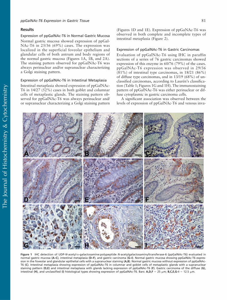
Normal gastric mucosa showed expression of ppGal-NAc-T6 in 25/36 (69%) cases. The expression waslocalized in the superficial foveolar epithelium andglandular cells of both antrum and body regions ofthe normal gastric mucosa (Figures 1A, 1B, and 2A).The staining pattern observed for ppGalNAc-T6 wasalways perinuclear and/or supranuclear characterizinga Golgi staining pattern.
Expression of ppGalNAc-T6 in Intestinal Metaplasia
Intestinal metaplasia showed expression of ppGalNAc-T6 in 14/27 (52%) cases in both goblet and columnarcells of metaplastic glands. The staining pattern ob-served for ppGalNAc-T6 was always perinuclear and/or supranuclear characterizing a Golgi staining pattern
(Figures 1D and 1E). Expression of ppGaNAc-T6 wasobserved in both complete and incomplete types ofintestinal metaplasia (Figure 2).
Expression of ppGalNAc-T6 in Gastric Carcinomas
Evaluation of ppGalNAc-T6 using IHC in paraffinsections of a series of 76 gastric carcinomas showedexpression of this enzyme in 60/76 (79%) of the cases.ppGalNAc-T6 expression was observed in 29/36(81%) of intestinal type carcinomas, in 18/21 (86%)of diffuse type carcinomas, and in 13/19 (68%) of un-classified carcinomas, according to Laurén’s classifica-tion (Table 1; Figures 1G and 1H). The immunostainingpattern of ppGalNAc-T6 was either perinuclear or dif-fuse cytoplasmic in gastric carcinoma cells.
A significant association was observed between thelevels of expression of ppGalNAc-T6 and venous inva-
Figure 1 IHC detection of UDP-N-acetyl-D-galactosamine:polypeptide N-acetylgalactosaminyltransferase-6 (ppGalNAc-T6) evaluated innormal gastric mucosa (A–C), intestinal metaplasia (D–F), and gastric carcinoma (G–I). Normal gastric mucosa showing ppGalNAc-T6 expres-sion in the foveolar and glandular epithelial cells with a supranuclear staining (A,B). Normal gastric mucosa without expression of ppGalNAc-T6 (C). Intestinal metaplasia showing expression of ppGalNAc-T6 in columnar and goblet cells of metaplastic glands with a supranuclearstaining pattern (D,E) and intestinal metaplasia with glands lacking expression of ppGalNAc-T6 (F). Gastric carcinoma of the diffuse (G),intestinal (H), and unclassified (I) histological types showing expression of ppGalNAc-T6. Bars: A,D,F 5 25 mm; B,C,E,G–I 5 12.5 mm.
ppGalNAc-T6 Expression in Gastric Tissue 81
TheJourna
lof
Histoch
emistry&
Cytoc
hemistry

sion (Table 1). Most cases with low levels of ppGal-NAc-T6 expression showed absence of venous inva-sion (p,0.03). In addition, analysis of contingencysplit by tumor localization showed that venous inva-sion was more associated with ppGalNAc-T6 in tu-mors localized in the body region of the stomach(Table 2). Further logistic regression showed no deter-ministic relation in which venous invasion was solelyexplained by ppGalNAc-T6.
The expression of ppGalNAc-T6 was not associatedwith other clinico-pathological characteristics of gas-tric carcinomas.
Coexpression of Mucins (MUC5AC, MUC6, and MUC2)and ppGalNAc-T6 in Gastric Carcinomas
Analysis of the coexpression of mucins (MUC5AC,MUC6, andMUC2) and ppGalNAc-T6 in gastric carci-noma is shown in Table 3. The expression of MUC5AC
was inversely associated with the levels of expressionof ppGalNAc-T6. Most cases positive for MUC5ACshowed low expression of ppGalNAc-T6, whereasmost cases negative for MUC5AC showed high expres-sion of the enzyme (Table 3). No association was ob-served between the expression of ppGalNAc-T6 andthe expression of mucins MUC6 and MUC2 (Table 3).
DiscussionAlterations in mucin-type O-glycans are associatedwith malignant transformation. This study evaluatedthe pattern of expression of ppGalNAc-T6, a key en-zyme involved in O-glycan biosynthesis in normal,metaplastic, and neoplastic gastric tissues.
Our results showed that ppGalNAc-T6 was expressedboth in the foveolar epithelium and glands of both an-trum and body regions of the normal gastric mucosa.
Figure 2 IHC of ppGalNAc-T6 and mucins MUC5AC and MUC2, in normal gastric mucosa (A–C), complete intestinal metaplasia (D–F), andincomplete intestinal metaplasia (G–I). Foveolar epithelium of a normal gastric mucosa showing expression of ppGalNAc-T6 (A, arrowheads),MUC5AC (B, arrowheads), and lacking expression of MUC2 (C). Complete type of intestinal metaplasia expressing ppGalNAc-T6 (D, arrow-heads), MUC2 (F, arrowheads), and lacking expression of MUC5AC (E, arrows). Incomplete type of intestinal metaplasia expressing ppGalNAc-T6 (G, arrowheads), MUC5AC (H, arrowheads), and MUC2 (I, arrowheads). Bar 5 12.5 mm.
82 Gomes, Marcos, Berois, Osinaga, Magalhães, Pinto-de-Sousa, Almeida, Gärtner, Reis
TheJourna
lof
Histoch
emistry&
Cytoc
hemistry

In intestinal metaplasia, a precursor lesion of gastriccarcinoma, we observed a slightly lower percentage ofpositive cases. Intestinal metaplasia shows a highlyregulated pattern of expression of genes, reproducingthe differentiation characteristics of intestinal cells.This is the case of mucins such as MUC2 (Reis et al.1999) and a modified pattern of mucin-type O-glycanssuch as Sialyl-Tn. The percentage of cases expressingppGalNAc-T6 observed in intestinal metaplasia mayreflect the differentiation characteristics of these cellsand may explain the modified pattern of mucin-typeO-glycans observed in intestinal metaplasia (Davidet al. 1992; Ferreira et al. 2006; Mesquita et al. 2006).
Studies in vitro have shown that ppGalNAc-Ts dis-play site specificity and different kinetic properties
toward O-glycosylation sites in mucins, including theMUC1 tandem repeat (Wandall et al. 1997; Hassanet al. 2000b). These characteristics of ppGalNAc-Tssuggest that the members of this family of enzymeshave different functions and that the repertoire ofppGalNAc-Ts expressed in a given cell may determinethe pattern ofO-glycan attachment, especially the densityof glycosylation of the MUC1 tandem repeat (Bennettet al. 1998; Hassan et al. 2000b).
This study is, to the best of our knowledge, the firstevaluation of the expression of ppGalNAc-T6 in gas-tric carcinomas. This evaluation of ppGalNAc-T6 ofgastric carcinomas was possible because of the capabil-ity of MAb T6.3 to recognize formalin-fixed paraffin-embedded sections (Berois et al. 2006). The evaluationof ppGalNAc-T6 in 76 gastric carcinomas showedexpression of this enzyme in 79% of the cases. Ourresults showed that the expression of ppGalNAc-T6
Table 1 Summary of data on the expression level of ppGalNAc-T6and clinico-pathologic features in 76 gastric carcinoma cases
Expression of ppGalNAc-T6a
CategoryNumber ofcases (%)
Positivecases (%) Low (%) High (%) p
Age (years),40 1 (1.3) 1 (100.0) 1 (100.0) 0 (0.0) 0.4040–65 40 (52.6) 32 (80.0) 21 (65.6) 11 (34.4).65 35 (46.1) 27 (77.1) 14 (51.9) 13 (48.1)
Laurén’s classificationDiffuse 21 (27.6) 18 (85.7) 12 (66.7) 6 (33.3) 0.20Intestinal 36 (47.4) 29 (80.6) 19 (65.5) 10 (34.5)Unclassified 19 (25.0) 13 (68.4) 5 (38.5) 8 (61.5)
Ming’s classificationb
Expanding 28 (37.3) 21 (75.0) 11 (52.4) 10 (47.6) 0.42Infiltrative 47 (62.7) 38 (80.9) 24 (63.2) 14 (36.8)
SurvivalShort survival 67 (88.2) 53 (79.1) 32 (60.4) 21 (39.6) 0.87Long survival 9 (11.8) 7 (77.8) 4 (57.1) 3 (42.9)
Lymphatic invasionc
Absent 4 (5.4) 3 (75.0) 3 (100.0) 0 (0.0) 0.14Present 70 (94.6) 55 (78.6) 31 (56.4) 24 (43.6)
Nodal metastasisc
Absent 15 (20.2) 11 (73.3) 7 (63.6) 4 (36.4) 0.71Present 59 (79.7) 47 (79.7) 27 (57.4) 20 (42.6)
Venous invasionAbsent 33 (43.4) 25 (75.8) 19 (76.0) 6 (24.0) 0.03Present 43 (56.6) 35 (81.4) 17 (48.6) 18 (51.4)
Stagingd
Early 15 (20.8) 11 (73.3) 8 (72.7) 3 (27.3) 0.32Advance 57 (79.2) 46 (80.7) 26 (56.5) 20 (43.5)
Tumor localizationc
Cardia 18 (24.3) 11 (61.1) 8 (72.7) 3 (27.3) 0.50Body 27 (36.5) 23 (85.2) 12 (52.2) 11 (47.8)Antrum 29 (39.2) 24 (82.8) 15 (62.5) 9 (37.5)
Total 76 (100.0) 60 (78.9) 36 (60.0) 24 (40.0)
aExpression of ppGalNAc-T6 was scored as Low expression (,25% of positivestaining cells) and High expression (.25% of positive staining cells).bIn one case, the growth pattern was not classified according to Ming’s classi-fication because both infiltrative and expanding areas were observed.cIn two cases, there was no information regarding lymphatic invasion, nodalmetastasis, and tumor localization.dStaging as early (IA and IB) and advance (II–IV) was done based on the pTNMas described in Materials and Methods. In four cases, the pTNM classificationwas not available.ppGalNAc-T6, UDP-N-acetyl-D-galactosamine:polypeptide N-acetylgalactos-aminyltransferase-6.
Table 3 Summary of data on the coexpression level of mucins(MUC5AC, MUC6, and MUC2) and ppGalNAc-T6 in gastriccarcinoma cases
Expression of ppGalNAc-T6a
MucinsNumber ofcases (%)
Positivecases (%) Low (%) High (%) p
MUC5ACb
Negative 22 (30.6) 17 (77.3) 6 (35.3) 11 (64.7) 0.02Positive 50 (69.4) 39 (78.0) 27 (69.2) 12 (30.8)
MUC6c
Negative 28 (63.6) 21 (75.0) 13 (61.9) 8 (38.1) 0.51Positive 16 (36.4) 12 (75.0) 6 (50.0) 6 (50.0)
MUC2d
Negative 32 (58.2) 24 (54.5) 13 (54.2) 11 (45.8) 0.70Positive 23 (41.8) 20 (45.4) 12 (60.0) 8 (40.0)
aExpression of ppGalNAc-T6 was scored as low expression (,25% of positivestaining cells) and high expression (.25% of positive staining cells).bMUC5AC mucin expression was evaluated in 72 gastric carcinoma cases.cMUC6 mucin expression was evaluated in 44 gastric carcinoma cases.dMUC2 mucin expression was evaluated in 55 gastric carcinoma cases.ppGalNAc-T6, UDP-N-acetyl-D-galactosamine:polypeptide N-acetylgalactos-aminyltransferase-6.
Table 2 Expression level of ppGalNAc-T6 according to thepresence of venous invasion and localization of the tumor inthe stomach
Expression of ppGalNAc-T6b
Tumor localizationaNumber ofcases (%) Low (%) High (%) p
Cardia (n511)Absent venous invasion 3 (27.3) 2 (66.7) 1 (33.3) 0.99Present venous invasion 8 (72.7) 6 (75.0) 2 (25.0)
Body (n523)Absent venous invasion 9 (39.1) 8 (88.9) 1 (11.1) 0.01Present venous invasion 14 (60.9) 4 (28.6) 10 (71.4)
Antrum (n524)Absent venous invasion 13 (54.2) 9 (69.2) 4 (30.8) 0.67Present venous invasion 11 (45.8) 6 (54.5) 5 (45.5)
aIn two cases, there was no information regarding tumor localization.bExpression of ppGalNAc-T6 was scored as low expression (,25% of positivestaining cells) and high expression (.25% of positive staining cells).ppGalNAc-T6, UDP-N-acetyl-D-galactosamine:polypeptide N-acetylgalactos-aminyltransferase-6.
ppGalNAc-T6 Expression in Gastric Tissue 83
TheJourna
lof
Histoch
emistry&
Cytoc
hemistry

in gastric carcinomas was associated with the pres-ence of venous invasion. In addition, our results alsoshowed a particular association of venous invasion withppGalNAc-T6 expression in tumors localized in thebody region of the stomach.
Different levels of ppGalNAc-T3 have been detectedin patients with various types of carcinomas (Shibaoet al. 2002; Gu et al. 2004; Ishikawa et al. 2004;Miyahara et al. 2004; Yamamoto et al. 2004; Landerset al. 2005; Inoue et al. 2007), and this has been asso-ciated with a poor prognosis. ppGalNAc-T6 exhibits ahigh sequence homology to ppGalNAc-T3, and in vitrostudies have shown that both enzymes display similaracceptor substrate specificities (Bennett et al. 1999b).Our results suggest that ppGalNac-T6 may also playa role in the biological characteristics of gastric carci-noma cells, most probably through the variation inmucin O-glycosylation. This is in line with previousstudies showing that gastric carcinoma cell lines ex-pressing different set of ppGalNAc-Ts are associatedwith variable patterns of mucin glycosylation (Marcoset al. 2003). The abnormal expression of ppGalNAc-T6 in gastric carcinomas, and the mucin glycoformsproduced by this enzyme, could induce changes in cel-lular functions including adhesion and invasion.Although a preliminary evaluation of coexpression be-tween ppGalNAc-T6 and simple mucin-type carbo-hydrate antigens (Tn and sialyl-Tn) in a subseries ofgastric carcinomas did not show a direct association(data not shown), we cannot exclude that the expres-sion of ppGalNAc-T6 contributes to a different levelof O-glycan site occupancy in the mucin tandemrepeats. Our results warrant further study on the re-lationship between ppGalNAc-T6 expression and al-terations in the O-glycan density and expression ofcarbohydrate antigens in gastric carcinomas (Marcoset al. 2003). This evaluation should also include otherglycosyltransferases that can contribute to the synthesisof these carbohydrate antigens. In addition, future func-tional in vitro studies will clarify the biological role ofppGalNAc-T6 overexpression in gastric carcinomacells. Furthermore, the expression of the mucin proteincore is tightly regulated in gastric mucosa. Our resultsshowed that ppGalNAc-T6 is inversely associated withthe expression of mucin MUC5AC. This result maystem from characteristics of differentiation of the cellsexpressing either protein. No association was observedwith the other mucins evaluated. Finally, ppGalNAc-T6expression was observed in both complete and incom-plete intestinal metaplasia cases (Reis et al. 1999).
In summary, we showed that (a) ppGalNAc-T6 isexpressed in normal gastric mucosa in both antrumand body regions; (b) in intestinal metaplasia, there isexpression of ppGalNAc-T6 in 52% of the cases; (c) ingastric carcinomas, the expression of ppGalNAc-T6 isheterogeneous, with most cases (79%) expressing
ppGalNAc-T6; and (d) ppGalNAc-T6 expression ingastric carcinomas is associated with venous invasion.Our results provide evidence that ppGalNAc-T6 is anovel IHC marker associated with venous invasion ingastric carcinomas and contributes to the understand-ing of the molecular mechanisms that underlie aberrantglycosylation during gastric carcinogenesis and gas-tric carcinoma.
Acknowledgments
This work was supported by Fundação para a Ciência e aTecnologia (FCT) (PTDC/CTM/65330/2006 and PTDC/CVT/65537/2006); financiado no âmbito Programa OperacionalCiência e Inovação 2010 do Quadro Comunitário de Apoio IIIe comparticipado pelo FEDER; and Association for Interna-tional Cancer Research (AICR Grant 05-088). J.G. (SFRH/BD/40563/2007), N.T.M. (SFRH/BD/11764/2003), andA.M. (SFRH/BD/36339/2007) acknowledge FCT for finan-cial support.
We thank Leonor David and Ulla Mandel for suggestions,Mário Seixas for statistical analysis, and Nuno Mendes fortechnical assistance.
Literature Cited
Bennett EP, Hassan H, Clausen H (1996) cDNA cloning and expres-sion of a novel human UDP-N-acetyl-alpha-D-galactosamine.Polypeptide N-acetylgalactosaminyltransferase, Gal-NAc-T3. JBiol Chem 271:17006–17012
Bennett EP, Hassan H, Hollingsworth MA, Clausen H (1999a) Anovel human UDP-N-acetyl-D-galactosamine:polypeptide N-acetylgalactosaminyltransferase, GalNAc-T7, with specificity forpartial GalNAc-glycosylated acceptor substrates. FEBS Lett 460:226–230
Bennett EP, Hassan H, Mandel U, Hollingsworth MA, Akisawa N,Ikematsu Y,MerkxG, et al. (1999b) Cloning and characterization ofa close homologue of human UDP-N-acetyl-alpha-Dgalactosamine:polypeptide N-acetylgalactosaminyltransferase-T3, designatedGalNAc-T6. Evidence for genetic but not functional redundancy.J Biol Chem 274:25362–25370
Bennett EP, Hassan H, Mandel U, Mirgorodskaya E, Roepstorff P,Burchell J, Taylor–Papadimitriou J, et al. (1998) Cloning of ahuman UDP-N-acetyl-alpha-D-Galactosamine:polypeptide N-acetylgalactosaminyltransferase that complements other GalNAc-transferases in complete O-glycosylation of the MUC1 tandemrepeat. J Biol Chem 273:30472–30481
Berois N, Mazal D, Ubillos L, Trajtenberg F, Nicolas A, Sastre-GarauX, Magdelenar H, et al. (2006) UDP-N-acetyl-D-galactosamine:polypeptide N-acetylgalactosaminyltransferase-6 as a new immu-nohistochemical breast cancer marker. J Histochem Cytochem54:317–328
Brooks SA, Carter TM, Bennett EP, Clausen H, Mandel U (2007)Immunolocalisation of members of the polypeptide N-acetylgalac-tosaminyl transferase (ppGalNAc-T) family is consistent with bio-logically relevant altered cell surface glycosylation in breastcancer. Acta Histochem 109:273–284
Cheng L, Tachibana K, Iwasaki H, Kameyama A, Zhang Y, KubotaT, Hiruma T, et al. (2004) Characterization of a novel humanUDP-GalNAc transferase, pp-GalNAc-T15. FEBS Lett 566:17–24
Clausen H, Bennett EP (1996) A family of UDP-GalNAc: polypep-tide N-acetylgalactosaminyl-transferases control the initiation ofmucin-type O-linked glycosylation. Glycobiology 6:635–646
David L, Nesland JM, Clausen H, Carneiro F, Sobrinho-Simões M(1992) Simple mucin-type carbohydrate antigens (Tn, sialosyl-Tnand T) in gastric mucosa, carcinomas and metastases. APMISSuppl 27:162–172
84 Gomes, Marcos, Berois, Osinaga, Magalhães, Pinto-de-Sousa, Almeida, Gärtner, Reis
TheJourna
lof
Histoch
emistry&
Cytoc
hemistry

Ferreira B, Marcos NT, David L, Nakayama J, Reis CA (2006) Termi-nal alpha1,4-linked N-acetylglucosamine in Helicobacter pylori-associated intestinal metaplasia of the human stomach and gastriccarcinoma cell lines. J Histochem Cytochem 54: 585–591
Freire T, Berois N, Sóñora C, Varangot M, Barrios E, Osinaga E(2006) UDP-N-acetyl-D-galactosamine:polypeptide N-acetyl-galactosaminyltransferase 6 (ppGalNAc-T6) mRNA as a potentialnew marker for detection of bone marrow-disseminated breastcancer cells. Int J Cancer 119:1383–1388
Gu C, Oyama T, Osaki T, Li J, Takenoyama M, Izumi H, Sugio K,et al. (2004) Low expression of polypeptide GalNAc N-acetyl-galactosaminyl transferase-3 in lung adenocarcinoma: impact onpoor prognosis and early recurrence. Br J Cancer 90:436–442
Guo JM, Zhang Y, Cheng L, Iwasaki H, Wang H, Kubota T,Tachibana K, et al. (2002) Molecular cloning and characterizationof a novel member of the UDP-GalNAc:polypeptide N-acetyl-galactosaminyltransferase family, pp-GalNAc-T12. FEBS Lett524:211–218
Hagen FK, Ten Hagen KG, Beres TM, Balys MM, VanWuyckhuyseBC, Tabak LA (1997) cDNA cloning and expression of a novelUDP-N-acetyl-D-galactosamine:polypeptide N-acetylgalactos-aminyltransferase. J Biol Chem 272:13843–13848
Hanisch FG, Reis CA, Clausen H, Paulsen H (2001) Evidence forglycosylation-dependent activities of polypeptide N-acetylgalac-tosaminyltransferases rGalNAc-T2 and -T4 on mucin glycopep-tides. Glycobiology 11:731–740
Hassan H, Bennett EP, Mandel U, Hollingsworth MA, Clausen H(2000a) Control of mucin-type-O-glycosylation: O-glycan occu-pancy is directed by substrate specificities of polypeptide GalNAc-transferases. In Ernst B, Hart GW, Sinaÿ P, eds. Carbohydrates inChemistry and Biology, A Comprehensive Handbook. New York,Wiley-VCH, 273–292
Hassan H, Reis CA, Bennett EP, Mirgorodskaya E, Roepstorff P,Hollingsworth MA, Burchell J, et al. (2000b) The lectin domainof UDP-N-acetyl-D-galactosamine: polypeptide N-acetylgalactos-aminyltransferase-T4 directs its glycopeptide specificities. J BiolChem 275:38197–38205
Hollingsworth MA, Swanson BJ (2004) Mucins in cancer: protectionand control of the cell surface. Nat Rev Cancer 4:45–60
Homa FL, Hollander T, Lehman DJ, Thomsen DR, Elhammer AP(1993) Isolation and expression of a cDNA clone encoding a bovineUDP-GalNAc:polypeptide N-acetylgalactosaminyltransferase.J Biol Chem 268:12609–12616
Hsu SM, Faine L, Fanger H (1981) A comparative study of the per-oxidase-antiperoxidase method and the avidin-biotin complexmethod for studying polypeptide hormones with radioimmuno-assay antibodies. Am J Clin Pathol 75:734–738
Inoue T, Eguchi T, Oda Y, Nishiyama K, Fujii K, Izumi H, KohnoK, et al. (2007) Expression of GalNAc-T3 and its relationshipswith clinicopathological factors in 61 extrahepatic bile duct car-cinomas analyzed using stepwise sections - special reference toits association with lymph node metastases-. Mod Pathol 20:267–276
Ishikawa M, Kitayama J, Nariko H, Kohno K, Nagawa H (2004)The expression pattern of UDP-N-acetyl-alpha-d-galactosamine:polypeptide N-acetylgalactosaminyl transferase-3 in early gastriccarcinoma. J Surg Oncol 86:28–33
Kohsaki T, Nishimori I, Nakayama H, Miyazaki E, Enzan H,Nomoto M, Hollingsworth MA, et al. (2000) Expression ofUDP-GalNAc:polypeptide N-acetylgalactosaminyltransferase iso-zymes T1 and T2 in human colorectal cancer. J Gastroenterol35:840–848
Landers KA, BurgerMJ, TebayMA, Purdie DM, Scells B, SamaratungaH, Lavin MF, et al. (2005) Use of multiple biomarkers for a molec-ular diagnosis of prostate cancer. Int J Cancer 114:950–956
Laurén P (1965) The two histological main types of gastric carci-noma: diffuse and so-called intestinal-type carcinoma. Acta PatholMicrobiol Scand 64:31–49
Lesuffleur T, Zweibaum A, Real FX (1994) Mucins in normal andneoplastic human gastrointestinal tissues. Crit Rev Oncol Hematol17:153–180
Mandel U, Hassan H, Therkildsen MH, Rygaard J, Jakobsen MH,Juhl BR, Dabelsteen E, et al. (1999) Expression of polypeptideGalNAc-transferases in stratified epithelia and squamous cellcarcinomas: immunohistological evaluation using monoclonalantibodies to three members of the GalNAc-transferase family.Glycobiology 9:43–52
Marcos NT, Cruz A, Silva F, Almeida R, David L, Mandel U, ClausenH, et al. (2003) Polypeptide GalNAc-transferases, ST6GalNAc-transferase I, and ST3Gal-transferase I expression in gastric carci-noma cell lines. J Histochem Cytochem 51:761–771
Mesquita P, Raquel A, Nuno L, Reis CA, Silva LF, Serpa J, VanSeuningen I, et al. (2006) Metaplasia: a transdifferentiation pro-cess that facilitates cancer development: the model of gastric in-testinal metaplasia. Crit Rev Oncog 12:3–26
Ming S-C (1977) Gastric carcinoma. A pathobiological classifica-tion. Cancer 39:2475–2485
Miyahara N, Shoda J, Kawamoto T, FurukawaM, Ueda T, TodorokiT, Tanaka N, et al. (2004) Expression of UDP-N-acetyl-alpha-D-galactosamine-polypeptide N-acetylgalactosaminyltransferase iso-zyme 3 in the subserosal layer correlates with postsurgical survivalof pathological tumor stage 2 carcinoma of the gallbladder. ClinCancer Res 10:2090–2099
Pinto-De-Sousa J, David L, Seixas M, Pimenta A (2001) Clinico-pathologic profiles and prognosis of gastric carcinomas fromthe cardia, fundus/body and antrum. Dig Surg 18:102–110
Rajpert-De Meyts E, Poll SN, Goukasian I, Jeanneau C, Herlihy AS,Bennett EP, Skakkebaek NE, et al. (2007) Changes in the profileof simple mucin-type O-glycans and polypeptide GalNAc-trans-ferases in human testis and testicular neoplasms are associatedwith germ cell maturation and tumour differentiation. VirchowsArch 451:805–814
Reis CA, David L, Carvalho F,Mandel U, de Bolós C,MirgorodskayaE, Clausen H, et al. (2000) Immunohistochemical study of theexpression of MUC6 mucin and co-expression of other secretedmucins (MUC5AC and MUC2) in human gastric carcinomas.J Histochem Cytochem 48:377–388
Reis CA, David L, Correa P, Carneiro F, de Bolós C, Garcia E,Mandel U, et al. (1999) Intestinal metaplasia of human stomachdisplays distinct patterns of mucin (MUC1, MUC2, MUC5AC,and MUC6) expression. Cancer Res 59:1003–1007
Reis CA, David L, Nielsen PA, Clausen H,Mirgorodskaya K, RoepstorffP, Sobrinho-Simoes M (1997) Immunohistochemical study ofMUC5AC expression in human gastric carcinomas using a novelmonoclonal antibody. Int J Cancer 74:112–121
Reis CA, David L, Seixas M, Burchell J, Sobrinho-Simoes M (1998a)Expression of fully and under-glycosylated forms of MUC1 mucinin gastric carcinoma. Int J Cancer 79:402–410
Reis CA, Sørensen T, Mandel U, David L, Mirgorodskaya K,Roepstorff P, Kihlberg J, et al. (1998b) Development and character-ization of an antibody directed to an alpha-N-acetyl-D-galactos-amine glycosylated MUC2 peptide. Glycoconj J 15:51–62
Schwientek T, Bennett EP, Flores C, Thacker J, Hollmann M, ReisCA, Behrens J, et al. (2002) Functional conservation of subfami-lies of putative UDP-N-acetylgalactosamine:polypeptide N-acetyl-galactosaminyltransferases inDrosophila, Caenorhabditis elegans,and mammals. One subfamily composed of l(2)35Aa is essential inDrosophila. J Biol Chem 277:22623–22638
Shibao K, Izumi H, Nakayama Y, Ohta R, Nagata N, NomotoM, Matsuo K, et al. (2002) Expression of UDP-N-acetyl-alpha-Dgalactosamine- polypeptide galNAc N-acetylgalactosaminyltransferase-3 in relation to differentiation and prognosis in pa-tients with colorectal carcinoma. Cancer 94:1939–1946
Sutherlin ME, Nishimori I, Caffrey T, Bennett EP, Hassan H, MandelU, Mack D, et al. (1997) Expression of three UDP-N-acetylalpha-D-galactosamine:polypeptide GalNAc N-acetylgalactosaminyltransferases in adenocarcinoma cell lines. Cancer Res 57:4744–4748
Ten Hagen KG, Bedi GS, Tetaert D, Kingsley PD, Hagen FK, BalysMM, Beres TM, et al. (2001) Cloning and characterization of aninth member of the UDP-GalNAc:polypeptide N-acetylgalac-tosaminyltransferase family, ppGaNTase-T9. J Biol Chem 276:17395–17404
ppGalNAc-T6 Expression in Gastric Tissue 85
TheJourna
lof
Histoch
emistry&
Cytoc
hemistry

Ten Hagen KG, Fritz TA, Tabak LA (2003) All in the family: theUDP-GalNAc:polypeptide N-acetylgalactosaminyltransferases.Glycobiology 13:1R–16R
Ten Hagen KG, Hagen FK, Balys MM, Beres TM, Van WuyckhuyseB, Tabak LA (1998) Cloning and expression of a novel, tissuespecifically expressed member of the UDP-Gal-NAc:polypeptideN-acetylgalactosaminyltransferase family. J Biol Chem 273:27749–27754
Ten Hagen KG, Tetaert D, Hagen FK, Richet C, Beres TM, GagnonJ, Balys MM, et al. (1999) Characterization of a UDP-Gal-NAc:polypeptide N-acetylgalactosaminyltransferase that displays gly-copeptide N-acetylgalactosaminyltransferase activity. J Biol Chem274:27867–27874
Wandall HH, HassanH,Mirgorodskaya E, Kristensen AK, RoepstorffP, Bennett EP, Nielsen PA, et al. (1997) Substrate specificities ofthree members of the human UDP-N-acetyl-alpha-Dgalactos-amine:polypeptide N-acetylgalactosaminyltransferase family,GalNAc-T1, -T2, and -T3. J Biol Chem 272:23503–23514
Wang H, Tachibana K, Zhang Y, Iwasaki H, Kameyama A,
Cheng L, Guo J, et al. (2003) Cloning and characterization ofa novel UDP-GalNAc:polypeptide N-acetylgalactosaminyltrans-ferase, pp-GalNAc-T14. Biochem Biophys Res Commun 300:738–744
White T, Bennett EP, Takio K, Sorensen T, Bonding N, Clausen H(1995) Purification and cDNA cloning of a human UDP-Nacetyl-alpha-D-galactosamine:polypeptide N-acetylgalactosaminyltrans-ferase. J Biol Chem 270:24156–24165
Yamamoto S, Nakamori S, Tsujie M, Takahashi Y, Nagano H, DonoK, Umeshita K, et al. (2004) Expression of uridine diphosphate N-acetyl-alpha-D-galactosamine: polypeptide N-acetylgalactosaminyltransferase 3 in adenocarcinoma of the pancreas. Pathobiology71:12–18
Zhang Y, Iwasaki H, Wang H, Kudo T, Kalka TB, Hennet T, KubotaT, et al. (2003) Cloning and characterization of a new humanUDP-N-acetyl-alpha-D-galactosamine:polypeptide N-acetylgalac-tosaminyltransferase, designated pp-GalNAc-T13, that is specifi-cally expressed in neurons and synthesizes GalNAc alpha-serine/threonine antigen. J Biol Chem 278:573–584
86 Gomes, Marcos, Berois, Osinaga, Magalhães, Pinto-de-Sousa, Almeida, Gärtner, Reis
TheJourna
lof
Histoch
emistry&
Cytoc
hemistry
Copyright © 2022 FDOKUMEN