Transcript of “Nina Teicholz on Saturated Fats & the Soft ...
Upload
independentCategory
view
1download
0
Hindawi Publishing CorporationJournal of Biomedicine and BiotechnologyVolume 2012, Article ID 915380, 8 pagesdoi:10.1155/2012/915380
Research Article
Curcumin Attenuates Gastric Cancer Induced byN -Methyl-N -Nitrosourea and Saturated Sodium Chloride in Rats
Kawiya Sintara,1 Duangporn Thong-Ngam,1 Suthiluk Patumraj,1 and Naruemon Klaikeaw2
1 Department of Physiology, Faculty of Medicine, Chulalongkorn University, Bangkok 10330, Thailand2 Department of Pathology, Faculty of Medicine, Chulalongkorn University, Bangkok 10330, Thailand
Correspondence should be addressed to Duangporn Thong-Ngam, [email protected]
Received 23 February 2012; Revised 31 March 2012; Accepted 1 April 2012
Academic Editor: Amr Amin
Copyright © 2012 Kawiya Sintara et al. This is an open access article distributed under the Creative Commons Attribution License,which permits unrestricted use, distribution, and reproduction in any medium, provided the original work is properly cited.
To determine effects of curcumin on N-methyl-N-nitrosourea (MNU) and saturated sodium chloride (s-NaCl)-induced gastriccancer in rats. Male Wistar rats were divided into 5 groups: control (CO), control supplemented with 200 mg/kg curcumin (CC),MNU + s-NaCl, MNU + s-NaCl supplemented with 200 mg/kg curcumin daily for the first 3 weeks (MNU + s-NaCl + C3W), andMNU + s-NaCl supplemented with curcumin for 20 weeks (MNU + s-NaCl + C20W). To induce stomach cancer, rats except forCO and CC were orally treated with 100 mg/kg MNU on day 0 and 14, and s-NaCl twice-a-week for the first 3 weeks. Theexperiment was finished and rats were sacrificed at the end of 20 weeks. Cancers were found in forestomachs of all rats in MNU + s-NaCl. The expressions of phosphorylated inhibitor kappaB alpha (phospho-IκBα), 8-hydroxy-2′-deoxyguanosine (8-OHdG), andcyclin D1 significantly increased in MNU + s-NaCl compared with CO. Curcumin treatments for 3 and 20 weeks reduced thecancer incidence resulting in a decrease of phospho-IκBα expression in benign tumor-bearing rats compared with MNU + s-NaCl.Curcumin treatment for 20 weeks also decreased 8-OHdG expression in benign tumor-bearing rats compared with MNU + s-NaCl. Curcumin can attenuate cancer via a reduction of phospho-IκBα and 8-OHdG expressions, which may play a promisingrole in gastric carcinogenesis.
1. Introduction
Gastric cancer can generate in any part of the stomach.Poorly detected, gastric cancer causes nearly one millionannual deaths worldwide [1, 2]. Gastric cancer is closely asso-ciated with dietary factors and Helicobacter pylori infection.Previous studies have reported that consumption of saltyfoods and N-nitroso compounds and a low intake of freshfruits and vegetables increases the risk of gastric cancer [3, 4].Hypertonic NaCl solutions induce gastric cancer in animalmodels through the enhancement of tissue damage resultingin cell proliferation [5]. Experimental animal models havealso provided support for the hypothesis that salt promotesgastric carcinogenesis induced by N-nitroso carcinogen, suchas N-methyl-N-nitro-N-nitrosoguanidine (MNNG) or N-methyl-N-nitrosourea (MNU) [6–9]. MNU, a mutagen andgenotoxic substance, is a potent inducer of cellular stressleading to chromosomal aberrations, point mutations, celldeath, and DNA damage [10]. Other studies reported that
administration of MNU by oral gavage induced gastric can-cer in rats and mice [11, 12]. Thus, the association betweensaturated NaCl and N-nitroso carcinogen could promotegastric carcinogenesis in rats by inflammation, mutation, andcompensatory cell proliferation.
A range of stimuli such as reactive oxygen species (ROS)and cytokines from inflammatory response can activateinhibitor kappaB kinase (IKK) complex resulting in inhibitorkappaB (IκB) phosphorylation and proteolysis. Phosphory-lation of IκBα elicits IκBα degradation, allowing the nucleartranslocation of nuclear factor kappaB (NF-κB) complex andactivation of target genes that are involved in the control ofcellular proliferation and carcinogenesis such as cyclin D1[13, 14]. The interaction of ROS with the nucleobases of theDNA strand, such as guanine, leads to the formation of8-hydroxy-2′-deoxyguanosine (8-OHdG) and structuralalteration in DNA. The 8-OHdG is a potential biomarker ofoxidative damage of DNA and a factor of initiation andpromotion of carcinogenesis [15]. These molecules play

2 Journal of Biomedicine and Biotechnology
a pivotal role in carcinogenesis and may be targets fortherapeutic approaches.
Chemoprevention is promising as a preventive approachfor cancers. Curcumin (diferuloylmethane), a polyphenolcompound, is an active ingredient of tumeric (Curcumalonga). Curcumin has chemopreventive properties. Impor-tantly, curcumin is safe for humans and animals [16].Curcumin shows beneficial effects in many cancers includingcolorectal cancer, breast cancer, skin cancer, and oral cancer[17]. Several signalling pathways implicated in carcinogen-esis including NF-κB signalling have been modulated bycurcumin treatment [18]. However, data concerning theeffect of curcumin on in vivo study of gastric cancer and keyproteins involved in carcinogenesis induced by MNU andsaturated NaCl (s-NaCl) have not been confirmed.
Therefore, the present study aims to examine the protec-tive effect of curcumin on gastric cancer in rats induced byMNU and s-NaCl administration. In addition, activation ofNF-κB, expressions of oxidative damage of DNA and cellcycle regulator cyclin D1 will be investigated.
2. Methods
2.1. Experimental Design. 6-week-old male Wistar rats(National Laboratory Animal Centre, Mahidol University,Bangkok, Thailand) were used. All experiments and proce-dures carried out on the animals were approved by the EthicsCommittee of the Faculty of Medicine, Chulalongkorn Uni-versity, Bangkok, Thailand. Rats were housed in a controlledtemperature room at 25 ± 1◦C under standard conditions(12-h dark-light cycle). Thirty rats were randomized into fivegroups (six rats each) as follows.
Control rats (CO) were fed citrate buffer, pH 4.5(1 mL/rat) orally via intragastric tube on days 0 and 14 of theexperiment. In addition, rats were fed normal saline(1 mL/rat) orally twice a week for the first 3 weeks of theexperiment. Corn oil (2.5 mL/kg) was administrated daily byintragastric tube for 20 weeks.
Control rats supplemented with curcumin (CC) were fedcitrate buffer, pH 4.5 and normal saline as previously des-cribed. 200 mg/kg curcumin (95% purified curcumin, Cay-man Chemical, MI, USA) was dissolved in corn oil and givendaily to rats by intragastric tube for 20 weeks.
MNU- and saturated NaCl-induced rats (MNU + s-NaCl) were treated for gastric carcinogenesis by MNU(Sigma-Aldrich, MO, USA) and s-NaCl (Merck, Germany)according to Thong-Ngam et al. [19]. Briefly, rats were fed100 mg/kg MNU (dissolved in citrate buffer, pH 4.5) viaintragastric tube on days 0 and 14 of the experiment. Inaddition, rats were fed s-NaCl (30% NaCl solution, 1 mL/rat)orally twice weekly for the first 3 weeks of the experiment.Corn oil was administrated daily by intragastric tube for 20weeks.
MNU- and s-NaCl-induced rats, supplemented with cur-cumin for 3 weeks (MNU + s-NaCl + C3W), were given withMNU and s-NaCl as previously describe. The 200 mg/kg cur-cumin was fed daily to rats by intragastric tube duringadministration of MNU and s-NaCl for the first 3 weeks.
MNU- and s-NaCl-induced rats, supplemented with cur-cumin for 20 weeks (MNU + s-NaCl+C20W), were inducedwith MNU and s-NaCl. The 200 mg/kg curcumin was feddaily to rats by intragastric tube for 20 weeks.
2.2. Stomach Tissues Preparation. After 20 weeks, all animalswere sacrificed by intraperitoneal injection of Thiopental(Abbott, Italy, 120 mg/kg) after overnight fasting. Then, thestomach was excised and divided into 2 parts symmetricallyalong the greater and lesser curves. One part was fixed inliquid nitrogen and kept at −80◦C for western blot analysis.Another part was fixed in 4% paraformaldehyde in phos-phate-buffered saline (PBS) for histological study.
2.3. Histopathological Study. The tissue was fixed with 4%paraformaldehyde fixed and paraffin embedded. Multiple2 μm-thick histological sections were stained with hemo-toxylin and eosin (H&E). The alterations of gastric epithelialcells and the incidence of gastric carcinogenesis were deter-mined by a pathologist. In addition, the cancer incidence wascalculated as percentage using the following formula:
Cancer incidence
= 100×(
numbers of cancer-bearing ratsnumbers of induced rats
).
(1)
2.4. Western Blot Analysis. The tissue sample (0.05 g) washomogenized in 0.5 mL of ice-cold lysis RIPA buffer (CellSignalling Technology Inc., MA, USA) with protease inhib-itor (Sigma-Aldrich) and phosphatase inhibitor (Sigma-Aldrich). The homogenate was sonicated for 15 seconds andcentrifuged at 11,000 g for 10 minutes at 4◦C. The super-natant was retained. Protein concentration was measuredwith the BCA Protein Assay Kit (Thermo Scientific). Theextracted proteins were mixed with loading buffer and boiledfor 5 min. Then, the extracted proteins (35 μg/lane) wereapplied to 10% sodium dodecyl sulfate polyacrylamide gelelectrophoresis. The separated proteins were transferred topolyvinylidene fluoride membrane (Pall Corporation, FL,USA). The blotted membrane was incubated with 5% nonfatdried milk in TBS (0.02 M Tris, pH 7.6, and 0.15 M NaCl) for1 hour at room temperature. Then, the membrane wasprobed with mouse monoclonal anti-phosphorylation ofinhibitor kappaB alpha (Phospho-IκBα, Ser32/36) antibody(1 : 1000; Cell Signalling Technology Inc.) overnight at 4◦C.Moreover, the membrane was also probed with anti-β-actinantibody (sc-47778, 1 : 5,000; Santa Cruz Biotechnology Inc.,CA, USA) for 1 hour at room temperature. After three-timewashing in TBS/0.01% Tween-20), the membrane was incu-bated with goat anti-mouse IgG HRP secondary antibody(1 : 4,000; Cayman Chemical) for 1 hour at room tempera-ture. The bands of protein expression were developed witha commercial chemiluminescence detection kit (AmershamECL plus western blotting system, GE Healthcare, UK). Theluminescence was exposed to film (Fujiflim, Japan). Expres-sion levels of proteins were quantified by ImageJ program(US National Institutes of Health, Bethesda, MA, USA).
Journal of Biomedicine and Biotechnology 3
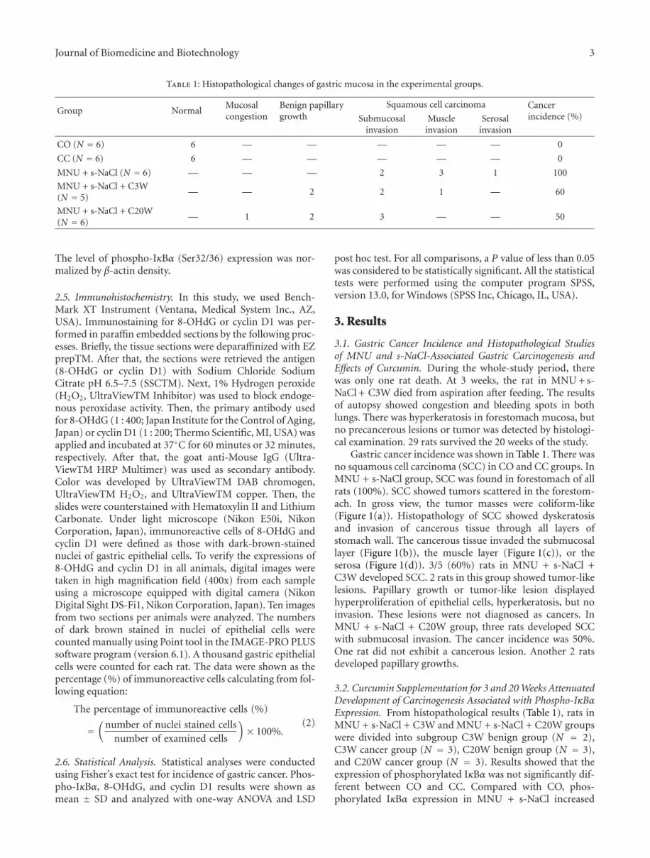
Table 1: Histopathological changes of gastric mucosa in the experimental groups.
Group NormalMucosalcongestion
Benign papillarygrowth
Squamous cell carcinoma Cancerincidence (%)Submucosal
invasionMuscle
invasionSerosal
invasion
CO (N = 6) 6 — — — — — 0
CC (N = 6) 6 — — — — — 0
MNU + s-NaCl (N = 6) — — — 2 3 1 100
MNU + s-NaCl + C3W(N = 5)
— — 2 2 1 — 60
MNU + s-NaCl + C20W(N = 6)
— 1 2 3 — — 50
The level of phospho-IκBα (Ser32/36) expression was nor-malized by β-actin density.
2.5. Immunohistochemistry. In this study, we used Bench-Mark XT Instrument (Ventana, Medical System Inc., AZ,USA). Immunostaining for 8-OHdG or cyclin D1 was per-formed in paraffin embedded sections by the following proc-esses. Briefly, the tissue sections were deparaffinized with EZprepTM. After that, the sections were retrieved the antigen(8-OHdG or cyclin D1) with Sodium Chloride SodiumCitrate pH 6.5–7.5 (SSCTM). Next, 1% Hydrogen peroxide(H2O2, UltraViewTM Inhibitor) was used to block endoge-nous peroxidase activity. Then, the primary antibody usedfor 8-OHdG (1 : 400; Japan Institute for the Control of Aging,Japan) or cyclin D1 (1 : 200; Thermo Scientific, MI, USA) wasapplied and incubated at 37◦C for 60 minutes or 32 minutes,respectively. After that, the goat anti-Mouse IgG (Ultra-ViewTM HRP Multimer) was used as secondary antibody.Color was developed by UltraViewTM DAB chromogen,UltraViewTM H2O2, and UltraViewTM copper. Then, theslides were counterstained with Hematoxylin II and LithiumCarbonate. Under light microscope (Nikon E50i, NikonCorporation, Japan), immunoreactive cells of 8-OHdG andcyclin D1 were defined as those with dark-brown-stainednuclei of gastric epithelial cells. To verify the expressions of8-OHdG and cyclin D1 in all animals, digital images weretaken in high magnification field (400x) from each sampleusing a microscope equipped with digital camera (NikonDigital Sight DS-Fi1, Nikon Corporation, Japan). Ten imagesfrom two sections per animals were analyzed. The numbersof dark brown stained in nuclei of epithelial cells werecounted manually using Point tool in the IMAGE-PRO PLUSsoftware program (version 6.1). A thousand gastric epithelialcells were counted for each rat. The data were shown as thepercentage (%) of immunoreactive cells calculating from fol-lowing equation:
The percentage of immunoreactive cells (%)
=(
number of nuclei stained cellsnumber of examined cells
)× 100%.
(2)
2.6. Statistical Analysis. Statistical analyses were conductedusing Fisher’s exact test for incidence of gastric cancer. Phos-pho-IκBα, 8-OHdG, and cyclin D1 results were shown asmean ± SD and analyzed with one-way ANOVA and LSD
post hoc test. For all comparisons, a P value of less than 0.05was considered to be statistically significant. All the statisticaltests were performed using the computer program SPSS,version 13.0, for Windows (SPSS Inc, Chicago, IL, USA).
3. Results
3.1. Gastric Cancer Incidence and Histopathological Studiesof MNU and s-NaCl-Associated Gastric Carcinogenesis andEffects of Curcumin. During the whole-study period, therewas only one rat death. At 3 weeks, the rat in MNU + s-NaCl + C3W died from aspiration after feeding. The resultsof autopsy showed congestion and bleeding spots in bothlungs. There was hyperkeratosis in forestomach mucosa, butno precancerous lesions or tumor was detected by histologi-cal examination. 29 rats survived the 20 weeks of the study.
Gastric cancer incidence was shown in Table 1. There wasno squamous cell carcinoma (SCC) in CO and CC groups. InMNU + s-NaCl group, SCC was found in forestomach of allrats (100%). SCC showed tumors scattered in the forestom-ach. In gross view, the tumor masses were coliform-like(Figure 1(a)). Histopathology of SCC showed dyskeratosisand invasion of cancerous tissue through all layers ofstomach wall. The cancerous tissue invaded the submucosallayer (Figure 1(b)), the muscle layer (Figure 1(c)), or theserosa (Figure 1(d)). 3/5 (60%) rats in MNU + s-NaCl +C3W developed SCC. 2 rats in this group showed tumor-likelesions. Papillary growth or tumor-like lesion displayedhyperproliferation of epithelial cells, hyperkeratosis, but noinvasion. These lesions were not diagnosed as cancers. InMNU + s-NaCl + C20W group, three rats developed SCCwith submucosal invasion. The cancer incidence was 50%.One rat did not exhibit a cancerous lesion. Another 2 ratsdeveloped papillary growths.
3.2. Curcumin Supplementation for 3 and 20 Weeks AttenuatedDevelopment of Carcinogenesis Associated with Phospho-IκBαExpression. From histopathological results (Table 1), rats inMNU + s-NaCl + C3W and MNU + s-NaCl + C20W groupswere divided into subgroup C3W benign group (N = 2),C3W cancer group (N = 3), C20W benign group (N = 3),and C20W cancer group (N = 3). Results showed that theexpression of phosphorylated IκBα was not significantly dif-ferent between CO and CC. Compared with CO, phos-phorylated IκBα expression in MNU + s-NaCl increased

4 Journal of Biomedicine and Biotechnology
(a)
MM
SM
M
500 μM
(b)
500 μM
MMSM
M
(c)
500 μM
M
(d)
Figure 1: Macroscopic (a) and microscopic (b–d, H&E staining, 40x) appearance of squamous cell carcinoma (SCC) in the forestomachs.The multiple polypoid tumors developed in the stomach after MNU and saturated NaCl administration (a). SCCs in the gastric epitheliumshow the invasion of cancerous tissue with dyskeratosis through submucosal layer (b), muscle layer (c), or serosa (d). Note: the arrowsindicate SCC with submucosal invasion (b), muscle-layer invasion (c), and stomach perforation (d). MM: muscularis mucosae, SM:submucosa, and M: muscle layer.
Table 2: The results of phospho-IκBα-relative expression, 8-OHdG-immunoreactive cells (%), and cyclin D1-immunoreactive cells (%).
Parameters/Group CO (N = 6) CC (N = 6)MNU + s-NaCl
(N = 6)MNU + s-NaCl + C3W (N = 5) MNU + s-NaCl + C20W (N = 6)
C3W benign(N = 2)
C3W cancer(N = 3)
C20W benign(N = 3)
C20W cancer(N = 3)
Phospho-IκBα 0.51± 0.11† 0.54± 0.18† 0.82± 0.18∗ 0.46± 0.04† 0.60± 0.29 0.51± 0.08† 1.16± 0.34∗∗,†
8-OHdG 44.42±3.41†† 40.39±3.53†† 53.06± 5.96∗∗ 49.28± 7.43 48.06± 3.47 44.99± 3.51† 47.77± 2.55
Cyclin D1 38.58±5.37†† 42.53±6.90†† 54.91± 7.93∗∗ 4.33± 1.27∗ 54.92± 3.87∗∗ 45.31± 6.29 53.82± 11.01∗∗
These data were presented as the mean ± SD. ∗Represented significant difference compared with CO group (P < 0.05). ∗∗Represented significant differencecompared with CO group (P < 0.01). †Represented significant difference compared with MNU + s-NaCl group (P < 0.05). ††Represented significantdifference compared with MNU + s-NaCl group (P < 0.01).
significantly (P = 0.010). Curcumin supplementations for 3and 20 weeks in C3W benign and C20W benign groupssignificantly declined the expression of phosphorylated IκBαcompared with MNU + s-NaCl (P = 0.028 and P = 0.031,resp.) (Table 2). The represented bands of phospho-IκBα andβ-actin were shown in Figure 2.
3.3. Curcumin Supplementation for 20 Weeks AttenuatedDevelopment of Carcinogenesis Associated with 8-OHdG andCyclin D1 Expressions. 8-OHdG and cyclin D1 expressions
were studied by immunohistochemistry and shown asnuclei-stained cells. The average percentages of immunore-active cells of all groups were shown in Table 2. Fromthe results, the mean percentages of 8-OHdG and cyclinD1-immunoreactive cells were not significantly differentbetween CO and CC. They were significantly increased inMNU + s-NaCl compared with CO (P = 0.002 and P =0.000, resp.). Curcumin supplementation for 20 weeksreduced gastric cancer incidence and significantly decreased8-OHdG expression in C20W benign group compared with

Journal of Biomedicine and Biotechnology 5
CO
CC
MN
U+
s-N
aCl
C3W
ben
ign
β-actin
Phospho-IκBα
(a)
CO
CC
MN
U+
s-N
aCl
C3W
can
cer
(b)
CO
CC
MN
U+
s-N
aCl
C20
W b
enig
n
β-actin
Phospho-IκBα
(c)M
NU
+s-
NaC
l
CO
CC
C20
W c
ance
r
(d)
Figure 2: Western blot analysis of phospho-IκBα expression. Curcumin supplementation in C3W benign group declined the expressionof phosphorylated IκBα compared with MNU + s-NaCl (a), whereas the expression of phosphorylated IκBα in C3W cancer group did notdecrease (b). Curcumin supplementation for 20 weeks in C20W benign decreased the expression of phosphorylated IκBα compared withMNU + s-NaCl (c), whereas the expression of phosphorylated IκBα in C20W cancer group did not decline (d). An antibody for β-actin wasused as an internal control.
MNU + s-NaCl (P = 0.015) (Table 2). The average percent-age of cyclin D1 stained cells in the C20W benign grouptended to reduce, but not reach a statistically significant levelwhen compared with MNU + s-NaCl (P = 0.062; Table 2).8-OHdG was expressed primarily in gastric epithelial cells(Figure 2(a)). This expression was increased in the mucosa ofSCC in both MNU + s-NaCl and C20W cancer groups (Fig-ures 3(b) and 3(c)). This enhanced expression was limitedto a small number of epithelial cells in C20W benign group(Figure 3(d)).
4. Discussion
This study demonstrated that oral gavage of MNU- and s-NaCl-induced a 100% cancer incidence in rats. The histo-logical results showed that curcumin could attenuate thegastric carcinogenesis induced by MNU and s-NaCl in rats.Administration of curcumin in both MNU + s-NaCl + C3Wand MNU + s-NaCl + C20W groups showed 40% and 50%reduction of cancer incidence, respectively. These observa-tions indicated that early administration of curcumin (dur-ing the first 3 weeks of cancer induction) might preventthe initiation of carcinogenesis. This is in agreement withprevious studies. Feeding 0.5 and 2.0% of commercial gradecurcumin in the diet during the initiation period (2 weeksbefore, during, and 1 week after benzo(a)pyrene administra-tion) reduced the number of mice with forestomach tumors[20]. Daily feeding of 1 and 2 g/mL radix curcumae extract
solution during MNNG administration for 40 weeks alsoshowed the reduction of tumor incidences in 10% NaCl andMNNG-induced gastric cancer in rats [21].
Phosphorylation of IκBα could imply the activation ofNF-κB, which plays a major role in carcinogenesis [13, 14]. Ingastric carcinoma patients, NF-κB activation correlated withIκBα phosphorylation and degradation [22, 23]. Our studyshowed that overexpression of phosphorylated IκBα wasassociated with cancer. This finding coincided with otherreports [24, 25]. Activation of NF-κB appeared to be a keystep of keratinocyte transformation into SCC in mice [24].49% of prostate adenocarcinoma patients showed NF-κBoverexpression that correlated with advanced tumor stage[25]. Curcumin has a chemopreventive property resulting insuppressing NF-κB activation. Many studies confirmed thata pivotal role of curcumin is inhibiting IKK activity withdeclining IκBα phosphorylation [26, 27]. Our results showedthat curcumin supplementations for 3 and 20 weeks sig-nificantly decreased phosphorylated IκBα in benign tumor-bearing rats. Curcumin supplementation in this study pre-vented carcinogenesis by declining IκBα phosphorylation.
8-OHdG is a potential biomarker of oxidative DNAdamage and a factor of initiation and promotion of carcino-genesis [15]. In this study, we also showed that 8-OHdGexpression significantly increased in the MNU + s-NaClgroup compared with CO group. This observation con-firmed many previous results obtained from various typesof cancers in patients. The level of 8-OHdG expression is

6 Journal of Biomedicine and Biotechnology
10 μM
LP
MM
(a)
10 μM
(b)
10 μM
(c)
10 μM
(d)
Figure 3: Immunohistochemical staining of 8-OHdG antibodies in the representative tissue specimens (400x): CO group (a), MNU +s-NaCl group (b), C20W cancer group (c), and C20W benign group (d). DAB stained immunoreactive cells (dark brown, arrows); nuclearcounterstaining was performed with Hematoxylin II and Bluing reagent. LP: laminar propria and MM: muscularis mucosae.
elevated in colorectal cancer [28], hepatocellular carcinoma[29], oral SCC [30], and gastric cancer patients [31]. Con-sidering the number of reports demonstrating a closerelation between 8-OHdG formation and carcinogenicity,including this study, it is likely that 8-OHdG formationsmight participate in carcinogen-induced forestomach SCC.Curcumin showed a potent scavenger of reactive species(RS), such as superoxide anion, hydroxyl radical, singlet oxy-gen, nitric oxide, and peroxynitrite [32]. The reduction ofRS could prevent the formation of 8-OHdG. This studydemonstrated that 200 mg/kg curcumin supplementationsfor 3 and 20 weeks in MNU + s-NaCl-induced SCCs dimin-ished 8-OHdG expression. In addition, 8-OHdG expressionin C20W benign group was significantly reduced comparedto MNU + s-NaCl. These results suggested that curcuminadministration could protect carcinogenesis against forma-tion of 8-OHdG.
Oxidative damage of DNA as well as deregulation of cellcycle control causes cancer. This study demonstrated thatthe immunoreactive cells of cyclin D1, the positive cell cycleregulator, significantly increased in MNU + s-NaCl com-pared with CO. The nuclear accumulation of cyclin D1 is anessential indicator of oncogenicity. Previous studies showedthat nuclear immunoreactivity for cyclin D1 positivelycorrelated with tumor cell proliferation of gastric cancerpatients [33]. Moreover, cyclin D1 overexpression occurredin patients with oral SCC [34]. Cyclin D1 expression in the
C20W benign subgroup tended to decrease, but not reach astatistically significant level when compared with MNU + s-NaCl. This study suggested that curcumin could not preventcarcinogenesis through improvement of dysregulation of cellcycle as shown by cyclin D1 accumulation.
5. Conclusion
MNU and s-NaCl led to increase of phosphorylated IκBα, 8-OHdG, and cyclin D1 that are associated with forestomachcarcinogenesis. Concomitant treatment with 200 mg/kg cur-cumin during the first three weeks or the entire study period(20 weeks) could decrease cancer incidence to 40% and 50%,respectively. This treatment shows a significant reductionof phosphorylated IκBα in benign rats. In addition, cur-cumin treatment for 20 weeks significantly reduces 8-OHdGexpression. Hence, administration of curcumin during theinitiation period could attenuate the incidence of cancer viareduction of phospho-IκBα and 8-OHdG expressions.
Acknowledgments
This paper was supported by the 90th Anniversary ofChulalongkorn University Fund (RatchadaphiseksomphotEndowment Fund), Chulalongkorn University and CerebosAward 2010, Cerebos (Thailand) Ltd.

Journal of Biomedicine and Biotechnology 7
References
[1] D. M. Parkin, F. I. Bray, and S. S. Devesa, “Cancer burden inthe year 2000. The global picture,” European Journal of Cancer,vol. 37, no. 8, pp. S4–S66, 2001.
[2] D. M. Parkin, “International variation,” Oncogene, vol. 23, no.38, pp. 6329–6340, 2004.
[3] C. P. Howson, T. Hiyama, and E. L. Wynder, “The decline ingastric cancer: epidemiology of an unplanned triumph,” Epi-demiologic Reviews, vol. 8, no. 1, pp. 1–27, 1986.
[4] M. Kobayashi, Y. Tsubono, S. Sasazuki, S. Sasaki, and S.Tsugane, “Vegetables, fruit and risk of gastric cancer in Japan:a 10-year follow-up of the JPHC study cohort I,” InternationalJournal of Cancer, vol. 102, no. 1, pp. 39–44, 2002.
[5] C. Furihata, H. Ohta, and T. Katsuyama, “Cause and effectbetween concentration-dependent tissue damage and tem-porary cell proliferation in rat stomach mucosa by NaCl, astomach tumor promoter,” Carcinogenesis, vol. 17, no. 3, pp.401–406, 1996.
[6] M. Tatematsu, M. Takahashi, and S. Fukushima, “Effectsin rats of sodium chloride on experimental gastric cancersinduced by N methyl N’ nitro N nitrosoguanidine or 4 nitro-quinoline 1 oxide,” Journal of the National Cancer Institute, vol.55, no. 1, pp. 101–106, 1975.
[7] M. Takahashi, A. Nishikawa, F. Furukawa, T. Enami, T.Hasegawa, and Y. Hayashi, “Dose-dependent promotingeffects of sodium chloride (NaCl) on rat glandular stomachcarcinogenesis initiated with N-methyl-N’-nitro-N-nitroso-guanidine,” Carcinogenesis, vol. 15, no. 7, pp. 1429–1432, 1994.
[8] S. Kato, T. Tsukamoto, T. Mizoshita et al., “High salt dietsdose-dependently promote gastric chemical carcinogenesisin Helicobacter pylori-infected Mongolian gerbils associatedwith a shift in mucin production from glandular to surfacemucous cells,” International Journal of Cancer, vol. 119, no. 7,pp. 1558–1566, 2006.
[9] W. K. Leung, K. C. Wu, C. Y. P. Wong et al., “Transgenic cyclo-oxygenase-2 expression and high salt enhanced susceptibilityto chemical-induced gastric cancer development in mice,”Carcinogenesis, vol. 29, no. 8, pp. 1648–1654, 2008.
[10] IARC, Some Aromatic Amines, Hydrazine and Related Sub-stances, N-Nitroso Compounds and Miscellaneous Alkylatingagents. IARC Monographs on the Evaluation of CarcinogenicRisk of Chemicals to Humans, International Agency forResearch on Cancer, Lyon, France, 1974.
[11] C. Furihata, E. Ikui, and T. Matsushima, “DNA damagingand cell proliferative activity of 1-methyl-1-nitrosourea in ratglandular stomach mucosa,” Mutation Research, vol. 348, no.4, pp. 169–173, 1995.
[12] D. J. Hoivik, J. S. Allen, H. G. Wall, J. B. Nold, R. T. Miller, andM. J. Santostefano, “Studies evaluating the utility of N-methyl-N-nitrosourea as a positive control in carcinogenicity studiesin the p53+/- mouse,” International Journal of Toxicology, vol.24, no. 5, pp. 349–356, 2005.
[13] M. Karin and F. R. Greten, “NF-κB: linking inflammation andimmunity to cancer development and progression,” NatureReviews Immunology, vol. 5, no. 10, pp. 749–759, 2005.
[14] H. J. Kim, N. Hawke, and A. S. Baldwin, “NF-κB and IKK astherapeutic targets in cancer,” Cell Death and Differentiation,vol. 13, no. 5, pp. 738–747, 2006.
[15] A. Valavanidis, T. Vlachogianni, and C. Fiotakis, “8-hydroxy-2’ -deoxyguanosine (8-OHdG): a critical biomarker of oxida-tive stress and carcinogenesis.,” Journal of EnvironmentalScience and Health C, vol. 27, no. 2, pp. 120–139, 2009.
[16] R. A. Sharma, A. J. Gescher, and W. P. Steward, “Curcumin:the story so far,” European Journal of Cancer, vol. 41, no. 13,pp. 1955–1968, 2005.
[17] G. Sa and T. Das, “Anti cancer effects of curcumin: cycle of lifeand death,” Cell Division, vol. 3, article 14, 2008.
[18] B. B. Aggarwal, Y. Takada, and O. V. Oommen, “From chemo-prevention to chemotherapy: common targets and commongoals,” Expert Opinion on Investigational Drugs, vol. 13, no. 10,pp. 1327–1338, 2004.
[19] D. Thong-Ngam, K. Sintara, M. Chayanupatkul, N. Klaikaew,and T. Chatsuwan, “The rat models of gastric cancer usingHelicobacter pylori infection, N-methyl-N-nitrosourea, andhigh salt induced carcinogenesis,” Thai Journal of Gastroen-terology, vol. 11, no. 1, pp. 41–48, 2010.
[20] M. T. Huang, Y. R. Lou, W. Ma, H. L. Newmark, K. R. Reuhl,and A. H. Conney, “Inhibitory effects of dietary curcuminon forestomach, duodenal, and colon carcinogenesis in mice,”Cancer Research, vol. 54, no. 22, pp. 5841–5847, 1994.
[21] B. Lu, L. Xu, L. Yu, and L. Zhang, “Extract of radix curcumaeprevents gastric cancer in rats,” Digestion, vol. 77, no. 2, pp.87–91, 2008.
[22] G. Levidou, P. Korkolopoulou, N. Nikiteas et al., “Expressionof nuclear factor κB in human gastric carcinoma: relationshipwith IκBa and prognostic significance,” Virchows Archiv, vol.450, no. 5, pp. 519–527, 2007.
[23] L. Wu, Z. Pu, J. Feng, G. Li, Z. Zheng, and W. Shen, “Theubiquitin-proteasome pathway and enhanced activity of NF-κB in gastric carcinoma,” Journal of Surgical Oncology, vol. 97,no. 5, pp. 439–444, 2008.
[24] M. Van Hogerlinden, B. L. Rozell, L. Ahrlund-Richter, and R.Toftgard, “Squamous cell carcinomas and increased apoptosisin skin with inhibited Rel/nuclear factor-κB signaling,” CancerResearch, vol. 59, no. 14, pp. 3299–3303, 1999.
[25] J. S. Ross, B. V. S. Kallakury, C. E. Sheehan et al., “Expressionof nuclear factor-κB and IκBα proteins in prostatic adenocar-cinomas: correlation of nuclear factor-κB immunoreactivitywith disease recurrence,” Clinical Cancer Research, vol. 10, no.7, pp. 2466–2472, 2004.
[26] C. Jobin, C. A. Bradham, M. P. Russo et al., “Curcumin blockscytokine-mediated NF-κB activation and proinflammatorygene expression by inhibiting inhibitory factor I-κB kinaseactivity,” Journal of Immunology, vol. 163, no. 6, pp. 3474–3483, 1999.
[27] D. Wang, M. S. Veena, K. Stevenson et al., “Liposome-encapsulated curcumin suppresses growth of head and necksquamous cell carcinoma in vitro and in xenografts throughthe inhibition of nuclear factor κB by an AKT-independentpathway,” Clinical Cancer Research, vol. 14, no. 19, pp. 6228–6236, 2008.
[28] D. Chang, F. Wang, Y. S. Zhao, and H. Z. Pan, “Evaluation ofoxidative stress in colorectal cancer patients,” Biomedical andEnvironmental Sciences, vol. 21, no. 4, pp. 286–289, 2008.
[29] M. Chuma, S. Hige, M. Nakanishi et al., “8-Hydroxy-2′-deoxy-guanosine is a risk factor for development of hepato-cellular carcinoma in patients with chronic hepatitis C virusinfection,” Journal of Gastroenterology and Hepatology, vol. 23,no. 9, pp. 1431–1436, 2008.
[30] G. Bahar, R. Feinmesser, T. Shpitzer, A. Popovtzer, and R. M.Nagler, “Salivary analysis in oral cancer patients: DNA andprotein oxidation, reactive nitrogen species, and antioxidantprofile,” Cancer, vol. 109, no. 1, pp. 54–59, 2007.
[31] X. Chen, J. Dub, and L. Gu, “Expression of 8-hydroxy-2-deoxyguanosine in gastric carcinomas,” Journal of NanjingMedical University, vol. 21, no. 1, pp. 11–14, 2007.
8 Journal of Biomedicine and Biotechnology
[32] V. P. Menon and A. R. Sudheer, “Antioxidant and anti-inflam-matory properties of curcumin,” Advances in ExperimentalMedicine and Biology, vol. 595, pp. 105–125, 2007.
[33] K. H. Lee, H. E. Lee, S. J. Cho et al., “Immunohistochemicalanalysis of cell cycle-related molecules in gastric carcinoma:prognostic significance, correlation with clinicopathologicalparameters, proliferation and apoptosis,” Pathobiology, vol. 75,no. 6, pp. 364–372, 2008.
[34] P. V. Angadi and R. Krishnapillai, “Cyclin D1 expression inoral squamous cell carcinoma and verrucous carcinoma: cor-relation with histological differentiation,” Oral Surgery, OralMedicine, Oral Pathology, Oral Radiology and Endodontology,vol. 103, no. 3, pp. e30–e35, 2007.

Submit your manuscripts athttp://www.hindawi.com
Stem CellsInternational
Hindawi Publishing Corporationhttp://www.hindawi.com Volume 2014
Hindawi Publishing Corporationhttp://www.hindawi.com Volume 2014
MEDIATORSINFLAMMATION
of
Hindawi Publishing Corporationhttp://www.hindawi.com Volume 2014
Behavioural Neurology
EndocrinologyInternational Journal of
Hindawi Publishing Corporationhttp://www.hindawi.com Volume 2014
Hindawi Publishing Corporationhttp://www.hindawi.com Volume 2014
Disease Markers
Hindawi Publishing Corporationhttp://www.hindawi.com Volume 2014
BioMed Research International
OncologyJournal of
Hindawi Publishing Corporationhttp://www.hindawi.com Volume 2014
Hindawi Publishing Corporationhttp://www.hindawi.com Volume 2014
Oxidative Medicine and Cellular Longevity
Hindawi Publishing Corporationhttp://www.hindawi.com Volume 2014
PPAR Research
The Scientific World JournalHindawi Publishing Corporation http://www.hindawi.com Volume 2014
Immunology ResearchHindawi Publishing Corporationhttp://www.hindawi.com Volume 2014
Journal of
ObesityJournal of
Hindawi Publishing Corporationhttp://www.hindawi.com Volume 2014
Hindawi Publishing Corporationhttp://www.hindawi.com Volume 2014
Computational and Mathematical Methods in Medicine
OphthalmologyJournal of
Hindawi Publishing Corporationhttp://www.hindawi.com Volume 2014
Diabetes ResearchJournal of
Hindawi Publishing Corporationhttp://www.hindawi.com Volume 2014
Hindawi Publishing Corporationhttp://www.hindawi.com Volume 2014
Research and TreatmentAIDS
Hindawi Publishing Corporationhttp://www.hindawi.com Volume 2014
Gastroenterology Research and Practice
Hindawi Publishing Corporationhttp://www.hindawi.com Volume 2014
Parkinson’s Disease
Evidence-Based Complementary and Alternative Medicine
Volume 2014Hindawi Publishing Corporationhttp://www.hindawi.com
Copyright © 2022 FDOKUMEN