Preparation and anti-cancer activity of polymer-encapsulated curcumin nanoparticles
Upload
independentCategory
view
0download
0
Published: August 10, 2011
r 2011 American Chemical Society 6183 dx.doi.org/10.1021/jm2004046 | J. Med. Chem. 2011, 54, 6183–6196
ARTICLE
pubs.acs.org/jmc
Curcumin Recognizes a Unique Binding Site of TubulinSoumyananda Chakraborti,† Lalita Das,† Neha Kapoor,‡ Amlan Das,§ Vishnu Dwivedi,‡ Asim Poddar,†
Gopal Chakraborti,§ Mark Janik,|| Gautam Basu,^ Dulal Panda,# Pinak Chakrabarti,† Avadhesha Surolia,‡,*and Bhabatarak Bhattacharyya*,†
†Department of Biochemistry, Bose Institute, Centenary Campus, P-1/12 CIT Scheme VIIM, Kolkata 700054, India‡Centre for Molecular Medicine, National Institute of Immunology, New Delhi 110012, India§Dr BC Guha Centre for Genetic Engineering & Biotechnology, University of Calcutta, Kolkata 700019, India
)Department of Chemistry, SUNY Fredonia, Fredonia, New York 14063, United States^Department of Biophysics, Bose Institute, P-1/12 CIT Scheme VIIM, Kolkata 700054, India#Department of Bioscience and Bioengineering, Indian Institute of Technology Bombay, Mumbai 400076, India
bS Supporting Information
’ INTRODUCTION
Tubulin is an essential eukaryotic protein that plays criticalroles in cell division and is an established target of anticancer drugdevelopment. This interest in tubulin as a chemotherapeutictarget has initiated the investigation of the molecular nature oftubulin�drug binding sites. Because a significant number ofanticancer drugs are antimicrotubule agents, new antimitoticnatural products are continuously being discovered and consid-ered for cancer chemotherapy. Although these drugs are struc-turally diverse,1 often they employ a common mechanism ofaction and, surprisingly, nature has repeatedly validated tubulinas a drug target.2 Both taxol and vinblastine show unique clinicalutilities. However, in both the cases resistance is exhibited bycancer cells at the later stages of treatment.3,4 Furthermore, bothdrugs have significant toxicity and other side effects on humanphysiology.5 Interestingly, there is compelling evidence fromepidemiological and other experimental studies that highlight theimportance of many dietary agents such as soyaisoflavone,resveratrol, genistein, napthoquinone plumbagin, and curcuminin reducing the risk of cancer and inhibition of the developmentand spread of tumors in experimental animals.6,7 The advantageof using such compounds for cancer treatment is their relativelynontoxic nature and availability in an ingestive form.8
Curcumin, a phytochemical known for its medicinal proper-ties, is currently in phase II clinical trials in patients with advanced
pancreatic cancer.9 Originally isolated from the rhizomes ofCurcuma longa, curcumin is characterized by a wide range ofmedicinal properties like antibacterial, antifungal, antiviral, anti-oxidative, anti-inflammatory, and antiproliferative.10 In additionto its broad range of bioactivities, curcumin is also known for itsstrong cancer preventive activity against a wide range of tumorcells11 and prevention of tumor initiation, promotion, metastasis,and angiogenesis in experimental animal systems.10,12 An im-portant mechanism by which curcumin inhibits tumorigenesis isby suppressing oncogenic cell proliferation by inducing apoptosisand arresting cell cycle progression.13 Recently, curcumin hasbeen shown to arrest cancer cells in the G2-M phase mainly byaltering the expression of many crucial genes responsible for theG2-M transition.14 Curcumin is also known to cause mitochon-drial damage and activate both caspase-dependent and caspase-independent apoptosis in various cancer cell lines.15,16 Throughrigorous screening processes and preclinical studies, curcuminhas emerged as a potential new chemopreventive agent due to itswide range of anticancer activity and, especially, the absence ofany associated toxicity even at high concentrations.10,17 As amultitarget molecule, a long list of targets/receptors for curcu-min has been reported.10 For example, it was suggested that
Received: April 5, 2011
ABSTRACT: Although curcumin is known for its anticarcino-genic properties, the exact mechanism of its action or theidentity of the target receptor is not completely understood.Studies on a series of curcumin analogues, synthesized toinvestigate their tubulin binding affinities and tubulin self-assembly inhibition, showed that: (i) curcumin acts as a bifunctional ligand, (ii) analogues with substitution at the diketone andacetylation of the terminal phenolic groups of curcumin are less effective, (iii) a benzylidiene derivative, compound 7, is moreeffective than curcumin in inhibiting tubulin self-assembly. Cell-based studies also showed compound 7 to be more effective thancurcumin. Using fluorescence spectroscopy we show that curcumin binds tubulin 32 Å away from the colchicine-binding site.Docking studies also suggests that the curcumin-binding site to be close to the vinblastine-binding site. Structure�activity studiessuggest that the tridented nature of compound 7 is responsible for its higher affinity for tubulin compared to curcumin.

6184 dx.doi.org/10.1021/jm2004046 |J. Med. Chem. 2011, 54, 6183–6196
Journal of Medicinal Chemistry ARTICLE
curcumin directly inhibits IKK and the 26S proteosome to blockNF-kβ activation.18 This conclusion was drawn on the basis ofcell-based studies with no report of in vitro purification of thetarget or the receptor.18
Recently, Gupta et al. have shown that curcumin inhibits HeLaand MCF-7 cell proliferation, disrupts microtubule assembly invitro, reduces GTPase activity, and partially inhibits colchicine’s
binding activity,19 strongly suggesting that curcumin interactswith dimeric tubulin or microtubules. More recently, Banerjeeet al. from the same laboratory reported that curcumin sup-presses the dynamic instability of microtubules in MCF-7 cells.20
The microtubule network in eukaryotic cells is an essentialcomponent of the cytoskeleton and plays a pivotal role in avariety of cell signaling events.21 A large number of antimitotic
Table 1. Spectral Properties, Physical Characteristics, andHalfMaximumPolymerization Inhibition Value of Different CurcuminAnalogues

6185 dx.doi.org/10.1021/jm2004046 |J. Med. Chem. 2011, 54, 6183–6196
Journal of Medicinal Chemistry ARTICLE
drugs bind tubulin or microtubules at one of the three character-ized tubulin ligand sites: taxol, colchicine, and vinca-bindingsites.22 Each site can accommodate compounds with verydifferent structures.23 Among the three binding sites, taxol-sitebinding drugs possess a unique ability to induce microtubuleassembly. Therefore, as an inhibitor of microtubule assembly,curcumin cannot be a likely candidate for the taxol-binding site oftubulin. Rather, curcumin probably targets one of the remainingtwo sites or a unique new site. Gupta et al. also reported thatcolchicine and podophyllotoxin partly inhibited the binding ofcurcumin to tubulin, while vinblastine had no effect on thecurcumin�tubulin interaction.19 Curcumin is a symmetric bi-dented ligand containing two α,β-unsaturated-diketone moietiesflanked by 4-hydroxy-3-methoxyphenyl groups (Table 1). Thesediketones at the middle and the hydroxyl group at the twoterminal phenyl rings are responsible for the instability ofcurcumin in aqueous solution.24 To probe the effect of perturb-ing the structure and chemical nature of curcumin on curcumin-binding to tubulin, we synthesized a number of curcuminanalogues (compounds 2�13; Table 1) and examined theirbinding with purified tubulin. These included curcumin and itsanalogues with an isoxazole ring compound 2, a pyrazole ringcompound 3, and substituted pyrazole ring, compounds 4�6, inplace of the dicarbonyl moiety, and a 4-hydroxy-3-methoxyphe-nyl in between the dicarbonyl moiety compound 7. We alsosynthesized an analogue where the phenolic groups in theterminal rings are acetylated compound 8, and a number ofsingle ring-curcumin analogues, compounds 9�13. Screening ofthe synthesized compounds, by determining their binding affi-nity for tubulin, their capacity to inhibit tubulin self-assembly,and cell-based study using fluorescence microscopy, has beenperformed. The thermodynamic parameters associated withcurcumin and compound 7 binding to tubulin have beendetermined using isothermal titration calorimetry (ITC) anddifferent fluorescence techniques. Fluorescence resonance en-ergy transfer (FRET) was observed between colchicine andcurcumin when colchicine�tubulin complex was titrated withcurcumin. Analysis of FRET results showed that curcumin bindstubulin at some unique position located about 32 Å away fromthe colchicine-binding site. A detailed comparison of interactionsof curcumin and the curcumin analogues with tubulin allowed us
to pinpoint the structure�function relationship of the curcu-min�tubulin interaction. In addition, using molecular modelingand FRET, we attempt to understand the relationship betweenthe curcumin-binding site of tubulin and two other canonicaldrug-binding sites on tubulin�colchicine and vinblastine.
’RESULTS
Binding of Curcumin Analogues to Tubulin PromoteDrugFluorescence. One of our main objectives was to synthesizevarious analogues of curcumin that would exhibit a higher affinitytoward tubulin, thus enhancing the inhibitory activity of curcu-min against cancer cell proliferation. Toward this goal, threetypes of curcuminoids25 were synthesized: (i) isoxazole deriva-tive of curcumin, compound 2, (ii) pyrazole derivatives ofcurcumin, compounds 3�6, and (iii) a benzylidiene derivativeof curcumin, compound 7. Apart from these, we also synthesizeda di-O-acetyl derivative of curcumin, compound 8. Binding ofcurcumin and its derivatives with purified tubulin was studiedusing fluorescence. Fluorescence spectra (Figure 1) of tubulin-bound curcumin analogues showed that curcumin and its twoanalogues, compounds 7 and 8, exhibit fluorescence emission at amuch higher wavelength compared to other analogues. Thesethree compounds possess an extended π�electron delocaliza-tion system and exhibit characteristic emission maxima near
Figure 1. Fluorescence spectra of different curcumin analogues inpresence of tubulin. The complexes were excited at their characteristicwavelength maxima, which were 330 nm for compounds 2�6, 427 nmfor curcumin and compound 8, and 370 nm for compound 7.
Figure 2. Plot of log[(Fo � F)/F] against log [Q] derived from thequenching of tubulin by compound 7 and curcumin respectively(Quencher in the plot). The equation of the fitted line is also shown.

6186 dx.doi.org/10.1021/jm2004046 |J. Med. Chem. 2011, 54, 6183–6196
Journal of Medicinal Chemistry ARTICLE
500 nm when bound to tubulin. The remaining curcuminanalogues exhibit characteristic fluorescence maxima near 400 nm.These are basically pyrazole, isaoxazole, and N-(substituted) phe-nylpyrazole curcumin (Table 1). The substitution in the diketonegroup in these molecules produces structural alterations that hinderextended conjugation, therefore resulting in a blue-shift in theabsorption maxima.Determination of Binding Constant and Thermody-
namics. Affinity constants of compound 7 and curcumin towardpurified tubulin were determined from fluorescence-monitoreddrug�tubulin titration using a modified Stern�Volmer equation(Figure 2). Compound 7 was characterized by a binding affinity(Ka = 2.5 � 106 M�1) toward tubulin that was five times highercompared to curcumin (Ka = 5.01� 105 M�1). To decipher thenature of the interaction of curcumin with tubulin, curcumin�tubulin binding was further studied using ITC. Figure 3 showsthe fitted titration data of tubulin with curcumin in PEM buffer at30 �C. The associated thermodynamic parameters (ΔG, ΔH,and ΔS) are presented in Table 2. The addition of curcuminresulted in an exothermic ligand binding event with an associa-tion constant of 4.5� 105M�1, compatible to the value obtainedfrom the fluorescence data. We could not measure the bindingconstant between compound 7 and tubulin using calorimeterydue to the poor solubility of compound 7 in aqueous solution.The tubulin�curcumin binding is characterized by simulta-neous participation of favorable van der Waals (negative ΔH)and hydrophobic interactions (positive ΔS). The heat capacitychange at constant pressure (ΔCp) was also determined usingKirchoff’s equation asΔCp = dΔH/dT. A plot of enthalpy change(ΔH) of curcumin�tubulin binding as a function of temperature
yieldedΔCp =�554.6 cal/(mol K) (Table 3). In general,ΔCp isnegative for drug�protein and protein�protein interactions.This happens due to the removal of water from the proteinsurface.26 A negative value of ΔCp, associated with curcuminbinding, indicates a surface�surface interaction between theprotein and the drug. Calorimetric titration also revealed thathalf-curcumin analogues, compounds 9�13, do not bind totubulin. The entropic contribution associated with the bindingreaction can be expressed as the sum of three terms.27
ΔStot ¼ ΔSsolv þ ΔSconf þ ΔSr=t ð1Þwhere ΔSsolv describes the change in entropy resulting fromsolvent release upon binding, the ΔSconf is the configurationalterm reflecting the reduction of rotational degrees of freedomaround torsion angles of proteins and ligand, andΔSr/t describesthe loss of translational and rotational degrees of freedomwhen acomplex is formed from two molecules free in solution. Thenumerical value of ΔSr/t is always close to the critical entropy of�8 cal K�1 mol�1.28
ΔSsolv can be expressed at a given temperature (T) byfollowing equation.
ΔSsolv ¼ ΔCp lnTTs�
� �ð2Þ
Where, Ts* is the temperature at which there is no solventcontribution to the hydrophobic entropy change and is equal to112 �C (385 K).29 UsingΔCp =�554.6 cal/(mol K), T = 303 K,and Ts* = 385 K, a value of 132.8 cal K�1 mol�1 is obtained forΔSsolv. The observed positive change in solvation entropy isattributed to solvent reorganization. As a rule of thumb, desolva-tion entropy is always favorable (positive) and is the predomi-nant force that drives binding of hydrophobic patch mediatedprotein�ligand association. Finally, the configurational entropyΔSconf is calculated from the following equation.
ΔSconf ¼ ΔStot � Ssolv � Sr=t ð3ÞThe conformational entropy change is always unfavorable in
protein�ligand binding event, as the binding process involvesthe loss of configurational degrees of freedom for both the drugmolecule and the proteinmolecule, resulting in a negative changein conformational entropy. The unfavorable effect can be mini-mized by introducing conformational constraint in the drugmolecule.30 For curcumin�tubulin binding, the change in con-formational entropy is�120.8 cal 3K
�1 mol�1. The large changein conformational entropy is attributed to the flexible nature ofcurcumin. It is evident from the above discussion that theunfavorable conformational entropy change is overcompensatedby a large favorable solvent contribution that leads to a smallentropic gain.Inhibition of Tubulin Polymerization by Curcumin Analo-
gues. Curcumin and its analogues effectively inhibit the self-assembly of tubulin, with half-maximum polymerization inhibition
Figure 3. Calorimetric titration of curcumin and its half analoguecompound 10 with tubulin. ITC data obtained from 25 injections of10 μL aliquots of curcumin into 0.030 mM tubulin in 50 mM PIPESbuffer (pH 7.0). The heat evolved per mol of curcumin against the molarratio (ligand:tubulin) for each injection has been plotted.
Table 2. Thermodynamics of Tubulin�Curcumin Binding at30 �C
parameter value (standard deviation)
N (drug:protein stoichiometry) 0.62 ( 0.01
Ka (binding constant, M�1) 4.5 ( 0.4 � 105
ΔH (binding enthalpy, kcal/mol) �6.62 ( 0.18
ΔStot (entropy change, cal/mol.K) 4
ΔSsolv (cal K�1 mol�1) 132.8
ΔSconf (cal K�1 mol�1) �120.8
ΔSr/t (cal K�1 mol�1) �8
ΔG (Free energy change, kcal/mol) �7.88
Table 3. Heat Capacity Change of Tubulin�CurcuminInteraction
temperature (K)
ΔH
(cal mol�1)
ΔS
(cal K�1 mol�1)
ΔCp
(cal K�1 mol�1)
293 �1154 20.7 �554.6
298 �5917 7.4
303 �6624 4

6187 dx.doi.org/10.1021/jm2004046 |J. Med. Chem. 2011, 54, 6183–6196
Journal of Medicinal Chemistry ARTICLE
values ranging from 16 to 60 μM.We estimated the half-maximumpolymerization inhibition values by studying the concentrationdependent inhibition behavior of each drug against tubulin self-assembly (Figure S1, Supporting Information 1). Compound 7wasfound to be the most effective in inhibiting tubulin polymerizationwith a half-maximumpolymerization inhibition value of 16( 1μM,whereas the half-maximum polymerization inhibition value ofcurcumin and compound 8 were 20 ( 1 and 60 ( 2 μM,respectively (Table 1). These three compounds (curcumin, com-pounds 7 and 8) were also spectroscopically alike (and differentfrom the others) in terms of their absorbance and fluorescence λmax(Figure 1). The half-maximum polymerization inhibition valuesseem to increase when the diketone group of curcumin is modifiedleading to the breakdown of the extended conjugation across themolecule. (Table 1)Cell-Based Study to Compare Potency of Curcumin and
Compound 7. Inhibition of Cell Proliferation and Delay Releaseof Cells from Mitotic Block. The effect of curcumin and itsanalogue, compound 7, on proliferation of human lung epithe-lium cancer (A549) cells and human cervical cancer (HeLa) cellswas assessed by MTT assay (described in Experimental Meth-ods). The dietary antioxidant curcumin, and its analogue, com-pound 7, inhibited the proliferation of the A549 and HeLa cellsafter 48 h of incubation, in a concentration-dependent manner.From the results of the MTT assay, it was found that compound7 was much more effective than curcumin. When treated withcurcumin the IC50 (50% inhibitory concentration) value forA549 was around 17 ( 1.6 μM, while for the HeLa cells, it wasaround 18 ( 1 μM, respectively. On the other hand, when thecells were treated with compound 7, IC50 value for A549 wasaround 7 ( 0.4 μM, while for the HeLa cells, it was around
6( 0.8 μM, respectively (Figures 4A,B). To investigate whethertreatment of curcumin and its derivative, compound 7, resultedin a G2/M block in cell cycle, we first performed the cell cycleanalysis with the HeLa cells, being treated with 5 μM of both thecompounds for 24 h. No blockages of the cell cycle progression inthe treated HeLa cells were observed in the G2/M phase (datanot shown). This result is consistent with that observed andreported by Banerjee et al.,20 where it was reported that treat-ment with curcumin does not cause any cell cycle arrest in theMCF-7 cells but potentially inhibits the release of the cells fromthe mitotic block induced by nocodazole.20 To check whethercompound 7 also inhibits the release of cells from mitoticphase of the mammalian cells induced by nocodazole, cell cycleanalysis of the nocodazole treated HeLa cells was performed inthe presence of both curcumin and compound 7 (Figure S2,Supporting Information). The HeLa cells were treated withnocodazole (1.5 μM) for 20 h, which resulted in their accumula-tion in the M phase of the cell cycle. Then the cells were washedwith fresh medium and subsequently incubated in medium withand without curcumin and compound 7 (5 μM each) separately.Nocodazole treatment resulted in the accumulation of 38% of thecells in the G2/M phase, while in the untreated cells the mitoticpopulation was only 6%. After the removal of the nocodazolecontaining medium, and incubation in the fresh medium for 8 h,the G2/M population decreased to 12%, suggesting the release ofthe cells from nocodazole induced mitotic block. Interestingly,when the nocodazole treated HeLa cells were incubated in thepresence of 5 μM of compound 7 for 8 h after removingnocodazole, 35% of the cells were found to be in the G2/Mphase of the cell cycle, but in the presence of 5 μM curcumin,only 18% of the cells were found to be in the G2/M phase. These
Figure 4. Inhibition of proliferation and induction of apoptosis in the cultured A549 and HeLa cells upon treatment with curcumin and compound 7.(A) Loss of viability of A549 cells with the increasing concentrations of curcumin and compound 7 (0�50 μM), when incubated for 48 h. Cell viabilitywas assessed by MTT assay. (B) Loss of viability of HeLa cells with the increasing concentrations of curcumin and compound 7 (0�50 μM), whenincubated for 48 h as assessed byMTT assay. Data are represented as mean( SEM (*P < 0.05) (untreated cell) vs treated cells where n = 4. (C) Dot plotrepresentation of Annexin-V-FITC-fluorescence (x-axis) vs PI-fluorescence (y-axis) of the apoptotic A549 (Annexin-V positive) cells, treated withcurcumin and compound 7 (0�5 μM) for 48 h. (D) Dot plot representation of Annexin-V-FITC-fluorescence (x-axis) vs PI-fluorescence (y-axis) of theapoptotic HeLa (Annexin-V positive) cells, treated with curcumin and compound 7 (0�5 μM) for 48 h.

6188 dx.doi.org/10.1021/jm2004046 |J. Med. Chem. 2011, 54, 6183–6196
Journal of Medicinal Chemistry ARTICLE
results clearly indicated that compound 7 inhibited the release ofcells from mitotic phase induced by nocodazole much moreeffectively than curcumin.Induction of Apoptosis.To compare the abilities of compound
7 and curcumin to induce apoptosis inmammalian cancerous celllines, FITC conjugated annexin-V/PI assay was performed withboth A549 and HeLa cells. After 48 h of treatment, the extent ofapoptosis in both cell lines was higher in the case of compound 7as compared to curcumin (Figures 4C,D). About 16% of cellswere found to be apoptotic (annexin-V positive) in the A549cells when treated with 5 μM curcumin for 48 h, while underthe same concentration of compound 7, about 45% of the A549were subjected to apoptosis (Figure 4C). Similarly, treatmentof the HeLa cells with 5 μM curcumin resulted in only 8% ofthe apoptotic cells. But when treated with 5 μM of compound7, about 38% of the HeLa cells were found to be apoptotic(Figure 4D).Further, it was also found that treatment with 5 μM of
compound 7 resulted in the aberration and fragmentation ofthe nuclear DNA of both A549 and HeLa cells as monitored bythe DAPI staining, but at the similar dose of curcumin, thenucleus of the cultured cells remained unaffected (Figure S3,Supporting Information).Disruption of Interphase Microtubule and Morphological
Aberrations. To observe the effect of curcumin and compound 7on the morphology of A549 and HeLa cells, phase contrastimages of the control and treated cells were recorded after 24 h ofincubation. Phase contrast images of the treated A549 and HeLacells indicated that their regular morphologies were altered in adose-dependent fashion when incubated with compound 7. At2.5 μM of compound 7, shrinkage of the cells were found to beinitiated after 24 h of incubation, and at a dose of 5 μM, the cellswere found to be contracted and rounded, but when the cellswere incubated with similar concentrations of curcumin, nosignificant alteration of the cellular morphology was observed(Figure S4, Supporting Information).
To investigate whether compound 7 is perturbing the cellularmicrotubules, and hence resulting in the cellular contraction, thestatus of the interphase microtubule network in the treated A549and HeLa cells was monitored. Confocal microscopic studiesrevealed that compound 7, but not curcumin, is effective indisrupting the microtubule network of both the A549 and HeLacells in a dose-dependent fashion at a lower range of concentra-tions. The interphase microtubules exhibited fibrous network-like structures in the untreated A549 cells (Figure 5A), but whenincubated with 2.5 μM of compound 7, the microtubules werecompletely depolymerized (Figure 5C). However, when the cellswere treated with the same concentrations of curcumin, nosignificant damage of the microtubule network was observed(Figure 5B).Similar trend was observed when the HeLa cells were treated
with 2.5 μM of compound 7. Intact microtubule network, asobserved in the untreated cells (Figure 5D), were found to bedistorted upon treatment with 2.5 μM of compound 7(Figure 5F), but the microtubule damage was not severe whenthe HeLa cells were treated with 2.5 μM curcumin (Figure 5E).These results clearly indicated that compound 7 is a much morepotent antimicrotubule agent than curcumin.Perturbation of Spindle Microtubule and Chromosomal
Organization. It has been reported that curcumin inhibitsmitosis and perturbs spindle microtubule organization in thecancer cell lines (HeLa andMCF-7) at a concentration of 10�36μM.19 To follow up this observation, we investigated the effect ofboth curcumin and compound 7 on spindle microtubules of bothA549 and HeLa cells at lower concentrations. In the untreatedA549 cells, normal bipolar spindles were observed with chromo-somes congregated in the form of compact metaphase plates(Figure 6A). At 2.5μM, curcumin showed no significant effect onspindle microtubules (Figures 6B), but when the cells weretreated with similar concentrations of compound 7, spindlemicrotubules were disorganized and accompanied by misalignedchromosomes from the metaphase plate (Figures 6C).
Figure 5. Disruption of microtubule network in the cultured A549 andHeLa cells upon treatment with curcumin and compound 7. CulturedA549 and HeLa cells were incubated in the presence of varyingconcentrations of curcumin and compound 7 for 24 h. The sampleswere stained for immunofluorescence using mouse monoclonal anti-α-tubulin antibody and corresponding rhodamine conjugated (red) sec-ondary antibody. Microtubule images of the control and treated A549cells (A�C) and HeLa cells (D�F) were taken under a Ziess confocalmicroscope (LSM 510 Meta). The results represent the best of datacollected from three experiments with similar results (n = 3).
Figure 6. Effects of curcumin and compound 7 on spindle microtubulesand chromosome organization of A549 and HeLa cells. Cultured A549and HeLa cells were incubated in the presence of varying concentrationsof curcumin and compound 7 (0�2.5 μM) for 24 h. The spindlemicrotubules were stained for immunofluorescence using mouse mono-clonal anti-α-tubulin antibody and corresponding rhodamine conju-gated (red) secondary antibody and the chromosomes were stained withDAPI (blue). Images of the spindle microtubule in the control andtreated A549 cells (A�C) and HeLa cells (D�F) were taken under aZiess confocal microscope (LSM 510 Meta). The results represent thebest of data collected from three experiments with similar results (n = 3).
6189 dx.doi.org/10.1021/jm2004046 |J. Med. Chem. 2011, 54, 6183–6196
Journal of Medicinal Chemistry ARTICLE
Similar results were observed when the cultured HeLa cellswere treated with curcumin and compound 7. Treatment of theHeLa cells with 2.5 μM of curcumin did not significantly perturbthe spindle microtubule organization (Figures 6E). However,when the cells were treated with similar concentrations ofcompound 7, perturbation of the spindle microtubules wasobserved (Figures 6F).
’DISCUSSION
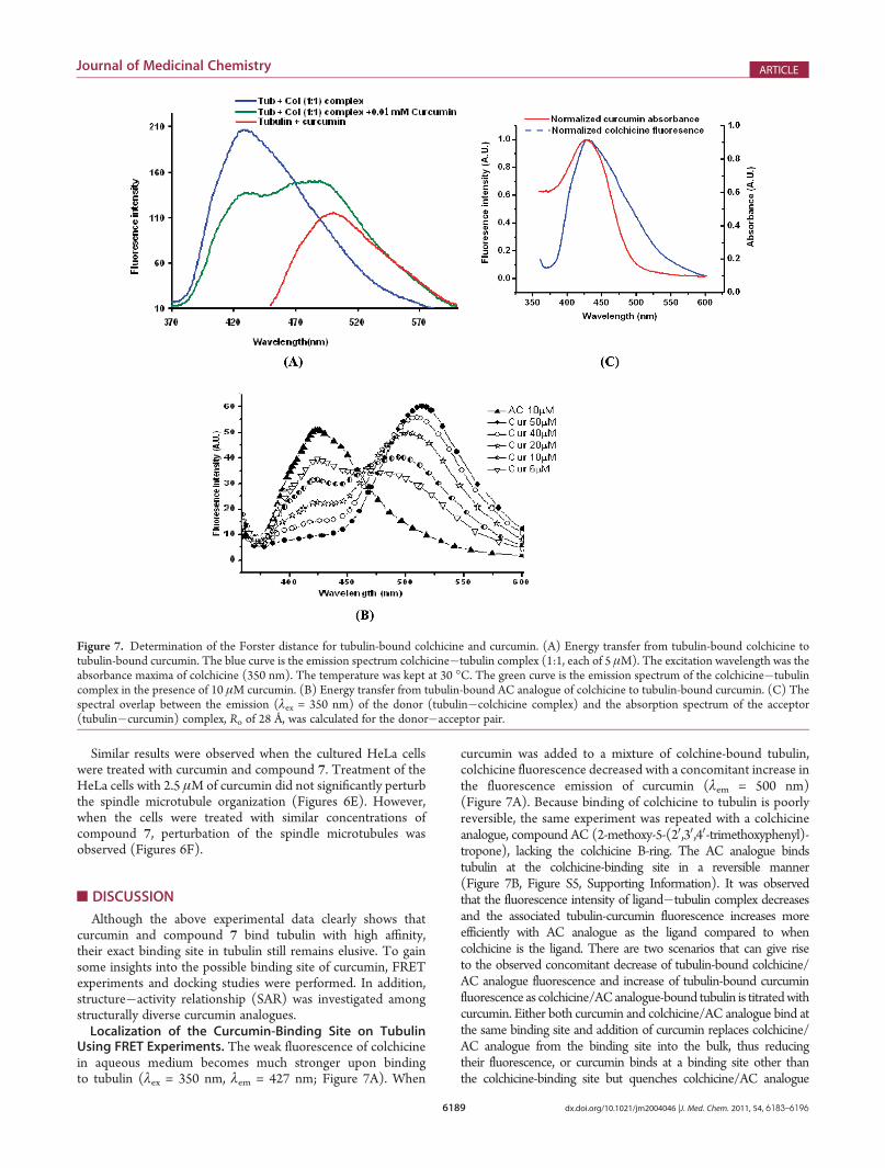
Although the above experimental data clearly shows thatcurcumin and compound 7 bind tubulin with high affinity,their exact binding site in tubulin still remains elusive. To gainsome insights into the possible binding site of curcumin, FRETexperiments and docking studies were performed. In addition,structure�activity relationship (SAR) was investigated amongstructurally diverse curcumin analogues.Localization of the Curcumin-Binding Site on Tubulin
Using FRET Experiments. The weak fluorescence of colchicinein aqueous medium becomes much stronger upon bindingto tubulin (λex = 350 nm, λem = 427 nm; Figure 7A). When
curcumin was added to a mixture of colchine-bound tubulin,colchicine fluorescence decreased with a concomitant increase inthe fluorescence emission of curcumin (λem = 500 nm)(Figure 7A). Because binding of colchicine to tubulin is poorlyreversible, the same experiment was repeated with a colchicineanalogue, compound AC (2-methoxy-5-(20,30,40-trimethoxyphenyl)-tropone), lacking the colchicine B-ring. The AC analogue bindstubulin at the colchicine-binding site in a reversible manner(Figure 7B, Figure S5, Supporting Information). It was observedthat the fluorescence intensity of ligand�tubulin complex decreasesand the associated tubulin-curcumin fluorescence increases moreefficiently with AC analogue as the ligand compared to whencolchicine is the ligand. There are two scenarios that can give riseto the observed concomitant decrease of tubulin-bound colchicine/AC analogue fluorescence and increase of tubulin-bound curcuminfluorescence as colchicine/ACanalogue-bound tubulin is titratedwithcurcumin. Either both curcumin and colchicine/AC analogue bind atthe same binding site and addition of curcumin replaces colchicine/AC analogue from the binding site into the bulk, thus reducingtheir fluorescence, or curcumin binds at a binding site other thanthe colchicine-binding site but quenches colchicine/AC analogue
Figure 7. Determination of the Forster distance for tubulin-bound colchicine and curcumin. (A) Energy transfer from tubulin-bound colchicine totubulin-bound curcumin. The blue curve is the emission spectrum colchicine�tubulin complex (1:1, each of 5 μM). The excitation wavelength was theabsorbance maxima of colchicine (350 nm). The temperature was kept at 30 �C. The green curve is the emission spectrum of the colchicine�tubulincomplex in the presence of 10 μM curcumin. (B) Energy transfer from tubulin-bound AC analogue of colchicine to tubulin-bound curcumin. (C) Thespectral overlap between the emission (λex = 350 nm) of the donor (tubulin�colchicine complex) and the absorption spectrum of the acceptor(tubulin�curcumin) complex, Ro of 28 Å, was calculated for the donor�acceptor pair.

6190 dx.doi.org/10.1021/jm2004046 |J. Med. Chem. 2011, 54, 6183–6196
Journal of Medicinal Chemistry ARTICLE
fluorescence by FRET (curcumin absorbance and colchicine/ACanalogue fluorescence show significant overlap).If the observed colchicine/AC analogue fluorescence quench-
ing indeed arises due to competitive binding between curcuminand colchicine/AC analogue, the addition of a suitable curcuminanalogue, incapable of participating in FRET with tubulin-boundAC analogue, would still quench the fluorescence of tubulin-bound AC analogue. We performed this experiment with com-pound 2 as the curcumin analogue because it exhibits insignif-icant overlap with the emission spectrum of tubulin-bound ACanalogue, ruling out any potential FRET between the two. Inaddition, unlike the parent compound curcumin, the molarextinction coefficient of compound 2 is negligible compared tothe AC analogue above 375 nm, ruling out direct excitation. Wedid not notice any fluorescence quenching (λex = 380 nm) of theAC analogue when compound 2was added to tubulin-bound ACanalogue (Figure S6, Supporting Information), establishing thatcompound 2 (and by extrapolation, curcumin) does not bind tothe colchicine-binding site of tubulin.
Having ruled out the binding of colchicine/AC analogue andcurcumin at the same binding site, we estimated the distancebetween tubulin-bound colchicine and curcumin from FRETstudies. The emission spectrum of tubulin-bound colchicine andthe absorption spectrum of curcumin show substantial overlap(Figure 7C), making the pair amenable to FRET studies. Thecharacteristic Forster distance for the pair was estimated as 28 Å(using k2 = 2/3, n = 1.336, and QD = 0.03, the overlap integral Jwas estimated as 8.5 � 10�14 M�1 cm3). Using this value of Ro(28 Å), the distance between tubulin-bound colchicine andtubulin-bound curcumin was estimated as 32 Å. Strictly speaking,donor�acceptor distance can be estimated from FRET experi-ments by the above method only when the acceptor shows noabsorption at the λex. In our case, curcumin shows a shoulder atλex (350 nm). To circumvent this problem, we also estimated theFRET efficiency for the colchicine�curcumin pair using analternate method that can exclude artifacts arising from theoverlap between acceptor and donor absorption spectra.31 Theestimated energy transfer efficiency (20%) was close to thatobtained using the direct method.
Figure 8. Molecular modeling of compound 7 (A) and curcumin bound tubulin (C) complex, the ligands curcumin and compound 7 (inmagenta) wereplaced between twoαβ-heterodimers (α in orange, andβ in green) and colchicine in red. (B) The local environment of compound 7 (in cyan) with a fewfunctionally important residues shown in blue.

6191 dx.doi.org/10.1021/jm2004046 |J. Med. Chem. 2011, 54, 6183–6196
Journal of Medicinal Chemistry ARTICLE
In summary, using FRET measurements, we have shownthat curcumin and its analogue, compound 2, do not bind tubulinat the colchicine-binding site. The binding site is about 32 Åaway from the colchicine-binding site. Among other canonicalbinding sites on tubulin, the vinblastine-binding site approxi-mately satisfies this distance constraint. Unfortunately, we failedto perform any direct binding assay (competition experiment)between curcumin and vinblastine as both the ligands inducetubulin aggregation (data not shown).Molecular Modeling of Compound 7 Binding Site on
Tubulin. FRET experiment clearly showed that curcumin bindstubulin about 32 Å away from the colchicine-binding site. Toconfirm the FRET results, molecular docking was performed tolocate the probable binding site of compound 7 on tubulin.In the docked structure, compound 7 binds at the interdimer
interface, between the α and the β-subunits of two α,β-dimers,close to the vinblastine binding site (Figure 8A). Residues 96�98from the β-subunit and 251�256 from the α-subunit constitutethe binding site. The pocket also contains H30-helix (residues105�110), T4 and T5 loops (residue 130�133 and 163�165),and the end of helix H110 (residues 407�411).32 The long-itudinal contacts between neighboring monomers in a micro-tubule are mostly mediated through residues 251�256 (end ofT7 loop and the beginning of H8 helix).33 Both the T5 and T7loops are highly dynamic in nature and can accommodate avariety of ligands by changing their conformational state. Tubulin�compound 7 interaction is mainly stabilized by van der Waalscontacts, although the involvement of hydrophobobic stackinginteractions between the phenyl ring of compound 7 and thearomatic rings of β-Trp 407 was also observed (Figure 8B).Interestingly, the calculated distance (34 Å) between colchicineand compound 7 is quite close to the experimentally determineddistance (32 Å). Molecular modeling of curcumin on tubulinalso reveals that both curcumin and compound 7 shares a closebinding site. (Figure 8C)Structure�Activity Relation of Curcumin�Tubulin Inter-
action. Curcumin is a flexible molecule, consisting of twopolyphenol rings connected to the ends of an α,β-unsaturateddiketone moiety. The substitution of diketone with isoxazole,pyrozole, and substituted pyrozole produced compounds thatshowed weaker (than curcumin) inhibition behavior againsttubulin polymerization, consistent with cell-based studies (datanot shown). These results indicate that the diketone moietyparticipates in the binding interaction between curcumin andtubulin. The interaction between different curcumin analoguesand tubulin has been studied using calorimetric as well asfluorescence spectroscopic techniques. The binding study byfluorescence spectroscopy identified two distinct groups withincurcuminoids. Both compounds, 7 and 8, have free diketonemoieties and show structural resemblance with curcumin. To-gether, they also show a characteristic emissionmaxima at amuchhigher wavelength compared to the rest of the analogues.Additionally, compound 7, which has a substituted polyphenolring in between the dicarbonyl moiety, was found to be mosteffective in inhibiting tubulin self-assembly (half-maximum po-lymerization inhibition value 16 μM) and bound tubulin with ahigher affinity compared to the parent compound curcumin(half-maximum polymerization inhibition value 20 μM). It ispossible that the extra steric hindrance caused by the substitutionof a polyphenol in between the diketones in compound 7,compared to curcumin, makes compound 7more conformation-ally constrained. As a result, compound 7 binding to tubulin is
associated with less conformational entropy loss than binding ofcurcumin to tubulin. Substitution of a polyphenol ring inbetween the diketones also produces a tridentate molecule thatcan be anchored to tubulin with higher affinity. It is interesting tonote that acetylation of the phenolic groups at the two termini ofcompound 8 produced an analogue which was less effective ininhibiting tubulin self-assembly (half-maximum polymerizationinhibition value 60 μM), indicating their participation duringbinding to tubulin. The structure�activity relationship studiesalso emphasize the role of the diketone and the phenolic OHgroups in compounds with biological activity. The half moleculesof curcumin (compounds 9�13) were inactive in bindingtubulin as revealed by ITC, indicating that curcumin acts as abifunctional ligand when it binds to tubulin. Recently, Qui et al.have shown18 that arylidene curcumins are more potent antic-ancer agents than curcumin. These drugs are reported to inhibitTNF-αmediated NF-kβ activation. Our observations reveal thatcurcumin and benzylidene analogues of curcumin target tubulinin both in vitro as well as in cell culture experiments. Compellingevidence are available regarding the role of microtubules as an up-regulator of TNF-α mediated NF-kβ signal transduction andgene expression.34 It is known that microtubule depolymeriza-tion triggers deactivation of NF-kβ signal transduction, suggest-ing that by blocking tubulin polymerization, curcumin analoguecan deactivate the NF-kβ mediated signal transductionpathway.35 The data presented here comprehensively show thatcurcumin analogues arrest cancer cell proliferation by binding totubulin.
’CONCLUSION
In this report, we have established that tubulin is one of thetargets of curcumin and elucidated the SAR between tubulin andcurcumin derivatives. The binding site of curcumin in tubulin hasbeen ascertained from both experimental and modeling studies.In summary, the enhanced activity of the benzylidene analoguesof curcumin is attributed to their higher binding affinity towardtubulin. Further modifications of benzylidene analogues maylead to alterations in their binding affinity and associated anti-cancer activity.
’EXPERIMENTAL METHODS
Piperazine-N,N0-bis(2-ethanesulphonic acid) (PIPES), guanosine-50-triphosphate (GTP), ethylene glycol-bis(β-aminoethyl ether) N,N,N0,N0-tetraacetic acid (EGTA), PMSF, demcolchicine, and vinblastinewere purchased from Sigma Chemical Co. All other reagents were ofanalytical grade.Chemistry. Synthesis of Compounds 1�13. All reagents and
solvents used were purchased from the best available commercialsources. Melting points were determined with Barnstead electrothermalapparatus and are uncorrected. Mass spectra were recorded on Micro-mass LCT kc 436 mass spectrometer using a Waters 1525 binary pump.All compounds were more than 99% pure as determined by ShimadzuLC-20AT HPLC system (Figure S7, Supporting Information). TheHPLC analyses were performed on Phenomenex C18 column (250 mm� 4.6 mm, 5 μm). The optimum HPLC separation was achieved onisocratic mode with a mobile phase composed of methanol, acetonitrile,and 0.6% acetic acid aqueous solution (25:20:55, v/v/v) at a flow rate of0.5 mL min�1. 1H and 13C NMR spectral analyses were performed onBruker Avance 300 MHz spectrometer with tetra-methylsilane as theinternal standard (δ ppm). The following abbreviations were used toexplain the multiplicities: s, singlet; d, doublet; t, triplet; dd, double

6192 dx.doi.org/10.1021/jm2004046 |J. Med. Chem. 2011, 54, 6183–6196
Journal of Medicinal Chemistry ARTICLE
doublet; m, multiplet, br,broad. Solvents and reagents were purifiedaccording to standard laboratory techniques.Synthesis of Curcumin Analogues (1�8). The detailed synthetic
methods of curcumin analogues 1�8 are reported by us in our earlierpublication.25
Synthesis of 3-Phenylprop-2-enoic Acid (9). Anhydrous acetic acid(6.3 mL, 0.1089 M) was added to a 100 mL three-necked round-bottomed flask equipped with thermometer and water cooled refluxcondenser. Boron tribromide (2.2 mL, 0.022 M) in anhydrous benzene(5 mL) was gradually added to the above solution in 30 min at 0�4 �C.The reaction mixture was allowed to stir for an hour at room tempera-ture and further for 5�6 h at 55�65 �C to expel hydrogen bromide gas.When the gas emission ceased, a solution of triacetyl borate in benzeneand acetic acid was obtained. Benzene and acetic acid were distilled offfrom the reaction mixture. The resulting residue was cooled to20�30 �C, and DMAP (1.3 g, 0.01M), pyridine (2.412 mL, 0.03M),and NMP (2 mL) were added successively while stirring. Benzaldehyde(Ia) (2.03 mL, 0.02 M) was then added to this reaction mixturecontaining triacetyl borate, which was stirred for 5 min followed byrefluxing for 12 h. The resulting mixture was diluted with water(70�80 mL) and pH of the solution adjusted to 9�10 with 20% NaOHsolution. The unreacted benzaldehyde was removed by distillation withwater under vacuum (30�40mmofHg).Water was added to the resultingresidue and cooled to room temperature, stirred, and filtered. The filtratewas brought to pH 1�2 with 20%HCl solution to precipitate out cinnamicacid. The filtrate was stirred for 2 h at 0�4 �C. The product was filtered,washed with ice-cold water (15�20mL), and dried to yield crude cinnamicacid (IIa). The crude product was recrystallized fromhot water (yield 1.93 g,65%; mp 132�135 �C (Scheme 1)).36
Synthesis of 3-(3,4-Dimethoxyphenyl)prop-2-enoic Acid (10). Insitu generated triacetyl borate, as synthesized above, in benzene and
acetic acid was cooled to 20�30 �C andDMAP (1.3 g, 0.01M), pyridine(2.412mL, 0.03M), andNMP (2mL) were added to it while stirring. Tothe above mixture, 3,4-dimethoxybenzaldehyde (Ib) (3.32 g, 0.02 M)was added stirred for 5 min. Benzene and acetic acid were removed bydistillation from the reaction mixture. The residue was heated at180�190 �C for 9 h and was further subjected to the series of treatmentsdescribed earlier for 9 to obtain crude 3,4-dimethoxycinnamic acid (IIb).Recrystallization from hot water yielded pure compound (yield 2.08 g,50%; mp 181�183 �C (Scheme 1)).
Synthesis of 3-(3,4-Dihydroxyphenyl)prop-2-enoic Acid (11). 3,4-Dimethoxycinnamic acid (IIb) (0.0624 g, 0.3 mM) was dissolved in drydichloromethane (3 mL) and cooled to �78 �C. A solution of BBr3 inDCM (1 M) (2.0 mL) was added dropwise to the above solution. Afterthe addition was over, the reaction mixture was brought to roomtemperature and stirred for 24 h. The reaction mixture was diluted withethyl acetate and its pH was adjusted to ∼8 with aqueous NaOH, andthe layers were separated. The organic layer was washed with water andbrine solution, dried over anhydrous Na2SO4, and vacuum evaporated.The crude product was further purified by column chromatographyusing silica gel (60�120 mesh size) with a 1:5 mixture of ethyl acetateand hexane to give of caffeic acid (IIIc) (yield 1.60 g, 44%; mp223�225 �C (Scheme 1)).
Synthesis of 3-(2-Hydroxyphenyl)prop-2-enoic Acid (12). DMAP(1.3 g, 0.01M), pyridine (2.412 mL, 0.03 M), and NMP (2 mL) wereadded to triacetyl borate generated in situ in benzene and acetic acid(described for IIa) at 20�30 �C. 2-Methoxybenzaldehyde (1d) (2.72 g,0.02 M) was added to the above mixture and stirred for 5�10 min,followed by distillation of benzene and acetic acid from the system. Theresulting residue was heated at 180�190 �C for 9 h and was furthersubjected to the series of treatments described for IIa to obtain2-methoxycinnamic acid (IId). The crude product was recrystallized
Scheme 1. Synthesis of Cinnamic Acids from Acetic Acid and Substituted Benzalaldehydes (IIa�b, IIIc�e)a
aReagents and Conditions: (1) BBr3, benzene, 55�65 �C, 5�6 h; (2) 4-DMAP, pyridine, NMP, 20�30 �C; (3) substituted benzaldehyde, 180�190 �C;(4) 20% HCl; (5) BBr3/DCM, �78�C to rt.

6193 dx.doi.org/10.1021/jm2004046 |J. Med. Chem. 2011, 54, 6183–6196
Journal of Medicinal Chemistry ARTICLE
from hot water with a melting point of 182�186 �C. 2-Methoxycin-namic acid (IId) (0.0534 g, 0.3 mM) thus synthesized was subjected todemethylation as described earlier for IIIc to obtain of 2-hydroxycinnamic acid (IIId) (yield 1.80 g, 54%; mp 217 �C, (Scheme 1)).Synthesis of 3-(2,4-Dihydroxyphenyl)prop-2-enoic Acid (13).To a
solution of triacetyl borate (generated in situ) in benzene and acetic acid(as described for (IIa) at 20�30 �C, DMAP (1.3 g, 0. 01 M), pyridine(2.412 mL, 0.03 M), and NMP (2 mL) were added. 2,4-Dimethoxybenzaldehyde (Ie) (3.32 g, 0.02 M) was added to it, and the system wasstirred for 5�10 min. This reaction mixture was then subjected to thesteps described earlier for the synthesis of IIa, which eventually yielded2,4-dimethoxycinnamic acid (IIe). The melting point of IIe afterrecrystallization from hot water was 192�194 �C. 2,4-Dimethoxycin-namic acid (IIe) was then taken through the demethylation proceduredescribed for the compound IIIc to obtain 2,4 dihydroxycinnamic acid(IIIe) (yield 1.60 g, 44%; mp 201�203 �C (Scheme 1)).3-Phenylprop-2-enoic Acid (9). λmax(MeOH): 273 nm. Retention
time (tR) in the reverse phase HPLC (RP-HPLC) carried out asdescribed for compound 9: tR = 20.38 min. δH (300 MHz, CDCl3)7.8 (1H, d, J = 16.2 Hz), 7.41�7.38 (5H, m), 6.45 (1H, d, J = 15.9 Hz).δC (75.4 MHz, CDCl3) 172.7, 147.0, 133.7, 132.5, 130.5, 127.91, 128.8,128.2, 117.0. ESI-MSm/z: calculated for C9H8O2: 148.05. ESI-MS m/z(�ve mode) found: [(M�H) + 2H2O-H] as precursor to product iontransition with m/z 180.95 adduct during ionization process. A dimericstructure of the above ion predominated the spectrum with m/z 362.8.Analysis (CHN) Found: C, 72.96%; H, 5.4%. Calculated: C, 72.94%; H,5.44% (Figure S7, Supporting Information) .3-(3,4-Dimethoxyphenyl)prop-2-enoic Acid) (10). λmax (MeOH):
285 nm. RP-HPLC tR = 13.87 min. δH (300 MHz, DMSO) 7.46 (1H, d,J = 15.9Hz), 7.09 (1H, J = 1.8Hz), 7.15 (1H, dd, J = 8.4, 2.4Hz), 6.9 (1H,d, J = 8.4 Hz), 6.3 (1H, d, J = 15.9 Hz), 3.72 (3H, d, J = 15 Hz). δC (75.4MHz, DMSO) 169.4, 151.5, 149.6, 145.5, 127.6, 123.6, 117.0, 112.3,110.9, 56.3. ESI-MS m/z: calculated for C11H12O4: 208.2. ESI-MS m/z(�vemode) found: a prominent signal atm/z 254was due to [(M�H)+(�OH) + (�OCH3)] as precursor. CHN analysis Found: C = 63.4%;H = 5.85%. Calculated: C = 63.45%; H = 5.81% (Figure S7, SupportingInformation) .3-(3,4-Dihydroxyphenyl)prop-2-enoic Acid) (11). λmax (MeOH):
320 nm. RP-HPLC tR = 7.56 min. δH (300 MHz, DMSO): 7.40 (1H, d,J = 15.6 Hz), 7.02 (1H, J = 1.8 Hz), 6.9 (1H, dd, J = 8.1, 1.8 Hz), 6.75(1H, d, J = 8.1 Hz), 6.18 (1H, d, J = 15.9 Hz). δC (75.4 MHz, DMSO)169.2, 148.3, 145.6, 125.2, 122.1, 116.2, 115.2, 114.9. ESI-MS m/z:calculated for C9H8O4: 180.05. ESI-MS (�ve mode) found: threeprominent signals at m/z 343, 397, and 451 were found that wereprobably due to dimerization and adduct formation. m/z = 343[(dimer of cinnamaldehyde) + (�OH)-H)], m/z = 397 [(dimer ofcinnamaldehyde) + 3(H2O) + (�OH) �H]. CHN analysis Found:C = 60.00%; H = 4.48%. Calculated: C = 60.00%; H = 4.48% (Figure S7,Supporting Information) .3-(2-Hydroxyphenyl)prop-2-enoic Acid (12). λmax (MeOH):
321 nm. RP-HPLC: tR = 12.37 min. δH (300 MHz, DMSO) 7.79(1H, d, J = 16.2 Hz), 7.49�7.46 (1H, m), 7.2�7.1 (4H, q), 6.4 (1H, d,J = 6.4 Hz). δC (75.4 MHz, DMSO) 169.6, 157.2, 141.08, 133.08,132.73, 129.62, 121.6, 120.7, 118.7, 117.02. ESI-MS m/z: calculated forC9H8O3: 164.1. ESI-MS (�ve mode) found: [(M � H) + (�OH) +2(�OCH3)] as precursor to product ion transition withm/z 242 adductduring ionization process. A dimeric structure of the above ion pre-dominated the spectrum with m/z at 484. CHN analysis Found: C =65.85%; H = 4.91%. Calculated: C = 65.85%; H = 4.91% (Figure S7,Supporting Information) .3-(2,4-Dihydroxyphenyl)prop-2-enoic Acid) (13). λmax (MeOH):
319 nm. RP-HPLC: tR = 7.71 min. δH (300 MHz, DMSO) 7.72 (1H, d,J = 15.9 Hz), 7.32 (1H, d, J = 8.1 Hz), 6.5 (1H, s), 6.3 (1H, dd, J = 1.2,8.4 Hz).δC (75.4MHz, DMSO) 169.8, 160.8, 158.5, 141.0, 130.8, 114.2,
113.3, 108.4, 102.9. ESI-MS m/z: calculated for C9H8O4: 180.05. ESI-MS (�ve mode) found: the main adduct appears due to a dimericcinnamaldehyde quasimolecular ion, possibly associated with one waterand three methanol molecules through hydrogen bonding at m/z 441.The other prominent adduct signals at m/z 230, 330, 417, 527, and 616appear due to water and methanol molecules associated with monomer,dimer, andmultimers in varying composition. CHN analysis Found: C =60.00%; H = 4.44%. Calculated: C = 60.00%; H = 4.48% (Figure S7,Supporting Information).Tubulin Isolation and Estimation. Microtubular proteins were
isolated from goat brains by two cycles of a temperature-dependentassembly disassembly process. Pure tubulin was isolated from micro-tubular proteins by two additional cycles of temperature-dependentpolymerization and depolymerization using 1 M glutamate buffer forassembly.37 The composition of the assembly buffer was 50 mM PIPES,pH 7, 1 mM EGTA, 0.5 mMMgCl2, and 0.5 mMGTP. The protein wasstored at�80 �C. The protein concentration was determined by Lowrymethod using bovine serum albumin as standard. Tubulin preparationsused in this study contained natural mixture of isoforms.38 Bothcalorimetry and fluorescence measurements were carried out with thisunfractionated tubulin, and therefore the binding parameters obtainedhere are averages for the different isoforms.Tubulin Polymerization Assay. Pure tubulin in PEM (50 mM
PIPES, pH 7, 1 mM EGTA, 0.5 mM MgCl2) buffer was polymerized at37 �C in the presence of 1 mMGTP. Polymerization was initiated using10% dimethyl sulfoxide, and the turbidity was measured by theabsorbance at 360 nm. A Shimadzu UV-160 double beam spectro-photometer, fitted with a temperature controlled circulating water bathaccurate to (0.2 �C, was used for this purpose. Half-maximumpolymerization inhibition values were calculated using concentrationof the drug that caused 50% inhibition of the polymer mass. Each testwas performed in triplicate, and the half-maximum polymerizationinhibition values reported represents the result of at least two repeti-tions. The slight difference in half-maximum polymerization inhibitionvalues between our results and others could be due to slight variations inexperimental conditions, drug purity, and solvent condition.Fluorescence Measurement. The binding of the ligands to the
protein was monitored by enhancement of curcumin fluorescence in thepresence of tubulin. Fluorescence spectra were recorded using a HitachiF-3000 fluorescence spectrophotometer connected to a constant tem-perature circulating water bath accurate to (0.2 �C. All fluorescencemeasurements were carried out in a 0.5 cm path-length quartz cuvette,and fluorescence values were corrected for the inner filter effect using thefollowing equation:
Fcor ¼ FobsantilogðAex þ AemÞ
2
� �
where Aex andAem are the absorbances of the ligand at the excitation andthe emission wavelength. The complexes were excited at their char-acteristic wavelength maxima, which were 330 nm for compounds 2�6,427 nm for curcumin and compound 8, and 370 nm for compound 7. Inall the experiment set up excitation and emission band-pass were 5 nm.
For small molecules binding to protein, the equilibrium between freeand bound molecules is given by the equation39
logðFo � FÞ
F¼ log Kb þ nlog½Q �
where Fo and F denotes the steady-state fluorescence intensities in theabsence and in the presence of the quencher (compound 7 andcurcumin); Kb is the binding constant, n is the number of binding sites,and [Q] is the concentration of compound 7 and curcumin, respectively.Isothermal Titration Calorimetry (ITC). ITC measurements
were performed on a VP-ITC Micro Calorimeter of Micro Cal Inc.,(MA, USA). Tubulin was dialyzed extensively against PEM buffer with

6194 dx.doi.org/10.1021/jm2004046 |J. Med. Chem. 2011, 54, 6183–6196
Journal of Medicinal Chemistry ARTICLE
GDP (to offer stabilization) and the ligand (curcumin and its halfanalogue) dissolved in the last dializant. The pH of the tubulin and theligand solutions was made identical before loading into the calorimeter.A typical titration involved 25 injections (10 injection in case ofcompound 10) of ligand (10 μL aliquots per shot), at 3 min intervals,into the sample cell (volume 1.4359 mL) containing tubulin. Thetitration cell was kept at a definite temperature and stirred continuouslyat 310 rpm. The data were then analyzed to determine the bindingstoichiometry (N), affinity constant (Ka), and thermodynamic para-meters of the reaction, using Origin 5.0 software.Cell Culture and Maintenance. Human lung epithelium adeno-
carcinoma cells (A549) and human cervical carcinoma cells (HeLa)were maintained in DMEM medium supplemented with 1 mML-glutamine, 10% fetal bovine serum, 50 μg/mL penicillin, 50 μg/mLstreptomycin, and 2.5 μg/mL amphotericin B. Cells were cultured at37 �C in a humidified atmosphere containing 5% CO2. Cells weregrown in tissue culture flasks until they were 80% confluent beforetrypsinisation with 1� trypsin and splitting. The morphology of controland treated cells was observed by Olympus inverted microscopemodel CKX41.Cell Proliferation Inhibition Assay (MTT Assay). Viability of
the A549 and HeLa cells, in the presence of varying concentrations ofcurcumin and compound 7were assessed byMTT assay. Cultured A549and HeLa cells were grown in 96-well culture plates (1� 104 cells perwell), treated with different concentrations of curcumin (0�50μM) andcompound 7 (0�50 μM), and incubated for 48 h. After incubation, 50μL of MTT (2 mg/mL) solution in PBS were added to each well. Thiswas incubated until a purple precipitate was visible. The absorbance wasmeasured on an ELISA reader (MultiskanEX, Lab systems, Helsinki,Finland) at a test wavelength of 570 nm and a reference wavelength of650 nm.Cell Cycle Analysis by Flow Cytometry. HeLa cells (1 � 106
cells/ml) were treated without and with 2 μM nocodazole for 20 h.Nocodazole was washed off, and fresh medium containing 5 μMcurcumin and 5 μM compound 7 was added. The untreated and treatedcells were incubated for 8 h, fixed in ice chilled methanol for at least 30min in 4 �C, and incubated for 4 h at 37 �C in a PBS solution containing 1mg/mL RNase A. The nuclear DNA was then labeled with propidiumiodide (PI). Cell cycle analysis was performed using the BectonDickinson FACS Caliber, and the data were analyzed using Cell Questprogram from Becton Dickinson.Analysis for Apoptosis by Flow Cytometry. Apoptosis was
measured using flow cytometry by annexin V (1μg/mL) and PI (0.5 μg/mL) double staining. Around 1� 106 A549 and HeLa cells were treatedwith curcumin (0�5 μM) and compound 7 (0�5 μM), respectively,and incubated for 48 h. Cells were then stained for 15 min at roomtemperature in the dark with fluorescein isothiocyanate (FITC)-con-jugated annexin V (1 μg/mL) and PI (0.5 μg/mL) in a Ca2+-enrichedbinding buffer. Annexin V-FITC and PI emissions were detected in theFL1 and FL2 channels of a FACSCaliber flow cytometer, using emissionfilters of 525 and 575 nm, respectively. For each sample, 10000 cells werecounted. Apoptosis analysis was performed using the Becton DickinsonFACS Caliber. The data were analyzed using Cell Quest program fromBecton Dickinson.Sample Preparation for Confocal Microscopy. Cultured
A549 and HeLa cells were grown at a density of 106 cells/mL andincubated in the presence of curcumin (0�2.5 μM) and compound 7(0�2.5 μM), respectively, for 24 h. Subsequently cells were washedtwice by PBS, fixed with 2% para-formaldehyde, and incubated withpermeable solution (0.1% Na citrate, 0.1% Triton) for 1 h. Cells werethen mildly washed with PBS, and the nonspecific binding sites wereblocked by incubating the cells with 5% BSA. Subsequently, cells wereincubated with antimouse monoclonal anti-α-tubulin antibody (1:200dilutions, Sigma, USA), followed by anti mouse rhodamine conjugated
IgG antibody (1:150 dilutions, GeNei, India) and DAPI (1 μg/mL).After incubation, cells were washed with PBS and viewed under a ZiessLSM 510 Meta confocal microscope.Fluoresence Resonance Energy Transfer (FRET). The effi-
ciency (E) of FRET between curcumin and colchicine, both tubulin-bound, was estimated from curcumin fluorescence quenching studies inpresence of tubulin (λex = 350 nm; λem = 427 nm; PEM buffer pH 7.0;30 �C). Fluorescence intensities of 5 μMcolchicine, in absence (Fo), andin presence of 10 μM of curcumin (F), yielded E = 1 � F/Fo. Thedistance (r) between tubulin-bound colchicine and curcumin wassubsequently estimated from the relationship: E = (Ro
6)/(Ro6 + r6),
where Ro is the Forster distance between the donor (colchicine) and theacceptor (curcumin). Ro (in Å) was estimated using the followingrelationship:
Ro ¼ 9:78� 103 k2n�4QD
ZFDðλÞεAðλÞλ4 dλ
� �" #1=6
where k2 is the orientation factor, n is the refractive index of themedium,QD is the fluorescence quantum yield of the donor in absence of theacceptor, FD(λ) is the corrected (and normalized) fluorescence intensityof the donor, and εA(λ) is the molar extinction coefficient of theacceptor.Molecular Modeling Study. The crystal structure tubulin com-
plexed with stathmin-like domain (PDB (Protein Data Bank) ID:1SA0)40 was used to obtain the model structure. The energy minimizedatomic coordinates of compound 7was generated using Corina (http://www.molecular-networks.com/online_demos/corina). Docking mod-els were obtained using the PatchDock.41 Patchdock is a geometry-basedmolecular docking algorithm. It divides interacting molecules surfacesinto patches according to the surface shape (concave, convex, or flat).Then it applies the geometric hashing algorithm to match concavepatches with convex patches and flat patches with flat patches andgenerates a set of candidate transformations. Each candidate transfor-mation is further evaluated by a set of scoring functions that estimateboth the shape complementarity and the atomic desolvation energy42 ofthe obtained complex. Finally, redundant solutions are discarded by theapplication of an rmsd (root-mean-square deviation) clustering. Patch-Dock is highly efficient because it utilizes advanced data structures andspatial pattern detection techniques which are based on matching oflocal patches. The local shape information is then extended andintegrated to achieve global solutions. The algorithm implicitly ad-dresses surface flexibility by allowing minor penetrations. The bestsolution was retained for further analysis. The solution obtained usingPatch Dock was further verified using other docking programs likeGOLD43 and Auto Dock (http://autodock.scripps.edu/resources).PyMol (http://www.pymol.org) was used for visualization and for theidentification of residues in the binding pocket (within a distance of 4.5Å of the drug).
’ASSOCIATED CONTENT
bS Supporting Information. Additional experimental de-tails; effect of different curcumin analogue on tubulin polymer-ization; comparative effect of curcumin and compound 7stimulated mitotic delay on nocodazole treated HeLa cells;nuclear aberration in A549 cells by compound 7; effect ofcurcumin and compound 7 on cellular morphology of HeLaand A549 cells; chemical structure of colchicines and ACcolchicines; compound 2 binding on tubulin�AC complex; 1HNMR, 13CNMR,MS, and HPLC spectra of single ring curcuminanalogue. This material is available free of charge via the Internetat http://pubs.acs.org.

6195 dx.doi.org/10.1021/jm2004046 |J. Med. Chem. 2011, 54, 6183–6196
Journal of Medicinal Chemistry ARTICLE
’AUTHOR INFORMATION
Corresponding Authors*For B.B.: phone, 91-33-2337-9544; E-mail: [email protected]. For A.S.: phone, 91-11- 26717121; E-mail, [email protected].
’ACKNOWLEDGMENT
This work is supported in part by grant from the Departmentof Atomic Energy (DAE) to B. Bhattacharyya as a Raja Ramannafellow.
’ABBREVIATIONS USED
PIPES, piperazine-N,N0-bis(2-ethanesulphonic acid); EGTA,ethylene glycol-bis(β-aminoethyl ether)N,N,N0,N0-tetraacetic acid;MgCl2, magnesium chloride; GTP, guanosine-50-triphosphate;PDB, Protein Data Bank; ITC, isothermal titration calorimetry;AC, [2-methoxy-5-(20,30,40-trimethoxyphenyl)tropone]; SAR,structure�activity relationship; FRET, fluorescence resonanceenergy transfer
’REFERENCES
(1) Correia, J. J.; Lobert, S. Physiochemical aspects of tubulin-interacting antimitotic drugs. Curr. Pharm. Des. 2001, 7, 1213–1228.(2) Jordan, A.; Hadfield, J. A.; Lawrence, N. J.; McGown., A. T.
Tubulin as a target for anticancer drugs: agents which interact with themitotic spindle. Med. Res. Rev. 1998, 18, 259–296.(3) Chen, G. K.; Dur�an, G. E.; Mangili, A.; Beketic-Oreskovic, L.;
Sikic, B. I. MDR1 activation is the predominant resistance mechanismselected by vinblastine in MES-SA cells. Br. J. Cancer 2000, 83, 892–896.(4) Orr, G. A.; Verdier-Pinard, P.; McDaid, H.; Horwitz, S. B.
Mechanisms of taxol resistance related to microtubules. Oncogene2003, 22, 7280–7295.(5) Pannacciulli, I.; Ballarino, P.; Castello, G.; Arboscello, E.; Botta,
M.; Tredici, S.; Lerza., R. In vitro toxicity of taxol based anticancer drugcombinations on human hemopoietic progenitors. Anticancer Res. 1999,19, 409–412.(6) Sun, W.; Wang, W.; Kim, J.; Keng, P.; Yang, S.; Zhang, H.; Liu,
C.; Okunieff, P.; Zhang, L. Anti-cancer effect of resveratrol is associatedwith induction of apoptosis via a mitochondrial pathway alignment. Adv.Exp. Med. Biol. 2008, 614, 179–186.(7) Mukherjee, S.; Acharya, B. R.; Bhattacharyya, B.; Chakrabarti, G.
Genistein arrests cell cycle progression of A549 cells at the G2/M phaseand depolymerizes interphase microtubules through binding to a uniquesite of tubulin. Biochemistry 2010, 49, 1702–1712.(8) Sarkar, F. H.; Li, Y. Harnessing the fruits of nature for the
development of multitargeted cancer therapeutics. Cancer Treat. Rev.2009, 35, 597–607.(9) Dhillon, N.; Aggarwal, B. B.; Newman, R. A.; Wolff, R. A.;
Kunnumakkara, A. B.; Abbruzzese, J. L.; Ng, C. S.; Badmaev, V.;Kurzrock, R. Phase II trial of curcumin in patients with advancedpancreatic cancer. Clin. Cancer. Res. 2008, 14, 4491–4499.(10) Aggarwal, B. B.; Sung, B. Pharmacological basis for the role of
curcumin in chronic diseases: an age-old spice with modern targets.Trends Pharmacol. Sci. 2009, 30, 85–94.(11) Goel, A.; Kunnumakkara, A. B.; Aggarwal, B. B. Curcumin as
“Curecumin”: from kitchen to clinic. Biochem. Pharmacol. 2008,75, 787–809.(12) Kawamori, T.; Lubet, R.; Steele, V. E.; Kelloff, G. J.; Kaskey,
R. B.; Rao, C. V.; Reddy, B. S. Chemopreventive effect of curcumin, anaturally occurring antiinflammatory agent, during the promotion/progression stages of colon cancer. Cancer. Res. 1999, 59, 597–601.(13) Mackenzie, G. G.; Queisser, N.; Wolfson, M. L.; Fraga, C. G.;
Adamo, A. M.; Oteiza, P. I. Curcumin induces cell-arrest and apoptosis
in association with the inhibition of constitutively active NF-kappaB andSTAT3 pathways in Hodgkin’s lymphoma cells. Int. J. Cancer 2008,123, 56–65.
(14) Van Erk, M. J.; Teuling, E.; Staal, Y. C.; Huybers, S.; VanBladeren, P. J.; Aarts, J. M.; Van Ommen, B. Time- and dose-dependenteffects of curcumin on gene expression in human colon cancer cells.J Carcinog. 2004, 3, 8.
(15) Jana, N. R.; Dikshit, P.; Goswami, A.; Nukina, N. Inhibition ofproteasomal function by curcumin induces apoptosis through mito-chondrial pathway. J. Biol. Chem. 2004, 279, 11680–11685.
(16) Anto, R. J.; Mukhopadhyay, A.; Denning, K.; Aggarwal, B. B.Curcumin (diferuloylmethane) induces apoptosis through activation ofcaspase-8, BID cleavage and cytochrome c release: its suppression byectopic expression of Bcl-2 and Bcl-xl. Carcinogenesis 2002, 23, 143–150.
(17) Aggarwal, B. B.; Kumar, A.; Bharti, A. C. Anticancer potential ofcurcumin: preclinical and clinical studies. Anticancer Res. 2003,23, 363–298.
(18) Qiu, X.; Du, Y.; Lou, B.; Zuo, Y.; Shao, W.; Huo, Y.; Huang, J.;Yu, Y.; Zhou, B.; Du, J.; Fu, H.; Bu, X. Synthesis and identification of new4-arylidene curcumin analogs as potential anticancer agents targetingnuclear factor-kB signaling pathway. J. Med. Chem. 2010, 53, 8260–8273.
(19) Gupta, K. K.; Bharne, S. S.; Rathinasamy, K.; Naik, N. R.; Panda,D. Dietary antioxidant curcumin inhibits microtubule assembly throughtubulin binding. FEBS J. 2006, 273, 5320–5332.
(20) Banerjee, M.; Singh, P.; Panda, D. Curcumin suppresses thedynamic instability of microtubules, activates the mitotic checkpoint andinduces apoptosis in MCF-7 cells. FEBS J. 2010, 277, 3437–3448.
(21) Fourest-Lieuvin, A.; Peris, L.; Gache, V.; Garcia-Saez, I.; Juillan-Binard, C.; Lantez, V.; Job, D. Microtubule regulation in mitosis: tubulinphosphorylation by the cyclin-dependent kinase Cdk1. Mol. Biol. Cell2006, 17, 1041–1050.
(22) Hearn, B. R.; Shaw, S. J.; Myles, D. C. Microtubule targetingagents. Compr. Med. Chem. II 2007, 7, 81–110.
(23) RayChaudhuri, A.; Tomita, I.; Mizuhashi, F.; Murata, K.;Ludue~na, R. F. Distinct and overlapping binding sites for IKP104 andvinblastine on tubulin. J. Protein Chem. 1998, 17, 685–690.
(24) Wang, Y. J.; Pan,M. H.; Cheng, A. L.; Lin, L. I.; Ho, Y. S.; Hsieh,C. Y.; Lin, J. K. Stability of curcumin in buffer solutions and character-ization of its degradation products. J. Pharm. Biomed. Anal. 1997,12, 1867–1876.
(25) Mishra, S.; Karmodiya, K.; Surolia, N.; Surolia, A. Synthesis andexploration of novel curcumin analog as antimalarial agents. Bioorg. Med.Chem. 2008, 16, 2894–2902.
(26) Sch€on, A.; Madani, N.; Smith, A. B.; Lalonde, J. M.; Freire, E.Some binding-related drug properties are dependent on thermodynamicsignature. Chem. Biol. Drug Des. 2011, 77, 161–165.
(27) Perozzo, R.; Folkers, G.; Scapozza, L. Thermodynamics ofprotein�ligand interactions: history, presence, and future aspects.J. Recept. Signal Transduction Res. 2004, 24, 1–52.
(28) Kauzmann, W. Some factors in the interpretation of proteindenaturation. Adv. Protein Chem. 1959, 14, 1–63.
(29) Baldwin, R. L. Temperature dependence of the hydrophobicinteraction in protein folding. Proc. Natl. Acad. Sci. U.S.A. 1986, 83,8069–8072.
(30) Freire, E. Do enthalpy and entropy distinguish first in class frombest in class? Drug Discovery Today 2008, 13, 869–874
(31) Eisinger, J. Intermolecular energy transfer in adrenocorticotro-pin. Biochemistry 1969, 8, 3902–3908.
(32) L€owe, J.; Li, H.; Downing, K. H.; Nogales, E. Refined structure ofalpha beta-tubulin at 3.5 Å resolution. J. Mol. Biol. 2001, 313, 1045–1057.
(33) Dorl�eans, A.; Gigant, B.; Ravelli, R. B.; Mailliet, P.; Mikol, V.;Knossow, M. Variations in the colchicine-binding domain provideinsight into the structural switch of tubulin. Proc. Natl. Acad. Sci. U.S.A.2009, 106, 13775–13779.
(34) Jackman, R. W.; Rhoads, M. G.; Cornwell, E.; Kandarian, S. C.Microtubule-mediated NF-kappaB activation in the TNF-alpha signal-ing pathway. Exp. Cell. Res. 2009, 315, 3242–3249.

6196 dx.doi.org/10.1021/jm2004046 |J. Med. Chem. 2011, 54, 6183–6196
Journal of Medicinal Chemistry ARTICLE
(35) Mackenzie, G. G.; Keen, C. L.; Oteiza, P. I. Microtubules arerequired for NF-kappaB nuclear translocation in neuroblastoma IMR-32cells: modulation by zinc. J. Neurochem. 2006, 99, 402–415.(36) Chiriac, I. C.; Tanasa, F.; Onciu, M. A Novel approach in
cinnamic acid synthesis: direct synthesis of cinnamic acids from aromaticaldehydes and aliphatic carboxylic acids in the presence of borontribromide. Molecules 2005, 10, 481–487.(37) Lin, C. M.; Hamel, E. Effects of inhibitors of tubulin polymer-
ization on GTP hydrolysis. J. Biol. Chem. 1981, 256, 9242–9245.(38) Banerjee, A.; Luduena, R. F. Kinetics of association and
dissociation of colchicine�tubulin complex from brain and renaltubulin. Evidence for the existence of multiple isotypes of tubulin inbrain with differential affinity to colchicine. FEBS Lett. 1987,219, 103–107.(39) Abou-Zied, O. K.; Al-Shihi, O. I. Characterization of subdomain
IIA binding site of human serum albumin in its native, unfolded, andrefolded states using small molecular probes. J. Am. Chem. Soc. 2008,130, 10793–10801.(40) Ravelli, R. B.; Gigant, B.; Curmi, P. A.; Jourdain, I.; Lachkar, S.;
Sobel, A.; Knossow, M. Insight into tubulin regulation from a complexwith colchicine and a stathmin-like domain.Nature 2004, 428, 198–202.(41) Schneidman-Duhovny, D.; Inbar, Y.; Nussinov, R.; Wolfson,
H. J. PatchDock and SymmDock: servers for rigid and symmetricdocking. Nucl. Acids. Res. 2005, 33, 363–367.(42) Benyamini, H.; Shulman-Peleg, A.; Wolfson, H. J.; Belgorodsky,
B.; Fadeev, L.; Gozin, M. Interaction of C(60)-fullerene and carboxyful-lerene with proteins: docking and binding site alignment. BioconjugateChem. 2006, 17, 378–386.(43) Jones, G.; Willett, P.; Glen, R. C.; Leach, A. R.; Taylor, R.
Development and validation of a genetic algorithm for flexible docking.J. Mol. Biol. 1997, 267, 727–748.
Copyright © 2022 FDOKUMEN