Research article Plant species accumulation curves ... - arXiv

Summary. Concurrently with cold-induced disintegration of microtubu-lar structures in the cytoplasm, gradual tubulin accumulation was ob-served in a progressively growing proportion of interphase nuclei intobacco BY-2 cells. This intranuclear tubulin disappeared upon rewarm-ing. Simultaneously, new microtubules rapidly emerged from the nuclearperiphery and reconstituted new cortical arrays, as was shown by im-munofluorescence. A rapid exclusion of tubulin from the nucleus duringrewarming was also observed in vivo in cells expressing GFP-tubulin.Nuclei were purified from cells that expressed GFP fused to an endo-plasmic-reticulum retention signal (BY-2-mGFP5-ER), and green-fluo-rescent protein was used as a diagnostic marker to confirm that thenuclear fraction was not contaminated by nuclear-envelope proteins.These purified, GFP-free nuclei contained tubulin when isolated fromcold-treated cells, whereas control nuclei were void of tubulin. Further-more, highly conserved putative nuclear-export sequences were identi-fied in tubulin sequences. These results led us to interpret theaccumulation of tubulin in interphasic nuclei, as well as its rapid nuclearexport, in the context of ancient intranuclear tubulin function during thecell cycle progression.
Keywords: Tobacco Bright Yellow 2; Microtubule; Nucleus; Plant cell;Tubulin.
Abbreviations: NLS nuclear-localization sequence; NES nuclear-exportsequence.
Introduction
The microtubular cytoskeleton plays a dual role during theprogression of the cell cycle. In interphase, microtubulesare exclusively found in the cytoplasm, where they partic-ipate in intracellular transport, distribution of organelles,
and maintenance of cell shape. Depending on the phase ofthe cell cycle, microtubules are organized in differentstructures – cortical microtubular arrays at G1, corticaland endoplasmic microtubules at S and G2 phase, pre-prophase band of microtubules at late G2 phase – being,however, always confined to the cytoplasm and separatedfrom the karyoplasm. When the nuclear envelope disinte-grates at the onset of mitosis, the organized scaffold ofmicrotubules disappears. The division spindle is estab-lished and the major role of microtubules is to ensure anequal distribution of genetic material between the twodaughter cells. After the completion of nuclear divisionand reformation of the nuclear envelope, a new cytoplas-mic interphase array of microtubules is reestablished(Hasezawa and Kumagai 2002).
The microtubular structures listed above differ not onlywith respect to their timing but also with respect to theirnucleation. In contrast to animal and plant cells, someprotists, fungi, and algae perform a so-called closed mito-sis, where nuclear division takes place within an intact nu-clear envelope (for a review, see Heath 1980). In cellswith closed mitosis, the entry of tubulin and microtubule-nucleating components into the nucleus at the onset of mi-tosis, as well as their exclusion at the end, must be understrict cell cycle control. For example, in Saccharomycescerevisiae, microtubules of the mitotic spindle are orga-nized inside the nucleus by a specialized structure, thespindle pole body (SPB) (Pereira et al. 1998). It has beenshown that the SPB assembles in the cytoplasm and istransported in a cell cycle-dependent manner into the nucleus via the nuclear-localization sequence (NLS) of
Protoplasma (2006) 227: 185–196DOI 10.1007/s00709-005-0139-x PROTOPLASMA
Printed in Austria
Intranuclear accumulation of plant tubulin in response to low temperature
K. Schwarzerová1,*, J. Petrásek1,2, K. C. S. Panigrahi 3, S. Zelenková1, Z. Opatrny1, and P. Nick4
1 Department of Plant Physiology, Faculty of Science, Charles University, Prague2 Institute of Experimental Botany, Academy of Sciences of the Czech Republic, Prague3 Institut für Biologie II, University of Freiburg, Freiburg4 Botanisches Institut 1, University of Karlsruhe, Karlsruhe
Received February 14, 2005; accepted June 4, 2005; published online May 3, 2006© Springer-Verlag 2006
* Correspondence and reprints: Department of Plant Physiology, Faculty ofScience, Charles University, Vinicná 5, 128 44 Prague 2, Czech Republic.E-mail: [email protected]
Spc98p protein (Pereira et al. 1998). Interestingly, �- and�-tubulin, the basic components of the mitotic spindle, arenot found in the nucleus of cells with closed mitosis dur-ing interphase. Therefore, in organisms with closed mito-sis, tubulin has to be transported into and out of thenucleus. Indeed, it has been shown in Aspergillus nidulansthat tubulin enters the nucleus immediately before the on-set of closed mitosis and is quickly excluded at the end(Ovechkina et al. 2003). Although the molecular mecha-nisms of nuclear tubulin transport remain to be elucidated,these results suggest that the movement of tubulin throughthe nuclear envelope during the cell cycle is an active andhighly regulated process.
The mitotic spindle microtubules of animals, higherplants (Embryobionta), and charophycean green algae(Charophyceae) interact with chromosomes after the nuclearenvelope has disintegrated at the onset of mitosis (Heath1980). Consequently, directed movement of tubulin throughthe nuclear envelope is not required for formation of the mitotic spindle. After nuclear envelope disintegration, themicrotubular spindle can exploit the pool of cytoplasmictubulin that is no longer withdrawn by compartmentaliza-tion. The spindle microtubules are nucleated from centro-somes in animal and algal cells (Wiese and Zheng 1999),but from rather diffuse microtubule-organizing centers(MTOCs) in the acentriolar cells of higher plants (Baskinand Cande 1990, Shimamura et al. 2004). In addition, thekinetochores of both animal and plant cells are endowedwith a microtubule-nucleating activity (Cande 1990). �-Tubulin, a minus-end nucleator of microtubule assembly,is an indispensable component of both centrosomes andplant MTOCs (Pereira and Schiebel 1997, Stoppin-Melletet al. 2000). In plant cells, the association of �-tubulinwith the prospective kinetochore sites already occurs dur-ing the G2 phase and thus precedes the disintegration ofthe nuclear envelope (Binarová et al. 2000). This indicatesthat, like the SPB of S. cerevisiae, the microtubule-nucle-ating component �-tubulin must be actively transportedthrough the intact nuclear envelope before mitosis. Again,neither the molecular mechanism of �-tubulin transportinto the nucleus is known nor whether other componentsof MTOCs persist in the nucleus during the whole cell cy-cle or have to be imported as well.
In our previous work, we followed the response of themicrotubular and actin cytoskeleton to low temperature intobacco cells (Pokorná et al. 2004). We observed a pro-gressive disintegration of microtubules during prolongedexposure of tobacco BY-2 cells to 0 °C and a reassemblyof microtubules during subsequent recovery at 25 °C. Inthe present work, we show by means of several methods
that cold-induced disintegration of microtubules is accom-panied by the entry of tubulin into the interphase nucleiand its gradual accumulation there. Upon rewarming,tubulin was quickly excluded from the nuclei and immedi-ately polymerized into cytoplasmic microtubules such thatfree tubulin became undetectable in most nuclei within afew minutes of incubation at 25 °C. The accumulation oftubulin in nuclei and its exclusion from them were shownby immunofluorescence staining and also in vivo in cellsthat expressed GFP–�-tubulin. Furthermore, increasinglevels of �-tubulin could be detected in isolated andhighly purified nuclei during cold treatment. We interpretthese findings first of all as evidence for a reversible per-meabilization of the nuclear envelope for tubulin, possiblyvia altered function of nuclear pore complexes (NPCs).Further, these data indicate intranuclear tubulin-bindingsites that are either unmasked or activated under certainenvironmental conditions. The subsequent quick exclu-sion of tubulin from interphase plant nuclei during rewarming could be explained by the recovery of tubulintransport between karyo- and cytoplasm and seems to bean active process. This is supported by the identificationof five putative signatures for nuclear export in sequencesof plant �- and �-tubulin. This transport might be func-tionally relevant for the dynamic changes in the nuclearenvelope at the beginning and end of mitosis.
Material and methods
Plant material and chemicals
The tobacco cell line BY-2 (Nicotiana tabacum L. cv. Bright Yellow 2[Nagata et al. 1992]) was cultured in medium containing 4.3 g ofMurashige–Skoog salts (Sigma, St. Louis, Mo., U.S.A.), 1 mg of thi-amine, 200 mg of KH2PO4, 100 mg of myo-inositol, 30 g of sucrose, and0.2 mg (0.9 �M) of 2,4-dichlorophenoxyacetic acid (2,4-D) per liter,pH 5.8. The transgenic BY-2 cell lines GT16, expressing GFP–�-tubulin(Kumagai et al. 2001), and BY-2-mGFP-ER, expressing mGFP5-ER(Petrásek et al. 2003), were maintained on the same medium supple-mented with 100 �g of kanamycin and 100 �g of cefotaxim per ml.Every 7 days, 1.5 ml of cells were transferred to 30 ml of fresh mediumand cultured in darkness at 25 °C on an orbital shaker (IKA KS501; IKALabortechnik, Staufen, Federal Republic of Germany; 120 rpm; orbitaldiameter, 30 mm). All chemicals were obtained from Sigma unlessstated otherwise.
Cold treatment and recovery experiments
Cells in the exponential phase of growth (3 days after inoculation) wereused for all experiments. Cell suspensions in Erlenmeyer flasks wereplaced in a bath of ice water to maintain the temperature at 0 °C andshaken on an orbital shaker at 100 rpm (IKA KS501) in darkness. For re-covery experiments, flasks were taken from the ice water and the cellswere immediately collected by filtering through a nylon mesh (mesh di-ameter, 20 �m). They were then resuspended in conditioned medium atoptimal temperature (25 °C) that had been obtained by sterile filtration
186 K. Schwarzerová et al.: Nuclear accumulation of plant tubulin
of 3-day-old BY-2 cells. Thereafter, the cells were cultivated at 25 °C.Samples of cells for cytological observation or extraction of proteinswere taken at specified time points during the cold treatment and subse-quent recovery at normal temperature.
Observation of tubulin exclusion from nuclei in vivo
The tobacco cell line expressing GFP-tubulin was cultivated at 0 °C asdescribed above. For observation under the confocal laser scanning mi-croscope, a drop of cell suspension was collected from the flask andplaced on a prewarmed (25 °C) microscopic slide. The drop of cell sus-pension was covered immediately with a coverslip and observed under aconfocal laser scanning microscope (Leica TCS NT, Heidelberg, FederalRepublic of Germany). The optimal confocal plane through the nuclearregion of a selected cell or cell file was defined and a time-lapse se-quence was recorded at this confocal plane. The time-lapse scanning wasrecorded over 20 min at 1 min intervals.
To measure the exclusion of tubulin from the nucleus and its accumu-lation in the cytoplasm around the nucleus during the recovery period,the first and last confocal sections from time-lapse series were analyzed.The intensity of the tubulin signal was measured by the Scion Imageprogram (Scion Corporation, Frederick, Md., U.S.A.) using a linearprobe (profile plot line width, 6) that was placed so as to cover the nu-clear region, the cytoplasm around the nucleus, and the vacuolar regionwith one probe. Intensity profiles were plotted onto graphs and the re-spective parts of curves were labeled as nucleus, cytoplasm, and vacuole.
Visualization of microtubules
Microtubules were visualized as described by Wick et al. (1981) with themodifications described by Mizuno (1992). Briefly, 3 ml of cell suspen-sion (at day 3 after inoculation) were prefixed for 10 min at 25 °C in3.7% (w/v) paraformaldehyde (PFA) in microtubule-stabilizing buffer(MSB), consisting of 50 mM piperazine-1,4-bis(2-ethanesulfonic acid),2 mM ethylene-bis(oxyethylenenitrilo)tetraacetic acid, and 2 mMMgSO4�7H2O, pH 6.9, followed by fixation in 3.7% (w/v) PFA with 1%Triton X-100 in MSB for 50 min. For cold-treated cells, fixation for thefirst 10 min was performed in fixation solution chilled to 0 °C. After di-gestion with an enzyme solution (1% [w/v] macerozyme and 0.1% [w/v]pectolyase) for 7 min at 25 °C, the cells were attached to poly-L-lysine-coated coverslips and treated with 1% (v/v) Triton X-100 in MSB for20 min. Subsequently, the cells were treated with 0.5% (w/v) bovineserum albumin (Fluka, Buchs, Switzerland) in phosphate-buffered saline(PBS) (8 g of NaCl, 0.2 g of KCl, 0.158 g of KH2PO4, 2.31 g ofNa2HPO4�12 H2O per liter) for 30 min and incubated with a monoclonalmouse antibody against �-tubulin (N356; Amersham BiosciencesEurope GmbH, Freiburg, Federal Republic of Germany) for 45 min at25 °C (1 : 500 dilution in PBS). After washing with PBS, a secondaryFITC (fluorescein isothiocyanate)- or TRITC (tetramethylrhodamineisothiocyanate)-conjugated anti-mouse antibody, diluted 1 : 80 in PBS,was applied for 1 h at 25 °C. The specimens were washed in PBS, em-bedded in 50% glycerol supplemented with 0.1 �g of bisbenzimide (2�-(4-ethoxyphenyl)-5-(4-methyl-1-piperazinyl), Hoechst 33258) to stainnuclei, and viewed immediately.
Microscopy and image processing
Immunolabeled samples were viewed under an epifluorescence micro-scope (Olympus Provis AX 70; Olympus Optical Co., Ltd., Japan)equipped with appropriate filter sets for the detection of TRITC (excita-tion at 510–550 nm, barrier filter at 590 nm), FITC (excitation at450–480 nm, barrier filter at 515 nm), and Hoechst 33258 fluorescence(excitation at 330–385 nm, barrier filter at 420 nm). Objective lensesused were Plan Apo (magnification, �40; numerical aperture, 0.85) orLUMPlan Fl (magnification, �40; numerical aperture, 0.80). The fluo-
rescence signal was grabbed with a monochromatic integrating charge-coupled-device camera (Cohu 4910; Cohu, Inc., Poway, Calif., U.S.A.)and Nomarski differential interference contrast (DIC) images were takenwith a 3-charge-coupled-devices color-video camera (Sony DXC-950P;Sony Corp., Tokyo, Japan). Images were digitally stored using the Lucia image analysis software (Laboratory Imaging, Prague, Czech Republic). Optical sections were obtained with a confocal laser scanningmicroscope (Leica TCS NT) equipped with an ArKr laser using filtersets for GFP (excitation at 488 nm, emission at 515–545 nm), FITC (ex-citation at 488 nm, emission at 515–545 nm), and TRITC (excitation at568 nm, emission at 590 nm). An objective lens Plan Apo (magnifica-tion, �63; numerical aperture, 1.2) was used for all observations.
For the determination of the percentage of cells containing tubulin innuclei during cold treatment, at least 200 cells were assessed in total foreach sample. During image grabbing, the setup of the frame grabber andthe intensity of the fluorescence lamp were optimized for control cellswith no signal in the nucleus and all other samples were grabbed withthis setup. Therefore, all obtained values are corrected to this “baseline”. Cells in which the tubulin signal was stronger in nuclei, but not innucleoli, as compared to the cytoplasm (refer to Figs. 1C, D and 2A, C�),were considered as containing tubulin in nuclei. All other cells were con-sidered as not containing tubulin in nuclei.
Isolation of nuclei
All procedures were performed at 4 °C unless stated otherwise. 100 mlof BY-2-mGFP5-ER cells in exponential phase (3 days after inoculation)were collected by filtering through a nylon mesh (mesh diameter,20 �m) and subsequently resuspended in 10 volumes of chilled buffer A(10 mM Tris-HCl, pH 6.8; complemented with 5 mM MgCl2, 10 mM �-mercaptoethanol, 0.15 M sucrose, 50% [v/v] glycerol, 1 mM phenyl-methylsulfonyl fluoride, 50 �M N-p-tosyl-L-phenylalanine chloromethylketone, and 0.6% [v/v] Triton X-100). Cells were homogenized in aWaring blender for 30 s and filtered through Miracloth (Calbiochem,Merck Biosciences Ltd., Nottingham, U.K.). Cell material retained bythe filter was collected and resuspended in 10 volumes of buffer A, ho-mogenized once again for 30 s, and filtered through Miracloth. Filtrateswere mixed and filtered through a nylon mesh (mesh diameter, 20 �m).The filtrate was centrifuged at 500 g for 5 min at 2 °C, and the resultingsupernatant was centrifuged again at 2000 g for 10 min at 2 °C. Analiquot of 100 �l was collected from the supernatant and stored at 20 °C as fraction I. The sediment containing nuclei was resuspendedcarefully in 1 ml of cold buffer A, and again an aliquot of 100 �l wascollected as fraction II. Nuclei were further purified by centrifugationthrough a 25% to 50% Percoll gradient prepared in buffer A at 7000 gfor 30 min at 2 °C. The interface which contained the nuclei was col-lected, resuspended in 15 ml of buffer A, and centrifuged at 4000 g for10 min at 2 °C. Sedimented nuclei were washed with buffer A and cen-trifuged at 2000 g for 10 min at 2 °C. Again, an aliquot of 100 �l wascollected from the supernatant as fraction III. The sedimented nuclei(fraction IV) were assayed under the microscope and proteins were ex-tracted and separated electrophoretically.
Protein electrophoresis and immunoblotting
Proteins from fractions I–IV were precipitated with 10% trichloroaceticacid and diluted in denaturing sample buffer (50 mM Tris-HCl, pH 6.9,2% [w/v] SDS [sodium dodecyl sulfate], 36% [w/v] urea, 30% [w/v]glycerol, 5% [v/v] �-mercaptoethanol, 0.5% [w/v] bromophenol blue).The total concentration of proteins was determined after staining withamido black (Popov et al. 1975). Protein samples were vortexed, boiledfor 5 min and separated by SDS polyacrylamide gel electrophoresis(PAGE) on 10% (v/v) acrylamide gels. The separated proteins weretransferred onto polyvinylidene difluoride membranes by electro-blot-ting (SemiDry, Bio-Rad) and probed with antibodies. For the Western
K. Schwarzerová et al.: Nuclear accumulation of plant tubulin 187

blot analyses, we used the mouse monoclonal anti-�-tubulin antibodyN356 (Amersham Biosciences) at 1 : 4000 dilution, the mouse mono-clonal anti-tyrosinated �-tubulin antibody TUB-1A2 (Sigma) at 1 : 800dilution, and the mouse monoclonal anti-GFP antibody (mixture of clone7.1 and 13.1; Boehringer Mannheim GmbH, Mannheim, Federal Repub-lic of Germany) at 1 : 1000 dilution. After incubation with an anti-mousehorseradish peroxidase-conjugated secondary antibody (ICN Biochemi-cals), the proteins were visualized by means of a chemiluminiscenceECL detection kit (Amersham Biosciences) on X-ray films (Foma,Hradec Králové, Czech Republic). The polyvinylidene difluoride mem-brane with the blotted proteins was stained with amido black.
Identification of potential nuclear-export sequences
All reported amino acid sequences of Arabidopsis thaliana �- and �-tubulin and three known amino acid sequences of Nicotiana tabacum�-tubulin (nucleotide database at National Center for Biotechnology In-formation website http://www.ncbi.nlm.nih.gov/) were scanned by eye toidentify nuclear-export sequences that correspond to a consensus as fol-lows: Z-X(1–4)-J-X(2–3)-Z-X-Z, where Z stands for Leu (L), Ile (I), orVal (V); J stands for Z, Met (M), or Phe (F); X stands for any (Mirskiet al. 2003).
Results
Tubulin accumulates in the nucleus during chilling
At the control temperature of 25 °C, the microtubular cy-toskeleton of interphase tobacco BY-2 cells was arrangedin the classical pattern of transversely oriented micro-tubules. Tubulin was present exclusively as polymerizedmicrotubules in the cortical cytoplasm, not in the nuclei,as observed by confocal laser scanning microscopy(Fig. 1A, A�; nucleus, Fig. 1A�). Cortical microtubuleswere clearly affected after 1 h of chilling at 0 °C (Fig. 1B)and a diffuse tubulin signal accumulated progressivelyaround the nuclei (Fig. 1B�, B�), although it was largelyexcluded from the karyoplasm. However, weak staining ofnuclei in Fig. 1B� might reflect early stages of intranu-clear tubulin accumulation. The first clear incidence of intranuclear tubulin was detected after around 4 h of chill-ing (Fig. 1G). During prolonged chilling (up to 12 h), thecortical microtubules disappeared progressively, whereasthe frequency of cells with intranuclear tubulin increasedsteadily, reaching approximately 50% after 12 h of chill-ing. Tubulin appeared to be present throughout the kary-oplasm in a diffuse or very fine punctate pattern but wasclearly absent from the nucleoli (Fig. 1C, C� for epifluo-rescence microscopy, Fig. 1D for confocal laser scanningmicroscopy). Cells that were presumably in mitosis at thetime of the shift to 0 °C exhibited depolymerized mitoticspindles, leading to a diffuse tubulin signal surroundingthe spiralized chromosomes (Fig. 1E, E�). The number ofcells with intranuclear tubulin increased during the 12 h ofchilling (Fig. 1G).
In order to test whether the elimination of cytoplasmicmicrotubules would generally result in accumulation oftubulin in nuclei, the cells were treated with 5 �M oryza-lin. Microtubules were effectively removed after 11 h ofthis treatment (Fig. 1F). A diffuse tubulin signal accumu-lated near the nuclei but clearly remained excluded fromthe karyoplasm as shown by double staining with Hoechst33258 (Fig. 1F�).
New microtubules are rapidly nucleated from nucleartubulin upon rewarming
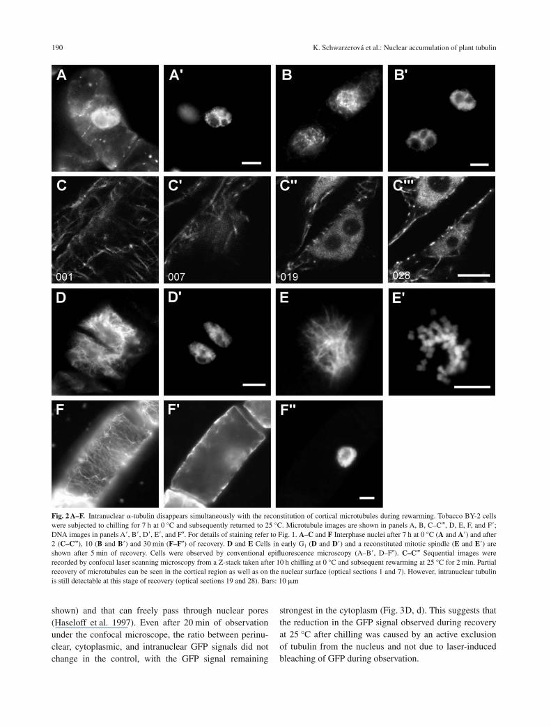
After 7 h of cold treatment, cells retained only very fewmicrotubules in the cortical region, but up to 25% of cellsexhibited a massive tubulin signal in the nuclei (Fig. 2A,A�). However, upon rewarming, new microtubules wereinitiated within a few seconds on the surface of the nuclei,and a reticular array developed around the nucleus within10 min after the temperature had been raised (Fig. 2B,B�). At the same time, the tubulin signal decreased in thenucleus. However, transient stages with newly polymer-ized microtubules in the cortical cytoplasm (Fig. 2C) andaround the nucleus (Fig. 2C�), and a persistent intranu-clear tubulin signal (Fig. 2C�, C) were observed as well.Note that even in such cells, tubulin remained excludedfrom the nucleoli. The re-formation of new microtubuleswas most pronounced at the nuclear surface of early G1
nuclei (Fig. 2D, D�). Similarly, upon rewarming, new mi-totic spindle microtubules were polymerized around thespiralized chromosomes (Fig. 2E, E�). Within 30 min ofincubation at 25 °C, the microtubular cytoskeleton hadcompletely recovered in all surviving cells (Fig. 2F, F�)and was almost indistinguishable from that of controlcells that had not been challenged by chilling (compareFig. 2A, A�). Conversely, the intranuclear tubulin had al-most completely disappeared (Fig. 2F�, F�).
Tubulin exclusion from nuclei can be followed in vivowithin a few minutes of rewarming
To observe tubulin accumulation during cold treatmentand its exclusion from nuclei to the cytoplasm during rewarming in vivo, we used the transgenic BY-2 cell lineGT16 that expresses GFP-tubulin (Kumagai et al. 2001).Tubulin accumulated in the nuclei of a gradually increas-ing number of transgenic cells during cultivation at 0 °C,as observed under the confocal microscope (Fig. 3A). Fluorescence intensity profiles measured within the linear probe showed that the GFP signal in cold-treatedcells was more intense in the nuclear region than in the
188 K. Schwarzerová et al.: Nuclear accumulation of plant tubulin

perinuclear cytoplasm (Fig. 3a). Upon rewarming, tubulinwas quickly excluded from the nucleus into the cytoplasm(Fig. 3B) and after 20 min at 25 °C, the GFP signal wasstronger in the cytoplasm than in the nucleus (Fig. 3b). Incontrol cells, the GFP-tubulin signal was detectable in
the perinuclear cytoplasm as a diffuse signal. However, amuch weaker GFP signal was always detectable in nuclei(Fig. 3C, c), which was possibly caused by unfused GFPthat is present at a low level in transgenic cells (detectedon Western blots using anti-GFP antibody, data not
K. Schwarzerová et al.: Nuclear accumulation of plant tubulin 189
Fig. 1 A–G. �-Tubulin accumulates in nuclei during the response to chilling (0 °C). Microtubules (A, A�, B, B�, C, D, E, and F) were visualized byimmunofluorescence with a monoclonal anti-�-tubulin antibody and a TRITC-conjugated secondary antibody, and DNA was stained with Hoechst33258 (A�, B�, C�, E�, and F�) at 0 (A–A�), 1 (B–B�), 7 (C, C� and E, E�), and 12 h (D) of chilling, and after 11 h of treatment with 5 �M oryzalin (Fand F�). Cells were observed by conventional epifluorescence microscopy (B–C�, E, and E�) and confocal laser scanning microscopy (A–A�, D, F,and F�). Panels A–A� and B–B� show two focal planes, one cortical (A and B) and one central (A�, A�, B�, and B�). Panel D shows a central confocaloptical section, the arrow indicates position of the nucleus. Panels E and E� show a residual mitotic spindle around spiralized metaphase chromo-somes. Panels F and F� show four merged optical sections (Z step size, 1 �m) through central regions of cells. Bars: 20 �m. G Frequency of cellswith intranuclear tubulin during cold treatment. Average values, n � 200 cells per sample
shown) and that can freely pass through nuclear pores(Haseloff et al. 1997). Even after 20 min of observationunder the confocal microscope, the ratio between perinu-clear, cytoplasmic, and intranuclear GFP signals did notchange in the control, with the GFP signal remaining
strongest in the cytoplasm (Fig. 3D, d). This suggests thatthe reduction in the GFP signal observed during recoveryat 25 °C after chilling was caused by an active exclusionof tubulin from the nucleus and not due to laser-inducedbleaching of GFP during observation.
190 K. Schwarzerová et al.: Nuclear accumulation of plant tubulin
Fig. 2 A–F. Intranuclear �-tubulin disappears simultaneously with the reconstitution of cortical microtubules during rewarming. Tobacco BY-2 cellswere subjected to chilling for 7 h at 0 °C and subsequently returned to 25 °C. Microtubule images are shown in panels A, B, C–C, D, E, F, and F�;DNA images in panels A�, B�, D�, E�, and F�. For details of staining refer to Fig. 1. A–C and F Interphase nuclei after 7 h at 0 °C (A and A�) and after2 (C–C), 10 (B and B�) and 30 min (F–F�) of recovery. D and E Cells in early G1 (D and D�) and a reconstituted mitotic spindle (E and E�) areshown after 5 min of recovery. Cells were observed by conventional epifluorescence microscopy (A–B�, D–F�). C–C Sequential images wererecorded by confocal laser scanning microscopy from a Z-stack taken after 10 h chilling at 0 °C and subsequent rewarming at 25 °C for 2 min. Partialrecovery of microtubules can be seen in the cortical region as well as on the nuclear surface (optical sections 1 and 7). However, intranuclear tubulinis still detectable at this stage of recovery (optical sections 19 and 28). Bars: 10 �m

Tubulin cofractionates with nuclei from cold-treated cells
The immunofluorescence and in vivo analyses showedthat tubulin accumulated in the nuclei of cold-treatedcells. To challenge this finding, protein extracts from iso-lated nuclei of the BY-2 cell line BY-2-mGFP5-ER(Petrásek et al. 2003) were analyzed by Western blottingusing monoclonal antibodies recognizing �-tubulin ingeneral, and tyrosinated �-tubulin in particular (for detailsrefer to Wiesler et al. [2002]). Whereas tubulin could not
be detected in control nuclei, it was clearly present in nu-clei from cold-treated cells (Fig. 4A, B). The nuclear sig-nal of �-tubulin and its tyrosinated form increasedprogressively with the duration of chilling (Fig. 4A, B,compare extracts taken after 8 and 12 h), in agreementwith the increased frequency of tubulin-containing nucleifound in the immunofluorescence study (Fig. 1G). Fig-ure 4C shows an amido black stain of proteins separatedby SDS-PAGE and blotted in parallel under the sameconditions.
Furthermore, we tested whether the tubulin signal
K. Schwarzerová et al.: Nuclear accumulation of plant tubulin 191
Fig. 3 A, B. Exclusion of tubulin from nucleiduring recovery in vivo. A and B Tobaccocells expressing GFP-tubulin were cultivatedat 0 °C for 8 h and then subsequently at25 °C for 20 min. A Confocal optical sectionin the nuclear region of cells cultivated for8 h at 0 °C. B The same confocal optical sec-tion of the same cell after 20 min of recoveryat 25 °C. a The intensity of the GFP signalwas maximal in nuclei of cold-treated cellssuggesting that tubulin accumulated in thenuclei during cold treatment. b Tubulin wasexcluded from the nuclei during recovery at25 °C and accumulated in the surroundingcytoplasm. C and D A control cell express-ing GFP-tubulin kept at 25 °C throughout theexperiment. C and D Confocal optical sec-tion in the nuclear region at time zero (C)and after 20 min (D). c The intensity of theGFP signal was maximal in the cytoplasmsurrounding nuclei; d this distribution ofGFP fluorescence did not change during the20 min of observation. vac Vacuole, cyt cyto-plasm, nuc nucleus

found in extracts from cold-treated nuclei was possiblycaused by contamination with nonnuclear, but sedi-mentable, proteins. The transgenic cell line BY-2-mGFP5-ER used in this experiment constitutively expressesmGFP5 fused at the N terminus to the signal peptide fromArabidopsis thaliana basic chitinase and at the C terminusto the endoplasmic-reticulum retention signal (the HDELpeptide) (Petrásek et al. 2003). This so-called mGFP5-ERfusion protein has been previously shown to localize tothe endoplasmic reticulum (Haseloff et al. 1997) (Fig. 5A)and thus it can serve as a marker for perinuclear mem-
192 K. Schwarzerová et al.: Nuclear accumulation of plant tubulin
Fig. 4 A–C. Detection of tubulin in protein extracts from isolated nuclei.A and B Immunoblots stained with antibodies directed against �-tubulin(A) and tyrosinated �-tubulin (B). C Amido black stain of proteins sepa-rated by SDS-PAGE and blotted in parallel under the same conditions.15 �g of total protein were loaded per lane
Fig. 5 A–D. Isolated nuclei are free from non-nuclear contamination. AThe tobacco cell line BY-2-mGFP5-ER expressing mGFP5 fused at theN terminus to the signal peptide from Arabidopsis thaliana basic chitin-ase and at the C terminus to the endoplasmic-reticulum retention signalHDEL. A projection of eight optical sections (Z step size, 0.6 �m)through a central part of cell is shown. B and C DIC image (B) and GFPsignal (C) of a nucleus isolated from a cell expressing mGFP5-ER main-tained under control conditions. D Immunoblot detection of GFP proteinin fractions collected during the isolation of nuclei. Fraction I, super-natant from the first centrifugation at 2000 g. Fraction II, nuclei beforeloading onto the Percoll gradient. Fraction III, supernatant from the lastcentrifugation at 2000 g. Fraction IV, sediment from the last centrifuga-tion at 2000 g (purified nuclei). 10 �g of total protein were loaded perlane. Note that all GFP remained in fraction I

brane structures. Nuclei were isolated from BY-2-mGFP5-ER cells that had been cultivated under standardconditions and were examined under the epifluorescencemicroscope. No GFP signal was found in these isolatednuclei (Fig. 5B, C). When different fractions from the iso-lation procedure were tested by Western blot analysis us-ing an anti-GFP antibody, the GFP signal was clearlypresent in the first supernatant, but absent in fraction IVthat contained the purified nuclei, irrespective of whetherthey had been isolated from control or from cold-treatedcells (Fig. 5D). Nevertheless, nuclei isolated from cold-treated cells by the same protocol contained abundanttubulin (Fig. 4A, B).
Both tubulin molecules contain putative nuclear-export sequences
Tubulins lack at least the canonical nuclear-import signals(data not shown). We screened various sequences of plant�- and �-tubulins for nuclear-export sequences (NES). Weidentified two putative NES in the �-tubulins and three in
the �-tubulins (Tables 1 and 2). Moreover, according toour preliminary studies, the potential NES motifs 2� and1� through 3� are also well conserved between plant andanimal tubulins, whereas the NES 1� seems to be plant-specific (data not shown).
Discussion
We observed that tubulin accumulated in interphase nucleiduring the response to prolonged chilling that progressivelyeliminated cortical microtubules, and that this phenomenonwas fully reversible when the temperature was restored to25 °C. Intranuclear accumulation of tubulin during chillingand its exclusion during rewarming was shown by immuno-fluorescence in situ, as well as in vivo using transgenic celllines expressing GFP–�-tubulin. Another transgenic cell linethat expresses a GFP marker for endoplasmic reticulum andnuclear envelope (mGFP5-ER) was used for the detection oftubulin in isolated nuclei. Nuclear preparations that werevoid of cytoplasmic contamination (as evident from thecomplete removal of the GFP signal) nevertheless contained
K. Schwarzerová et al.: Nuclear accumulation of plant tubulin 193
Table 1. Putative nuclear-export sequences in plant �-tubulinsa
Plant and �-tubulin Accession nr. Putative NES 1� Putative NES 2�
Arabidopsis thalianaTUA1 NP_176654 LGSLLLERLSV 149–159 LRFDGAINV 242–250TUA2 NP_175423 LGSLLLERLSV 149–159 LRFDGAINV 242–250TUA3 NP_197478 LGSLLLERLSV 149–159 LRFDGALNV 242–250TUA4 NP_171974 LGSLLLERLSV 149–159 LRFDGAINV 242–250TUA5 NP_197479 LGSLLLERLSV 149–159 LRFDGALNV 242–250TUA6 NP_849388 LGSLLLERLSV 149–159 LRFDGALNV 242–250
Nicotiana tabacumNtTUA AB052822 LGSLLLERLSV 149–159 LRFDGALNV 242–250NtTUA1 AJ421411 LGSLLLERLSV 149–159 LRFDGALNV 242–250NtTUA2 AJ421412 LGSLLLERLSV 149–159 LRFDGALNV 242–250
a Consensus sequence Z-X(1–4)-J-X(2–3)-Z-X-Z, where Z stands for Leu, Ile, or Val; J stands for Met,or Phe; X stands for any
Table 2. Putative nuclear-export sequences in Arabidopsis thaliana �-tubulinsa
�-Tubulin Accession nr. Putative NES 1� Putative NES 2� Putative NES 3�
TUB1 NP_177706 LQLERINV 43–50 ICFRTLKL 211–218 LNSDLRKLAV 247–256TUB2 NP_568959 LQLERINV 42–49 ICFRTLKL 210–217 LNSDLRKLAV 247–256TUB3 NP_568960 LQLERINV 42–49 ICFRTLKL 210–217 LNSDLRKLAV 247–256TUB4 NP_199247 LQLERIDV 42–49 ICFRTLKL 210–217 LNSDLRKLAV 247–256TUB5 NP_564101 LQLERINV 43–50 ICFRTLKL 211–218 LNSDLRKLAV 247–257TUB6 NP_196786 LQLERVNV 42–49 ICFRTLKL 210–217 LNSDLRKLAV 247–256TUB7 P29515 LQLERVNV 42–49 ICFRTLKL 210–217 LNSDLRKLAV 247–256TUB8 NP_568437 LQLERVNV 42–49 ICFRTLKL 210–217 LNSDLRKLAV 247–256TUB9 P29517 LQLERINV 42–49 ICFRTLKL 210–217 LNSDLRKLAV 247–256
a For consensus sequence, see footnote of Table 1
tubulin, but only when the nuclei had been prepared fromcold-treated cells. No tubulin signal was detected in nuclearpreparations from cells that had not been exposed to low temperature.
Hitherto, reports of intranuclear tubulin have beenscarce. This indicates that the separation of interphase andmitotic microtubular functions is correlated to a strictcompartmentalization of tubulin. Intranuclear tubulin hasbeen reported for the mouse cell line SV3T3 (Menko andTan 1980). More recently, nonassembled ��II-tubulin het-erodimers have been detected in cultured rat kidneymesangial cells (Walss et al. 1999). The �II isoform waspresent exclusively in nuclei and could be reversibly re-moved from the nuclei by vinblastine treatment (Walss-Bass et al. 2003). The authors suggested that nucleartubulin constitutes a pool of dynamic tubulin that mightassist the rapid cell proliferation characteristic of cancercells. In plant cells, intranuclear microtubule-like struc-tures have been documented in Aesculus hippocastanumL. by electron microscopy (Barnett 1991). However, thefunction of these intranuclear microtubules remainedenigmatic. In our study, the tubulin was apparently notpresent in the form of microtubules, but as nonassembledtubulin heterodimers.
Although intranuclear tubulin seems to be a fairly ex-otic phenomenon in animal and plant cells, it is quitecommon in primitive eukaryotes with closed mitosis,where the division spindle develops within the kary-oplasm surrounded by a nuclear envelope. This type ofmitosis is frequently found in certain protists, fungi, andalgae (for a review, see Heath 1980). There is a mountingbody of evidence that tubulin is imported into the nucleusalong with components of MTOCs just prior to the onsetof mitosis (Aspergillus nidulans [Ovechkina et al. 2003],spindle-pole body of Saccharomyces cerevisiae [Pereiraet al. 1998]). The precise timing of this nuclear importwith respect to the cell cycle suggests that, in these or-ganisms, the presence and absence in the nucleus of cy-toskeletal components that participate in mitosis iscarefully regulated and represents an important check-point within the cell cycle. In animal and plant cells, thedisassembly of the nuclear envelope that results in an im-mediate and perfect exposure of chromatin to tubulin(open mitosis) has evolved during evolution as a more ef-fective mechanism. However, even under conditions ofopen mitosis, some components of the spindle regulationmachinery are imported into the nucleus prior to mitosis.For instance, �-tubulin already appears in the nucleus ofVicia faba in the G2 phase (Binarová et al. 2000). It is im-portant to note that the antagonistic process – active ex-
clusion of ��-tubulin heterodimer from the nucleus –must occur at the transition from M to G1, when thedaughter nuclei become enclosed by a new nuclear mem-brane, not only in cells with closed mitosis, but also inthose with open mitosis. Moreover, independently of therespective type of mitosis, all interphase cells must effi-ciently exclude ��-tubulin from the nucleus or preventits entry because it is not found in interphase nuclei.These facts suggest that, in spite of substantial differ-ences between open and closed mitosis, some mecha-nisms, such as the import of cytoskeletal componentsregulating polymerization of microtubules at the begin-ning of mitosis and export of tubulin at the end, must bepreserved in all organisms in order to ensure proper cellcycle progression.
It remains to be asked how tubulin is actually kept outof the interphase nucleus. We show in the present workthat this mechanism is impaired during chilling at 0 °C.Transport between the cytoplasm and the nucleus is medi-ated by NPCs residing in the nuclear envelope. Transportof molecules through the nuclear pores can be either non-selective (i.e., passive) or selective (i.e., active, mediatedby numerous protein transporters). Passive diffusion islimited by the exclusion size of the NPC, which is approx-imately 50 kDa (for a review, see Talcott and Moore1999). Although molecules of up to 60 kDa are in princi-ple allowed to passively diffuse across NPC, the diffusionrate is very low for molecules over 50 kDa (Rose et al.2004). Therefore, it is unlikely that ��-tubulin het-erodimers (110 kDa) can pass the pores without specificinteraction with the nuclear transport machinery. Themassive increase in the pool of free tubulin dimers in thecytoplasm as a consequence of cold-induced eliminationof assembled microtubules (Pokorná et al. 2004) mightsimply overload highly selective transport through the nu-clear envelope such that tubulin “leaks” into the nucleus.However, an excess of free tubulin dimers generated byother means (oryzalin treatment, inhibiting the assemblyof dimers into microtubules) does not produce such an en-try of tubulin into the nucleus. This suggests that intranu-clear tubulin is specific to the cold response. Since lowtemperature affects biological membranes (Kacperska1999, Örvar et al. 2000), a possible explanation would bethat cold-induced membrane injuries allow a slow leakageof tubulin into the karyoplasm. However, passive leakageof tubulin into the karyoplasm occurring as a consequenceof membrane damage cannot explain its striking accumu-lation in the karyoplasm resulting in a clear concentrationgradient in response to chilling. The accumulation of in-tranuclear tubulin coupled with a much lower concentra-
194 K. Schwarzerová et al.: Nuclear accumulation of plant tubulin

tion in the surrounding cytoplasm suggests that it binds toan unknown intranuclear lattice. The affinity to this latticeseems to be rather strong since tubulin remained attachedeven during the isolation of nuclei.
The entry of tubulin into the nucleus seems to be per-fectly reversible when the cells are rewarmed, because itis not detectable in nuclei within minutes of recovery, asevident from immunofluorescence microscopy results andfrom live cells expressing GFP. Since this process is sorapid, it is unlikely that tubulin disappearance reflectsdegradation; the half-life of tubulin has been shown to becell cycle dependent, varying between 1 and 15 h inPhysarum polycephalum (Ducommun and Wright 1989).Even degradation of regulatory proteins (enzymes) usu-ally takes much longer. A specific mechanism that ex-cludes tubulin from the karyoplasm and is strongly (butreversibly) inhibited by low temperature must be involvedin the recovery processes. Rapid and complete exclusionof tubulin during the first minutes of rewarming is un-likely to be caused by simple diffusion since the tempera-ture dependency of diffusion is relatively low. Moreover,we observed that the intranuclear tubulin is rich in tyrosi-nated �-tubulin indicating that this has not been incorpo-rated into stable microtubules and thus might represent ahighly dynamic reservoir for the assembly of new micro-tubules (Wiesler et al. 2002).
The rapid recovery of tubulin exclusion from the nu-cleus demonstrates that the membranes of the nuclear en-velope are not damaged or perturbed irreversibly. Thetarget of chilling seems rather to be the NPCs and indi-cates that import and/or export of tubulin must be some-how regulated. Tubulin lacks at least the canonical NLS(Ovechkina et al. 2003 and unpubl. results). It is clear,however, that the standard pathways of protein importmust be complemented by additional import mechanisms,since several proteins lacking an obvious NLS can enterthe nucleus in both plant and animal cells (Evans et al.2004). Interestingly, the intranuclear tubulin in culturedrat kidney mesangial cells reported by Walss et al. (1999)entered nuclei only if the cells underwent mitosis (Walss-Bass et al. 2001). The authors suggested that tubulin wastrapped by nuclear factors at the end of mitosis rather thanbeing actually transported through the nuclear envelope.However, we searched for putative NES in tubulin mole-cules and identified 5 in the �- and �-tubulins. Though thefunctionality of these nuclear-export signals must be con-firmed experimentally, their existence is consistent with amodel in which tubulin is exported through the nuclearpores by means of the conventional nuclear transport ma-chinery, most of the components of which, such as im-
portins, exportins, Ran, RanGAP and others (for a review,see Dasso 2002), have also been identified in higherplants (Smith and Raikhel 1999, Merkle and Nagy 1997,Pay et al. 2002).
We interpret the presence of tubulin in the nuclei ofcold-treated cells in the following way. Tubulin is exclu-sively cytoplasmic during interphase, probably becauseits presence in the interphasic nucleus could induce se-rious disorders of cell-cycle regulation. We hypothesizethat the integrity of the nuclear membrane during inter-phase, when nuclear pores control the transport of molecules, is the main mechanism that ensures the se-questration of tubulin to the cytoplasm during inter-phase. The interaction of tubulin with chromatin iscrucial during mitosis. For closed mitosis, tubulin is im-ported into the nucleus by an unknown mechanism,whereas for open mitosis, no tubulin transport throughthe nuclear membrane is required since the nuclear en-velope breaks down at the onset of mitosis. However,when daughter nuclei are formed at the end of mitosis,new nuclear envelopes have to be reestablished and thepool of tubulin molecules from the disintegrated spindlehas to be removed from the karyoplasm. At this stage,tubulin is excluded from nuclei, possibly utilizing theNES export signals conserved in all tubulins. The in-tegrity of the nuclear envelope and the active transportthrough the nuclear pores are impaired during coldtreatment, resulting in intranuclear accumulation oftubulin. In our future work, we would like to verify thefunctionality of the different NES signatures in tubulinand to identify the corresponding binding partners ofthe nuclear transport machinery in both the cyto- andthe karyoplasm.
Acknowledgments
We thank S. Hasezawa for a kind gift of the transgenic BY-2 cell lineGT16. We thank Jan Marc for critical reading of the manuscript. Wealso thank Vlasta Sakarová for excellent technical assistance. Thiswork was supported by grants from the Ministry of Education, Youth,and Sports of the Czech Republic nr. VZ113100003 and nr. LN00A081(to Z.O.), by the Volkswagen Foundation Nachwuchsgruppenpro-gramm and the EC-FP5 grant nr. QLK5 CT-2000-00357 (to P.N.).
References
Barnett JR (1991) Microtubules in interphase nuclei of Aesculus hippocastanum L. Ann Bot 68: 159–165
Baskin TI, Cande WZ (1990) The structure and function of the mitoticspindle in flowering plants. Annu Rev Plant Physiol 41: 277–315
Binarová P, Cenklová V, Hause B, Kubátová E, Lysák M, Dolezel J,Bogre L, Dráber P (2000) Nuclear gamma-tubulin during acentriolarplant mitosis. Plant Cell 12: 433–442
K. Schwarzerová et al.: Nuclear accumulation of plant tubulin 195

Cande WZ (1990) Centrosomes: composition and reproduction. CurrOpin Cell Biol 2: 301–305
Dasso M (2002) The Ran GTPase: theme and variations. Curr Biol 12:R502–R508
Ducommun B, Wright M (1989) Variation of tubulin half-life during thecell cycle in the synchronous plasmodia of Physarum polycephalum.Eur J Cell Biol 50: 48–55
Evans DE, Bryant JA, Hutchinson C (2004) The nuclear envelope: acomparative overview. In: Evans DE, Hutchinson CJ, Bryant JA (eds)The nuclear envelope. BIOS Scientific Publishers, Abingdon, pp 1–8
Haseloff J, Siemering KR, Prasher DC, Hodge S (1997) Removal of acryptic intron and subcellular localization of green fluorescent proteinare required to mark transgenic Arabidopsis plants brightly. Proc NatlAcad Sci USA 94: 2122–2127
Hasezawa S, Kumagai F (2002) Dynamic changes and the role of the cy-toskeleton during the cell cycle in higher plant cells. Int Rev Cytol214: 161–191
Heath IB (1980) Variant mitoses in lower eucaryotes – indicators of theevolution of mitosis. Int Rev Cytol 64: 1–80
Kacperska A (1999) Plant responses to low temperature: signalling path-ways involved in plant acclimation. In: Margesin R, Schinner F (eds)Cold-adapted organisms. Springer, Berlin, pp 79–104
Kumagai F, Yoneda A, Tomida T, Sano T, Nagata T, Hasezawa S (2001)Fate of nascent microtubules organized at the M/G(1) interface, as vi-sualized by synchronized tobacco BY-2 cells stably expressing GFP-tubulin: time-sequence observations of the reorganization of corticalmicrotubules in living plant cells. Plant Cell Physiol 42: 723–732
Menko AS, Tan KB (1980) Nuclear tubulin of tissue culture cells.Biochim Biophys Acta 629: 359–370
Merkle T, Nagy F (1997) Nuclear import of proteins: putative importfactors and development of in vitro import systems in higher plants.Trends Plant Sci 2: 458–464
Mirski SEL, Bielawski JC, Cole SPC (2003) Identification of functionalnuclear export sequences in human topoisomerase II alpha and beta.Biochem Biophys Res Commun 306: 905–911
Mizuno K (1992) Induction of cold stability of microtubules in culturedtobacco cells. Plant Physiol 100: 740–748
Nagata T, Nemoto Y, Hasezawa S (1992) Tobacco BY-2 cell line as the“HeLa” cell in the cell biology of higher plants. Int Rev Cytol 132:1–30
Örvar BL, Sangwan V, Omann F, Dhindsa RS (2000) Early steps in coldsensing by plant cells: the role of actin cytoskeleton and membranefluidity. Plant J 23: 785–794
Ovechkina Y, Maddox P, Oakley CE, Xiang X, Osmani SA, Salmon ED,Oakley BR (2003) Spindle formation in Aspergillus is coupled totubulin movement into the nucleus. Mol Biol Cell 14: 2192–2200
Pay A, Resch K, Frohnmeyer H, Fejes E, Nagy F, Nick P (2002) PlantRanGAPs are localized at the nuclear envelope in interphase and asso-ciated with microtubules in mitotic cells. Plant J 30: 699–709
Pereira G, Schiebel E (1997) Centrosome-microtubule nucleation. J CellSci 110: 295–300
Pereira G, Knop M, Schiebel E (1998) Spc98p directs the yeast gamma-tubulin complex into the nucleus and is subject to cell cycle-depen-dent phosphorylation on the nuclear side of the spindle pole body.Mol Biol Cell 9: 775–793
Petrásek J, Cerná A, Schwarzerová K, Elckner M, Morris DA,Zazímalová E (2003) Do phytotropins inhibit auxin efflux by impair-ing vesicle traffic? Plant Physiol 131: 254–263
Pokorná J, Schwarzerová K, Zelenková S, Petrásek J, Janotová I,Capková V, Opatrny Z (2004) Sites of actin filament initiation and re-organization in cold-treated tobacco cells. Plant Cell Environ 27:641–653
Popov N, Schmitt S, Mathies H (1975) Eine störungsfreie Mikrome-thode zur Bestimmung des Proteingehaltes in Gewebshomogenaten.Acta Biol Germ 34: 144–146
Rose A, Patel S, Meier I (2004) Plant nuclear envelope proteins. In:Evans DE, Hutchinson CJ, Bryant JA (eds) The nuclear envelope.BIOS Scientific Publishers, Abingdon, pp 69–88
Shimamura M, Brown RC, Lemmon BE, Akashi T, Mizuno K,Nishihara N, Tomizawa KI, Yoshimoto K, Deguchi H, Hosoya H,Mineyuki Y (2004) Gamma-tubulin in basal land plants: characteriza-tion, localization, and implication in the evolution of acentriolar mi-crotubule organizing centers. Plant Cell 16: 45–59
Smith HMS, Raikhel NV (1999) Protein targeting to the nuclear pore.What can we learn from plants? Plant Physiol 119: 1157–1163
Stoppin-Mellet V, Peter C, Lambert AM (2000) Distribution of gamma-tubulin in higher plant cells: cytosolic gamma-tubulin is part of highmolecular weight complexes. Plant Biol 2: 290–296
Talcott B, Moore MS (1999) Getting across the nuclear pore complex.Trends Cell Biol 9: 312–318
Walss C, Kreisberg JI, Luduena RF (1999) Presence of the beta(II) iso-type of tubulin in the nuclei of cultured mesangial cells from rat kid-ney. Cell Motil Cytoskeleton 42: 274–284
Walss-Bass C, Kreisberg JI, Luduena RF (2001) Mechanism of localiza-tion of beta(II)-tubulin in the nuclei of cultured rat kidney mesangialcells. Cell Motil Cytoskeleton 49: 208–217
Walss-Bass C, Kreisberg JI, Luduena RF (2003) Effect of the antitumordrug vinblastine on nuclear beta(II)-tubulin in cultured rat kidneymesangial cells. Invest New Drug 21: 15–20
Wick SM, Seagull RW, Osborn M, Weber K, Gunning BES (1981) Im-munofluorescence microscopy of organized microtubule arrays instructurally stabilized meristematic plant cells. J Cell Biol 89:685–690
Wiese C, Zheng YX (1999) Gamma-tubulin complexes and their interac-tion with microtubule-organizing centers. Curr Opin Struct Biol 9:250–259
Wiesler B, Wang QY, Nick P (2002) The stability of cortical micro-tubules depends on their orientation. Plant J 32: 1023–1032
196 K. Schwarzerová et al.: Nuclear accumulation of plant tubulin
Copyright © 2022 FDOKUMEN