Quinone-amino acid conjugates targeting Leishmania amino acid transporters
Organic &Biomolecular Chemistry
PAPER
Cite this: Org. Biomol. Chem., 2014,12, 7993
Received 5th June 2014,Accepted 8th August 2014
DOI: 10.1039/c4ob01152j
www.rsc.org/obc
Pyrazole–oxadiazole conjugates: synthesis,antiproliferative activity and inhibition of tubulinpolymerization†
Ahmed Kamal,*a,d Anver Basha Shaik,a Sowjanya Polepalli,b Vangala Santosh Reddy,a
G. Bharath Kumar,a Soma Gupta,a K. V. S. Rama Krishna,c
Ananthamurthy Nagabhushana,e Rakesh K. Mishrae and Nishant Jainb
A number of pyrazole–oxadiazole conjugates were synthesized and evaluated for their ability to function
as antiproliferative agents on various human cancer cell lines. These conjugates are comprised of pyrazole
and oxadiazole scaffolds closely attached to each other without any spacer as two structural classes. The
Type I class has a trimethoxy substituent and the type II class has a 3,4-(methylenedioxy) substituent on
their A rings. Among these conjugates, 11a, 11d and 11f manifest potent cytotoxicity with IC50 values
ranging from 1.5 μM to 11.2 μM and inhibit tubulin polymerization with IC50 values of 1.3 μM, 3.9 μM and
2.4 μM respectively. The cell cycle assay showed that treatment with these conjugates results in accumu-
lation of cells in the G2/M phase and disrupts the microtubule network. Elucidation of zebrafish embryos
revealed that the conjugates cause developmental defects. Molecular docking simulations determined
the binding modes of these potent conjugates at the colchicine site of tubulin.
Introduction
In eukaryotic cells, the microtubules are hollow tubes ofα- and β-tubulin heterodimers. The microtubule system is animportant constituent of the cytoskeleton and functions inmany fundamental cellular processes, including chromo-some segregation during cell division, intracellular transport,development and maintenance of the cell shape, cell motilityand distribution of molecules on cell membranes.1–4 Inaddition, microtubules are involved in a host of cell signalingpathways which are responsible for cellular apoptosis. Themicrotubule dynamics are regulated by various important pro-teins such as kinesin and dynein.5,6 Many experiments haveidentified that microtubule dependence forces the underlyingchromosomal translocation and spindle formation.7 Hencealtered/aberrant microtubule dynamics result in the obstruction
of cell division, often at the metaphase. As a result, variousefforts aimed at blocking mitosis like inhibition of tubulinpolymerization by tubulin targeting agents have emerged as anattractive approach to cancer therapy.8,9
Microtubules possess three ligand-binding sites: vinca, col-chicine and taxol domains. Combretastatin A-4 (1) is one ofthe naturally occurring tubulin targeting agents that inhibittubulin polymerization through the colchicine domain of themicrotubule (Fig. 1).10 Among the common antitubulin
Fig. 1 Chemical structures of microtubule targeting agents: CA-4 (1),azapodophyllotoxin (2), nocodazole (3), anilinonicotinyl–oxadiazoleconjugate (4) and pyrazole–oxadiazole conjugates (5, 6).
†Electronic supplementary information (ESI) available: 1H NMR and 13C NMRspectra of the final compounds are provided. See DOI: 10.1039/c4ob01152j
aMedicinal Chemistry and Pharmacology, CSIR-Indian Institute of Chemical
Technology, Hyderabad 500 007, India. E-mail: [email protected];
Fax: (+)91-40-27193189; Tel: (+)91-40-27193157bCentre for Chemical Biology CSIR-Indian Institute of Chemical Technology,
Hyderabad 500 007, IndiacNuclear Magnetic Resonance Centre, CSIR-Indian Institute of Chemical Technology,
Hyderabad 500 007, IndiadDepartment of Medicinal Chemistry, National Institute of Pharmaceutical Education
and Research (NIPER), Hyderabad 500 037, IndiaeCSIR – Centre for Cellular and Molecular Biology, Hyderabad 500 007, India
This journal is © The Royal Society of Chemistry 2014 Org. Biomol. Chem., 2014, 12, 7993–8007 | 7993
Publ
ishe
d on
13
Aug
ust 2
014.
Dow
nloa
ded
by I
ndia
n In
stitu
te o
f C
hem
ical
Tec
hnol
ogy
(IIC
T),
Hyd
erab
ad o
n 26
/09/
2014
14:
35:2
5.
View Article OnlineView Journal | View Issue
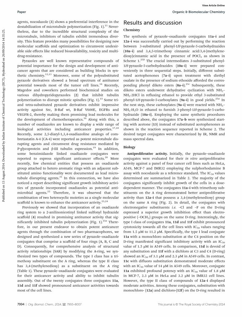
agents, nocodazole (3) shows a preferential interference in thedestabilization of microtubule polymerization (Fig. 1).11 Never-theless, due to the incredible structural complexity of themicrotubule, inhibitors of tubulin exhibit tremendous diver-sity. This feature provides many possibilities for designing newmolecular scaffolds and optimization to circumvent undesir-able side effects like reduced bioavailability, toxicity and multi-drug-resistance.
Pyrazoles are well known representative compounds ofpotential importance for the design and development of anti-cancer agents that are considered as building blocks in syn-thetic chemistry.12,13 Moreover, some of the polysubstitutedpyrazole derivatives showed a broad spectrum of antitumorpotential towards most of the tumor cell lines.14 Recently,Magedov and coworkers performed biochemical studies onvarious dihydropyridopyrazoles (2) that inhibit tubulinpolymerization to disrupt mitotic spindles (Fig. 1).15 Some tri-and tetra-substituted pyrazole derivatives exhibit impressiveactivity against Src, B-Raf wt, B-Raf V600E, EGFRs andVEGFR-2, thereby making them promising lead molecules forthe development of chemotherapeutics.16 Along with this, anumber of oxadiazoles are known to display a wide range ofbiological activities including anticancer properties.17,18
Recently, some 2,5-diaryl-1,3,4-oxadiazoline analogs of com-bretastatin A-4 (CA-4) were reported as potent microtubule dis-rupting agents and circumvent drug resistance mediated byP-glycoprotein and β-III tubulin expression.19 In addition,some benzimidazole linked oxadiazole conjugates werereported to express significant anticancer effects.20 Morerecently, few chemical entities that possess an oxadiazolegroup attached to hetero aromatic rings with an adjacent sub-stituted amino functionality were documented as lead micro-tubule disrupting agents.21 In this connection, we have alsonoticed a report describing significant growth inhibitory activi-ties of pyrazole incorporated oxadiazoles as potential anti-microbial agents.22 Therefore, it was observed that thecombination of two heterocyclic moieties as a single molecularscaffold is known to enhance the anticancer activity.23,24
Previously we showed that incorporation of an oxadiazolering system to a 2-anilinonicotinyl linked sulfonyl hydrazidescaffold (4) resulted in promising antitumor activity that sig-nificantly inhibited tubulin polymerization (Fig. 1).25a There-fore, in our present endeavor to obtain potent anticanceragents through the combination of two pharmacophores, wedesigned and synthesized a new series of pyrazole–oxadiazoleconjugates that comprise a scaffold of four rings (A, B, C andD). Consequently, for comprehensive analysis of structuralactivity relationships (SAR) by modifying the A-ring, we syn-thesized two types of compounds. The type I class has a tri-methoxy substituent on the A ring, whereas the type II classhas 3,4-(methylenedioxy) as a substituent on the A ring(Table 1). These pyrazole–oxadiazole conjugates were evaluatedfor their anticancer activity and ability to inhibit tubulinassembly. Out of the twenty conjugates three conjugates 11a,11d and 11f showed pronounced anticancer activities towardsmost of the cell lines.
Results and discussionChemistry
The synthesis of pyrazole–oxadiazole conjugates 11a–i and12a–i was successfully carried out by performing the reactionbetween 3-substituted phenyl-1H-pyrazole-5-carbohydrazides(10a–i) and 3,4,5-trimethoxy cinnamic acid/3,4-(methylene-dioxy)cinnamic acid in the presence of POCl3 as shown inScheme 1.25b The crucial intermediates 3-substituted phenyl-1H-pyrazole-5-carbohydrazides (10a–i) were prepared con-veniently in three sequential steps. Initially, different substi-tuted acetophenones (7a–i) upon treatment with diethyloxalate in the presence of sodium ethoxide afforded the corres-ponding phenyl diketo esters (8a–i).25c Subsequently, thesediketo esters underwent dehydrative cyclization with NH2–
NH2·2HCl in refluxing ethanol to provide ethyl 3-substitutedphenyl-1H-pyrazole-5-carboxylates (9a–i) in good yields.25a Inthe next step, these carboxylates (9a–i) were reacted with NH2–
NH2·H2O in ethanol to furnish 3-phenyl-1H-pyrazole-5-carbo-hydrazide (10a–i). Employing the same synthetic proceduresdescribed above, the conjugates 17a–b were synthesized start-ing with acetone (13) instead of substituted acetophenones asshown in the reaction sequence reported in Scheme 2. Thedesired target conjugates were characterized by IR, NMR andmass spectral data.
Biology
Antiproliferative activity. Initially, the pyrazole–oxadiazoleconjugates were evaluated for their in vitro antiproliferativeactivity against a panel of four cancer cell lines such as HeLa,A549, MCF-7 and IMR32 employing the sulphorhodamine Bassay with nocodazole as a reference standard. The IC50 valuesdetermined are summarized in Table 2. The majority of theconjugates significantly inhibit growth of the cells in a dose-dependent manner. The conjugates 11a–i with trimethoxy sub-stituents on the A ring demonstrated better antiproliferativeactivity than 12a–i that possess a 3,4-(methylenedioxy) groupon the same A ring (Fig. 2). In detail, the conjugates withelectronegative substituents i.e. –Cl and –F on the D-ringexpressed a superior growth inhibition effect than electro-positive (–OCH3) groups on the same D-ring. Interestingly, thetype I class of conjugates 11a, 11d and 11f exhibit pronouncedcytotoxicity towards all the cell lines with IC50 values rangingfrom 1.5 μM to 11.2 μM. Specifically, the type I lead conjugate11a with a monochloro substitution at the C4 position on theD-ring manifested significant inhibitory activity with an IC50
value of 1.5 μM in A549 cells. In comparison, 11d is devoid ofany substitution and 11f with a dichloro at C3 and C4 (D-ring)showed an IC50 of 3.1 μM and 2.3 μM in A549 cells. In contrast,11c with difluoro substitution demonstrated moderate effectswith an IC50 value of 14 μM in A549 cells. Moreover, conjugate11a exhibited profound potency with an IC50 value of 1.8 μMin MCF-7, 2.3 μM in HeLa and 3.2 μM in IMR32 cell lines.However, the type II class of compounds of 12a–i displayedmoderate activities. Among these conjugates, substitution withmonochloro- (12a) and dichloro-(12f ) on the D-ring resulted in
Paper Organic & Biomolecular Chemistry
7994 | Org. Biomol. Chem., 2014, 12, 7993–8007 This journal is © The Royal Society of Chemistry 2014
Publ
ishe
d on
13
Aug
ust 2
014.
Dow
nloa
ded
by I
ndia
n In
stitu
te o
f C
hem
ical
Tec
hnol
ogy
(IIC
T),
Hyd
erab
ad o
n 26
/09/
2014
14:
35:2
5.
View Article Online
an increased cytotoxicity against most of the cell lines. Further-more, conjugate 12d devoid of any substitutions manifestedan activity of 10 μM (IC50) towards A549 cells. Notably, thepresence of bulky groups on the D-ring diminished the activi-ties of the compounds in the type II class. Similarly, conju-gates of the type I class, such as 11h harboring trimethoxysubstitution on the D-ring, abrogated the potency of the conju-gate. Based on these observations, removal of the D-ring in17a and 17b substantially reduced the potency, suggesting thatthe D-ring is essential for the optimal activity of these conju-gates. Therefore, these results suggest that the trimethoxy sub-stituent of the A-ring conjugated to chloro or un-substituted atthe C-4 position of the D-ring is the most potent agentbetween the two structural classes of these conjugates.
Dot-blot analysis for cyclin-B1. Previously our studiesshowed that 2-anilino-nicotinyl 1,3,4-oxadiazoles, with a tri-methoxy phenyl group, an oxadiazole and pyridine moiety,function as potent anti-tubulin agents. Therefore, we hypo-thesize that these conjugates induce cytotoxicity through cellcycle arrest at the G2/M phase. Accumulation of cyclin-B1 is anindicator of G2/M arrest.26 Hence, we treated A549 cells with
all the conjugates at 5 μM concentration for a duration of 24 hand performed dot-blot analysis for cyclin-B1. Interestingly,11a and 11f exhibited a marked increase in cyclin-B1 proteinlevels similar to colchicine and CA-4 treated cells. In addition,11d, 11g, and 11h showed a slight increase in cyclin-B1protein. Therefore, these results support the suggestion thatcongeners, 11a, 11f, of pyrazole–oxadiazole conjugates mani-fest anti-proliferative effects through cell cycle arrest at mitosis(Fig. 3).
Effect on tubulin polymerization. Since conjugates of thetype I class of compounds harbor trimethoxy phenyl, oxadia-zole and pyrazole moieties, we presumed that these conjugatescan prevent tubulin assembly. Based on our SAR analysis anddot-blot results we selected 11a and 11f as potent congeners.Intriguingly, 11d treated cells showed a modest increase incyclin B1 protein levels. In contrast, 11d exhibited potent cyto-toxicity compared to the other conjugates in the series. Conse-quently, we elucidated the ability of 11a, 11d and 11f to inhibittubulin assembly. Thus, we incubated varying concentrationsof the lead conjugates with tubulin to determine the IC50
values for inhibition of tubulin polymerization. As expected,
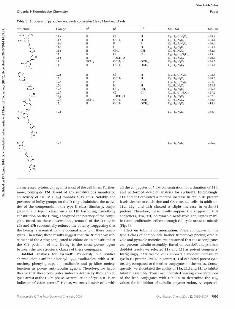
Table 1 Structures of pyrazole–oxadiazole conjugates 11a–i, 12a–i and 17a–b
Structure Compd R1 R2 R3 Mol. for Mol. wt
11a H Cl H C22H19ClN4O4 438.811b H OCH3 H C23H22N4O5 434.411c H F F C22H18F2N4O4 440.411d H H H C22H20N4O4 404.411e H CH3 CH3 C24H24N4O4 432.411f H Cl Cl C22H18Cl2N4O4 473.311g H –OCH2O– C23H20N4O6 448.411h OCH3 OCH3 OCH3 C25H26N4O7 494.511i H OCH3 OCH3 C24H24N4O6 464.4
12a H Cl H C20H13ClN4O3 392.812b H OCH3 H C21H16N4O4 388.312c H F F C20H12F2N4O3 394.312d H H H C20H14N4O3 358.312e H CH3 CH3 C22H18N4O3 386.412f H Cl Cl C20H12Cl2N4O3 427.212g H –OCH2O– C21H14N4O5 402.312h OCH3 OCH3 OCH3 C23H20N4O6 448.412i H OCH3 OCH3 C22H18N4O5 418.4
17a — — — C17H18N4O4 342.3
17b — — — C15H12N4O3 296.2
Organic & Biomolecular Chemistry Paper
This journal is © The Royal Society of Chemistry 2014 Org. Biomol. Chem., 2014, 12, 7993–8007 | 7995
Publ
ishe
d on
13
Aug
ust 2
014.
Dow
nloa
ded
by I
ndia
n In
stitu
te o
f C
hem
ical
Tec
hnol
ogy
(IIC
T),
Hyd
erab
ad o
n 26
/09/
2014
14:
35:2
5.
View Article Online
11a, which potentially inhibited cell growth, also significantlydown-regulated tubulin polymerization with an IC50 value of1.3 µM when compared to 11d and 11f. However, 11d and 11f
also inhibited tubulin assembly at an IC50 value of 3.9 µM and2.41 µM respectively (Table 3 and Fig. 4).
Effect on cell cycle arrest. Anti-microtubule agents areknown to induce cell cycle arrest in the G2/M phase of the cellcycle. Flow cytometry is routinely employed to determine thepopulation of cells in each phase of the cell cycle by measuringthe DNA content of individual cells.
Since conjugates 11a, 11d and 11f bind tubulin and inhibitcell growth, we analyzed for their ability to arrest cells at theG2/M phase. Therefore, A549 cells were treated with 5 μM con-centration of representative compounds for 24 h. Sub-sequently, harvested cells were subjected to flow cytometryanalysis. The lead conjugate 11a showed an increase of cells inthe G2/M phase to 77.3%. In comparison, cells accumulated to75.02% and 75.07% on 11d and 11f treatments in the G2/Mphase. Moreover, the cell number significantly decreased in
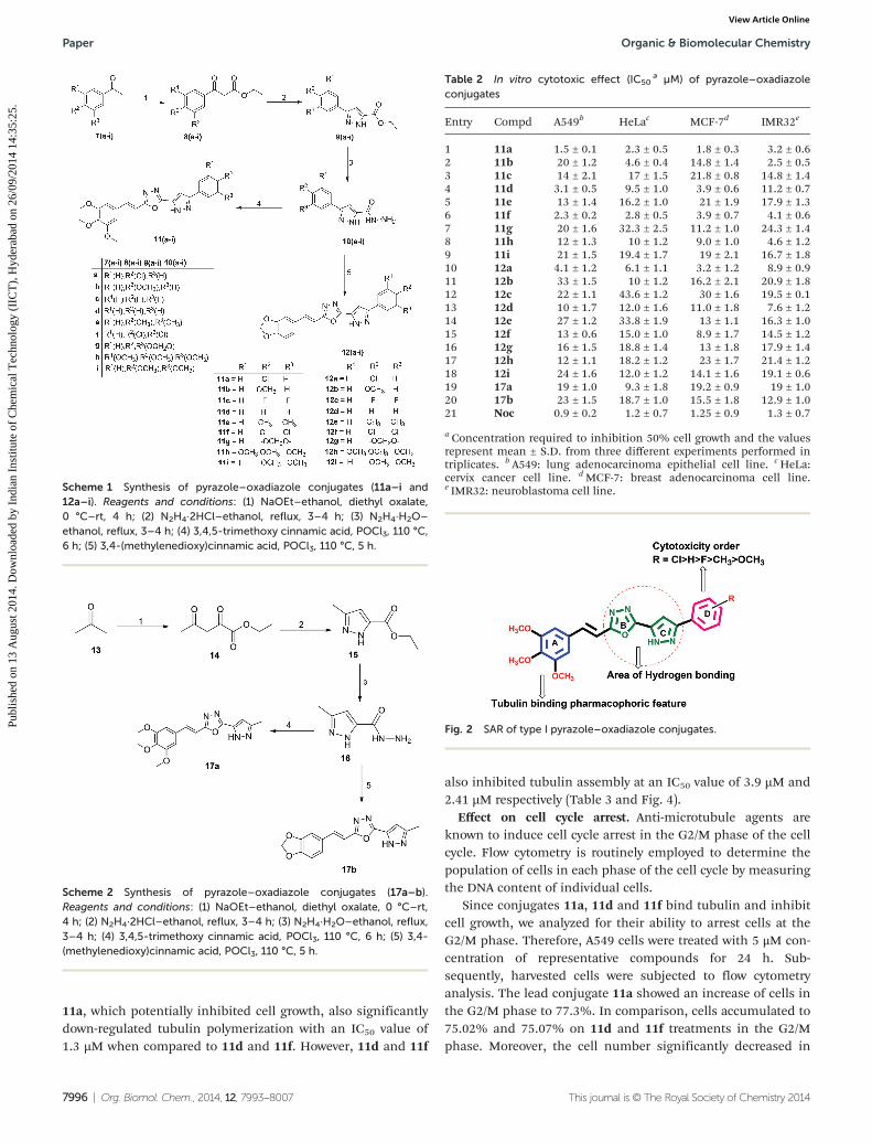
Scheme 1 Synthesis of pyrazole–oxadiazole conjugates (11a–i and12a–i). Reagents and conditions: (1) NaOEt–ethanol, diethyl oxalate,0 °C–rt, 4 h; (2) N2H4·2HCl–ethanol, reflux, 3–4 h; (3) N2H4·H2O–
ethanol, reflux, 3–4 h; (4) 3,4,5-trimethoxy cinnamic acid, POCl3, 110 °C,6 h; (5) 3,4-(methylenedioxy)cinnamic acid, POCl3, 110 °C, 5 h.
Table 2 In vitro cytotoxic effect (IC50a µM) of pyrazole–oxadiazole
conjugates
Entry Compd A549b HeLac MCF-7d IMR32e
1 11a 1.5 ± 0.1 2.3 ± 0.5 1.8 ± 0.3 3.2 ± 0.62 11b 20 ± 1.2 4.6 ± 0.4 14.8 ± 1.4 2.5 ± 0.53 11c 14 ± 2.1 17 ± 1.5 21.8 ± 0.8 14.8 ± 1.44 11d 3.1 ± 0.5 9.5 ± 1.0 3.9 ± 0.6 11.2 ± 0.75 11e 13 ± 1.4 16.2 ± 1.0 21 ± 1.9 17.9 ± 1.36 11f 2.3 ± 0.2 2.8 ± 0.5 3.9 ± 0.7 4.1 ± 0.67 11g 20 ± 1.6 32.3 ± 2.5 11.2 ± 1.0 24.3 ± 1.48 11h 12 ± 1.3 10 ± 1.2 9.0 ± 1.0 4.6 ± 1.29 11i 21 ± 1.5 19.4 ± 1.7 19 ± 2.1 16.7 ± 1.810 12a 4.1 ± 1.2 6.1 ± 1.1 3.2 ± 1.2 8.9 ± 0.911 12b 33 ± 1.5 10 ± 1.2 16.2 ± 2.1 20.9 ± 1.812 12c 22 ± 1.1 43.6 ± 1.2 30 ± 1.6 19.5 ± 0.113 12d 10 ± 1.7 12.0 ± 1.6 11.0 ± 1.8 7.6 ± 1.214 12e 27 ± 1.2 33.8 ± 1.9 13 ± 1.1 16.3 ± 1.015 12f 13 ± 0.6 15.0 ± 1.0 8.9 ± 1.7 14.5 ± 1.216 12g 16 ± 1.5 18.8 ± 1.4 13 ± 1.8 17.9 ± 1.417 12h 12 ± 1.1 18.2 ± 1.2 23 ± 1.7 21.4 ± 1.218 12i 24 ± 1.6 12.0 ± 1.2 14.1 ± 1.6 19.1 ± 0.619 17a 19 ± 1.0 9.3 ± 1.8 19.2 ± 0.9 19 ± 1.020 17b 23 ± 1.5 18.7 ± 1.0 15.5 ± 1.8 12.9 ± 1.021 Noc 0.9 ± 0.2 1.2 ± 0.7 1.25 ± 0.9 1.3 ± 0.7
a Concentration required to inhibition 50% cell growth and the valuesrepresent mean ± S.D. from three different experiments performed intriplicates. b A549: lung adenocarcinoma epithelial cell line. cHeLa:cervix cancer cell line. dMCF-7: breast adenocarcinoma cell line.e IMR32: neuroblastoma cell line.
Scheme 2 Synthesis of pyrazole–oxadiazole conjugates (17a–b).Reagents and conditions: (1) NaOEt–ethanol, diethyl oxalate, 0 °C–rt,4 h; (2) N2H4·2HCl–ethanol, reflux, 3–4 h; (3) N2H4·H2O–ethanol, reflux,3–4 h; (4) 3,4,5-trimethoxy cinnamic acid, POCl3, 110 °C, 6 h; (5) 3,4-(methylenedioxy)cinnamic acid, POCl3, 110 °C, 5 h.
Fig. 2 SAR of type I pyrazole–oxadiazole conjugates.
Paper Organic & Biomolecular Chemistry
7996 | Org. Biomol. Chem., 2014, 12, 7993–8007 This journal is © The Royal Society of Chemistry 2014
Publ
ishe
d on
13
Aug
ust 2
014.
Dow
nloa
ded
by I
ndia
n In
stitu
te o
f C
hem
ical
Tec
hnol
ogy
(IIC
T),
Hyd
erab
ad o
n 26
/09/
2014
14:
35:2
5.
View Article Online
the S phase. In contrast, vehicle or DMSO treated cells showeda majority of the population in the G1 phase (67.7%) and 11a,11d and 11f treated cells resulted in a significant increase ofcyclin-B1 protein, compared to DMSO treated cells (Fig. 5).Thus treatment with the pyrazole–oxadiazole conjugates
resulted in a clear G2/M phase arrest with a concomitantdecrease of cells in other phases of the cell cycle and inductionof cyclin-B1 protein.27
Effect on the microtubule network. Spindle poisons exerttheir action through the disruption of chromosome separationduring mitosis. The pyrazole–oxadiazole conjugates signifi-cantly decreased cell growth, inhibited tubulin assembly andstalled cells in the G2/M phase of the cell cycle. Therefore, weevaluated their ability to disrupt the microtubule network incells. Consequently, A549 cells were treated with 11a, 11d and11f at 5 μM concentration for 24 h. Immunofluorescence ana-lysis reveals that cells exhibited a round morphology typical ofmitotic arrest. Moreover, the chromatin was condensed in thenuclei, suggesting a metaphase arrest (Fig. 6).
Microtubules exhibit dynamic equilibrium with free tubulinmonomers in the cells. Pharmacological agents exploit thisproperty of microtubules to exert their anticancer effects.28
Since these conjugates disrupt tubulin polymerization, we elu-cidated their effect on cytosolic tubulin. Thus, A549 cells weretreated with 5 μM concentration of 11a, 11d and 11f for 24 h.CA-4 and colchicines were also treated in the same experimentas the standard reference. Subsequently, these soluble andpolymerized fractions were collected and subjected to westernblot analysis. Immunoblot results show that the cells exposedto these conjugates 11a, 11d, 11f contain more tubulin in thesoluble fraction of cells when compared to the insoluble frac-tion. Hence, increased tubulin in the soluble fraction of cells
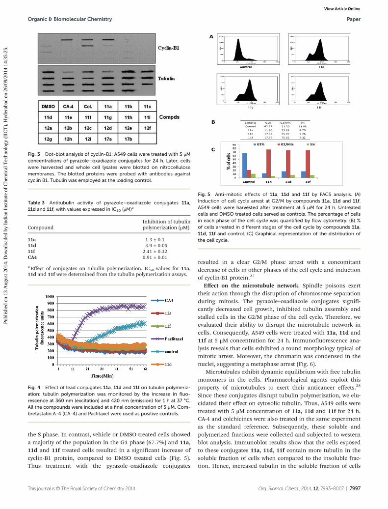
Fig. 3 Dot-blot analysis of cyclin-B1: A549 cells were treated with 5 μMconcentrations of pyrazole–oxadiazole conjugates for 24 h. Later, cellswere harvested and whole cell lysates were blotted on nitrocellulosemembranes. The blotted proteins were probed with antibodies againstcyclin B1. Tubulin was employed as the loading control.
Table 3 Antitubulin activity of pyrazole–oxadiazole conjugates 11a,11d and 11f, with values expressed in IC50 (μM)a
CompoundInhibition of tubulinpolymerization (µM)
11a 1.3 ± 0.111d 3.9 ± 0.0511f 2.41 ± 0.32CA4 0.91 ± 0.01
a Effect of conjugates on tubulin polymerization. IC50 values for 11a,11d and 11f were determined from the tubulin polymerization assays.
Fig. 4 Effect of lead conjugates 11a, 11d and 11f on tubulin polymeriz-ation: tubulin polymerization was monitored by the increase in fluo-rescence at 360 nm (excitation) and 420 nm (emission) for 1 h at 37 °C.All the compounds were included at a final concentration of 5 µM. Com-bretastatin A-4 (CA-4) and Paclitaxel were used as positive controls.
Fig. 5 Anti-mitotic effects of 11a, 11d and 11f by FACS analysis. (A)Induction of cell cycle arrest at G2/M by compounds 11a, 11d and 11f.A549 cells were harvested after treatment at 5 µM for 24 h. Untreatedcells and DMSO treated cells served as controls. The percentage of cellsin each phase of the cell cycle was quantified by flow cytometry. (B) %of cells arrested in different stages of the cell cycle by compounds 11a,11d, 11f and control. (C) Graphical representation of the distribution ofthe cell cycle.
Organic & Biomolecular Chemistry Paper
This journal is © The Royal Society of Chemistry 2014 Org. Biomol. Chem., 2014, 12, 7993–8007 | 7997
Publ
ishe
d on
13
Aug
ust 2
014.
Dow
nloa
ded
by I
ndia
n In
stitu
te o
f C
hem
ical
Tec
hnol
ogy
(IIC
T),
Hyd
erab
ad o
n 26
/09/
2014
14:
35:2
5.
View Article Online
treated with pyrazole–oxadiazole conjugates corroborated wellwith the inhibition of tubulin polymerization and arrest ofcells in the G2/M phase (Fig. 7).
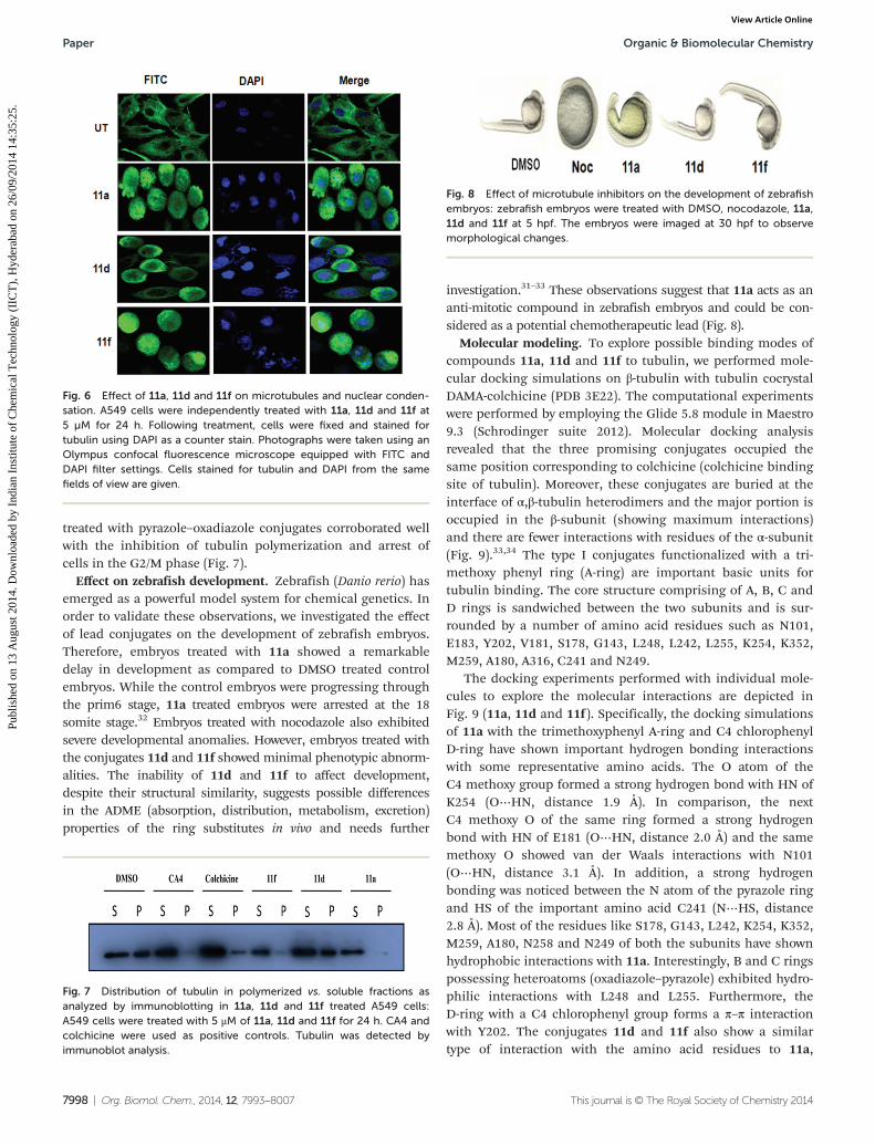
Effect on zebrafish development. Zebrafish (Danio rerio) hasemerged as a powerful model system for chemical genetics. Inorder to validate these observations, we investigated the effectof lead conjugates on the development of zebrafish embryos.Therefore, embryos treated with 11a showed a remarkabledelay in development as compared to DMSO treated controlembryos. While the control embryos were progressing throughthe prim6 stage, 11a treated embryos were arrested at the 18somite stage.32 Embryos treated with nocodazole also exhibitedsevere developmental anomalies. However, embryos treated withthe conjugates 11d and 11f showed minimal phenotypic abnorm-alities. The inability of 11d and 11f to affect development,despite their structural similarity, suggests possible differencesin the ADME (absorption, distribution, metabolism, excretion)properties of the ring substitutes in vivo and needs further
investigation.31–33 These observations suggest that 11a acts as ananti-mitotic compound in zebrafish embryos and could be con-sidered as a potential chemotherapeutic lead (Fig. 8).
Molecular modeling. To explore possible binding modes ofcompounds 11a, 11d and 11f to tubulin, we performed mole-cular docking simulations on β-tubulin with tubulin cocrystalDAMA-colchicine (PDB 3E22). The computational experimentswere performed by employing the Glide 5.8 module in Maestro9.3 (Schrodinger suite 2012). Molecular docking analysisrevealed that the three promising conjugates occupied thesame position corresponding to colchicine (colchicine bindingsite of tubulin). Moreover, these conjugates are buried at theinterface of α,β-tubulin heterodimers and the major portion isoccupied in the β-subunit (showing maximum interactions)and there are fewer interactions with residues of the α-subunit(Fig. 9).33,34 The type I conjugates functionalized with a tri-methoxy phenyl ring (A-ring) are important basic units fortubulin binding. The core structure comprising of A, B, C andD rings is sandwiched between the two subunits and is sur-rounded by a number of amino acid residues such as N101,E183, Y202, V181, S178, G143, L248, L242, L255, K254, K352,M259, A180, A316, C241 and N249.
The docking experiments performed with individual mole-cules to explore the molecular interactions are depicted inFig. 9 (11a, 11d and 11f ). Specifically, the docking simulationsof 11a with the trimethoxyphenyl A-ring and C4 chlorophenylD-ring have shown important hydrogen bonding interactionswith some representative amino acids. The O atom of theC4 methoxy group formed a strong hydrogen bond with HN ofK254 (O⋯HN, distance 1.9 Å). In comparison, the nextC4 methoxy O of the same ring formed a strong hydrogenbond with HN of E181 (O⋯HN, distance 2.0 Å) and the samemethoxy O showed van der Waals interactions with N101(O⋯HN, distance 3.1 Å). In addition, a strong hydrogenbonding was noticed between the N atom of the pyrazole ringand HS of the important amino acid C241 (N⋯HS, distance2.8 Å). Most of the residues like S178, G143, L242, K254, K352,M259, A180, N258 and N249 of both the subunits have shownhydrophobic interactions with 11a. Interestingly, B and C ringspossessing heteroatoms (oxadiazole–pyrazole) exhibited hydro-philic interactions with L248 and L255. Furthermore, theD-ring with a C4 chlorophenyl group forms a π–π interactionwith Y202. The conjugates 11d and 11f also show a similartype of interaction with the amino acid residues to 11a,
Fig. 6 Effect of 11a, 11d and 11f on microtubules and nuclear conden-sation. A549 cells were independently treated with 11a, 11d and 11f at5 µM for 24 h. Following treatment, cells were fixed and stained fortubulin using DAPI as a counter stain. Photographs were taken using anOlympus confocal fluorescence microscope equipped with FITC andDAPI filter settings. Cells stained for tubulin and DAPI from the samefields of view are given.
Fig. 7 Distribution of tubulin in polymerized vs. soluble fractions asanalyzed by immunoblotting in 11a, 11d and 11f treated A549 cells:A549 cells were treated with 5 μM of 11a, 11d and 11f for 24 h. CA4 andcolchicine were used as positive controls. Tubulin was detected byimmunoblot analysis.
Fig. 8 Effect of microtubule inhibitors on the development of zebrafishembryos: zebrafish embryos were treated with DMSO, nocodazole, 11a,11d and 11f at 5 hpf. The embryos were imaged at 30 hpf to observemorphological changes.
Paper Organic & Biomolecular Chemistry
7998 | Org. Biomol. Chem., 2014, 12, 7993–8007 This journal is © The Royal Society of Chemistry 2014
Publ
ishe
d on
13
Aug
ust 2
014.
Dow
nloa
ded
by I
ndia
n In
stitu
te o
f C
hem
ical
Tec
hnol
ogy
(IIC
T),
Hyd
erab
ad o
n 26
/09/
2014
14:
35:2
5.
View Article Online
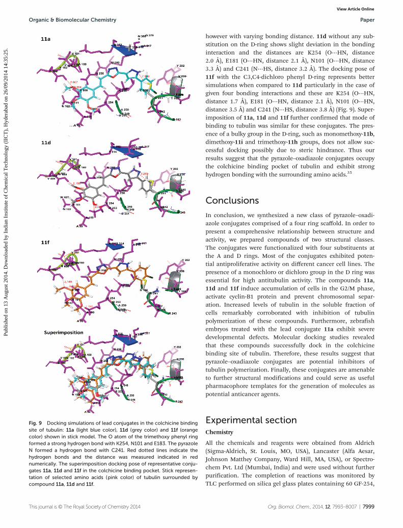
however with varying bonding distance. 11d without any sub-stitution on the D-ring shows slight deviation in the bondinginteraction and the distances are K254 (O⋯HN, distance2.0 Å), E181 (O⋯HN, distance 2.1 Å), N101 (O⋯HN, distance3.3 Å) and C241 (N⋯HS, distance 3.2 Å). The docking pose of11f with the C3,C4-dichloro phenyl D-ring represents bettersimulations when compared to 11d particularly in the case ofgiven four bonding interactions and these are K254 (O⋯HN,distance 1.7 Å), E181 (O⋯HN, distance 2.1 Å), N101 (O⋯HN,distance 3.5 Å) and C241 (N⋯HS, distance 3.8 Å) (Fig. 9). Super-imposition of 11a, 11d and 11f further confirmed that mode ofbinding to tubulin was similar for these conjugates. The pres-ence of a bulky group in the D-ring, such as monomethoxy-11b,dimethoxy-11i and trimethoxy-11h groups, does not allow suc-cessful docking possibly due to steric hindrance. Thus ourresults suggest that the pyrazole–oxadiazole conjugates occupythe colchicine binding pocket of tubulin and exhibit stronghydrogen bonding with the surrounding amino acids.35
Conclusions
In conclusion, we synthesized a new class of pyrazole–oxadi-azole conjugates comprised of a four ring scaffold. In order topresent a comprehensive relationship between structure andactivity, we prepared compounds of two structural classes.The conjugates were functionalized with four substituents atthe A and D rings. Most of the conjugates exhibited poten-tial antiproliferative activity on different cancer cell lines. Thepresence of a monochloro or dichloro group in the D ring wasessential for high antitubulin activity. The compounds 11a,11d and 11f induce accumulation of cells in the G2/M phase,activate cyclin-B1 protein and prevent chromosomal separ-ation. Increased levels of tubulin in the soluble fraction ofcells remarkably corroborated with inhibition of tubulinpolymerization of these compounds. Furthermore, zebrafishembryos treated with the lead conjugate 11a exhibit severedevelopmental defects. Molecular docking studies revealedthat these compounds successfully dock in the colchicinebinding site of tubulin. Therefore, these results suggest thatpyrazole–oxadiazole conjugates are potential inhibitors oftubulin polymerization. Finally, these conjugates are amenableto further structural modifications and could serve as usefulpharmacophore templates for the generation of molecules aspotential anticancer agents.
Experimental sectionChemistry
All the chemicals and reagents were obtained from Aldrich(Sigma-Aldrich, St. Louis, MO, USA), Lancaster (Alfa Aesar,Johnson Matthey Company, Ward Hill, MA, USA), or Spectro-chem Pvt. Ltd (Mumbai, India) and were used without furtherpurification. The completion of reactions was monitored byTLC performed on silica gel glass plates containing 60 GF-254,
Fig. 9 Docking simulations of lead conjugates in the colchicine bindingsite of tubulin: 11a (light blue color), 11d (grey color) and 11f (orangecolor) shown in stick model. The O atom of the trimethoxy phenyl ringformed a strong hydrogen bond with K254, N101 and E183. The pyrazoleN formed a hydrogen bond with C241. Red dotted lines indicate thehydrogen bonds and the distance was measured indicated in rednumerically. The superimposition docking pose of representative conju-gates 11a, 11d and 11f in the colchicine binding pocket. Stick represen-tation of selected amino acids (pink color) of tubulin surrounded bycompound 11a, 11d and 11f.
Organic & Biomolecular Chemistry Paper
This journal is © The Royal Society of Chemistry 2014 Org. Biomol. Chem., 2014, 12, 7993–8007 | 7999
Publ
ishe
d on
13
Aug
ust 2
014.
Dow
nloa
ded
by I
ndia
n In
stitu
te o
f C
hem
ical
Tec
hnol
ogy
(IIC
T),
Hyd
erab
ad o
n 26
/09/
2014
14:
35:2
5.
View Article Online
and visualization was achieved by a UV light or iodine indi-cator. Column chromatography was performed with Merck60–120 mesh silica gel. NMR spectra were recorded on BrukerUXNMR/XWIN-NMR (300 MHz) or Inova Varian-VXR-unity(400, 500 MHz) instruments. Chemical shifts (δ) are reportedin ppm downfield from an internal TMS standard. ESI spectrawere recorded on a Micro mass Quattro LC using ESI+ softwarewith a capillary voltage 3.98 kV and an ESI mode positive iontrap detector. High-resolution mass spectra (HRMS) wererecorded on a QSTAR XL Hybrid MS–MS mass spectrometer.Melting points were determined with electrothermal meltingpoint apparatus, and are uncorrected.
Preparation of ethyl 2,4-dioxo-4-(substituted phenyl)butano-ates 8a–i. Initially sodium ethanolate was prepared in situ anddiethyl oxalate (1.0 mol) was added slowly at 0 °C. The stirringwas continued for 15 minutes followed by the addition ofdifferent acetophenones 7(a–i) (1.0 mol) slowly in small por-tions, maintaining the temperature at 0 °C. After completionof addition, the stirring was continued for 4 h at room temp-erature. The progress of the reaction was monitored by TLCusing ethyl acetate and hexane as the mobile phase. After com-pletion of reaction, the reaction mixture was neutralized usingdilute H2SO4 solution and further extracted with ethyl acetatefollowed by evaporation of the solvent under reduced pressureto obtain solid products 8(a–i) (yield 80–90%). These weretaken as such for the next step without purification.
Preparation of ethyl 3-substituted phenyl-1H-pyrazole-5-car-boxylates 9a–i. To each of the ethyl 2,4-dioxo-4-(substitutedphenyl)butanoates 8(a–i) (1.0 mol) produced in the previousstep was added hydrazine dihydrochloride (N2H4·2HCl)(1.5 mol) in ethanol and refluxed for 3–4 h. After completionof the reaction, the ethanol was removed under reducedpressure and then 150–200 ml of water was added to theresidue followed by extraction with ethyl acetate (50 ml × 4).The organic layer was dried on anhydrous Na2SO4 and thesolvent was removed to obtain a crude compound that wasfurther purified by column chromatography using an ethylacetate and hexane solvent system (3 : 7). The pure compounds9(a–i) were eluted with 30–40% of ethyl acetate with goodyields.
Ethyl 3-(4-chlorophenyl)-1H-pyrazole-5-carboxylate 9a. Paleyellow colored solid (yield 80.0%): Rf = 0.3 (25% ethyl acetate–hexane); 1H NMR (300 MHz, CDCl3); δ 1.34–1.42 (t, 3H, J1 =6.9 Hz, J2 = 7.9 Hz, –CH3), 4.33–4.41 (q, 2H, J1 = 6.9 Hz, J2 =7.9 Hz, CH2), 7.04 (s, 1H, ArH), 7.36 (d, 2H, J = 7.9 Hz, ArH),7.70 (d, 2H, J = 7.9 Hz, ArH) ppm; MS (ESI) m/z 251 [M + H].
Ethyl 3-(4-methoxyphenyl)-1H-pyrazole-5-carboxylate 9b.Pale yellow colored solid (yield 80.0%): Rf = 0.3 (30% ethylacetate–hexane); 1H NMR (300 MHz, CDCl3); δ 1.22–1.36 (t, 3H,J1 = 7.5 Hz, –CH3), 3.81 (s, 3H, –OCH3), 4.20–4.35 (q, 2H, J1 =4.5 Hz, –CH2), 6.88–6.90 (m, 3H, ArH), 7.60 (d, 2H, J = 9.0 HzArH), 8.33 (brs, 1H, –NH) ppm; MS (ESI) m/z 248 [M + H].
Ethyl 3-(3,4-difluorophenyl)-1H-pyrazole-5-carboxylate 9c.Yellow colored solid (yield 75.0%): Rf = 0.3 (20% ethyl acetate–hexane); 1H NMR (200 MHz, CDCl3); δ 1.22–1.32 (t, 3H, J1 =7.1 Hz, J2 = 7.5 Hz, –CH3), 4.20–4.33 (q, 2H, J = 7.1 Hz, –CH2),
7.02 (s, 1H, ArH), 7.22–7.43 (m, 2H, ArH), 7.72 (d, 1H, J = 6.7 Hz,ArH), 9.31 (brs, 1H, –NH) ppm; MS (ESI) m/z 253 [M + H].
Ethyl 3-phenyl-1H-pyrazole-5-carboxylate 9d. Pale yellowcolored solid (yield 85.0%): Rf = 0.3 (25% ethyl acetate–hexane);1H NMR (500 MHz, CDCl3); δ 1.35–1.40 (t, 3H, J1 = 6.9 Hz,–CH3), 4.32–4.41 (q, 2H, J1 = 6.9 Hz, J2 = 7.9 Hz, –CH2), 7.04 (s,1H, ArH), 7.36 (d, 2H, J = 7.9 Hz, ArH), 7.36 (d, 3H, J = 7.9 Hz,ArH), 9.31 (brs, 1H, –NH) ppm; MS (ESI) m/z 217 [M + H].
Ethyl 3-(3,4-dichlorophenyl)-1H-pyrazole-5-carboxylate 9e.Yellow colored solid (yield 75.0%): Rf = 0.3 (20% ethyl acetate–hexane); 1H NMR (300 MHz, CDCl3); δ 1.39–1.45 (t, 3H, J =6.5 Hz, –CH3), 4.33–4.46 (q, 2H, J = 7.4 Hz, –CH2), 7.16 (s, 1H,ArH), 7.49–7.63 (m, 2H, ArH), 7.90–7.94 (m, 1H, ArH), 8.20(brs, 1H, –NH) ppm; MS (ESI) m/z 285 [M + H].
Ethyl 3-(3,4-dimethylphenyl)-1H-pyrazole-5-carboxylate 9f.Yellow colored solid (yield 80.0%): Rf = 0.3 (25% ethyl acetate–hexane); 1H NMR (200 MHz, CDCl3); δ 1.22–1.32 (t, 3H, J =5.8 Hz, –CH3), 2.25 (s, 6H, –CH3), 4.17–4.30 (q, 2H, J = 5.8 Hz,–CH2), 6.93 (s, 1H, ArH), 7.07 (d, 1H, J = 8.1 Hz, ArH), 7.39(d, 1H, J = 6.9 Hz, ArH), 7.47 (s, 1H, ArH), 8.20 (brs, 1H, –NH)ppm; MS (ESI) m/z 245 [M + H].
Ethyl 3-(benzo[d][1,3]dioxol-5-yl)-1H-pyrazole-5-carboxylate9g. Pale yellow colored solid (yield 85.0%): Rf = 0.3 (40% ethylacetate–hexane); 1H NMR (400 MHz, CDCl3); δ 1.35–1.42 (t, 3H,J = 7.5 Hz, –CH3), 4.35–4.45 (q, 2H, J = 6.7 Hz, –CH2), 6.0 (s,2H, OCH2O), 6.84 (d, 1H, J = 8.3 Hz, ArH), 6.99 (s, 1H, ArH),7.21–7.26 (m, 2H, ArH) ppm; MS (ESI) m/z 261 [M + H].
Ethyl 3-(3,4,5-trimethoxyphenyl)-1H-pyrazole-5-carboxylate9h. Yellow colored solid (yield 80.0%): Rf = 0.3 (40% ethylacetate–hexane); 1H NMR (300 MHz, CDCl3); δ 1.34–1.39 (t, 3H,J = 7.4 Hz, –CH3), 3.87 (s, 3H, –OCH3), 3.88 (s, 6H, –OCH3),4.33–4.45 (q, 2H, J1 = 7.4 Hz, –CH2), 7.00 (s, 2H, ArH), 7.04 (s,1H, ArH) ppm; MS (ESI) m/z 307 [M + H].
Ethyl 3-(3,4-dimethoxyphenyl)-1H-pyrazole-5-carboxylate 9i.Pale yellow colored solid (yield 80.0%): Rf = 0.3 (30% ethylacetate–hexane); 1H NMR (300 MHz, CDCl3); δ 1.23–1.31 (t, 3H,J = 7.1 Hz, –CH3), 3.85 (s, 3H, –OCH3), 3.90 (s, 3H, –OCH3),4.19–4.32 (q, 2H, J = 7.1 Hz, –CH2), 6.88 (d, 1H, J1 = 7.1 Hz,ArH), 6.94 (s, 1H, ArH), 7.21–7.28 (m, 1H, ArH), 7.29–7.34 (m,1H, ArH), 9.67 (brs, 1H, –NH) ppm; MS (ESI) m/z 277 [M + H].
Preparation of 3-substituted phenyl-1H-pyrazole-5-carbohy-drazides 10a–i. To the ethyl 3-substituted phenyl-1H-pyrazole-5-carboxylates 9(a–i) obtained in the above step was addedhydrazine hydrate (4.0 mol) in ethanol at room temperatureand heated to reflux for 3–4 h. The progress of the reactionwas monitored by TLC using ethyl acetate and hexane (1 : 1) asthe solvent system. The reaction mixture was allowed to attainroom temperature and then the solvent was distilled off underreduced pressure. Then the residue was dissolved in water andextracted twice with ethyl acetate, dried over anhydrous Na2SO4
and the solvent was removed under reduced pressure to affordthe corresponding compound 10(a–i) as a crystalline solid. Thecarbohydrazides obtained by this method were pure and weretaken as such for the next step without further purification.
Preparation of (E)-2-(3-substituted phenyl-1H-pyrazol-5-yl)-5-(3,4,5-trimethoxystyryl)-1,3,4-oxadiazole 11a–i. To each of the
Paper Organic & Biomolecular Chemistry
8000 | Org. Biomol. Chem., 2014, 12, 7993–8007 This journal is © The Royal Society of Chemistry 2014
Publ
ishe
d on
13
Aug
ust 2
014.
Dow
nloa
ded
by I
ndia
n In
stitu
te o
f C
hem
ical
Tec
hnol
ogy
(IIC
T),
Hyd
erab
ad o
n 26
/09/
2014
14:
35:2
5.
View Article Online
3-substituted phenyl-1H-pyrazole-5-carbohydrazides 10(a–i)(1.0 mol) produced in the above step was added (E)-3-(3,4,5-tri-methoxyphenyl)acrylic acid (1.0 mol) in POCl3 and stirred at atemperature of 110 °C for 6 h. The completion of the reactionwas monitored by TLC in the ethyl acetate and hexane solventsystem and then ice cold water was added to the reactionmixture followed by extraction with ethyl acetate (50 ml × 4).The organic layer so obtained was washed with saturatedNaHCO3 solution and dried on anhydrous Na2SO4. Ethylacetate was distilled off under vacuum to produce a crude com-pound and this was purified by column chromatography usingethyl acetate and hexane resulting in pure compounds (E)-2-(3-substituted phenyl-1H-pyrazol-5-yl)-5-(3,4,5-trimethoxy-styryl)-1,3,4-oxadiazole 11(a–i) in good yields (60–70%).
(E)-2-(3-(4-Chlorophenyl)-1H-pyrazol-5-yl)-5-(3,4,5-trimethoxy-styryl)-1,3,4-oxadiazole 11a. This compound was preparedusing the procedure described above by the addition of 3-(4-chlorophenyl)-1H-pyrazole-5-carbohydrazide 10a (236 mg,1.0 mmol) and (E)-3-(3,4,5-trimethoxyphenyl)acrylic acid(238 mg, 1.0 mmol). The compound was obtained as a yellowcolored solid. Yield: 300 mg (68.4%); mp: 225–227 °C; 1H NMR(300 MHz, DMSO-d6); δ 3.79 (s, 3H, –OCH3), 3.90 (s, 6H,–OCH3), 6.94 (s, 2H, ArH), 7.18 (d, 1H, J = 18.3 Hz, -trans H),7.45 (d, 2H, J = 8.4 Hz, ArH), 7.59 (d, 1H, J = 16.2 Hz, -trans H),7.79 (d, 2H, J = 8.4 Hz, ArH), 7.90 (s, 1H, ArH); 13C NMR(75 MHz, DMSO-d6): δ 54.1, 58.2, 101.3, 103.4, 107.2, 125.2,127.1, 128.4, 131.5, 137.2, 151.2, 161.8 ppm; IR (KBr) (νmax/cm−1): ν = 3428, 2925, 2846, 1611, 1582, 1503, 1456, 1311,1251, 1176, 1125, 1029, 1003 cm−1; MS (ESI) 439 [M + H] m/z[M + H]; HR-MS (ESI) m/z for C22H20N4O4Cl calculated m/z:439.11676, found m/z: 439.11597.
(E)-2-(3-(4-Methoxyphenyl)-1H-pyrazol-5-yl)-5-(3,4,5-trimethoxy-styryl)-1,3,4-oxadiazole 11b. This compound was preparedusing the procedure described above by the addition of 3-(4-methoxyphenyl)-1H-pyrazole-5-carbohydrazide 10b (232 mg,1.0 mmol) and (E)-3-(3,4,5-trimethoxyphenyl)acrylic acid(238 mg, 1.0 mmol). The compound was obtained as a paleyellow colored solid. Yield: 298 mg (68%); mp: 189–192 °C; 1HNMR (300 MHz, DMSO-d6); δ 3.56 (s, 3H, –OCH3), 3.88 (s, 3H,–OCH3), 3.94 (s, 6H, –OCH3), 6.49 (s, 1H, ArH), 6.96 (s, 1H,ArH), 7.01–7.05 (m, 2H, ArH), 7.11 (s, 1H, ArH), 7.71 (d, 1H, J =15.8 Hz, -trans H), 7.95–7.99 (m, 2H, ArH), 8.42 (d, 1H, J =15.8 Hz, -trans H); 13C NMR (75 MHz, DMSO-d6): δ 55.3, 55.8,97.6, 104.9, 114.4, 120.9, 122.4, 122.9, 133.3, 128.3, 132.8,155.8, 161.2 ppm; IR (KBr) (νmax/cm
−1): ν = 3208, 3160, 2932,2841, 1625, 1582, 1530, 1509, 1418, 1362, 1241, 1133, 1091,1052 cm−1; MS (ESI) m/z 435 [M + H]; HR-MS (ESI) m/z forC23H23N4O5 calculated m/z: 435.16630, found m/z: 435.16665.
(E)-2-(3-(3,4-Difluorophenyl)-1H-pyrazol-5-yl)-5-(3,4,5-tri-methoxystyryl)-1,3,4-oxadiazole 11c. This compound wasprepared using the procedure described above by the additionof 3-(3,4-difluorophenyl)-1H-pyrazole-5-carbohydrazide 10c(238 mg, 1.0 mmol) and (E)-3-(3,4,5-trimethoxyphenyl)acrylicacid (238 mg, 1.0 mmol). The compound was obtained as abrown colored solid. Yield: 295 mg (67%); mp: 150–152 °C; 1HNMR (300 MHz, DMSO-d6); δ 3.81 (s, 3H, –OCH3), 3.92 (s, 6H,
–OCH3), 6.91 (s, 2H, ArH), 7.09–7.42 (m, 1H, ArH), 7.21 (d, 1H,J = 16.0 Hz, -trans H), 7.52–7.73 (m, 1H, ArH), 7.62 (d, 1H, J =16.0 Hz, -trans H), 7.75–7.88 (m, 1H, ArH), 7.96 (s, 1H, ArH);13C NMR (75 MHz, DMSO-d6): 54.1, 58.1, 101.6, 103.3, 103.4,107.2, 107.4, 112.7, 112.8, 116.2, 128.3, 137.0, 137.2, 151.2,161.4, 161.7 δ ppm; IR (KBr) (νmax/cm
−1): ν = 3112, 3049, 2942,1648, 1612, 1583, 1468, 1419, 1335, 1236, 1156, 1040 cm−1; MS(ESI) m/z 441 [M + H]; HR-MS (ESI) m/z for C22H19N4O4F2 cal-culated m/z: 441.13689, found m/z: 441.13681.
(E)-2-(3-Phenyl-1H-pyrazol-5-yl)-5-(3,4,5-trimethoxystyryl)-1,3,4-oxadiazole 11d. This compound was prepared using theprocedure described above by the addition of 3-phenyl-1H-pyr-azole-5-carbohydrazide 10d (202 mg, 1.0 mmol) and (E)-3-(3,4,5-trimethoxyphenyl)acrylic acid (238 mg, 1.0 mmol). Thecompound was obtained as a pale yellow colored solid. Yield:270 mg (66.8%); mp: 205–207 °C; 1H NMR (300 MHz, DMSO-d6); δ 3.86 (s, 3H, –OCH3), 3.93 (s, 6H, –OCH3), 6.89 (s, 2H,ArH), 7.10 (d, 1H, J = 16.2 Hz, -trans H), 7.20 (s, 1H, ArH),7.40 (d, 1H, J = 7.1 Hz, ArH), 7.43–7.52 (t, 1H, J = 7.5 Hz, ArH),7.63 (d, 1H, J = 16.4 Hz, -trans H), 7.67–7.71 (m, 1H, ArH),7.80 (d, 2H, J = 7.1 Hz, ArH); 13C NMR (75 MHz, DMSO-d6):δ 55.7, 59.9, 102.4, 104.8, 108.8, 125.2, 128.2, 128.6, 130.0,138.6, 138.9, 152.91, 158.8, 163.3 ppm; IR (KBr) (νmax/cm
−1):ν = 3427, 3150, 3110, 3013, 2851, 1644, 1603, 1521, 1447,1233, 1035 cm−1; MS (ESI) m/z 405 [M + H]; HR-MS (ESI)m/z for C22H20N4O4 calculated m/z: 405.15573, found m/z:405.15483.
(E)-2-(3-(3,4-Dimethylphenyl)-1H-pyrazol-5-yl)-5-(3,4,5-tri-methoxystyryl)-1,3,4-oxadiazole 11e. This compound wasprepared using the procedure described above by the additionof 3-(3,4-dimethylphenyl)-1H-pyrazole-5-carbohydrazide 11e(230 mg, 1.0 mmol) and (E)-3-(3,4,5-trimethoxyphenyl)acrylicacid (238 mg, 1.0 mmol). The compound was obtained as apale yellow colored solid. Yield: 280 mg (64.8%); mp:222–224 °C; 1H NMR (300 MHz, DMSO-d6); δ 2.29 (s, 6H,–CH3), 2.33 (s, 6H, –CH3), 3.80 (s, 3H, –OCH3), 3.92 (s, 6H,–OCH3), 6.90 (s, 3H, ArH), 6.96 (d, 2H, J = 5.4 Hz, ArH), 7.08 (d,1H, J = 16.2 Hz, -trans H), 7.12–7.26 (m, 1H, ArH), 7.45 (d, 1H,J = 16.2 Hz, -trans H), 7.48–7.67 (m, 2H, ArH); 13C NMR(75 MHz, DMSO-d6): δ 19.1, 19.3, 55.7, 59.9, 101.8, 104.8,109.0, 109.1, 122.6, 126.3, 129.7, 130.0, 138.0, 138.6, 152.8,163.0 ppm; IR (KBr) (νmax/cm
−1): ν = 3143, 3000, 2938, 2835,1647, 1583, 1528, 1504, 1451, 1419, 1333, 1244, 1186, 1154,1125, 1038 cm−1; MS (ESI) m/z 434 [M + H]; HR-MS (ESI) m/zfor C24H25N4O4 calculated m/z: 433.18703, found m/z:433.18632.
(E)-2-(3-(3,4-Dichlorophenyl)-1H-pyrazol-5-yl)-5-(3,4,5-tri-methoxystyryl)-1,3,4-oxadiazole 11f. This compound wasprepared using the procedure described above by the additionof 3-(3,4-dichlorophenyl)-1H-pyrazole-5-carbohydrazide 10f(271 mg, 1.0 mmol) and (E)-3-(3,4,5-trimethoxyphenyl)acrylicacid (238 mg, 1.0 mmol). The compound was obtained as abrown colored solid. Yield: 315 mg (66.5%); mp: 158–160 °C;1H NMR (400 MHz, DMSO-d6); δ 3.89 (s, 3H, –OCH3), 3.90 (s,6H, –OCH3), 6.76 (s, 2H, ArH), 6.80 (s, 1H, ArH), 6.93 (d, 1H,J = 15.8 Hz, -trans H), 6.98 (d, 1H, J = 7.1 Hz, ArH), 7.26 (s, 1H,
Organic & Biomolecular Chemistry Paper
This journal is © The Royal Society of Chemistry 2014 Org. Biomol. Chem., 2014, 12, 7993–8007 | 8001
Publ
ishe
d on
13
Aug
ust 2
014.
Dow
nloa
ded
by I
ndia
n In
stitu
te o
f C
hem
ical
Tec
hnol
ogy
(IIC
T),
Hyd
erab
ad o
n 26
/09/
2014
14:
35:2
5.
View Article Online
ArH), 7.40 (d, 1H, J = 15.8 Hz, -trans H), 7.52 (d, 1H, J = 8.2 Hz,ArH); 13C NMR (75 MHz, DMSO-d6): δ 54.1, 58.2, 103.2, 107.5,128.4, 136.3, 137.0, 137.1, 137.3, 151.2, 160.9, 161.4,162.1 ppm; IR (KBr) (νmax/cm
−1): ν = 3049, 3000, 2939, 2835,1647, 1583, 1529, 1503, 1466, 1419, 1334, 1245, 1186, 1125,1045 cm−1; MS (ESI) m/z 511 [M + K].
(E)-2-(3-(Benzo[d][1,3]dioxol-5-yl)-1H-pyrazol-5-yl)-5-(3,4,5-tri-methoxystyryl)-1,3,4-oxadiazole 11g. This compound was pre-pared using the procedure described above by the addition of3-(benzo[d][1,3]dioxol-5-yl)-1H-pyrazole-5-carbohydrazide 10g(246 mg, 1.0 mmol) and (E)-3-(3,4,5-trimethoxyphenyl)acrylicacid (238 mg, 1.0 mmol). The compound was obtained as abrown red colored solid. Yield: 294 mg (65.6%); mp:199–202 °C; 1H NMR (300 MHz, DMSO-d6); δ 3.84 (s, 3H,–OCH3), 3.94 (s, 6H, –OCH3), 6.05 (s, 2H, –OCH2O–), 6.86 (d,1H, J = 7.9 Hz, ArH), 6.94 (d, 1H, J = 16.8 Hz, -trans H), 7.09 (s,2H, ArH), 7.16 (d, 2H, J = 8.9 Hz, ArH), 7.60 (d, 1H, J = 15.8 Hz,-trans H), 7.67–7.69 (m, 1H, ArH); 13C NMR (75 MHz, DMSO-d6): δ 55.8, 60.0, 101.2, 102.0, 104.8, 105.7, 108.5, 109.2, 119.3,130.1, 138.1, 138.8, 147.5, 147.8, 153.0, 162.6 ppm; IR (KBr)(νmax/cm
−1): ν = 3050, 2983, 1645, 1583, 1529, 1504, 1461,1418, 1334, 1240, 1188, 1125, 1035 cm−1; MS (ESI) m/z 449 [M+ H]; HR-MS (ESI) m/z for C23H21N4O6 calculated m/z:449.14556, found m/z: 449.14569.
(E)-2-(3-(3,4,5-Trimethoxyphenyl)-1H-pyrazol-5-yl)-5-(3,4,5-tri-methoxystyryl)-1,3,4-oxadiazole 11h. This compound was pre-pared using the procedure described above by the addition of3-(3,4,5-trimethoxyphenyl)-1H-pyrazole-5-carbohydrazide 11h(292 mg, 1.0 mmol) and (E)-3-(3,4,5-trimethoxyphenyl)acrylicacid (238 mg, 1.0 mmol). The compound was obtained as ayellow colored crystal. Yield: 345 mg (69.8%); mp: 184–185 °C;1H NMR (400 MHz, DMSO-d6); δ 3.70 (s, 9H, –OCH3), 3.85 (s,9H, –OCH3), 7.08–7.25 (m, 4H, ArH), 7.37–7.50 (m, 2H, J =16.9 Hz, trans H, ArH), 7.63 (d, 1H, J = 16.3 Hz, trans H);13C NMR (75 MHz, DMSO-d6): δ 56.0, 60.0, 103.0, 105.4, 109.2,124.0, 130.2, 137.7, 138.9, 153.1, 153.3, 159.1, 163.6 ppm;IR (KBr) (νmax/cm
−1): ν = 3115, 2947, 2832, 1683, 1612, 1587,1545, 1463, 1427, 1377, 1346, 1237, 1132, 1070 cm−1; MS (ESI)m/z 495 [M + H]; HR-MS (ESI) m/z for C25H27N4O7 calculatedm/z: 495.18944, found m/z: 495.18350.
(E)-2-(3-(3,4-Dimethoxyphenyl)-1H-pyrazol-5-yl)-5-(3,4,5-tri-methoxystyryl)-1,3,4-oxadiazole 11i. This compound was pre-pared using the procedure described above by the additionof 3-(3,4-dimethoxyphenyl)-1H-pyrazole-5-carbohydrazide 10i(262 mg, 1.0 mmol) and (E)-3-(3,4,5-trimethoxyphenyl)acrylicacid (238 mg, 1.0 mmol). The compound was obtained as abrown colored solid. Yield: 315 mg (67.8%); mp: 158–160 °C;1H NMR (300 MHz, DMSO-d6); δ 3.89 (s, 3H, –OCH3), 3.91 (s,9H, –OCH3), 3.93 (s, 3H, –OCH3), 6.76 (s, 1H, ArH), 6.80 (d,2H, J = 6.2 Hz, ArH), 6.92 (d, 1H, J = 16.3 Hz, -trans H), 6.98 (d,1H, J = 16.3 Hz, -trans H), 7.04–7.08 (m, 1H, ArH), 7.19–7.25(m, 1H ArH), 7.39–7.45 (m, 1H ArH); 13C NMR (75 MHz,DMSO-d6): δ 55.9, 60.0, 105.2, 109.3, 130.2, 138.1, 138.8, 153.0,162.9, 163.9 ppm; IR (KBr) (νmax/cm
−1): ν = 3150, 2883, 1660,1645, 1553, 1529, 1500, 1461, 1418, 1287, 1240, 1188, 1125,1033 cm−1; MS (ESI) m/z 465 [M + H].
Preparation of (E)-2-(2-(benzo[d][1,3]dioxol-5-yl)vinyl)-5-(3-substituted phenyl-1H-pyrazol-5-yl)-1,3,4-oxadiazole 12a–i. Toeach of the 3-substituted phenyl-1H-pyrazole-5-carbohydra-zides 10(b–i) (1.0 mol) produced in the above step was added(E)-3-(benzo[d][1,3]dioxol-5-yl)acrylic acid (1.0 mol) in POCl3and stirred at a temperature of 110 °C for 5 h. The completionof the reaction was monitored by TLC in an ethyl acetate andhexane solvent system and then ice cold water was added tothe reaction mixture followed by extraction with ethyl acetate(50 ml × 4). The organic layer so obtained was washed withsaturated NaHCO3 solution and dried on anhydrous Na2SO4.Ethyl acetate was distilled off under vacuum to produce acrude compound and this was further purified by columnchromatography using ethyl acetate and hexane to afford purecompounds of each (E)-2-(2-(benzo[d][1,3]dioxol-5-yl)vinyl)-5-(3-substituted phenyl-1H-pyrazol-5-yl)-1,3,4-oxadiazole 12(a–i)in good yields (65–75%).
(E)-2-(2-(Benzo[d][1,3]dioxol-5-yl)vinyl)-5-(3-(4-chlorophenyl)-1H-pyrazol-5-yl)-1,3,4-oxadiazole 12a. This compound was pre-pared using the procedure described above by the addition of3-(4-chlorophenyl)-1H-pyrazole-5-carbohydrazide 10a (236 mg,1.0 mmol) and (E)-3-(benzo[d][1,3]dioxol-5-yl)acrylic acid(192 mg, 1.0 mmol). The compound was obtained as a browncolored solid. Yield: 263 mg (67%); mp: 164–166 °C; 1H NMR(300 MHz, DMSO-d6); δ 6.04 (s, 2H, –OCH2O–), 6.81 (d, 1H, J =16.4 Hz, -trans H), 6.84 (d, 2H, J = 7.7 Hz, ArH), 7.0 (d, 1H, J =8.1 Hz, ArH), 7.09–7.21 (m, 2H, ArH), 7.41 (d, 1H, J = 16.4 Hz,-trans H), 7.46 (s, 2H, ArH), 7.64–7.84 (m, 1H, ArH); 13C NMR(75 MHz, DMSO-d6): δ 101.4, 106.0, 108.0, 108.3, 124.0, 129.0,137.8, 147.9, 148.6, 162.8, 163.9 ppm; IR (KBr) (νmax/cm
−1): ν =2924, 2854, 1743, 1638, 1603, 1579, 1530, 1496, 1445, 1355,1316, 1255, 1255, 1227, 1097, 1033, 965 cm−1; MS (ESI) m/z 393[M + H].
(E)-2-(2-(Benzo[d][1,3]dioxol-5-yl)vinyl)-5-(3-(4-methoxyphenyl)-1H-pyrazol-5-yl)-1,3,4-oxadiazole 12b. This compound wasprepared using the procedure described above by the additionof 3-(4-methoxyphenyl)-1H-pyrazole-5-carbohydrazide 10b(232 mg, 1.0 mmol) and (E)-3-(benzo[d][1,3]dioxol-5-yl)acrylicacid (192 mg, 1.0 mmol). The compound was obtained as ayellow colored solid. Yield: 235 mg (65.6%); mp: 186–189 °C;1H NMR (500 MHz, DMSO-d6); δ 3.81 (s, 3H, –OCH3), 6.09 (s,2H, –OCH2O–), 6.98 (d, 1H, J = 7.9 Hz, ArH), 7.02 (d, 2H, J =7.7 Hz, ArH), 7.16–7.40 (m, 3H, J = 15.2 Hz, -trans H, ArH), 7.53(s, 1H, ArH), 7.60 (d, 1H, J = 16.4 Hz, -trans H), 7.81 (d, 2H, J =7.7 Hz, ArH); 13C NMR (75 MHz, DMSO-d6): δ 55.2, 101.4,101.8, 106.1, 106.2, 107.8, 108.4, 114.4, 120.7, 124.2, 126.8,129.1, 137.5, 138.5, 143.9, 148.0, 148.8, 159.2, 159.5,163.6 ppm; IR (KBr) (νmax/cm
−1): ν = 3195, 2922, 2852, 1608,1501, 1447, 1257, 1360, 1308, 1177, 1119, 1032, 965 cm−1; MS(ESI) m/z 389 [M + H]; HR-MS (ESI) m/z for C21H17N4O4 calcu-lated m/z: 389.12443, found m/z: 389.12518.
(E)-2-(2-(Benzo[d][1,3]dioxol-5-yl)vinyl)-5-(3-(3,4-difluorophenyl)-1H-pyrazol-5-yl)-1,3,4-oxadiazole 12c. This compound wasprepared using the procedure described above by the additionof 3-(3,4-difluorophenyl)-1H-pyrazole-5-carbohydrazide 10c(238 mg, 1.0 mmol) and (E)-3-(benzo[d][1,3]dioxol-5-yl)acrylic
Paper Organic & Biomolecular Chemistry
8002 | Org. Biomol. Chem., 2014, 12, 7993–8007 This journal is © The Royal Society of Chemistry 2014
Publ
ishe
d on
13
Aug
ust 2
014.
Dow
nloa
ded
by I
ndia
n In
stitu
te o
f C
hem
ical
Tec
hnol
ogy
(IIC
T),
Hyd
erab
ad o
n 26
/09/
2014
14:
35:2
5.
View Article Online
acid (192 mg, 1.0 mmol). The compound was obtained as ayellow colored solid; yield: 250 mg (63.4%); mp: 210–212 °C;1H NMR (300 MHz, DMSO-d6); δ 6.04 (s, 2H, –OCH2O–), 6.88(s, 1H, ArH), 6.99 (d, 1H, J = 16.6 Hz, -trans H), 7.09 (s, 1H,ArH), 7.23 (s, 1H, ArH), 7.42–7.68 (m, 1H, ArH), 7.59 (d, 1H, J =16.4 Hz, -trans H), 7.72–7.84 (m, 1H, ArH), 7.88 (s, 2H, ArH),13.93 (brs, 1H, –NH); 13C NMR (75 MHz, DMSO-d6): δ 101.4,103.6, 106.1, 107.7, 108.4, 114.5, 117.3, 118.4, 122.3, 123.4,124.3, 129.0, 138.7, 139.7, 148.0, 148.9, 163.7 ppm; IR (KBr)(νmax/cm
−1): ν = 3116, 3055, 2921, 2853, 1637, 1609, 1503,1494, 1491, 1446, 1362, 1246, 1036 cm−1; MS (ESI) m/z 395[M + H]; HR-MS (ESI) m/z for C20H13F2N4O3 calculatedm/z:395.09502, found m/z: 395.09491.
(E)-2-(2-(Benzo[d][1,3]dioxol-5-yl)vinyl)-5-(3-phenyl-1H-pyrazol-5-yl)-1,3,4-oxadiazole 12d. This compound was prepared usingthe procedure described above by the addition of 3-phenyl-1H-pyrazole-5-carbohydrazide 10d (202 mg, 1.0 mmol) and (E)-3-(benzo[d][1,3]dioxol-5-yl)acrylic acid (192 mg, 1.0 mmol). Thecompound was obtained as a saffron colored solid. Yield:235 mg (65.5%); mp: 220–222 °C; 1H NMR (400 MHz, DMSO-d6); δ 6.04 (s, 2H, –OCH2O–), 6.87 (d, 1H, J = 8.1 Hz, ArH), 6.92(d, 1H, J = 16.2 Hz, -trans H), 7.05–7.11 (m, 2H, ArH), 7.20 (s,1H, ArH), 7.35–7.48 (m, 4H, ArH), 7.61 (d, 1H, J = 16.4 Hz,-trans H), 7.77 (d, 1H, J = 7.1 Hz, ArH); 13C NMR (75 MHz,DMSO-d6): δ 101.5, 102.8, 106.2, 107.7, 108.4, 124.3, 125.4,128.7, 129.0, 129.1, 138.6, 148.0, 148.8, 163.7 ppm; IR (KBr)(νmax/cm
−1): ν = 3448, 3120, 3069, 2995, 2956, 2828, 1677,1610, 1516, 1337, 1247, 1207, 1167 cm−1; MS (ESI) m/z 359[M + H]; HR-MS (ESI) m/z for C20H15N4O3 calculated m/z:359.11387, found m/z: 359.11377.
(E)-2-(2-(Benzo[d][1,3]dioxol-5-yl)vinyl)-5-(3-(3,4-dimethylphe-nyl)-1H-pyrazol-5-yl)-1,3,4-oxadiazole 12e. This compound wasprepared using the procedure described above by the additionof 3-(3,4-dimethylphenyl)-1H-pyrazole-5-carbohydrazide 10e(230 mg, 1.0 mmol) and (E)-3-(benzo[d][1,3]dioxol-5-yl)acrylicacid (192 mg, 1.0 mmol). The compound was obtained as apale yellow colored solid. Yield: 265 mg (68.6%); mp:187–189 °C; 1H NMR (400 MHz, DMSO-d6); δ 2.28 (s, 3H,–CH3), 2.32 (s, 3H, –CH3), 6.08 (s, 2H, –OCH2O–), 6.91 (s, 1H,ArH), 7.07–7.32 (m, 4H, J = 16.0 Hz, -trans H, ArH), 7.40 (s, 1H,ArH), 7.58 (d, 3H, J = 16.0 Hz, -trans H, ArH); 13C NMR(75 MHz, DMSO-d6): δ 19.0, 19.2, 101.1, 101.8, 105.7, 108.0,122.5, 123.5, 126.2, 128.7, 129.6, 136.5, 136.6, 138.2, 147.9,148.7, 163.3 ppm; IR (KBr) (νmax/cm
−1): ν = 3170, 3068, 2918,2851, 1663, 1522, 1498, 1485, 1418, 1360, 1253, 1169,1031 cm−1; MS (ESI) m/z 387 [M + H]; HR-MS (ESI) m/z forC22H19N4O3 calculated m/z: 387.14517, found m/z: 387.14565.
(E)-2-(2-(Benzo[d][1,3]dioxol-5-yl)vinyl)-5-(3-(3,4-dichlorophe-nyl)-1H-pyrazol-5-yl)-1,3,4-oxadiazole 12f. This compound wasprepared using the procedure described above by the additionof 3-(3,4-dichlorophenyl)-1H-pyrazole-5-carbohydrazide 10f(271 mg, 1.0 mmol) and (E)-3-(benzo[d][1,3]dioxol-5-yl)acrylicacid (192 mg, 1.0 mmol). The compound was obtained as ayellow colored solid. Yield: 285 mg (66.7%); mp: 186–188 °C;1H NMR (400 MHz, DMSO-d6); δ 6.01 (s, 2H, –OCH2O–),6.82–6.86 (m, 1H, ArH), 6.82 (d, 1H, J = 16.0 Hz, -trans H), 6.99
(d, 1H, J = 8.0 Hz, ArH), 7.05 (s, 2H, ArH), 7.26 (s, 3H, ArH),7.40 (d, 1H, J = 16.0 Hz, -trans H); 13C NMR (75 MHz, DMSO-d6): δ 101.4, 106.0, 108.0, 108.3, 124.0, 129.0, 137.8, 148.6,147.9, 162.8, 163.9 ppm; IR (KBr) (νmax/cm
−1): ν = 3421,2916, 1652, 1635, 1604, 1559, 1499, 1447, 1318, 1256, 1228,1102, 1036 cm−1; MS (ESI) m/z 427 [M + H]; HR-MS (ESI) m/zfor C20H13Cl2N4O3 calculated m/z: 427.03592, found m/z:427.03582.
(Z)-5-Methoxy-3-((3-(3,4,5-trimethoxyphenyl)-1H-pyrazol-5-yl)-methylene)indolin-2-one 12g. This compound was preparedusing the procedure described above by the addition of3-(benzo[d][1,3]dioxol-5-yl)-1H-pyrazole-5-carbohydrazide 10g(246 mg, 1.0 mmol) and (E)-3-(benzo[d][1,3]dioxol-5-yl)acrylicacid (192 mg, 1.0 mmol). The compound was obtained as asaffron colored solid; yield: 270 mg (67%); mp: 196–198 °C; 1HNMR (300 MHz, DMSO-d6); δ 6.01 (s, 4H, OCH2O), 6.53 (d, 1H,J = 15.5 Hz, -trans H), 6.81 (d, 2H, J = 7.7 Hz, ArH), 7.0 (d, 1H,J = 7.7 Hz, ArH), 7.05 (s, 1H, ArH), 7.45–7.63 (m, 4H, J = 15.5Hz, -trans H, ArH); 13C NMR (75 MHz, DMSO-d6): δ 101.4,106.2, 108.5, 117.4, 123.5, 128.9, 139.8, 147.9, 148.6, 163.8,167.7 ppm; IR (KBr) (νmax/cm
−1): ν = 3198, 3020, 1637, 1601,1483, 1443, 1365, 1251, 1101, 1037 cm−1; MS (ESI) m/z 403[M + H].
(E)-2-(2-(Benzo[d][1,3]dioxol-5-yl)vinyl)-5-(3-(3,4,5-trimethoxy-phenyl)-1H-pyrazol-5-yl)-1,3,4-oxadiazole 12h. This compoundwas prepared using the procedure described above by theaddition of 3-(3,4,5-trimethoxyphenyl)-1H-pyrazole-5-carbohy-drazide 11h (292 mg, 1.0 mmol) and (E)-3-(benzo[d][1,3]-dioxol-5-yl)acrylic acid (192 mg, 1.0 mmol). The compoundwas obtained as a light yellow colored solid. Yield: 300 mg(66.9%); mp: 228–230 °C; 1H NMR (300 MHz, DMSO-d6); δ 3.84(s, 3H, –OCH3), 3.94 (s, 6H, –OCH3), 6.05 (s, 2H, –OCH2O–),6.86 (d, 1H, J = 7.9 Hz, ArH), 6.94 (d, 1H, J = 16.8 Hz, -trans H),7.09 (s, 2H, ArH), 7.16 (d, 1H, J = 8.9 Hz, ArH), 7.60 (d, 1H, J =15.8 Hz, -trans H), 7.67–7.69 (m, 1H, ArH); 13C NMR (75 MHz,DMSO-d6): δ 54.3, 58.5, 99.8, 101.0, 101.3, 104.3, 106.0, 106.7,122.2, 127.4, 136.2, 136.8, 146.5, 147.3, 151.6, 161.9 ppm;IR (KBr) (νmax/cm
−1): ν = 3136, 3103, 2924, 1638, 1589, 1527,1503, 1490, 1468, 1253, 1123, 1036 cm−1; MS (ESI) m/z 449[M + H]; HR-MS (ESI) m/z for C23H21N4O6 calculated m/z:449.14556, found m/z: 449.14424.
(E)-2-(2-(Benzo[d][1,3]dioxol-5-yl)vinyl)-5-(3-(3,4-dimethoxy-phenyl)-1H-pyrazol-5-yl)-1,3,4-oxadiazole 12i. This compoundwas prepared using the procedure described above by theaddition of 3-(3,4-dimethoxyphenyl)-1H-pyrazole-5-carbohydra-zide 10i (262 mg, 1.0 mmol) and (E)-3-(benzo[d][1,3]dioxol-5-yl)acrylic acid (192 mg, 1.0 mmol). The compound wasobtained as a light yellow colored solid. Yield: 265 mg (63.3%);mp: 172–174 °C; 1H NMR (500 MHz, DMSO-d6); δ 3.78 (s, 3H,–OCH3), 3.85 (s, 3H, –OCH3), 6.09 (s, 2H, –OCH2O–), 6.99 (s,1H, ArH), 7.02 (s, 1H, ArH), 7.13–7.31 (m, 2H, ArH), 7.36 (s,1H, ArH), 7.45 (d, 2H, J = 16.7 Hz, -trans H), 7.36 (s, 1H, ArH),7.45 (d, 1H, J = 16.7 Hz, -trans H); 13C NMR (75 MHz, DMSO-d6): δ 55.6, 59.7, 101.5, 102.2, 106.2, 107.8, 108.5, 109.2, 112.0,117.9, 121.2, 124.3, 129.1, 138.6, 149.0, 149.1, 159.0,163.6 ppm; IR (KBr) (νmax/cm
−1): ν = 3117, 2917, 1684, 1620,
Organic & Biomolecular Chemistry Paper
This journal is © The Royal Society of Chemistry 2014 Org. Biomol. Chem., 2014, 12, 7993–8007 | 8003
Publ
ishe
d on
13
Aug
ust 2
014.
Dow
nloa
ded
by I
ndia
n In
stitu
te o
f C
hem
ical
Tec
hnol
ogy
(IIC
T),
Hyd
erab
ad o
n 26
/09/
2014
14:
35:2
5.
View Article Online
1549, 1515, 1461, 1416, 1337, 1244, 1187, 1127, 1079,1039 cm−1; MS (ESI) m/z 419 [M + H]; HR-MS (ESI) m/z forC22H19N4O5 calculated m/z: 419.13500, found m/z: 419.13453.
Preparation of ethyl 2,4-dioxopentanoate 14. Initiallysodium ethanolate was prepared in situ and diethyl oxalate(1.0 mol) was added slowly at 0 °C. The stirring was continuedfor 15 minutes followed by addition of acetone (13) (1.0 mol)dropwise maintaining the temperature at 0 °C. After com-pletion of addition the stirring was continued for 4 h at roomtemperature. The reaction mixture was neutralized by theaddition of dilute H2SO4 solution and further extracted withethyl acetate to obtain product 14 (yield 80–90%) which wastaken as such for the next step without purification.
Preparation of ethyl 3-methyl-1H-pyrazole-5-carboxylate15. The produced ethyl 2,4-dioxopentanoate (14) (1.0 mol) wasadded to hydrazine dihydrochloride (N2H4·2HCl) (1.5 mol) inethanol and refluxed for 3–4 h. The progress of the reactionwas monitored by TLC using an ethyl acetate–hexane solventsystem. The ethanol was distilled under vacuum and thenwater was added to the residue followed by extraction withethyl acetate (50 ml × 4). The organic layer was dried on anhy-drous Na2SO4 and the solvent was evaporated to obtain acrude compound that was further purified by column chrom-atography using an ethyl acetate and hexane solvent system.The pure compound (15) was eluted with 25–30% of ethylacetate with good yields. Yellow colored solid (yield 80.0%):Rf = 0.3 (30% ethyl acetate–hexane); 1H NMR (300 MHz,CDCl3); δ 1.32–1.41 (t, 3H, J = 6.7 Hz, –CH3), 2.36 (s, 3H,–CH2), 4.32–4.44 (q, 2H, J1 = 6.7 Hz, J1 = 7.5 Hz, –CH2), 7.04 (s,1H, ArH) ppm; MS (ESI) m/z 155 [M + H].
Preparation of 3-methyl-1H-pyrazole-5-carbohydrazide16. To the ethyl 3-methyl-1H-pyrazole-5-carboxylate (15)obtained in the above step was added hydrazine hydrate(4.0 mol) in ethanol at room temperature and then heated toreflux for 3–4 h. The completion of the reaction was monitoredby TLC using an ethyl acetate and hexane (1 : 1) system. Thereaction mixture was allowed to attain room temperature andthen the solvent was distilled off under reduced pressure. Theresidue was dissolved in water and extracted twice with ethylacetate, dried over anhydrous Na2SO4 and the solvent wasremoved under reduced pressure to afford the compound (16)as a crystalline solid. The carbohydrazide obtained by thismethod was pure and was taken as such for the next stepwithout further purification.
(E)-2-(3-Methyl-1H-pyrazol-5-yl)-5-(3,4,5-trimethoxystyryl)-1,3,4-oxadiazole 17a. To the 3-methyl-1H-pyrazole-5-carbohy-drazide 16 (154 mg, 1.0 mol) produced in the above step wasadded (E)-3-(3,4,5-trimethoxyphenyl)acrylic acid (238 mg,1.0 mol) in POCl3 and stirred at a temperature of 110 °C for6 h. The completion of the reaction was monitored by TLC inan ethyl acetate and hexane solvent system and then ice coldwater was added to the reaction mixture followed by extractionwith ethyl acetate (50 ml × 4). The organic layer so obtainedwas washed with saturated NaHCO3 solution and dried onanhydrous Na2SO4. Ethyl acetate was distilled off undervacuum to produce a crude compound and this was further
purified by column chromatography using ethyl acetate andhexane to yield a yellow colored pure compound (E)-2-(3-methyl-1H-pyrazol-5-yl)-5-(3,4,5-trimethoxystyryl)-1,3,4-oxadia-zole (17a) in good yield: 240 mg (70%); mp: 15–152 °C; 1HNMR (400 MHz, DMSO-d6); δ 2.57 (s, 3H, –CH3), 3.85 (s, 3H,–OCH3), 3.89 (s, 6H, –OCH3), 6.71 (s, 2H, ArH), 6.76 (s, 1H,ArH), 6.86 (d, 1H, J = 16.8 Hz, -trans H), 7.36 (d, 1H, J = 15.8Hz, -trans H); 13C NMR (75 MHz, DMSO-d6): δ 8.9, 54.2, 58.4,103.2, 107.6, 128.5, 136.5, 137.1, 137.3, 137.5, 151.4, 160.9,161.6, 162.3 ppm; IR (KBr) (νmax/cm
−1): ν = 3503, 3049, 2999,2940, 2834, 1641, 1583, 1503, 1466, 1334,1245, 1125 cm−1; MS(ESI) m/z 343 [M + H].
(E)-2-(2-(Benzo[d][1,3]dioxol-5-yl)vinyl)-5-(3-methyl-1H-pyrazol-5-yl)-1,3,4-oxadiazole 17b. To the 3-methyl-1H-pyrazole-5-carbo-hydrazide 16 (154 mg, 1.0 mol) produced in the above step wasadded (E)-3-(benzo[d][1,3]dioxol-5-yl)acrylic acid (192 mg,1.0 mmol) in POCl3 and stirred at a temperature of 110 °C for5 h. The completion of the reaction was monitored by TLC inan ethyl acetate and hexane solvent system and then ice coldwater was added to the reaction mixture followed by extractionwith ethyl acetate (50 ml × 4). The organic layer so obtainedwas washed with saturated NaHCO3 solution and dried onanhydrous Na2SO4. Ethyl acetate was distilled off undervacuum to produce a crude compound and this was furtherpurified by column chromatography using ethyl acetate andhexane to afford a pale yellow colored pure compound (E)-2-(2-(benzo[d][1,3]dioxol-5-yl)vinyl)-5-(3-methyl-1H-pyrazol-5-yl)-1,3,4-oxadiazole (17b) in good yield: 200 mg (67.5%); mp:210–212 °C; 1H NMR (400 MHz, DMSO-d6); δ 2.55 (s, 3H,–CH3), 5.99 (s, 2H, –OCH2O), 6.74 (d, 1H, J = 8.0 Hz, ArH), 6.79(s, 1H, ArH), 6.95 (d, 1H, J = 8.0 Hz, ArH), 7.04 (d, 1H, J = 16.0Hz, -trans H), 7.24 (s, 1H, ArH), 7.35 (d, 1H, J = 16.0 Hz, -transH); 13C NMR (75 MHz, DMSO-d6): δ 8.7, 99.5, 104.1, 106.4,106.6, 122.0, 127.2, 135.9, 146.1, 146.8, 160.8, 162.1 ppm;IR (KBr) (νmax/cm
−1): ν = 3448, 3120, 3069, 2995, 2956, 2828,1677, 1610, 1516, 1337, 1247, 1207, 1167 cm−1; MS (ESI) m/z297 [M + H].
Biology
Cell cultures, maintenance and antiproliferative evaluation.All the cell lines used in this study were purchased from theAmerican Type Culture Collection (ATCC, United States). A549,MCF-7, HeLa and IMR32 were grown in Dulbecco’s modifiedEagle’s medium (containing 10% FBS under a humidifiedatmosphere of 5% CO2 at 37 °C). Cells were trypsinized whensub-confluent from T25 flasks/60 mm dishes and seeded in96-well plates. The pyrazole–oxadiazole conjugates were evalu-ated for their in vitro antiproliferative activity in four differenthuman cancer cell lines. A protocol of 48 h continuous drugexposure was used and a SRB cell proliferation assay wasemployed to estimate cell viability or growth. The cell lineswere grown in their respective media containing 10% fetalbovine serum and were seeded in 96-well microtiter plates in200 μL aliquots at plating densities depending on the doublingtime of individual cell lines. The microtiter plates were incu-bated at 37 °C, 5% CO2, 95% air, and 100% relative humidity
Paper Organic & Biomolecular Chemistry
8004 | Org. Biomol. Chem., 2014, 12, 7993–8007 This journal is © The Royal Society of Chemistry 2014
Publ
ishe
d on
13
Aug
ust 2
014.
Dow
nloa
ded
by I
ndia
n In
stitu
te o
f C
hem
ical
Tec
hnol
ogy
(IIC
T),
Hyd
erab
ad o
n 26
/09/
2014
14:
35:2
5.
View Article Online
for 24 h prior to addition of experimental drugs. Aliquots of2 μL of the test compounds were added to the wells alreadycontaining 198 μL of cells, resulting in the required final drugconcentrations. For each compound, four concentrations (0.1,1, 10 and 100 μM) were evaluated, and each was done in tripli-cate wells. Plates were incubated further for 48 h, and theassay was terminated by the addition of 100 μL of 10% (wt/vol)cold trichloroacetic acid and incubated at 4 °C for 1 h. Thesupernatant was discarded. The plate was washed four timeswith tap water and was allowed to air dry. The cells were thenstained with 0.057% SRB dissolved in 1% acetic acid for30 min at room temperature. Unbound SRB was washed awaywith four washes of 1% acetic acid. The plate was againallowed to air dry and the bound SRB stain, representing sur-viving cells, was dissolved in 50 μL of Tris base (10 mM). Theoptical density was determined at 510 nm using a microplatereader (Enspire, Perkin Elmer, USA). Percent growth was calcu-lated on a plate by plate basis for test wells relative to controlwells. The above determination was repeated thrice. Thegrowth inhibitory effects of the compounds were analyzed bygenerating dose response curves as a plot of the percentage ofsurviving cells versus compound concentration. The sensitivityof the cancer cells to the test compound was expressed interms of IC50, a value defined as the concentration of the com-pound that produced 50% reduction as compared to thecontrol absorbance. IC50 values are indicated as means ± SD ofthree independent experiments.25c
Dot-blot assay. A549 cells were treated with 5 μM concen-tration of 11a–i, 12a–i and 17a–b for 24 h. Subsequently, cellswere harvested and proteins were quantified using AmidoBlack followed by densitometry analysis. Equal amounts ofproteins were blotted on a nitrocellulose membrane using Bio-Dot SF microfiltration apparatus (Bio-Rad). Briefly, the nitro-cellulose membrane and 3 filter papers (Whatmann 3) weresoaked in 1× TBS solution for 10 min. Later, the filter papersand membrane were arranged in the apparatus and connectedto a vacuum pump (Millipore). The membranes were rehy-drated using 100 μl of 1× TBS by vacuum filtration. Sub-sequently, 50 μl of samples were blotted on the membranesand washed with 200 μl of 1× TBS by application of vacuum.The blot was blocked with 5% blotto for 1 h at room tempera-ture. Immunoblot analysis was performed as described pre-viously using the UVP, biospectrum 810 imaging system.25c
Tubulin polymerization assay. An in vitro assay for monitor-ing the time-dependent polymerization of tubulin to microtu-bules was performed employing a fluorescence-based tubulinpolymerization assay kit (BK011, Cytoskeleton, Inc.) accordingto the manufacturer’s protocol. The reaction mixture in a finalvolume of 10 µl in PEM buffer (80 mM PIPES, 0.5 mM EGTA,2 mM MgCl2, pH 6.9) in 384 well plates contained 2 mg ml−1
bovine brain tubulin, 10 µM fluorescent reporter, and 1 mMGTP in the presence or absence of test compounds at 37 °C.Tubulin polymerization was followed by monitoring the fluo-rescence enhancement due to the incorporation of a fluo-rescence reporter into microtubules as polymerizationproceeds. Fluorescence emission at 420 nm (excitation wave-
length was 360 nm) was measured for 1 h at 1 min intervals ina multimode plate reader (Tecan M200). To determine the IC50
values of the compounds against tubulin polymerization, thecompounds were pre-incubated with tubulin at varying con-centrations (0.001, 0.01, 0.1, 1, 10 and 100 μM). Assays wereperformed under similar conditions to those employed forpolymerization assays as described above.27
Analysis of the cell cycle. A549 cells in 60 mm dishes wereincubated for 24 h in the presence or absence of test com-pounds 11a, 11d and 11f at 5 μM concentration. Cells were har-vested with Trypsin-EDTA, fixed with ice-cold 70% ethanol at4 °C for 30 min, ethanol was removed by centrifugation andcells were stained with 1 ml of DNA staining solution [0.2 mgof propidium iodide (PI), and 2 mg RNase A] for 30 min asdescribed earlier. The DNA contents of 20 000 events weremeasured using a flow cytometer (BD FACSCanto II). Histo-grams were analyzed using a FCS express 4 plus.29
Immunohistochemistry of tubulin and analysis of nuclearmorphology. A549 cells were seeded on glass cover slips, andincubated for 24 h in the presence or absence of test com-pounds 11a, 11d and 11f at a concentration of 5 μM. Cellsgrown on cover slips were fixed in 3.5% formaldehyde in phos-phate-buffered saline (PBS) pH 7.4 for 10 minutes at roomtemperature. Cells were permeabilized for 6 minutes in PBScontaining 0.5% Triton X-100 (Sigma) and 0.05% Tween-20(Sigma). The permeabilized cells were blocked with 2% BSA(Sigma) in PBS for 1 h. Later, the cells were incubated with aprimary antibody for tubulin from Sigma at 1 : 200 dilution ina blocking solution for 4 h at room temperature. Subsequentlythe antibodies were removed and the cells were washed thricewith PBS. Cells were then incubated with the FITC labeledanti-mouse secondary antibody (1 : 500) for 1 h at room temp-erature. Cells were washed thrice with PBS and mounted in amedium containing DAPI. Images were captured using theOlympus confocal microscope FLOW VIEW FV 1000 series andanalyzed with the FV10ASW 1.7 series software.
Western blot analysis of soluble versus polymerized tubulin.Cells were seeded in 12-well plates at 1 × 10⁵ cells per well incomplete growth medium. Following treatment of cells withthe respective compounds (11a, 11d and 11f ) for a duration of24 h, cells were washed with PBS and subsequently solubleand insoluble tubulin fractions were collected. To collect thesoluble tubulin fractions, cells were permeabilized with 200 μLof pre-warmed lysis buffer [80 mM Pipes-KOH (pH 6.8), 1 mMMgCl2, 1 mM EGTA, 0.2% Triton X-100, 10% glycerol, 0.1%protease inhibitor cocktail (Sigma-Aldrich)] and incubated for3 min at 30 °C. Lysis buffer was gently removed, and mixedwith 100 µL of 3 × Laemmli’s sample buffer (180 mM Tris-ClpH 6.8, 6% SDS, 15% glycerol, 7.5% β-mercaptoethanol and0.01% bromophenol blue). Samples were immediately heatedto 95 °C for 3 min. To collect the insoluble tubulin fraction,300 μL of 1 × Laemmli’s sample buffer was added to theremaining cells in each well, and the samples were heated to95 °C for 3 min. Equal volumes of samples were run on anSDS-10% polyacrylamide gel and were transferred to a nitrocel-lulose membrane employing semidry transfer at 50 mA for 1 h.
Organic & Biomolecular Chemistry Paper
This journal is © The Royal Society of Chemistry 2014 Org. Biomol. Chem., 2014, 12, 7993–8007 | 8005
Publ
ishe
d on
13
Aug
ust 2
014.
Dow
nloa
ded
by I
ndia
n In
stitu
te o
f C
hem
ical
Tec
hnol
ogy
(IIC
T),
Hyd
erab
ad o
n 26
/09/
2014
14:
35:2
5.
View Article Online
Blots were probed with mouse anti-human α-tubulin, dilutedto 1 : 2000 ml (Sigma) and stained with the rabbit anti-mousesecondary antibody coupled with horseradish peroxidasediluted to 1 : 5000 ml (Sigma). Bands were visualized using anenhanced chemiluminescence protocol (Pierce) and radio-graphic film (Kodak).
Zebrafish screening. Wild type zebrafish (Danio rerio) wereraised and maintained at 28.5 °C with 14 h : 10 h light : darkcycles. 2–3 pairs of zebrafish were synchronously mated andembryos were pooled. The embryos were dechlorinated and3–4 embryos were dispensed in 200 µl of E3 embryo medium(5 mM NaCl, 0.17 mM KCl, 0.44 mM CaCl2 and 0.68 mMMgCl2) into each well of a 96 well plate. The embryos weretreated with compounds 11a, 11d and 11f (10 & 25 μM), noco-dazole (5 μM) or DMSO (1%) at 5 hpf and incubated at 28.5 °C.About 20 embryos were screened for each compound in bio-logical replicates. They were observed after 28 hpf with a Leicastereomicroscope. Only those compounds that resulted insimilar phenotypic defects in most of the embryos were con-sidered as active.29
Molecular modeling. The Glide 5.8 module in Maestro 9.3(Schrodinger suite 2012) was employed to dock pyrazole–oxa-diazole conjugates in the colchicine binding site of tubulin.30
The structures of the compounds were drawn using a 2Dsketcher and converted to the 3D structure in Maestro 9.3.Using the LigPrep (2.5) module the drawn ligands’ geometrywas optimized using the Optimized Potentials for LiquidSimulations-2005 (OPLS-2005) force field. To fix the geometryof the drawn conjugates, we have determined the chiralityfrom the 3D structure from workspace in LigPrep. Thus it gen-erated ligands with fixed stereocenters. The crystal structure(PDB code 3E22) was obtained from the RCSB protein databank (http://www.rcsb.org/pdb). The protein preparationwizard of the Schrodinger suite was used to prepare proteins.The proteins were preprocessed separately by deleting the sub-strate cofactor as well as the crystallographically observedwater molecules (water without H bonds), correcting the mis-takes in the PDB file, optimizing hydrogen bonds. After assign-ing the charge and protonation state, finally energyminimization with a root mean square deviation (RMSD) valueof 0.30 Å was performed using the OPLS2005 force field. Thereceptor grid was generated at the active site of the proteincrystal structure with a length of 20 Å. After completion ofprotein and ligand preparation, each ligand is docked in theprepared protein individually. Glide XP (Extra Precision) wasemployed for all docking analyses and was performed in glideFlexible docking with default parameters. The best dockedposes were selected as the ones with the lowest Glide Score;the more negative the Glide Score, the more favorable thebinding.31,32
Acknowledgements
A.B.S., G.B.K. and V.S.R. acknowledge CSIR-UGC, New Delhifor the award of a senior research fellowship. We also acknowl-
edge CSIR for financial support under the 12th Five Year planproject “Affordable Cancer Therapeutics (ACT)” (CSC0301) and“Small Molecules in Lead Exploration (SMiLE)” (CSC0111). Wethank Dr K. Ravinder for assistance with zebrafish assays.
Notes and references
1 R. Heald and E. Nogales, J. Cell Sci., 2002, 115, 3–4.2 M. A. Jordan and L. Wilson, Nat. Rev. Cancer, 2004, 4,
253–265.3 J. Lowe, H. Li, K. Downing and E. Nogales, J. Mol. Biol.,
2001, 313, 1045–1057.4 B. Gigant, C. Wang, R. B. G. Ravelli, F. Roussi,
M. O. Steinmetz, P. A. Curmi, A. Sobel and M. Knossow,Nature, 2005, 435, 519–522.
5 Z. Chen, P. J. Merta, N. H. Lin, S. K. Tahir, P. Kovar,H. L. Sham and H. Zhang, Mol. Cancer Ther., 2005, 4,562–568.
6 R. D. Vale, Cell, 2003, 112, 467–480.7 J. Howard and A. A. Hyman, Curr. Opin. Cell Biol., 2007, 19,
31–35.8 T. J. Mitchison and E. D. Salmon, Nat. Cell Biol., 2001, E17–
E21. Review. Erratum in: Nat. Cell Biol., 2001, 3(5), 530.9 Q. Li and H. L. Sham, Expert Opin. Ther. Pat., 2002, 12,
1663–1702.10 C. M. Lin, H. H. Ho, G. R. Pettit and E. Hamel, Biochemis-
try, 1989, 28, 6984–6991.11 R. J. Vasquez, B. Howell, A. M. Yvon, P. Wadsworth and
L. Cassimeris, Mol. Biol. Cell, 1997, 8(6), 973–985.12 A. Sachse, L. Penkova, G. Noel, S. Dechert, O. A. Varzatskii,
I. O. Fritsky and F. Meyer, Synthesis, 2008, 800–806.13 C. A. Dvorak, D. A. Rudolph, S. Ma and N. I. Carruthers,
J. Org. Chem., 2005, 70, 4188–4190.14 S. A. Rostom, Bioorg. Med. Chem., 2010, 18(7), 2767–2776.15 I. V. Magedov, L. Frolova, M. Manpadi, Ud. Bhoga, H. Tang,
N. M. Evdokimov, O. George, K. H. Georgiou, S. Renner,M. Getlik, T. L. Kinnibrugh, M. A. Fernandes, S. Van slam-brouck, W. F. Steelant, C. B. Shuster, S. Rogelj, W. A. vanOtterlo and A. Kornienko, J. Med. Chem., 2011, 54(12),4234–4246.
16 B. Abu Thaher, M. Arnsmann, F. Totzke, J. E. Ehlert,M. H. Kubbutat, C. Schächtele, M. O. Zimmermann,P. Koch, F. M. Boeckler and S. A. Laufer, J. Med. Chem.,2012, 55, 961–965.
17 H. Z. Zhang, S. Kasibhatla, J. Kuemmerle, W. Kemnitzer,K. Ollis-Mason, L. Qiu, C. Crogan-Grundy, B. Tseng,J. Drewe and S. X. Cai, J. Med. Chem., 2005, 48(16),5215–5223.
18 A. Kamal, D. Dastagiri, M. J. Ramaiah, E. V. Bharathi,J. S. Reddy, G. Balakishan, P. Sarma, S. N. Pushpavalli,M. Pal-Bhadra, A. Juvekar, S. Sen and S. Zingde, Bioorg.Med. Chem., 2010, 18(18), 6666–6677.
19 L. Lee, L. M. Robb, M. Lee, R. Davis, H. Mackay, S. Chavda,B. Babu, E. L. O’Brien, A. L. Risinger, S. L. Mooberry andM. Lee, J. Med. Chem., 2010, 53(1), 325–334.
Paper Organic & Biomolecular Chemistry
8006 | Org. Biomol. Chem., 2014, 12, 7993–8007 This journal is © The Royal Society of Chemistry 2014
Publ
ishe
d on
13
Aug
ust 2
014.
Dow
nloa
ded
by I
ndia
n In
stitu
te o
f C
hem
ical
Tec
hnol
ogy
(IIC
T),
Hyd
erab
ad o
n 26
/09/
2014
14:
35:2
5.
View Article Online
20 M. Rashid, A. Husain and R. Mishra, Eur. J. Med. Chem.,2012, 54, 855–866.
21 L. Eugene, C. Piatnitski, S. K. Alexander, O. Xiaohu,C. Xiaoling, P. Vatee, W. Ying, T. M. Carolina andF. D. Jacqueline, ACS Med. Chem. Lett., 2010, 1, 488–492.
22 P. Horrocks, M. R. Pickard, H. H. Parekh, S. P. Patelb andR. B. Pathak, Org. Biomol. Chem., 2013, 11, 4891–4898.
23 A. Kamal, G. Ramakrishna, P. Raju, A. V. Rao,A. Viswanath, V. L. Nayak and S. Ramakrishna, Eur. J. Med.Chem., 2011, 46, 2427–2435.
24 P. Singh, M. Kaur and W. Holzer, Eur. J. Med. Chem., 2010,45(11), 4968–4982.
25 (a) A. Kamal, Y. V. V. Srikanth, T. B. Shaik, M. Naseer,A. Khan, Md. Ashraf, M. Kashi Reddy, K. Anil Kumar andS. V. Kalivendi, Med. Chem. Commun., 2011, 2, 819–823;(b) A. Kamal, A. B. Shaik, G. Narender Reddy, C. G. Kumar,J. Joseph, G. B. Kumar, U. Purushotham and G. N. Sastry,Med. Chem. Res., 2014, 23, 2080–2092; (c) A. Kamal,A. B. Shaik, N. Jain, C. Kishore, A. Nagabhushana,B. Supriya, G. B. Kumar, S. S. Chourasiya, Y. Suresh,R. K. Mishra and A. Addlagatta, DOI: 10.1016/j.ejmech.2013.10.077.
26 P. S. Frisa and J. W. Jacobberger, PLoS One, 2009, 4(9),e7064.
27 A. S. Kumar, M. A. Reddy, N. Jain, C. Kishor, T. R. Murthy,D. Ramesh, B. Supriya, A. Addlagatta, S. V. Kalivendi andB. Sreedhar, Eur. J. Med. Chem., 2013, 60, 305–324.
28 A. M. Minotti, S. B. Barlow and F. Cabral, J. Biol. Chem.,1991, 266, 3987–3994.
29 C. Riccardi and I. Nicoletti, Nat. Protocols, 2006, 1(3), 1458–1461.
30 (a) Glide version 5.8, Schrödinger, LLC, New York, NY, 2012;(b) Maestro, version 9.3, Schrödinger, LLC, New York, NY,2012.
31 C. B. Kimmel, W. W. Ballard, S. R. Kimmel, B. Ullmannand T. F. Schilling, Dev. Dyn., 1995, 203, 253–310.
32 R. D. Murphey, H. M. Stern, C. T. Straub and L. I. Zon,Chem. Biol. Drug Des., 2006, 68(4), 213–219.
33 H. S. Moon, E. M. Jacobson, S. M. Khersonsky,M. R. Luzung, D. P. Walsh, W. Xiong, J. W. Lee, P. B. Parikh,J. C. Lam, T. W. Kang, G. R. Rosania, A. F. Schier andY. T. Chang, J. Am. Chem. Soc., 2002, 124, 11608–11609.
34 M. A. Reddy, N. Jain, D. Yada, C. Kishore, J. R. Vangala,R. P. Surendra, A. Addlagatta, S. V. Kalivendi andB. Sreedhar, J. Med. Chem., 2011, 54(19), 6751–6760.
35 R. Romagnoli, P. G. Baraldi, A. Brancale, A. Ricci,E. Hamel, R. Bortolozzi, G. Basso and G. Viola, J. Med.Chem., 2011, 54(14), 5144–5153.
Organic & Biomolecular Chemistry Paper
This journal is © The Royal Society of Chemistry 2014 Org. Biomol. Chem., 2014, 12, 7993–8007 | 8007
Publ
ishe
d on
13
Aug
ust 2
014.
Dow
nloa
ded
by I
ndia
n In
stitu
te o
f C
hem
ical
Tec
hnol
ogy
(IIC
T),
Hyd
erab
ad o
n 26
/09/
2014
14:
35:2
5.
View Article Online
Copyright © 2022 FDOKUMEN


![4-[(2,4-Difluorophenyl)hydrazinylidene]-3-methyl-5-oxo-4,5-dihydro-1 H -pyrazole-1-carbothioamide](https://static.fdokumen.com/doc/165x107/63448600596bdb97a9087f01/4-24-difluorophenylhydrazinylidene-3-methyl-5-oxo-45-dihydro-1-h-pyrazole-1-carbothioamide.jpg)