Synthesis, antimicrobial evaluation and QSAR studies of gallic acid derivatives
Upload
independentCategory
view
0download
0
IOP PUBLISHING NANOTECHNOLOGY
Nanotechnology 20 (2009) 125101 (9pp) doi:10.1088/0957-4484/20/12/125101
Synthesis and characterization ofpolyamidoamine dendrimer-coatedmulti-walled carbon nanotubes and theirapplication in gene delivery systemsBifeng Pan1, Daxiang Cui1,6, Ping Xu1, Cengiz Ozkan2, Gao Feng1,Mihri Ozkan3, Tuo Huang1, Bingfeng Chu4, Qing Li1, Rong He1
and Guohan Hu5,6
1 Department of Bio-Nano-Science and Engineering, National Key Laboratory of Nano/MicroFabrication Technology, Key Laboratory for Thin Film and Microfabrication of Ministry ofEducation, Institute of Micro-Nano Science and Technology, Shanghai JiaoTong University,800 Dongchuan Road, Shanghai 200240, People’s Republic of China2 Mechanical Engineering Department, University of California Riverside, 900 UniversityAvenue-Riverside, CA 92521, USA3 Electrical Engineering Department, University of California Riverside, 900 UniversityAvenue, Riverside, CA 92521, USA4 Department of Stomatology, General Hospital of PLA, 28 Fuxing Road, Beijing100853,People’s Republic of China5 Department of Neurosurgery of Changzheng Hospital, 415 Fengyang Road, Second MilitaryMedical University, Shanghai 20003, People’s Republic of China
E-mail: [email protected] and [email protected]
Received 23 May 2008, in final form 17 January 2009Published 3 March 2009Online at stacks.iop.org/Nano/20/125101
AbstractWith the aim of improving the amount and delivery efficiency of genes taken by carbon nanotubes intohuman cancer cells, different generations of polyamidoamine dendrimer modified multi-walled carbonnanotubes (dMNTs) were fabricated, and characterized by high-resolution transmission electronmicroscopy, atomic force microscopy, x-ray photoelectron spectroscopy, Raman spectroscopy, Fouriertransform infrared spectroscopy and thermogravimetric analysis, revealing the presence of dendrimercapped on the surface of carbon nanotubes. The dMNTs fully conjugated with FITC-labeled antisensec-myc oligonucleotides (asODN), those resultant asODN–dMNTs composites were incubated withhuman breast cancer cell line MCF-7 cells and MDA-MB-435 cells, and liver cancer cell line HepG2cells, and confirmed to enter into tumor cells within 15 min by laser confocal microscopy. Thesecomposites inhibited the cell growth in time- and dose-dependent means, and down-regulated theexpression of the c-myc gene and C-Myc protein. Compared with the composites of CNT–NH2–asODNand dendrimer–asODN, no. 5 generation of dendrimer-modified MNT–asODN composites exhibitmaximal transfection efficiencies and inhibition effects on tumor cells. The intracellular gene transportand uptake via dMNTs should be generic for the mammalian cell lines. The dMNTs have potentials inapplications such as gene or drug delivery for cancer therapy and molecular imaging.
S Supplementary data are available from stacks.iop.org/Nano/20/125101
(Some figures in this article are in colour only in the electronic version)
6 Author to whom any correspondence should be addressed.
0957-4484/09/125101+09$30.00 © 2009 IOP Publishing Ltd Printed in the UK1

Nanotechnology 20 (2009) 125101 B Pan et al
1. Introduction
Antisense therapy is an important form of treatment for geneticdisorders or tumors. Antisense oligodeoxynucleotides canbind the start location of mRNA translation inside cells,block translation of target mRNA into protein and finallyinhibit target gene expression at protein levels [1–5]. Todate, antisense therapy is still limited in application in clinicaltherapy because of two existing problems: rapid degradationby exo- and endo-nucleases and poor diffusion across the cellmembrane [6–8]. Although some methods such as chemicallymodified oligonucleotides, oligonucleotides bound to virusor synthesized carriers, siRNA, microRNA, etc, have beenexplored to solve these problems [9–12], so far no optimalsolution is found. Therefore, looking for more effectivealternative delivery systems is of importance to solve thepresent problems.
Carbon nanotubes (CNTs), because of their uniquemechanical, physical and chemical properties, have beenactively investigated for their applications in various fieldsincluding molecular electronics, medical chemistry andbiomedical engineering [13–15]. Carbon nanotubes canbe functionalized to achieve improved biological propertiesand functions [16, 17]. Recently, some reports [18–22]show that carbon nanotubes can take various cargos such assmall peptides, streptavidin and nucleic acids and penetratemammalian cell membranes into the cytoplasm via sidewallfunctionalization. However, the amount of genes or peptidesconjugated by each CNT is still limited. More important, thesegenes or peptides taken by CNTs have to be released out andthen take effect in the cancer cells. As is known, a CNT is aforeign body to cells, the mechanism of the effects of CNTson cells is not still well clarified [13, 22], and therefore, thestrategy of using less CNTs to take more genes or drugs intocells is an excellent choice to protect target cells.
Dendrimers are one class of novel special organicmolecules: they can take different functional groups througha series of chemical modifications, and their interior cavitiescan serve as storage areas for a lot of genes or drugs because ofthe proton sponge effect [23–25]. Dendrimers are one kind ofgood nonviral gene delivery system because of their advantagesof simplicity of use and ease of mass production compared toviral vectors with inherent risk [26, 27]. Our previous studiesconfirmed that the polyamidoamine (PAMAM) dendrimer-modified magnetic nanoparticles can markedly enhance theefficiency of gene delivery systems [28], dendrimer-modifiedsingle-walled carbon nanotubes can reduce the cytotoxicityof CNTs and enhance the cellular uptake of the CNTs [29],CNTs can be filled with biomolecules such as DNA andpeptides [30–32] and different generations of dendrimers canbe grown on the surface of CNTs [33]. CNTs show potentialtoxicity to cells, animals and the environment [13, 22, 23].However, to date, no report is closely associated with the useof dendrimer-modified CNTs as a gene delivery system.
Herein we selected antisense c-myc oligonucleotides asthe research target, synthesized and characterized No. 1–5 generations of dendrimer-modified multi-walled carbonnanotubes (dMNTs) and investigated the dMNTs’ penetrating
efficiency and effects on breast cancer MCF-7 cells, MDA-MB-435 and liver cancer HepG2 cells with the aim ofevaluating the transfection efficiency and therapeutic effectsof dMNTs as a delivery system of FITC-labeled antisensec-myc oligonucleotides(asODN) for cancer therapy. Ourresults showed that dMNT–asODN composites can moveacross cellular membranes and enter into the cytoplasmwithin 15 min, inhibiting cell proliferation in dose- andtime-dependent means, and down-regulating the expressionof the c-myc gene and C-Myc protein. Compared withthe composites of MNT–NH2–asODN and dendrimer–asODN,G5.0 dendrimer-modified MNTs exhibit maximal transfectionefficiency and inhibition effects on cancer cells. The dMNTsmarkedly enhance the inhibition effects of antisense c-mycon cancer cells. The intracellular gene transport and uptakevia dendrimer-modified carbon nanotubes are generic for themammalian cell lines. This gene delivery strategy mayallow for more rational design of transfection reagents andapplications in cancer therapy and molecular imaging in thenear future.
2. Experimental details
2.1. Materials’ source
Ethylenediamine, thionyl chloride (SOCl2) and methylacrylatewere purchased from Aldrich Chemical Company. Multi-walled carbon nanotubes with 99.9% purity were obtainedfrom Shenzhen Nanoport Company (Shenzhen, China).Human breast cancer cell lines MCF-7 and MDA-MB-435,as well as liver cancer cell line HepG2, were obtainedfrom the American Type Collection Company. DMEM(Gibco BRL, Gaithersburg, MD, USA) were supplementedwith 10% fetal calf serum (FCS, Gibco BRL), penicillin(100 U ml−1), streptomycin (100 μg ml−1), L-glutamine2 mM (ICN Biomedicals, Costa Mesa, CA, USA) andamphotericin B 2.5 μg ml−1 (Sigma-Aldrich). FITC-labeled antisense c-myc oligonucleotides (asODNs) werepurchased from Shanghai Sangon Biotechnology Company.The antisense c-myc sequence was complementary to theinitiation five codons of human c-myc mRNA: 5′-AAC GTTGAG GGG CAT-3′; and the control oligomer sequence(nsODN) is complementary to the sequence of the antisenseoligomer: 5′-TTG CAA CTC CCC GTA-3′. One StepRNA PCR kit was purchased from TaKaRa BiotechnologyCo. Ltd. The primers to amplify c-myc were designedaccording to the c-myc sequence; their sequences are asfollows: upstream primer: 5′-CCGTATTTCTAGTCTTG-3′, downstream primer: 5′-GAGAAGGCGCTGGAGTCTTG-3′, yield length is 590 bp. GAPDH (internal control),forward primer 5′-CCA CCC ATG GCA AAT TCC ATGGCA-3′, reverse primer 5′-TCT AGA CGG GTC AGGTCC ACC-3′, yield length is 180 bp. Agarose was fromSigma (St Louis, USA). 3-(4,5-dimethyl-2-thiazolyl)-2,5-diphenyl-2H-tetrazolium bromide (MTT) was obtained fromDojin Laboratories (Kumamoto, Japan). Rabbit anti-humanpolyclonal antibody against C-Myc and horseradish peroxidase(HRP)-conjugated goat anti-rabbit secondary antibody were
2

Nanotechnology 20 (2009) 125101 B Pan et al
from Invitrogen Co., monoclonal anti-β-actin clone AC-15(product no. A5441), anti-mouse IgG Cy3 conjugate secondaryantibody (product no. C2181) and DAPI were purchased fromSigma Inc. Enhanced chemiluminescence kits were fromAmersham Company (Germany).
2.2. Synthesis of dendrimer-functionalized multi-walledcarbon nanotubes
Different generations of dendrimer-modified MNTs wereprepared according to our previous report [34]. The stepsare as follows: MNTs were added to 60% aqueous nitricacid. The mixture was placed in an ultrasonic bath for60 min and then stirred for 24 h while being boiled underreflux. The mixture was then vacuum-filtered through a0.22 μm Millipore polycarbonate membrane and subsequentlywashed with distilled water until the pH of the filtrate wasapprox. 7. The filtered solid was dried under vacuum for24 h at 70 ◦C, yielding MWNT–COOH. The dried MWNT–COOH was suspended in SOCl2 and stirred for 48 h at 70 ◦C.The solution was filtered, washed with anhydrous THF anddried under vacuum at room temperature for 48 h, generatingMWNT–COCl. The dried MWNT–COCl was mixed withethylenediamine at a ratio of 1:2 and stirred for 48 h at80 ◦C. The resulting solid was separated by vacuum-filtrationusing a 0.22 μm Millipore polycarbonate membrane filterand subsequently washed with water, generating MWNT–NH2. 50 mg dried CNT–NH2 was dispersed in 20 ml of20% methylacrylate aqueous solution. The suspension wasimmersed in a sonicating water bath at 25 ◦C for 3 h. Theparticles were then washed with water. After washing, 20 mlof a 1:1 methanol–ethylenediamine solution was then addedto the complex and the mixture was allowed to proceed underthe same conditions. Stepwise growth using methylacrylateand ethylenediamine was repeated until No. 1–5 generationsof dendrimer-modified MNTs were achieved. The dendrimer-modified MWNTs (dMNTs) were then washed three timeswith 25 ml distilled water and were saved at 4 ◦C for furtherusage (see schematic S1 in supporting data available atstacks.iop.org/Nano/20/125101). The MNT–NH2 and G5.0
PAMMAM dendrimer were prepared according to previousreports [29, 34, 35].
2.3. Characterization of dendrimer-modified multi-walledcarbon nanotubes
High-resolution transmission electron microscopy (HR-TEM,Hitachi H-700H) was used to characterize dMNTs. Fouriertransform infrared (FT-IR) spectroscopy of dMNTs wasconducted with an FTS135 infrared spectrometer (BIO-RAD,USA) and zeta potential was measured with Zetasizer 2000instruments (Malvern Co., UK). TGA measurement of dMNTswas performed by a TGA 2850 thermogravimetric analyzer(TA instruments) under N2 in the temperature range 30–700 ◦C with an increasing rate of 5 ◦C min−1. Atomic forcemicroscopy (AFM) imaging of dMNTs was performed bytapping mode with standard Si/N tips and a NanoscopeIII (Digital Instruments/Veeco Metrology Group, USA).Resonance Raman spectra of dMNTs were measured on a
Raman infinity spectrophotometer (France) at a resolution of4 cm−1; the 488 nm line with a power of 50 mW froman argon ion laser was used as the excitation source. X-ray photoelectron spectroscopy (XPS) analysis of dMNTs wasperformed with an Axis Ultra spectrometer (Kratos, UK), usingMono Al KR (1486.71 eV) radiation at a power of 225 W(15 mA, 15 kV). To compensate for surface charge effects,binding energies were calibrated using the C 1s hydrocarbonpeak at 284.8 eV.
2.4. Conjugation of asODNs with dMNTs, MNT-NH2 anddendrimer
The pH of the solution was adjusted to 7.4. A suspension of0.025 mg ml−1 dMNTs was mixed with 1 μM FITC-labeledantisense c-myc oligonucleotides for 2 h at room temperature,0.025 mg ml−1 MNT–NH2 was mixed with 1 μM asODNs for2 h at room temperature and 1 μM asODN was mixed with500 μl G5.0 dendrimer for 2 h at room temperature. Thenthese mixtures were centrifuged for 10 min at 12 000 rpm andthese precipitates including asODNs were collected and theirvolumes were adjusted to 1000 μl and they were kept in 4 ◦Cfor further usage.
2.5. Electrophoretic shift assay and zeta potential analysis
The suspensions of 0.2 μg of different generations ofdendrimer-modified MNTs were respectively mixed with5.0 nmol asODNs or control nsODNs (the free asODNs andnsODNs were used as controls) and were added into thesample wells in a 1% agarose gel in TAE buffer containingethidium bromide. The gel was run for 1 h at 90 V and thenphotographed under UV light using a UVP gel documentationsystem (Upland, CA). Each sample was run in duplicate. Thezeta potential of each sample was measured with Zetasizer2000 instruments (Malvern Co., UK) [36].
2.6. Cellular morphological observation by laser confocalmicroscope and HR-TEM
All the tumor cells were cultured at 37 ◦C in a humidifiedatmosphere of 5% CO2. The MCF-7 cells, MDA-MB-435cells and HepG2 cells were seeded into 6-well plateswith fibronection covered slides and cultured for 24 h, andthen 100 μl of FITC-labeled asODN–dMNTs composites wasadded into the 6-well plates, then the slides were observed bya Zeiss LSM 510 laser confocal microscope. The remainingcells in the 6-well plates were collected and were embeddedand made into TEM specimens, and then were observed viaHR-TEM.
2.7. Immunofluorescent staining analysis
MDA-MB-435 cells with 20 μg ml−1 FITC-labeled asODN–dMNTs were cultured on sterile coverslips at 37 ◦C in ahumidified atmosphere of 5% CO2 in air and cultured for24 h. The cells were then washed briefly in PBS, fixedwith −20 ◦C methanol for 10 min and with −20 ◦C acetonefor 1 min. The coverslips were washed twice in PBS, and
3

Nanotechnology 20 (2009) 125101 B Pan et al
blocked with PBS containing 0.1% BSA for 10 min at roomtemperature followed by draining. The cell-side-up of thecoverslips was incubated with monoclonal anti-β-actin cloneAC-15(1:2000) in PBS containing 1% BSA for 60 min andwas washed for three times in PBS. The cell-side-up of thecoverslips was incubated with anti-mouse IgG Cy3 conjugateantibody, at the recommended dilution, in PBS containing 1%BSA for 30 min and then was washed for three times in PBS.Then, the cellular nuclei were stained with DAPI for 1 h.One drop of aqueous mounting medium was added on thecoverslip and inverted carefully on a glass slide. The cells wereobserved by a fluorescence microscope with appropriate filtersand photographs taken [23].
2.8. Cell viability and proliferation assays
MCF-7, MDA-MB-435 and HepG2 cells were cultured inDMEM medium supplemented with 10% FBS and 1%penicillin–streptomycin at 37 ◦C in a humidified atmosphereof 5% CO2 for 48 h. These cells were collected and addedinto 24-well plates at the concentration of 5000 cells/well, andcontinued to culture for 24 h. Then the 100 μl dMNT–asODNcomplexes, 100 μl MNT–NH2–asODN and 100 μl G5.0
dendrimer–asODN composites were added into the 24-wellplates, not added into the control wells, and continued toculture for three days. MTT (5 mg ml−1) was preparedin PBS and 20 μl was added to each well, and the cellswere incubated for 4 h at 37 ◦C. Then the medium wasremoved, 200 μl dimethyl sulfoxide was added to each welland the optical density (OD) was read at 515 nm. The cellviability was calculated by the following formula: cell viability(%) = OD (optical density) of the treated cells/OD of thenon-treated cells. The cell numbers were counted by usingthe Trypan blue dye exclusion method and the percentageof cell growth was calculated as a ratio of the numbers ofPAMAM–asODN treated cells and control cells with 0.5%DMSO vehicle [23, 27].
2.9. Semiquantitative RT–PCR analysis
Cellular total RNAs were extracted by using the totalRNA extraction kit (Promega Inc.,USA) according tothe manufacturer’s instructions. Single-strand cDNA wassynthesized with oligo(dT) primer in a 20 μl reaction from5 mg of total RNA using the SuperScript PreamplificationSystem for First Strand cDNA synthesis system (PromegaCompany, USA) and diluted up to 80 μl. PCR was thenperformed with 1 μl of cDNA for 1 cycle at 94 ◦C for 2 min,followed by 1 cycle at 94 ◦C for 30 s, 60 ◦C for 30 s and72 ◦C for 3 min repeated 30 times, using gene-specific primersand Taq polymerase. PCR products were analyzed by 1%agarose gel electrophoresis with ethidium bromide; GAPDHwas used as the internal control to confirm equal amounts ofthe templates [27].
2.10. Western blotting analysis
Cells were lysed in a protein lysis buffer (50 mM trispH 7.4, 150 mM NaCl, 1 mM EDTA, 1 mM EGTA, 5%
2-mercaptoethanol, 1% NP-40, 0.25% sodium deoxycholate,5 mg ml−1 leupeptin, 5 mg ml−1 aprotinin, 10 mg ml−1 soy-bean trypsin inhibitor and 0.2 mM phenylmethyl sulfonylflu-oride). Protein concentrations were determined using theBradford method. Equal amounts of sample lysate wereseparated by sodium dodecylsulfate–polyacrylamide gel elec-trophoresis (SDS-PAGE) and electrophoretically transferredonto polyvinylidene difluoride (PVDF) membranes (Millipore,Germany). The membrane was blocked with 0.1% BSA in aTBST buffer (20 mM tris, pH 7.4, 150 mM NaCl and 0.1%Tween-20) and incubated overnight at 4 ◦C with a rabbit anti-human, anti-c-Myc monoclonal antibody. Subsequently, themembrane was washed with a TBST buffer and incubated witha horseradish peroxidase-conjugated secondary antibody. Theenhanced chemiluminescence kits (ECL) used were from theAmersham Company (UK). β-actin was used as a negativecontrol [37].
2.11. Statistical analysis
All data are presented in this paper as means result ± S.D.The significance of the results was statistically analyzed by aone-way analysis of variance (ANOVA) with Tukey’s multiplecomparison for pairwise comparison. Statistical significancewas set at P < 0.05. All figures shown in this paper wereobtained from three independent experiments with similarresults.
3. Results and discussion
3.1. Characterization of dMNTs
Figure 1 shows the results of different generations ofdendrimer-modified MNTs characterized by HR-TEM, TGAand FT-IR. As shown in figure 1(a), MNTs have a smoothsurface and a diameter of 50 nm or so. As shown infigures 1(b) and (c), the grafted dendrimers can be clearlyobserved by HR-TEM and AFM. Also this can be furtherconfirmed by TGA analysis, as shown in figure 1(e). Thegrafted dendrimer content of the dMNTs can be calculatedfrom TGA from the weight loss between 20 and 800 ◦C(figure 1(d)). The quantity of dendrimer grown on theMNTs increased as the generation of the dendrimer increasedfrom 1.0 to 5.0. The weight losses at 800 ◦C on TGAplots show the average weight loss of 7.5%, 11.2%, 17.5%,22.3% and 28.5% for dMNTs (generation 1–5), respectively.The dendrimer modification process was also proven bycomparison of FT-IR spectra of the dMNTs and MNTs asshown in figure 1(e). For amine-terminated MNTs (CNT–NH2), at around 3500 cm−1 there is a strong characteristicband, at 1730 cm−1 no carboxyl stretching is detected, whileat 1646, 1560 and 1462 cm−1 there are bands indicativeof –CO–NH– stretching. After reaction with methylacrylate,G0.5 dMNTs were formed, which is shown by the bands at1656, 1543 and 1438 cm−1, which are characteristic of –CO–NH– stretching. –COO– stretching for the ester groupswas observed at 1732 cm−1, indicating the formation of–COOCH3– terminated MNTs. When the ethylenediaminereacted with the ester-terminated G0.5 dendrimer-modified
4

Nanotechnology 20 (2009) 125101 B Pan et al
a
100
90
80
700 200 400
Temperature C°
Rel
ativ
e w
eigh
t (%
)
600 800
Figure 1. Characterization results of MNTs and dMNTs. (a) HR-TEM image of MNTs, (b) HR-TEM image of G5.0 dMNTs, arrow indicatesthe dendrimer layer, (c) AFM image of G5.0 dMNTs, (d) TGA curves of MNT–NH2 and G1.0 to G5.0 dMNTs, (e) FT-IR spectra of dMNTs.CNT = pristine MNTs, CNT–NH2 = amine-terminated MNTs (MNT–NH2), G0.5 dCNT = G0.5 PAMAM dendrimer-modified MNTs andG1.0 dMNT = G1.0 PAMAM dendrimer-modified MNTs.
MNTs, the FT-IR spectrum shows amide bands at 1647, 1550and 1460 cm−1. No ester stretching band at 1732 cm−1 isdetected, indicating the formation of G1.0 dendrimer-coatedMNTs. Furthermore, a strong band at 3500 cm−1 indicatesthere are many amine groups on the surface of G1.0 dMNTs.The dMNTs were also characterized by AFM, Raman spectraand XPS, as shown in figures S1–S4 in supporting data(available at stacks.iop.org/Nano/20/125101).
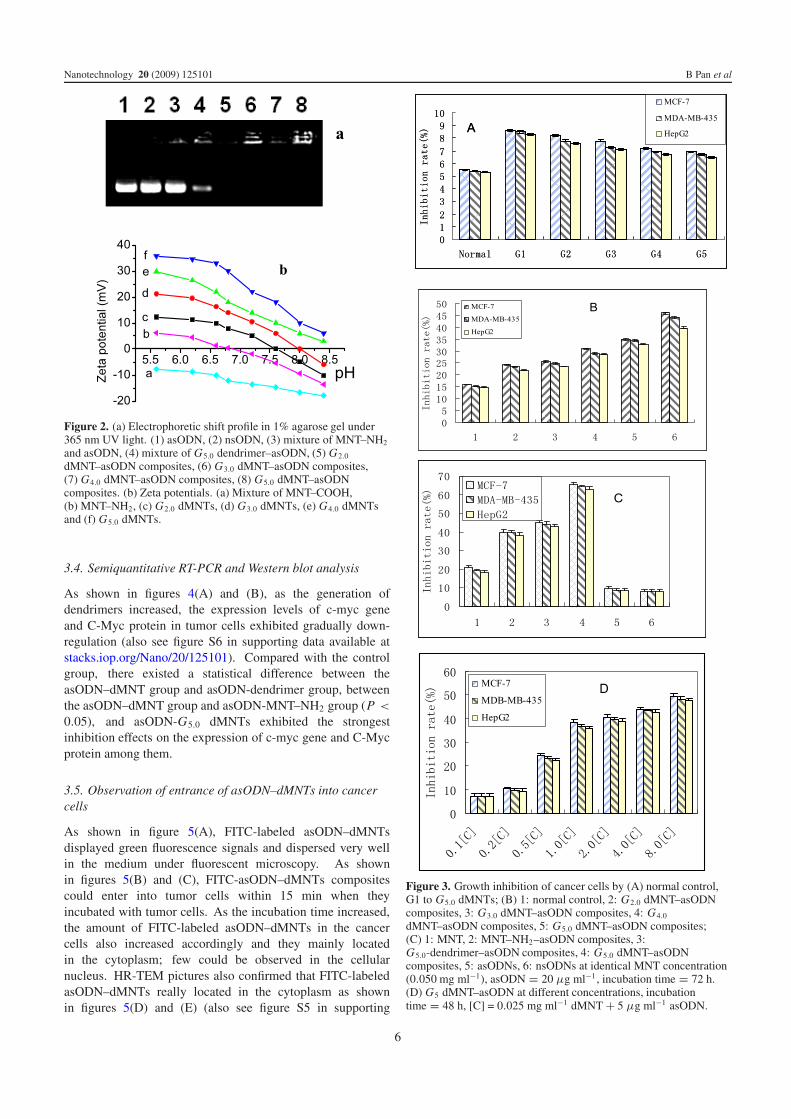
3.2. Electrophoretic shift assay and zeta potential analysis
Figure 2(a) shows the electrophoretic shift assay result ofasODNs in the absence and presence of G2.0 to G5.0-dMNTs,which showed that the amine-terminated dMNT compositescould bind with asODNs and formed stable asODN–dMNTcomposites. As shown in figure 2(b), dMNTs displayedpositive charge properties: as the generation of dendrimersincreased, the amount of positive charges displayed bydMNTs also increased accordingly, indicating the amountof NH2 groups of dMNTs also increased accordingly; theamount of asODNs with negative charge absorbed by dMNTsalso increased correspondingly. G5.0 dMNTs absorbed themaximal amount of asODNs. Positively charged asODN–dMNTs were very easily attached to negatively charged cellmembranes to improve the endocytosis [27, 28]; thereforethe positively charged surface of asODN–dMNTs is key forcellular internalization.
3.3. Cell availability and proliferation assay
As shown in figure 3(A), different generations of dMNTs caninhibit the growth of tumor cells with the inhibition rate of<9%: G5.0 dMNTs exhibited lowest cellular toxicity to tumorcells. Whereas MNTs markedly inhibit the growth of tumorcells with an inhibition rate of 20% or so, therefore dendrimerscan markedly improve the biocompatibility of MNTs, similarto the previous report [28].
As shown in figure 3(B), as the generation of dendrimersincreased, the inhibition rate of asODN–dMNT compositesgradually increased, and there existed a statistical differenceamong different generations of dMNTs (P < 0.05): G5.0
dMNT–asODN composites have the maximum inhibition ratefor tumor cells. Under identical conditions, as shown infigure 3(C), the control MNT–NH2–asODN composites alsoexhibit inhibition effects on the tumor cells, but the inhibitionrates were markedly lower than those caused by dMNT–asODN composites, and there existed a statistical differencebetween dMNT–asODN composites and control groups suchas asODN-MNT–NH2 or dendrimer–asODN composites (P <
0.01), which highly suggest that dMNTs have advantages overMNTs, MNT–NH2 and dendrimer in the asODN delivery.
As shown in figure 3(D), G5.0 dMNTs inhibited the growthof tumor cells in dose-dependent means. This is becausethe amount of asODN–dMNTs entering into cancer cells alsoincreased as the amount of asODN–dMNTs in the mediumincreased.
5
Nanotechnology 20 (2009) 125101 B Pan et al
Figure 2. (a) Electrophoretic shift profile in 1% agarose gel under365 nm UV light. (1) asODN, (2) nsODN, (3) mixture of MNT–NH2
and asODN, (4) mixture of G5.0 dendrimer–asODN, (5) G2.0
dMNT–asODN composites, (6) G3.0 dMNT–asODN composites,(7) G4.0 dMNT–asODN composites, (8) G5.0 dMNT–asODNcomposites. (b) Zeta potentials. (a) Mixture of MNT–COOH,(b) MNT–NH2, (c) G2.0 dMNTs, (d) G3.0 dMNTs, (e) G4.0 dMNTsand (f) G5.0 dMNTs.
3.4. Semiquantitative RT-PCR and Western blot analysis
As shown in figures 4(A) and (B), as the generation ofdendrimers increased, the expression levels of c-myc geneand C-Myc protein in tumor cells exhibited gradually down-regulation (also see figure S6 in supporting data available atstacks.iop.org/Nano/20/125101). Compared with the controlgroup, there existed a statistical difference between theasODN–dMNT group and asODN-dendrimer group, betweenthe asODN–dMNT group and asODN-MNT–NH2 group (P <
0.05), and asODN-G5.0 dMNTs exhibited the strongestinhibition effects on the expression of c-myc gene and C-Mycprotein among them.
3.5. Observation of entrance of asODN–dMNTs into cancercells
As shown in figure 5(A), FITC-labeled asODN–dMNTsdisplayed green fluorescence signals and dispersed very wellin the medium under fluorescent microscopy. As shownin figures 5(B) and (C), FITC-asODN–dMNTs compositescould enter into tumor cells within 15 min when theyincubated with tumor cells. As the incubation time increased,the amount of FITC-labeled asODN–dMNTs in the cancercells also increased accordingly and they mainly locatedin the cytoplasm; few could be observed in the cellularnucleus. HR-TEM pictures also confirmed that FITC-labeledasODN–dMNTs really located in the cytoplasm as shownin figures 5(D) and (E) (also see figure S5 in supporting
Figure 3. Growth inhibition of cancer cells by (A) normal control,G1 to G5.0 dMNTs; (B) 1: normal control, 2: G2.0 dMNT–asODNcomposites, 3: G3.0 dMNT–asODN composites, 4: G4.0
dMNT–asODN composites, 5: G5.0 dMNT–asODN composites;(C) 1: MNT, 2: MNT–NH2–asODN composites, 3:G5.0-dendrimer–asODN composites, 4: G5.0 dMNT–asODNcomposites, 5: asODNs, 6: nsODNs at identical MNT concentration(0.050 mg ml−1), asODN = 20 μg ml−1, incubation time = 72 h.(D) G5 dMNT–asODN at different concentrations, incubationtime = 48 h, [C] = 0.025 mg ml−1 dMNT + 5 μg ml−1 asODN.
6

Nanotechnology 20 (2009) 125101 B Pan et al
Figure 4. (A) RT-PCR result of c-myc and GAPDH (internal control) mRNA expression at 72 h after cell incubation. lane 1: normal control;lane 2: G5 dMNTs; lane 3: MNT–NH2–asODN; lane 4: G5.0 dendrimer–asODN; lane 5: G3 dMNT–asODN; lane 6: G4 dMNT–asODN; lane7: G5 dMNT–asODN. (B) Western blotting result of C-Myc and beta-actin (internal control) protein expression at 72 h after cell incubation.Lane 1: normal control; lane 2: G5 dMNTs; lane 3: MNT–NH2–asODN; lane 4: G5 dendrimer–asODN; lane 5: G3 dMNT–asODN; lane 6:G4 dMNT–asODN; lane 7: G5 dMNT–asODN.
Figure 5. (A) Fluorescent microscopy image of FITC-asODN–dMNTs in medium, bar 500 nm; (B) laser confocal microscopy images ofMCF-7 cells incubated with FITC-asODN–dMNTs for 5 min, 10 min and 15 min, respectively. Scale bar: 5.0 μm. (C) Tridimensionalimmunofluorescent images of FITC-asODN–dMNTs in MDA-MB-435 cells. Blue: DAPI stained cell nucleus. Red: cellular actin framework,scale bar: 5.0 μm. ((D) and (E)) HR-TEM images of as-ODN-dMNT composites in the cytoplasm of MCF-7 cells. (E) is the magnified imageof local frame in (D).
data available at stacks.iop.org/Nano/20/125101). All theseresults fully demonstrate that asODN–dMNTs composites canenter into tumor cells highly efficiently and do not affect theinhibition function of asODNs on cancer cells. The dendrimer–asODNs and MNT–NH2–asODNs also can enter into cancercells within 1 h: however, their amounts in cancer cellswere markedly lower than those of asODN–dMNTs under theidentical incubation time (data not shown), which suggeststhat dMNTs may have maximum gene delivery efficiencyamong them.
3.6. Potential mechanism
As mentioned above, our results show that dMNTs can takeantisense c-myc oligonucleotides into cancer cells highlyefficiently and can release asODNs inside cancer cells. The
released asODNs can bind the start location of c-myc mRNAtranslation in the cancer cells, block translation of c-mycmRNA into protein and inhibit expression of c-Myc proteins,finally inhibiting the growth of cancer cells. Therefore, thedMNTs should be a highly efficient gene delivery system forantisense therapy.
Regarding the mechanism of entrance of asODN–dMNTscomposites into cancer cells, we consider dendrimer-modifiedMNTs exhibit positive charges, can absorb the asODNs withnegative charge into their interior cavities and finally forma thin film wrapped on the surface of MNTs, as shownin figure 5(A). As the generation of dendrimers increases,dMNTs absorb more asODNs and form closer thin filmscovering on the surface of MNTs, similar to Nam et al’sreports [38]. The dendrimer on the surface of MNTs can formtransient nanoscale holes in the membrane of cancer cells,
7

Nanotechnology 20 (2009) 125101 B Pan et al
similar to Banaszak Holl et al’s reports [39–41], markedlydecreasing the integrity of the cell membranes. Then asODN–dMNTs quickly enter into the cytoplasm, In the cellularmicroenvironment, under the action of different enzymes andions, asODNs can be released out from the interior cavities ofdendrimers and then bind the start sites of mRNA translationand inhibit protein synthesis. Because dMNTs can change thecellular microenvironment within a short time, and absorbedsome ions, changing the pH of the environment, protectingasODNs from degradation caused by in vivo enzymes andprolonging the asODN’ action time inside cancer cells, whichfinally markedly enhance the function of asODNs in cancercells. Kostarelos et al [42] reported that cellular uptake offunctionalized carbon nanotubes is independent of functionalgroup and cell type. Conversely, our results show thatcellular uptake of dendrimer-functionalized MNTs is highlydependent on the number of positive charges of functionalgroups and independent of cell types. The course of entranceof asODN–dMNTs composites into cancer cells may be anenergy-independent nanoendocytotic pathway, similar to thenanosyringe model [43].
Regarding the cellular toxicity of dMNTs, Banaszak Hollet al’s reports showed that dendrimer is not toxic to cellsup to 500 nM [39–41]. We consider that dMNTs’ toxicityshould be closely associated with the number of surfacecharges of dendrimers: molecules with negative charges suchas asODNs, peptides and antibodies, can counteract the partialpositive charges of dendrimers and can then reduce the cellulartoxicity of dendrimers. However, the number of positivecharges of dendrimers is also closely associated with genedelivery efficiency. Therefore, finding a balance betweencellular toxicity and delivery efficiency is very important fordeveloping the dMNTs-based gene delivery system.
Regarding the function of MNTs in the dMNT deliverysystem, MNTs own holes, which can be filled with genesor drugs. More importantly, MNTs own special physical,chemical and mechanical properties and can easily enter intothe cytoplasm. A combination of dendrimer and MNTs canreduce the cytotoxicity of MNTs and simultaneously improvethe delivery efficiency of dendrimer and MNTs. MNTs canabsorb near-infrared light and release the heat energy to killcancer cells or design the switch to control the release ofdrugs inside cancer cells [13]. In addition, MNTs can protectasODNs from degradation caused by in vivo enzymes andprolong the action time of asODNs in cancer cells, so MNTscan enhance the therapeutic effects of asODNs, and haveintensive application prospects in cancer therapy in the nearfuture.
4. Conclusions
PAMAM dendrimer-modified MNTs may be good genetransporters for mammalian cell lines and antisense therapy.As the generation of PAMAM dendrimer increases, the amountof asODNs absorbed by dMNTs also increased accordingly.The asODN-G5.0 dMNTs exhibit the maximal deliveryefficiency and inhibition effects on cancer cells among MNT–NH2–asODN composites, dendrimer–asODN composites and
dMNT–asODN composites, which may be helpful to solve thecurrent problem in antisense therapy. Further work will focuson clarifying distribution and the metabolism course of dMNTsin cancer cells. For asODNs, short peptides, antibodies andsiRNA, the dMNTs should be general, high efficient deliverysystems, and have great potential in applications such as geneor drug delivery for cancer therapy and molecular imaging inthe near future.
Acknowledgments
This work is supported by China National 973 Project(no. 2005CB723400-G), the 863 project (no. 2007AA022004),the National Natural Scientific Fund (No. 20661075 and30672147) and the Shanghai Fund of Scientific andTechnology (No. 072112006 and 0752nm024).
References
[1] Nishiyama N et al 2004 Nat. Mater. 4 934[2] Pack D W, Hoffman A S, Pun S and Stayton P S 2005 Nat. Rev.
Drug Discov. 4 581[3] Fuessel S, Herrmann J, Ning S, Kotzsch M, Kraemer K,
Schmidt U, Hakenberg O W, Wirth M P and Meye A 2006Cancer Lett. 232 243
[4] Kim S H, Mok H M, Jeong J H, Kim S W and Park T G 2006Bioconjug. Chem. 17 241
[5] Ondeyka J G et al 2006 J. Nat. Prod. 69 377[6] Ezquer F, Nunez M T and Israel Y 2005 Biochem. Pharmacol.
69 1559[7] Maier M A et al 2006 J. Med. Chem. 49 2534[8] Tarkanyi I, Horvath A, Szatmari I, Eizert H, Vamosi G,
Damjanovich S, Segal-Bendirdjian E and Aradi J 2005FEBS Lett. 579 1411
[9] Kipshidze N, Moses J, Shankar L R and Leon M 2001 Curr.Opin. Mol. Ther. 3 265
[10] Shadidi M and Sioud M 2003 FASEB J. 17 256[11] Bonora G M, Ivanova E, Zarytova V, Burcovich B and
Veronese F M 1997 Bioconjug. Chem. 8 793[12] Choy J H, Kwak S Y, Jeong Y J and Park J S 2000 Angew.
Chem. Int. Edn 39 4042[13] Cui D 2007 J. Nanosci. Nanotechnol. 7 1298[14] Liu Y, Wu D, Zhang W, Jiang X, He C, Chung T S, Goh S H
and Leong K W 2005 Angew. Chem. Int. Edn 44 4782[15] Cui D, Pan B, Zhang H, Gao F, Wang J, He R and Toru A 2008
Anal. Chem. 80 7996[16] Pantarotto D, Singh R, McCarthy D, Erhardt M, Briand J,
Prato M, Kostarelos K and Bianco A 2004 Angew. Chem.Int. Edn 43 5242
[17] Lu Q, Moore J M, Huang G, Mount A S, Rao A M, Larcom L Land Ke P C 2004 Nano Lett. 4 2473
[18] Kam N W S and Dai H 2005 J. Am. Chem. Soc. 127 6021[19] Kam N W S, Jessop T C, Wender P A and Dai H 2004 J. Am.
Chem. Soc. 126 6850[20] Kam N W S, Liu Z and Dai H 2005 J. Am. Chem. Soc.
127 12492[21] Cui D, Tian F, Coyer S R, Wang J, Gao F, He R, Pan B and
Zhang Y 2007 J. Nanosci. Nanotechnol. 7 1639[22] Benito J M, Garcia M G, Mellet C O, Baussanne I, Defaye J and
Fernandez J M G 2004 J. Am. Chem. Soc. 126 10355[23] Cui D, Tian F, Ozkan C S, Mao W and Gao H 2005 Toxicol.
Lett. 155 77[24] Dhanikula R S and Hildgen P 2006 Bioconjug. Chem. 17 29[25] Majoros I J, Myc A, Thomas T, Mehta C B and Baker J R 2006
Biomacromolecules 7 572
8

Nanotechnology 20 (2009) 125101 B Pan et al
[26] Jimenez-Contreras R 2008 Nanotechnology ResearchDevelopments ed B Pan and D Cui (Berlin: Springer)chapter 1 (Advance and Application Prospect ofDendrimers) pp 7–95
[27] Lee J W, Kim B K, Kim H, Han S C, Shin W S and Jin S H2006 Macromolecules 39 2418
[28] Pan B, Cui D, Shen Y, Ozkan C S, Gao F, He R, Li Q,Xu P and Huang T 2007 Cancer Res. 67 8156
[29] Pan B, Cui D, Xu P, Huang T, Li Q, He R and Gao F 2007J. Biomed. Pharm. Eng. 1 13
[30] Gao H, Kong Y, Cui D and Ozkan C S 2003 Nano Lett. 3 471[31] Kong Y, Cui D, Ozkan C S and Gao H 2003 Proc.
Biomicroelectromech. Syst. 773 111[32] Cui D, Ozkan C S, Ravindran S, Kong Y and Gao H 2004
Mech. Chem. Biosyst. 1 113[33] Pan B, Cui D, Gao F and He R 2006 Nanotechnology 17 2483[34] Cui D, Zhang H, Wang Z, Asahi T and Osaka T 2008 ECS
Trans. 13 111[35] Bhattacharyya S, Kymakis E and Amaratunga G A J 2004
Chem. Mater. 16 4819
[36] Zhang Y, Yang M, Portney N G, Cui D, Budak G, Ozbay E,Ozkan M and Ozkan C S 2008 Biomed. Microdev.10 321
[37] Cui D, Jin G, Gao T, Sun T, Tian F, Estrada G G, Gao H andSarai A 2004 Cancer Epidemiol. Biomarkers Prevent. 131136
[38] Nam H Y, Hahn H J, Nam K, Choi W H, Jeong Y S, Kim D Eand Park J S 2008 Int. J. Pharm. 363 199
[39] Mecke A, Uppuluri S, Sassanella T M, Lee D K,Ramamoorthy A, Baker J R, Orr B G andBanaszak Holl M M 2004 Chem. Phys. Lipids 132 3
[40] Hong S, Bielinska A U, Mecke A, Keszler B, Beals J L, Shi X,Balogh L, Orr B G, Baker J R and Banaszak Holl M M 2004Bioconjug. Chem. 15 774
[41] Hong S, Leroueil P R, Janus E K, Peters J L, Kober M M,Islam M T, Orr B G, Baker J R and Banaszak Holl M M2006 Bioconjug. Chem. 17 728
[42] Kostarelos K et al 2007 Nat. Nanotechnol. 2 108[43] Lopez C F, Nielsen S O, Moore P B and Klein M L 2004 Proc.
Natl Acad. Sci. 101 4431
9
Copyright © 2022 FDOKUMEN