HPMA copolymer-aminoellipticine conjugates: mechanism of action.
207
HPMA copolymer - aminoellipticine conjugates: Mechanism of action by Richard Keane, BPharm A thesis submitted to the University of London in partial fulfilment of the requirements for the degree of Doctor of Philosophy Centre for Polymer Therapeutics The School of Pharmacy University of London Or 9
-
Upload
khangminh22 -
Category
Documents
-
view
1 -
download
0
Transcript of HPMA copolymer-aminoellipticine conjugates: mechanism of action.

HPMA copolymer - aminoellipticine conjugates:
Mechanism of action
b y
Richard Keane, BPharm
A thesis submitted to the University of London in partial fulfilment of the requirements for the degree of Doctor of Philosophy
Centre for Polymer TherapeuticsThe School of Pharmacy University of London
Or 9
ProQuest Number: 10104773
All rights reserved
INFORMATION TO ALL USERS The quality of this reproduction is dependent upon the quality of the copy submitted.
In the unlikely event that the author did not send a complete manuscript and there are missing pages, these will be noted. Also, if material had to be removed,
a note will indicate the deletion.
uest.
ProQuest 10104773
Published by ProQuest LLC(2016). Copyright of the Dissertation is held by the Author.
All rights reserved.This work is protected against unauthorized copying under Title 17, United States Code.
Microform Edition © ProQuest LLC.
ProQuest LLC 789 East Eisenhower Parkway
P.O. Box 1346 Ann Arbor, Ml 48106-1346

This work is dedicated to my family and to Yuk.
11

Acknowledgements
I would firstly like to acknowledge my supervisor. Prof. Ruth Duncan without whose
support and helpful advice this thesis would not have been possible. I would like to
also especially thank Dr. Stephanie Gac-Breton, Dr. Frances Searle and Dr. Steve
Brocchini for their supervision and advice, especially during the early phases of this
work. For many a thought provoking discussion I would like to thank Prof. Tom
Connors, Prof. Helmut Ringsdorf and Dr. Dale Hreczuk-Hirst. I would like to
acknowledge the National Cancer Institute, U.S.A. for their funding of this project also.
Many people within the Centre for Polymer Therapeutics have helped me with learning
and understanding the techniques used in this work and I thank them sincerely for all
their assistance. In particular Dr. Yee-Nee Sat, Dr. Navid Malik and Mr. John Latigo
for their help with learning the in vivo techniques and Ms. RhondaLea MacDonald and
Dr. Keith Anderson for teaching me cell culture. I would like to thank my colleagues
Dr. Antony Godwin and Mr. Ryan Tomlinson for helping me with my understanding of
the chemistry side of things. A special word of thanks also to Ms. Samantha Kneller
for her assistance in a number of these studies and for teaching me the ‘joy’ of making
tritosomes.
I would like to acknowledge the collaborators at the Gray Cancer Institute, particularly
Mr. Ian Wilson and Dr. Gillian Tozer for their patience and willingness to help me with
the window chamber experiments.
I would like to thank all my friends and family for their support through my PhD. My
colleagues Ruth Musila and Nicola Pattrick as well as all the other friends I have
mentioned above and others who have passed through CPT. Many people have helped
me a lot throughout my PhD and I would like to apologise to anyone I may have
omitted.
Last, but by no means least, I would like to thank Yuk-Fung Chau for all her love and
support, especially through the rough times.
Ill

Abstract
Over the past two decades cancer chemotherapy has resulted in a small number of
previously fatal cancers becoming curable. Many cancers, particularly the so-called
solid tumours, do not respond well to conventional chemotherapy. To maximise
tumour targeting and minimise host tissue toxicity a large number of drug delivery
systems have been proposed. Polymer-anticancer conjugates based on N-(2-
hydroxypropyl) methacrylamide (HPMA) have recently entered early clinical trial. It
has been shown that HPMA copolymer conjugates preferentially extravasate into solid
tumours and are retained there by a process known as the ‘enhanced permeability and
retention’ (EPR) effect.
A natural anticancer agent, derived from ellipticine, namely 6-(3 -aminopropy 1)
ellipticine (APE) was selected for conjugation to HPMA copolymers. In this study a
series of HPMA copolymer-APE conjugates were synthesised, containing a variety of
drug loadings (1.07-6.10%w/w) conjugated via the tetrapeptide linker (Gly-Phe-Leu-
Gly). These conjugates were designed to be localised in tumours following injection
and to be taken up by tumour cells via the process of endocytosis before liberating
APE, mediated by cathepsin B present in the lysosome. These conjugates were shown
to form complex intramolecular micelles in solution which resulted in the conjugates of
a high drug loading showing reduced APE release in vitro (20%/5h) compared to the
medium and low drug loading conjugates (55%/5h), suggesting hindered enzyme
access. All conjugates showed a marked reduction in haemolysis, a common problem
with ellipticines, compared to APE alone. Anti-tumour activity was observed in the s.c.
B16F10 murine melanoma model and also in the CORL-23 human non small cell lung
carcinoma xenograft in mice, particularly for the conjugate of medium APE loading.
This thesis also examined the extravasation and intratumoural distribution of HPMA
copolymer-anticancer conjugates using HPMA copolymer doxorubicin (PKl) as a
model conjugate in the rat dorsal window chamber model.
IV

Contents
Page number
Title iDedication iiAcknowledgements illAbstract ivContents vList of Figures ixList of tables xiiiAbbreviations xiv
Chapter 1 General Introduction 1
1.1 General Introduction 2
1.2 Drug delivery systems in cancer therapy 41.2.1 Polymeric implants for controlled release and drug targeting 41.2.2 Antibodies 51.2.3 Liposomes 7
1.3 Polymer therapeutics 81.3.1 Polymeric drugs 101.3.2 Polymer-protein conjugates 101.3.3 Polymeric micelles 111.3.4 Polymer-drug conjugates 12
1.3.4.1 Polymeric backbone 121.3.4.2 Linkers for drug conjugation 171.3.4.3 Polymer-drug conjugates in clinical trial and 19
development
1.4 Biological rationale for the design of HPMA copolymer-anticancer 28 conjugates
1.4.1 Biocompatibility and pharmacokinetics of HPMA copolymers 281.4.2 Optimisation of linker design 291.4.3 The Enhanced Permeability and Retention (EPR) Effect 301.4.4 Intracellular pharmacokinetics 33
1.5 Natural products in Cancer 351.5.1 Natural products in clinical use 351.5.2 Ellipticines 39
1.6 Aims of the study 42
Chapter 2 Materials and General Methods 44
2.1 Materials 452.1.1 Chemicals 452.1.2 Cell culture 45
2.2 Equipment 46

2.2.1 Analytical equipment 462.2.2 Cell culture equipment 462.2.3 In vivo equipment and suppliers 462.2.4 Miscellaneous and general equipment 46
23 General methods 472.3.1 In vitro methods 47
2.3.1.1 Growing cells from frozen vials 472.3.1.2 Day to day maintenance of cells 472.3.1.3 Cell counting 482.3.1.4 Freezing cells 482.3.1.5 Growth curve 482.3.1.6 Cell cytotoxicity using the MTT assay 492.3.1.7 Cell preparation for subcutaneous (s.c.) injection 49
into mice2.3.2 Evaluation of anti-tumour activity 492.3.3 Analytical methods 51
2.3.3.1 HPLC determination of APE 512.3.3.2 HPLC determination of free and total doxorubicin 53
2.3.4 General biochemical methods 562.3.4.1 Isolation and standardisation of rat liver lysosomes 56
(tritosomes)2.3.4.2 Protein determination using the bicinchoninic acid assay 592.3.4.3 Standardisation of lysosomal enzyme activity in 60
tritosomes2.3.4.4 Studying drug release from HPMA copolymer GFLG 60
conjugates using tritosomes2.3.5 Statistics 63
Chapter 3 Synthesis and characterisation of HPMA copolymer-APE 64conjugates
3.1 Introduction 65
3.2 Methods 753.2.1 Synthesis of HPMA copolymer-APE conjugates 753.2.2 Determination of APE content in conjugates 783.2.3 Gel Permeation Chromatography (GPC) 78
3.2.5 Determination of solubility of free APE and HPMA copolymer-APE 78
3.3 Results 813.3.1 Conjugation characteristics of HPMA copolymer conjugates 813.3.2 Conjugation of APE to HPMA copolymers 833.3.3 GPC analvsis of HPMA copolvmer-APE conjugates 83
3.3.5 Solubility of HPMA copolymer-APE conjugates 88
3.4 Discussion 90
VI

Chapter 4 Effect of APE loading on physicochemical and biological 92properties of HPMA copolymer APE conjugates in vitro
4.1 Introduction 934.1.1 Polymer conformation in solution 934.1.2 Drug release from HPMA copolymer-APE conjugates 954.1.3 Haemolytic activity of HPMA copolymer-APE conjugates 954.1.4 Cytotoxicity of HPMA copolymer-AJ^E conjugates 96
4.2 Methods 974.2.1 Conformational studies using the hydrophobic probe, pyrene 974.2.2 In vitro cytotoxicity against B16F10 murine melanoma 974.2.3 Rat red blood cell lysis assay 984.2.4 Tritosome-mediated release of APE 98
4.3 Results 1014.3.1 HPMA copolymer-APE conformation in solution 1014.3.2 Tritosome-mediated release of APE 1014.3.3 Haematocompatibility of HPMA copolymer-APE conjugates 1094.3.4 In vitro cytotoxicity 114
4.4 Discussion 114
Chapter 5 Effect of APE loading on anti-tumour activity of HPMA 122copolymer GFLG-APE conjugates
5.1 Introduction 123
5.2 Methods 1265.2.1 Cell preparation 1265.2.2 Establishment of s.c. tumours 1265.2.3 Determination of APE MTD in tumour-bearing mice 1265.2.4 Anti-tumour data expression 128
5.3 Results 1285.3.1 Determination of APE MTD in tumour-bearing mice 1285.3.2 Anti-tumour activity of HPMA copolymer-APE conjugates 128
against B16F10 melanoma
5.4 Discussion 137
Chapter 6 The window chamber model: evaluation of the 140intratumoural distribution and mechanism of action of PKl
6.1 Introduction 141
6.2 Methods 1446.2.1 Window chamber surgery 1446.2.2 Tumour examination and PKl administration 1466.2.3 Determination of doxorubicin content of tumours 148
6.3 Results 148
Vll
6.4 Discussion 152
Chapter 7 General discussion 156
References 163
Appendix 190
vin

List of Figures
Figure 1.1 Schematic of “polymer therapeutics”.
Figure 1.2 Schematic of a polymer-drug conjugate.
Figure 1.3 Graph showing Mw and Mn for a polymer.
Figure 1.4 The structure of dextran doxorubicin.
Figure 1.5 Structures of PKl (A) and PK2 (B).
Figure 1.6 Structures of poly (L-glutamic acid)-paclitaxel (A) and HPMA
copolymer paclitaxel (B).
Figure 1.7 Structures of HPMA copolymer camptothecin (A) and HPMA
copolymer platinate (B).
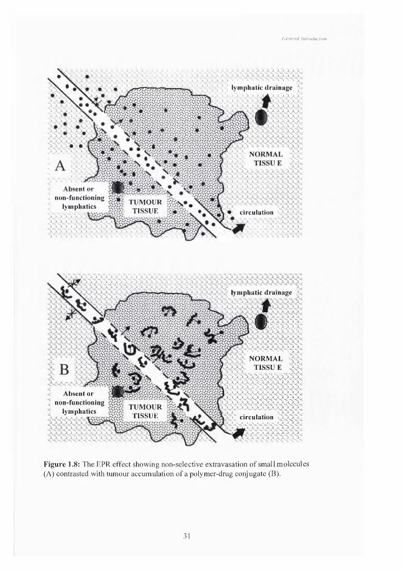
Figure 1.8 The EPR effect showing non-selective extravasation of small
molecules (A) contrasted with tumour accumulation of a polymer-
drug conjugate (B).
Figure 1.9 Schematic of intracellular release of pendant drugs with respect to a
low molecular weight anticancer drug.
Figure 1.10 Anticancer drugs approved in the U.S.A. in 1994 and their origin
(adapted from Cragg et al, 1997).
Figure 1.11 General structure of the ellipticines showing some of the common
sites of derivatisation and the structures of ellipticine, Celliptium,
Datelliptium and APE.
Figure 2.1 Metabolism of MTT by viable cells.
Figure 2.2 Typical HPLC chromatograms obtained during APE quantitation
using method 1(A) and method 2 (B).
Figure 2.3 A standard curve of determination of APE in the HPLC system.
Figure 2.4 Typical HPLC chromatograms obtained during quantitation of
doxorubicin using free (A) and total (B) methods.
Figure 2.5 Typical standard curve used to determine doxorubicin using HPLC
analysis.
Figure 2.6 Typical BSA standard curve used for lysosomal protein content
determination. The equation of the straight line is used to determine
the protein content of tritosomes.
IX

Figure 2.7 Typical example of NAp release from the low molecular weight
substrate, n-Benzoyl-Phe-Val-Arg-NAp, in a tritosome activity
assay. The equation is used to calculate the cathepsin B activity of
the tritosomes.
Figure 3.1 Three dimensional structure of APE (NSC176328).
Figure 3.2 Synthesis of HPMA copolymer GFLG-ONp precursor by free
radical polymerisation.
Figure 33 Aminolysis of the HPMA copolymer GFLG-ONp using APE as an
example of a suitable drug with a free primary amine.
Figure 3.4 Reaction schemes showing both the undesired (hydrolysis) reaction
and the desired (aminolysis) reactions where R is HPMA copolymer-
GFLG and Ri is a reactant with a free amine e.g. APE or
doxorubicin.
Figure 3.5 Reaction scheme showing free radical polymerisation of HPMA with
methacryloylated-GFLG-doxorubicin monomers to produce a
conjugate similar to PKl (St*astny et al, 1996; Section 3.1).
Figure 3.6 Method used by Caiolfa et al (2000) to synthesise an HPMA
copolymer GFLG-camptothecin conjugate.
Figure 3.7 ATRP-mediated copolymerisation of activated HPMA monomers to
produce a * universal* precursor (Godwin et al, 2001).
Figure 3.8 The UV spectra of APE, HPMA copolymer GFLG-l-amino-2-
propanol and HPMA copolymer GFLG-APE (RK3).
Figure 3.9 Standard curve for APE in PBS measured using UV absorption at
296nm.
Figure 3.10 Hydrolysis of the HPMA copolymer GFLG-ONp (CPT-03).
Figure 3.11 TLC of a typical HPMA copolymer-APE reaction mixture.
Figure 3.12 GPC chromatogram of an aminolysed HPMA copolymer and the
equivalent HPMA copolymer-APE conjugate (RK4).
Figure 4.1
Figure 4.2
Figure 4.3
Figure 4.4
The structure of pyrene and its fluorescence spectrum (0.2mg/l in
water) using an excitation wavelength of 330nm showing the
intensity peaks Ii (372nm) and I3 (392nm).
Haemolytic activity of dextran and poly(ethylene)imine.
B16F10 growth curve.
Cytotoxicity of dextran and poly(l-lysine) against B16F10 cells.
X
Figure 4.5 Fluorescence spectra of pyrene in the presence of APE and increasing
concentrations of HPMA copolymer GFLG-aminopropanol.Figure 4.6 Fluorescence spectra of pyrene in the presence of increasing
concentrations of the HPMA copolymer-APE conjugates.
Figure 4.7 Relationship between pyrene 1] and HPMA copolymer conjugate
concentration.
Figure 4.8 Relationship between the I1/I3 ratio of pyrene and HPMA copolymer
conjugate concentration.
Figure 4.9 Release of APE from HPMA copolymer-APE conjugates in the
presence (+) or absence (-) of tritosomes.
Figure 4.10 The effect of substrate concentration on tritosome-mediated release
of APE from HPMA copolymer-APE and doxorubicin from PKl.
Figure 4.11 The effect of APE loading on tritosome-mediated release of APE
from HPMA copolymer-APE and doxorubicin from PKl.
Figure 4.12 Haemolytic activity of APE, ellipticine and HPMA copolymer-APE
conjugates.
Figure 4.13 In vitro cytotoxicity of APE and HPMA copolymer-APE conjugates
against B16F10 cells.
Figure 4.14 Proposed conformation of HPMA copolymer-APE and HPMA
copolymer-aminopropanol conjugates.
Figure 5.1 Growth curve of CORL23 cells grown from a seeding density of
1 x1 0 cells per well.
Figure 5.2 Toxicity of 20 and 25mg/kg doses observed as 20% body weight loss
in the MTD determination of APE in mice bearing s.c. B16F10.
Figure 5.3 Body weight and anti-tumour activity of saline, APE and HPMA
copolymer GFLG-APE conjugates.
Figure 5.4 Effect of saline (control), APE and HPMA copolymer GFLG-APE
conjugates of diBerent loading on organ weight of organs removed
at the end of an anti-tumour study.
Figure 5.5 Effect of saline and HPMA copolymer GFLG-APE (RK2) on mouse
weight and tumour area.
Figure 6.1 The type of window chambers available (from Jain, 2001).
Figure 6.2 Schematic of C clamp from a front and side view.
Figure 6.3 Picture of one side of a window chamber frame (A) and of the
microscope set-up for imaging (B).
XI

Figure 6.4 Trans-illuminated picture of a window chamber preparation under
the lowest magnification (xl.6).
Figure 6.5 A series of still images taken from two studies showing time-
dependent distribution of PKl following injection of 4mg/kg
(doxorubicin equivalence).
Figure 6.6 A series of still images taken from two further studies showing time-
dependent distribution of PKl following injection of 4mg/kg
(doxorubicin equivalence).
Figure 6.7 Intratumoural distribution of liposomes (described in Section 63) of
approximately 90nm in LS174T tumours implanted in dorsal
window chambers on SCID mice showing characteristic
perivascular deposits (from Yuan et al, 1994)
Xll

List of Tables
Table 1.1 Methods for overcoming the current limitations of cancer
chemotherapy.
Table 1.2 Polymers used to prepare polymer-anticancer conjugates.
Table 1.3 Examples of biodegradable linkers utilised in drug conjugates for
anticancer therapy.
Table 1.4 Polymer-drug conjugates developed for cancer chemotherapy and
their stage of development.
Table 1.5 Ellipticine derivatives which have undergone pre-clinical and/or
clinical trial but are not yet marketed.
Table 3.1 Characteristics of the HPMA copolymer precursors used in the
synthesis of HPMA copolymer-APE conjugates.
Table 3.2 Characteristics of the HPMA copolymer-APE conjugates
synthesised in this study.
Table 33 Molecular weight of HPMA copolymer-APE conjugates compared
to poly(ethylene-oxide) standards.
Table 4.1 Characteristics of the tritosome batches.
Table 5.1 PKl activity in a panel of in vivo tumour models (Duncan et al,
1992).
Table 5.2 Anti-tumour activity of APE and HPMA copolymer GFLG-APE
conjugates against s.c. B16F10 murine melanoma.
Table 53 The effect of APE loading on the anti-tumour activity of HPMA
copolymer GFLG-APE conjugates against B16F10 melanoma.
Table 5.4 Activity of RK2 (4.34%w/w APE) against the CORL23 human
tumour xenograft in nu/nu mice
Table 5.5 Comparison between HPMA copolymer GFLG-APE, S16020-2,
S30972-1 and PKl administered i.p. in the B16F10 model.
Table 6.1 Comparison of present study with previous accounts for the total
amount of doxorubicin (dox) present in blood and tumour samples.
X lll

Abbreviations
AML Acute myeloid leukaemiaAPE 6-(3 - Aminopropy l)-ellipticineATRP Atom transfer radical polymerisationDIVEMA Divinylether maleic acidDMF Dimethyl formamideDMSO Dimethyl sulphoxideEDTA Ethylenediaminetetraacetic acidEPR Enhanced permeability and retention effectPCS Foetal calf serumFTIR Fourrier-transform infrared spectroscopyGFLG Glycine-Phenylalanine-Leucine-GlycineGPC Gel permeation chromatographyGSH Reduced glutathioneHAMA Human anti-mouse antibody formationHPLC High performance liquid chromatographyHPMA V-(2-Hydroxypropyl)methacrylamideIC50 Inhibitory concentration for 50% of cellsi.p. Intraperitoneali.v. IntravenousLDH Lactate dehydrogenaseMPEG Monomethoxy poly(ethylene glycol)MTD Maximum tolerated doseMTT 1 -[4,5-Dimethylthiazol-2-yl]-2,5-diphenyl tétrazolium
bromideMw Weight average molar massMn Number average molar massNAp p-NitroanilideNCI National Cancer InstituteONp p-NitrophenolPBS Phosphate buffered salinePEG poly (Ethylene glycol)PKl HPMA copolymer Gly-Phe-Leu-Gly-doxorubicinPK2 HPMA copolymer Gly-Phe-Leu-Gly-
doxorubicin/galactosamineRES Reticulo-endothelial systemRf Reference factor for TTCRI Refractive indexs.c. SubcutaneousSD Standard deviationSE Standard errorSMANCS Poly(styrene-co-maleic acid)-neocar2dnostatinTLC Thin layer chromatographyTNF Tumour necrosis factorUV UltravioletVEGF Vascular endothelial growth factor
XIV

General inlroduction
Chapter One
General Introduction

General Introduction
1.1 General Introduction
Globally approximately 10 million people are diagnosed with cancer each year
and this number is estimated to double by the year 2020 because of the world’s ageing
population. Cancer is the cause of 6 million deaths every year worldwide (Sikora,
1999). In the United Kingdom a half of all cancer deaths are caused by just three
cancer types - lung, large bowel and prostate (men) or breast (female) (CRC
CancerStats, 2000). These are so-called solid tumours and the main modalities of their
treatment are surgery and radiotherapy.
Drug chemotherapy has resulted in a number of cancers becoming largely
curable such as Hodgkin’s disease and childhood leukaemia and it is hoped that this
success in the treatment of the so-called soft tissue tumours can be emulated with
improved treatments for solid tumours. Effective treatment of soft tissue tumours
results from the fact that they have characteristics of a disseminated disease. Solid
tumours, however, form a more localised tumour mass and therefore it is often difficult
to achieve the high local drug concentration essential to achieve effective treatment and
simultaneously minimise side-effects (reviewed in De Vita et al, 1993). The main
difficulties associated with systemic chemotherapy of solid tumours are: achieving a
sufficient local drug concentration (reviewed by Jain, 1989); achieving drug penetration
throughout the tumour mass (Tunggal et al, 1999) and not least the development of
resistance to therapy (reviewed by Krishna & Mayer, 2000). A number of strategies
have been employed as means of overcoming these difficulties (Table 1.1). They
include use of combination chemotherapy of different agents acting via different
mechanisms of action, development of more effective analogues of existing agents,
synthesis or identification of new compounds and not least the use of drug delivery
systems to optimise tumour targeting and minimise drug access to sites of toxicity.
Drug delivery systems have the potential advantage that the technology used may be
applied to a broad spectrum of agents. One promising new approach being developed
for improved tumour targeting is the use of polymer-drug conjugates and a number of
these systems have recently entered Phase I/II clinical trials (reviewed by Duncan,
2000).
Those polymer-drug conjugates in clinical testing utilise established anticancer
agents but this approach may potentially benefit anticancer agents which have failed

General introduction
Table 1.1: Methods for overcoming the current limitations of cancer chemotherapy.
Key: MOPP (mechlorethamine, vincristine, procarbazine, prednisone) CHOP
(cyclophosphamide, doxorubicin, vincristine, prednisone).
Type of therapy
Examples Condition treated or stage of development
References
Combinationchemotherapy
MOPP
CHOP
Hodgkin's disease
Non-Hodgkin'slymphoma
reviewed by McCaffrey & Bajorin, 1998
Analogues of existing agents
Temozolamide (analogue of dacarbazine)
Epirubicin (an anthracycline)
Malignant glioma
Breast cancer
Martindale, 1993
Novel cytotoxic compounds (e.g. natural products)
Ecteinascidin 743
Combretastatin A4
Phase I
Phase I
Ryan et al, 2001
Tozer et al, 1999
Biologies Interferon a-2a
Interleukin-2
Kaposi's sarcoma
Metastatic renal Cell carcinoma
Martindale, 1993
Drug delivery systems
Liposomes
Antibodies
Polymer-drugconjugates
Various tumour typesGregoriadis, 1995
Buske et al, 1999
Duncan, 2000
General inlrociuclion
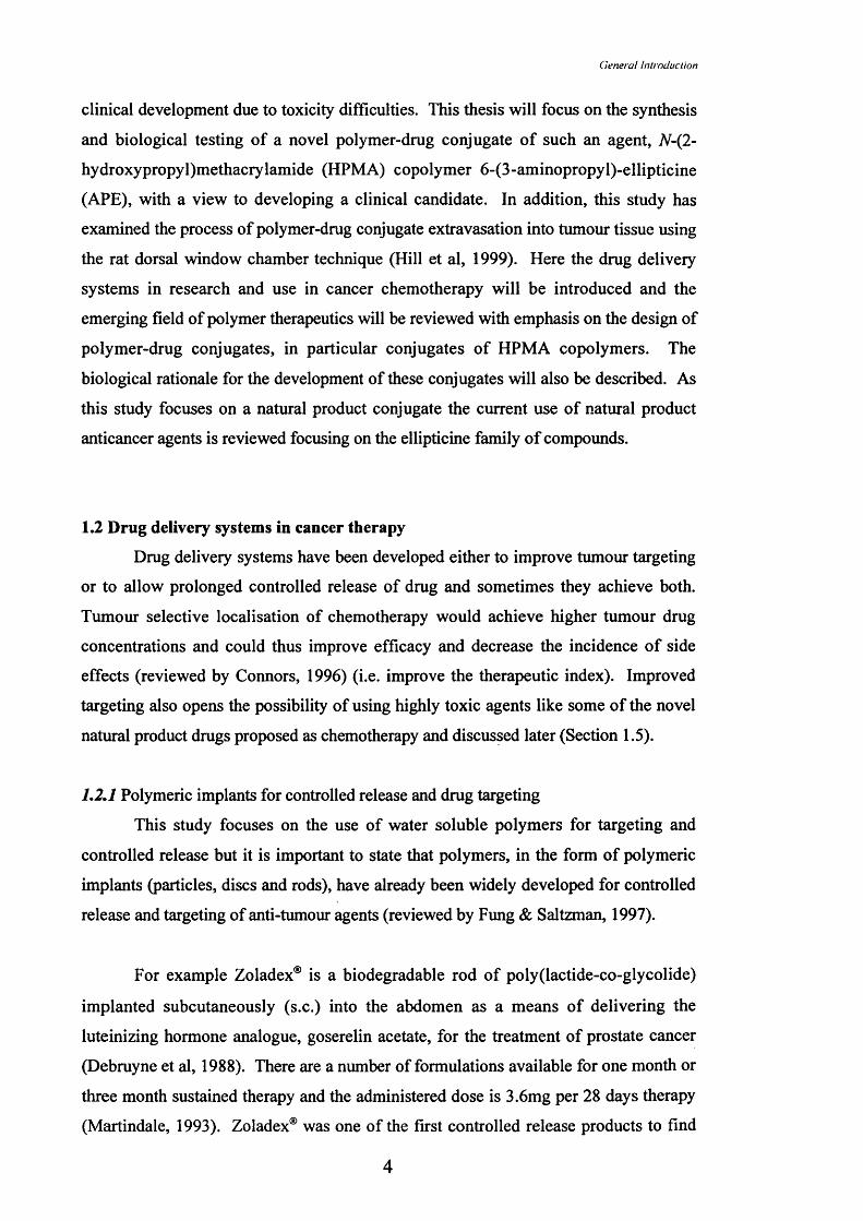
clinical development due to toxicity difficulties. This thesis will focus on the synthesis
and biological testing of a novel polymer-drug conjugate of such an agent, #-(2-
hydroxypropyl)methacrylamide (HPMA) copolymer 6-(3-aminopropyl)-ellipticine
(APE), with a view to developing a clinical candidate. In addition, this study has
examined the process of polymer-drug conjugate extravasation into tumour tissue using
the rat dorsal window chamber technique (Hill et al, 1999). Here the drug delivery
systems in research and use in cancer chemotherapy will be introduced and the
emerging field of polymer therapeutics will be reviewed with emphasis on the design of
polymer-drug conjugates, in particular conjugates of HPMA copolymers. The
biological rationale for the development of these conjugates will also be described. As
this study focuses on a natural product conjugate the current use of natural product
anticancer agents is reviewed focusing on the ellipticine family of compounds.
1.2 Drug delivery systems in cancer therapy
Drug delivery systems have been developed either to improve tumour targeting
or to allow prolonged controlled release of drug and sometimes they achieve both.
Tumour selective localisation of chemotherapy would achieve higher tumour drug
concentrations and could thus improve efficacy and decrease the incidence of side
effects (reviewed by Connors, 1996) (i.e. improve the therapeutic index). Improved
targeting also opens the possibility of using highly toxic agents like some of the novel
natural product drugs proposed as chemotherapy and discussed later (Section 1.5).
L 2 ,l Polymeric implants for controlled release and drug targeting
This study focuses on the use of water soluble polymers for targeting and
controlled release but it is important to state that polymers, in the form of polymeric
implants (particles, discs and rods), have already been widely developed for controlled
release and targeting of anti-tumour agents (reviewed by Fung & Saltzman, 1997).
For example Zoladex® is a biodegradable rod of poly(lactide-co-glycolide)
implanted subcutaneously (s.c.) into the abdomen as a means of delivering the
luteinizing hormone analogue, goserelin acetate, for the treatment of prostate cancer
(Debruyne et al, 1988). There are a number of formulations available for one month or
three month sustained therapy and the administered dose is 3.6mg per 28 days therapy
(Martindale, 1993). Zoladex® was one of the first controlled release products to find

General Introduction
use in cancer chemotherapy. Gliadel is a polymeric implant (wafer) used to achieve
“local” targeting in the treatment of malignant glioma. The biodegradable wafer
Gliadel® contains carmustine (3.85%w/w) incorporated into a copolymer matrix
composed of 1,3-bis(p-carboxyphenoxy)propane and sebacic acid (20:80 molar ratio)
(Dang et al, 1996). After surgical removal of glioma Gliadel® wafers are placed in the
surgical site. This treatment has been shown to prolong patient survival by 50%
following malignant glioma removal (Brem et al, 1995). This is a method of restricting
delivery to a tumour site as carmustine is rapidly metabolised and penetrates just 2mm
into the surrounding brain tissue and therefore toxicity to healthy tissue is abrogated
(reviewed by Wang et al, 1999).
Localised delivery of chemotherapy is only useful for treatment of local disease
but would not be able to treat metastatic disease. For effective treatment of metastatic
disease it is necessary to target tumours after intravenous (i.v.) administration. Several
systems have been developed using this principle, including antibodies, liposomes and
polymer therapeutics. These systems are described in more detail in the following
sections.
L2,2 Antibodies
Antigen-antibody reactions are potentially highly specific so the use of
monoclonal antibodies as an active targeting system was seen as a wonderful
opportunity to achieve tumour-specific targeting (Kohler & Milstein, 1975). It was a
natural extension of this concept to attach drugs via a linker to the antibodies to achieve
active tumour targeting (reviewed by Trail & Bianchi, 1999). Since then many
anticancer drugs, immunotoxins and radioactive agents have been conjugated to
antibodies for use as cancer chemotherapeutic agents.
These include conjugates of immunotoxins such as a Fab’ fragment of a
monoclonal anti-CD22 antibody conjugated to deglycosylated ricin-A chain which gave
partial responses in 38% of evaluable patients in a Phase I study against patients with
refractory B-cell lymphoma (Vitetta et al, 1991). Antibody conjugates containing
radionuclides have also been widely studied such as [^^*I]iodide which has been
conjugated to a number of antibodies (e.g. anti-CD20 which was studied in a Phase I
trial against B-cell non-Hodgkin’s lymphoma and showed 50% complete remission and
a further 29% partial responses in patients (Kaminski et al, 1996)). Antibodies have

General Inlroduction
also been conjugated to classical cytotoxic agents such as doxorubicin. An example is
doxorubicin conjugated to the BR96 antibody via a hydrazone linkage (Firestone et al,
1996) which gave a maximum tolerated dose (MTD) of 25mg/m^ (doxorubicin
equivalent), when administered by i.v. infusion every three weeks, in a recent Phase I
study against metastatic colon and breast cancer (Saleh et al, 2000). The first antibody-
drug conjugate to enter clinical use in 2000 was the anti-CD33 calicheamicin
immunoconj ugate (Mylotarg™) which is used for the treatment of acute myeloid
leukaemia (AML). CD33 is an antigen present on maturing normal haematopoietic and
AML cells but not on normal haematopoietic cells. Calicheamicin, an anti-tumour
antibiotic (Lee et al, 1987), is released upon internalisation of the construct into CD33
positive cells killing these cells whilst sparing normal stem cells. In this case the target
antigen is expressed on a high percentage of desired target cells (about 90%) (Sievers et
al, 1999).
In recent years interest has grown in the use of antibodies that can stimulate an
immune response against the tumour. It has become apparent that the body is capable
of removing a large tumour burden in this manner (Kranz et al, 1998). Herceptin® is an
antibody to the human epidermal growth factor receptor (HER-2), often over-expressed
in breast cancer, and has been developed both in single agent trials but also particularly
in combination with established agents (doxorubicin and paclitaxel). It has shovm
responses in clinical trials itself but is especially useful in combination chemotherapy
(reviewed by Baselga, 2001).
Originally antibodies were developed using murine systems but these mouse
antibodies were immunogenic as they were recognised as foreign proteins.
Administration gave rise to the so-called human anti-mouse antibody formation
(HAMA) which resulted in rapid clearance of antibodies, particularly on repeated
administration (reviewed by Buske et al, 1999). The development of ‘humanised’ or
chimeric antibodies has significantly reduced this problem. The main difficulties
remaining for antibodies as carriers are the general lack of specific identifiable targets
within tumours, poor tumour penetration and the difficulty of maintaining antibody
reactivity for antibody-drug conjugates. Drug loading capacity can also be limited
(reviewed by Kranz et al, 1998).

General introduction
Recent years have seen an increasing number of studies using antibodies in
combination with liposomes (reviewed by Allen & Moase, 1996) and polymer
conjugates (reviewed by Rihova, 1998). The aims of these studies being to improve
tumour targeting and in the case of polymer conjugates reduce antibody
immunogenicity.
L2.3 Liposomes
Liposomes were first proposed by Bangham (1965) and they were amongst the
earliest systems developed for tumour targeting. Liposomes are self-assembling
colloidal particles consisting of a lipid bilayer which encloses a fraction of the
surrounding aqueous medium. This medium usually contains the drug of interest which
is carried either in the bilayer or in the aqueous core (reviewed by Allen, 2000). The
limitations of liposomes include: rapid clearance by the reticulo-endothelial system
(RES) (Papahadjopoulos et al, 1991), extravasation into a tumour in an unpredictable
manner (Uster et al, 1998 and Forssen et al, 1996) and there are issues of long-term
physicochemical stability (reviewed by Gregoriadis, 1995). Some of these difficulties
have been overcome with the addition of a poly (ethylene glycol) (PEG) coating. These
liposomes, known now as Stealth® liposomes, have improved stability and reduced
recognition by the RES and thus have longer circulation times (Papahadjopoulos et al,
1991). Liposomes can be prepared to have a large range of sizes and therefore size can
be tailored as required.
DaunoXome® and Caelyx®/Doxil® are licensed medicines and their liposomes
contain daunorubicin and doxorubicin respectively. DaunoXome® consists of a
liposom e o f approxim ately lOOnm diam eter prepared from
distearoylphosphatidylcholine : cholesterol in a molar ratio of 2 : 1 and daunorubicin. It
is licensed for first line therapy of HIV-associated Kaposi’s sarcoma at a dose of
40mg/m^ of daunorubicin every 2 weeks (reviewed by Forssen, 1997). In a Phase III
trial against HIV-associated Kaposi’s sarcoma DaunoXome® displayed comparable
response rates and survival time to the standard treatment (ABV - doxorubicin,
bleomycin, vincristine) but showed a significant reduction in side effect profile (Gill et
al, 1996). Caelyx®/Doxil® is a liposome with a diameter of around lOOnm containing
doxorubicin, mainly in the central core. Drug is contained within a lipid bilayer of
phosphatidylcholine: cholesterol: methoxyPEG(MPEG)-distearolylglycerol-
phosphoethanolamine in a ratio of 3: 1: 1. The presence of the MPEG designates this
7

General inlroduction
formulation as being a Stealth® liposome with benefits as previously outlined.
Caelyx®/Doxil® (50mg/m^ every three weeks) was administered in a Phase II study to
35 patients with ovarian cancer who did not respond to paclitaxel or platinum-based
regimens. Nine responses were seen and the treatment was well tolerated (Muggia et
al, 1997). Both Caelyx®/Doxil® and DaunoXome® produced decreases in the
incidences of anthracycline-associated side effects in clinical trial (see Section 1.5.1 for
these side effects). However Caelyx®/Doxil® caused palmar-plantar erythrodysesthesia
(or hand-foot syndrome), which was also observed in the past after continuous infusion
of doxorubicin. This is caused by extravasation of liposomes from peripheral
capillaries with subsequent local release of anthracycline (Gabizon et al, 1994). Other
liposomal systems containing anticancer drugs are being developed (reviewed by Allen
& Moase, 1996) but a full review of these systems is beyond the scope of this thesis.
Although both antibodies and liposomes have been researched widely over a
long period of time there are still relatively few products on the market and both suffer
from a number of disadvantages as outlined above. Another family of macromolecular
carriers developed over the last two decades are the water soluble polymer-drug
conjugates (Duncan & Kopecek, 1984; Duncan, 1992; Putnam & Kopecek, 1995;
Duncan et al, 1996; Kopecek et al, 2000 and Duncan, 2000). This approach was
originally followed as polymers have widespread biomedical use e.g. as artificial
prostheses, contact lenses, plasma expanders and pharmaceutical excipients (reviewed
in Polymers: Biomaterials and Medical Applications, 1989), and thus they have been
proven to be non-immunogenic and to cause no or minimal adverse effects in the body
(reviewed by Duncan, 1992).
1.3 Polymer therapeutics
Polymer therapeutics have been defined as polymeric drugs, polymer protein
conjugates, polymeric micelles and polymer drug conjugates (reviewed by Brocchini &
Duncan, 1999) and see Figure 1.1. These classes of polymer therapeutic will be briefly
reviewed before focusing on polymer-drug conjugates as anticancer agents, the topic of
this study.
8

(leneral Introdiiciion
vfcS» PROTEIN
Polymeric drug Polymer-protein conjugate
© ■■■
0
Polymeric micelle Polymer-drug conjugate
Figure 1.1: Schematic of “polymer therapeutics”.

General fntrocluclion
1.3.1 Polymeric drugs
As mentioned above polymers are widely used in biomedical applications and
also as polymeric implants for use in cancer chemotherapy (Section 1.2.1). Recently
some soluble polymers with intrinsic biological activity have been developed as
chemical entities in themselves. For example Copaxone® (a random copolymer of L-
alanine, L-glutamate, L-lysine and L-tyrosine units of a molecular weight of 4-
13,000Da also known as glatiramer acetate) has recently been approved by Regulatory
Authorities for use in the treatment of multiple sclerosis (Blumhardt, 2000). In cancer
chemotherapy no polymeric drugs are licensed as anticancer agents. However some
show anti-tumour activity. Pluronic F68 PEG suppresses growth of induced (using
azoxymethane) colonic tumours in rats and mice (Pamaud et al, 2001). Additionally it
has long been known that polyanions can display immunomodulatory properties
(Ottenbrite et al, 1983) which give them potential as anticancer and antiviral agents.
1.3.2 Polymer-protein conjugates
Proteins have widespread potential in the treatment of cancer and many other
conditions. The major difficulties associated with the use of proteins as drugs has been
their immunogenicity, poor stability, biodegradability, and also, in common with small
molecular weight agents, poor localisation at the target site (reviewed by Me Cafferty &
Glover, 2000). Polymer-protein conjugation has been developed as a means of
‘shielding’ the protein from the immune system, reducing degradation and also to
prolong blood circulation times. In addition certain polymer-protein conjugates show
tumour localisation by means of the enhanced permeability and retention (EPR) effect
which will be discussed in Section 1.4.3. The most common polymer component is
PEG and PEGylation is used primarily as a means of increasing blood circulation times.
Proteins which seem to have benefited from conjugation to PEG include granulocyte-
macrophage colony stimulating factor, various interferons and interleukins (reviewed
by Bailon & Berthold, 1998). In addition PEG-asparaginase (Oncaspar®) is an
anticancer conjugate which is in clinical use. Asparaginase depletes asparagine levels,
upon which certain leukaemic cells rely for survival. The use of free asparaginase as an
anticancer treatment has been hampered by its limited half-life and the high incidence
of allergic reactions. PEG modification increases asparaginase half-life (20-fold) and
decreases asparaginase immunogenicity (reviewed by Nucci et al, 1991). An increasing
number of PEG-protein conjugates are being developed but a full review of this field is
beyond the scope of this thesis.
10

General Introduction
One other polymer-protein conjugate is worthy of mention. This is styrene
maleic acid conjugated to the anti-tumour protein neocarzinostatin (SMANCS). This
conjugate had a blood circulation half-life 10-fold higher than neocarzinostatin alone.
More significantly the conjugate gave a tumour to blood ratio of 5 within 19h of
administration and such a level was not achieved for neocarzinostatin alone
demonstrating passive tumour targeting of this conjugate by the EPR effect (Matsumura
& Maeda, 1986). SMANCS is approved in Japan for the treatment of primary
hepatocellular carcinoma. Intra-arterial administration in the lipid contrast agent
Lipoidal® in patients with unresectable hepatoma showed remarkable anti-tumour
activity (95% reduction in tumour size and prolonged survival) vdth minimal incidence
of side-effects (Konno et al, 1983).
1,33 Polymeric micelles
Micelles can be defined as colloidal dispersions consisting of amphiphilic
molecules. These amphiphiles aggregate in solution to form micelles when present at a
concentration above the critical micelle concentration and many micelle-utilising
formulations are in pharmaceutical use as they solubilise poorly water-soluble drugs
(reviewed by Jones & Leroux, 1999). Polymeric amphiphiles form micelles in the same
way if the polymer consists of at least two blocks of different hydrophobic/hydrophilic
character. Drugs are incorporated, depending on their hydrophilicity, into either the
hydrophobic core or the outer shell of the polymeric micelle or they can be covalently
attached to the polymer (e.g. in the case of imaging agents for example ["^Injindium
(Trubetskoy & Torchilin, 1995)).
The most widely used polymeric micelles are the Pluronic®s which are triple
copolymers of hydrophilic ethylene oxide units with hydrophobic propylene oxide
units. Several Pluronic® micelles have been shown to improve drug efficacy against
multidrug resistant cells in vitro. The mechanism of this action has not been fully
clarified but two possibilities have been suggested. Either the Pluronic® interacts with
the cellular membrane directly (reviewed by Erukova et al, 2000) or they interact with
multidrug resistance p-glycoprotein (Alakhov et al, 1996). However both mechanisms
may be involved in combination. A number of anticancer drugs have been incorporated
into Pluronic® systems. For example a doxorubicin-containing Pluronic® showed
increased survival and decreased toxicity in a number of animal models (Batrakova et
11

General Introduction
al, 1996) and increased in vitro cytotoxicity against multidrug resistant cells (Venne et
al, 1996).
A polymeric micelle consisting of a combination of bound and entrapped drug is
poly(ethylene oxide-aspartate) block copolymer doxorubicin, first proposed by
Yokoyama et al (1990). In this case a block copolymer of poly (ethylene oxide) linked
to poly(aspartate) was used to form a micelle with doxorubicin bound to the
poly(aspartate) via amide linkages. These micelles also contain free doxorubicin which
is closely associated with the bound doxorubicin. In biodistribution experiments in C26
colon adenocarcinoma-bearing CDFl mice this construct was shown to produce a 10-
fold increase in tumour accumulation and a 4-fold decrease in peak cardiac levels
compared to free doxorubicin (important because of anthracycline cardiotoxicity see
Section 1.5.1) (Kwon et al, 1994). Anti-tumour activity has recently been reported in
the same tumour model with significant tumour volume reduction compared to
doxorubicin alone and no evident toxicity (Kataoka et al, 2000).
Both the Pluronic® doxorubicin micelles (Supratek website, 2001) and the poly
(ethylene oxide)-poly (aspartate)-doxorubicin micelles (Nakanishi et al, 2001) are
currently in early clinical testing.
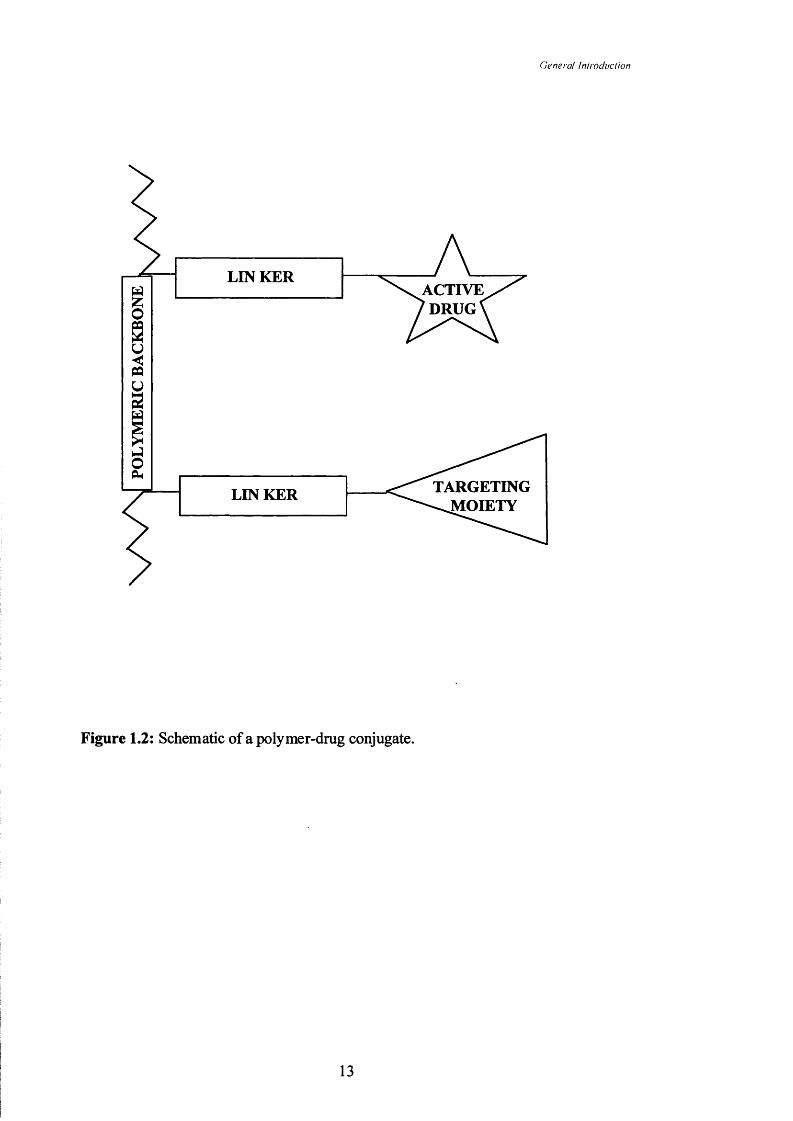
1.3,4 Polymer-drug conjugates
The concept of conjugating low molecular weight anticancer drugs to a
polymeric backbone was first proposed by Ringsdorf (1975). It was suggested that
drug would be bound via a suitable linker to the polymer backbone and additionally
targeting moieties could be present (Figure 1.2). Since then there has been a lot of
interest in this field, evident from the increasing number of polymer-drug conjugates
which have entered clinical Phase I/II testing (reviewed by Duncan, 2000). The three
main components of a polymer-drug conjugate will now be discussed.
1.3.4.1 Polymeric backbone:
The polymeric backbone is the major component of a polymer-drug conjugate
and must perform a number of important fimctions. The ideal polymeric backbone
would:
12
General Iniracliiction
LIN KERACTIVE^DRUG<
TARGETING^^ O IE T Y
LIN KER
Figure 1.2: Schematic of a polymer-drug conjugate.
13

General IntroducHon
1. Be soluble in physiological conditions and capable of solubilising poorly water-
soluble drugs
2. Possess functionality which allows conjugation of sufficient quantities of drug
3. Be monodisperse
4. Not be toxic and would have non-toxic breakdown products (if biodegradable)
5. Not be immunogenic and would be capable of ‘shielding’ immunogenic drugs
6. Be biodegradable or be of a size which would allow excretion from the body
7. Meet industrial constraints such as ease and cost of production and ease of
characterisation (reviewed by Duncan et al, 1996)
The importance of these individual factors varies depending on the drug of
interest and its characteristics. Solubility and solubilisation characteristics of the
polymer are important because the majority of anticancer agents have very poor
aqueous solubility, particularly those of natural origin (reviewed by Cragg et al, 1997).
The polymer should possess functionality allowing drugs to be conjugated and it is
important also that it can carry a sufficient drug load to be active, hence also the
importance of drug potency.
Synthetic polymers typically exist as a range of molecular masses (Figure 1.3)
and this is characterised by their polydispersity. Polydispersity is expressed as M^/M».
Weight-average molar mass (M w) is defined as the sum of the products of the molar
mass of each fraction multiplied by its weight fraction. Number-average molar mass
(M n) is defined as the sum of the products of the molar mass of each fraction multiplied
by its mole fraction. Therefore the closer the polydispersity is to 1.0 the more uniform
the polymer molecular weight (Young & Lovell, 1996).
The polymer backbone should either be of such a size that it can be excreted
from the body, usually by glomerular filtration in the kidneys, or be biodegradable to
such an extent. This size characteristic very much depends on the polymer in question
but for example HPMA copolymers display a renal threshold of -45kDa (Seymour et
al, 1987).
A large number of polymers have been explored as carriers for the delivery of
therapeutic agents (Table 1.2). Many of these polymers are non-biodegradable and
therefore the molecular weight which can be safely used is limited by renal clearance.
However, a number of polymers have been synthesised to contain biodegradable
elements in the backbone, particularly using low molecular weight PEGs (Pechar et al,
14

Genera! IntroducHan
4.0-M„ =100000Da
XM„ =199900DaI 3.0-
IIIg, 2.0-*o
i1.0-
Molar mass of polymer molecules (xlO g mol^)
Figure 1.3: Graph showing and M„ for a polymer.
Note: In this case the polydispersity would be 199900/100000 = 1.999.
15

General Inlroduciion
Table 1.2: Polymers used to prepare polymer-anticancer conjugates
Polymer Source PotentiallyBiodegradable
Drug carrying capacity
References
dextran Natural Yes Good reviewed by Mehvar, 2 0 0 0
chitosan Natural Yes Good reviewed by Dodane & Vilivalam, 1998
poly (L-lysine) Semisynthetic
Yes Good Shen & Ryser, 1981
poly (L- glutamic acid)
Semisynthetic
Yes Good Hirano et al, 1979
poly(amidoamines)
Synthetic Yes Good Ranucci et al, 1991
HPMAcopolymers
Synthetic No Good Duncan, 1992
poly (styrene-co-maleicacid)
Synthetic No Poor Matsumura & Maeda, 1986
PEG Synthetic No Poor reviewed by Nucci et al, 1991
Divinylether maleic acid copolymers (DIVEMA)
Synthetic No Good Przybylski et al, 1978
16

General introduction
2 0 0 0 ) in order to prepare block copolymers.
The means by which the drug is conjugated to the polymer is also of vital
importance. Most drugs must escape from the lysosomal compartment to exert their
biological activity. This release from the conjugate is essential for activity (reviewed
by Brocchini & Duncan, 1999). Many different types of polymer-drug linkage have
been used to allow cleavage by enzymes or other local physiological conditions (e.g.
hydrolysis at low pH) (reviewed by Soyez et al, 1996) and these will be reviewed next.
1.3.4.2 Linkers for drug conjugation
Careful consideration must be given to the linker design of an anticancer
conjugate. The linker should be stable in the bloodstream and only release the drug
intratumourally at a rate optimal for its mechanism of action. A number of linker
chemistries have been used (Table 1.3) to benefit from the tumour environment.
Linkers have been used which hydrolyse to liberate drug (e.g. esters and carbonates)
(reviewed by Greenwald, 1997), for example the poly(glutamic acid)-paclitaxel
conjugate which has recently entered Phase I clinical trial (Sludden et al, 2001).
The tumour extracellular environment is acidic in nature and acid-mediated
cleavage can also be useful within the endocytic pathways of the cell (endosomes and
lysosomes; see Section 1.4.4). A number of linkers have been developed to capitalise
on this phenomenon (e.g. cis-aconityl and hydrazone linkers). An HPMA copolymer
conjugate containing doxorubicin bound via a cw-aconityl linkage released 60% of the
doxorubicin over 40h in a buffer at pH5 (Choi et al, 1999).
Linkers which release drug under the reductive conditions found in the centre of
solid tumours and in the endocytic pathway of tumour cells have also been used. Ryser
& Shen (1978) synthesised methotrexate-poly(L-lysine) conjugates using a disulphide
linkage to allow drug release under reductive conditions.
The most widely investigated method of conjugation has been the use of
peptidic linkers (Duncan et al, 1983 and Subr et al, 1992). The Gly-Phe-Leu-Gly
(GFLG) spacer used to prepare HPMA copolymer-anticancer conjugates (see Section
1.3.4.2) was developed to be cleaved by cathepsin B, one of the thiol-dependent
proteinase enzymes present in the lysosome (reviewed by Dean, 1977). Cathepsins (in
particular cathepsins B and D (reviewed by Schwartz, 1995)) have been implicated in
17

General Introduction
Table 1.3: Examples of biodegradable linkers utilised in drug conjugates for anticancer
therapy.
Linker Cleavage conditions References
peptidic cathepsin B Subr et al, 1992
carbamate B-lactamase Senter et al, 1995
cw-aconityl acid hydrolysis Shen & Ryser, 1981
hydrazone acid hydrolysis Firestone et al, 1996
esters generalised hydrolysis Lietal, 1998
urea bonds generalised hydrolysis Ohyaetal, 1991
disulphide reductive conditions Ryser & Shen, 1978
18

Genera! Introduction
tumour metastasis and spread and are also present in the extracellular compartment of
many tumours. Therefore polymer-drug conjugates which contain this linker could
potentially liberate free drug both within the cell and extracellularly in a tumour. The
HPMA copolymer GFLG-doxorubicin conjugate (PKl) is in Phase II clinical trial
(Vasey et al, 1999) and combinations of these GFLG linkers and terminal ester bonds
(Caiolfa et al, 2000) or hydrazone bonds (Etrych et al, 2001) have been used to link
anti-tumour agents to HPMA copolymers.
A number of polymer-drug conjugates have progressed into clinical trial and
many are in pre-clinical development also. These will be reviewed before moving on to
focussing on the conjugates of HPMA copolymers, which are the subject of this thesis.
1.3.4.3 Polymer-drug conjugates in clinical trial and development
Those polymer-drug conjugates in either pre-clinical development or Phase 1/11
clinical trial are listed in Table 1.4. Many of the first conjugates tested clinically
contained doxorubicin. In a Phase 1 trial, a conjugate of dextran (70kDa molecular
weight) and doxorubicin (Figure 1.4) was given as a single dose i.v. administered every
3 to 4 weeks. The conjugate MTD was 40mg/m^ (doxorubicin-equivalent) (Danhauser-
Reidl et al, 1993) indicating that this conjugate was more toxic than free doxorubicin,
which is typically used at a clinical dose of 60-80mg/m^ (Martindale, 1993).
In contrast HPMA copolymer-doxorubicin conjugates (PKl and PK2; Figure
1.5) were less toxic than doxorubicin. PKl (FCE28068) is now in Phase 11 testing
(Vasey et al, 1999). PKl consists of an HPMA copolymer backbone (28kDa)
conjugated to doxorubicin via a GFLG linker and contains 8.5%w/w of doxorubicin.
Dose limiting toxicities in the Phase 1 clinical trial were febrile neutropenia and
mucositis (typical side-effects of doxorubicin administration) (Martindale, 1993). The
MTD of PKl given every three weeks was 320mg/m^ (doxorubicin-equivalent) and this
is 4-5 times higher than for free doxorubicin. Cardiotoxicity is the major toxicity of
doxorubicin (see Section 1.5.1) and cumulative doses are limited to 450-550mg/m^.
However, PKl was administered at cumulative doses of up to 1680mg/m^ with no
evidence of cardiotoxicity. PKl also showed anti-tumour activity with two partial and
two minor responses observed in the cohort of 36 patients. The patients entered the
trial with histologically confirmed solid tumours that were refractory to conventional
treatments.
19
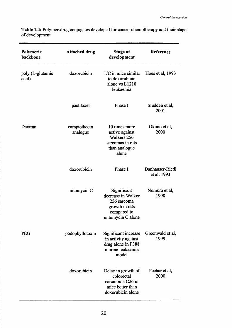
General Introduction
Table 1.4; Polymer-drug conjugates developed for cancer chemotherapy and their stage of development.
Polymericbackbone
Attached drug Stage of development
Reference
poly (L-glutamic acid)
doxorubicin T/C in mice similar to doxorubicin alone vs L1210
leukaemia
Hoes et al, 1993
paclitaxel Phase I Sludden et al, 2 0 0 1
Dextran camptothecinanalogue
1 0 times more active against Walkers 256
sarcomas in rats than analogue
alone
Okuno et al, 2 0 0 0
doxorubicin Phase I Danhauser-Riedl etal, 1993
mitomycin C Significant decrease in Walker
256 sarcomai growth in rats compared to
mitomycin C alone
Nomura et al, 1998
PEG podophyllotoxin Significant increase in activity against drug alone in P388 murine leukaemia
model
Greenwald et al, 1999
doxorubicin Delay in growth of colorectal
carcinoma C26 in mice better than
doxorubicin alone
Pechar et al, 2 0 0 0
20

General inlroduciion
PEG(contd.)
paclitaxel Improved activity and reduced
toxicity against P388 leukaemia model in mice
Pendri et al, 1998
Chitosan 5-fluorouracil Improved antitumour activity
against P388 leukaemia model in
mice
Ohyaetal, 1991
doxorubicin Conjugate showed increase in activity
against P388 leukaemia model in
mice
Zunino et al, 1987
DIVEMA cyclophosphamide Almost 3 times increase in survival
inL 1 2 1 0
leukaemia model in mice
Hirano et al, 1980
HPMA copolymers doxorubicin Phase I/II Vasey et al, 1999
daunorubicin Significant Walker sarcoma growth
delay in rats
Cassidy et al, 1989
paclitaxel Phase I Meerum Terwogt et al,
2001
camptothecin Phase I Caiolfa et al, 2000
melphalan Similar reduction in Walker sarcoma tumours in rats to melphalan alone
Duncan et al, 1991
21

Genera! Inlroduciion
HPMA copolymers mesochiorin C6
(contd.)Shows enhanced
tumour accumulation in
OVCAR-3 ovarian carcinoma
xenograft in nu/nu mice
Shiah et al, 1999
emetine Similar T/C value to drug alone in
B16F10 melanoma model in mice
Dimitrijevic & Duncan, 1998
platinate Phase I Gianasi et al, 1999
5-fluorouracil Shows drug release from the polymer
Putnam & Kopecek, 1995
22

Genera! Introciiiction
Figure 1.4: The structure of dextran doxorubicin.
23

Genera! Inlroduciion
% -]-
r
r 1 r r rtp o j
r 95
1 r S
k j
r90 L r°
rrÇHOH
1r r rr° ÇHOH r°
r ° J
CM C- C CH, 2 CH>
OCHs
Figure 1.5; Structures of PKl (A) and PK2 (B).
B
24

General Introduction
A similar conjugate with additional galactosamine residues (PK2 / FCE28069)
incorporated to promote liver targeting via the asialoglycoprotein receptor has also
undergone Phase I clinical testing in patients with primary or secondary liver cancer for
whom conventional treatments had failed (Palmer et al, 2001). In this case dose
limiting toxicities were febrile neutropenia, fatigue and mucositis (typical side-effects
of doxorubicin administration) (Martindale, 1993). When given every three weeks by
short infusion the MTD of PK2 was 160mg/m^ (doxorubicin-equivalent) again with no
incidence of cardiotoxicity. PK2 administration produced three partial responses and
eleven patients had stable disease in the cohort of 23 patients. A gamma camera
imaging analogue of PK2 confirmed significant liver targeting, when patients were
imaged using single photon emission computed tomography (Julyan et al, 1999).
HPMA copolymer-paclitaxel (PNUl 66945) (Figure 1.6) (20,000 times more
water soluble than paclitaxel alone) has also entered Phase I trial (Huinink et al, 1998).
No dose limiting toxicities were noted up to a dose of 196mg/m^ (paclitaxel-
equivalent), the usual paclitaxel dose being 250mg/m^ (Martindale, 1993). However
this study was halted prematurely as a parallel study in rats demonstrated severe
neurotoxicity of the conjugate compared to that of paclitaxel alone and during the trial
also there was a case of grade 3 neurotoxicity observed (Meerum Terwogt et al, 2001).
One partial response was observed in this study.
Another conjugate containing paclitaxel in early clinical testing is based on poly
(L-glutamic acid) (Sludden et al, 2001; Figure 1.6). The poly (L-glutamic acid)-
paclitaxel (Mw 36kDa) uses a simple ester linkage for drug conjugation and has a drug
content of 21%w/w. It is considerably more water soluble than paclitaxel alone (Li et
al, 1998). Doses of up to 266mg/m^ (paclitaxel-equivalent) have been administered but
there was evidence of serious neutropenia at this dose. Generally the conjugate was
reported to be well tolerated and the trial is ongoing.
Most recently HPMA copolymer conjugates containing a camptothecin and a
platinate have entered Phase I evaluation (Figure 1.7). HPMA copolymer-camptothecin
(consisting of polymer conjugated to camptothecin via a terminal ester linkage showed
significant in vivo anti-tumour activity against a HT29 human colon carcinoma
xenograft model (growth inhibition coupled with long term survivors where none were
seen in the group treated with camptothecin alone (Caiolfa et al, 2000)).
25

General Iniracliiction
N— CH G-N— CH GM— CH G
CH,CH,CHj
HNCH,
Na I —iw o
CH OHOH
NH
H,C
H,C
r r r
r r r\rJ »
fi. fL rJ 1 I ' r
rÎ
r Îr r
ÇHj
CH,
•CH,
Hf
B
Figure 1.6: Structures of poly (L-glutamic acid)-paclitaxel (A) and HPMA copolymer paclitaxel (B).
26

General Introduction
B
C H ,
L o
ÇHzCHOH
CH3
LCH»
TL oIr(ÇHzk0 = 0
1 = 0I
NH
CHz
NH
rÇHOH
C H s
r rs. î f i . 1
r°. 95 rr r1ÇHz
1ÇHz1
ÇHOH
C H jrNH
‘l’- s r s —
I in /-\rA
c o NH*
Figure 1.7: Structures of HPMA copolymer camptolhecin (A) and HPMA copolymer platinate (B).
27

General Inlroduciion
An HPMA copolymer-platinate conjugate also recently started Phase I clinical
trial (Gianasi et al, 1999). This conjugate contains an aminomalonato chelate of cis-
diammineplatinum (II) conjugated to HPMA copolymer GFLG and containing
7.5%w/w of platinum equivalence (Stewart et al, 2000). A number of HPMA
copolymer-platinate conjugates were described which showed up to 60-fold higher
levels of platinum compared to cisplatin in a B I6 F I0 s.c. tumour in mice also
demonstrating anti-tumour activity in this model. A recent press release describing the
ongoing Phase I clinical trial with this conjugate stated that doses of up to 1440mg/m^
(platinum equivalent) had been administered with no dose limiting toxicities observed.
This is 22 times more platinum than the usual administered dose of cisplatin (Access
Pharmaceuticals Inc. website, 2001).
The early promise of those polymer-drug conjugates that have entered clinical
development bodes well for the future development of second generation conjugates.
The clinical results seen for PKl and PK2 agree with preclinical animal studies in terms
of pharmacokinetics and reduced drug toxicity. As can be seen in Table 1.4 most of the
polymer-drug conjugates which have progressed to Phase I/II clinical trial utilise the
HPMA copolymer backbone. This is why this polymer was chosen here to prepare
novel polymer-natural product conjugates. The advantages of HPMA copolymer
conjugates and the biological rationale for their design is briefly discussed below.
1.4 Biological rationale for the design of HPMA copolymer-anticancer conjugates
It is important to better understand how those conjugates in development exert
anti-tumour activity in order to design, synthesise and optimise new conjugates.
Polymers used for drug conjugation must be safe, target drug selectively to the tumour
and release active drug intratumourally usually following endocytic uptake. Here
polymer-drug conjugate biodistribution and tumour targeting v ill be described and the
mechanisms of endocytic uptake of conjugate discussed.
1,4,1 Biocompatibility and pharmacokinetics of HPMA copolymers
As HPMA copolymers are not biodegradable conjugates of molecular weight of
> 45kDa would accumulate in the body (Seymour et al, 1987). Therefore the HPMA
copolymer-drug conjugates taken into clinical trial were all of a molecular weight of
approximately 30kDa (Duncan, 2000). However, as outlined earlier HPMA
2 8

General inlroduciion
copolymers do have many of the characteristics of an ideal polymeric carrier. They are
very water soluble and consequently are capable of solubilising poorly-water soluble
agents such as doxorubicin and paclitaxel. Neither PKl nor PK2 caused antibody
production when administered i.v., intraperitoneal (i.p.) or orally to two inbred strains
of mice. Doxorubicin bone marrow toxicity was also considerably decreased by
polymer conjugation as shown by the colony-forming unit-spleen assay (Rihova et al,
1989). Additionally HPMA copolymers did not activate either the classical or the
alternative complement pathways at therapeutic concentrations (Simeckova et al, 1986).
No evidence of HPMA copolymer-related toxicity was seen during clinical evaluation.
The Phase I trials of PKl and PK2 have also demonstrated that these HPMA
copolymer conjugates carry a sufficient loading of doxorubicin to produce anticancer
activity in chemotherapy refractory patients.
L4,2 Optimisation of linker design
As discussed previously a number of linkers have been used in the design of
polymer-drug conjugates (Section 1.3.4.2). The peptides have been the most widely
examined and were designed to be cleaved in the lysosomal compartment of the cell.
All the HPMA copolymer-anticancer conjugates in clinical development utilise a
peptidic linker, in most cases the GFLG linker, designed to be cleaved by cathepsin B.
A series of HPMA copolymer peptide linkers terminated with /?-nitroanilide (NAp)
were tested for the stability of the linker in rat serum and plasma, monitored by release
of NAp. None of the linkers tested released more than 5% in either serum or plasma
and the GFLG linker released less than 2% in either test solution (Rejmanova et al,
1985). While the susceptibility of the terminal bond may depend on the structure of the
conjugated drug, this is an indication that the bond will be stable in plasma and serum
as release upon injection would be undesirable.
A number of di-, tri- and tetra-peptidic linkers linker were then selected to
synthesise a series of HPMA copolymer-doxorubicin/daunorubicin conjugates which
were examined for their liberation of drug when co-incubated with tritosomes (rat liver
lysosomes) and for their activity in a mouse tumour model (i.p. L1210 leukaemia
model) (Subr et al, 1992). A GLFG linker showed the most complete release
(~100%/50h) but was toxic to mice in the anti-tumour experiment. The GFLG linker
showed good release (~70%/50h) and showed some anti-tumour activity. There was no
evidence of inappropriate release of doxorubicin in the Phase I clinical trials of PKl
and PK2 as such release would be expected to manifest itself as cardiotoxicity. Also
29

General Inlroduciion
some anti-tumour activity was observed in these trials. For these reasons the GFLG
linker was selected as a suitable linker for this study.
L 4 3 The Enhanced Permeability and Retention Effect
HPMA copolymer-anticancer conjugates have been shown to accumulate in
tumours by the EPR effect. PKl, for example, was administered to C57 black mice
with s.c. B16F10 melanoma at 5mg/kg (doxorubicin-equivalent) (Seymour et al, 1994).
PKl showed a greater than 15-fold increase in doxorubicin concentration in the tumour
48h after i.v. administration compared to free doxorubicin administered at the same
dose. As PKl can be administered at higher doses than free doxorubicin, because of
reduced cardiotoxicity, a higher dose of 18mg/kg was administered in this same study
and resulted in a greater than 70-fold increase in doxorubicin in the tumour, compared
to the 5mg/kg free doxorubicin dose. The EPR effect will now be discussed.
The EPR effect was first described by Matsumura and Maeda (1986). They
observed that the polymer protein conjugate SMANCS accumulated in tumour tissue
more than neocarzinostatin alone. Furthermore tumours lack a functional lymphatic
system. They proposed that this effect could be similar for other macromolecules and
they would extravasate into tumour tissue passively and be retained there because of
this poor lymphatic drainage. The tumour vascular system has many structural and
functional abnormalities (Konerding et al, 1999). Structural abnormalities include the
abnormal vessel wall, abnormal architecture and abnormal vascular density due to
unregulated angiogenesis (reviewed by Carmeliet & Jain, 2000). The underlying
functional abnormalities result in this increased vascular permeability, altered flow
characteristics and altered morphological characteristics. It has been known for some
time that neovascularisation in tumours results in distorted blood vessels (reviewed by
Rubin and Casarett, 1966) and that these blood vessels are sometimes incomplete with
large intracellular gaps. These gaps allow macromolecules to pass into the tumour
interstitium whereas in normal vasculature this would not be possible due to the tight
continuous intracellular nature of the normal blood vessel (reviewed by Jain & Baxter,
1988) (Figure 1.8). The EPR effect is mediated by imbalances in the level of many
endogenous factors such as vascular endothelial growth factor (VEGF), bradykinin,
tumour necrosis factor (TNF) as well as a host of cytokine and related factors such as
the hypoxic character of tumours (Maeda et al, 1992). In non-tumour tissue if
30
( iericrcil liiirodiicH an
^ lymphatic drainage ;
TUMOUR "V"" #non-functioning
lymphatics
m ;TISSUE "•> circulation
■-. - - . - -. ............
4 # # #
‘i : 4 S 4 x Blymphatic drainage 4
TUMOURTISSUE
L ■< '-1 < -1 •< -L V •-• •-* •<
Absent or non-functioning
lymphatics
-444S4-?-.r-_L ■.; v : •-•: -. A
4 4 ^4 4 4 4 4 ^ 4 4 4 4 4
illNORMAL 44<
TISSUE 4 : 4
Pil:C circulation ' A aX a;l##s
Figure 1.8: The EPR effect showing non-selective extravasation o f small m olecules (A) contrasted with tumour accum ulation o f a polymer-drug conjugate (B).
31

General inlroduciion
macromolecules extravasate they are brought back to the general circulation by
drainage mediated by the lymphatic system. Lymphatic drainage from tumours is
severely restricted or absent and therefore once a macromolecule has been captured in a
tumour by the EPR effect it remains within the tumour interstitium (Maeda &
Matsumura, 1989).
Macromolecular drug delivery systems can take advantage of these unique
physiological circumstances found in tumours to passively target them to the tumour by
the EPR effect. What is interesting in the case of the HPMA copolymers is that it has
been shown that their EPR-mediated targeting is governed solely by their plasma
concentration across a wide range of molecular weights (15-800kDa) (Seymour et al,
1995). The HPMA copolymer drug conjugates in clinical development have been
shown to be of a physical diameter of around 6 -8 nm (Duncan et al, 1996). They are
thus smaller in dimension than the liposomal systems. Many liposomal systems,
however, show poor penetration into the tumour mass, with even those liposomes in
clinical use, of a diameter of approximately 90nm, showing marked restriction in
penetration beyond the close proximity of the blood vessels (Uster et al, 1998). The
size dependence of tumour extravasation is being examined (Yuan et al, 1995) as
liposomes of up to 400nm have been shown to extravasate in a human colon
adenocarcinoma LS174T model but those of 600nm don’t. It is thought that these
localised deposits of liposomes containing drug then act as a slow drug release system
(Yuan et al, 1994). The extent to which polymer-drug conjugates extravasate has never
been characterised in terms of intratumoural distribution and this will be examined
using PKl as a model polymer-drug conjugate in Chapter 6 of this thesis.
After the HPMA copolymer-anticancer conjugate has been passively retained in
the tumour the cytotoxic agent needs to be liberated in order to be active. For those
polymer-drug conjugates containing the linkers which are susceptible to cathepsin B,
described in Section 1.3.4.2, the conjugate needs to be taken up by the cell and
trafficked to the lysosome in order to be exposed to cathepsin B and release active drug.
Therefore the process of endocytosis will now be described as it applies to polymer-
drug conjugates.
32

Genera! Inlroduciion
1,4,4 Intracellular pharmacokinetics
Low molecular weight anticancer agents show a widespread distribution in the
body following systemic administration (Figure 1.8). This is also true at the cellular
level and as most anticancer agents tend to be lipophilic they can traverse the cell
membrane non-specifically. Macromolecular drug delivery systems, however, are
incapable of traversing the cell membrane due to their size and are therefore reliant on
the process of endocytosis to enter the cell (De Duve et al, 1974). Endocytosis can be
broadly described as two mechanisms of uptake by cells of extracellular fluid.
Phagocytosis can be defined as the process by which specialised cells such as
macrophages engulf foreign matter for processing, usually by the RES, and is related to
large particles (>500nm diameter). Pinocytosis can be defined as the process of
engulfing small portions of the surrounding fluid and processing it through endosomes
and into lysosomes and is usually initiated by clathrin-coated pits and is common to
almost all cell types (reviewed by Mellman, 1996).
Figure 1.9 shows the differences in subcellular localisation of low molecular
weight and a polymer-drug conjugate. Polymer-drug conjugates have to go through a
number of stages before the cytotoxic agent can get to its site of action, if that site is
intracellular which in the case of many anticancer agents is the nucleus. They firstly
need to be taken up by the cell and therefore need to be of a physical diameter which is
not too large (Mellman, 1996). The attached anticancer agent needs to be stable in the
acidic milieu of the intracellular compartment (Ohkuma & Poole, 1978) and also needs
to be not broken down by the enzymes it encounters, of which the lysosome is thought
to contain up to 70 types (Dean, 1977). In the case of the HPMA copolymer-anticancer
conjugates the drug needs to be cleaved from the polymeric backbone in order to escape
from the lysosome to reach its intended target by crossing the lysosomal membrane. In
most cases it will need to be a free drug, without any residual linker in order to do so
and to exert its effect within the cell (reviewed by Lloyd, 2000).
The reliance of polymer-drug conjugates on endocytosis also provides a means
of circumventing the common resistance mechanism of p-glycoprotein-mediated
expulsion of anticancer agents back into the extracellular milieu which is a common
mechanism of resistance to conventional anticancer agents (reviewed by Krishna &
Mayer, 2000). PKl, for example, has shown cytotoxic activity towards human ovarian
carcinoma cells which are resistant to doxorubicin both in vitro (Minko et al, 1998) and
33

Clenera/ hi/rotliicUon
Low molecular weight anticancer agent • •
Polymer conjugate
ENDOSOME
LYSOSOME
: E
TARGET
Lysosomal enzymes (E) (over 40 identified) e.g. phosphorases, sulpatases, oxidases, hydrolases,peptidases (e.g.cathepsins)
Figure 1.9: Schematic of intracellular release of pendant drugs with respect to a low molecular weight anticancer drug.
34

General Inlroduciion
in vivo in animal models (Kopecek et al, 2000). These results demonstrate that polymer-
drug conjugates offer the possibility of overcoming such resistance.
The choice of polymeric backbone and linker are thus governed by biological as
well as physicochemical characteristics and both need to be taken into account in order
to design novel conjugates. The aspects of polymer-drug conjugates described so far
demonstrate the suitability of HPMA copolymer GFLG conjugates as the system of
choice for novel conjugates. The next section describes those natural products which
show anticancer activity and the reason why an analogue of ellipticine, namely APE,
was chosen for conjugation to HPMA copolymer GFLG in this thesis.
1.5 Natural products in Cancer
Natural products have always been at the fore in the development of anticancer
agents. In the U.S.A (1994) more than 50% of anticancer drugs on the market were
either of natural origin, derived from natural sources or were semi-synthetic natural
products (reviewed by Cragg et al, 1997) (Figure 1.10). Natural products are also a
widespread source of novel structures and, in the case of anticancer agents, of novel
modes of action which might circumvent resistance, penetration and targeting issues
described earlier (reviewed by Newman et al, 2000).
L5.1 Natural products in clinical use
A number of natural products are used in cancer treatment and there are a
number of classes of these compounds which have been widely studied. Some of the
major interesting classes will now be described.
The anthracyclines are very commonly used chemotherapeutic agents as they
exhibit a broad spectrum of activity against many solid tumours as well as lymphomas
and leukaemias and include doxorubicin and daunorubicin (reviewed by De Vita et al,
1993). They are thought to have a number of potential mechanisms of anti-tumour
activity but are usually described as being topoisomerase 11 inhibitors and to form
damaging free radicals (reviewed by Gewirtz, 1999). This radical formation has been
implicated in the major toxicity of the anthracyclines, notably cardiotoxicity. This
cardiotoxicity results from cumulative dosing of doxorubicin which is usually given at a
dose of 60-80mg/m^ alone or 30-40mg/m^ when given with agents having overlapping
35

( k ’n cra l Inirndiiclion
Synthetic source natural model
Biologic
Natural
Synthetic
+ + + + *
Naturallyderived
Figure 1.10: Anticancer drugs approved in the U.S.A. in 1994 and their origin (adapted from Cragg et al, 1997).
36

General fnlroduction
toxicity by fast running intravenous infusion every three weeks. The maximum
recommended cumulative dosage is 450-550mg/m^ as above this significant ventricular
failure and dysrrhythmias commonly occur. Other common adverse events include
myelosuppression, mucositis and local extravasation at the site of administration
resulting in severe necrosis (Martindale, 1993). PKl (Figure 1.5; Section 1.3.4.3)
demonstrated the feasibility of conjugating anthracydines to HPMA copolymers via a
GFLG linker and this was the first polymer-drug conjugate to enter clinical
development.
The vinca alkaloids are another class of clinically used natural products and
vinblastine and vincristine are the best known and are in routine clinical use.
Analogues of these compounds vinorelbine and vindesine are also approved for use in
many European countries (reviewed by Newman et al, 2000). These agents act by
binding to tubulin and inhibiting microtubule assembly thereby blocking the formation
of the mitotic spindle resulting in cell division being suspended in mitosis and causing
cell death by apoptosis (Lobert et al, 1996). So far there are no reports of polymer-drug
conjugates of the vinca alkaloids, although vincristine encapsulated in a glucuronide-
modified liposome showed significant tumour growth delay in a mouse model
(Tokudome et al, 1996).
Paclitaxel is another agent that acts on the tubulin polymerisation process.
Paclitaxel, however, acts in a different way to the other so-called spindle poisons in that
it stabilises microtubules and prevents depolymerisation back to tubulin whereas the
others, such as the vinca alkaloids, bind to tubulin and prevent the formation of
microtubules (reviewed by Wall & Wani, 1995). As described in Section 1.3.4.3 both
HPMA copolymer and poly (L-glutamic acid)-paclitaxel conjugates (Figure 1.6) have
entered Phase I clinical trial (Meerum Terwogt et al, 2001 and Sludden et al, 2001
respectively). Both conjugates show a remarkable increase in paclitaxel solubility.
However the Phase I trial involving the HPMA copolymer-paclitaxel conjugate was
halted prematurely because of excessive neurotoxicity; worse than that of paclitaxel
alone. This could have been caused by the unstable nature of the ester linkage leading
to premature release of paclitaxel at unexpected sites producing toxicity. Paclitaxel is a
potent and clinically useful drug and therefore further investigation of polymer-
paclitaxel conjugates is justified and indeed the clinical trial of the poly (L-glutamic
acid) conjugate is ongoing.
37

General Introduction
Another class of natural products of importance are the camptothecins.
Camptothecin was initially discovered in 1958 but only became of interest when its
specificity for the topoisomerase I enzyme was discovered and it underwent clinical
trials in the 1960’s. Some anti-tumour activity was reported but severe side-effects
halted further trials (reviewed by Potmesil, 1994). Further analogues were developed
and the analogues topotecan and irinotecan are now in routine clinical use with a
number of other camptothecin analogues in clinical development (Newman et al, 2000).
As described earlier a HPMA copolymer-camptothecin construct (Figure 1.7) has
recently entered Phase I/II clinical trial (Caiolfa et al, 2000 and Zamai et al, 2001).
A number of biologic agents are also in clinical use in cancer therapy such as
the colony stimulating factors used in adjuvant therapy (e.g. Filgrastim), interleukin-2
(a lymphokine which stimulates proliferation of lymphocytes) and interferons which
can have an anti-proliferative effect (Martindale, 1993).
Natural products are a useful source of novel therapeutics and new natural
products are constantly entering clinical trial. An example of a novel natural product
which recently underwent Phase I clinical trial is dolastatin 10. This is a peptidic drug
and binds to tubulin at a different site to the other antimitotic agents previously outlined
causing inhibition of tubular polymerisation and leading to apoptosis. The initial Phase
I trial was promising and Phase II trials are currently in progress (Madden et al, 2000).
Combretastatin A4 is isolated from the South African tree Combretum cqffrum and has
been shown to have a high affinity for tubulin causing destabilisation of the tubulin
polymers in the cytoskeleton (Woods et al, 1995). Where this compound has been
shown to have activity is as an antivascular agent. The disodium phosphate derivative
of this drug has been shown to induce vascular shutdown very rapidly in experimental
tumours and has recently entered Phase I clinical trial in the U.K. (Tozer et al, 1999).
The disodium phosphate modification of combretastatin is a means of ensuring
activation in the tumour where alkaline phosphatase activity is increased and the drug
will be liberated locally. This is a novel mechanism of action for an anticancer drug
and it has been shown not to induce resistance in a number of in vitro and in vivo
models (Kerbel, 1997). Therapies with unique mechanisms of action, such as this, are
very important for the development of future anticancer therapies and in particular for
combination chemotherapy.
38

General inlroduclion
1,5,2 Ellipticines
The majority of the polymer-drug conjugates which have been developed so far
have utilised anticancer agents which are already on the market (e.g. doxorubicin,
paclitaxel, mitomycin C and platinates. Table 1.4). However the potential candidates
for polymer conjugation should not be limited to these widely used anticancer agents.
The approach, now validated with these known compounds, could be applied to other
potent cytotoxic agents which initially failed early pre-clinical and clinical testing. The
candidate compound chosen for study in this thesis is from one such family of
compounds, the ellipticines, which were identified as interesting compounds some time
ago. Many of the ellipticine analogues failed early preclinical or clinical testing due to
poor formulatory properties and severe toxicities.
Ellipticines (5, 1 l-dimethyl-6H-pyrido(4,3-b)carbazoles; Figure 1.11) are anti
tumour alkaloids isolated from the Ochrosia elliptica tree (Goodwin et al, 1959). The
potential of these agents as anti-tumour agents was first identified in 1967 (Dalton et al,
1967). The mechanism of action of ellipticine and its subsequent analogues is
multifactorial. However most analogues act as DNA intercalators and topoisomerase II
inhibitors, supported by their cross-resistance to other topoisomerase II inhibitors
(Charcosett et al, 1988). It is interesting to note that a derivative of ellipticine, termed
T-215, was the first anticancer drug shown to induce apoptosis of tumour cells in
connection with mutant p53 protein. Another derivative, 9-hydroxyellipticine, also
promotes apoptosis by inhibiting phosphorylation of mutant p53 protein (reviewed by
Ohashi & Oki, 1996).
Celliptium (elliptinium acetate; Figure 1.11) in a Phase II study showed activity
in advanced breast cancer and is marketed as second line treatment for advanced breast
cancer (Rouesse et al, 1993). Datelliptium (2-diethylaminoethyl-9-hydroxyellipticine,
Figure 1.11) is also marketed and showed activity in a Phase I study against breast
cancer (Khayat et al, 1992). Celliptium was shown in previous studies to cause serious
immune-mediated haemolytic reactions (Rouesse et al, 1985) but this was abrogated in
the reported Phase I study by changing the dosing schedule. These compounds have
only entered clinical use relatively recently given how long ellipticines have been
studied. A large series of derivatives of ellipticine have been synthesised in this time
and a number of these have entered pre-clinical and clinical development (Table 1.5).
39

(Jenera! Inlrocliiclion
R11R9
R6
R5
R2
Compound R2 R5 R6 R9 R ll
Ellipticine H H NH H CH3
Celliptium CH] CH3 H OH CH3
Datelliptium H2N(C2H5)2 CH3 H OH CH3
APE H CH3 NCH2CH2CH2NH2 H CH3
Figure 1.11: General structure of the ellipticines showing some of the common sites of derivatisation and the structures of ellipticine, Celliptium, Datelliptium and APE.
40

General Introduction
Table 1.5: Ellipticine derivatives which have undergone pre-clinical and/or clinical trial
but are not yet marketed.
Compound Status Results Reference
Retelliptine Phase I Dose limiting cardiac toxicity
Cappelaere et al, 1993
Pazellipticine Pre-clinical Needs bioactivation for activity
Balosso et al, 1991 Vilarem et al, 1986
Elliprabin Phase I Terminated due to hepatic toxicity
Ohashi & Oki, 1996
RPI-6 Pre-clinical 1000-fold as active as doxorubicin against
some lung cancer cell lines
Ruckdeschel & Archer, 1989
S-16020 Phase I Still ongoing. Selected as a
particularly active derivative
Le Mee et al, 1998
41

General Introduction
The major toxicity of the ellipticines has been cardiotoxicity. They can cause an
initial tachycardia and hypotensive shock on administration, followed by long term
bradycardia linked with an increased force of contractility. Haemolysis is also a
common adverse event (reviewed by Ohashi & Oki, 1996), and there has been evidence
of hepatotoxicity (Donato et al, 1992) and renal toxicity (Raguenez-Viotte et al, 1988).
An immune response, observed as anti-elliptinium immunoglobulin G-type antibodies,
has also been seen after administration of Celliptium (Alberici et al, 1986). A number
of these toxicities are exacerbated by the problem of poor solubility of the ellipticines in
aqueous solutions.
Ellipticine and its analogues therefore seemed to be good candidates for
polymer-drug conjugation. It was hoped that conjugation to HPMA copolymers would
improve water solubility, improve tumour targeting by the EFR effect and moreover
reduce non-specific toxicity as seen in the case of PKl (Vasey et al, 1999). The
ellipticine derivative APE was selected because of its potent in vitro cytotoxicity
(Inhibitory concentration for 50% cell killing (IC50) of 3.2x10'^M in the National
Cancer Institute (NCI) screen (NCI website, 2001). APE was also selected because of
its chemical suitability for synthesising an HPMA copolymer-APE conjugate using an
HPMA copolymer precursor and a simple aminolysis reaction which vdll be described
in Chapter 3.
1.6 Aims of the study
When this project started it had become clear that PKl was already showing
clinical potential and that the approach of HPMA copolymer conjugation might be
applicable to other (novel and more interesting) anticancer agents. The broad aims of
this thesis were therefore to investigate further the mechanism of the EPR effect by
understanding better the extravasation characteristics of PK l. This was studied using
an in vivo visualisation system of a tumour (rat dorsal window chamber model; Chapter
6).
The most important aim was to try to design an HPMA copolymer-natural
product conjugate that could progress into early clinical trial. The ellipticine derivative
APE was chosen as the model for this work. First it was necessary to synthesise and
characterise HPMA copolymer-APE conjugates using two peptidyl spacers (GO,
thought to be lysosomally non-degradable and GFLG chosen as a degradable linker).
42

General introduction
To enable study of the effect of drug loading compounds were synthesised containing
l-6.1%w/w APE (Chapter 3). First the in vitro physicochemical and biocompatibility
(cytotoxicity and haematocompatibility) were studied and the effect of drug loading on
the rate of drug liberation by lysosomal enzymes was studied (Chapter 4). Then
selected conjugates were evaluated in vivo using murine and xenograft tumour models
to examine the therapeutic index (Chapter 5).
43

Materials and Genera! Methods
Chapter Two
Materials and General Methods
44

Materials and Genera! Methods
2.1 Materials
2,L I Chemicals
All chemicals were obtained from Sigma-Aldrich Ltd. (Dorset, U.K.) or Fisher
Chemicals Ltd. (Leicestershire, U.K.) and were of analytical or reagent grade unless
otherwise stated. HPMA copolymer-GG-p-nitrophenol (ONp) containing
approximately 5mol% peptidyl side-chains and HPMA copolymer-GFLG-ONp of
approximately 1, 5 and 10mol% peptidyl side-chains were supplied by Polymer
Laboratories (Church Stretton, U.K.). APE was kindly donated by the NCI (Maryland,
U.S.A.). Daunomycin hydrochloride was kindly donated by Rhone Poulenc (France).
PKl and doxorubicin hydrochloride were kindly donated by Pharmacia (Milan, Italy).
All solvents designated dry were dispensed from stock containers under argon gas. All
solvents used in High Pressure Liquid Chromatography (HPLC) and Gel Permeation
Chromatography (GPC) applications were filtered through a 0.22pm filter and
subsequently sonicated for 5-lOmin before use.
2d,2 Cell culture
B16F10 murine melanoma cells were kindly donated by Prof. Ian Hart (St.
Thomas’ Hospital, London, U.K.). CORL23 human non small cell lung carcinoma
cells were purchased from ECACC (European Collection of Cell Cultures, Centre for
Applied Microbiology & Research, Salisbury, Wiltshire, U.K.). Tissue culture grade
dimethyl sulphoxide (DMSO), 3-(4,5-dimethylthiazol-2-yl)-2,5-diphenyl tétrazolium
bromide (MTT) and trypan blue were purchased from Sigma (Dorset, U.K.). Foetal
Calf Serum (FCS), trypsin with ethylenediaminetetraacetic acid (EDTA) and RPMI
1640 media were purchased from Gibco BRL Life Technologies (Paisley, U.K.).
Phosphate buffered saline (PBS) was obtained from Oxoid Unipath Ltd. (Hampshire,
U.K.) and 0.9% NaCl (normal saline) was prepared using sodium chloride from BDH
(Dorset, U.K.) and distilled water and both these solutions were autoclave sterilised
before use in cell culture work.
45

Materials and Genera! Methods
2.2 Equipment
2.2.1 Analytical equipment
The Ultra Violet (UV)-visible spectrophotometers used were a UV-1601
purchased from Shimadzu Scientific Instruments (Milton Keynes, U.K.) and a Cary IG
UV-visible spectrophotometer purchased from Varian Ltd. (Surrey, U.K.). 717plus
Autosampler and C l8 reverse phase pBondapak columns were from Waters Ltd.
(Hertfordshire, U.K.). Mobile phase was delivered using a PU-980 Intelligent HPLC
pump from Jasco Inc. (Essex, U.K.). Fluoromonitor III fluorescence detector fitted
with fluorescence lamp (excitation 480nm, emission 560nm) was from LDC/Milton
Roy (U.K.) for doxorubicin determination. For APE determination a Dynamax-60Â Cn
column from Varian Analytical Instruments Ltd. (California, U.S.A.) was used. A
Spectroflow 783 UV detector from Kratos Analytical (Milton Keynes, U.K.) and an LC
1255 fluorodetector from GBC Scientific Equipment (Illinois, U.S.A.) were used as
detectors. All chromatography data was recorded and analysed using PowerChrom
hardware and software (version 2.0.7).
2.2.2 Cell culture equipment
The 96 well plate spectrophotometers used were from Titertek Multiskan Plus
(EFLAB, Finland) and a Tecan Sunrise instrument (Tecan Ltd., Berkshire, U.K.). All
cell culture work was carried out in a Class II biological safety cabinet.
2.2.3 In vivo equipment and suppliers
L.I.P. Services and Equipment Ltd. (West Yorkshire, U.K.) supplied
heparin/lithium blood tubes. All nu/nu mice were housed in an isolator unit (Isolator
type 12162 (M20)) which was purchased from Harlan Ltd. (Oxon, U.K.). Other
procedures involving nu/nu mice were carried out on a laminar flow bench. Needles
and syringes were from Becton Dickinson (Oxford, U.K.). All animals were purchased
from Bantin and Kingman Ltd. (Hull, U.K.) or Harlan UK Ltd. (Bicester, Oxon, U.K.).
Gases were from BOC Ltd. (Surrey, U.K.) and isoflurane was obtained from Abbott
Laboratories Ltd. (Kent, U.K.).
2.2.4 Miscellaneous and general equipment
The freeze-drying system for lyophilising samples was a Flexi-Dry freeze drier
from FTS Systems (New York, U.S.A.) attached to a double stage high vacuum pump
46

Materials and Genera! Methods
from Javac Ltd. (Melbourne, Australia). Dialysis membranes Spectra/Por® (Cellulose
ester) were from Spectrum Laboratories Inc. (California, U.S.A.) with a molecular
weight cut-off of 3,000Da. All low speed centrifuge work was carried out using a
Varifuge 3.0RS from Heraeus Instruments (Osterode, Germany). All other centrifuge
work was carried out using an Allegra 6 KR, Avanti J-25 centrifuge or an Optima LE-
80K ultracentrifuge from Beckman Coulter (California, U.S.A.).
2.3 General methods
23.1 In vitro methods
Cell line usage was conducted based on the principles outlined in the UKCCCR
Guidelines for the use of Cell Lines in Cancer Research (2000). All cell manipulations,
except centrifugation steps, were carried out in a Class II biological safety cabinet. Any
equipment brought into the cabinet was sterile and sprayed with a solution of ethanol
(70%) and water. All solutions in contact with cells were heated to 37°C in a water
bath.
2.3.1.1 Growing cells from frozen vials
Both cell types (B16F10 and CORL23) were grown from frozen by heating
vials to 37°C in a water bath and as soon as they had defrosted they were added to 9ml
of RPMI-1640 media containing 10% FCS in a sterile container. This cell suspension
was then centrifuged at l,000g for 5min at room temperature and the supernatant liquid
discarded to remove any residual DMSO. 10ml of the same media was then added and
the cells were disaggregated using a syringe and needle added to a 75cm^ tissue culture
flask and this was then put into a 37°C incubator with a humidified atmosphere and 5%
CO2.
2.3.1.2 Day to day maintenance of cells
Both cell types were maintained in the same media as outlined before in a
75cm^ vented tissue culture flask. Cells were examined every day for confluence and at
75-95% confluence were split 1:10 as follows. The old media was removed and the
cells washed with 10ml of sterile PBS twice. Trypsin (0.25%)/EDTA (ImM) solution
(1ml) was then added and the cells returned to the incubator for 3-5min until the cells
had detached from the surface of the flask and disaggregated. Fresh media (9ml) was
47

Materials and Genera! Methods
added to this cell suspension. Subsequently 9ml of this cell suspension was removed
and 9ml of fresh media was added to complete a 1:10 split. The 9ml of cell suspension
removed was used to produce further flasks of cells by adding 1ml to a fresh flask
containing 9ml of fresh media.
2.3.1.3 Cell counting
To count cells 50pl of a cell suspension was taken and added to 50pl of a
solution of 0.2% trypan blue. This suspension was left for 2-3min, then pipetted onto a
haemocytometer with a cover slide. A cell count was made by counting the cells,
which were not blue, in 4 of the grids on the haemocytometer and averaging the result.
This value was then doubled to allow for the dilution by tiypan blue and the number of
cells calculated by multiplying this value by IxlO' to get a final value of viable cells
per ml of a suspension. Any cells which did not exclude trypan blue and were therefore
blue were not counted and assumed to be non viable as intact cells should not take up
this dye.
2.3.1.4 Freezing cells
Cells were periodically frozen as follows. Cells were trypsinised as detailed
previously made up to 10ml with media and centrifuged at l,000g for 5min at room
temperature. Cells were also counted. After centrifugation the cells were made up to a
concentration of IxlO^cells/ml in a freezing media (90%FCS with 10%DMSO) and
immediately frozen to -80°C and either continually stored at this temperature or
transferred to a liquid nitrogen cell bank until required.
2.3.1.5 Growth curve
Cells were trypsinised as described in Section 2.3.1.2 and made up to a volume
of 10ml with fresh media. A 50pl sample was taken and the cell number counted as
described in Section 2.3.1.3. The rest of the suspension was centrifuged at 1,000g for
5min at room temperature. The supernatant liquid was decanted and the cells made up
to a concentration of IxlO^cells/ml using media. Aliquots of this cell suspension
(lOOpl) were added to each well of a 96 well plate which was then returned to the
incubator overnight to allow the cells to attach to the surface and form a monolayer.
The next day, and on each subsequent day, 20pl of MTT (5mg/ml in sterile PBS and
filter sterilised through a 0 .2 2 pm filter) was added to each well in one row of wells and
the cells were incubated for 5h at 37°C. Viable cells transform MTT, in the
48
Maleriah and Genera! Methods
mitochondria, to an insoluble formazan derivative in a time and cell number dependent
manner (Sgouras and Duncan, 1990 and Figure 2.1). After 5h all cell debris and media
were removed and the formazan crystals dissolved in lOOpl of spectrophotometric
grade DMSO at 37°C for 30min. The absorbance (550nm) for each well was measured
using a micro-titre plate spectrophotometer reader. The DMSO, containing formazan
crystals, was then removed from each well. Fresh media or PBS was then added to
each of these wells to prevent the DMSO from evaporating and causing toxicity to the
remaining cells.
2.3.1.6 Cell cytotoxicity using the MTT assay
To determine drug cytotoxicity cells were plated into 96 well micro-titer plates
in the same manner as for a growth curve. After 24h, when the cells had adhered,
media was removed and the drug to be tested, dissolved in media, was added. The cells
were returned to the incubator for a further 67h. After this time MTT solution (20pl;
5mg/ml in PBS sterile filtered) was added to each well and the plates re-incubated for a
further 5h. The formazan crystals were dissolved in DMSO as before and read at
550nm using a micro-titer plate reader. Cells grown in media alone were used as a
reference for 100% viability. The absorbance of the other wells was calculated against
the average absorbance of the reference wells and calculated as a percentage of this
value.
2.3.1.7 Cell preparation for s.c. injection into mice
Injection (s.c.) of B16F10 and CORL23 cells as cell suspensions was used to
establish tumours in C57B1 (B16F10) or nude mice (CORL23). Prior to injection cells
were prepared by trypsinising and counting as described above. The cells were then
made up to the appropriate volume (IxlO^cells/ml for B16F10 or IxlO^cells/ml for
CORL23) in sterile saline solution.
2,3,2 Evaluation of anti-tumour activity
All in vivo work involving tumours was carried out according to the UKCCCR
Guidelines for the Welfare of Animals in Experimental Neoplasia (1998). In all of
these experiments the animals were allowed food and water ad libitum.
The B16F10 cell line grows s.c. upon injection of IxlO^cells into the nape of the
neck of C57BL black male mice. After injection the tumours were allowed to establish
49

Maleriah and Genera! Methods
MTT (water soluble)
CK
CH.
f V c-M
1NH
Formazan (water insoluble)
CH.
Figure 2.1: Metabolism of MTT by viable cells.
50

Materials and Genera! Methods
to a palpable size of between 25-50mm^ as determined by the measurement of the two
longest diameters. The animals were then randomised by assigning each mouse to a
separate group, when the tumours reached the correct size, and then sequentially adding
each mouse to the next group until each group contained at least 5 mice. The test drugs
or saline, as a control, were injected i.p. into the animals as a single dose on the day the
tumour reached the correct size. On each subsequent day each animal was weighed and
the tumour measured. If the animal had lost more than 20% of body weight or was
obviously sick (lethargic, unkempt and unresponsive) or the tumour had reached
289mm^ the animal was humanely killed by CO2 asphyxiation. All organs and tumours
were retained, weighed and examined for gross morphological changes.
The CORL23 cell line also grows s.c. upon injection of IxlO^cells into the nape
of the neck of nu/nu male mice. Nude mice were housed in an isolator at all times. For
manipulations, such as tumour measurement or injections, the animals were removed in
a box with a filter on top to a laminar flow bench. All items added to the isolator, such
as food and water, were purchased sterilised by irradiation and the exterior sprayed with
a virucidal solution. After injection of the cell suspension tumours were allowed to
establish to a palpable size of between 25-50mm^ as determined by the measurement of
the two longest diameters. The animals were then randomised in the same manner as
for the C57B1 mice above. The test drugs or saline, as a control, were injected i.p. into
the animals as a single dose on the day the tumour reached the correct size. On each
subsequent day each animal was weighed and the tumour measured. If the animal had
lost more than 2 0 % of body weight or was obviously sick (lethargic, unkempt and
unresponsive) or the tumour had reached 289mm^ the animal was humanely killed by
CO2 asphyxiation. All organs and tumours were retained, weighed and examined for
gross morphological changes.
23,3 Analytical methods
2.3.3.1 HPLC determination of APE
A HPLC method was used to determine the free APE content of conjugates as
well as APE released in tritosome release studies. All solutions were kept on ice
throughout the course of the experiment to minimise potential degradation and original
test solutions were in polypropylene tubes. A solution of 0.1 mg/ml was used to make
various standard concentrations of APE (0-lpg/ml) in distilled water. Conjugates for
51

Materials and Genera! Methods
analysis were dissolved in distilled water at a known concentration. Samples from
tritosome release studies were used as the lOOpl taken at each timepoint as described in
Section 2.3.4. Two methods were used in these studies as it was found that the first
method did not counteract the buffering capacity of proteins in tritosome release
studies. The first method was used for analysis of conjugates while the second method
was used for all biological samples.
Extraction and HPLC analysis of non-biological samples:
To the test solutions lOOpl of doxorubicin (0.6mg/ml) was added as the internal
standard and they were then made up to 1ml using distilled water. To this a small
amount (usually 5pi) of O.IM NaOH was added to make the solution pH 10. Then 5ml
of 4:1 chloroform : isopropanol was added to each tube. The organic layer was
subsequently removed into fresh polypropylene tubes. This organic solution was
evaporated by blowing nitrogen gas at the surface of the liquid. The residue was
dissolved in 200pl of methanol : distilled water (60 : 40v/v) which was also the mobile
phase used. 2 0 pl of this solution was injected either manually or using an autosampler
onto a C l8 pBondapak reverse phase column with mobile phase delivered by a HPLC
pump delivering mobile phase at Iml/min. Detection was by means of a UV
absorbance detector set to 296nm. Data were recorded and analysed using the
PowerChrom system.
Extraction and HPLC analysis of biological samples:
To the test solutions 200pl of quinaldine red (0.1 mg/ml in methanol) as the
internal standard was added and they were made up to 1ml using distilled water. To
this lOOpl of ammonium formate IM at pH8.5 was added. This ammonium formate
was used to counteract any protein-mediated buffering. To these solutions 5ml of 4 : 1
dichloromethane : isopropanol was added. The tubes were then centrifuged at 2,000g
for lOmin at 4°C to aid separation. The organic layer was removed into glass test tubes
which had been silanized by treatment with a solution of 93:7 toluene :
dichloromethylsilane and then washed with ethanol and allowed to dry (as per the
method of Fraier et al, 1995). This organic solution was evaporated by blowing
nitrogen gas at the surface of the liquid. The residue was dissolved in 70% acetonitrile
in distilled water which contained 0.035% trifiuoroacetic acid, which was also the
mobile phase. This solution (20pl) was injected using an autosampler onto a Dynamax-
60Â Cn column with mobile phase delivered by a HPLC pump delivering the mobile
52

Materials and Genera! Methods
phase at 0.6ml/min. Detection was achieved using a UV absorbance detector set to
296nm and a fluorescence detector in series using wavelengths of 286nm excitation and
480nm emission. Data were recorded and analysed using the PowerChrom system.
Figure 2.2 gives example chromatograms and Figure 2.3 shows standard curves from
such HPLC determinations of APE
2.S.3.2 HPLC determination of free and total doxorubicin
This method was adopted from Wedge (1991) and was used to examine the free
and total doxorubicin content of organs as well as the doxorubicin liberated from PKl
in tritosome release studies. Samples from these studies were stored in polypropylene
tubes in the dark at 4°C before analysis to minimise degradation. On the day of
extraction they were removed but stored on ice for the entire process of extraction to
minimise any potential degradation. In the free doxorubicin method it is free
doxorubicin and daunomycin which are being detected. In the total doxorubicin
method it is aglycone doxorubicin and daunomycin, because the acid hydrolysis
liberates these species, being detected and also the retention time is increased.
Determination of free doxorubicin content:
To each tube 25pl of a lOpg/ml solution of daunomycin (lOpg/ml dissolved in
distilled water) was added. The tubes were then vortexed to mix the contents and made
up to 1ml with distilled water and revortexed. The solution was buffered by adding
150pl of a IM solution of ammonium formate adjusted to pH8.5 with ammonium
hydroxide and the solution revortexed. To each tube 5ml of a 4 : 1 solution of
chloroform : propan-2-ol was added and the solutions mixed. These were then
centrifuged at l,000g for 30min at 4°C and the organic layer removed and evaporated
under nitrogen gas to dryness. The residue was redissolved in lOOpl of methanol and
injected, using an autosampler, onto a C l8 reverse phase pBondapak column with a
mobile phase delivered at Iml/min of propan-2-ol 29% in distilled water adjusted to
pH3.2 using o-phosphoric acid. Detection was by a fluorescence detector (excitation
480nm; emission 560nm) and data were analysed using PowerChrom system.
Determination of total doxorubicin content:
To each tube 25pi of a lOpg/ml solution of daunomycin (lOpg/ml dissolved in
distilled water) was added. The tubes were then vortexed to mix the contents and made
up to 1ml with distilled water and revortexed. To each solution 1ml of a 2M solution of
53

A la ie n a ls amJ ( jcn era l M ethods
APE
e3
t:I DoxorubicinU
ii
31 4 52Retention time (min)
B
'c3
Ë3kSE
I- w3kBI-3
10 -
5 -
Ellipticinei
Quinaldine Red
“I 1------1----- 1----- 1----- 1 1----- 1—12 13 14 15 16 17 18 19
Retention time (min)
Figure 2.2: Typical HPLC chromatograms obtained during APE quantitation using method 1(A) and m ethod 2 (B).
Note: In both cases the internal standard (Doxorubicin (60pg/m l) (A) and Quinaldine Red (20pg/m l) (B)) are at a set concentration with increasing amounts o f APE (0-4 pg/ml for A and 0-lpg/m l for B) and/or ellipticine (0- Ipg/m l).
54

Materials and General Methods
500-]
400-
I« 300-II y = 0.31213 + 97.092X
200 -ms<
100-
1 30 2 4 5APE concentration (/#g/ml)
60-1
50-
li 40oo£Ic3
30
I< 20 -y = 2.2219e-2 + 46.586x
10
0.0 1.00.2 0.4 0.80.6APE concentration (//g/ml)
Figure 2.3: A standard curve of determination of APE in the HPLC system.
Note: Panel a is from a study using Method 1 whilst b is from a study using Method 2. Data represent mean ±SD (n=3).
55

Materials and Genera! Methods
hydrochloric acid was added. These tubes were then heated to 80°C for 30min. These
solutions were buffered by adding 1.5ml of a IM solution of ammonium formate
adjusted to pH8.5 and the solution revortexed. 1ml of 2M sodium hydroxide was then
added to each tube to neutralise the acid. To each tube was added 5ml of a 4 : 1
solution of chloroform : propan-2-ol and the solutions mixed. These were then
centrifuged at l,000g for 30min at 4°C and the organic layer removed and evaporated
under nitrogen gas to dryness. The residue was redissolved in lOOpl of methanol and
20pl injected, using an autosampler, onto a C l8 reverse phase pBondapak column with
a mobile phase delivered at Iml/min of propan-2-ol 29% in distilled water adjusted to
pH3.2 using o-phosphoric acid. Detection was by means of a fluorescence detector as
outlined in Section 2.2 above. Data were collected and analysed using the PowerChrom
system. Sample chromatograms and standard curves from the HPLC determinations
using each of these methods are shown in Figures 2.4 and 2.5.
2,3.4 General biochemical methods
2.3.4.1 Isolation and standardisation of rat liver lysosomes (tritosomes)
As described in Section 1.3.4 the tetrapeptide linker GFLG is designed to be
cleaved by cathepsin B, a lysosomal enzyme. To study release of drug from this linker
it was necessary to isolate rat liver lysosomal enzymes, so-called ‘tritosomes’.
Tritosomes were prepared using the detergent Triton WR1339 (Tyloxapol) to
allow separation of lysosomes from other intracellular organelles of similar density
(Trouet, 1974). Male Wistar rats (5 weighing 250-300g) were injected i.p. with
tyloxapol (20% v/v in 0.15M NaCl) at a dose of either Iml/lOOg of body weight of
tyloxapol (Sigma grade) (batches RKT1-RKT5) or 0.5ml/100g of tyloxapol
(SigmaUltra grade) (batches RKT6 -8 ). The animals were then left for 4 days to allow
the detergent to accumulate in the liver lysosomes. On day 3 rats were starved
overnight before being humanely killed by CO2 asphyxiation and the liver removed.
For the remainder of the experiment all tissue was kept on ice.
The livers were crudely cut into small pieces using scissors and the pulp pushed
through a sieve of mesh size of approximately Imm^. The pulp was then suspended in
0.25M sucrose (5ml/g) and homogenised using a smooth-walled glass tube using a
Citenco homogeniser attached to a teflon pestle with 0.019cm clearance from the glass
56

Materials and (ieneral Methods
Doxorubicin5 0 -
5 4 0 -Daunomycin
= 20 -
32Retention time (min)
Doxorubicin
B
2 0 -
Daunomycin
10 -
4 5 6Retention time (min)
Figure 2.4: Typical HPLC chromatograms obtained during quantitation of doxorubicin using free (A) and total (B) methods.
Note: To determine free doxorubicin, daunomycin is used as an internal standard whilst for the determination of total doxorubicin (liberated from PK l) the daunomycin and doxorubicin are converted to the aglycone form. In both cases the internal standard (daunomycin) is used at a concentration o f 250ng/ml. Doxorubicin concentrations shown are 20 to 500ng/ml.
57

Mcllerials and General Methods
4
3
2
y = 5.49776-3 + 6.6024e-3x
1
0100 400 6000 200 300 500
Doxorubicin concentration (ng/ml)
o doxorubicin from PKl y = 0.63145 + 3.1981 e-2x
15
Ë8II3 • free doxorubicin
y = 0.25459 + 3.4863e-2x10
g<
0&0 100 400 500 600200 300
Doxorubicin concentration (ng/mi)
Figure 2.5: Typical standard curve used to determine doxorubicin using HPLC analysis.
Note: Panel a is a determination of free doxorubicin and b is a determination of total doxorubicin. Panel b shows standard curves determined using the hydrolysis method for free doxorubicin and PKl at the same doxorubicin equivalent concentration. Data represent mean ±SD (n=3).
58

Materials and Genera! Methods
at approximately 300rpm. This procedure was carried out in order to separate and
break open the liver cells. This suspension was then centrifuged at 700g for lOmin at
4°C in 50ml centrifuge tubes (capped polycarbonate tubes). The supernatants were
saved and the pellets were resuspended in 0.25M sucrose (3ml/g) and centrifuged in the
same tubes at 500g for lOmin at 4°C and again the supernatants were saved. These
centrifugation steps removed debris such as intact cells and nuclei. The pooled
supernatants were centrifuged at 14,000rpm for lOmin at 4°C in fresh tubes and the
supernatants discarded. The pellets were resuspended in 0.25M sucrose (5ml/g) and
centrifuged again at 14,000rpm for lOmin at 4°C and the supernatant discarded. This
step was carried out to pellet the lysosomes and mitochondria which have similar
density. The pellets were resuspended in a small volume (l-2ml) of 45% sucrose and
the volume made up to 56ml with 45% sucrose (if 6 tubes were to be used) or 28ml (if 3
tubes were to be used). This suspension (9ml) was added to Ultra-Clear™ centrifuge
tubes. On top of this layers of 34.5% sucrose (8 ml) and 14.3% sucrose (4ml) were
added sequentially to create a discontinuous density gradient. These tubes were then
centrifuged at 21,000rpm for 2h 4min (the extra 4 minutes allowing the centrifuge to
get to the correct speed) at 4°C and the tritosomes removed, using a pasteur pipette,
from the interface of the 34.5% and 14.3% layers. The isolated lysosomes were
separated into 1ml aliquots and then frozen until required. The isolated lysosomes
(tritosomes) concentrate at this interface because of the increased buoyant density
caused by tyloxapol. Tritosomes were characterised for their protein content and
enzyme activity. As tritosomes are intact organelles it is necessary to rupture the
membrane before use therefore triton X-100 (0.2%v/v) was used in the buffer of the
following assays.
2.3.4.2 Protein determination using the bicinchoninic acid assay
Bicinchoninic acid is a chromogenic reagent for copper (I) and proteins are
known to reduce copper (II) to copper (I) in a concentration-dependent manner (Smith
et al, 1985). This method was used to determine the protein content of the tritosomes.
First, BCA reagent was prepared by mixing 20ml of reagent A (bicinchoninic acid)
with 400pl of reagent B (copper(II) sulphate pentahydrate 4%) and 40.8pi of triton X-
100 and bovine serum albumin (BSA) (1 mg/ml in distilled water) was used as a
standard. Various concentrations of BSA (O-lOpg/ml) or tritosomes (2, 5 and lOpl)
were added to the wells of a 96 well flat-bottom plate. BCA reagent (200pl) was added
to each well and the plate was incubated at 37°C for 20min. Using a UV plate reader
59

Materials and Genera! Methods
the absorbance was taken at 550nm for each well and the protein content of the
tritosomes was determined from the BSA standard curve (Figure 2.6).
2.3.4.3 Standardisation of lysosomal enzyme activity in tritosomes
In order to standardise the thiol-protease (cathepsin B) activity in each batch of
tritosomes a low molecular weight substrate (n-benzoy 1-Phe-Val-Arg-NAp) was used as
follows. Firstly, the bound NAp content of the substrate in the stock solution was
determined by UV determination in a solution of citrate phosphate buffer containing
0.2%v/v triton X-100 as follows. Buffer was made up by adding a solution of 0.2M
citric acid in distilled water to a 0.2M solution of disodium hydrogen orthophosphate 12
hydrate in distilled water until the pH was 5.5. The appropriate volume of triton X-100
was added and the pH rechecked. The bound content of NAp in the standard solution
was determined using the molar extinction coefficient value of bound NAp (extinction
coefficient (e)3i5nm = 12,700). Then reaction solutions were made as follows. Each
cuvette contained lOOpl of a lOmM solution of EDTA in buffer to chelate divalent ions
which are known to inhibit thiol proteases and lOOpl of a 50mM solution of reduced
glutathione (GSH) in buffer to activate the lysosomal thiol-dependent proteinases
(Duncan, 1986). Substrate solution (30pl) and either 30pl of DMSO (for the blank) or
30pl of tritosomes (for the active cell) and the remainder as buffer were added with
tritosomes added last to commence the reaction. These solutions were mixed and
allowed to react for 5min at 37°C. The absorbance at 410nm was determined every
30sec over 15min at 37°C (Figure 2.7). The rate of NAp release was used to determine
enzyme activity. Using this value, in combination with the protein content, thiol
protease activity of tritosomes was expressed as NAp release in nmol/minute/mg
protein. This allowed standardisation of the amount of tritosome activity used in the
drug release studies.
2.3.4.4 Studying drug release from HPMA copolymer GFLG conjugates using
tritosomes
As outlined previously the purpose of isolating tritosomes was to study in vitro
the rate of release of drug from conjugates containing the GFLG linker. Citrate
phosphate buffer (0.2M; pH5.5) containing 0.2%v/v triton X-100 was prepared as
described before. EDTA (lOmM) and GSH (50mM) solutions were also freshly
prepared.
For the release studies lOOpl of these solutions of EDTA and GSH were added
60

Materials and Cenerai Methods
0.51
0.4
sc(Q■e 0.2o.8< y = 2.3041 e-2 + 4.5684e-2x
0. 1 -
0 .0 ^0 12102 4 6 8
BSA concentration (//g/ml)
Figure 2.6; Typical BSA standard curve used for lysosomal protein content determination. The equation of the straight line is used to determine the protein content of tritosomes.
Note: Data represent mean ±SD (n=8 ).
61
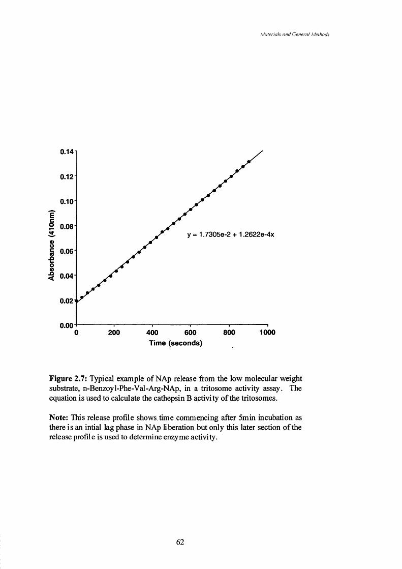
Materials and Genera! Methods
0.141
0.12 -
0.10
Ec® 0.06
y = 1.7305e-2 + 1.2622e-4xsg■s 0.06
o%§ 0.04
0.02
0.0010008000 200 400 600
Time (seconds)
Figure 2.7: Typical example of NAp release from the low molecular weight substrate, n-Benzoy 1-Phe-Val-Arg-NAp, in a tritosome activity assay. The equation is used to calculate the cathepsin B activity of the tritosomes.
Note: This release profile shows time commencing after 5min incubation as there is an intial lag phase in NAp liberation but only this later section of the release profile is used to determine enzyme activity.
62

Materials and Genera! Methods
to 15ml polypropylene tubes along with sample concentrations (usually 50|ig drug
equivalence) of HPMA copolymer-APE or PKl all made up using the citrate phosphate
buffer. To commence the reaction a specified activity of tritosomes was added to make
the solution 1ml using the citrate phosphate buffer to adjust as necessary. Tritosomes
were added last and this was taken as being time zero. At each time-point (0, 5, 15,
30minutes and then 1,2, 3, 4 and 5 hours) lOOpl of reaction mixture was removed using
a pipette and frozen immediately in liquid nitrogen in a fresh polypropylene and kept in
a freezer until analysis. The reaction tubes were incubated at 37°C for the duration of
the experiment. Analysis was by extraction followed by HPLC determination as
detailed in Section 2.3.3.1 for APE and 2.3.3.2 for doxorubicin samples.
2,3,5 Statistics
All error bars describing in vitro studies are the standard error of the mean (SE).
The error bars in all other studies are standard deviation (SD) of the mean. Statistical
significance was calculated using the students’ t-test for small sample sizes (two tailed).
Statistical significance was defined as a p value <0.05. All equations of a straight line
were determined using Cricket Graph software (version 1.3.2). All calculations were
performed using Microsoft Excel 98 software.
63

Synthesis and characterisation o f HPMA copolymer-APE conjugates
Chapter Three
Synthesis and characterisation o f HPMA copolymer-APE conjugates
64

Synthesis and characterisation o f HI’MA copolymer-APE corijugates
3.1 Introduction
Ellipticines (Figure 1.11) are potent cytotoxic agents as their IC50 values are in
the range of 0.3-0.8pM towards a variety of malignant cell lines in vitro (Devraj et al,
1996). As mentioned in Section 1.5.2 their mechanisms of action include DNA
intercalation (Elcock et al, 1996), topoisomerase inhibition (Froelich-Ammon et al,
1995) and DNA alkylation (reviewed by Sainsbury, 1990). Early clinical development
was limited by poor drug solubility and in vivo host toxicities including haemolytic
activity, decreased heart rate and hepatotoxicity (Donato et al, 1992). Two ellipticine
derivatives did, however, show interesting clinical activity against a number of solid
tumours (Section 1.5.3). These observations led to the choice of ellipticines, and APE
in particular, as interesting candidates for evaluation in the form of polymer-dmg
conjugates.
APE (NSC 176328) is a derivative of ellipticine which retains cytotoxicity with
an average IC50 across the NCI anticancer screen of 3.2x10’ M. This is comparable to
ellipticine (NSC71795; 7.5x10'’M), doxorubicin (NSC123127; l.SxlO'^M) and
paclitaxel (NSC125973; 1.3xlO'*M) in the same screen. APE showed a similar
spectrum of activity to ellipticine being most toxic against cell lines derived from
leukaemias, colon, lung and breast tumours (NCI website, 2001). APE is also
particularly suited to conjugation to a HPMA copolymer precursor because of its free
amine and the fact that this amine is removed from the main ellipticine structure (Figure
3.1) and therefore sterically amenable to conjugation by aminolysis.
Biocompatible, water-soluble polymers such as HPMA copolymers provide a
useful platform for tumour-selective drug delivery (Section 1.3.4.3). Conjugation of
doxorubicin, paclitaxel and camptothecin to HPMA copolymers has been shown to
improve the drug water solubility and reduce drug toxicity. Additionally, after i.v.
administration such conjugates selectively accumulate in solid tumour tissue by the
EPR effect (described in Section 1.4.1). In this study a series of HPMA copolymer-
APE conjugates were synthesised with the aim of improving the water-solubility of
APE and also improving its therapeutic index.
Previously HPMA copolymer-drug conjugates have been synthesised by four
routes:
65

S ') nrhcMs itn d i hnnK i<‘n.\<iin>n o f IH ’M I cnpolvnier-. IP/ ' con jii^olcs
Free primary amine
%
m m
Figure 3.1: Three dimensional structure o f APE (N SC l 76328).
66

Synthes is und characlerisalion o f HPMA copolymer-APE conjugates
1) Polymer analogous reaction using aminolysis of HPMA copolymer-ONp precursors
The most widely used method has been the use of an HPMA copolymer
precursor containing HPMA copolymer bearing peptidyl spacers with a terminal ONp.
Firstly the HPMA copolymer precursor is prepared by copolymerisation of HPMA and
A^-methacryloylated oligopeptide-ONp (Figure 3.2) using a free radical polymerisation
method. Drugs containing an aliphatic amino group can displace ONp by aminolysis
(Rejmanova et al, 1977; Figure 3.3) and this approach was used to prepare PKl and
PK2 (reviewed by Kopecek & Duncan, 1987). The advantages of this method are:
A. This is a simple aminolysis reaction and as long as the drug to be
attached has only one accessible aliphatic group then cross-linking
should not occur.
B. The polymer Mw can be standardised and the ‘universal precursor’ used
to prepare conjugates of different drug loading.
C. The ‘universal precursor’ can be used to prepare conjugates with drug
and targeting groups by sequential aminolysis.
The difficulties with this method are:
A. In the case of poorly reactive drugs the drug needs to be stable in basic
conditions as basic conditions are often required to facilitate the reaction.
B. There is a competing hydrolysis reaction leading to the generation of
side products (Figure 3.4).
C. The conjugate is a random copolymer and therefore there could be an
uneven distribution of drug along the polymer chain and between
different polymer chains.
D. Free drug will always be present in the conjugate (even if this is a small
contaminant).
Because of some of these difficulties other methods of preparation have been
explored.
2) Copolymerisation of methacryloylated drug-bearing monomers
St’astny et al (1996) described the preparation of HPMA copolymer GFLG-
doxorubicin conjugates by first synthesising a monomer of methacryloylated-GFLG-
doxorubicin and then copolymerising this with HPMA using free radical polymerisation
to produce a conjugate of similar structure to PKl (Figure 3.5). This synthetic
67
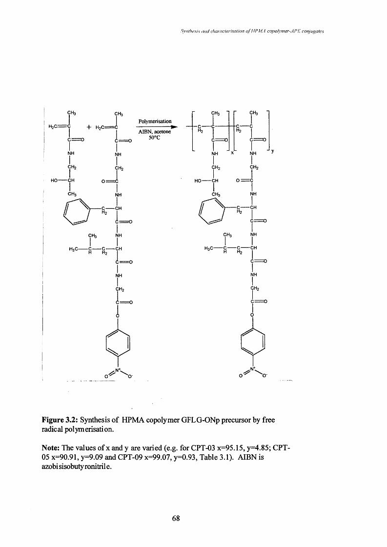
Syiuhesi.s and chai aclerisation o f HPMA copolymer-APF, conjugates
PolymerisationH2C Ç -|" HoC
AIBN, acetone 50°C
ÇH3 - ÇH3 -
— c — c1■0— c
Hz Hz 1
C = o1
_ I
Figure 3.2: Synthesis of HPMA copolymer GFLG-ONp precursor by free radical polymerisation.
Note: The values of x and y are varied (e.g. for CPT-03 x=95.15, y=4.85; CPT- 05 x=90.91, y=9.09 and CPT-09 x=99.07, y=0.93. Table 3.1). AIBN is azobi sisobuty ronitril e.
68

Synl/iesis and characterisal ion o f HPMA copolymer-AP E conjugates
ÇH3
C —
IrÇH2
CHOH
CH3
ÇH3
NH
ICH
1 = 0Ir
CH C
NH CH3
i=:NH
CH-
CH3
Figure 3.3: Aminolysis of the HPMA copolymer GFLG-ONp using APE as an example of a suitable drug with a free primary amine.
Note: The values of x and y are varied as described in Table 3.1.
69

Svii/lie.si.s and chaiaclerisation o f HPMA coi)olynier-AP E conjuf^aies
HYDROLYSISR.
HO.
‘H
N\
O ' 0
T
>+
OH
o ^ \
AMINOLYSIS R
NHz .
\ Q-
R
HN
IRi
>c = o
+
OH
\
Figure 3.4: Reaction schemes showing both the undesired (hydrolysis) reaction and the desired (aminolysis) reactions where R is HPMA copolymer- GFLG and Rj is a reactant with a free amine e.g. APE or doxorubicin.
70

Synlhesis and characlerisalion o f HP,VIA coi?olynier-AP E conjugates
CH, CH,
H 2 C = C H , C = (
r °
Polymerisation )AIBN, acetone
50°C
NH
CH
HO CH
CH
NH
ÇH2
0 = 1
■NS5S== (p= 0
CH3 NH
NH
I■CH
NH
CH,
■ s r r1 = 0
NH
CH,
CH,
srr1 = 0
NH
ICHz
HO CH 0= C
I ICHr, NH
■CH
CH3 r
0 " >CH , NH
( j != 0
NH
CH
OHCH
NH
OH OCH3 Q OH OCH.
OH OHHOHO
Figure 3.5: Reaction scheme showing free radical polymerisation of HPMA with methaciyloylated-GFLG-doxorubicin monomers to produce a conjugate similar to PKl (St’astny et al, 1996; Section 3.1).
71

Synthesis and characlerisalion o f HPMA copolymer-APE conjugales
approach has also been utilised to obtain cross-linked HPMA copolymer GFLG
conjugates containing both doxorubicin and antibodies for targeting (Ulbrich et al,
2000). This synthetic route avoids high levels of free drug in the product (as the
potential contaminant is methacryloylated GFLG-doxorubicin) but poor control during
polymerisation leads to high polydispersity (approximately 2-3). When this method
was used to conjugate antibodies there was evidence of high molecular weight species
containing higher amounts of antibody (Ulbrich et al, 2000).
3) Use o f‘MAG’ precursors
An alternative method has been used by Pharmacia to prepare HPMA
copolymer-anticancer conjugates and the HPMA copolymer-G-Cô-G-camptothecin
conjugate recently brought into Phase I clinical trial (Zamai et al, 2001). The synthesis
involves firstly the preparation of a peptidyl camptothecin derivative which is then
bound by aminolysis to an HPMA copolymer-G-ONp (called a ‘MAG’) precursor
(Figure 3.6). It has been suggested that these conjugates have a low free drug content
and that the drug is equally distributed across high and low molecular weight species
present in the conjugate (Rizzo, 2000). However the polydispersity is higher than seen
for PKl (1.5-1.7) and there are free peptidyl-drug species (e.g. FLG-camptothecin) in
the final product (Caiolfa et al, 2000).
4) Synthesis of a universal precursor of low polydispersity
Recent work by Godwin et al (2001) has sought to overcome the problem of
HPMA copolymer conjugate polydispersity by developing a new polymerisation
method. All the methods above utilise free radical polymerisation while this method
uses an atom-transfer radical polymerisation (ATRP) method. This method produces
an activated homopolymer which can then be used to prepare polymer-drug conjugates
(Figure 3.7) with a polydispersity of 1.1-1.2, thus potentially giving better defined
HPMA copolymer-drug conjugates more suitable for regulatory approval.
The potential disadvantages of this method are again free linker drug species in
the final product similar to those seen in the use of ‘MAG’ precursors outlined above.
Also incomplete reaction of the polymer precursor with drug (or side reactions /
hydrolysis) would lead to poorly-defined polymer species.
As outlined in Section 1.5.3 ellipticines are thought to act mainly on targets
within the nucleus of the cell (reviewed by Sainsbury, 1990) and therefore intracellular
72

Synf/iesi.s and characlerisalion o f HPMA copolymer-APE conjugales
o + B o c -H ie -L e u -G ly -O N p
CH, CH,
L . ' L .
NH1 J
NH
L .
o==c
CH,
in
CH, NH
T - ^0 = 0 o
CH, NH
H3 C —I C C— CH
NH
ÇH,
NH
CH,
= \ IcL—Cin,
rCH,
cL— OH
B o c -P h e -L e u -G ly
+
CH, CH,
cH, 1 H, 1
1 ° \
- r ■CH,
HO— in
NH1CH,
0:s; 1in. ONp
Figure 3.6: Method used by Caiolfa et al (2000) to synthesise an HPMA copolymer GFLG-camptothecin conjugate.
73

Synthesis and chairiclerisalion o f HPMA copolyiner-AP E conjugales
CH3C
D C -Hz
. C ^ .OHCH2
CuBr2,2'-bipyridine
DMSO
ÇH3
C C-Hz Ii = o
NHz-linker-drug
ÇH3
•C C-Hz I
NH
ÇH3
C c -Hz I
(j;=GNH
DRUG
R-NH2ÇH3
C C-Hz I
ÇH3
c — c- Hz I
^ = 0
NH
DRUG
Figure 3.7: ATRP-mediated copolymerisation of activated HPMA monomers to produce a ‘universal’ precursor (Godwin et al, 2001).
74

Synthesis and characterisation o f HPMA copolymer-APE conjugates
release from the HPMA copolymer backbone would be essential to tbeir activity. For
this reason it seemed reasonable to hypothesise that the GFLG linker used to produce
PKl (which is degraded by lysosomal thiol-dependent proteases (Section 1.3.4.2))
would be most suitable for an HPMA copolymer-APE conjugate. HPMA copolymer
precursors containing 1 and 1 0 mol% linker content on a backbone of a similar
molecular weight were also selected as a means of synthesising conjugates with varying
degrees of drug loading. Varying the drug loading of conjugates would be expected to
affect various characteristics of the constructs due to the chemical nature of the
components and this feature would need to be optimised to select a lead candidate for
future development. Conjugates containing a diglycyl linker were also synthesised as
this linker is usually not degraded by lysosomal enzymes when a bulky substituent is
present as the terminal group (Subr et al, 1992) and is potentially a useful comparator,
particularly in drug release studies.
3.2 Methods
3.2.1 Synthesis of HPMA copolymer-APE conjugates
It was considered important to first carefully standardise the aminolysis reaction
to determine APE reaction kinetics. During drug conjugation ONp can also be
displaced non-specifically by simple hydrolysis (Figure 3.4). This is particularly true
when a strong base like triethylamine is added. Basic conditions greatly accelerate both
aminolysis and hydrolysis so it is very important to use dry solvents. DMSO, in
particular, can contain a high percentage of water. To quantitate non-specific
hydrolysis an HPMA copolymer precursor (CPT-03; Table 3.1) was dissolved (5mg/ml)
in either water or a mix of dimethyl formamide (DMF)ZDMSO in the presence and
absence of triethylamine. The change in UV absorbance (400nm) with time was
measured to determine how much ONp was liberated. Free ONp species absorb at this
wavelength. To quantitate the total possible ONp liberated an excess of l-amino-2-
propanol was added. Release of ONp was expressed as a percentage of the total ONp
present.
The HPMA copolymer-APE conjugates were synthesised essentially using the
method outlined in Searle et al (2001). The following is a typical synthesis.
75

Synthesis and characterisation o f HPMA copolytner-APE conjugates
HPMA copolymer GFLG-ONp (200mg; CPT-03, Table 3.1) was dissolved in
dry DMF (5ml). APE.2HC1 (18mg) was dissolved to 5mg/ml in dry DMSO (which
would be equivalent to a maximum of 75% displacement of ONp from the precursor).
A sample (5 pi) of each solution was retained for thin layer chromatographic (TLC)
analysis. The APE solution was added to the solution of HPMA copolymer and mixed.
Aliquots of a 1/lOOv/v solution of triethylamine in dry DMSO were added over 30min
(1.9ml total volume) with further 5 pi aliquots of reaction mixture being retained at
various time-points for TLC analysis. TLC was performed using chloroform : methanol
: triethylamine 8 : 1 : 1 by volume as the mobile phase. Any unreacted APE could be
seen at a retention factor (Rf) value of 0.4 while bound APE remained at the origin.
Spots were visualised using a UV lamp. During the reaction the absorbance (400nm)
was also measured (taking lOpl of the reaction mixture and diluting to 1ml with PBS)
to monitor ONp release (as in Section 3.2.4). The reaction was allowed to run
overnight in a dark cupboard and the next day an absorbance reading was taken as
before at 400nm. 1 -Aminopropan-2-ol (1/100 v/v) in DMF (440pi) was then added to
react with any remaining ONp groups.
Conjugates containing different drug loading were prepared using HPMA
copolymer GFLG-ONp precursors, CPT-09 and CPT-05. Conjugates containing the
GG linker were synthesised using CPT-04 (Table 3.1). In each case the same weight of
precursor (200mg) and sufficient APE to displace 75% of the bound ONp were used.
Otherwise the synthesis was identical to that outlined above. After completing the
reaction with 1 -aminopropan-2-ol the absorbance at 400nm was again determined.
The organic solvents were evaporated in a glass flask heated to approximately
30°C using a paraffin bath attached to a high vacuum pump with a liquid nitrogen trap
to retain all evaporated solvents. The residual film was dissolved in approximately
20ml of distilled water and added into a SpectraPor® cellulose dialysis membrane with
a molecular weight cut-off of 2,000Da, pre-soaked in distilled water to remove any
residual sodium azide which is used as a preservative agent. This was then dialysed
against approximately 51 of distilled water with frequent water changes for three days to
remove any low molecular weight impurities including unbound APE. Finally the
conjugate solution was poured into a round-bottomed flask, frozen using liquid nitrogen
and then lyophilised to a constant mass as outlined in Section 2.2.4.
76

Synthesis and characlerisalion o f HPMA copolymer-APE conjugales
Table 3.1: Characteristics of the HPMA copolymer precursors used in the synthesis of
HPMA copolymer-APE conjugates.
Precursor Peptidic linker ONp content (mol%)^
CPT-03 GFLG 4.85
CPT-04 GG 5.05
CPT-05 GFLG 9.09
CPT-09 GFLG 0.93
These values were supplied by Polymer Laboratories and were determined by them
using a UV absorbance method.
77

Synthesis and characlerisalion o f HPMA copolymer-AP li conjugates
3.2.2 Determination of APE content in conjugates
The HPLC method used to determine the free APE content of the conjugates
was described in Section 2.3.3.1. To determine total APE content a simple UV assay
was used. The UV spectrum of APE shows a maximal absorbance at 296nm which is
not interfered with by any UV absorbance of the HPMA copolymer precursors (Figure
3.8). A standard curve was prepared by determining the absorbance at 296nm of APE
samples (0-5pg/ml) in PBS using a quartz cuvette (Figure 3.9). Conjugates were
dissolved in PBS and similarly absorbance (296nm) was determined. Values were
compared to the standard curve and the total APE content in the conjugate determined.
3.2.3 Gel Permeation Chromatography
HPMA copolymer-APE molecular weight was assessed by comparison with
poly(ethylene oxide) standards (18.3 to 124.7kDa) but ideally HPMA copolymer-APE
standards should be used. GPC analysis was performed using two TSK-gel columns in
series (G3000 PW followed by G2000 PW) with a guard colunm (ProgeF^ PWXL) and
differential refractometer and UV-visible detectors connected in series. Tris buffer
(Tris 0.05M, NaCl 0.5M) was used as the mobile phase (Iml/min). Conjugates (20pl of
a 5mg/ml solution in eluant buffer) were injected onto the colunms using a 20pl loop
and manual injection. PowerChrom software was used for recording the detector output
signal and for analysis of the data.
3,2,5 Determination of solubility of free APE and HPMA copolymer-APE
Firstly to assess the solubility of APE a solution of 0.1 M PBS was prepared and
78

SmiiIu'ms (HuJ clhirdclcnsiilKjn oj HI’M. \ copolymer-. iPI: con/i/yaic.s
2.51
2.0(0
I€(0
APEHPMA GFLG-APE HPMAGFLG-1A2P
Sc(0€8n< 0.5
0.0200 250 350 400300
Wavelength (nm)
Figure 3.8: The UV spectra of APE, HPMA copolymer GFLG-1 -amino-2- propanol and HPMA copolymer GFLG-APE (RK3).
79

Synthesis and characterisation o f HPMA copofymer-A PE conjugates
0.81
0 .6 -
EcCO8
owJO<
0 .2 -
0 .0 ^0 1 2 4 63 5
APE concentration (/[/g/ml)
Figure 3.9: Standard curve for APE in PBS measured using UV absorption at 296nm.
Note: Data represent mean ± SD (n=3).
80

Synthesis and characterisalioii o f HPMA copolymer-APE conjugates
heated to 37°C. A standard (O.lmg/ml) solution of APE in PBS was also prepared and
a standard curve constructed using a UV-visible spectrophotometer taking triplicate
measurements at a wavelength of 296nm as in Figure 3.9. PBS (5ml) was added to a
glass container accurately. Powdered APE was added until the solution became a dark
colour. The pH of this solution was adjusted to 7.4 using small volumes of a 0.1 M
NaOH solution. The resulting solution was allowed to equilibrate at 37°C for lOmin
and the pH was taken again to ensure that the APE.2HC1 had not changed the solution
pH. This solution was filtered using a syringe and 0.45pm filter. The resulting filtrate
(lOpl) was diluted to 1ml with PBS at 37°C and absorbance at 296nm measured, in
triplicate.
To assess the solubility of HPMA copolymer GFLG-APE 2ml of PBS at 37°C
was measured accurately into a glass container. Approximately 50mg of HPMA
copolymer GFLG-APE (Conjugate RK3; Table 3.2) as a powder was added to the PBS
solution and allowed to remain at 37°C for lOmin. The pH of this solution was adjusted
to 7.4 again using small volumes of 0.1 M NaOH. This solution was filtered as before
(despite there being no visible particles in the solution) to allow for equivalent losses.
This solution (lOpl) was made up to 1ml with PBS as before (37°C) and the absorbance
at 296nm measured in triplicate.
33 Results
3,3.1 Conjugation characteristics of HPMA copolymer conjugates
It was found that considerable care had to be taken during drug conjugation by
aminolysis due to the competing hydrolysis reaction (Figure 3.4; Figure 3.10). For this
reason dry solvents dispensed under argon (DMF/DMSO) were always used in the
synthesis of HPMA copolymer-APE conjugates. Triethylamine, a strong base, has been
used before to activate the doxorubicin hydrochloride giving the free base necessary for
preparation of PKl (Rihova et al, 1989). As APE is also supplied as a hydrochloride
salt triethylamine was also used to generate the free base needed for the synthesis of
HPMA copolymer-APE conjugates. Unfortunately triethylamine also accelerates the
competing hydrolysis reaction, re-enforcing the need for dry solvents (Figure 3.10).
81

Synlhfsis and characlerisalion o f HPMA copolymer-APE conjugales
Addition of 1-amino-2-propanol120
Water+triethylamineDMF/DMSO+triethylamine100
80
Water60Q .
Zo40
20DMF/DMSO
15000 500 1000Time (min)
Figure 3.10: Hydrolysis of the HPMA copolymer GFLG-ONp (CPT-03).
82

Synthesis and characlerisalion o f HPMA copolymer-APE conpigates
3.3.2 Conjugation of APE to HPMA copolymers
During the reaction the disappearance of free APE and appearance of conjugate
was followed by TLC. Free APE had an Rf of 0.4; bound APE remained at the origin
while ONp had an Rf of 0.9 (Figure 3.11). It was found in a previous study that a molar
ratio of 1:0.75 of ONpiAPE gave the most efficient conjugation (Searle et al, 2001).
Therefore all conjugates were synthesised using these conditions. Using the HPMA
copolymer precursors (Table 3.1) three batches (synthesised using 200mg of HPMA
copolymer precursor) of each conjugate were prepared to ensure the reproducibility of
the reaction (Table 3.2). The recovery of conjugates at the end of the synthesis was
between llOmg and ISOmg giving an overall recovery/yield compared to initial mass of
conjugate of approximately 65%.
In addition the conjugates contained a small quantity of ellipticine (<0.1%w/w
of conjugate). This impurity is a theoretical breakdown product of APE. The HPMA
copolymer-APE conjugates (Table 3.2) contained less than 0.1%w/w free APE (the
exception to this was the batch RK6 where there were problems during purification and
it contained a high percentage of free APE).
3.3.3 GPC analysis of HPMA copolymer-APE conjugates
GPC was used as a method of size determination using poly(ethylene oxide)
standards. Table 3.3 shows the apparent molecular weight of the conjugates when
compared to poly(ethylene oxide) standards. These are clearly incorrect based on the
molecular weight of the precursors used and the possible reasons for this will be
discussed in Section 3.4. The conjugates were shown to consist of an apparently single
species using this method. The chromatograms of the HPMA copolymer-APE
conjugates suggest a lack of cross-linked species, as expected given the nature of the
aminolysis reaction with the only potential source of cross reactivity being a drug with
two free amines. Figure 3.12 shows an example of one such GPC trace and shows two
HPMA copolymer species, namely a HPMA copolymer GG-APE (RK4) and a HPMA
copolymer GG-aminopropanol (where the HPMA copolymer precursor has been
aminolysed using 1 -amino-2-propanol). The HPMA copolymer GG-aminopropanol
showed no UV absorbance (296nm) as described earlier (Figure 3.8) and gave a
refractive index profile similar to that of the HPMA copolymer GG-APE conjugate
(RK4) which showed a UV peak at 296nm. This UV peak is of a similar profile to the
refractive index (Rl) trace and this indicates that the APE is bound across the different
83

Synthesis and characterisation o f HPMA copolymer-APE conjugates
Table 3.2; Characteristics of the HPMA copolymer-APE conjugates synthesised in this
study.
Code Precursor TotalAPE
(%wAv)^
APE(mol%)
Free APE (%w/w total
APE)^^
Impurities (%wAv conjugate)
APE^^ ellipticine^^
RKl CPT-03 3.07 1.7 0.043 0.001 0.005
RK2 CPT-03 4.34 2.4 0.148 0.006 0.042
RK3 CPT-03 3.79 2.1 >0.017 >0.001 0.009
RK4 CPT-04 4.27 2.2 0.017 0.001 0.035
RK5 CPT-04 4.62 2.3 0.051 0.002 0.024
RK6 CPT-04 4.90 2.5 12.176 0.597 0.088
RK7 CPT-09 1.07 0.5 7.742 0.082 0.011
RK8 CPT-09 1.20 0.6 7.653 0.092 0.012
RK9 CPT-09 1.21 0.6 4.321 0.052 0.011
RKIO CPT-05 6.10 3.7 0.077 0.005 0.042
RKll CPT-05 6.01 3.7 0.092 0.007 0.035
RK12 CPT-05 5.85 3.5 0.058 0.003 0.051
Determined using UV analysis (Section 3.2.2)
Determined using extraction and HPLC analysis (Section 2.3.3.1)
84

Syinhasis and characlerisalion o f HPMA copolymer-APE conjugates
Solvent front
O o
O o o 0
^ — X— X (X) (X
ONp (yellow) Rf 0.9
APE (purple under UV) Rf 0.4
HPMA-APERfO
Polymer APE 7.5min 17.5min 30min (yellow)
Figure 3.11; TLC of a typical HPMA copolymer-APE reaction mixture.
85

Synthesis and characterisation o f HP MA copolymer-APE conjugates
Table 3.3: Molecular weight of HPMA copolymer-APE conjugates compared to
poly(ethylene-oxide) standards.
Conjugate %w/w APE Mw (Da) Polydlspersity
HPMA GFLG-APE (RK8) 1.20 32068 2.2
HPMA GFLG-APE (RK3) 3.79 974 1.4
HPMA GFLG-APE (RKIO) 6.10 692 1.2
HPMA GG-APE (RK4) 4.27 9907 1.3
8 6

Syiil/ie.si.'i and characlerisation o f HPMA copolynier-A P E conjugales
HPMA copolymer GG-l-amino-2-propanol (5mg/ml)
R.I.
U.V. 296nm
20 24
Time (min)
HPMA copolymer GG-APE RK4 (5mg/ml)
R.I.
U.V. 296nm
2420
Time (min)VtVo
Figure 3.12: GPC chromatogram of an aminolysed HPMA copolymer and the equivalent HPMA copolymer-APE conjugate (RK4).
Note: The UV detector was placed in series with a differential refractometer. Vo indicates the exclusion volume of the columns and Vt the total elution volume.
87

Synthesis and characterisation o f HPMA copolymer-APE conjiigates
sizes of conjugate species and is not preferentially binding to long or short species.
3,3,5 Solubility of HPMA copolymer-APE conjugates
A solubility experiment was carried out on an HPMA copolymer-APE
conjugate (RK3). It was found that, at physiological pH (7.4) and at 37°C in PBS, RK3
(625pg/ml) was at least ten-fold more soluble than APE alone (62pg/ml) in terms of
equivalence of APE. This experiment demonstrates the ability of HPMA copolymer
conjugates to solubilise poorly-water soluble drugs. This is an important obstacle to
overcome in order to use such agents in vivo and therefore this was a useful benefit of
conjugation to the HPMA copolymer.
3.4 Discussion
In this study HPMA copolymer GFLG and GG-APE conjugates were
successfully synthesised using a standard aminolysis reaction (Rejmanova et al, 1977;
Figure 3.3). The ease of conjugation was predictable as the 3 dimensional structure of
APE (Figure 3.1) shows that the primary amino group is free from any steric hindrance,
standing well outside the plane formed by the unsaturated rings. Conjugates were
synthesised to contain a range of APE loadings (I.07-6.I0%w/w APE; Table 3.2).
Maintenance of high concentrations of reagents in dry solvents and dropwise addition
of the triethylamine to free the 3’-amino group of APE slowly from its hydrochloride
salt (such that the copolymer remained in excess during the reaction) was critical for
successful conjugation. Also the use of an APEiONp ratio of 75:100 gave good
8 8

Synl/iexis and characterisation o f HPMA copotymer-AP E conjugates
89

Syn/hesis and characterisalion o f HPMA copolymer-APE conjugates
reaction yields (55 to 80% of the maximum expected). A drug loading of 2.4mol% was
achieved for APE, which is similar to that achieved for doxorubicin when synthesising
PKl (2.5mol%; Subretal, 1992).
A number of methods of purification have been employed in the past to remove
low molecular weight impurities from HPMA copolymer drug conjugates. These
include gel filtration to remove free doxorubicin from PKl (Rihova et al, 1989) and
precipitation to remove peptidyl-camptothecin from HPMA copolymer-camptothecin
(Caiolfa et al, 2000). In this study simple dialysis using a large volume of distilled
water was able to remove most low molecular weight impurities. The high sensitivity
of the HPLC analytical procedure showed that only small amounts of free APE and
ellipticine were present in the conjugates (Table 3.2). Impurities detected represented <
0.1% of the conjugate by weight in most cases. Further optimisation of the reaction
conditions is necessary to reduce the free APE content in the conjugates made from the
precursor CPT05. This polymeric precursor has a small number of reactive ONp
groups and therefore a greater percentage of reactive groups may be lost to any
hydrolysis reaction mediated by water. Also the higher ratio of GFLG-ONp/APE to
hydrophilic HPMA may have led to greater stearic hindrance in the reaction. The
minimal free drug in most of the conjugates is particularly important for biological
testing such as in vitro cytotoxicity and haematocompatibility (Chapter 4) where free
drug has been shown to interfere in previous studies on PKl and doxorubicin (Wedge,
1991).
GPC analysis has been shown to be difficult as a method of determining the
molecular weight of HPMA copolymer-APE conjugates using the poly(ethylene-oxide)
standards (Table 3.3). Interestingly the higher the drug loading the lower the apparent
molecular weight of the conjugate and therefore perhaps drug loading is playing a role
in this determination. The use of other standards may be helpful but ideally HPMA
copolymer-APE standards would be used in such a study. However, GPC did show that
APE was conjugated to the HPMA copolymers uniformly across the molecular weight
range of the conjugates (Figure 3.12). Non-uniformity of conjugation has been reported
previously with the HPMA copolymer-doxorubicin/antibody conjugates of Ulbrich et al
(2000). GPC also showed that the HPMA copolymer-APE conjugates exist as single
species which are not cross-linked.
90

Synihcsis and characterisalion o f HPMA copolymer-APE conjugates
A major problem encountered with previous ellipticine derivatives is their
limited solubility (Djuric et al, 1992). In this study it was found that the maximum
solubility of APE at room temperature and 37°C was 44.5pg/ml and 62.4pg/ml
respectively. Drug conjugation to HPMA copolymers dramatically improved solubility
(approximately 10-fold). This was not unexpected from the previous studies on HPMA
copolymer-paclitaxel which was 20,000 times more water soluble than paclitaxel alone
(Huinink et al, 1998). Paclitaxel alone is extremely water insoluble and therefore this
dramatic increase in solubility is significant. The HPMA copolymer-APE conjugate
(RK3) was still soluble at an APE-equivalent concentration of 625.2pg/ml. APE is not
as water insoluble as paclitaxel, and is comparable to doxorubicin in its water solubility.
Therefore a 10-fold increase in solubility of APE, similar to the 10-fold increase in
doxorubicin solubility in PKl, was felt to be sufficient for the studies which were
planned in vitro and ultimately in vivo.
Following the successful synthesis and characterisation of HPMA copolymer-
APE conjugates in this study these conjugates need to be evaluated in vitro for drug
release and biocompatibility before they can be evaluated as anti-tumour conjugates.
The effect of drug loading on these characteristics needs to be determined in order to
identify a lead optimal loading for progression to in vivo studies. To do this firstly the
conjugates will be compared for their solution properties before being evaluated for
their ability to release free APE, haematocompatibility and in vitro cytotoxicity. These
studies will be described in Chapter 4.
91

E(fecl o f A PE loading on physicochemical and biologicalproperlies o f HPMA copolymer-APE conjugales in vino
Chapter Four
Effect o f APE loading on physicochemical and biological properties o f HPMA copolymer-APE conjugates in vitro
9 2

Effcd o f APE loading on physicochemical and hioiogica!properlies o f HPMA copolymer-APE conjugales in viiro
4.1 Introduction
Following the synthesis of the HPMA copolymer-APE conjugates described in
Chapter 3 it was necessary to evaluate their physicochemical and in vitro biological
properties (APE release by lysosomal enzymes, haemolytic activity and cytotoxicity),
focusing particularly on the effect of drug loading on these parameters before
progressing to in vivo anti-tumour testing. Before evaluating the effect of drug loading
on biological properties of the conjugates it was considered necessary to characterise
the structure of conjugates in solution. Specific techniques were chosen to allow
conjugate characterisation and also to determine the effect of APE loading on each
parameter. The rationale for selection of the methods used is briefly given below.
4.1.1 Polymer conformation in solution
A number of methods have been utilised historically to study polymer
conformation in solution. These include viscosimetry (Frenoy, 1986), potentiometric
titrations (Pederson et al, 1971), calorimetry (Biltonen & Friere, 1978), Raman
spectroscopy (Brown et al, 1972), circular dichroism (Mezo et al, 2000), flow light
scattering (Lee & Muller, 1999) and fluorimetric determinations (Ringsdorf et al,
1991). Many of these methods (particularly viscosimetry, potentiometric titration and
calorimetry) were developed to evaluate polymers as plastics and pharmaceutical
excipients, therefore large samples are often required and these methods are not
particularly sensitive to minor changes in polymer conformation. Raman spectroscopy,
circular dichroism and flow light scattering are more sensitive techniques but are quite
specific to certain chemical and structural changes which are used to elucidate polymer
conformational changes and do not detect more general changes in conformation. For
this reason it was decided to use a fluorescent probe to try and understand the
conformation of HPMA copolymer-APE conjugates in aqueous solution.
The use of fluorimetric methods to determine conformation rely on the change
of fluorescence of a fluorescent probe when sitting within the polymer coil or simply
free in solution. Many studies have used the hydrophobic fluorescent probe pyrene
(Figure 4.1) which is extremely water insoluble (approx 0.2mg/l; Gautier, 1995) but has
a strong fluorescence signal, which consists of three major emission peaks when excited
at a wavelength of 330nm (Figure 4.1). These peaks represent the diffuse excimer
emission centred at 382nm and two vibrational bands at 372nm (Ii) and 392nm (I3).
The relative intensities of Ii and I3 are extremely sensitive to the local solution
93

EJfed o j APE loading on physicochemical and biologicalproperlies q! HPMA copolynier~APE conjugales in vitro
C3
2IXk .
aoocQOgo3
7
6
5
4
3
2
1
n IIII MinJJjimmuMiiMHip
5000 ^350 400 450
Emission waveiength (nm)
Figure 4.1: The structure of pyrene and its fluorescence spectrum (0.2m g/1 in water) using an excitation wavelength of 330nm showing the intensity peaks I, (372nm) and I3 (392nm).
94

E(fcci oJ'APE loading on physicochemical and biologicalproperlies o f HPMA copolymer-APE conjugales in vino
microenvironment. The ratio (I1/I3) is therefore used to describe the polarity of a
solution. An increase in this ratio is indicative of increased polarity and therefore a
more hydrophilic environment for pyrene, the opposite also being the case (Winnik et
al, 1991 and Gautier, 1995). The strong fluorescent signal of pyrene also allows the
detection of small changes in intensity of the signal. A decrease in intensity indicates
increased pyrene localisation in the solution as pyrene molecules will become self-
quenching when closely associated.
4.1.2 Drug release from HPMA copolymer-APE conjugates
Intracellular release of APE from HPMA copolymer-APE conjugates is
essential for anti-tumour activity. This relies on effective access of cathepsin B to the
GFLG linker and would theoretically be affected by solution conformation. Thus it was
essential to characterise APE release from conjugates with different drug loading. This
can be done by firstly isolating rat liver lysosomes as described in Section 2.3.4. This
method has been used previously to examine the release of doxorubicin from PKl (Subr
et al, 1992) and will be used here as the same linker (GFLG) is used in the HPMA
copolymer-APE conjugates
4.1.3 Haemolytic activity of HPMA copolymer-APE conjugates
As mentioned previously ellipticines have been shown to cause severe
haematotoxicity and minor immunogenicity upon i.v. administration (reviewed by
Ohashi & Oki, 1996). Therefore it would be desirable for an HPMA copolymer-APE
conjugate to be non-haematotoxic and non-immunogenic in order to facilitate parenteral
administration. There has been extensive testing of the biocompatibility, immunogenic
potential and toxicology of PKl (Rihova et al, 1983; Simeckova et al, 1986; Rihova et
al, 1989 and Duncan et al, 1998). These studies have shown that PKl was not
immunogenic (Rihova et al, 1989) and indeed other studies have shown that HPMA
copolymers can reduce the immunogenicity of conjugated immunogenic proteins such
as transferrin (Flanagan, 1987). Blood is the first biological environment encountered
following parenteral administration and therefore any administered drug needs to be
stable and non-toxic in this environment. As conjugation to HPMA copolymers has
been shown to reduce immunogenicity of bound proteins and drugs and as ellipticines
have only been shown to be mildly immunogenic (Alberici et al, 1986) screening for
immunogenicity of the HPMA copolymer-APE conjugates was not considered
necessary in the first instance. However it was felt to be important to determine the
95
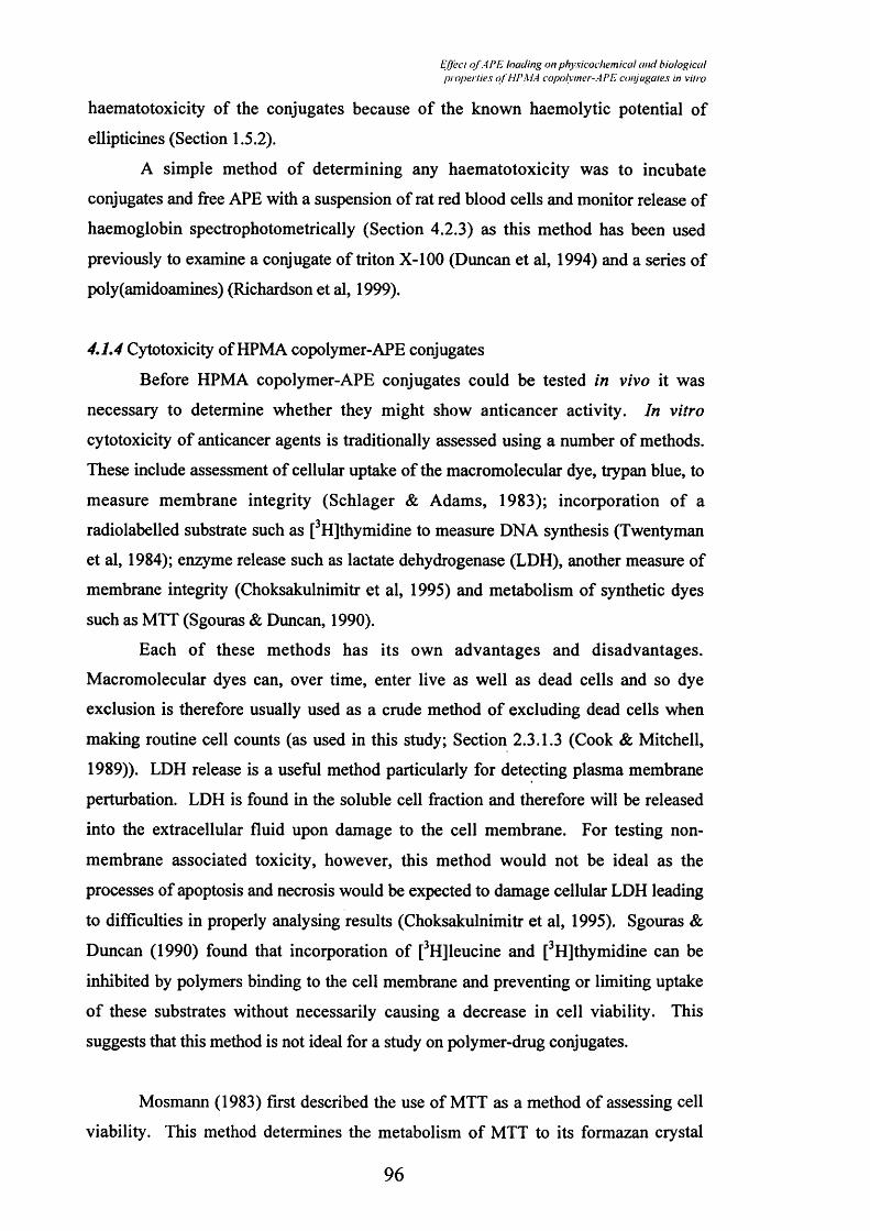
E(l'ecl o f A P E lo a d in g on p h y s ic o c h e m ic a l a n d b io lo g ic a l p r o p e r l ie s o f H P M A co p o lym er -A P E co n ju g a le s in v ilro
haematotoxicity of the conjugates because of the known haemolytic potential of
ellipticines (Section 1.5.2).
A simple method of determining any haematotoxicity was to incubate
conjugates and free APE with a suspension of rat red blood cells and monitor release of
haemoglobin spectrophotometrically (Section 4.2.3) as this method has been used
previously to examine a conjugate of triton X-100 (Duncan et al, 1994) and a series of
poly(amidoamines) (Richardson et al, 1999).
4,1,4 Cytotoxicity of HPMA copolymer-APE conjugates
Before HPMA copolymer-APE conjugates could be tested in vivo it was
necessary to determine whether they might show anticancer activity. In vitro
cytotoxicity of anticancer agents is traditionally assessed using a number of methods.
These include assessment of cellular uptake of the macromolecular dye, trypan blue, to
measure membrane integrity (Schlager & Adams, 1983); incorporation of a
radiolabelled substrate such as [^Hjthymidine to measure DNA synthesis (Twentyman
et al, 1984); enzyme release such as lactate dehydrogenase (LDH), another measure of
membrane integrity (Choksakulnimitr et al, 1995) and metabolism of synthetic dyes
such as MTT (Sgouras & Duncan, 1990).
Each of these methods has its own advantages and disadvantages.
Macromolecular dyes can, over time, enter live as well as dead cells and so dye
exclusion is therefore usually used as a crude method of excluding dead cells when
making routine cell counts (as used in this study; Section 2.3.1.3 (Cook & Mitchell,
1989)). LDH release is a useful method particularly for detecting plasma membrane
perturbation. LDH is found in the soluble cell fraction and therefore will be released
into the extracellular fluid upon damage to the cell membrane. For testing non
membrane associated toxicity, however, this method would not be ideal as the
processes of apoptosis and necrosis would be expected to damage cellular LDH leading
to difficulties in properly analysing results (Choksakulnimitr et al, 1995). Sgouras &
Duncan (1990) found that incorporation of [^H]leucine and [^Hjthymidine can be
inhibited by polymers binding to the cell membrane and preventing or limiting uptake
of these substrates without necessarily causing a decrease in cell viability. This
suggests that this method is not ideal for a study on polymer-drug conjugates.
Mosmann (1983) first described the use of MTT as a method of assessing cell
viability. This method determines the metabolism of MTT to its formazan crystal
96

Effect o f A PE hading on physicochemical and biologicalproperties o f HPMA copolymer-APE conjugates in vitro
(reaction described in Section 2.3.1.6) which is carried out in the mitochondria. The
formazan crystals are then determined using a colourimetric determination at 550nm.
However there have been some reported difficulties associated with this assay. Some
cell lines do not generate sufficient formazan for detection. There are also issues
relating to the stability and solubility of the formazan metabolite in organic solvent
systems (Sgouras & Duncan, 1990). Coloured drugs, for example doxorubicin, can
interfere with the absorbance readout from an MTT assay (Wedge, 1991). However
these problems can be overcome by using appropriate controls and suitable solvents
(Sgouras & Duncan, 1990) and therefore this assay was to evaluate the in vitro
cytotoxicity of the HPMA copolymer-APE conjugates containing different drug
loadings (Section 2.3.1.6).
The aims of the studies described in this Chapter were to select suitable HPMA
copolymer-APE conjugates for progression to anti-tumour studies in mouse models and
to understand the effect of APE loading on solution conformation, tritosome-mediated
APE release, haematocompatibility and in vitro cytotoxicity.
4.2 Methods
4.2.1 Determination of conjugate conformation using pyrene
Pyrene was dissolved to its maximum solubility in distilled water (0.2mg/l;
6xlO’ molL‘*) and the solution filtered (using a syringe and 0.22pm filter) to remove
any microcrystals. This stock solution was then used to prepare HPMA copolymer-
APE and HPMA copolymer-aminopropanol conjugate solutions (3ml; 0-1 mg/ml) in
triplicate in cuvettes. The fluorescence spectrum of each sample was then recorded
(excitation = 330nm and an emission scan of 350 to 450nm using a band pass of 4nm
on both excitation and emission slits). The solution containing only pyrene was used to
set the fluorescence maximum at 75% of the maximum detectable using this machine.
This maximum was at 372nm in the spectrum taken within the 350 to 450nm range.
4.2.2 Tritosome-mediated release of APE from HPMA copolymer-APE conjugates
Tritosomes were isolated and assayed for protein content and enzyme activity
using a standard substrate as described in Section 2.3.4. To determine thiol-protease
degradation of HPMA copolymer-APE conjugates incubations were set up as described
97

Effect o f A PE loading on physicochemical and hioiogica!properlies o f HPMA copolymer-APE conjugates in vitro
in Section 2.3.4.4. A sample from any batch of tritosomes was retested the day before
using that batch in a release study. Liberated APE (and in the case of control studies
using PKl, doxorubicin) were quantified using the organic extraction and HPLC
methods described in Sections 2.3.3.1 and 2.3.3.2 respectively.
4.2.3 Rat red blood cell lysis assay
Blood was obtained from male Wistar strain rats (250-350g body weight) by
immediate cardiac puncture following asphyxiation with carbon dioxide and transferred
into a heparin/lithium tube and kept on ice to minimise any damage to the erythrocytes.
The blood was centrifuged at lOOOg for lOmin at 4°C, the supernatant discarded and
some of the heparin beads carefully removed using a plastic pipette and then fresh pre
chilled (4°C) PBS (2ml) added. The red blood cell suspension was then centrifuged
again at lOOOg for lOmin at 4°C and the process repeated twice until all the heparin
beads had been removed. After the final centrifugation step the supernatant was
removed and the pellet used to make a 2%w/v suspension of red blood cells, again
using pre-chilled PBS.
Red blood cells (lOOpl of this 2%w/v suspension) were added to each well of a
96 well plate which already contained lOOpl solutions of substances to be tested
dissolved in PBS, or PBS alone as a control. To induce 100% lysis a l%v/v solution of
triton X-100 in PBS was used as a positive control (Duncan et al, 1994). These plates
were then incubated for Ih at 37°C, and then they were centrifuged (1500g; lOmin).
Intact red blood cells pellet while any haemoglobin released remains in the supernatant.
The supernatant was then pipetted into a fresh 96 well plate and assayed for
haemoglobin at 550nm using a micro-titre platereader. The average absorbance of the
wells containing triton X-100 was taken as 100% lysis and the absorbance of all other
wells related to this value as a 100% haemoglobin release. Dextran was used as a
negative control (0-5mg/ml) and poly(ethylene)imine (0-5mg/ml; Mw approximately
750kDa) was used as a positive control (Duncan et al, 1991). An example of the
haemolysis profiles of these reference compounds is shown in Figure 4.2 and both were
used in each experiment to ensure reproducibility of the method.
4.2.4 In vitro cytotoxicity of HPMA copolymer-APE conjugates
First a growth curve was constructed for B16F10 cells (Figure 4.3) (methods as
described in Section 2.3.1.5) and the doubling time of these cells was approximately
48h. Further experiments used cells in the exponential growth phase
98

Effect o f A PE loading on physicochemical and biologicalproperlies ofhU'MA copolymer-APE conjugates in vitro
lOOlpoly(ethylene)imine
80
2c8OaR
20-
dextran
.001 .01 .1 101Concentration (mg/ml)
Figure 4.2: Haemolytic activity of dextran and poly (ethyl ene)imine.
Note: Data represent mean ± SD (n=6).
99

Effect o f APE loading on physicochemical and biologicalproperties o f HPMA copolymer-APE conjugates in vitro
E
II<
1.41
1.2
1.0
0 .8 -
0.6 -
0.43002001000
Time (h)
Figure 43: B16F10 growlh curve.
Note: Data represent mean ± SD (n=6).
100

Effed o f APE loading on physicochemical and biologicalproperlies o f HPMA copolymer-APE conjugales in vitro
(i.e. the second or third day following sub-culture). Cytotoxicity of APE and HPMA
copolymer-APE conjugates was determined using the method described in Section
2.3.1.6. Dextran (0-5mg/ml; Mw approximately 41kDa) was used as a negative control
as it has been shown to be non-toxic and poly(L-lysine) (0-5mg/ml; Mw approximately
57kDa) was used as a positive control as it has been shown to be cytotoxic previously
(Sgouras & Duncan, 1990). An example of a typical cytotoxicity profile for these
control compounds is shown in Figure 4.4 and these reference controls incorporated
into every experiment to ensure reproducibility.
43 Results
4.3.1 HPMA copolymer-APE conformation in solution
The Ii (372nm) and I3 (392nm) values for each conjugate were used to calculate
the overall reduction in signal intensity, as a measurement of the localisation of pyrene
in the solution. The I1/I3 ratio was also calculated for each concentration of conjugate
to examine the local solution microenvironment characteristics.
Both HPMA copolymer aminopropanol (Figure 4.5) and HPMA copolymer-
APE conjugates (Figure 4.6) caused a decrease in the overall fluorescence intensity of
the spectrum. This can be clearly seen by plotting the pyrene Ii value against conjugate
concentration (Figure 4.7). The HPMA copolymer GFLG-aminopropanol conjugate
showed little decrease in fluorescence over this concentration range (Figures 4.5 and
4.7). The HPMA copolymer GFLG-APE conjugate with the lowest loading (RK9)
produces little decrease in pyrene Ii over the concentration range used (Figures 4.6 and
4.7). However the HPMA copolymer GG-APE (RK4) and the other HPMA copolymer
GFLG-APE conjugates (RK3 and RKIO) all cause a considerable decrease in the
pyrene fluorescence intensity (Figures 4.6 and 4.7). The pyrene I1/I3 ratio seen on
addition of increasing concentrations of HPMA copolymer conjugates is shown in Figure 4.8. Figure 4.5 demonstrates also that APE (60pg/ml) does not itself cause any
sigmflcant quenching of the pyrene solution.
4.3.2 Tritosome-mediated release of APE
Six batches of tritosomes were prepared for use in these studies and their protein
content and thiol protease activity is summarised in Table 4.1. Batches RKTl, 2 and 4
were not used for tritosome release studies because of their low activity and protein
content.
101

liffcct o f A PE loading on physicochemical and biologicalproperlies o f HPMA copolvmer-AP E conjugales in vilro
lOOl
90
80'^ Dextran
I8O
20
10 Poly(l-lysine)
101.001 .01 .1Concentration (mg/ml)
Figure 4.4; Cytotoxicity of dextran and poly (1-lysine) against B16F10 cells.
Note: Data represent mean ± SD (n=6).
102

r.ffert of-I !’E loaJiny. on physicochemical and hiolo}>iailproperlies ol i i i ’MA copoh iner-Al’h conjugales in vilro
âc3
Ia8
I§
7
6
cPyrene solution alone Pyrene solution + APE (60|jg/ml)
4
3
2
1
0350 400 450 500
Wavelength (nm)2 .5-1
2.0 -1 mg/ml
0.4mg/ml
0.1 mg/ml
0.025mg/ml
Polymer alone (no pyrene)1
Iu.
0 .5 -
0.0350 400 450
Wavelength (nm)
Figure 4.5: Fluorescence spectra of pyrene in the presence of APE and increasing concentrations of HPMA copolymer GFLG-aminopropanol.
Note: Panel a shows pyrene in the absence and presence of APE(60pg/ml) and panel b shows HPMA copolymer GFLG- aminopropanol.
103

i2c3
£!5S8c8v>
2o3U_
■sw0)o§£o3U.
/ //(■( 7 nj / / ' / ; l iHidin^ tJii p h v s i c o c h v n i i c d l a n d h io lo j^ icd l
pro iH d i i c s o f III’ \ l I ( n p d h n u ’i - M ' i . coniu^i iU's in v i i r o
0.025mg/ml
0.1 mg/ml
0.4mg/ml
1 mg/ml
Conjugate alone (no pyrene)
350 400 450
W avelength (nm)7
6
50.025mg/ml 0.1 mg/ml
0.4mg/ml 1 mg/ml
Conjugate alone (no pyrene)
4
3
2
1
0 + -350 400 450
W avelength (nm)
Figure 4.6: Fluorescence spectra o f pyrene in the presence o f increasing concentrations o f the HPMA copolymer-APE conjugates.
Note: Panel a shows RK9 (HPMA copolym er GFLG-APE; 1.21%w/w APE) and Panel b shows RKl (HPMA copolym er GFLG-APE; 3.07% w/w APE).
104

I'lJfCi III I /V . II» / i / ivMciH h f i n i a i l u n d hui ii i^icti i
p r i i /n ’i iii's , ' / / / / ' U I i n p i ih n ic r-AI 'I . l O i ijuna lc s 1» v ii rn
i2c3
2!5à8§£o3
1.51
0.025mg/ml
0.1 mg/ml
0.4mg/ml 1 mg/ml
Conjugate alone (no pyrene)0.5^
0.0350 400 450
c3
2I§
Io3
W avelength (nm)
0.025mg/ml
0.1 mg/ml
0.4mg/ml
1 mg/ml
Conjugate alone (no pyrene)
400W avelength (nm)
450
Figure 4.6: Fluorescence spectra o f pyrene in the presence o f increasing concentrations o f the HPMA copolymer-APE conjugates.
Note: Panel c shows RKIO (HPMA copolym er GFLG-APE; 6.10% w/w APE) and Panel d shows RK4 (HPMA copolym er GG-APE; 4.27% w/w APE).
105

Effect o f APE loading on physicochemical and biologicalproperties of HPMA copolymer-APE conjugates in vilro
8001
c3
I€
600
S 400- 81£o2 200-
UL
0.80.60.0 0.2 0.4
HPMAGFLG-ApRK9
RK4RK3RK10
Concentration (mg/ml)
Figure 4.7: Relationship between pyrene Ij and HPMA copolymer conjugate concentration.
Note: Data represent mean ±SD (n=3).
106

Effccl o f APE loadinfi on f^hysicocliennccil and biologicalpiojK i livs (d 'lll’MA copolymer-APE conjugales in vilro
1.0 -
o?S
0.9-
0.8
0.70.0 0.2 0.6 1.00.4 0.8
RKIO
HPMAGFLG-Ap
RK4
Concentration (mg/ml)
Figure 4.8: Relationship between the Ij/Ij ratio of pyrene and HPMA copolymer conjugate concentration.
Note: Data represent mean ±SD (n=3).
107

Effed o f A PE loading on physicochemical and hioiogica!properlies o f HPMA copolymer-APE conjugates in vilro
Table 4.1: Characteristics of the tritosome batches.
Code Protein content (mg/ml) Activity (nM/min/ml)*
RKTl^ 0.3 10.5
RKT2^ 0.4 4.9
RKT3 0.8 29.9
RKT4^ 0.5 17.7
RKT7 1.0 114.7
RKT8 1.5 62.9
* Activity is expressed as nanomoles of NAp liberated/min/ml (see Section 2.3.4)
Not used in degradation studies
108
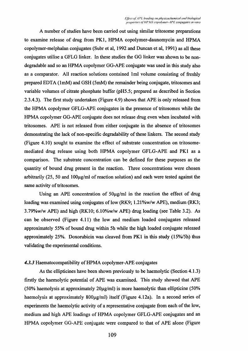
Effect o f APE loading on physicochemical and biologicalproperlies o f HPMA copolymer-APE conjugates in vitro
A number of studies have been carried out using similar tritosome preparations
to examine release of drug from PKl, HPMA copolymer-daunomycin and HPMA
copolymer-melphalan conjugates (Subr et al, 1992 and Duncan et al, 1991) as all these
conjugates utilise a GFLG linker. In these studies the GG linker was shown to be non-
degradable and so an HPMA copolymer GG-APE conjugate was used in this study also
as a comparator. All reaction solutions contained 1ml volume consisting of freshly
prepared BDTA (ImM) and GSH (5mM) the remainder being conjugate, tritosomes and
variable volumes of citrate phosphate buffer (pH5.5; prepared as described in Section
2.3.4.3). The first study undertaken (Figure 4.9) shows that APE is only released from
the HPMA copolymer GFLG-APE conjugates in the presence of tritosomes while the
HPMA copolymer GG-APE conjugate does not release drug even when incubated with
tritosomes. APE is not released from either conjugate in the absence of tritosomes
demonstrating the lack of non-specific degradability of these linkers. The second study
(Figure 4.10) sought to examine the effect of substrate concentration on tritosome-
mediated drug release using both HPMA copolymer GFLG-APE and PKl as a
comparison. The substrate concentration can be defined for these purposes as the
quantity of bound drug present in the reaction. Three concentrations were chosen
arbitrarily (25, 50 and lOOpg/ml of reaction solution) and each were tested against the
same activity of tritosomes.
Using an APE concentration of 50pg/ml in the reaction the effect of drug
loading was examined using conjugates of low (RK9; 1.21%w/w APE), medium (RK3;
3.79%w/w APE) and high (RKIO; 6.10%w/w APE) drug loading (see Table 3.2). As
can be observed (Figure 4.11) the low and medium loaded conjugates released
approximately 55% of bound drug within 5h while the high loaded conjugate released
approximately 25%. Doxorubicin was cleaved from PKl in this study (15%/5h) thus
validating the experimental conditions.
4,3,3 Haematocompatibility of HPMA copolymer-APE conjugates
As the ellipticines have been shown previously to be haemolytic (Section 4.1.3)
firstly the haemolytic potential of APE was examined. This study showed that APE
(50% haemolysis at approximately 20pg/ml) is more haemolytic than ellipticine (50%
haemolysis at approximately 800pg/ml) itself (Figure 4.12a). In a second series of
experiments the haemolytic activity of a representative conjugate from each of the low,
medium and high APE loadings of HPMA copolymer GFLG-APE conjugates and an
HPMA copolymer GG-APE conjugate were compared to that of APE alone (Figure
109

EJJi’ct o f API', loadinf’ on pfiy.ficocl-ieniical and biologicalpropcriies oi HPMA copniymer-APP. conjugales in vitro
1001
80-
<0 60 o£111Q.^ 40
20
100 2 4 6 8
RK3 (+)
RK4 (-) RK4 (+) RK3 (-)
Time (hours)
Figure 4.9: Release of APE from HPMA copolymer-APE conjugates in the presence (+) or absence (-) of tritosomes.
Note: RK3 is HPMA copolymer GFLG-APE (3.79%w/w APE). RK4 is HPMA copolymer GG-APE (4.27%w/w APE). Tritosome-containing tubes had tritosome batch RKT3 at an activity of 20ng/min/tube. Data represent mean ±SD (n=3)
110

Ejfecl o f A PE loading on physicochemical and biologicalfvoperlies oj I IP MA copolymer-AP E conjugates in vitro
lOOl
80-0)s£conEisQ.<
2 0 -
0 300100 200
RK3 25fiq
RK3 50//g
RK3 lOO^g
PK1 25##g
PK1 5(V#g PK1 100#/g
Time (mlns)
Figure 4.10: The effect of substrate concentration on tritosome-mediated release of APE from HPMA copolymer-APE and doxorubicin from PKl.
Note: RK3 is HPMA copolymer GFLG-APE (3.79%w/w APE). Tritosome- containing tubes had tritosome batch RKT7 at an activity of 20ng/min/tube. Data represent mean ±SD (n=3).
I l l

EJJect o f A Pfi loading on physicocheinical and hioiogicaipropej iies oJ hiPMA capolymer-API: conjugales in vitro
701
60-
« SO §2 40-
ÎÙ3 30 o.<
20 -
10 -
0 100 300200
RK3
RK9
RK10PKl
RK4
Time (mlns)
Figure 4.11; The effect of APE loading on tritosome-mediated release of APE from HPMA copolymer-APE and doxorubicin from PKl.
Note: RK3 is HPMA copolymer GFLG-APE (3.79%w/w APE). RK4 is HPMA copolymer GG-APE (4.27%w/w APE). RK9 is HPMA coploymer GFLG-APE (1.21%w/w APE). RKIO is HPMA copolymer GFLG-APE (6.10%w/w APE). Tritosome-containing tubes had tritosome batch RKT7 at an activity of 20ng/min/tube. Data represent mean ±SD (n=3)
112

liffc’ct o f A I’f lauding on physicochemical and hioiogicaiproperties ot fU’MA copolymer-AFE conjugales in vilro
1201
100 -
80oc8
60-o
I 40-
2 0 -
.001 .01 .1 1 10
■o Ellipticine APE
Concentration (mg/ml)
1201
100 -
APE
RK4
RK2
RK1020-
RK9
.01 .1 1 10 100 1000 10000Concentration (APE-equivalent //g/ml)
Figure 4.12: Haemolytic activity of APE, ellipticine and HPMA copolymer- APE conjugates.
Note: Panel a shows the comparative haemolysis caused by APE and ellipticine. Panel b shows the effect of drug loading on haemolysis. Data represent mean ±SE (n=6)
113

Effect o f APE hading on physicochemical and biologicalproperiies o f HPMA copolymer-APE conjugates in vitro
4.12b). All conjugates of different drug loading showed less than 20% haemolysis in
this assay up to lOOpg/ml of APE equivalence while APE alone gave over 80% lysis at
this concentration.
4,3,4 In vitro cytotoxicity
The in vitro cytotoxicity data can be expressed in either of two ways, either in
terms of concentration of conjugates or as APE-equivalent concentration. The graph
showing the concentration of conjugates versus APE alone shows that all the HPMA
copolymer-APE conjugates are at least several hundred-fold less cytotoxic than free
APE (Figure 4.13a). However, when this data is expressed in terms of APE-
equivalence this gap narrows, particularly for the HPMA copolymer GFLG-APE
conjugate of low APE loading and the HPMA copolymer GG-APE conjugate (Figure
4.13b). The remaining conjugates are still at least 100-fold less toxic than free APE.
4.4 Discussion
Pyrene has been used to give a myriad information on the
hydrophobic/hydrophilic characteristics of a local solution environment. As described
earlier the pyrene molecules in a solution containing areas of hydrophobicity would be
distributed in high local concentrations in these areas. This would lead to self-
quenching of the fluorescent signal which is easily detected. The intensity ratio ( I 1 / I 3 )
has been shovm to decrease in more hydrophobic environments (e.g. 1.4 in water; 0.5 in
hexane) and is therefore a sensitive measure of local solution hydrophobicity (Gautier,
1995). By dissolving a range of concentrations of HPMA copolymer-APE conjugates
in a saturated solution of pyrene it was possible to examine the presence of areas of
hydrophobicity in the HPMA copolymer-APE conjugates in solution by examining the
fluorescence intensity of pyrene.
The coupling of a hydrophobic anticancer drug to the hydrophilic HPMA
copolymer precursor was thought to affect the solution characteristics of the resultant
conjugate but this has never been examined previously. This study sought to examine
this effect and showed some unexpected results. The FL component of the GFLG
linker has been shown to influence the solution characteristics of conjugates
significantly. The conjugates seem to form micellar structures at low concentrations
which is not unexpected considering the hydrophobicity of APE. A previous study
114

I'ffcct o f A PE loading on physicochemical and hioiogicaiproperiies oj HPMA copoiynier-APE conjugates in vilro
120
100
c8o&>,
RKIO
RK5APE
.0001 .001 .01 .1Concentration (mg/ml)
120
100
RK3I8O
80 RKIO
60-
I 40
RK5RK7APE
20
1000.01 .1 1 10 100Concentration (APE-equivalent //g/mi)
Figure 4.13: In vitro cytotoxicity of APE and HPMA copolymer-APE conjugates against B16F10 cells.
Note: Panel a shows the data in terms of concentration of APE and conjugates. Panel b shows the data in terms of APE-equivalent concentration. Data represent mean ±SE (n=6)
115

Effed nf.A PE loading on physicochemical and biologicalproperiies o f HPMA copolymer-APE conjugales in vino
showed that pyrenyl molecules conjugated to poly(A^-isopropylacrylamides) did indeed
form such micelles (Ringsdorf et al, 1991). Also any micellar effect would be expected
to be exacerbated by 7i-7t stacking which is a common phenomenon between planar
aromatic structures such as doxorubicin (Menozzi et al, 1984) and would be expected to
occur with APE because of its similar planar aromatic structure. If the polymeric
backbone is sufficiently flexible and hydrophilic (such as HPMA) then the hydrophobic
drug could form these stacked conformations even after conjugation (Ilhan et al, 1999).
There is no evidence of drug loading affecting the pyrene I 1 / I 3 ratio in the
presence of the HPMA copolymer GFLG-APE conjugates but there may be differences
in the size of the hydrophobic ‘pockets’ formed which were not detected using this
approach. The clinical relevance of such a micellar structure is yet to be examined, if
indeed these micellar structures reflect the situation following i.v. administration. To
definitively determine the conformation of these conjugates it would be necessary to do
structure property relationships of micelle stability. The stability of polymeric micelles
is very strongly structure-dependent (Ringsdorf et al, 1992) and the equilibrium
between an open chain coil and micelle forming units within the aqueous phase is very
sensitive to the composition of the entire polymer (Ringsdorf et al, 1991).
Examining Figure 4.8 it is interesting to observe that the HPMA copolymer GG-
APE conjugate (RK4) seems to provide the most hydrophobic environment for the
pyrene molecules. This is understandable though as the polymer has a clear distinction
between its hydrophilic component (HPMA copolymer GG) and its only undisturbed
hydrophobic part (APE) (represented in Figure 4.14). What is interesting is that the
HPMA copolymer GFLG-APE conjugate of medium APE loading (RK3) seems to
offer a less hydrophobic surrounding for pyrene than the comparable HPMA copolymer
GFLG-aminopropanol. Again, a possible explanation might be that the APE containing
conjugate, although more hydrophobic, cannot form a stable micelle due to the two
glycyl units disturbing a defined separation of hydrophobic and hydrophilic domains.
This could be explained by the formation of FL loops in both cases, the hydrophilic
aminopropanol and HPMA copolymer GG elements stacking on the outside for the
aminopropanol conjugate while the conformation of RK3 is more disturbed by the
hydrophobic APE (represented in Figure 4.14). This is a speculative explanation but
considering the sensitivity of micelle stability it may not be completely outlandish.
When considering the I 1 / I 3 ratio data the three HPMA copolymer GFLG-APE
conjugates of variable drug loading (RK3, RK9 and RKIO) do not differ much because
116

/.7A ’( / o f I / ’ /. l(iuJin<^ nn /> livsic()('h e iiiica l ( /f id h io io g ic a i
/>i '>/>ci iic^ III l i r \ l I I o p a lv iiic r - i r I I coif/i/f^aic.s in v iiro
HPMA copolymer GG-APE Most stable micelle
HPMA copolymer GFLG-Ap Less stable micelle
HPMA copolymer GFLG-APE Least stable micelle
Figure 4.14: Proposed conformation o f HPMA copolym er-APE and HPM A copolym er-am in opropanol conjugates.
Note: The shaded area represents the area o f hydrophobicity central to each proposed m icelle.
117

Effect nf.A PE loading nn physicochemical and biologicalproperties o f HPMA copolymer-APE conjugates in vitro
pyrene will sense the presence of hydrophobicity but not the size of these hydrophobic
pockets (Figure 4.8). This may be the only difference between these conjugates.
The stability of the GFLG and GG linkers used in this study have been
examined in rat plasma and serum (Rejmanova et al, 1985). A variety of different tri-
and tetra-peptide linkers as well as one each of a penta- and a hexa-peptide linker were
tested by detecting, using UV spectroscopy, the release of NAp from the terminal group
of the linkers. The GFLG linker was chosen for the present study to synthesise HPMA
copolymer-APE conjugates for the reasons outlined in Section 1.3.4.2. This linker
released 1.5 and 1.7% of total NAp in plasma and serum respectively in a 5h time
period. No significant release in the circulation has been reported for those polymer-
drug conjugates which utilise this linker and PKl, for example, has been shown to
release only small amounts of doxorubicin in the circulation of patients in clinical trial
(Thomson et al, 1999).
Release of drug from the HPMA copolymer conjugates must occur for these
conjugates to have anti-tumour activity. The tritosome-mediated APE release studies
reported here have demonstrated the stability of the GFLG linked APE in the absence
of tritosomes. This lack of non-thiol protease release of APE is also confirmed by the
observed lack of haematotoxicity and in vitro cytotoxicity of the HPMA copolymer-
APE conjugates. There seemed to be a limit of cathepsin B access in the case of the
HPMA copolymer GFLG-APE conjugate of highest loading which showed
approximately half the release of that seen from the low and medium loaded conjugates.
Interestingly APE was released more rapidly from the low and medium loaded
conjugate than doxorubicin was cleaved from PKl under the same conditions at the
same substrate concentration perhaps indicating that APE allows cathepsin B easier
access to the terminal bond than doxorubicin. However as the ideal profile of drug
exposure is likely to vary depending on the drug it is not known whether quicker release
is a benefit. This has been demonstrated with the toxicity reported in animal studies of
an HPMA copolymer GLFG-doxorubicin conjugate which releeised doxorubicin more
rapidly than the GFLG linker in tritosome release studies (Subr et al, 1992).
This present study also demonstrated that at these concentrations of substrate
and enzyme activity of tritosomes the enzymes were not saturated. This was not
unexpected as previous studies on PKl cited substrate concentrations of doxorubicin-
equivalence of approximately 600pg/ml (Subr et al, 1992).
1 1 8

Effed o f APE loading on physicochemical and biologicalproperlies o f HPMA copolymer-APE conjugales in vitro
For a polymer-drug conjugate to be administered by the parenteral route it must
be shown to not cause toxicity in the bloodstream either as the intact conjugate or by
inappropriate release of active drug. Many anticancer drugs have been shown to
extravasate at the site of administration into subcutaneous or subdermal tissue causing
severe tissue necrosis because of the high local concentration of a toxic agent. For this
reason potent anticancer agents are usually administered as slow infusions of low
concentrations (British National Formulary, 2001) and those polymer-drug conjugates
in clinical trial have also been administered in this way (Vasey et al, 1999 and Meerum
Terwogt et al, 2001). It has been known for some time that macromolecules such as
polymer-drug conjugates have longer half-lifes in the circulation than the equivalent
low molecular weight compounds (reviewed by Duncan & Spreafico, 1994). Therefore
a polymer-drug conjugate will be exposed to the blood and surrounding tissue for a
longer period of time relative to administered free drug.
Toxicity could potentially occur by a number of mechanisms following
administration. The polymer-drug conjugate could itself be toxic if the drug is still
active when conjugated. The linker may be unstable and result in inappropriate drug
release into the circulation hence causing toxicity.
As described in Section 4.1.3 it is well recognised that one of the major
difficulties associated with the clinical use of ellipticines has been their tendency to be
haematotoxic. HPMA copolymer conjugates of anticancer drugs have been shown to
abrogate many of the usual toxicities of the administered drug in previous studies. The
HPMA copolymer-APE conjugates were all significantly less toxic than APE alone in
this haemolysis assay. This demonstrates the ability of HPMA copolymers to protect
the drug from causing such toxicities while APE was shown to be haemolytic as
expected from reports on other ellipticines (Lee, 1976). This study showed that APE is
more haemolytic than ellipticine itself and therefore parenteral administration of APE
could lead to haematotoxicity. The observed lack of haemolysis of the HPMA
copolymer GFLG-APE conjugates bodes well for future administration of these
conjugates in in vivo anti-tumour studies.
In the in vitro cytotoxicity assays there was some evidence of cytotoxicity for
the two APE-containing conjugates which had been shown to adopt an open solution
conformation in the pyrene studies. Some, but not all, of this toxicity could be
explained by the presence of free drug in the HPMA copolymer-APE conjugate but
119

Effect o f APE loading on physicochemical and hioiogicaiproperties o f HPMA copoiymer-APE conjugates in vitro
Other mechanisms seem to play a role. The classical method of testing anticancer
agents is to investigate their in vitro cell cytotoxicity against a range of cell lines and
hence determine specificity towards certain cell and hence tumour types. This method
is still widely used and indeed is the basis for testing compounds in the NCI screening
process (Cragg et al, 1999). This simple test is suitable only for molecules which are
active in their own right but is limited in the case of those molecules and systems
requiring activation in some manner (e.g. prodrugs or drug delivery systems).
Polymer-drug conjugates rely on uptake by fluid-phase pinocytosis, in the
absence of other carrier mechanisms (e.g. receptor-mediated uptake) as described in
Section 1.4.4. The rate of uptake by this type of endocytosis is approximately 2pl of
the surrounding medium being taken up per mg of protein per hour incubation (Duncan
& Kopecek, 1984). Therefore only a small amount of conjugate will be taken up over
the time course of a classical 72h cytotoxicity assay. As well as uptake the conjugate
needs to be trafficked to the lysosome and the drug released before cytotoxic activity
can occur. Therefore polymer-drug conjugates invariably show no or minimal toxicity
in these assays although one must be careful to examine the role of free drug in any
observed conjugate toxicity. It was still considered useful to evaluate the cytotoxicity
of these conjugates as any unexpected toxicity could be determined.
Toxicity could be indicative of the presence of a significant quantity of free
drug, instability of the linker, another biological process being capable of liberating free
drug or perhaps an indication of activity of the drug even while conjugated. If one
examines the cytotoxicity profile in terms of equivalent concentration of APE then all
the HPMA copolymer-APE conjugates were less toxic than the parent APE as would be
expected from their differing modes of cellular uptake. The HPMA copolymer GFLG-
APE conjugate with the lowest loading (RK7) had a higher concentration of free APE
than the other conjugates and a portion of its apparent toxicity can be explained by the
presence of this free drug. However this does not completely explain the cytotoxicity
of this conjugate. One possible explanation may be that the conjugate retains some
activity towards the cell membrane and combined with the more open structure of this
conjugate as determined in the conformation study the APE may be more exposed.
Another possible explanation would be that because it forms a more open structure in
solution this conjugate is interacting more closely with the cellular membrane and being
internalised and therefore cleaved more quickly within the time scale of the experiment.
This could also explain the cytotoxicity seen for the HPMA copolymer GG-APE
1 2 0

E ffect o f A PE lo a d in g on p h y s ic o c h e m ic a l a n d b io lo g ic a l p r o p e r t ie s o f H PM A co p o ly m e r -A P E c o n ju g a te s in v itro
conjugate (RK4) as it also has been shown to adopt a more open structure in the
solution conformation studies (Section 4.3.1).
The only conjugate which showed haemolytic activity was the HPMA
copolymer GG-APE conjugate, RK4. This, in combination with the cytotoxicity of this
conjugate, could suggest there being some element of surface activity mediated by the
more open solution structure of this conjugate. It is important to note that while this
haemolysis data seems to contradict the cytotoxicity of the HPMA copolymer GFLG-
APE conjugates of low drug loading (RK7/RK9) cytotoxicity is a 72h assay while this
red blood cell lysis assay was over Ih. In addition to this red blood cells do not have a
process of endocytosis and their membranes have been shown to be more stable than
other cellular membranes because of their different lipid composition and the presence
of elevated amounts of cholesterol (reviewed by Weinstein, 1969). This would not
therefore preclude increased uptake by endocytosis or some element of surface activity
playing some role in the cytotoxicity of these two conjugates (namely an HPMA
copolymer GFLG-APE conjugate of low drug loading or an HPMA copolymer GG-
APE conjugate).
In summary HPMA copolymer-APE conjugates form complex micellar
structures in solution. APE is only liberated by thiol proteases and therefore no in vitro
cytotoxicity or haematotoxicity was observed. This reduction in haematotoxicity is
important because of the clinical difficulties with haematotoxicity observed with the use
of ellipticine derivatives (Section 1.5.2). There was no effect of drug loading on
haematotoxicity but there seemed to be some effect on the in vitro cytotoxicity of the
conjugates. These studies do not preclude investigation of any of the HPMA
copolymer GFLG-APE conjugates in anti-tumour studies in vivo. Therefore the next
study tested these HPMA copolymer GFLG-APE conjugates in vivo.
121

Effect o f APE loading on anti tumour activity o fHPMA copolymer GFLG-APE conjugates
Chapter Five
Effect o f APE loading on anti-tumour activity o f HPMA copolymer GFLG-APE conjugates
1 2 2

Effect o f A PE loading on antiiumour activity o fHPMA copolymer GFLG-APE conjugates
5.1 Introduction
Conjugation of APE to HPMA copolymers reduced APE haematotoxicity and
APE was shown to be released from the conjugates in vitro using thiol proteases
isolated as lysosomes (Chapter 4). These observations justified testing HPMA
copolymer-APE conjugates in vivo against murine tumour models. As described in
Section 4.4 many of the anticancer agents in development are first tested against a panel
of tumour cell lines in vitro to determine firstly whether they are active and secondly
the spectrum of their anticancer activity. This approach is not suitable as a screen for
drugs requiring activation such as prodrugs or polymer-drug conjugates due to the
inherent limitations of these assays discussed in Section 4.4. It has been shown that
HPMA copolymer conjugates are preferentially retained in solid tumours by the EPR
effect (Section 1.4.3) and that HPMA copolymer paclitaxel, camptothecin and platinate
have increased activity against a variety of solid tumours. PKl has also shown activity
in a number of murine and human xenograft tumour models in vivo which were not
sensitive to doxorubicin alone (Duncan et al, 1992; Table 5.1).
The models used for pre-clinical evaluation of PKl were mainly mouse models
(Table 5.1) and were subsequently shown to be good predictors of the activity and
reduction in toxicity observed in the Phase I human clinical trial (Vasey et al, 1999). In
these mouse studies PKl showed increased activity over doxorubicin alone and a lower
incidence of toxicity allowing higher doses to be administered (Duncan et al, 1992).
Therefore mouse tumour models were chosen as suitable for examining anti-tumour
activity of HPMA copolymer GFLG-APE conjugates. In the first instance a s.c.
B16F10 murine melanoma model in C57B1 male mice was chosen as this model has
been widely used to study other HPMA copolymer conjugates (Duncan et al, 1992 and
Gianasi et al, 1999). These s.c. B16F10 tumours take approximately 10 days to develop
to a size of 25-50mm^. When the tumour is not treated animals usually progress to the
maximum allowable tumour area within a further 7 days. Therefore this is a
particularly aggressive model and any anti-tumour activity in this model is promising.
The B16F10 melanoma has shown significant and sustained accumulation of PKl and
HPMA copolymer-platinates by the EPR effect (Seymour et al, 1994 and Gianasi et al,
1999). Indeed Seymour et al (1994) showed that PKl administered i.v. at a doxorubicin
equivalent dose of 5mg/kg produced a greater than 10-fold increase in total drug levels
in the tumour after Ih compared to that seen following administration of free
doxorubicin given at the same dose. Also, following a lag phase during which PKl is
123

Effect o f APE loading on antiiumour activity o fHPMA copolymer GFLG-APE conjugates
Table 5.1: PKl activity in a panel of in vivo tumour models (Duncan et al, 1992).
Hostanimal
Cell line and
origin
Site Route of administration
(dosing schedule)
Doxorubicin^^ PKl tt
Dose(mg/kg)
T/C(%)
Dose(mg/kgequiv.)
T/C(%)
CDFlmice
L1210murine
leukaemia
i.p. i.p.(Days 1,2, 3)
5 214 30 430
CDFlmice
L1210murine
leukaemia
i.p. i.v.(Days 1,2,3)
22 183 100 233
CDFlmice
M5076murine
reticulosarcoma
s.c. i.v.(Days 5, 9,15)
10 125 20 319
CDFlmice
M5076murine
reticulosarcoma
s.c. i.p.(Days 5, 9, 15)
10 102 20 >264
CDFlmice
M5076murine
reticulosarcoma
i.v. i.v.(Days 3, 7,11)
10 148 30 >244
Wistarrats
^Walkersarcoma
s.c. i.p.(Day 0)
10 88 10 >196
DBA2mice
^P388murine
leukaemia
s.c. i.p.(Days 0,1,2)
3 117 18 >312
C57BL/ 6J mice
^B16F10murine
melanoma
s.c. i.p.(Days 0 ,1,2)
5 133 36 >320
nu/numice
^LS174Thuman
colorectalcarcinomaxenograft
s.c. i.p.(Days 0,1,2)
5 70 18 200
'tumours established before treatment commenced
^^Doses and T/C(%) shown represent the maximum anti-tumour activity seen in each
experiment where a range of doses were typically administered
124

Effect nf.A PE loading on antitumonr acth'ity o fHPMA copolymer GFLG-.4PE conjugates
trafficked to the lysosome and doxorubicin is released, higher levels of free doxorubicin
were maintained in the tumour over the subsequent 24h at this equal dose and higher
doses of PKl gave further improvements in tumour levels of doxorubicin. Therefore
this tumour model displays the typical characteristics of the EPR effect (Section 1.4.3).
It was hoped that HPMA copolymer-APE conjugates could benefit from this change in
pharmacokinetic profile mediated by the nature of the HPMA copolymer conjugate.
The LS174T tumour model is a human colorectal xenograft model and was used
in the evaluation of PKl (Table 5.1). Such xenograft models have become vital to our
understanding of human tumours since the development of mice lacking in T
lymphocytes to eradicate immune reaction to the implantation or administration of
human cancer cells (Van Putten, 1987). For that reason it was decided that a pilot study
would be carried out on an HPMA copolymer GFLG-APE conjugate using a human
non-small cell lung carcinoma model (CORL23) in nu/nu mice. This model has
previously been used to evaluate PKl activity and the polymer-directed enzyme
prodrug therapy (PDEPT) concept. Satchi (1999) showed that PKl had reduced
activity compared to doxorubicin alone (T/C% 115 vs 130) when administered i.v. at an
equal doxorubicin-equivalent dose of lOmg/kg. However there was no PKl-associated
toxicity at this low dose and therefore dose escalation would be expected to lead to
increased activity. CORL23 was therefore selected as a suitable xenograft model for
testing HPMA copolymer-APE conjugates. These tumours, when injected s.c., are
allowed to grow to a size of 25-50mm^ (within 12 to 15 days) before administration of
treatment. Untreated animals usually progress to the maximum allowable tumour area
within approximately 30 days.
The extravasation of macromolecules into a variety of tumours in mice has been
characterised based on the tumour size for a number of these model systems (Sat,
1999). Both PKl and the macromolecular dye, Evans Blue, were injected as probes
into tumour-bearing mice and the accumulation in each of the tumours measured. For
many of the tumours examined smaller tumours took up more of the probes as these
have been shown to be more angiogenic in nature and therefore to have a more leaky
vasculature (Carmeliet & Jain, 2000). This was the case with the B16F10 model which
showed uptake of approximately 14% dose/g of tumour after Ih for tumours weighing
approximately 50mg and approximately 1% dose/g of tumour after Ih for tumours
weighing approximately lOOOmg. Therefore the B16F10 model is an example of a
125

E ffect o f A P E lo a d in g o n an titu m o n r a c t iv i ty o f H P M A co p o ly m e r G F L G -A P E co n ju g a te s
tumour which shows tumour size-dependent uptake. The CORL23 model was also
examined in a similar study and showed non tumour size-dependent uptake with
approximately 8% dose/g of tumour across the weight range studied (10-500mg).
Therefore these two tumour models were selected to examine the anti-tumour
activity of the HPMA copolymer GFLG-APE conjugates of different APE loading.
5.2 Methods
5.2.1 Cell preparation
The growth curve for B16F10 cells has been described in Section 4.2.4. A
growth curve was also constructed for the CORL23 cells using the method described in
Section 2.3.1.5 (Figure 5.1). These cells therefore have a doubling time of
approximately 48h and all cell suspensions ‘Were thus prepared on day 2. Cell
suspensions were prepared in saline using the method described in Section 2.3.1.7 when
the cells were in the exponential growth phase.
5.2.2 Establishment of s.c. tumours
Tumours were established using cell suspensions in saline of IxlO^cells/ml
(B16F10) or IxlO^cells/ml (CORL23) as described in Section 2.3.1.7. Of these
suspensions lOOpl was injected s.c. into the nape of the neck of the lightly anaesthetised
C57B1 male mice (6-8 weeks old; approximately 25g body weight) or nu/nu male mice
(6-8 weeks old; approximately 25g body weight) respectively. Tumours were allowed
to establish to an area of 25-50mm^ and animals were then randomised and APE,
conjugate or saline (control) injected i.p. under light anaesthesia as described in Section
2.3.2.I.
5.2.3 Determination of APE MTD in tumour-bearing mice
The MTD can be defined as the highest dose which can be administered without
significant toxicity to animals being observed. B16F10 melanoma cells were injected
s.c. as for anti-tumour studies and tumours were allowed to establish to a diameter of
25-50mm^ as described in Section 2.3.2.1. APE was then administered i.p. at a dose of
25mg/kg which was the reported MTD i.p. of APE for mice (NCI, personal
communication). The animals were then monitored for tumour size and weight every
126

Effed o f A PE loading on aniitumovr a d iv id o fPi PM A copolymer (ih LG-AP E conjugales
1.41
1.2
ScCO
0.8-o&<
0.6-
0.40 300200100
Time (h)
Figure 5.1: Growth curve of CORL23 cells grown from a seeding density of 1X1 O'* cell s per well.
Note: Data represent mean ±SD (n=6).
127

Effect o f A PE loading on aniitumour acth'ity- o fHPMA copolymer GFLG-APE conjugates
day and animals were killed when tumours reached a size of 289mm^ or when the
weight of the animal dropped below 80% of initial body weight. The dose was then
adjusted if toxicity was observed to find a dose which did not cause significant toxicity.
5,2,4 Anti-tumour data expression
The measure of anti-tumour activity determined here was the average survival
time of the treated group (T) divided by the average survival time of the control group
(C) expressed as a percentage. This method has been used by the NCI and a substance
is usually considered active if the T/C% is greater than 125. Toxic deaths are quoted as
a combination of animals who were too sick to carry on the experiment and those
animals found dead in cages during the experiment. These animals are not included in
T/C% determinations.
5.3 Results
5.3.1 Determination of APE MTD in tumour-bearing mice
The initial dose of 25mg/kg APE i.p. gave one toxic death observed as a 20%
loss in body weight from the two animals and therefore it was decided to reduce the
dose to 20mg/kg. This dose also gave one toxic death and so it was decided to further
reduce the dose to lOmg/kg which gave no toxic deaths. Therefore, for the purposes of
these studies, the MTD of APE was taken to be lOmg/kg. This is shown in Figure 5.2
as toxicity was observed as greater than 20% body weight loss in at least one animal of
the two tested and this was the case for the groups dosed at 20 and 25mg/kg.
5.3.2 Anti-tumour activity of HPMA copolymer-APE conjugates against B16F10
melanoma
Following this study to determine the MTD dose an anti-tumour experiment was
carried out in the B16F10 model testing three doses of APE and HPMA copolymer
GFLG-APE using a single dose i.p. on the day tumours reached an area of 25-50mm^.
This study utilised an HPMA copolymer GFLG-APE conjugate made from CPT-03
(Table 3.1) and containing 5.5%w/w APE (less than 0.04% of which was free) and is
reported in Searle et al (2001). The results of this study are reported in Table 5.2 as this
gave the information on which further studies were carried out using the HPMA
copolymer GFLG-APE conjugates containing different loading of APE, described in
128

EJ'fcd o f I I ’E lo a d in g o n a n l i lu m o ti r a c l t v ih ’ o f
l i r \ l . \ c o /jo ly m c i- ( i f ' L d - d P r, c n iy u g u le s
130i
120 Saline
APE lOmg/kg
110 -
Î■S’ 1001 E(0
APE 20mg/kg
APE 25mg/kg
90
80
0 2 4 126 10 148days survived after treatment
Figure 5.2: Toxicity of 20 and 25mg/kg doses observed as 20% body weight loss in the MTD determination of APE in mice bearing s.c. B16F10.
Note: Saline is shown as a control which shows no toxicity. Symbols represent mean ±SD (n=2).
129

Effect o f APE loading nn antiiumour activity o fHPMA copolymer GFLG-APE conjugates
Table 5.2: Anti-tumour activity of APE and HPMA copolymer GFLG-APE conjugates
against s.c. B16F10 murine melanoma.
Treatment Dose(mg/kg)
Mean time to tumour
progression ±SE
T/C (%) ±SE
Toxic deaths
saline - 7.7 ± 0.4 100 0/14
APE 1 9.8 ± 0.6 126.7 ±8.0'’ 0/9
APE 5 9.0 ± 0.8 116.7 ±10.9^® 0/10
APE 7.5 7.2 ± 0.9 93.3 ±11.1'^® 1/5
HPMA copolymer -APE*
1 11.0±0.7 142.6 ±9.6' 1/14
HPMA copolymer -APE*
5 9.9 ±1.4 128.0 ±17.6^® 0/8
HPMA copolymer -APE*
10 8.8±1.1 110.0 ±13.9'^® 1/5
* doses are indicated in APE-equivalent. not significant, ° p<0.01, p<0.001
(Student’s t test vs saline controls).
130

FJfed o f A PE loading on antiiumour activity o fHPMA copolymer GFl.G-APE conjugates
this study. On increasing the dose of APE and HPMA copolymer GFLG-APE an
unexpected result was encountered in that the T/C% was less than for the 1 mg/kg dose
(Table 5.2).
From the study described previously it was decided to examine the anti-tumour
activity of the HPMA copolymer GFLG-APE conjugates containing different loading
using a single i.p. dose of 5mg/kg of APE equivalence. The results of this study are
shown in Table 5.3. No significant anti-tumour activity was noted (using Student’s t
test) although the T/C% value for RK2 was 135. This shows that while there is some
activity for RK2 and RK8 (119%) there is none observed for the highest drug loading
conjugate RKIO or for APE alone (in accordance with Searle et al, 2001) at this dose.
This could potentially be due to the reduced release of APE from the RKIO conjugate
observed in the tritosome release studies (Chapter 4). None of the treatments showed
toxicity observed as weight loss of greater than 20% (Figure 5.3) and no gross organ
toxicity was noted on examination of liver, heart, lungs, kidneys and spleen in any
animals in this study (Figure 5.4). This opens the possibility of using higher doses of
conjugates provided the problem of dose escalation can be elucidated.
The conjugate which gave the best activity here was the conjugate containing a
medium loading of APE (RK2; 4.34%w/w APE) and so this would seem to be the best
drug loading for further anti-tumour studies. For this reason a pilot study was
conducted against the CORL23 human tumour xenograft in nu/nu mice using the same
conjugate of medium drug loading (RK2). This was a simple study comparing the
HPMA copolymer GFLG-APE conjugate to saline treatment to elucidate any activity of
the conjugate in this tumour model. The results of the study are shown in Table 5.4.
As can be observed RK2 is significantly more active than saline treatment. This tumour
is slower growing than the B16F10 melanoma and so any differences in tumour growth
can be observed more easily. The tumour growth for the mice treated with RK2 is very
much slower than the control group (Figure 5.5). The difficulties associated with toxic
deaths are clearly demonstrated here with one toxic death in both the control (saline)
and treated (RK2) groups. Neither group showed significant weight loss during the
study (Figure 5.5) and neither group showed any gross abnormalities to liver, lung,
heart, kidneys or spleen upon examination.
131

E (fect o f A P E lo a d in g on a n titu m o n r a c t iv i ty o f H PM A c o p o ly m e r G F L G -A P E co n ju g a te s
Table 5.3: The effect of APE loading on the anti-tumour activity of HPMA copolymer
GFLG-APE conjugates against B16F10 melanoma.
Note: These conjugates contained 1.20%w/w RK8, 4.34%w/w RK2 and 6.10%w/w
RKIO of APE and each had less than 0.1%w/w free APE (Table 3.2).
Treatment Dose(mg/kg)
Initial tumour area mm^ (mean ± SD)
Days survived after
treatment (mean±SD)
T/C % (±SD)
Toxicdeaths
Saline - 27.5, 30,27.5, 36,25.5 (29.3 ±4.1)
6, 7, 9, 6, 8 (7.2 ±1.3)
100 (± 18.1)
0/5
APE 5 52.5,25,25,27.56, 23.4 (30.7 ±12.3)
4,10,4, 6,6 (6 ±2.4)
101(±32.1)^*
2/5
RK8* 5 30.25,25,42, 36,27.5 (32.2 ± 6.9)
6,10,11,6,10 (8.6 ±2.4)
119 (± 33.4)^®
0/5
RK2* 5 25, 27.5, 25,27.5, 26.95 (26.4 ±1.3)
6, 10, 12, 7, 10 (9 ±2.4)
135 (± 28.6)' ®
1/5
RKIO* 5 24.75,31.2, 30,31.35, 26.52 (28.8 ±3)
8, 6, 7,4, 8 (6.6 ±1.7)
100 (± 13.3)' ®
1/5
* doses are indicated in APE-equivalent. not significant (Student’s t test vs saline
controls).
132
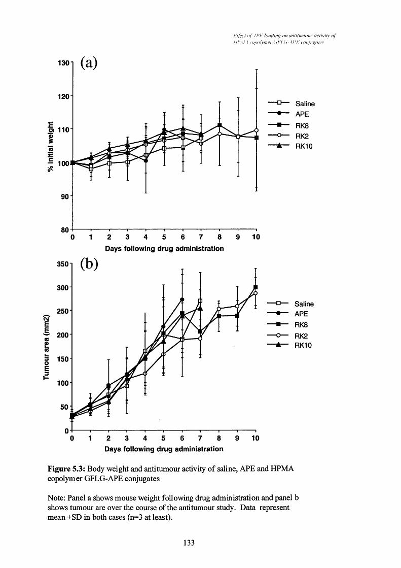
I 'l'fL’ct o f I I’i'. loading on an/i/unioiir acUvity o fiH'MA i (//)olympr ( ii 'l.(i-. U '/'. conjugales
1301
120 -
o) 110
100
90
803 8 9 101 2 4 5 6 70
Days following drug administration
3501
300
250
200
5 150
100
50:
8 9 101 3 40 2 5 6 7
Saline
Saline
Days following drug administration
Figure 5.3: Body weight and anti tumour activity of saline, APE and HPMA copolymer GFLG-APE conjugates
Note: Panel a shows mouse weight following drug administration and panel b shows tumour are over the course of the anti tumour study. Data represent mean ±SD in both cases (n=3 at least).
133

i l fcc! <>! I r i . lo u d in v o n u n iiiu m o iii' a c i i r ih o f
! i! ‘\ i I c o p o ly n ic r ( i i t l ’ i. con /ii^a/c .'i
■ Lungs ea Kidneys m Liver 0 Spleen □ Heart
saline APE RK8Treatment
RK2 RK10
Figure 5.4: Effect o f saline (control), APE and HPM A copolym er GFLG-APE conjugates o f different loading on organ weight o f organs rem oved at the end o f an anti tum our study.
Note: Data represent mean ±SD (n=3 at least).
134

Effect o f APE loading on antiiumour activity o fHPMA copolymer GFLG-APE conjugates
Table 5.4: Activity of RK2 (4.34%w/w APE) against the CORL23 human tumour
xenograft in nu/nu mice.
Treatment Dose(mg/kg)
Initial tumour area mm^ (mean ± SD)
Days survived after
treatment (mean±SD)
T/C % (±SD)
Toxicdeaths
Saline - 27, 30,27.5,25, 27.5, 30
(27.8 ±1.9)
35, 7,40, 32, 28, 28
(28.3 ±11.4)
100 (± 15.6)
1/6
RK2* 1 40, 25, 36,25,25,25 (29.3 ± 6.8)
47,46, 32,51, 75, 45
(49.3 ±14.1)
162 (± 38.7)'’
1/6
* doses are indicated in APE-equivalent. p<0.05 (Student’s t test vs saline controls).
135

I'l'fi’Ct of. I rr. loading on an/ilunioiir ac/ivih.’ ofU rM 1 i-o/MilMucr (il-'i.(i-. \P !■'. conjiiga/e.s
Day
1201
110 -
100(0
co
90
8040 500 10 20 30
Saline
RK2
1000
800o>(03O 6003
400
200
40 500 10 20 30
Saline
RK2
Day
Figure 5.5: Effect of saline and HPMA copolymer GFLG-APE (RX2) on mouse weight and tumour area.
Note: Panel a shows the effect of drug administration on mouse body weight and b shows the effect on tumour area over the course of the anti tumour study. Data represent mean ±SD in both cases (n=3 at least).
136

E(fect o f A PE loading on anlitumoiir activity o fHPMA copolymer GFLG-APE conjugates
5.4 Discussion
The early study of HPMA copolymer GFLG-APE reported in Searle et al (2001)
and described here in Table 5.1 was encouraging as the T/C% for HPMA copolymer
GFLG-APE at a dose of 1 mg/kg was significant against the control treatment, saline
(p<0.001; Student’s t test). The later study, described in Table 5.3 showed some
activity for the conjugates containing low and medium drug loadings but it was not
statistically significant when using the 5mg/kg dose of APE equivalence. It is
important to note, however, that a single dose i.p. study of PKl showed low activity in
this tumour model (O’Hare et al, 1993). Increased activity of PKl in the B16F10
model was only evident upon increasing dose and frequency of dosing (Duncan et al,
1992 and Seymour et al, 1994). The observed sustained reduction in tumour growth
over time for such a low dose of APE equivalence of the conjugates described here was
I promising This is particularly true when considering the high repeated doses
necessary for activity of the newly developed ellipticine analogues (SI6020-2 and
S30972-1) in the B16 model (Malonne et al, 2000).
The data obtained in this thesis using single doses of HPMA copolymer GFLG-
APE are compared with the later studies using PKl and the reported activity of the
ellipticine analogues which are undergoing clinical testing in Table 5.5. It is interesting
to note that an altered dosing regimen of PKl administration showed significant activity
in the B16F10 model and therefore dosing regimen optimisation is a vital aspect of the
development of polymer-drug conjugates. It is necessary to optimise such regimens if
the potential of polymer-anticancer drug conjugates is going to be fully realised. The
anti-tumour activity of the ellipticine analogues are obtained at cumulative doses of up
to 360mg/kg administered over three weeks. Therefore the anti-tumour activity
observed in this model for the HPMA copolymer GFLG-APE conjugates at 1 mg/kg of
drug equivalence is | promising These results also show how difficult it is to treat the
B16F10 model as even high doses of the active ellipticine analogues, which are in
clinical trial, gave no long term survivors in this model. Long term survivors were only
observed in the highest dose of PKl in the small number of animals tested.
The results of the pilot study in the CORL23 human tumour xenograft model are
also interesting for many of the same reasons. The low dose of 1 mg/kg of APE
equivalence gave a sustained decrease in the rate of tumour growth during this study. A
full study is warranted in this model based on this pilot study and the activity observed.
137

E ffect o f A P E lo a d in g on a n ti tu m ou r a c t iv i ty o f H P M A c o p o ly m e r G F L G -A P E co n ju g a te s
Table 5.5: Comparison between HPMA copolymer GFLG-APE, SI6020-2, S30972-1
and PKl administered i.p. in the B16F10 model.
Note: The HPMA copolymer GFLG-APE conjugates contained 1.20%w/w RK8,
4.34%w/w RK2 and 6.10%w/w RKIO
Drug Dose* T/C (%) Long-termsurvivors
Reference
RK8* 5 119 0/5
5 135 0/5
RKIO* 5 100 0/5
816020-2** 10 139 0/9
20 161 0/9
40 53 0/9Malonne et al.
830972-1** 10 137 0/9 2000
20 184 0/9
40 148 0/9
PKl" 18 209 0/10
27 293 0/4 Duncan et al.1992
36 320 3/3
* doses of conjugates are in drug-equivalence
* single dose administered on day of tumour establishment
** Nine doses administered in the three weeks following tumour establishment
® Three doses administered on days 0,1 and 2 of tumour establishment
138

Ed'ect n f APE loading on anlitumoiir activity o fHPMA copolymer CFLC-APE conjugates
In summary HPMA copolymer GFLG-APE conjugates have shown activity in
two tumour models in vivo. The conjugate of medium drug loading showed the best
activity in the B16F10 study. No toxicity of the HPMA copolymer GFLG-APE
conjugates was evident in any of these studies so there is the possibility of dose
escalation. However attempts to give increased doses seemed to give lower activity
than the 1 mg/kg dose. This phenomenon needs to be examined to determine what is
giving this inexplicable result. All the studies reported here have used the i.p. route of
drug administration and therefore the comparison between i.p. dosing and i.v. dosing
needs to be examined to optimise this feature of conjugate testing.
There were unexpected toxic deaths in the study of Searle et al (2001) with one
toxic death reported at a dose of 1 mg/kg APE equivalence of the HPMA copolymer
GFLG-APE conjugate. It is difficult sometimes to attribute early deaths in these studies
to toxicity as there are compounding factors. The B16F10 tumour has been shown to
be a good tumour model of metastasis (reviewed by Glinsky & Glinsky, 1996) and
therefore unexpected métastasés could be fatal. Another difficulty is that the C57B1
strain of male mice are particularly aggressive and fighting is not uncommon and it is
therefore sometimes difficult to distinguish toxic deaths from those caused by fighting.
The possible explanations for the decreased activity of HPMA copolymer
GFLG-APE conjugates with increased doses are that the compounds may have
precipitated in the peritoneal cavity; there was increased liver clearance; there was a
preferred accumulation site other than the tumour or that there was dramatic decrease of
the release of APE from the conjugate when the concentration of polymer increases
(Searle et al, 2001).
HPMA copolymer GFLG-APE conjugates have therefore demonstrated anti
tumour activity in these early studies but further optimisation of dosing schedules and
routes of administration are necessary before these conjugates can be considered for
further development. The lead candidates for such studies would seem to be the
conjugates containing a medium APE loading (3.07-4.34%w/w).
139

The M'indow chamber model: visualisation o fthe inlralumoural distribution o f PK /
Chapter Six
The window chamber model: visualisation o f the intratumoural distribution o f PKl
140

The window chamber model: visucilisaiion o fthe innatumoura! dislrihulion o f PK !
6.1 Introduction
It has been shown that polymer-drug conjugates benefit from the EPR effect
resulting in increased tumour accumulation compared to low molecular weight
anticancer agents (Section 1.4.2). This has been proven for a number of polymer-drug
conjugates using a variety of murine tumour models using both tumour excision and
drug content determination by various methods (e.g. PKl; Duncan et al, 1992) as well
as various imaging studies (e.g. HPMA copolymer-camptothecin; Caiolfa et al, 2000).
In the case of PKl imaging trials have also been carried out as a side-arm to the Phase 1
clinical study using ^^^1-labelled PKl (Vasey et al, 1999). While this particular isotope
was found to be not optimal for imaging, tumour accumulation was noted and
importantly no significant accumulation was observed at unexpected sites. This
reduction in non-tumour deposition was also shown by the decrease in cardiotoxicity of
PKl compared to doxorubicin alone, suggesting decreased PKl accumulation in the
heart. Future studies using methods such as gamma camera and positron emission
tomography have been suggested as means of better characterising the EPR effect. One
method which has been reported has been the use of an ^^^1-labelled PK2 conjugate in a
clinical imaging study using single photon emission computed tomography (Julyan et
al, 1999). This shows that there are many promising methods of imaging polymer-drug
conjugates in future studies and helping our understanding of the EPR effect in humans.
However the pattern and extent of intratumoural distribution of such polymer-
drug conjugates has not been reported. It has been shown previously that antibodies
(reviewed by Jain & Baxter, 1988) and liposomes (Yuan et al, 1995) do extravasate
well into tumour tissue but penetrate poorly beyond the immediate vicinity of the blood
vessels. This has been attributed to factors such as the elevated interstitial pressure of
tumours as well as slow interstitial diffusion of these systems. This is a vital aspect of
drug delivery as lack of penetration of anticancer agents into solid tumours has been
shown to result in low exposure of certain cell populations to the cytotoxic agent and to
lead to resistance to therapy (reviewed by Tungaal et al, 1999). One method that has
been most widely used to examine the structure of tumours has been the use of excised
tumour sections from solid tumours. This has been used as a means of examining the
vasculature of tumours (Konerding et al, 1999) as well as detecting tumour markers
(reviewed by Fenton et al, 1999). However to use this method to examine
intratumoural drug distribution would require the use of many animals to determine the
time course and extent of extravasation and, due to practical limitations, the earliest
141

The winJnw chamber model: visualisation o fthe inii arumoural distribulion ofP K /
time points immediately following parenteral administration, particularly in the first
minute, cannot be studied.
One method which has gained widespread use recently, particularly to examine
tumour blood flow characteristics, has been the window chamber model. This method
was first developed in 1982 (reviewed in Li et al, 2000) and has been used in a number
of studies on blood flow characteristics of tumours since then (reviewed by Jain, 1998).
A number of variations exist of the window chamber (described in Jain, 2001; Figure
6.1), the rat and mouse window chambers being most widely used to examine tumour
characteristics. Perhaps one of the most powerful aspects of this technique is that the
tumour can be visualised in real time and video captured and re-examined to, for
example, study blood flow characteristics. In addition one animal can be used to
examine the complete time course of intratumoural distribution of a drug or marker
therefore reducing the effect of inter-animal variation as long as a suitable number of
animals are used to allow for any gross variations. This technique has recently been
used to examine the migration of implanted tumour cells towards the pre-existing blood
supply in the surrounding tissue (Li et al, 2000) and also to examine the antivascular
activity of combretastatin A4-phosphate (Tozer et al, 2001). In the first study the
tumour cells used expressed green fluorescent protein and therefore tumours were
examined using fluorescence microscopy while in the second study simple light
microscopy was used. This underlines the versatility of this model as almost any
imaging method can potentially be utilised for visualisation.
PKl is the most widely studied of the polymer-drug conjugates and its EPR
characteristics have been described earlier (Section 1.3). PKl was therefore selected as
a better probe than using an HPMA copolymer-APE conjugate to establish this method
of visualising polymer-drug conjugates and study their intratumoural distribution using
the window chamber model. The inherent fluorescence properties of doxorubicin
provide an ideal probe for visualising PKl distribution. A P22 carcinosarcoma tumour
in BD9 rats was selected as the tumour to use for these studies. This model has been
used previously to study the mechanism of action of vasoactive compounds such as
combretastatin A4 phosphate (Tozer et al, 2001). These window chambers can be
monitored for up to 3h to examine any change in distribution pattern.
After this time period tumours can be re-examined under light microscopy to
determine whether there were any gross changes in blood flow. This would help
142

/ / / ( ' uutow c h d i v h e r m o d e l : e v a l i ia i /n i i o f
ihe in ir t i l i i i i i o i i ru id i s i r th i i tK jn o f / ' K /
B
Figure 6.1: The type o f window chambers available (from Jain, 2001).
Note: The rabbit ear window cham ber (A) has mostly been used for physiological measurements o f blood flow while the rat and m ouse skin fold (B) and cranial (C) m odels have been used more for tumour studies.
143

The window chamber model: visualisation o fdie intratumoural distribution o f PK I
determine whether doxorubicin was being released near the vasculature, due to poor
penetration of the conjugate, and then causing a local cytotoxic effect.
6.2 Methods
6,2.1 Window chamber surgery
Male BD9 rats (weighing 150-200g) were prepared for surgery by first being
anaesthetised using lOmg/kg fluanisone containing 0.32mg/kg fentanyl citrate and
5mg/kg midazolam administered i.p. They were then kept under anaesthesia for the
duration of the surgery. All hair on the back of each rat was then removed using hair
removal cream and standard clippers. The rat was then transferred to the surgical area,
wrapped in sterile drapes leaving the back exposed, onto a heated pad and full aseptic
technique was used from this point onwards. The skin of the back was then tented
upwards and sutured into a C clamp using 4/0 Ethilon sutures to stretch the skin slightly
and then keep the skin stretched. The two ends of each suture, on one side of the C
clamp, were then clamped using artery forceps to prevent them from slipping and the
suture tightened to tent the skin (Figure 6.2). One side of the aluminium frame of the
window chamber was then used to mark where the window would be on each side. A
fibre optic light is then used to trans-illuminate the skin. The bottom edge of the circle
where the window will be was then sliced using a scalpel. A fine toothed forceps was
used to lift up the thick tissue layer and a scissors was used to make a hole in the centre
of this incision. The scissors were then opened and closed carefully to free the outer
layers of skin, leaving the deepest fascial layer. The remainder of the circle was then
cut using the scissors. The animal was turned around and the other side was prepared in
the same manner, while protecting the exposed tissue using a sterile gauze soaked in
warm saline. When this was completed the window chamber frame was used to line up
and punch holes through the skin to allow its attachment on both sides. Both sides of
the window chamber were then lined up and stainless steel screws used to join the
frames together with the skin between. Before fully tightening the screws the areas of
exposed fascia were adjusted, if necessary, so that they were in the correct position in
the centre of the window. The window chamber frames were then further re-enforced
by suturing them together.
To implant tumours firstly the areas of fascia were dried using sterile swabs.
The tumour fragments were then implanted on or near a blood vessel, usually near the
144

I'hc window chamber model: evaluation ofthe inlralumoural dislrihulion o f dK /
itainless steel screw holes
suture holes
C clamp y stainless steel screw
I /suture
pulled to tighten suture
artery forceps
rat dorsal skin area
Figure 6.2: Schematic of C clamp from a front and side view.
145

The window chamber model: visucilisaiion o fthe inlralumoural dislrihulion o f P K l
top of the window to allow for any movement of the fragment which tends to fall
downwards. The tumour fragments were early generation (generation 0-10) P22
carcinosarcoma fragments removed from a donor rat and sliced, in media, into pieces
with a diameter of approximately 0.5mm using a scalpel and kept in warm media until
use. Saline was slowly added to the window, so as not to disturb the fragment, and then
a glass window was carefully placed on top of the saline, so as not to introduce air
bubbles. This window was then secured using a C-shaped spring. The same procedure
was carried out on the other side of the window, except no tumour was implanted. The
C clamp was removed and antibiotic cream was applied to the edges of the window
chamber frame. Dextrose saline solution (3ml) was administered to the rat i.p. while
the animals were allowed to recover on a heated pad. Subsequently animals were kept
in a warm room (32-34°C) until the day of the experiment.
6,2.2 Tumour examination and PKl administration
Following surgery tumours took approximately 7-14 days to develop to a
diameter of approximately 4mm, when they would be used in an experiment. For the
window chamber visualisation studies the rats were again anaesthetised using the same
regimen as before and kept under anaesthesia for the duration of the study. The
window chamber frame was attached to a modified microscope stand to allow
visualisation. Rats were monitored for rectal temperature, which was maintained at 35-
37°C using a thermostatically controlled heating pad beneath the rat and an overhead
infrared lamp.
Before PKl was administered the tumour was examined using a light
microscope first under low and then high power to find an area within the tumour
containing typical vasculature. For fluorescence visualisation of PKl the excitation
source was of 546 +/- 5nm wavelength, passed through a 580nm dichroic cube on an
inverted Nikon Diaphot 200 fluorescence microscope. The microscope setup and a
picture of a window chamber are shown in Figure 6.3. Emission was visualised
through a 580nm long pass filter. The processes of extravasation and distribution were
viewed and recorded using a video system (Sony DSR-30P digital videocassette
recorder) coupled to a 3-CCD colour camera (JVC KYF-55) or to a monochrome
intensified CCD camera (Phillips FM800 fibre optic coupled to an XX1666/CJ10 image
intensifier). The sensitivity was set to minimise inherent fluorescence prior to injection.
146

window
The '1 tihl(>\v c h a m h e r m o d e l e w ih u itio n o f
th e in li i i lu m o i ir a i ( ii s in h i i l io n o f i ’T !
heatedblanket
B
stage
Figure 6.3: Picture o f one side o f a window cham ber fram e (A) and o f the m icroscope set-up for imaging (B).
147

The w in d o w c h a m b e r m ode l: v isu a lisa tio n o f th e in tra tu m o u ra l d is tr ib u tio n o f P K /
To begin the experiment PKl in PBS was injected via an intravenous cannula in a tail
vein.
6,2.3 Determination of doxorubicin content of tumours
At the end of the 3h experiments blood was removed from the animals using a
cannulated tail artery or using cardiac puncture following anaesthetic overdose.
Tumours were excised from the window using a scalpel. Samples were stored in a
freezer until analysis. Total and free doxorubicin levels in the blood and tumour
samples were determined by extraction and HPLC as described previously (Section
2.3.3.2)
6.3 Results
Before any further experiments could be started it was necessary to deduce a
suitable dose of PKl which could be administered. The first dose attempted was
lOmg/kg of doxorubicin equivalence. However as PKl was being injected the
fluorescence was already being visualised in the tumour and the injection stopped. It
was calculated that 4mg/kg doxorubicin had been administered and therefore this dose
was used for further studies.
Figure 6.4 shows an example of what the window looks like under light
microscopy at the lowest magnification (xl.6). Figures 6.5 and 6.6 show a series of still
images taken from each of four studies (all at magnification x20) all utilising 4mg/kg of
doxorubicin equivalence of PKl. The first image in each series shows the area of
tumour selected, under light microscopy, as being suitable for further study viewed
from the surface adjacent to the fascial layer. These areas contain a number of blood
vessels which are believed to be mainly venules but it is difficult to fully characterise a
tumour blood vessel. These areas are towards the centre of the tumour in all cases and
contain vessels which are easily distinguishable using light microscopy.
At the time of injection the fluorescence image of all tumours examined was
dark in the field studied due to adjustment of the gain. Immediately after PKl was
injected (lOsec) fluorescence can be seen localised in nearly all the blood vessels within
the same field (second image in each series). After Imin PKl was seen to extravasate
148

//;<■ H 7 « (A n i c h i im h c r n u u k ! f r t i / iu i l i o i i n f
th e m i l ( i i u m n u r u l ( l i s in h i i l in n o f I’K I
blood vessels
B JECfïVEm # OLt NGTH
tumour
0 0 : 0 0 : 2 6 - O C
springs
Figure 6.4: Trans-illuminated picture of a window chamber preparation under the lowest magnification (x 1.6).
149
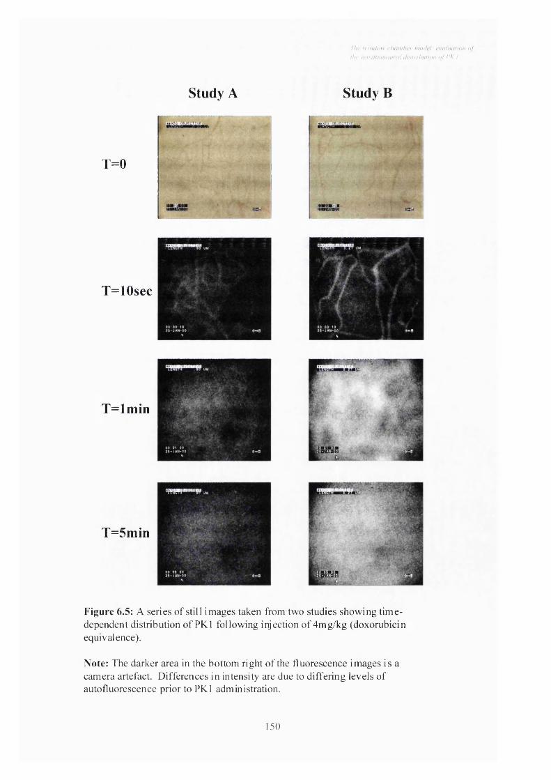
Tiw w hhIum < h ( in iher nuxieT cva l iu i l i o i i o f
i h f inl' (Hin ilo iin ji ( i i s l r ih id io n n! TT /
Study A Study B
T=0
T=10sec
T=lm in
T=5min
TW!TU""'Wi
Figure 6.5: A series of still images taken from two studies showing time- dependent distribution of PKl following injection of 4mg/kg (doxorubicin equivalence).
Note: The darker area in the bottom right of the fluorescence images is a camera artefact. Differences in intensity are due to differing levels of auto fluorescence prior to PKl administration.
150

The \i iriiltw chamber madeT cvahialiaii o fthe iiuralimioural (hsinhiitinn of TK /
Study C Study D
T=0
T=10sec
T=lm in
T=5min
Figure 6.6: A series of still images taken from two further studies showing time-dependent distribution of PKl following injection of 4mg/kg (doxorubicin equivalence).
Note: The darker area in the bottom right of the fluorescence images is a camera artefact. Differences in intensity are due to differing levels of autofluorescence prior to PKl administration.
151
The window chamber model: visualisation o fihe intratumoural distribution o f PK /
with fluorescence beginning to distribute evenly throughout the tumour (third image in
each series). After lOmin there was an almost homogenous distribution of fluorescence
throughout the tumour with no evidence of areas of non-accessibility (fourth image in
each series). The dark area in the bottom right area of all fluorescent images can be
attributed to a camera artefact. This profile of uniform distribution remained
throughout the duration of the experiment, up to 3h which is the longest time permitted
under the animal project licence.
At the end of the 3h studies no gross changes in tumour blood flow were
discernible. Analysis of blood and P22 carcinosarcoma tumour samples following PKl
administration (3h) showed total conjugate levels of 3.5 and 1.1% dose/ml or g
respectively. There was no evidence of doxorubicin being released from PKl within
this time period, as no free doxorubicin was detected in the tumour or blood samples by
this HPLC analysis method.
6.4 Discussion
This study clearly demonstrates that PKl extravasates very rapidly and is
quickly and homogeneously distributed intratumourally. This is in contrast to studies
using rhodamine labelled liposomes (containing egg phosphatidylcholine : cholesterol:
polyethylene glycol: distearoylphosphatidylethanolamine / rhodamine labelled
distearoylphosphatidylethanolamine in a molar ratio of 10: 5: 0.8: 0.1; approximately
90nm diameter). This study utilised a human colon adenocarcinoma (LS174T)
xenograft implanted into window chambers on severe combined immunodeficient
(SCID) mice and showed perivascular deposits of fluorescent spots (<20pm in
diameter) (Figure 6.7; Yuan et al, 1994).
The lack of obvious vascular effects of PKl contrasts with combretastatin A4
phosphate which causes vascular shutdown in this same model after 20min which is
clearly visible using light microscopy (Tozer et al, 2001). These observations support
the distribution profile observed in these studies showing widespread intratumoural
distribution of PKl. The principle site of PKl activity would therefore seem to be
intratumoural rather than at the level of the tumour endothelium.
152

The II in d o u clKinihi-r m n d e l e v u l iu i t i o n o f
the in l r o i n i i i o u r a l i l ist i ih n ii n n o j t 'K /
blood vessel
rhodamine fluorescence
Figure 6.7: Intratumoural distribution o f liposomes o f approxim ately 90nm in LS174T tumours implanted in dorsal window cham bers on SCID m ice showing characteristic perivascular deposits (from Yuan et al, 1994)
153

The M'indow chamber model: visucilisaiion o fthe intratum oural distribution o f PK /
It is known that to exert its anti-tnmour effects doxorubicin must be released
from the PKl conjugate and the GFLG linker was designed specifically to be cleaved
by intralysosomal thiol-dependent proteinases (Duncan et al, 1983). The time
dependence of doxorubicin release from PKl in s.c. B16F10 tumours previously
suggested that lysosomotropic delivery is an essential step for PKl activation (Seymour
et al, 1994). The results of this study are not inconsistent with those of published
studies in mice (Table 6.1) taking into account that these earlier studies used a Ih time
point and blood clearance of PKl has been shown to follow an exponential profile (Sat,
1999).
Future studies aim to investigate the extravasation characteristics in larger
tumours (here the P22 carcinosarcoma used had a size range of 1.8 to 5.4mm diameter)
and other tumour types in order to confirm the results seen here. If this is the case then
other HPMA copolymer conjugates (such as HPMA copolymer GFLG-APE) could be
tested to see if this intratumoural distribution profile is a general effect of HPMA
copolymer-conjugates. The effect of polymer conformation on these characteristics
will need to be examined also using novel polymeric architectures, such as dendrimers,
as a means of optimising these structures and polymer-drug conjugates generally for
tumour extravasation and intratumoural distribution. The next stage of development
would be to integrate these studies with clinical imaging studies in humans to determine
the clinical relevance of these observations.
If clinical imaging studies do confirm this distribution profile one clinical
implication of this study would be in the treatment of métastasés using HPMA
copolymer-anticancer conjugates. Métastasés are difficult to treat using conventional
surgery and radiotherapy methods and are often the cause of fatalities in patients with
solid tumours. These are small tumours and therefore would be expected to have an
irregular vasculature which would be similar in size to the tumours visualised here.
HPMA copolymer-anticancer conjugates could therefore be employed as an effective
treatment for métastasés, perhaps in combination with conventional chemotherapy.
154

The window chamber model: visualisalion o fthe inlralumoural dislrihulion o f PK /
Table 6.1: Comparison of present study with previous accounts for the total amount of
doxorubicin (dox) present in blood and tumour samples.
Species Strain-Tumour
type
Time post injection
Total dox %dose/ml
blood
Total dox % dose/g tumour
Reference
Rat* BD9-P22
3h 3.5 1.07
Mouse DBA2-None
60 min 18 ND Seymour et al, 1990
Mouse Balb/c- Meta 7
60 min ND 2.5 Sat, 1999
Mouse nu/nu-MAXF449
60 min ND 1.0 Sat, 1999
Mouse nu/nu- PAXF 546
60 min ND 2.3 Sat, 1999
Mouse C57B1/B16F10
60 min 20 5Sat, 1999 Seymour
etal, 1994
* Present study described in Section 6.3
ND denotes not determined
155

Genera! discussion
Chapter Seven
General discussion
156

General discussion
The continuing success of the progression of classical polymer-drug conjugates
to clinical trial expounds the potential of this strategy to improve the pharmacokinetic,
toxicological and ultimately clinical anticancer activity of the attached drugs (reviewed
by Duncan et al, 2001). What is also interesting is that many of these conjugates have
shown responses in Phase I clinical trials in chemotherapy refractory patients, which is
something that is usually only observed in less than 5% of patients entering Phase I
trials. It was hoped that the principles derived from studying those conjugates, which
contain clinically used drugs, could be applied to other non-classical anticancer
compounds. This thesis describes a polymer-drug conjugate synthesised using such a
non-clinically studied anticancer drug, namely APE. The effect of drug loading of a
polymer-drug conjugate has not been described before for the polymer-drug conjugates
in clinical development and therefore this aspect of conjugate structure was also
examined. This thesis has also, for the first time, visualised the tumoural extravasation
of a polymer-drug conjugate (PKl), using the rat dorsal window chamber model.
The synthesis of a polymer-drug conjugate needs to be closely controlled to
synthesise a conjugate with the lowest quantity of free drug possible in a reproducible
manner. A series of HPMA copolymer-APE conjugates containing a range of drug
loading have been synthesised in this study, all of which contain less than 0.1%w/w
APE present as the free drug. These conjugates consist of three groups of HPMA
copolymer GFLG-APE conjugates containing low (0-1.21%w/w APE), medium (3.07-
4.34%w/w APE) and high (5.85-6.10%w/w APE) drug loadings. A series of HPMA
copolymer GG-APE conjugates were also synthesised (4.27-4.90%w/w APE) for use as
a comparator in drug release studies. This thesis demonstrates the importance of
examining these HPMA copolymer conjugates, in this case HPMA copolymer-APE, as
new chemical entities and not simply as a drug delivery system, as the physicochemical
nature of the attached drug and the degree of drug loading can significantly influence in
vitro and in vivo properties of the conjugate.
The anti-tumour activity of HPMA copolymer GFLG-APE observed in the
B16F10 and CORL23 in vivo models in this thesis demonstrates the anticancer potential
of these conjugates. The observed activity of HPMA copolymer GFLG-APE at low
doses of APE equivalence in the s.c. B16F10 model is particularly relevant with the
recent study of Malonne et al (2000) examining the activity of SI6020-2 and S30972-1
(ellipticine derivatives) in this same model. These compounds showed anti-tumour
activity in the B16F10 model but using considerably higher doses administered
157

General discussion
repeatedly than those used in this thesis. The anti-tumour activity observed in the pilot
study against the s.c. CORL23 xenograft model using the HPMA copolymer GFLG-
APE conjugate compared to saline alone is particularly interesting with the recently
described lack of activity of PKl administered as a single dose in this model (Satchi et
al, 2001). The reduced activity of HPMA copolymer GFLG-APE conjugates with
increasing dose observed in this thesis in the B16F10 study, however, will need to be
resolved before further studies using HPMA copolymer-APE conjugates could be
attempted with an optimised dosing regimen. Selection of appropriate tumour models
is also particularly important in the evaluation of polymer-drug conjugates as the EPR-
mediated uptake and tumoural levels of cathepsin B are vital for the activity of these
conjugates. A full anti-tumour study, in a panel of tumour models such as that used to
evaluate PKl, will need to be carried out, using an optimised dosing regimen, to more
fully examine the activity of HPMA copolymer GFLG-APE conjugates.
Is APE the best drug for conjugation?
APE was chosen as the natural anticancer drug to be used in this study for the
reasons outlined in Section 3.1. In retrospect perhaps a more potent agent may have
been chosen to better examine the benefits of conjugation to the HPMA copolymer
conjugate. However APE was chemically suitable for the strategy of conjugation by
aminolysis to the HPMA copolymer precursor and ellipticines have interesting
anticancer activity but also marked toxicities, which conjugation was shown to largely
overcome. In the tumour models examined HPMA copolymer-APE conjugates also
had increased activity over free APE at low doses, demonstrating the importance of
increased tumour targeting mediated by the HPMA copolymer.
Is HPMA the best polymer to use?
For many years the polymer of choice for the design of polymer-drug
conjugates has been HPMA based on its proven lack of toxicity and excellent
solubilisation properties. It was therefore the best polymer to use in this study to design
HPMA copolymer conjugates of naturally-derived non-classical anticancer agents.
Indeed conjugates of other non-classical anticancer agents have been recently described
using HPMA copolymers. These include an HPMA copolymer GFLG-
158

Genera! discussion
aminopropylgeldanamycin conjugate, geldanamycin being a benzoquinone ansamycin
anticancer antibiotic (Kasuya et al, 2001).
HPMA is not perhaps the ideal polymer, however, as its lack of degradability
limits the molecular weight to below that of the renal threshold. Therefore the design
of degradable polymers will be vital to the future use of these conjugates. In our group
alone systematic studies have been ongoing to identify suitable polymers for this
purpose. In the last few years, a potentially degradable carrier poly(L-glutamic acid)
conjugated to paclitaxel (Li et al, 1998) and more recently camptothecin (Singer et al,
2001) have been described, with a molecular weight of approximately 50kDa. These
conjugates have shown good pre-clinical activity and poly(L-glutamic) paclitaxel has
shown good activity in a Phase I trial (Sludden et al, 2001).
What have we learned about polymer-drug linkers?
The importance of the choice of a suitable linker in a polymer-drug conjugate
has recently been re-enforced by the toxicity reported at clinical trial of the ‘MAG’-
camptothecin conjugate (Twelves, 2002), described in Section 3.1. These conjugates
contained camptothecin linked by a spacer with a terminal ester bond, which may be
releasing the drug inappropriately. Therefore in this thesis HPMA copolymer-APE
conjugates have used the GFLG linker, chosen on the basis of the widespread use and
benefits of this linker, described in Section 1.3.4.2. However, this linker has been used
for a long period of time and I believe that there is scope for the development of novel
linkers, such as suitable acid-labile or slow hydrolytically-labile linkers. These can be
designed to capitalise on some of the basic tumour characteristics which are constantly
being discovered. This would help broaden the choice of anticancer agent which could
be conjugated as its most potent form, rather than the use of derivatives which may not
be as active which are currently conjugated.
What is the influence of drug loading on polymer-drug conjugates?
Only one range of drug loading has been reported for each of the polymer-drug
conjugates in development. The data obtained in this thesis show that HPMA
copolymer-APE conjugates form complex structures in solution. It may be expected
that this will be the case for many of the other polymer-drug conjugates being examined
due to the hydrophobicity, in the majority of cases, of the pendent drug. The
159

Genera! discussion
significance of conjugate conformation needs to be more closely examined for all the
polymer-drug conjugates to optimise this feature of design. In this study there was a
noticeable effect of conformation observed with the HPMA copolymer GFLG-APE
conjugate of highest drug loading showing reduced APE release in tritosome release
studies with subsequent reduced in vivo activity in the B16F10 tumour model.
Understanding more about the EPR effect
Many are still sceptical about the benefits of EPR-mediated targeting and
intratumoural distribution of polymer-drug conjugates. Therefore the second aspect of
this study has been to utilise the rat dorsal window chamber model to visualise, for the
first time, the distribution profile of HPMA copolymer conjugates following
extravasation into a solid tumour (using PKl as a model compound with suitable
properties for visualisation). This study showed that PKl extravasated more rapidly
than might have been expected from previous studies and then was distributed widely
throughout the tumour. There was no evidence of limited distribution within the
tumours examined. Studies on other tumour types vWll be important to characterise and
understand this effect fully. The tumours examined are relatively small and therefore
this favourable distribution may not exist for large solid tumours in man. Sat (1999)
has shown that such small tumours show better EPR-mediated targeting. However,
métastasés are often the cause of cancer deaths and this increased accumulation and
distribution of polymer-drug conjugates throughout the tumour mass may become
important in the treatment of metastatic tumours. This could also be extrapolated to the
treatment of small tumours which often develop and re-establish following the removal
of a larger tumour. This would strengthen the case for the examination of combination
chemotherapy using polymer-drug conjugates.
The work reported here is just the beginning and future studies must utilise the
window chamber technology to examine the extravasation and intratumoural
distribution of other polymer-drug conjugates. Another use of this model would be to
examine the effect of polymeric architecture on extravasation as new polymer
architectures are being developed for use as polymer-anticancer drug conjugates (such
as dendrimers (Malik et al, 1999)).
The continuing development of novel methods of visualisation, such as the
window chamber model, greatly aid our understanding of the development and
potential treatment of tumours. This is particularly important with the development of
160

Genera! discussion
novel methods of examining human tumours which will be used to standardise the
animal models and characterise the relevance of the EPR effect in humans.
Will HPMA copolymer GFLG-APË become a clinical candidate?
The potential of HPMA copolymer GFLG-APE conjugates is yet to be
fully determined. The reasons for the reduction in anti-tumour activity with increasing
dose has to be elucidated, using further anti-tumour studies. These studies are
warranted based on the activity observed at very low doses of APE equivalence in the
two tumour models studied. Examination of the body distribution and EPR-mediated
localisation of HPMA copolymer-APE conjugates would need to be completed,
especially to determine the effect of drug loading and dose on these characteristics.
Combining these studies with window chamber examination of HPMA copolymer-APE
would help give a clearer idea of how this particular conjugate is behaving in vivo.
Studies using the window chamber model and other visualisation techniques in
the cancer field will, I believe, lead to a better understanding of the conjugate
characteristics required to successfully target anticancer drugs to the tumour using the
EPR effect. Combining this type of information with the large amount of studies and
early clinical trials which have already been reported for polymer-drug conjugates will
result in better rational design of the next generation of anticancer polymer-drug
conjugates.
The synthesis of more closely defined polymeric structures, particularly in terms
of polydispersity and reduction in free drug content will also aid the development of the
next generation of polymer-drug conjugates. New treatment strategies, such as the
development of the two step PDEPT approach (Satchi et al, 2001) and the development
of novel polymeric architectures ensures an exciting future for polymer-drug conjugates
as anti-tumour agents.
Using polymer-drug conjugates to treat other diseases
Finally as polymer-drug conjugates have been shown to be biocompatible their
use need not be limited to cancer and this more widespread interest in the development
of polymer-drug conjugates can only benefit the field of polymer therapeutics. Other
161

General discussion
conjugates have recently been described for use in diseases other than cancer.
Examples of this are an HPMA copolymer 8-aminoquinoline antileishmanial conjugate
(Nan et al, 2001) and an HPMA copolymer lectin anti-ulcerative colitis / Barrett’s
oesophagus conjugate (Wroblewski et al, 2001) which have been recently described.
This demonstrates the potential of polymer-drug conjugates in other diseases based on
the clinical experience and considerable preclinical data with HPMA copolymers. Here
the development of degradable polymers vvdll help, particularly for those diseases where
repeated dosing, over long periods, will be necessary and the use of non-degradable
polymers would be undesirable such as in the treatment of inflammation, where there
may also be hyperpermeability to macromolecules.
1 6 2

References
References
163

References
Access Pharmaceuticals Ltd. website (2001) www.accesspharma.com
Alakhov VY, Moskaleva EY, Batrakova EV and Kabanov AV (1996)
Hypersensitization of multidrug resistant human ovarian carcinoma cells by pluronic
P85 block copolymer. Bioconj. Chem., 7: 209-216
Alberici GF, Fellous R, Bidart JM, Troalen F, Mondesir JM, Goodman A, Ho DH and
Bohuon C (1986) Human antibodies to the antineoplastic drug elliptinium:
characterization and structure-activity relationships. J. Allergy Clin. Immunol, 77(4):
624-630
Allen TM and Moase EH (1996) Therapeutic opportunities for targeted liposomal drug
delivery, yfi/v. Drug Del Rev., 21: 117-133
Allen TM (2000) Solving drug delivery problems with liposomal carriers. In Controlled
Drug Delivery: designing technologies for the future ACS Symposium Series 752, Park
K and Mrsny R (eds) pp 100-109. American Chemical Society, Washington, U.S.A.
Bailon P and Berthold W (1998) Polyethylene glycol-conjugated pharmaceutical
proteins. Pharm. Scl Technol To., 1(8): 352-356
Balosso J, Giocanti N and Favaudon V (1991) Additive and supraadditive interaction
between ionizing-radiation and pazelliptine, a DNA topoisomerase inhibitor, in chinese-
hamster V-79 fibroblasts. Cancer Res., 51(12): 3204-3211
Bangham AD, Standish HM and Watkins JC (1965) Diffusion of univalent ions across
the lamellae of swollen phospholipids. J. Mol Biol, 13: 238-252
Baselga J (2001) Clinical trials of Herceptin® (trastuzumab). Eur. J. Cancer, 37: SI8-24
Batrakova EV, Dorodnych TY, Klinskii EY, Kliushnenkova EN, Shemchukova OB,
Goncharova ON, Arjakov SA, Alakhov VY and Kabanov AV (1996) Anthracycline
antibiotics non-covalently incorporated into the block copolymer micelles: In vivo
evaluation of anti-cancer activity. Br. J. Cancer, 74(10): 1545-1552
164

References
Biltonen RL and Friere E (1978) Thermodynamic characterization of conformational
states of biological macromolecules using differential scanning calorimetry. CRC Crit.
Rev. Biochem., 5(2): 85-124
Blumhardt L (2000) Phase III studies with the synthetic copolymer glatiramer acetate: a
new treatment for multiple sclerosis. Proc. Intl. Symp. Polym. Ther.: 24
Brem H, Piantadosi S, Burger PC, Walker M, Selker R, Vick NA, Black K, Sisti M,
Brem S, Mohr G, Muller P, Morawetz R and Schold SC (1995) Placebo-controlled trial
of safety and efficacy of intraoperative controlled delivery by biodegradable polymers
of chemotherapy for recurrent gliomas. Lancet, 345(8956): 1008-1012
British National Formulary (2001) Number 41. British Medical Association and Royal
Pharmaceutical Society of Great Britain, London, U.K.
Brocchini S and Duncan R (1999) Pendent drugs, release from polymers. In
Encyclopedia o f Controlled Drug Delivery, Mathiowitz E (ed) pp 786-816. John Wiley
& Sons, New York, U.S.A.
Brown KG, Erfurth SC, Small EW and Peticolas WL (1972) Conformationally
dependent low-frequency motions of proteins by laser Raman spectroscopy. Proc. Natl.
Acad. Sci. U.S.A., 69(6): 1467-1469
Buske C, Feuring-Buske M, Unterhalt M and Hiddemann W (1999) Monoclonal
antibody therapy for B cell non-hodgkin’s lymphomas: emerging concepts of a tumour-
targeted strategy. Eur. J. Cancer, 35(4): 549-557
Caiolfa VR, Zamai M, Fiorino A, Frigerio E, Pellizzoni C, d’Argy R, Ghiglieri A,
Castelli MG, Farao M, Pesenti E, Gigli M, Angelucci F and Suarato A (2000) Polymer-
bound camptothecin: initial biodistribution and antitumour activity studies. J. Control.
Rel, 65: 105-119
Cappelaere P, Démaillé MC, Azab M and Degardin M (1993) Phase I study of
retelliptine dihydrochloride (RTE) using daily i.v. infusion for 3 days. Proc. Am. Soc.
Clin. Oncol, 12: 129
165

References
Carmeliet P and Jain RK (2000) Angiogenesis in cancer and other diseases. Nature^
407: 249-257
Cassidy J, Duncan R, Morrison GJ, Strohalm J, Plocova D, Kopecek J and Kaye SB
(1989) Activity of N-(2-hydroxypropyl)methacrylamide copolymers containing
daunomycin against a rat tumor model. Biochem. Pharmacol., 38(6): 875-879
Charcosett JY, Saucier JM and Jacquemin-Sablon A (1988) Reduced DNA
topoisomerase II activity and drug-stimulated DNA cleavage in 9-hydroxyellipticine
resistant cells. Biochem. Pharmacol., 37(11): 2145-2149
Choi W-M, Kopeckova P, Minko T and Kopecek J (1999) Synthesis of HPMA
copolymer containing adriamycin bound via an acid-labile spacer and its activity
toward human ovarian carcinoma cells. J. Bioact. Compat. Polym., 14: 447-456
Choksakulnimitr S, Masuda S, Tokuda H, Takakura Y and Rashida M (1995) In vitro
cytotoxicity of macromolecules in different cell culture systems. J. Control. Rel., 34:
233-241
Connors TA (1996) New anti-cancer agents. Anti-Cancer Drug., 7(SuppL 3): 179-185
Cook JA and Mitchell VB (1989) Viability measurements - cell systems. Anal.
Biochem, 179(1): 1-7
Cragg GM, Boyd MR, Khanna R, Newman DJ and Sausville EA (1999) Natural
product drug discovery and development: the United States National Cancer Institute
role. In Phytochemicals in Human Health Protection, Nutrition and Plant Defense,
Romeo (ed) pp 1-29. Plenum publishers: New York, U.S.A.
Cragg GM, Newman DJ and Snader KM (1997) Natural products in drug discovery and
development. J. Nat. Prod., 60: 52-60
CRC Cancerstats (2000): Mortality-UK, The Cancer Research Campaign, U.K.
166
References
Dalton LK, Demerac S, Elmes BC, Lorder JW, Swan JM and Teitel T (1967) Synthesis
of the tumor-inhibitory alkaloids ellipticine, 9-methoxyellipticine and related
pyrido[4,3-b]carbazoles. Aust. J. Chem.^ 20: 2715-2727
Dang W, Daviau T, Ying P, Zhao Y, Nowotnik D, Clow CS, Tyler B and Brem H
(1996) Effects of Gliadel® wafer initial molecular weight on the erosion of wafer and
release of BCNU. J. Control. Rel., 42: 83-92
Danhauser-Reidl S, Hausmann E, Schick HD, Bender R, Dietzfelbinger H, Rastetter J
and Hanauske AR (1993) Phase I clinical and pharmacokinetic trial of dextran
conjugated doxorubicin (AD-70, DOX-OXD). Invest. New Drugs, 11(2-3): 187-195
Dean RT (1977) Lysosomes. In The Institute o f Biology’s studies in Biology no. 84 pp 1-
54. Edward Arnold (Publishers) Ltd: London U.K.
Debruyne FMJ, Denis L, Lunglmayer G, Mahler C, Newling DWW, Richards B,
Robinson MRG, Smith PH, Weil EHJ and Whelan P (1988) Long-term therapy with a
depot luteinizing-hormone-releasing hormone analog (Zoladex) in patients with
advanced prostatic-carcinoma. J. Urol., 140(4): 775-777
De Duve C, De Barsy T, Poole B, Trouet A, Tulkens P and Van Hoof F (1974)
Commentary. Lysosomotropic agents. Biochem. Pharmacol., 23(18): 2495-2531
De Vita VT, Heilman S and Rosenberg SA (1993) In Cancer: principles and practice o f
oncology (4* ed), JB Lippincott Company, Philadelphia, U.S.A.
Devraj R, Jurayi J, Fernandez JA, Barrett JF and Cushman M (1996) Synthesis of a
series of cytotoxic 2-acyl-1,2-dihydroellipticines which inhibit topoisomerase II.
Anticancer Drug Des., 11(4): 311-324
Dimitrijevic S and Duncan R (1998) Synthesis and characterization of V-(2-
hydroxypropyl)-methacrylamide (HPMA) copolymer-emetine conjugates. J. Bioact.
Compat. Polym., 13: 165-178
167

References
Djuric Z, Everett CK and Valeriote FA (1992) DNA damage and cytotoxicity in L1210
cells by ellipticine and a structural analogue, N-2-(diethylaminoethyl)-9-
hydroxyellipticinium chloride. Cancer Res., 52: 1515-1519
Dodane V and Vilivalam VD (1998) Pharmaceutical applications of chitosan. Pharm.
Sci. Technol To., 1(6): 246-253
Donato MT, Goethals F, Gomez-Lechon MJ, Deboyser D, DeCoster I, Roberfroid M
and Castell JV (1992) Toxicity of the antitumoral drug datelliptium in hepatic cells: use
of models in vitro for the prediction of toxicity in vivo. Toxicol In Vitro, 6(4): 295-302
Duncan R, Cable HC, Lloyd JB, Rejmanova P and Kopecek J (1983) Polymers
containing enzymatically degradable bonds, 7: design of oligopeptide side-chains in
poly[JV-(2-hydroxypropyl)methacrylamide] copolymers to promote efficient
degradation by lysosomal enzymes. Makromol Chem., 184: 1997-2008
Duncan R and Kopecek J (1984) Soluble synthetic polymers as potential drug carriers.
Adv. Polym. Scl, 57: 52-101
Duncan R (1986) Lysosomal degradation of polymers used as drug carriers. CRC Crit.
Rev. Biocompat., 2(2): 127-145
Duncan R, Hume IC, Yardley HJ, Flanagan PA, Ulbrich K, Subr V and Strohalm J
(1991) Macromolecular prodrugs for use in targeted cancer chemotherapy: melphalan
covalently coupled to A^-(2-hydroxypropyl)methacrylamide copolymers. J. Control
Rel, 16:121-136
Duncan R (1992) Drug-polymer conjugates: potential for improved chemotherapy.
Anti-Cancer Drugs, 3: 175-210
Duncan R, Ferruti P, Sgouras D, Tuboku-Metzger A, Ranucci E and Bignotti F (1994)
A polymer-triton X-100 conjugate capable of pH-dependent red blood cell lysis: a
model system illustrating the possibility of drug delivery within acidic intracellular
compartments. J. Drug Targeting, 2: 341-347
168

References
Duncan R and Spreafico F (1994) Polymer conjugates - pharmacokinetic
considerations for design and development. Clin. Pharmacokinet., 27(4): 290-306
Duncan R, Dimitrijevic S and Evagorou EG (1996) The role of polymer conjugates in
the diagnosis and treatment of cancer. STP Pharm. Sci., 6(4): 237-263
Duncan R, Coatsworth JK and Burtles S (1998) Preclinical toxicology of a novel
polymeric antitumour agent: HPMA copoiymer-doxorubicin (PKl). Human. Exp.
Toxicol., 17: 93-104
Duncan R (2000) Polymer therapeutics into the 21*‘ century. In Controlled Drug
Delivery: designing technologies for the future ACS Symposium Series 752, Park K and
Mrsny R (eds) pp 350-363. American Chemical Society, Washington, U.S.A.
Duncan R, Gac-Breton S, Keane R, Musila R, Sat YN, Satchi R and Searle F (2001)
Polymer-drug conjugates, PDEPT and PELT: basic principles for design and transfer
from the laboratory to clinic. J. Control. Rel, 74: 135-146
Elcock AH, Rodger A and Richards WG (1996) Theoretical studies of the intercalation
of 9-hydroxyellipticine in DNA. Biopolymers, 39(3): 309-326
Erukova VY, Krylova OO, Antonenko YN and Melik-Nubarov NS (2000) Effect of
ethylene oxide and propylene oxide block copolymers on the permeability of bilayer
lipid membranes to small solutes including doxorubicin. Biochim. Biophys. Acta, 1468:
73-86
Etrych T, Jelinkova M, Rihova B and Ulbrich K (2001) New HPMA copolymers
containing doxorubicin bound via pH-sensitive linkage: synthesis and preliminary in
vitro and in vivo biological properties. J. Control Rel, 73: 89-102
Fenton BM, Paoni SF, Lee J, Koch CJ, Lord EM (1999) Quantification of tumour
vasculature and hypoxia by immunohistochemical staining and HbOz saturation
measurements. British J. Cancer, 79(3/4): 464-471
169

References
Firestone RA, Willner D, Hofstead SJ, King HD, Kaneko T, Braslawsky GR,
Greenfield RS, Trail PA, Lasch SJ, Henderson AJ, Casazza AM, Hellstrom I and
Hellstrom KE (1996) Synthesis and antitumor activity of the immunoconjugate BR96-
Dox. J. Control Rel, 39: 251-259
Flanagan PA (1987) Evaluation of the pinocytic uptake and cellular processing of
antibody-N-(2-hydroxypropyl)methacrylamide copolymer conjugates and estimation of
their potential use in “targeted” drug delivery - PhD thesis. University of Keele U.K.
Forssen EA, Malé-Brune R, Adler-Moore JP, Lee MJA, Schmidt PG, Krasieva TB,
Shimizu S and Tromberg BJ (1996) Fluorescence imaging studies for the disposition of
daunorubicin liposomes (DaunoXome) within tumor tissue. Cancer Res., 56: 2066-
2075
Forssen EA (1997) The design and development of DaunoXome® for solid tumor
targeting in vivo. Drug Del Rev., 24: 133-150
Fraier D, Frigerio E, Pianezzola E, Strolin Benedetti M, Cassidy J and Vasey P (1995)
A sensitive procedure for the quantitation of free and N-(2-hydroxypropyl)
methacrylamide polymer-bound doxorubicin (PKl) and some of its metabolites, 13-
dihydrodoxorubicin, 13-dihydrodoxorubicinone and doxorubicinone, in human plasma
and urine by reversed-phase HPLC with fluorimetric detection. J. Pharm. Biomed.
Anal, 13(4/5): 625-633
Frenoy JP (1986) Effect of physical environment on the conformation of ricin.
Influence of low pH. Biochem. J., 240(1): 221-226
Froelich-Ammon SJ, Patchan MW, Osheroff N and Thompson RB (1995)
Topoisomerase II binds to ellipticine in the absence or presence of DNA. J. Biol
Chem., 270(25): 14998-15004
Fung LK and Saltzman WM (1997) Polymeric implants for cancer chemotherapy. Adv.
Drug Del Rev., 26: 209-230
170

References
Gabizon A, Catane R, Uziely B, Kaufman B, Safra T, Cohen R, Martin F, Huang A and
Barenholz Y (1994) Prolonged circulation time and enhanced accumulation in
malignant exudates of doxorubicin encapsulated in polyethylene-glycol coated
liposomes. Cancer Res., 54: 987-992
Gautier S (1995) Hydrophobisation du vecteur poly(L-lysine citramide): comportement
physico-chimique et aptitude a solubiliser des molecules lipophiles dans l’eau - PhD
thesis. University of Montpellier France
Gewirtz DA (1999) A critical evaluation of the mechanisms of action proposed for the
antitumor effects of the anthracycline antibiotics adriamycin and daunorubicin.
Biochem. Pharmacol., 57: 727-741
Gianasi E, Wasil M, Evagorou EG, Keddle A, Wilson G and Duncan R (1999) HPMA
copolymer platinates as novel antitumour agents: in vitro properties, pharmacokinetics
and antitumour activity in vivo. Eur. J. Cancer, 35(6): 994-1002
Giavazzi R, Albini A, Bussolino F, DeBraud F, Presta M, Ziche M and Costa A (2000)
The biological basis for antiangiogenic therapy. Eur. J. Cancer, 36: 1913-1918
Gill PS, Wemz J, Scadden DT, Cohen P, Mukwaya GM, Von Roenn JH, Jacobs M,
Kempin S, Silverberg I, Gonzales G, Rarick MU, Myers AM, Shepherd F, Sawka C,
Pike MC and Ross ME (1996) Randomized Phase III trial of liposomal daunorubicin
versus doxorubicin, bleomycin and vincristine in AIDS-related Kaposi’s sarcoma. J.
Clin. Oncol., 14: 2353-2364
Glinsky GY and Glinsky VY (1996) Apoptosis and metastasis: a superior resistance of
metastatic cancer cells to programmed cell death. Cancer Lett., 101:43-51
Godwin A, Hartenstein M, Muller AHE and Brocchini S (2001) Narrow molecular
weight distribution precursors for polymer-drug conjugates. Angew. Chem., 113(3):
614-617
Goodwin S, Smith AF and Homing EC (1959) Alkaloids of Ochrosia elliptica Labill. J.
Am. Chem. Soc., 81: 1903-1908
171
References
Greenwald RB (1997) Drug delivery systems: anticancer prodrugs and their polymeric
conjugates. Exp. Opin. Ther. Patents^ 7(6): 601-609
Greenwald RB, Conover CD, Pendri A, Choe YH, Martinez A, Wu D, Guan S, Yao Z
and Shum KL (1999) Drug delivery of anticancer agents: water soluble 4-poly(ethylene
glycol) derivatives of the lignan, podophyllotoxin. J. Control. Rel., 61: 281-294
Gregoriadis G (1995) Engineering liposomes for drug delivery: progress and problems.
Trends BiotechnoL, 13(12): 527-537
Hillery A (2000) Anti-vascular targeting: a novel approach to cancer treatment. Pharm.
Sci. Technol. To., 3(1): 4
Hirano T, Klesse W and Ringsdorf H (1979) Polymeric derivatives of activated
cyclophosphamide as drug delivery systems in anti-tumor chemotherapy. Makromol.
Rev., 180: 1125-1131
Hirano T, Ringsdorf H and Zaharko DS (1980) Antitumor activity of monomeric and
polymeric cyclophosphamide derivatives compared with in vitro hydrolysis. Cancer
Res., 40(7): 2263-2267
Hoes CJT, Grootoonk J, Duncan R, Hume IC, Bhakoo M, Bouma M, Bouma JMW and
Feijen J (1993) Biological properties of adriamycin bound to biodegradable polymeric
carriers. J. Control. Rel., 23(1): 37-54
Huinink WWT, Terwogt JM, Dubbelman R, Valkenet L, Zurlo MG, Schellens JHM
and Beijnen JH (1998) Phase I and pharmacokinetics study of PNUl66945, a polymer
formulated paclitaxel. Proc. 3' Intl. Symp. Polym. Ther.: 12
Ilhan F, Gray M, Blanchette K and Rotelio VM (1999) Control of polymer solution
structure via intra- and intermolecular aromatic stacking. Macromolecules, 32: 6159-
6162
172

References
Jain RK and Baxter LT (1988) Mechanisms of heterogeneous distribution of
monoclonal antibodies and other macromolecules in tumors: significance of elevated
interstitial pressure. Cancer Res.y 48: 7022-7032
Jain RK (1989) Delivery of novel therapeutic agents in tumors: physiological barriers
and strategies. J. Natl. Cancer Inst,, 81(8): 70-76
Jain RK (1998) The next frontier of molecular medicine: delivery of therapeutics. Nat.
Med., 4(6): 655-657
Jain RK (2001) Delivery of molecular medicine to solid tumors: lessons from in vivo
imaging of gene expression and function. J. Control. Rel., 74(1-3): 7-25
Jones M-C and Leroux J-C (1999) Polymeric micelles - a new generation of colloidal
drug carriers. Eur. J. Pharm. Biopharm., 48: 101-111
Julyan PJ, Seymour LW, Ferry DR, Daryani S, Boivin CM, Doran J, David M,
Anderson D, Christodoulou C, Young AM, Hesslewood S and Kerr DJ (1999)
Preliminary clinical study of the distribution of HPMA copolymers bearing doxorubicin
and galactosamine. J. Control. Rel., 57(3): 281-290
Kaminski MS, Zasadny KR, Francis IR, Fenner MC, Ross CW, Milik AW, Estes J,
Tuck M, Regan D, Fisher S, Glenn SD and Wahl RL (1996) Iodine-131-anti-Bl
radioimmunotherapy for B-cell lymphoma. J. Clin. Oncol., 14(7): 1974-1981
Kasuya Y, Lu Z-R, Kopeckova P, Minko T, Tabibi SE and Kopecek J (2001) Synthesis
and characterization of HPMA copolymer-aminopropylgeldanamycin conjugates. J.
Control. Rel, 74: 203-211
Kataoka K, Matsumoto T, Yokoyama M, Okano T, Sakurai, Fukushima S, Okamoto K
and Kwon OS (2000) Doxorubicin-loaded poly(ethylene glycol)-poly(P-benzyl-L-
aspartate) copolymer micelles: their pharmaceutical characteristics and biological
significance. J Control Rel, 64: 143-153
173

References
Keating MJ, Holmes R, Lemer S and Ho DH (1993) L-asparaginase and PEG
asparaginase - past, present and future. Leuk. Lymphoma^ lO(Suppl): 153-157
Kerbel RS (1997) A cancer therapy resistant to resistance. Nature, 390: 335-336
Khayat D, Borel C, Azab M, Paraisot D, Malaurie E, Bouloux C and Weil M (1992)
Phase-I study of datelliptium chloride, hydrochloride given by 24-h continuous
intravenous-infusion. Cancer Chemo. Pharmacol, 30(3): 226-228
Kohler G and Milstein C (1975) Continuous culture of fused cells secreting antibody of
predefined specificity. Nature, 256: 495-497
Konerding MA, Malkusch W, Klapthor B, Van Ackem C, Fait E, Hill SA, Parkins C,
Chaplin DJ, Presta M and Denekamp J (1999) Evidence for characteristic vascular
patterns in solid tumours: quantitative studies using corrosion casts. British J. Cancer,
80(5/6): 724-732
Konno T, Maeda H, Iwai K, Tashiro S, Maki S, Morinaga T, Mochinaga M, Hiraoka T
and Yokoyama I (1983) Effect of arterial administration of high-molecular-weight
anticancer agent SMANCS with lipid lymphographic agent on hepatoma: a preliminary
report. Eur. J. Clin. Oncol, 19(8): 1053-1065
Kopecek J and Duncan R (1987) Targetable polymeric prodrugs. J. Control Rel, 6:
315-327
Kopecek J, Kopeckova P, Minko T and Lu Z-R (2000) HPMA copolymer-anticancer
drug conjugates: design, activity and mechanism of action. Eur. J. Pharm. Biopharm.,
50: 61-81
Kranz DM, Manning TC, Rund LA, Cho BK, Gruber MM and Roy EJ (1998) Targeting
tumor cells with bispecific antibodies and T cells. J. Control Rel, 53: 77-84
Krishna R and Mayer LD (2000) Multidrug resistance (MDR) in cancer: mechanisms,
reversal using modulators of MDR and the role of MDR modulators in influencing the
pharmacokinetics of anticancer drugs. Eur. J. Pharm. Sci., 11: 265-283
174

References
Kwon G, Suwa S, Yokoyama M, Okano T, Sakurai Y and Kataoka K (1994) Enhanced
tumor accumulation and prolonged circulation times of micelle-forming poly(ethylene
oxide-aspartate) block copolymer-adriamycin conjugates. ^ Control Rel, 29: 17-23
Lee IP (1976) A possible mechanism of ellipticine-induced hemolysis. J. Pharmacol
Exp. Ther., 196(3): 525-535
Lee MD, Dunne TS, Chang CC, Ellestad GA, Siegel MM, Morton GO, McGahren WJ
and Borders DB (1987) Calichemicins, a novel family of antitumor antibiotics. 2.
Chemistry and structure of calichemicin-gamma-1. J. Am. Chem. Soc., 109(11): 3466-
3468
Lee EC and Muller SJ (1999) Flow light scattering studies of polymer coil
conformation in solutions under shear: effect of solvent quality. Polymer, 40: 2501-
2510
Le Mee S, Pierre A, Markovits J, Atassi G, Jacquemin-Sablon A and Saucier J-M
(1998) SI6020-2, a new highly cytotoxic antitumor olivacine derivative: DNA
interaction and DNA topoisomerase II inhibition. Mol Pharmacol., 53: 213-220
Li C, Yu D-F, Newman RA, Cabral F, Stephens LC, Hunter N, Milas L and Wallace S
(1998) Complete regression of well-established tumors using a novel water-soluble
poly(l-glutamic acid)-paclitaxel conjugate. Cancer Res., 58: 2404-2409
Li C, Price JE, Milas L, Hunter NR, Ke S, Yu D-F, Chamsangavej C and Wallace S
(1999) Antitumour activity of poly(L-glutamic acid)-paclitaxel on syngeneic and
xenografted tumors. Clin. Cancer Res., 5: 891-897
Li C-Y, Shan S, Huang Q, Braun RD, Lanzen J, Hu K, Lin P and Dewhirst MW (2000)
Initial stages of tumor cell-induced angiogenesis: evaluation via skin window chambers
in rodent models. J. Natl Cancer Inst., 92(2): 143-147
Lloyd JB (2000) Lysosome membrane permeability: implications for drug delivery.
Adv. Drug Del. Rev., 41: 189-200
175

References
Lobert S, Vulevic B and Correia JJ (1996) Interaction of vinca alkaloids with tubulin: a
comparison of vinblastine, vincristine and vinorelbine. Biochemistry, 35(21): 6806-
6814
Madden T, Tran HT, Beck D, Huie R, Newman RA, Pusztai L, Wright JJ and
Abbruzzese JL (2000) Novel marine-derived anticancer agents: a Phase I clinical,
pharmacological and pharmacodynamic study of dolastatin 10 (NSC 376128) in
patients with advanced solid tumors. Clin. Cancer Res., 6: 1293-1301
Maeda H, Seymour LW and Miyamoto Y (1992) Conjugates of anticancer agents and
polymers: advantages of macromolecular therapeutics in vivo. Bioconj. Chem., 3(5):
351-362
Maeda H and Matsumura Y (1989) Tumoritropic and lymphotropic principles of
macromolecular drugs. Crit. Rev. Ther. Drug Carrier Syst., 6(3): 193-210
Maeda H, Wu J, Sawa T, Matsumura Y and Hori K (2000) Tumor vascular
permeability and the EPR effect in macromolecular therapeutics: a review. J. Control.
Rel, 65: 271-284
Malik N, Evagorou E and Duncan R (1999) Dendrimer-platinate: a novel approach to
cancer chemotherapy. Anti-Cancer Drugs, 10: 767-776
Malonne H, Farinelle S, Decaestecker C, Gordower L, Fontaine J, Chaminade F,
Saucier J-M, Atassi 0 and Kiss R (2000) In vitro and in vivo pharmacological
characterizations of the antitumor properties of two new olivacine derivatives, S16020-
2 and S30972-1. Clin. Cancer Res., 6: 3774-3782
Martindale (1993) 30* edition, Reynolds JE (ed). The Pharmaceutical Press, London
U.K.
Matsumura Y and Maeda H (1986) A new concept for macromolecular therapeutics in
cancer chemotherapy: Mechanism of tumoritropic accumulation of proteins and the
antitumor agent smancs. Cancer Res., 46(121): 6387-6392
176

References
McCafferty J and Glover DR (2000) Engineering therapeutic proteins. Curr. Opin.
Struct. Biol, 10: 417-420
McCaffrey JA and Bajorin DF (1998) Therapy for good risk germ cell tumors. Semin.
Oncol, 25(2): 186-193
Meerum Terwogt JM, ten Bokkel Huinink WW, Schellens JHM, Schot M, Mandjes
I AM, Zurlo MG, Rocchetti M, Rosing H, Koopman FJ and Bejnen JH (2001) Phase I
clinical and pharmacokinetic study of PNUl66945, a novel water-soluble polymer-
conjugated prodrug of paclitaxel. Anti-Cancer Drugs, 12: 315-323
Mehvar R (2000) Dextrans for targeted and sustained delivery of therapeutic and
imaging agents. J. Control Rel., 69: 1-25
Mellman I (1996) Endocytosis and molecular sorting. Annu. Rev. Cell Dev. Biol, 12:
575-625
Menozzi M, Valentini L, Vannini E and Arcamone F (1984) Self-association of
doxorubicin and related-compounds in aqueous-solution. J. Pharm. Scl, 73(6): 766-770
Mezo G, Remenyi J, Kajtar J, Barna K, Gaal D and Hudecz F (2000) Synthesis and
conformational studies of poly(L-lysine) based branched polypeptides with ser and
glu/leu in the side chains. J. Control Rel., 63: 81-95
Minko T, Kopeckova P, Pozharov and Kopecek J (1998) HPMA copolymer bound
adriamycin overcomes MDRl gene encoded resistance in a human ovarian carcinoma
cell line. J. Control Rel., 54: 223-233
Mosmann T (1983) Rapid colorimetric assay for cellular growth and survival:
application to proliferation and cytotoxicity assays. J. Immunol Meth., 65: 55-63
177

References
Muggia FM, Hainsworth JD, Jeffers S, Miller P, Groshen S, Tan M, Roman L, Uziely
B, Muderspach L, Garcia A, Burnett A, Greco FA, Morrow CP, Paradiso LJ and Liang
LJ (1997) Phase II study of liposomal doxorubicin in refractory ovarian cancer:
antitumor activity and toxicity modification by liposomal encapsulation. J. Clin. Oncol,
15(3): 987-993
Nakanishi T, Fukushima S, Okamoto K, Suzuki M, Matsumura Y, Yokoyama M,
Okano T, Sakurai Y and Kataoka K (2001) Development of the polymer micelle carrier
system for doxorubicin. J. Control Rel, 74: 295-302
Nan A, Dhammika Nanayakkara NP, Walker LA, Yardley V, Croft SL and Ghandehari
H (2001) W-(2-hydroxypropyl)methacrylamide (HPMA) copolymers for targeted
delivery of 8-aminoquinoline antileshmanial drugs. J. Control Rel, 77: 233-243
NCI (National Cancer Institute) website (2001) www.nci.nih.gov
Newman DJ, Cragg GM and Snader KM (2000) The influence of natural products upon
drug discovery. Nat. Prod. Rep.., 17: 215-234
Nomura T, Saikawa A, Morita S, Sakeda T, Yamashita F, Honda K, Takakura Y and
Hashida M (1998) Pharmacokinetic characteristics and therapeutic effects of mitomycin
C-dextran conjugates after intratumoural injection. J. Control Rel., 52: 239-252
Nucci ML, Shorr R and Abuchowski A (1991) The therapeutic value of poly (ethylene
glycol)-modified proteins, Drug Del Rev., 6: 133-151
O’Hare KB, Duncan R, Strohalm J, Kopeckova P, Kopecek J and Ulbrich K (1993)
Macromolecular prodrugs containing doxorubicin and melanocyte stimulating hormone
in vitro and in vivo evaluation against murine melanoma. J. Drug Target., 1: 217-230
Ohashi M and Oki T (1996) Ellipticine and related anticancer agents. Exp. Opin. Ther.
Patents, 6(12): 1285-1294
178

References
Ohkuma S and Poole B (1978) Fluorescence probe measurement of the intralysosomal
pH in living cells and the perturbation of pH by various agents. Proc. Natl. Acad. Sci.
U.S.A., 75(7): 3327-3331
Ohnama T, Holland JF and Meyer P (1972) Erwinia carotovora asparaginase in
patients with prior anaphylaxis to asparaginase from E. coli. Cancer, 30: 376-381
Ohya Y, Huang TZ, Ouchi T, Hasegawa K, Tamura J, Kadowaki K, Matsumoto T,
Suzuki S and Suzuki M (1991) Synthesis and antitumor activity of a-1 , 4-
polygalactosamine and N-acetyl-a-1, 4-polygalactosamine immobilized 5-fluorouracils
through hexamethylene spacer groups via urea, urea bonds. J. Control. Rel., 17: 259-
266
Okuno S, Harada M, Yano T, Yano S, Kiuchi S, Tsuda N, Sakamura Y, Imai J,
Kawaguchi T and Tsujihara K (2000) Complete regression of xenografted human
carcinomas by camptothecin analogue-carboxymethyl dextran conjugate (T-0128).
Cancer Res., 60(11): 2988-2995
Ottenbrite RM, Kuus K and Kaplan AM (1983) Characteristic biological effects of
anionic polymers. In Polymers in Medicine: biomedical and pharmacological
applications, Chiellini E and Giusti P (eds) pp 3-22. Plenum Press, New York, U.S.A.
Palmer DH, Seymour LW, Ferry DR, Hussain SA, Hesslewood S, Julyan PJ, Poyner R,
Doran J, Young AM, Burtles S and Kerr DJ (2001) Targeting drugs to the liver: clinical
evaluation of polymer-bound doxorubicin for the treatment of liver cancer. Br. J.
Cancer, 85(Suppl 1): #7.7
Papahadjopoulos D, Allen TM, Gabizon A, May hew E, Matthay K, Huang SK, Lee
KD, Woodle MC, Lasic DD, Redemann C and Martin FJ (1991) Sterically stabilized
liposomes: improvements in pharmacokinetics and antitumor therapeutic efficacy.
Proc. Natl. Acad. Sci. U.S.A., 88: 11460-11464
Pamaud G, Tache S, Peiffer G and Corpet DE (2001) Pluronic F68 block polymer, a
very potent suppressor of carcinogenesis in the colon of rats and mice. Br. J. Cancer,
84(1): 90-93
179

References
Pechar M, Ulbrich K, Subr V, Seymour LW and Schacht EH (2000) Poly(ethylene
glycol) multiblock copolymer as a carrier of anti-cancer drug doxorubicin. Bioconj.
Chem.,ll(2): 131-139
Pederson D, Gabriel D and Hermans J (1971) Potentiometric titration of poly-L-lysine:
the coil to beta transition. Biopolymers, 10(11): 2133-2145
Pendri A, Conover CD and Greenwald RB (1998) Antitumor activity of paclitaxel-2’-
glycinate conjugated to poly(ethylene glycol): a water-soluble prodrug. Anti-Cancer
Drug Design, 13(5): 387-395
Polymers: Biomaterials and Medical Applications (1989) Kroschwitz JI (ed). John
Wiley & Sons, New York, U.S.A.
Potmesil M (1994) Camptothecins: from bench research to hospital wards. Cancer Res.,
54: 1431-1439
Przybylski M, Zaharko DS, Chirigos MA, Adamson RH, Schultz RM and Ringsdorf H
(1978) DIVEMA-methotrexate: immune-adjuvant role of polymeric carriers linked to
antitumor agents. Cancer Treat. Rep., 62(11): 1837-1843
Putnam D and Kopecek J (1995) Enantioselective release of 5-fluorouracil from N-(2-
hydroxypropyl)methacrylamide-based copolymers via lysosomal-enzymes. Bioconj.
Chem., 6(4): 483-492
Raguenez-Viotte G, Dadoun C, Buchet P, Ducas-Telle T and Fillastre JP (1988) Renal
toxicity of the antitumour drug N-methyl-9-hydroxyellipticinium acetate in the Wistar
rat. Arch. Toxicol., 61: 292-297
Ranucci E, Spagnoli G, Ferruti P, Sgouras D and Duncan R (1991) Poly(amidoamine)s
with potential as drug carriers: degradation and cellular toxicity. J. Biomat. Sci., Polym.
E^.,2: 303-315
180

References
Rejmanova P, Labsky J and Kopecek J (1977) Aminolyses of monomeric and
polymeric /7-nitrophenyl esters of methacryloylated amino acids. Makromol. Chem.,
178:2159-2168
Rejmanova P, Kopecek J, Duncan R and Lloyd JB (1985) Stability in rat plasma and
serum of lysosomally degradable oligopeptide sequences in jV-(2-
hydroxypropyl)methacrylamide copolymers. Biomaterials, 6: 45-48
Richardson S, Ferruti P and Duncan R (1999) Poly(amidoamine)s as potential
endosomolytic polymers: evaluation in vitro and body distribution in normal and
tumour-bearing animals. J. Drug Target., 6(6): 391-404
Rihova B, Ulbrich K, Kopecek J and Mancal P (1983) Immunogenicity of N-(2-
hydroxypropyl)-methacrylamide copolymers - potential hapten or drug carriers. Folia.
Microbiol., 28: 217-227
Rihova B, Bilej M, Vetvicka V, Ulbrich K, Strohalm J, Kopecek J and Duncan R
(1989) Biocompatibility of A^-(2-hydroxypropyl) methacrylamide copolymers
containing adriamycin. Biomaterials, 10: 335-342
Rihova B (1998) Receptor-mediated targeted drug or toxin delivery. Adv. Drug Del.
Rev., 29: 273-289
Ringsdorf H (1975) Structure and properties of pharmacologically active polymers. J.
Polym. Sci. Polymer Symp., 51: 135-153
Ringsdorf H, Venzmer J and Winnik FM (1991) Fluorescence studies of
hydrophobically modified poly(N-isopropylacrylamides). Macromolecules, 24: 1678-
1686
Ringsdorf H, Simon J and Winnik FM (1992) Hydrophobically-modified poly(W-
isopropylacrylamides) in water: a look by fluorescence techniques at the heat-induced
phase transition. Macromolecules, 25: 7306-7312
181

References
Ringsdorf H, Sackmann E, Simon J and Winnik FM (1993) Interactions of liposomes
and hydrophobically-modified poly-(N-isopropylacrylamides) : an attempt to model the
cytoskeleton. Biochim. Biophys. Acta, 1153: 335-344
Rizzo V (2000) Validated characterization of polymer-drug conjugates. Proc. 4* Intl.
Symp. Polym. Ther., 17
Rouesse J, LeChevalier T, Caille P, Mondesir J, Sancho-Gamier H, May-Levin F,
Spielmann M, Dejager R and Amiel J (1985) Phase II study of ellipticinium in
advanced breast cancer. Cancer Treat. Rep., 69: 707-708
Rouesse J, Spielmann M, Turpin F, Le Chevalier T, Azab M and Mondesir JM (1993)
Phase II study of elliptinium acetate salvage treatment of advanced breast cancer. Eur.
J. Cancer, 29A(6): 856-859
Rubin P and Casarett G (1966) Microcirculation of tumors. I. Anatomy, function and
necrosis. Clin. Radiol., 17: 220-229
Ruckdeschel JC and Archer S (1989) Rational design of a novel ellipticine derivative
with significant activity against human lung cancer cell lines. Proc. Am. Assoc. Cancer
Res., 30: A2420
Ryan DP, Supko JG, Bder JP, Seiden MY, Demetri G, Lynch TJ, Fischman AJ, Davis J,
Jimeno J and Clark JW (2001) Phase I and pharmacokinetic study of ecteinascidin 743
administered as a 72-hour continuous intravenous infusion in patients with solid
malignancies. Clin. Cancer Res., 7(2): 231-242
Ryser HJ and Shen WC (1978) Conjugation of methotrexate to poly(l-lysine) increases
drug transport and overcomes drug resistance in cultured cells. Proc. Natl. Acad. Sci.
U.S.A., 75(8): 3867-3870
Sainsbury M (1990) Ellipticines. In The chemistry o f antitumour agents, Wilman DEV
(ed) pp 410-435. Blackie & Son Ltd., London, U.K.
182

References
Saleh MN, Sugarman S, Murray J, Ostroff JB, Healey D, Jones D, Daniel CR, LeBherz
D, Brewer H, Onetto N and LoBuglio AF (2000) Phase I trial of the anti-Lewis Y drug
immunoconjugate BR96-doxorubicin in patients with Lewis Y-expressing epithelial
tumors./. Clin. Oncol, 18(11): 2282-2292
Sat YN (1999) Factors that influence tumour targeting by the enhanced permeability
and retention (EPR) effect - PhD thesis. University of London U.K.
Satchi R (1999) PDEPT: polymer directed enzyme prodrug therapy - PhD thesis.
University of London U.K.
Satchi R, Connors TA and Duncan R (2001) PDEPT: polymer-directed enzyme prodrug
therapy I. HPMA copolymer-cathepsin B and PKl as a model combination. Br. J.
Cancer, 85(7): 1070-1076
Schlager SI and Adams AC (1983) Use of dyes and radioisotopic markers in cyto
toxicity tests. Meth. Enzymol, 93: 233-245
Schwartz MK (1995) Tissue cathepsins as tumor markers. Clin. Chim. Acta, 237: 67-78
Searle F, Gac-Breton S, Keane R, Dimitrijevic S, Brocchini S, Sausville EA and
Duncan R (2001) Y-(2-Hydroxypropyl)methacrylamide copolymer-6-(3-aminopropyl)-
ellipticine conjugates. Synthesis, in vitro and preliminary in vivo evaluation. Bioconj.
Chem., 12(5): 711-718
Senter PD, Svensson HP, Schreiber GJ, Rodriguez JL and Vrudhula VM (1995)
Poly (ethylene glycol)-doxorubicin conjugates containing beta-lactamase-sensitive
linkers. Bioconj. Chem., 6(4): 389-394
Seymour LW, Duncan R, Strohalm J and Kopecek J (1987) Effect of molecular weight
(Mw) of V-(2-hydroxypropyl)methacrylamide copolymers on body distribution and rate
of excretion after subcutaneous, intraperitoneal and intravenous administration to rats.
J. Biomed. Mat. Res., 21: 1341-1358
183

References
Seymour LW, Ulbrich K, Steyger PS, Brereton M, Subr V, Strohalm J and Duncan R
(1994) Tumour tropism and anti-cancer efficacy of polymer-based doxorubicin
prodrugs in the treatment of subcutaneous murine B16F10 melanoma. British J.
Cancer, 70: 636-641
Seymour LW, Miyamato Y, Maeda H, Brereton M, Strohalm J, Ulbrich K and Duncan
R (1995) Influence of molecular weight on passive tumour accumulation of a soluble
macromolecular drug carrier. Eur. J. Cancer, 31A(5): 766-770
Sgouras D and Duncan R (1990) Methods for the evaluation of biocompatibility of
soluble synthetic polymers which have potential for biomedical use: 1-use of the
tetrazolium-based colorimetric assay (MTT) as a preliminary screen for evaluation of in
vitro cytotoxicity. J. Mater. Sci.- Mater. Med, 1(2): 61-68
Shen W-C and Ryser HJ-P (1981) Poly(L-lysine) has different membrane transport and
drug-carrier properties when complexed with heparin. Proc. Natl. Acad. Sci. U.S.A.,
78(12): 7589-7593
Shiah J-G, Sun Y, Peterson CM and Kopecek J (1999) Biodistribution of free and W-(2-
hydroxypropyl)methacrylamide copolymer-bound mesochlorin ee and adriamycin in
nude mice bearing human ovarian carcinoma OVCAR-3 xenografts. J. Control. Rel.,
61: 145-157
Sievers EL, Appelbaum FR, Spielberger RT, Forman SJ, Flowers D, Smith FO,
Shannon-Dorcy K, Berger MS and Bernstein ID (1999) Selective ablation of acute
myeloid leukemia using antibody-targeted chemotherapy: a Phase I study of an anti-
CD33 calicheamicin immunoconjugate. Blood, 11: 3678-3684
Sikora K (1999) Developing a global strategy for cancer. Eur. J. Cancer, 35(14): 1870-
1877
Simeckova J, Rihova B, Plocova D and Kopecek J (1986) The activity of complement
in the presence of N-(2-hydroxypropyl)methacrylamide copolymers. J. Bioact. Compat.
Polym., 1: 20-31
184

References
Singer JW, Bhatt R, Tulinsky J, Buhler KR, Heasley E, Klein P and de Vries P (2001)
Water-soluble poly-(L-glutamic acid)-Gly-camptothecin conjugates enhance
camptothecin stability and efficacy in vivo. J. Control Rel, 74: 243-247
Skoog DA and Leary JJ (1992) Infrared absorption spectroscopy In Principles o f
instrumental analysis, Skoog DA and Leary JJ (Eds) p252-291. Saunder College
Publishing, Florida, U.S.A.
Sludden J, Boddy AV, Griffin MJ, Robson L, Todd R, Cassidy J, Bissett D, Main M,
Brannan MD, Elliott S, Verrill M and Calvert H (2001) Phase I and pharmacological
study of CT-2103, a poly(L-glutamic acid)-paclitaxel conjugate. Proc. Am. Assoc.
Cancer Res., 42: #2883
Smith PK, Krohn RI, Hermanson GT, Mallia AK, Gartner FH, Provenzano MD,
Fujimoto EK, Goeke NM, Olson BJ and Klenk DC (1985) Measurement of protein
using bicinchoninic acid. Anal Biochem., 150: 76-85
Soyez H, Schacht E and Vanderkerken S (1996) The crucial role of spacer groups in
macromolecular prodrug design. Adv. Drug Del Rev., 21(2): 81-106
St’astny M, Rihova B, Strohalm J and Ulbrich K (1996) Comparison of the binding
kinetics of antibody-targeted V-(2-hydroxypropyl) methacrylamide (HPMA)-bound
doxorubicin in vitro and in vivo. J. Control Rel, 42: 229-236
Stewart DR, Callahan EH, Jacob JE, Rice JR, Shannon KF, St John JV and Nowotnik
DP (2000) Preclinical development of the polymer platinate AP5280. Proc. Intl.
Symp. Polym. Ther., 57
Subr V, Strohalm J, Ulbrich K, Duncan R and Hume IC (1992) Polymers containing
enzymatically degradable bonds, XII. Effect of spacer structure on the rate of release of
daunomycin and adriamycin from poly [N-(2-hydroxypropyl)-methacrylamide]
copolymer drug carriers in vitro and antitumour activity measured in vivo. J. Control
R e l ,n : 123-132
Supratek website (2001) www.supratek.com
185

References
Takakura Y, Takagi A, Hashida M and Sezaki H (1987) Disposition and tumor-
localization of mitomycin-C dextran conjugates in mice. Pharm. Res., 4(4): 293-300
Thomson AH, Vasey PA, Murray LS, Cassidy J, Fraier D, Frigerio E and Twelves C
(1999) Population pharmacokinetics in phase I drug development: a phase I study of
PKl in patients with solid tumours. British J. Cancer, 81(1): 99-107
Tokudome Y, Oku N, Doi K, Namba Y and Okada S (1996) Antitumour activity of
vincristine encapsulated in glucuronide-modified long-circulating liposomes in mice
bearing Meth A sarcoma. Biochim. Biophys. Acta, 1279: 70-74
Tozer GM, Prise VE, Wilson J, Locke RJ, Vojnovic B, Stratford MRL, Dennis MF and
Chaplin DJ (1999) Combretastatin A-4 phosphate as a tumor vascular-targeting agent:
Early effects in tumors and normal tissues. Cancer Res., 59: 1626-1634
Tozer GM, Prise VE, Wilson J, Cemazar M, Shan SQ, Dewhirst MW, Barber PR,
Vojnovic B and Chaplin DJ (2001) Mechanisms associated with tumor vascular shut
down induced by combretastatin A4-phosphate: Intravital microscopy and measurement
of vascular permeability. Cancer Res., 61(17): 6413-6422
Trail PA and Bianchi AB (1999) Monoclonal antibody drug conjugates in the treatment
of cancer. Curr. Opin. Immunol, 11: 584-588
Trouet A (1974) Isolation of modified liver lysosomes. Meth. Enzymol., 31: 323-329
Trubetskoy VS and Torchilin VP (1995) Use of polyethylene-lipid conjugates as long-
circulating carriers for delivery of therapeutic and diagnostic agents. Adv. Drug Del
Rev., 16:311-320
Tungaal JK, Cowan DSM, Shaikh H and Tannock IF (1999) Penetration of anticancer
drugs through solid tissue: a factor that limits the effectiveness of chemotherapy for
solid tumours. Clin. Cancer Res., 5: 1583-1586
186

References
Twelves C (2002) Clinical experience with MAG-CPT. Proc. 5* Intl. Symp. Polym.
Ther., 21
Twenty man PR, Walls GA and Wright KA (1984) The response of tumor-cells to
radiation and cytotoxic drugs - a comparison of clonogenic and isotope uptake assays.
British J. Cancer, 50(5): 625-631
UKCCCR guidelines for the use of cell lines in cancer research (2000). British J.
Cancer, 82(9): 1495-1509
Ulbrich K, Subr V, Strohalm J, Plocova D, Jelinkova M and Rihova B (2000)
Polymeric drugs based on conjugates of synthetic and natural macromolecules I.
Synthesis and physico-chemical characterisation. J. Control ReL, 64(1-3): 63-79
United Kingdom co-ordinating committee on cancer research (UKCCCR) guidelines
for the welfare of animals in experimental neoplasia (second edition) (1998). Br. J.
Cancer, 77(1): 1-10
Uster PS, Working PK and Vaage J (1998) Pegylated liposomal doxorubicin (DOXIL®,
CAELYX®) distribution in tumour models observed with confocal laser scanning
microscopy. Int. J. Pharm., 162: 77-86
Van Putten LM (1987) In Clinical evaluation o f antitumour therapy, Muggia FM and
Rozencweig (Eds) pi 7. Nijhoff, Boston, U.S.A.
Vasey PA, Kaye SB, Morrison R, Twelves C, Wilson P, Duncan R, Thomson AH,
Murray LS, Hilditch TE, Murray T, Burtles S, Fraier D, Frigerio E and Cassidy J
(1999) Phase I clinical and pharmacokinetic study of PKl [N-(2-
hydroxypropyl)methacrylamide copolymer doxorubicin]: First member of a new class
of chemotherapeutic agents - Drug-polymer conjugates. Clin. Cancer Res., 5(1): 83-94
Venne A, Li SM, Mandeville R, Kabanov A and Alakhov V (1996) Hypersensitizing
effect of pluronic L61 on cytotoxic activity, transport and subcellular distribution of
doxorubicin in multiple drug-resistant cells. Cancer Res., 56(16): 3626-3629
187
References
Vilarem MJ, Riou JF, Multon E, Gras MP and Larsen CJ (1986) The in vitro
involvement of topoisomerase II in the activity of aza-ellipticine analogues is not
correlated with drug activity on isolated nuclei. Biochem. Pharmacol., 35(13): 2087-
2095
Vitetta ES, Stone M, Amlot P, Fay J, May R, Till M, Newman J, Clark P, Collins R,
Cunningham D, Ghetie V, Uhr JW and Thorpe PE (1991) Phase-I immunotoxin trial in
patients with B-cell lymphoma. Cancer Res., 51: 4052-4058
Wall ME and Wani MC (1995) Camptothecin and Taxol: Discovery to clinic -
Thirteenth Bruce F. Cain Memorial award lecture. Cancer Res., 55: 753-760
Wang C-H, Li J, Teo CS and Lee T (1999) The delivery of BCNU to brain tumors. J.
Control Rel, 61: 21-41
Wedge SR (1991) Mechanism of action of polymer-anthracydines; potential to
overcome multidrug resistance - PhD thesis. University of Keele U.K.
Weinstein RS (1969) Electron microscopy of surfaces of red cell membranes. In Red
Cell Membrane: Structure and Function, Jamieson GA and Greenwalt TJ (eds). J.B.
Lipincott Company, Philadelphia, U.S.A.
Winnik FM, Winnik MA, Ringsdorf H and Venzmer J (1991) Bis( 1 -pyrenylmethy 1)
ether as an excimer-forming probe of hydrophobically modified poly(iV-
isopropylacrylamides) in water. J. Phys. Chem., 95: 2583-2587
Woods JA, Hadfield JA, Pettit GR, Fox BW and McGowan AT (1995) The interaction
with tubulin of a series of stilbenes based on combretastatin A-4. Br. J. Cancer, 71(4):
705-711
Wroblewski S, Berenson M, Kopeckova P and Kopecek J (2001) Potential of lectin-N-
(2-hydroxypropyl)methacrylamide copolymer-drug conjugates for the treatment of pre-
cancerous conditions. J. Control Rel, 74: 283-293
188

References
Yokoyama M, Miyauchi M, Yamada N, Okano T, Sakurai Y, Kataoka K and Inoue S
(1990) Polymer micelles as novel drug carrier: adriamycin-conjugated poly(ethylene
glycol)-poly(aspartic acid) block copolymer. J. Control Rel, 11: 269-278
Young RJ and Lovell PA (1996) In Introduction to Polymers, Young RJ and Lovell PA
(eds). Chapman & Hall, London, U.K.
Yuan F, Dellian M, Fukumura D, Leunig M, Berk DA, Torchilin VP and Jain RK
(1995) Vascular permeability in a human tumor xenograft: molecular size dependence
and cutoff size. Cancer Res., 55(17): 3752-3756
Yuan F, Leunig M, Huang SK, Berk DA, Papahadjopoulos D and Jain RK (1994)
Microvascular permeability and interstitial penetration of sterically stabilized (Stealth)
liposomes in a human tumor xenograft. Cancer Res., 54: 3352-3356
Zamai M, Ghiglieri A, Geroni CM, Farao M, Pesenti E, Suarato A, Vandeven M,
Gratton E and Caiolfa VR (2001) Camptothecin co-polymer conjugates: A novel
perspective in topoisomerase I-based anti-tumour therapy. Proc. Am. Assoc. Cancer
Res., 42: #749
Zunino F, Pratesi G and Pezzoni G (1987) Increased therapeutics efficacy and reduced
toxicity of doxorubicin linked to pyran copolymer via the side chain of the drug.
Cancer Treat. Rep., 71(4): 367-373
189

Appendix
Appendix
190

Appendix
List of abstracts and publications
Papers
Searle F, Gac-Breton S, Keane R, Dimitrijevic S, Brocchini S, Sausville EA and
Duncan R (2001) N-(2-Hydroxypropyl)methacrylamide copolymer- 6 - ( 3 -
aminopropyl) - ellipticine conjugates. Synthesis, in vitro and preliminary in vivo
evaluation. Bfoco/y. Chem., 12(5): 711-718
Duncan R, Gac-Breton S, Keane R, Musila R, Sat YN, Satchi R and Searle F (2001)
Polymer-drug conjugates, PDEPT and PELT: basic principles for design and transfer
from the laboratory to clinic. J. Control Rel., 74: 135-146
Keane RS, Wilson J, Vojnovic B, Sausville EA, Tozer GM and Duncan R (2002)
Extravasation of PK1/FCE28068 in a P22 Carcinosarcoma tumour: preliminary studies
using the rat window chamber model, in preparation
Keane R and Duncan R (2002) The effect of drug loading on solution conformation,
drug release, haematotoxicity and in vitro cytotoxicity of N-(2-hydroxypropyl)
methacrylamide copolymer-6-(3-aminopropyl)-ellipticine conjugates, in preparation
Abstracts
Gac-Breton S, Keane R, Searle F, Sausville EA and Duncan R (2000) HPMA
copolymer-aminopropylellipticine: a novel anticancer conjugate. Proc. 4 Inti Symp
Polym. Ther., 60
Keane R, Wilson I, Tozer G and Duncan R (2000) Extravasation of PK1/FCE28068 in
a P22 Carcinosarcoma tumour: preliminary studies using the window chamber model.
Proc. 4* Inti Symp. Polym. Ther., 76
Keane R, Gac-Breton S, Searle F and Duncan R (2000) The effect of drug loading on in
vitro characteristics of N-(2-hydroxypropyl)methacrylamide-6-(3-aminopropyl)
ellipticine anticancer conjugates. J. Pharm. Pharmacol, 52 (SuppL): 52
191

Appendix
Keane R, Sausville EA and Duncan R (2001) Investigation of the effect of drug loading
on the properties of HPMA copolymer 6-(3-aminopropyl)-ellipticine: a novel anticancer
conjugate. Anticancer Res., 21(Suppl.)
192