Why Mechanism Matters
9
Review began 11/14/2021 Review ended 12/07/2021 Published 12/07/2021 © Copyright 2021 Muscat et al. This is an open access article distributed under the terms of the Creative Commons Attribution License CC-BY 4.0., which permits unrestricted use, distribution, and reproduction in any medium, provided the original author and source are credited. Why Mechanism Matters: A Literature Review of Simultaneous Ipsilateral Tibial Tuberosity Avulsion and Patella Fracture With Case Report Joseph Muscat , Aroon Baskaradas , Govind Dhillon , Vashist Motkur , Raj Thakrar 1. Trauma and Orthopaedics, East and North Hertfordshire Trust, Stevenage, GBR 2. Orthopaedics and Trauma, Royal Surrey County Hospital, Guildford, GBR Corresponding author: Joseph Muscat, [email protected] Abstract Simultaneous ipsilateral tibial tuberosity avulsion and patella fractures are rare in adults. They are often associated with patients who have underlying bone disease and other medical co-morbidities. Here we describe a case where this injury was attributed to direct trauma and demonstrate our department’s management for such an injury. In addition to our case report, we have performed a systematic literature review to identify other cases of the same injury. Only four other cases have been reported. Here we summarise and compare the management and outcome measures reported in each case. All patients are managed differently, yet all authors report satisfactory outcomes. With this being a relatively rare injury, further research is required to establish a gold standard for management of such patients. Categories: Orthopedics, Trauma Keywords: unilateral and extensor mechanism disruption, ipsilateral, bifocal, tibial tuberosity avulsion, patella Introduction Tibial tuberosity fractures are more typically seen in the paediatric and adolescent population, particularly in athletic male adolescents approaching skeletal maturity [1]. In the adult population these fractures are rare [2]. Patellar fractures are complex injuries that impact mobility due to disruption of the extensor mechanism. There are favourable outcomes for function in patients that are selected for operative management [3]. Knee injuries commonly result in injury of the extensor mechanism, it is key for a gait, stability and activity of daily living [4]. A crucial part of the extensor mechanism is formed by the patellar tendon which originates from the patella inferior pole and then inserts tibial tuberosity [4]. The extensor mechanism is based on dynamic and static forces centred around the patellar and its tendinous insertion into the tibial tuberosity [5]. When the extensor mechanism is repaired there are significantly higher rates of adverse outcomes if they are mobilised early, however, prolonged immobilisation contributes to decline of mobility, decreased quality of life and increased financial burden [6]. Studies on patellar fractures have estimated that approximately 10 weeks would be required to achieve bony healing if patients were able to mobilise early, which puts a high workload on any surgical fixation [7]. Extensor mechanism disruption can occur in a variety of patterns to injuries in combination of the patella, patella ligament and tibial tubercle. Cases of bifocal extensor mechanism disruption are rare, particularly those which involve a fracture of both the patella and tibial tuberosity [8]. Kang et al. have previously described a case of simultaneous ipsilateral tibial tuberosity avulsion and patella fracture in an elderly male patient. They proposed a classification system for types of extensor mechanism disruption with the type 4 pattern described as “avulsion fracture of the tibial tubercle with avulsion fracture of the inferior pole of the patella” [8]. A summary of their classification system is shown below: Type 1: avulsion fracture of the tibial tubercle with avulsion of the patella ligament from the tibial tubercle; Type 2: avulsion fracture of the inferior pole of the patella with avulsion of the patella ligament from the tibial tubercle; Type 3: avulsion fracture of the tibial tubercle with rupture of the quadriceps tendon; Type 4: avulsion fracture of the tibial tubercle with avulsion fracture of the inferior pole of the patella; Type 5: rupture of the quadriceps tendon with avulsion of the patella tendon from the patella Given the unique injury and significant impact on extensor mechanism and mobility, it is important that further research is done to collate these cases and compare management and outcomes. In this paper, we focus on type 4 injuries as proposed by Kang et al., we have performed a literature review on reported cases 1 2 1 1 1 Open Access Case Report DOI: 10.7759/cureus.20232 How to cite this article Muscat J, Baskaradas A, Dhillon G, et al. (December 07, 2021) Why Mechanism Matters: A Literature Review of Simultaneous Ipsilateral Tibial Tuberosity Avulsion and Patella Fracture With Case Report. Cureus 13(12): e20232. DOI 10.7759/cureus.20232
-
Upload
khangminh22 -
Category
Documents
-
view
0 -
download
0
Transcript of Why Mechanism Matters
Review began 11/14/2021 Review ended 12/07/2021 Published 12/07/2021
© Copyright 2021Muscat et al. This is an open access articledistributed under the terms of the CreativeCommons Attribution License CC-BY 4.0.,which permits unrestricted use, distribution,and reproduction in any medium, providedthe original author and source are credited.
Why Mechanism Matters: A Literature Review ofSimultaneous Ipsilateral Tibial TuberosityAvulsion and Patella Fracture With Case ReportJoseph Muscat , Aroon Baskaradas , Govind Dhillon , Vashist Motkur , Raj Thakrar
1. Trauma and Orthopaedics, East and North Hertfordshire Trust, Stevenage, GBR 2. Orthopaedics and Trauma, RoyalSurrey County Hospital, Guildford, GBR
Corresponding author: Joseph Muscat, [email protected]
AbstractSimultaneous ipsilateral tibial tuberosity avulsion and patella fractures are rare in adults. They are oftenassociated with patients who have underlying bone disease and other medical co-morbidities. Here wedescribe a case where this injury was attributed to direct trauma and demonstrate our department’smanagement for such an injury. In addition to our case report, we have performed a systematic literaturereview to identify other cases of the same injury. Only four other cases have been reported. Here wesummarise and compare the management and outcome measures reported in each case.
All patients are managed differently, yet all authors report satisfactory outcomes. With this being a relativelyrare injury, further research is required to establish a gold standard for management of such patients.
Categories: Orthopedics, TraumaKeywords: unilateral and extensor mechanism disruption, ipsilateral, bifocal, tibial tuberosity avulsion, patella
IntroductionTibial tuberosity fractures are more typically seen in the paediatric and adolescent population, particularlyin athletic male adolescents approaching skeletal maturity [1]. In the adult population these fractures arerare [2]. Patellar fractures are complex injuries that impact mobility due to disruption of the extensormechanism. There are favourable outcomes for function in patients that are selected for operativemanagement [3]. Knee injuries commonly result in injury of the extensor mechanism, it is key for a gait,stability and activity of daily living [4]. A crucial part of the extensor mechanism is formed by the patellartendon which originates from the patella inferior pole and then inserts tibial tuberosity [4]. The extensormechanism is based on dynamic and static forces centred around the patellar and its tendinous insertioninto the tibial tuberosity [5]. When the extensor mechanism is repaired there are significantly higher rates ofadverse outcomes if they are mobilised early, however, prolonged immobilisation contributes to decline ofmobility, decreased quality of life and increased financial burden [6]. Studies on patellar fractures haveestimated that approximately 10 weeks would be required to achieve bony healing if patients were able tomobilise early, which puts a high workload on any surgical fixation [7].
Extensor mechanism disruption can occur in a variety of patterns to injuries in combination of the patella,patella ligament and tibial tubercle. Cases of bifocal extensor mechanism disruption are rare, particularlythose which involve a fracture of both the patella and tibial tuberosity [8]. Kang et al. have previouslydescribed a case of simultaneous ipsilateral tibial tuberosity avulsion and patella fracture in an elderly malepatient. They proposed a classification system for types of extensor mechanism disruption with the type 4pattern described as “avulsion fracture of the tibial tubercle with avulsion fracture of the inferior pole of thepatella” [8]. A summary of their classification system is shown below:
Type 1: avulsion fracture of the tibial tubercle with avulsion of the patella ligament from the tibial tubercle;
Type 2: avulsion fracture of the inferior pole of the patella with avulsion of the patella ligament from thetibial tubercle;
Type 3: avulsion fracture of the tibial tubercle with rupture of the quadriceps tendon;
Type 4: avulsion fracture of the tibial tubercle with avulsion fracture of the inferior pole of the patella;
Type 5: rupture of the quadriceps tendon with avulsion of the patella tendon from the patella
Given the unique injury and significant impact on extensor mechanism and mobility, it is important thatfurther research is done to collate these cases and compare management and outcomes. In this paper, wefocus on type 4 injuries as proposed by Kang et al., we have performed a literature review on reported cases
1 2 1 1 1
Open Access CaseReport DOI: 10.7759/cureus.20232
How to cite this articleMuscat J, Baskaradas A, Dhillon G, et al. (December 07, 2021) Why Mechanism Matters: A Literature Review of Simultaneous Ipsilateral TibialTuberosity Avulsion and Patella Fracture With Case Report. Cureus 13(12): e20232. DOI 10.7759/cureus.20232
of simultaneous ipsilateral tibial tuberosity and patella fractures in adults, in addition to presenting our owncase report of a patient seen and managed in our unit.
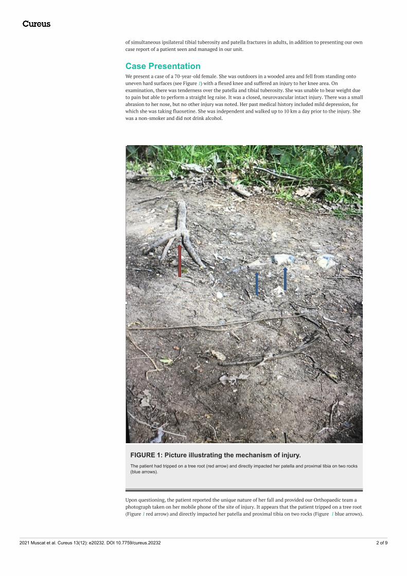
Case PresentationWe present a case of a 70-year-old female. She was outdoors in a wooded area and fell from standing ontouneven hard surfaces (see Figure 1) with a flexed knee and suffered an injury to her knee area. Onexamination, there was tenderness over the patella and tibial tuberosity. She was unable to bear weight dueto pain but able to perform a straight leg raise. It was a closed, neurovascular intact injury. There was a smallabrasion to her nose, but no other injury was noted. Her past medical history included mild depression, forwhich she was taking fluoxetine. She was independent and walked up to 10 km a day prior to the injury. Shewas a non-smoker and did not drink alcohol.
FIGURE 1: Picture illustrating the mechanism of injury.The patient had tripped on a tree root (red arrow) and directly impacted her patella and proximal tibia on two rocks(blue arrows).
Upon questioning, the patient reported the unique nature of her fall and provided our Orthopaedic team aphotograph taken on her mobile phone of the site of injury. It appears that the patient tripped on a tree root(Figure 1 red arrow) and directly impacted her patella and proximal tibia on two rocks (Figure 1 blue arrows).
2021 Muscat et al. Cureus 13(12): e20232. DOI 10.7759/cureus.20232 2 of 9
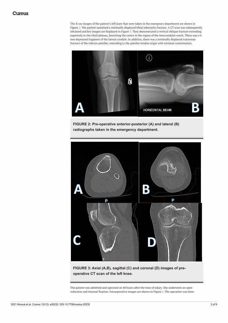
The X-ray images of the patient’s left knee that were taken in the emergency department are shown inFigure 2. The patient sustained a minimally displaced tibial tuberosity fracture. A CT scan was subsequentlyobtained and key images are displayed in Figure 3. They demonstrated a vertical oblique fracture extendingsuperiorly to the tibial plateau, breeching the cortex in the region of the intercondylar notch. There was a 6mm depressed fragment of the lateral condyle. In addition, there was a minimally displaced transversefracture of the inferior patellar, extending to the patellar tendon origin with minimal comminution.
FIGURE 2: Pre-operative anterior-posterior (A) and lateral (B)radiographs taken in the emergency department.
FIGURE 3: Axial (A,B), sagittal (C) and coronal (D) images of pre-operative CT scan of the left knee.
The patient was admitted and operated on 48 hours after the time of injury. She underwent an openreduction and internal fixation. Intraoperative images are shown in Figure 4. The operation was done
2021 Muscat et al. Cureus 13(12): e20232. DOI 10.7759/cureus.20232 3 of 9
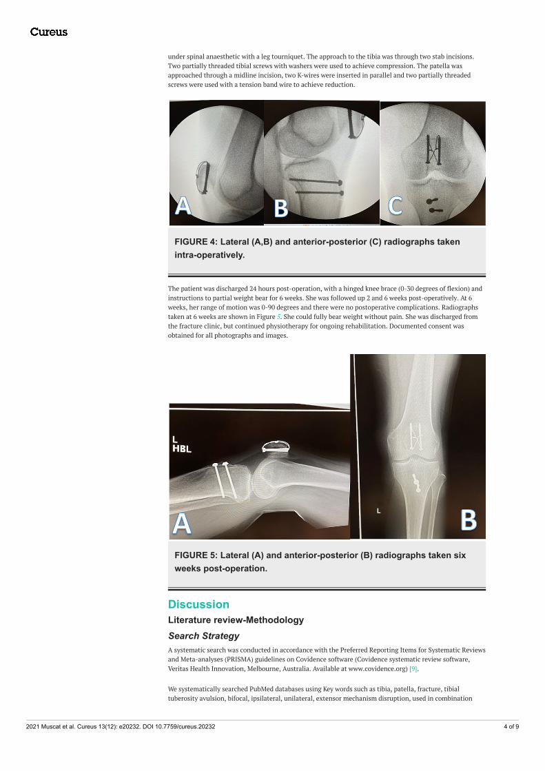
under spinal anaesthetic with a leg tourniquet. The approach to the tibia was through two stab incisions.Two partially threaded tibial screws with washers were used to achieve compression. The patella wasapproached through a midline incision, two K-wires were inserted in parallel and two partially threadedscrews were used with a tension band wire to achieve reduction.
FIGURE 4: Lateral (A,B) and anterior-posterior (C) radiographs takenintra-operatively.
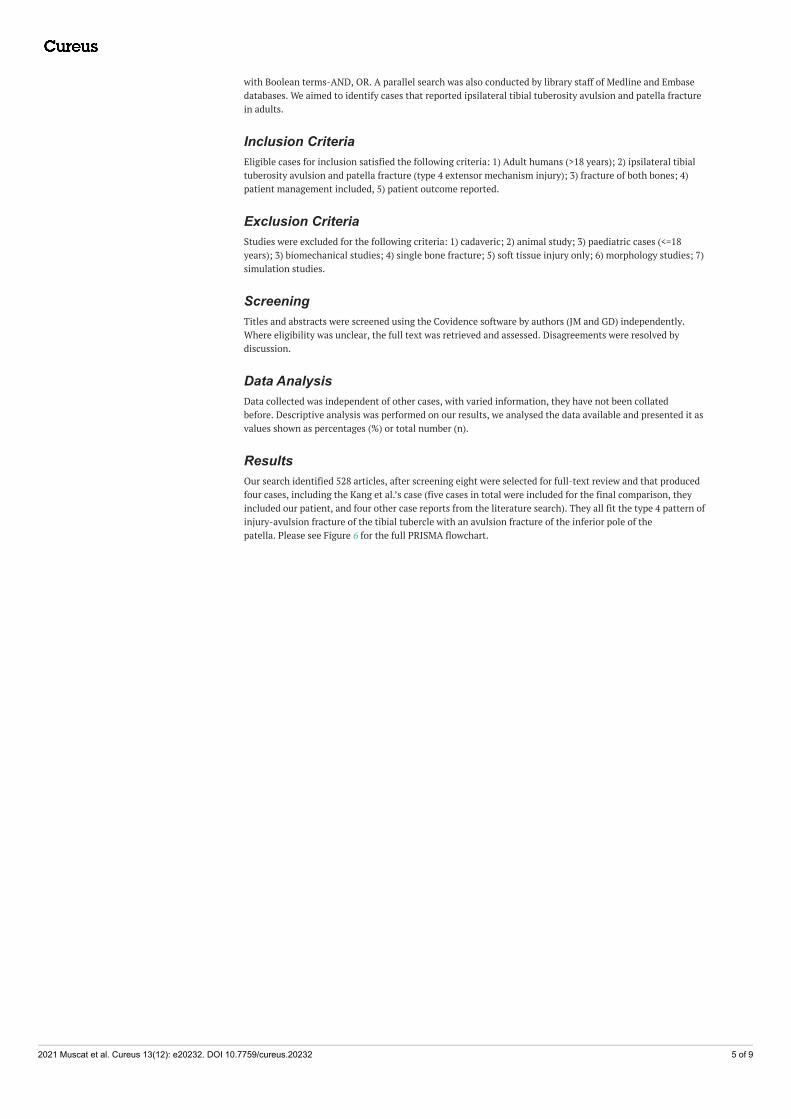
The patient was discharged 24 hours post-operation, with a hinged knee brace (0-30 degrees of flexion) andinstructions to partial weight bear for 6 weeks. She was followed up 2 and 6 weeks post-operatively. At 6weeks, her range of motion was 0-90 degrees and there were no postoperative complications. Radiographstaken at 6 weeks are shown in Figure 5. She could fully bear weight without pain. She was discharged fromthe fracture clinic, but continued physiotherapy for ongoing rehabilitation. Documented consent wasobtained for all photographs and images.
FIGURE 5: Lateral (A) and anterior-posterior (B) radiographs taken sixweeks post-operation.
DiscussionLiterature review-MethodologySearch StrategyA systematic search was conducted in accordance with the Preferred Reporting Items for Systematic Reviewsand Meta-analyses (PRISMA) guidelines on Covidence software (Covidence systematic review software,Veritas Health Innovation, Melbourne, Australia. Available at www.covidence.org) [9].
We systematically searched PubMed databases using Key words such as tibia, patella, fracture, tibialtuberosity avulsion, bifocal, ipsilateral, unilateral, extensor mechanism disruption, used in combination
2021 Muscat et al. Cureus 13(12): e20232. DOI 10.7759/cureus.20232 4 of 9
with Boolean terms-AND, OR. A parallel search was also conducted by library staff of Medline and Embasedatabases. We aimed to identify cases that reported ipsilateral tibial tuberosity avulsion and patella fracturein adults.
Inclusion CriteriaEligible cases for inclusion satisfied the following criteria: 1) Adult humans (>18 years); 2) ipsilateral tibialtuberosity avulsion and patella fracture (type 4 extensor mechanism injury); 3) fracture of both bones; 4)patient management included, 5) patient outcome reported.
Exclusion CriteriaStudies were excluded for the following criteria: 1) cadaveric; 2) animal study; 3) paediatric cases (<=18years); 3) biomechanical studies; 4) single bone fracture; 5) soft tissue injury only; 6) morphology studies; 7)simulation studies.
ScreeningTitles and abstracts were screened using the Covidence software by authors (JM and GD) independently.Where eligibility was unclear, the full text was retrieved and assessed. Disagreements were resolved bydiscussion.
Data AnalysisData collected was independent of other cases, with varied information, they have not been collatedbefore. Descriptive analysis was performed on our results, we analysed the data available and presented it asvalues shown as percentages (%) or total number (n).
ResultsOur search identified 528 articles, after screening eight were selected for full-text review and that producedfour cases, including the Kang et al.’s case (five cases in total were included for the final comparison, theyincluded our patient, and four other case reports from the literature search). They all fit the type 4 pattern ofinjury-avulsion fracture of the tibial tubercle with an avulsion fracture of the inferior pole of thepatella. Please see Figure 6 for the full PRISMA flowchart.
2021 Muscat et al. Cureus 13(12): e20232. DOI 10.7759/cureus.20232 5 of 9
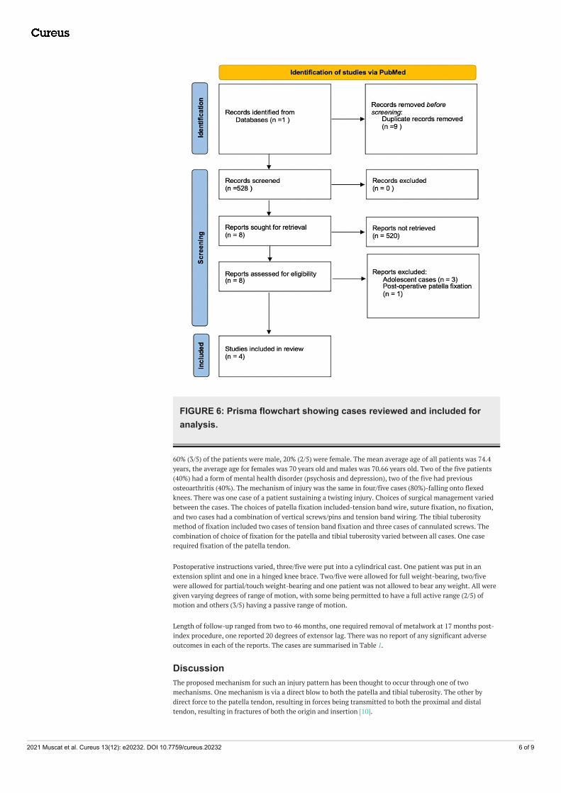
FIGURE 6: Prisma flowchart showing cases reviewed and included foranalysis.
60% (3/5) of the patients were male, 20% (2/5) were female. The mean average age of all patients was 74.4years, the average age for females was 70 years old and males was 70.66 years old. Two of the five patients(40%) had a form of mental health disorder (psychosis and depression), two of the five had previousosteoarthritis (40%). The mechanism of injury was the same in four/five cases (80%)-falling onto flexedknees. There was one case of a patient sustaining a twisting injury. Choices of surgical management variedbetween the cases. The choices of patella fixation included-tension band wire, suture fixation, no fixation,and two cases had a combination of vertical screws/pins and tension band wiring. The tibial tuberositymethod of fixation included two cases of tension band fixation and three cases of cannulated screws. Thecombination of choice of fixation for the patella and tibial tuberosity varied between all cases. One caserequired fixation of the patella tendon.
Postoperative instructions varied, three/five were put into a cylindrical cast. One patient was put in anextension splint and one in a hinged knee brace. Two/five were allowed for full weight-bearing, two/fivewere allowed for partial/touch weight-bearing and one patient was not allowed to bear any weight. All weregiven varying degrees of range of motion, with some being permitted to have a full active range (2/5) ofmotion and others (3/5) having a passive range of motion.
Length of follow-up ranged from two to 46 months, one required removal of metalwork at 17 months post-index procedure, one reported 20 degrees of extensor lag. There was no report of any significant adverseoutcomes in each of the reports. The cases are summarised in Table 1.
DiscussionThe proposed mechanism for such an injury pattern has been thought to occur through one of twomechanisms. One mechanism is via a direct blow to both the patella and tibial tuberosity. The other bydirect force to the patella tendon, resulting in forces being transmitted to both the proximal and distaltendon, resulting in fractures of both the origin and insertion [10].
2021 Muscat et al. Cureus 13(12): e20232. DOI 10.7759/cureus.20232 6 of 9
Chautems et al. [11] were the first to report on such a double bony avulsion fracture in an adult. Themechanism of injury was a fall onto the knee in a 90-year-old female. Concurrent opposing forces on theligament in the context of osteoporotic bone quality were described as the cause of the bifocaldisruption. This paper required translation into English.
Yoon et al. [12] described a case in a 50-year-old who sustained an avulsion fracture of the patella and tibialtuberosity in an ectopic ossified patella tendon following a twisting injury. They suggest that the geometryof the fracture pattern could be related to the ectopic ossified patella tendon.
Kang et al.’s [8] case was that of an 84-year-old male who was involved in a motorcycle accident. They alsoattribute the injury pattern to a direct force onto the patella tendon in a highly flexed knee, resulting inforce to both the proximal and distal parts of the tendon, which led to the avulsion fractures. More recently,MacDonald et al. [13] also describe a case of a type 4 injury in a 56-year-old male, who also fell onto a flexedknee. They suggest that poor bone quality, due to smoking and antipsychotic medication history, attributedto the injury pattern.
Our patient was relatively young and still very active, with no previously documented bone or kneepathology. It was noted that the patient was taking fluoxetine. Several studies have demonstrated anassociation between antidepressant use and bone loss in post-menopausal women [10,14]. This certainlycould have been an attributing factor, given the low energy mechanism of injury. When stress is applied tothe patella tendon, this increases strain at the bone-tendon interface of the tendon origin and insertion andcan result in an avulsion fracture, particularly in poor bone [13]. However, in our patient, there was clearevidence from the mechanism described by the patient of direct force being applied to both the patella andtibial tuberosity, rather than contraction of the patella tendon resulting in avulsions. Our patient’s fracturepattern was more severe than those previously described, including comminution of the patella, in additionto intraarticular extension of the tibial plateau. As noted, different management strategies were employed ineach case, but all reported satisfactory outcomes. The cases are summarised in Table 1.
2021 Muscat et al. Cureus 13(12): e20232. DOI 10.7759/cureus.20232 7 of 9
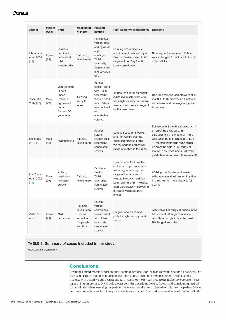
AuthorPatient(Age)
PMHMechanismof injury
Fixationmethod
Post operative instructions Outcome
Chautemset al. 2001[11]
Female(90)
Diabetic—non-insulin -dependent,mildosteoarthritis
Fall ontoflexed knee
Patella: twovertical pinsand figure-of-eightcerclage.Tibialtuberosity:three staplesand cerclagewire
Loading under extensionsplint protection from Day 4.Passive flexion limited to 60degrees from Day 8 untilbone consolidation
No complication reported. Patientwas walking at 6 months with the aidof two sticks
Yoon et al.2007 [12]
Male(72)
Osteoarthritisin bothknees.Previousright-sidedfemurfracture 50years ago
Twistinginjury toknee
Patella:tension bandwire Tibialtuberosity:tension bandwire. Patellatendon: fixedwithabsorbablesutures
Immobilised in full extensioncylindrical plaster cast withfull weight-bearing for severalweeks, then passive range ofmotion exercises
Required removal of metalwork at 17months. At 46 months, no functionalimpairment and radiological signs ofbony union
Kang et al.2013 [8]
Male(84)
HypertensionFall ontoflexed knee
Patella:suturefixation Tibialtuberosity:cannulatedscrews
Long leg cast for 6 weeksand non-weight-bearing.Then commenced partialweight-bearing and activerange of motion in the knee
Follow-up at 2 months showed bonyunion of the tibia, but 2 mmdisplacement of the patella. Therewas 20 degrees of extensor lag. At12 months, there was radiologicalunion of the patella, full range ofmotion in the knee and a Saltzmanpatellofemoral score of 92 (excellent)
MacDonaldet al. 2021[13]
Male(56)
Autism,psychosis,long-termsmoker
Fall ontoflexed knee
Patella: nofixation.Tibialtuberosity:cannulatedscrews
Cylinder cast for 2 weeks,and later hinged knee bracefollowing, increasing therange of flexion every 2weeks. Toe-touch weight-bearing for the first 4 weeks,then progressively allowed toincrease weight-bearingstatus
Walking comfortably at 8 weekswithout aids and full range of motionin the knee. At 1 year, back to fullactivity
Author’scase
Female(70)
Milddepression
Fall ontoflexed knee—directimpact tothe patellaand tibia
Patella:verticalscrews andtension bandwire. Tibialtuberosity:cannulatedscrews
Hinged knee brace andpartial weight-bearing for 6weeks
At 6 weeks her range of motion in theknee was 0-90 degrees and shecould bear weight fully with no aids.Discharged from clinic
TABLE 1: Summary of cases included in the study.PMH: past medical history
ConclusionsGiven the limited reports of such injuries, common protocols for the management in adults do not exist. Ourcase demonstrates that open reduction and internal fixation of both the tibial tuberosity and patellafracture, with partial weight-bearing and restricted knee flexion can produce a satisfactory outcome. Thesetypes of injuries are rare. One should always consider underlying bone pathology and contributing medicalco-morbidities when assessing the patient. Understanding the mechanism of exactly how the patient fell canhelp understand how such an injury may have been sustained. Open reduction and internal fixation of both
2021 Muscat et al. Cureus 13(12): e20232. DOI 10.7759/cureus.20232 8 of 9
the tibial tuberosity and patella, with partial weight-bearing and reduced knee flexion is one potentialmanagement strategy for such an injury. Further research is required to further establish a recommendedtreatment method based on comparable outcomes.
Additional InformationDisclosuresHuman subjects: Consent was obtained or waived by all participants in this study. Conflicts of interest: Incompliance with the ICMJE uniform disclosure form, all authors declare the following: Payment/servicesinfo: All authors have declared that no financial support was received from any organization for thesubmitted work. Financial relationships: All authors have declared that they have no financialrelationships at present or within the previous three years with any organizations that might have aninterest in the submitted work. Other relationships: All authors have declared that there are no otherrelationships or activities that could appear to have influenced the submitted work.
References1. McKoy BE, Stanitski CL : Acute tibial tubercle avulsion fractures . Orthop Clin North Am. 2003, 34:397-403.
10.1016/S0030-5898(02)00061-52. Raad M, Ndlovu S, Hǿgsand T, Ahmed S, Norris M: Fracture of tibial tuberosity in an adult with Paget's
disease of the bone - An interesting case and review of literature. Trauma Case Rep. 2021, 32:100440.10.1016/j.tcr.2021.100440
3. Matthews B, Hazratwala K, Barroso-Rosa S: Comminuted patella fracture in elderly patients: a systematicreview and case report. Geriatr Orthop Surg Rehabil. 2017, 8:135-44. 10.1177/2151458517710517
4. Deopujari S, Kiel J: Knee Extensor Mechanism Injuries. StatPearls (ed): StatPearls Publishing, TreasureIsland, FL; 2020. https://europepmc.org/article/nbk/nbk554587.
5. Gwinner C, Märdian S, Schwabe P, Schaser KD, Krapohl BD, Jung TM: Current concepts review: fractures ofthe patella. GMS Interdiscip Plast Reconstr Surg DGPW. 2016, 5:1-15. 10.3205/iprs000080
6. Serino J, Mohamadi A, Orman S, et al.: Comparison of adverse events and postoperative mobilizationfollowing knee extensor mechanism rupture repair: a systematic review and network meta-analysis. Injury.2017, 48:2793-9. 10.1016/j.injury.2017.10.013
7. Bostrom MP, Asnis SE, Ernberg JJ, Wright TM, Giddings VL, Berberian WS, Missri AA: Fatigue testing ofcerclage stainless steel wire fixation. J Orthop Trauma. 1994, 8:422-8. 10.1097/00005131-199410000-00009
8. Kang S, Chung PH, Kim YS, Lee HM, Kim JP: Bifocal disruption of the knee extensor mechanism: a casereport and literature review. Arch Orthop Trauma Surg. 2013, 133:517-21. 10.1007/s00402-013-1696-7
9. Covidence systematic review software, Veritas Health Innovation, Melbourne, Australia . (2014). Accessed:November 9, 2021: https://www.covidence.org/.
10. Bolton JM, Targownik LE, Leung S, Sareen J, Leslie WD: Risk of low bone mineral density associated withpsychotropic medications and mental disorders in postmenopausal women. J Clin Psychopharmacol. 2011,31:56-60. 10.1097/JCP.0b013e3182075587
11. Chautems R, Michel J, Barraud GE, Burdet A: Avulsion osseuse bifocale du tendon rotulien chez un adulte: àpropos d'un cas [Bifocal avulsion of the patellar tendon in an adult: a case report]. Revue de chirurgieorthopedique et reparatrice de l'appareil moteur. 2001, 87:388-91.
12. Yoon JR, Kim TS, Kim HJ, Noh HK, Oh JK, Yoo JC: Simultaneous patellar tendon avulsion fracture from bothpatella and tibial tuberosity: a case report. Knee Surg Sports Traumatol Arthrosc. 2007, 15:225-7.10.1007/s00167-006-0168-9
13. MacDonald DR, Neilly DW, Stevenson I: Simultaneous ipsilateral tibial tuberosity and patellar fractures inan adult patient. J Clin Orthop Trauma. 2021, 14:P139-41. 10.1016/j.jcot.2020.06.010
14. Diem SJ, Blackwell TL, Stone KL, Yaffe K, Haney EM, Bliziotes MM, Ensrud KE: Use of antidepressants andrates of hip bone loss in older women: the study of osteoporotic fractures. Arch Intern Med. 2007, 167:1240-5. 10.1001/archinte.167.12.1240
2021 Muscat et al. Cureus 13(12): e20232. DOI 10.7759/cureus.20232 9 of 9