Mechanism of CuA assembly
8
Mechanism of Cu A assembly Luciano A Abriata 1,2 , Lucia Banci 2 , Ivano Bertini 2 , Simone Ciofi-Baffoni 2 , Petros Gkazonis 2,3 , Georgios A Spyroulias 3 , Alejandro J Vila 1 , and Shenlin Wang 2 1Instituto de Biología Molecular y Celular de Rosario, Consejo Nacional de Investigaciones Científicas y Técnicas, Facultad de Ciencias Bioquímicas y Farmacéuticas, Universidad Nacional de Rosario, Suipacha 531, (S2002LRK) Rosario, Argentina. 2Magnetic Resonance Center CERM and Department of Chemistry, University of Florence, Scientific Campus, 50019, Sesto Fiorentino, Florence, Italy. 3Department of Pharmacy, University of Patras, Panepistimioupoli-Rion, GR-26504 Patras, Greece. Abstract Copper is essential for proper functioning of cytochrome c oxidases, and therefore for cellular respiration in eukaryotes and many bacteria. Here we show that a new periplasmic protein (PCu A C) selectively inserts Cu(I) ions into subunit II of Thermus thermophilus ba 3 oxidase to generate a native Cu A site. The purported metallochaperone Sco1 is unable to deliver copper ions; instead, it works as a thiol-disulfide reductase to maintain the correct oxidation state of the Cu A cysteine ligands. Cu A is a dinuclear copper site within the soluble domain of subunit II (Cox2) of bacterial and eukaryotic cytochrome c oxidases (CcOs), whose function is to convey electrons from a soluble cytochrome c to the catalytic heme a 3 -Cu B center of CcO (refs. 1,2). The proper assembly of the Cu A site is essential for the catalytic machinery of a functional oxidase. Several proteins have been identified as key players in the delivery of metal ions to the Cu A site3, but the detailed molecular mechanisms and the specific roles of each protein are poorly understood4. In prokaryotes two protein families have been proposed to be involved in Cu A site formation. The first includes proteins that are able to bind Cu(I) through methionine and histidine residues arranged in a highly conserved H(M)X 10 MX 21 HXM motif 5 (referred to as periplasmic Cu A chaperone (PCu A C) hereafter). The second consists of the Sco proteins, whose mechanism of action in Cu A assembly as thioredoxins or metallochaperones is still debated6. These proteins (PCu A C and Sco) are often found in the same bacterial operon, and most of the identified operons that encode Sco also contain a gene for Cox2 (ref. 7). PCu A C and Sco proteins occur together in the Vibrio cholerae bacterium (http://string.embl.de). The Cu A -containing subunit II from Thermus thermophilus ba 3 oxidase (Tt Cu A hereafter), T. thermophilus PCu A C (Tt PCu A C) and T. thermophilus Sco1 (Tt Sco1), which are all located in the bacterial periplasm, were expressed as truncated soluble versions in which the transmembrane helices (and the signal peptide region in Tt PCu A C) were not included, thus Correspondence should be addressed to I.B. ([email protected]). AUTHOR CONTRIBUTIONS L.A.A. cloned and expressed Tt Sco1 and Tt Cu A proteins and contributed to the NMR structure of Tt Sco1 and NMR titrations; P.G. cloned and expressed PCu A C protein and performed redox experiments; S.W. solved the structures of PCu A C and contributed to the NMR titrations and NMR structure of Tt Sco1; S.C.-B. supervised and coordinated the acquisition of NMR data and structures, and the performance of titration experiments; I.B., L.B., G.A.S. and A.J.V. developed and directed the project and contributed to the writing. All authors were involved in the discussion of the biochemical meaning of the experiments. Published online at http://www.nature.com/naturechemicalbiology/ Reprints and permissions information is available online at http://npg.nature.com/reprintsandpermissions/ NIH Public Access Author Manuscript Nat Chem Biol. Author manuscript; available in PMC 2009 April 1. Published in final edited form as: Nat Chem Biol. 2008 October ; 4(10): 599–601. doi:10.1038/nchembio.110. NIH-PA Author Manuscript NIH-PA Author Manuscript NIH-PA Author Manuscript
Transcript of Mechanism of CuA assembly

Mechanism of CuA assembly
Luciano A Abriata1,2, Lucia Banci2, Ivano Bertini2, Simone Ciofi-Baffoni2, PetrosGkazonis2,3, Georgios A Spyroulias3, Alejandro J Vila1, and Shenlin Wang2
1Instituto de Biología Molecular y Celular de Rosario, Consejo Nacional de Investigaciones Científicas yTécnicas, Facultad de Ciencias Bioquímicas y Farmacéuticas, Universidad Nacional de Rosario, Suipacha531, (S2002LRK) Rosario, Argentina.
2Magnetic Resonance Center CERM and Department of Chemistry, University of Florence, ScientificCampus, 50019, Sesto Fiorentino, Florence, Italy.
3Department of Pharmacy, University of Patras, Panepistimioupoli-Rion, GR-26504 Patras, Greece.
AbstractCopper is essential for proper functioning of cytochrome c oxidases, and therefore for cellularrespiration in eukaryotes and many bacteria. Here we show that a new periplasmic protein (PCuAC)selectively inserts Cu(I) ions into subunit II of Thermus thermophilus ba3 oxidase to generate a nativeCuA site. The purported metallochaperone Sco1 is unable to deliver copper ions; instead, it worksas a thiol-disulfide reductase to maintain the correct oxidation state of the CuA cysteine ligands.
CuA is a dinuclear copper site within the soluble domain of subunit II (Cox2) of bacterial andeukaryotic cytochrome c oxidases (CcOs), whose function is to convey electrons from a solublecytochrome c to the catalytic heme a3-CuB center of CcO (refs. 1,2). The proper assembly ofthe CuA site is essential for the catalytic machinery of a functional oxidase. Several proteinshave been identified as key players in the delivery of metal ions to the CuA site3, but the detailedmolecular mechanisms and the specific roles of each protein are poorly understood4. Inprokaryotes two protein families have been proposed to be involved in CuA site formation. Thefirst includes proteins that are able to bind Cu(I) through methionine and histidine residuesarranged in a highly conserved H(M)X10MX21HXM motif 5 (referred to as periplasmic CuAchaperone (PCuAC) hereafter). The second consists of the Sco proteins, whose mechanism ofaction in CuA assembly as thioredoxins or metallochaperones is still debated6. These proteins(PCuAC and Sco) are often found in the same bacterial operon, and most of the identifiedoperons that encode Sco also contain a gene for Cox2 (ref. 7). PCuAC and Sco proteins occurtogether in the Vibrio cholerae bacterium (http://string.embl.de).
The CuA-containing subunit II from Thermus thermophilus ba3 oxidase (Tt CuA hereafter), T.thermophilus PCuAC (Tt PCuAC) and T. thermophilus Sco1 (Tt Sco1), which are all locatedin the bacterial periplasm, were expressed as truncated soluble versions in which thetransmembrane helices (and the signal peptide region in Tt PCuAC) were not included, thus
Correspondence should be addressed to I.B. ([email protected]).AUTHOR CONTRIBUTIONSL.A.A. cloned and expressed Tt Sco1 and Tt CuA proteins and contributed to the NMR structure of Tt Sco1 and NMR titrations; P.G.cloned and expressed PCuAC protein and performed redox experiments; S.W. solved the structures of PCuAC and contributed to theNMR titrations and NMR structure of Tt Sco1; S.C.-B. supervised and coordinated the acquisition of NMR data and structures, and theperformance of titration experiments; I.B., L.B., G.A.S. and A.J.V. developed and directed the project and contributed to the writing. Allauthors were involved in the discussion of the biochemical meaning of the experiments.Published online at http://www.nature.com/naturechemicalbiology/Reprints and permissions information is available online at http://npg.nature.com/reprintsandpermissions/
NIH Public AccessAuthor ManuscriptNat Chem Biol. Author manuscript; available in PMC 2009 April 1.
Published in final edited form as:Nat Chem Biol. 2008 October ; 4(10): 599–601. doi:10.1038/nchembio.110.
NIH
-PA Author Manuscript
NIH
-PA Author Manuscript
NIH
-PA Author Manuscript

resulting in constructs of 136, 172 and 121 amino acids, respectively (Supplementary Methodsand Supplementary Fig. 1 online).
Tt PCuAC binds one equivalent of Cu(I), as also observed for the homologous protein DR1885(ref. 5). The Cu(I) binding affinity, measured by competition experiments with DTT followedby 1H-15N HSQC, is (2.2 ± 0.1) × 10−13 M (Supplementary Fig. 2, Supplementary Data andSupplementary Methods online). Upon addition of Cu(I) up to a 1:1 metal/protein ratio, themost affected region includes residues His46, Met61, Met83 and His85 and the neighboringresidues (Supplementary Fig. 2), thus defining the copper binding ligands (similar to theMet3His ligand set found for DR1885 in homologous positions).
Sco proteins, in contrast to the Tt PCuAC protein family, are able to bind both Cu(I) and Cu(II) ions8–10. Cu(I) binding followed by NMR confirmed the involvement of metal ligandsconserved in all Sco1 proteins (Cys47, Cys51 and His137) (Supplementary Fig. 2 andSupplementary Data). Apo-Tt Sco1 showed weaker Cu(I) binding capabilities compared withits eukaryotic homologs (a lower limit of ~10−10 M was estimated for the Kd of Cu(I)–Tt Sco1).Cu(II)–Tt Sco1 presents spectroscopic features resembling those of the human and Bacillussubtilis homologs8,10,11 (Supplementary Data and Supplementary Fig. 3 online).
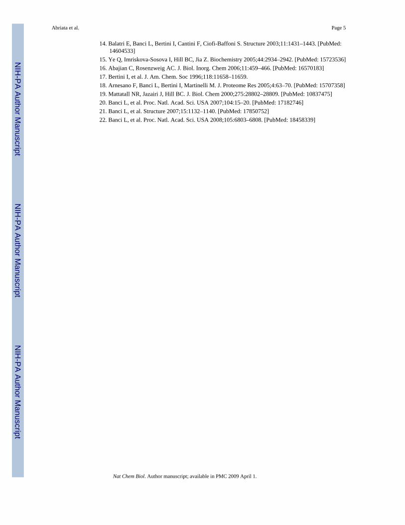
The solution structures of apo-Tt PCuAC, Cu(I)–Tt PCuAC and apo-Tt Sco1 were determinedusing NMR-derived restraints (Supplementary Methods). The NMR spectra of apo- and Cu(I)–Tt PCuAC variants show two sets of signals in a 65:35 ratio (Supplementary Fig. 3) for 23and 24 residues, respectively, which originate from a cis-trans isomerization of the Gly13-Pro14 peptide bond. The two structures resulting from this isomerism in both apo- and Cu(I)–Tt PCuAC were obtained (Fig. 1a–d and Supplementary Tables 1 and 2 online). The structuraldifferences induced by this isomerization are localized in loops 1 and 8 and do not affect Cu(I) binding (Fig. 1a–d). Tt PCuAC is arranged in a cupredoxin-like fold (Fig. 1a–d), except thatstrands µ4 and µ5 form an extended, flexible solvent-exposed µ-hairpin that is longer than theone reported for the homologous DR1885 protein5. The Cu(I) ion is coordinated in a tetrahedralarrangement to the sulfur atoms of Met61 and Met85, the Nδ 1 atom of His46 and the Nε2atom of His83 (Fig. 1a,c). The structure of apo-Tt Sco1 in the reduced form adopts thethioredoxin-like fold already observed for all Sco1 homologs12–16 (Fig. 1e andSupplementary Table 3 online). Loop 8 (which includes the metal ligand His137) adopts anextended conformation in Tt Sco1 (Fig. 1e) resembling that observed for the human and yeastproteins, in contrast with the shorter (and less extended) loop 8 present in Bs Sco1. The cysteinemetal ligands are solvent exposed (as in other Sco1 proteins), which is in agreement with theobservation of rapid air oxidation.
The availability of the NMR resonance assignments of the apo and the metallated forms of thetwo possible copper donors, as well as that of the CuA-containing soluble fragment of the T.thermophilus ba3 oxidase, allowed us to investigate, through NMR, copper uptake by theCuA fragment. We simultaneously monitored the occurrence of copper transfer and theformation of the correct metallated form, and also identified the copper donor protein, bydetecting the resulting apo state of the protein that had transferred the copper ions. This strategyalso allows the identification of possible transient intermediates.
Addition of Cu(I) to reduced 15N apo-Tt CuA under anaerobic conditions gives rise to the fullymetallated protein in the reduced state (Supplementary Data and Supplementary Fig. 4 online).Exposure of this species to oxygen resulted in the formation of a purple species with thecharacteristic electronic spectrum and 1H NMR signals of the oxidized, mixed-valence CuAcenter (Supplementary Fig. 4)17. This indicates that the CuA center can be formed in vitrowithout the assistance of any protein when the cysteine residues of the CuA center are reduced.The affinity of both copper ions in Tt CuA is in the femtomolar range based on competition
Abriata et al. Page 2
Nat Chem Biol. Author manuscript; available in PMC 2009 April 1.
NIH
-PA Author Manuscript
NIH
-PA Author Manuscript
NIH
-PA Author Manuscript

studies with DTT (Supplementary Methods) and is thus higher than that of Tt PCuAC and TtSco1. However, because Cu(I) is not freely available in the periplasmic space, a Cu(I)chaperone is needed to deliver two Cu(I) ions to the apo-CuA protein.
We initially explored the possible role of Sco1 as a Cu(I) or Cu(II) donor to apo-CuA. Noevidence of copper uptake by the CuA protein or of metal depletion of Tt Sco1 was observed,which suggests that Tt Sco1 is not responsible for the direct delivery of Cu(I) or Cu(II) ionsinto apo-Tt CuA.
Genomic studies have suggested that Tt PCuAC homologs could be involved in copper transportin the periplasmic space of bacteria5,18, and we decided to test this previously unexploredhypothesis. When apo-Tt CuA was added to a solution of 15N Cu(I)–Tt PCuAC, a new set ofresonances corresponding to apo-Tt PCuAC was observed (Fig. 2a). A step-wise titration ledto the progressive disappearance of the signals corresponding to Cu(I)–Tt PCuAC, with theconcomitant increase of signals from the apo form (Fig. 2a). This process was complete at aTt CuA/Tt PCuAC ratio of 1:2 (Fig. 2a). The complementary experiment to characterize themetallated species was performed by adding two equivalents of unlabeled Cu(I)–Tt PCuACto 15N apo-Tt CuA, which led to the typical 1H-15N HSQC pattern of native Cu(I)2–Tt CuA(Fig. 2b). A step-wise titration disclosed the formation of an intermediate species when lessthan one equivalent of Cu(I)–Tt PCuAC was added to the CuA domain (Fig. 2b,c). This speciesthen converted into the dimetallated Cu(I)2–Tt CuA upon further addition of the donor protein(Fig. 2c). This intermediate species displayed resonances with chemical shifts differing fromthose of both the apo- and the fully metallated species (Fig. 2b), and it did not show any linebroadening with respect to the other forms of the CuA domain. This behavior, together withthe observation that no intermediate was identified by monitoring the copper release from Cu(I)–Tt PCuAC to apo-Tt CuA, allowed us to rule out the detection of a possible complex betweenthe two proteins, and suggested that this intermediate corresponds to a singly metallated CuAspecies. We conclude that Tt PCuAC is capable of transferring two Cu(I) ions to the reducedapo-Tt CuA site sequentially, thereby eliciting the formation of the binuclear CuA center in thereduced state (Fig. 2c). Aerobic oxidation of this mixture leads to the mixed-valence, oxidizedCuA center, as revealed by its characteristic electronic spectrum (Supplementary Fig. 4).
Sco1 is able to bind Cu(II) ions. The possible direct transfer of Cu(II) ions was explored byfollowing the titration of a sample of 15N-labeled Cu(II)–Tt Sco1 with unlabeled apo-Tt CuA.There was no evidence of the formation of apo-Tt Sco1 in the 1H-15N HSQC spectra, thusrevealing the inability of this protein to transfer Cu(II) ions to the CuA domain. When 2equivalents of unlabeled Cu(I)–Tt PCuAC were added to the mixture of 15N-labeled Cu(II)–Tt Sco1 and 15N-labeled apo-Tt CuA, the reduced Cu(I)2–Tt CuA center was formed(Supplementary Fig. 5 online). This result indicates that (i) the presence of Cu(II)–Tt Sco1does not prevent Cu(I)–Tt PCuAC from transferring Cu(I), and (ii) as the final product is thereduced Cu(I)2–Tt CuA center, the mechanism cannot involve the concerted action of both Cu(II)–Tt Sco1 and Cu(I)–Tt PCuAC, which would give rise to a mixed-valence, oxidized CuAsite.
Sco1, which has already been shown to be essential for copper upload into the CuA site in B.subtilis19, is characterized by a thioredoxin-like fold. We therefore investigated the possiblethiol-disulfide oxidoreductase activity of Tt Sco1 during copper uptake. Oxidized apo-TtCuA, in which the two cysteine ligands had been aerobically oxidized to form a disulphidebond (ox-apo-Tt CuA hereafter), was not able to bind copper ions when Cu(I)–Tt PCuAC wasadded. However, when 15N-labeled ox-apo-Tt CuA was titrated with a 2:1 mixture of Cu(I)–Tt PCuAC and reduced apo-Tt Sco1, the backbone resonances of the native Cu(I)2–Tt CuAappeared (Fig. 3a). Exposure of the reaction sample to atmospheric oxygen allowed thedevelopment of the typical UV-vis spectrum of the oxidized, mixed-valence CuA center, which
Abriata et al. Page 3
Nat Chem Biol. Author manuscript; available in PMC 2009 April 1.
NIH
-PA Author Manuscript
NIH
-PA Author Manuscript
NIH
-PA Author Manuscript

confirmed that the native site had formed. A 4-acetamide-4′- amleimidylstilbene-2,2′-disulfonic acid, disodium salt (AMS)-reacted SDS gel of the reaction mixture (run undernonreducing conditions, Supplementary Methods) indicated that, at a Tt CuA/Tt Sco1/TtPCuAC ratio of 1:1:2, Tt Sco1 is mostly in the oxidized state (Fig. 3b). Similarly, in a 1:1 TtCuA/Tt Sco1 mixture, Tt Sco1 is essentially in an oxidized state, whereas Tt CuA is in a reducedstate (Fig. 3b). These experiments show that Tt Sco1 is able to reduce the disulfide bond of ox-apo-Tt CuA protein, which demonstrates that this protein behaves as a thiol-disulfide reductasein in vitro CuA assembly.
Although it is possible that the in vivo pathway is more complex, the structural characterizationof Tt PCuAC and of its copper binding properties and its ability to selectively and sequentiallydeliver two Cu(I) ions to apo-Tt CuA, thus giving rise to the native Cu(I)2–Tt CuA site invitro, strongly supports the annotation of this protein as a periplasmic Cu(I) chaperone. Ourdata also indicate that Tt Sco1 is able to reduce the disulfide bond of the CuA center, thusallowing the CuA site to accept Cu(I) ions from Tt PCuAC. The mechanism of bacterial CuAassembly therefore consists of a sequential insertion of two Cu(I) ions donated by ametallochaperone once the disulfide bond of the CuA center is reduced by a thioredoxin. Ineukaryotes the assembly of the CuA site is different, as Scos have a larger affinity for Cu(I)and may act as both chaperones and thioredoxins12,20–22.
Supplementary MaterialRefer to Web version on PubMed Central for supplementary material.
ACKNOWLEDGMENTSThis work was supported by the European Commission (European Network of Research Infrastructures for ProvidingAccess and Technological Advancements in Bio-NMR contract n° 026145, SPINE2-Complexes contract n° LSHG-CT-2006-031220 and Marie Curie host fellowships for early stage research training n° MEST-CT-2004-504391, NMRin Inorganic Structural Biology) and by a grant from Ente Cassa di Risparmio di Firenze. Work in Rosario (Argentina)was supported by the US National Institutes of Health (R01-GM068682), the Howard Hughes Medical Institute andAgencia Nacional de Promoción Científica y Tecnoló gica (PME2003-0026 and PICT2002-01-11625) grants to A.J.V.L.A.A. thanks Consejo Nacional de Investigaciones Científícas y Técnicas for a doctoral fellowship. We thank D.Winge (Departments of Medicine and Biochemistry, University of Utah Health Sciences Center, University of Utah)for kindly providing the expression plasmid for Tt Sco1.
References1. Ostermeier C, Iwata S, Michel H. Curr. Opin. Struct Biol 1996;6:460–466. [PubMed: 8794157]2. Maneg O, Malatesta F, Ludwig B, Drosou V. Biochim Biophys. Acta 2004;1655:274–281. [PubMed:
15100042]3. Carr HS, Winge DR. Acc. Chem. Res 2003;36:309–316. [PubMed: 12755640]4. Cobine PA, Pierrel F, Winge DR. Biochim. Biophys. Acta 2006;1763:759–772. [PubMed: 16631971]5. Banci L, et al. Proc. Natl. Acad. Sci. USA 2005;102:3994–3999. [PubMed: 15753304]6. Khalimonchuk O, Winge DR. Biochim. Biophys. Acta 2008;1783:618–628. [PubMed: 18070608]7. Banci L, Bertini I, Cavallaro G, Rosato A. J. Proteome Res 2007;6:1568–1579. [PubMed: 17300187]8. Horng YC, et al. J. Biol. Chem 2005;280:34113–34122. [PubMed: 16091356]9. McEwan AG, et al. FEBS Lett 2002;518:10–16. [PubMed: 11997009]10. Andruzzi L, Nakano M, Nilges MJ, Blackburn NJ. J. Am. Chem. Soc 2005;127:16548–16558.
[PubMed: 16305244]11. Imriskova-Sosova I, et al. Biochemistry 2005;44:16949–16956. [PubMed: 16363808]12. Banci L, et al. Proc. Natl. Acad. Sci. USA 2006;103:8595–8600. [PubMed: 16735468]13. Williams JC, et al. J. Biol. Chem 2005;280:15202–15211. [PubMed: 15659396]
Abriata et al. Page 4
Nat Chem Biol. Author manuscript; available in PMC 2009 April 1.
NIH
-PA Author Manuscript
NIH
-PA Author Manuscript
NIH
-PA Author Manuscript
14. Balatri E, Banci L, Bertini I, Cantini F, Ciofi-Baffoni S. Structure 2003;11:1431–1443. [PubMed:14604533]
15. Ye Q, Imriskova-Sosova I, Hill BC, Jia Z. Biochemistry 2005;44:2934–2942. [PubMed: 15723536]16. Abajian C, Rosenzweig AC. J. Biol. Inorg. Chem 2006;11:459–466. [PubMed: 16570183]17. Bertini I, et al. J. Am. Chem. Soc 1996;118:11658–11659.18. Arnesano F, Banci L, Bertini I, Martinelli M. J. Proteome Res 2005;4:63–70. [PubMed: 15707358]19. Mattatall NR, Jazairi J, Hill BC. J. Biol. Chem 2000;275:28802–28809. [PubMed: 10837475]20. Banci L, et al. Proc. Natl. Acad. Sci. USA 2007;104:15–20. [PubMed: 17182746]21. Banci L, et al. Structure 2007;15:1132–1140. [PubMed: 17850752]22. Banci L, et al. Proc. Natl. Acad. Sci. USA 2008;105:6803–6808. [PubMed: 18458339]
Abriata et al. Page 5
Nat Chem Biol. Author manuscript; available in PMC 2009 April 1.
NIH
-PA Author Manuscript
NIH
-PA Author Manuscript
NIH
-PA Author Manuscript
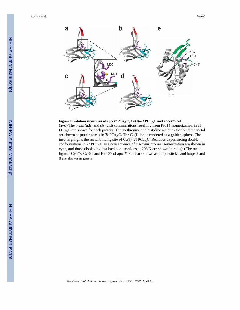
Figure 1. Solution structures of apo-Tt PCuAC, Cu(I)–Tt PCuAC and apo-Tt Sco1(a–d) The trans (a,b) and cis (c,d) conformations resulting from Pro14 isomerization in TtPCuAC are shown for each protein. The methionine and histidine residues that bind the metalare shown as purple sticks in Tt PCuAC. The Cu(I) ion is rendered as a golden sphere. Theinset highlights the metal binding site of Cu(I)–Tt PCuAC. Residues experiencing doubleconformations in Tt PCuAC as a consequence of cis-trans proline isomerization are shown incyan, and those displaying fast backbone motions at 298 K are shown in red. (e) The metalligands Cys47, Cys51 and His137 of apo-Tt Sco1 are shown as purple sticks, and loops 3 and8 are shown in green.
Abriata et al. Page 6
Nat Chem Biol. Author manuscript; available in PMC 2009 April 1.
NIH
-PA Author Manuscript
NIH
-PA Author Manuscript
NIH
-PA Author Manuscript
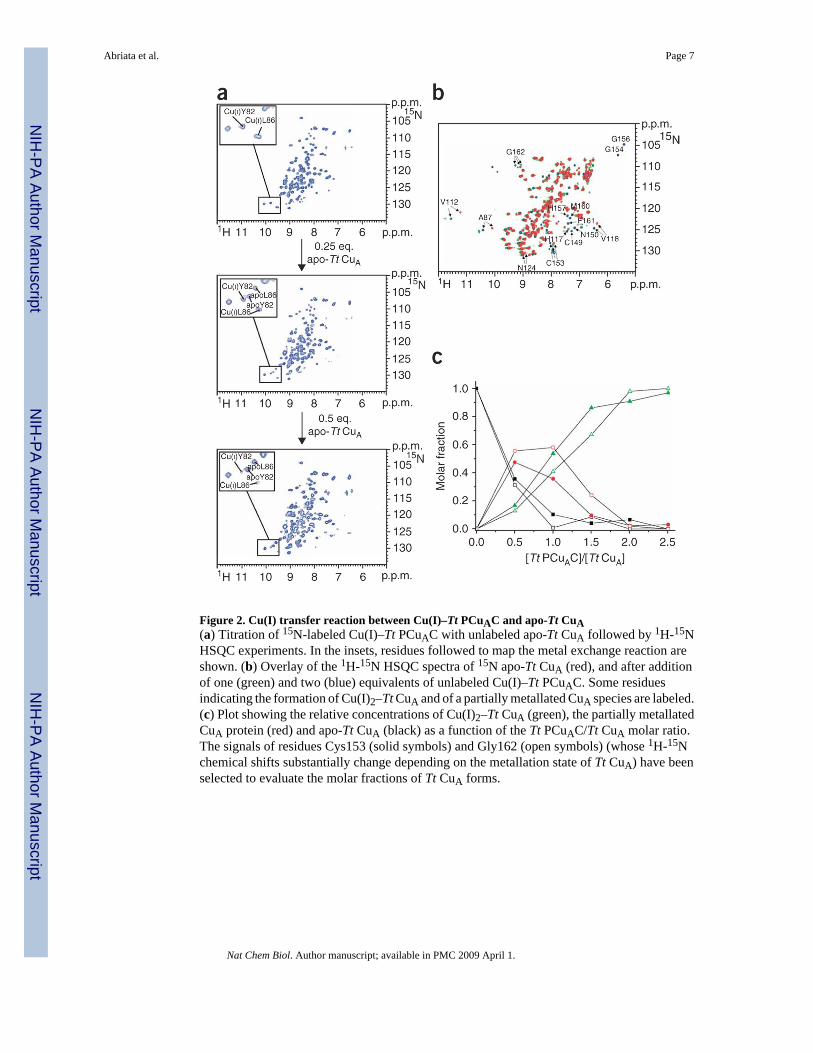
Figure 2. Cu(I) transfer reaction between Cu(I)–Tt PCuAC and apo-Tt CuA(a) Titration of 15N-labeled Cu(I)–Tt PCuAC with unlabeled apo-Tt CuA followed by 1H-15NHSQC experiments. In the insets, residues followed to map the metal exchange reaction areshown. (b) Overlay of the 1H-15N HSQC spectra of 15N apo-Tt CuA (red), and after additionof one (green) and two (blue) equivalents of unlabeled Cu(I)–Tt PCuAC. Some residuesindicating the formation of Cu(I)2–Tt CuA and of a partially metallated CuA species are labeled.(c) Plot showing the relative concentrations of Cu(I)2–Tt CuA (green), the partially metallatedCuA protein (red) and apo-Tt CuA (black) as a function of the Tt PCuAC/Tt CuA molar ratio.The signals of residues Cys153 (solid symbols) and Gly162 (open symbols) (whose 1H-15Nchemical shifts substantially change depending on the metallation state of Tt CuA) have beenselected to evaluate the molar fractions of Tt CuA forms.
Abriata et al. Page 7
Nat Chem Biol. Author manuscript; available in PMC 2009 April 1.
NIH
-PA Author Manuscript
NIH
-PA Author Manuscript
NIH
-PA Author Manuscript
Figure 3. Cu(I) transfer from Cu(I)–Tt PCuAC to oxidized apo-Tt CuA in the presence of reducedapo-Tt Sco1(a) 1H-15N HSQC overlay of oxidized apo-Tt CuA (red), Cu(I)2–Tt CuA (blue) and the finalmixture of the titration between 15N-labeled oxidized apo-Tt CuA and unlabeled, reduced apo-Tt Sco1 and Cu(I)–Tt PCuAC in a 1:1:2 ratio, in the absence of DTT (black). (b) Left panel:AMS-reacted, nonreducing SDS gel of different aliquots from the NMR titrationbetween 15N-labeled oxidized apo-Tt CuA and a 1:2 mixture of unlabeled, reduced apo-Tt Sco1and Cu(I)–Tt PCuAC (shown in a). Four steps of the NMR titration are reported. The band ofTt CuA protein at 14.8 kDa is very close to that of Tt PCuAC, which indeed has a very similarmolecular weight (13.2 kDa). Starting materials and the Tt PCuAC and Tt CuA proteins in theirdifferent redox states are also reported as a reference. Right panel: AMS-reacted, nonreducingSDS gel of a protein mixture containing oxidized apo-Tt CuA and reduced apo-Tt Sco1 in a1:1 ratio. The proteins in their different redox states are also reported as a reference. Molecularweight markers are shown in first lanes.
Abriata et al. Page 8
Nat Chem Biol. Author manuscript; available in PMC 2009 April 1.
NIH
-PA Author Manuscript
NIH
-PA Author Manuscript
NIH
-PA Author Manuscript


![[123doc vn] - triet-ly-kinh-doanh-cua-kinh-do](https://static.fdokumen.com/doc/165x107/6328c381fa4f06ee8e0531c2/123doc-vn-triet-ly-kinh-doanh-cua-kinh-do.jpg)