ovel insight into the mechanism of cellulosome assembly and ...
140
UNIVERSIDADE TÉCNICA DE LISBOA Faculdade de Medicina Veterinária ovel insight into the mechanism of cellulosome assembly and plant cell wall hydrolysis in anaerobic bacteria Benedita Andrade Pinheiro CONSTITUIÇÃO DO JURÍ ORIENTADOR PRESIDENTE Doutor Carlos Mendes Godinho Andrade Fontes Reitor da Universidade Técnica de Lisboa CO-ORIENTADOR VOGAIS Doutor José António Mestre Prates Doutora Isabel Maria Godinho de Sá Nogueira Doutor JoséAntónio Mestre Prates Doutor Carlos Mendes Godinho de Andrade Fontes Doutor Victor Manuel Diogo de Oliveira Alves Doutora Ana Luísa Moreira de Carvalho 2009 LISBOA
-
Upload
khangminh22 -
Category
Documents
-
view
3 -
download
0
Transcript of ovel insight into the mechanism of cellulosome assembly and ...

UNIVERSIDADE TÉCNICA DE LISBOA
Faculdade de Medicina Veterinária
�ovel insight into the mechanism of
cellulosome assembly and plant cell wall hydrolysis
in anaerobic bacteria
Benedita Andrade Pinheiro
CONSTITUIÇÃO DO JURÍ ORIENTADOR
PRESIDENTE Doutor Carlos Mendes Godinho Andrade Fontes
Reitor da Universidade Técnica de Lisboa CO-ORIENTADOR
VOGAIS Doutor José António Mestre Prates
Doutora Isabel Maria Godinho de Sá Nogueira
Doutor JoséAntónio Mestre Prates
Doutor Carlos Mendes Godinho de Andrade Fontes
Doutor Victor Manuel Diogo de Oliveira Alves
Doutora Ana Luísa Moreira de Carvalho
2009
LISBOA

UNIVERSIDADE TÉCNICA DE LISBOA
Faculdade de Medicina Veterinária
�ovel insight into the mechanism of
cellulosome assembly and plant cell wall hydrolysis
in anaerobic bacteria
Benedita Andrade Pinheiro
Tese de Doutoramento em Ciência e Tecnologia Animal
CONSTITUIÇÃO DO JURÍ ORIENTADOR
PRESIDENTE Doutor Carlos Mendes Godinho Andrade Fontes
Reitor da Universidade Técnica de Lisboa CO-ORIENTADOR
VOGAIS Doutor José António Mestre Prates
Doutora Isabel Maria Godinho de Sá Nogueira
Doutor JoséAntónio Mestre Prates
Doutor Carlos Mendes Godinho de Andrade Fontes
Doutor Victor Manuel Diogo de Oliveira Alves
Doutora Ana Luísa Moreira de Carvalho
2009
LISBOA
iii
À minha família
iv
ACK�OWLEDGEME�TS/AGRADECIME�TOS
Quero agradecer:
À Faculdade de Medicina Veterinária da Universidade Técnica de Lisboa, pelo acolhimento e por todos os meios materiais e humanos disponibilizados.
Ao Professor Doutor Carlos Fontes pela orientação, pela sua constante disponibilidade e pelos seus ensinamentos. Além de orientador, um grande amigo.
Ao Professor Doutor José Prates, pela sua disponibilidade e pelos seus conselhos científicos.
Ao Professor Doutor Luís Ferreira, pela simpatia com a qual me acolheu no seu grupo de investigação.
To Professor Harry Gilbert, Mark Proctor, David Bolam and Carl Morland, from the University of Newcastle upon Tyne, for their hospitality, support and guidance during the various times I went to their laboratory. I would also like to thank Professor Harry Gilbert and his team for their collaboration in the various cohesin-dockerin and esterases studies.
À Professora Maria João Romão e ao Shabir Najmudin, da Faculdade de Ciências e Tecnologia da Universidade Nova de Lisboa, por terem colaborado na resolução da estrutura endo-1,4-β-D-xylanase 10B em complexo com xilose.
To Professor Kazuo Sakka, from the Mie University, Japan, and his team; to Professor Gideon Davies, from University of York, and his team; to Professor Henri-Pierre Fierobe, from the Laboratoire de Chimie Bactérienne, France; and to Professor Edward Bayer, from the The Weizmann Institute of Science, Israel; to all the co-authors of the papers in which this thesis is based.
Aos colegas e amigos dos Laboratórios de Nutrição e de Bioquímica, nomeadamente a Susana, a Márcia, a Teresa, a Joana, a Vânia, a Catarina, a Patrícia, a Helena, a Paula, o Fernando, a Virgínia, a Zé, a Inês, a Maria, a Cristina, pela amizade, pelo apoio e boa disposição. Agradeço pela ajuda imprescindível prestada ao longo destes quatro anos e também durante a escrita da tese.
À minha família por me encorajarem a seguir e a realizar os meus sonhos.
v
This work was funded by Fundação para a Ciência e a Tecnologia, grant
SFRH/BD/25439/2005
Co-funded by POCTI/BIA-PRO/59118/2004 and PPCDT/BIA-PRO/59118/2004 from
Ministério da Ciência, Tecnologia e Ensino Superior

vi
vii
ABSTRACT
�ovel insight into the mechanism of cellulosome assembly and plant cell wall hydrolysis in
anaerobic bacteria
Cellulosomes are one of nature’s most elaborate and highly efficient nanomachines. These cell
bound multi-enzyme complexes orchestrate the deconstruction of cellulose and hemicellulose, two of
the most abundant polymers on earth, thus playing a major role in carbon turnover. Integration of
cellulosomal components occurs via highly ordered protein:protein interactions between cohesins and
dockerins, whose specificities allow the precise incorporation of cellulases and hemicellulases onto a
molecular scaffold. Clostridium thermocellum and C. cellulolyticum cellulosomes have been
extensively characterized and constitute the paradigm for the organization of cellulases and
hemicellulases in multi-enzyme complexes by thermophilic and mesophilic anaerobic bacteria,
respectively. The recent sequencing of C. thermocellum and C. cellulolyticum genomes allowed the
identification of the complete set of cohesins, dockerins and cellulosomal domains encoded by these
bacteria. Here, several unresolved issues concerning cohesin-dockerin specificity, cellulosome
assembly and the role of cellulosomal catalytic components in plant cell wall hydrolysis will be
explored. The ligand specificities of some newly identified C. thermocellum cohesin and dockerin
domains were described (Chapter 2). A novel cell-bound protein, termed OlpC, which contains a type
I cohesin domain was discovered in C. thermocellum. A restricted set of dockerins were shown to
interact, primarily, with OlpC. All the remaining dockerin containing polypeptides expressed by C.
thermocellum are directed to cellulosomes. Significantly, the structure of two C. cellulolyticum
cohesin-dockerin complexes revealed that, as it was previously reported for C. thermocellum,
mesophilic dockerins also express a dual binding mode for cohesins (Chapter 3). Initial crystallization
studies with the two N-terminal domains of C. thermocellum cellulosomal xylanase Xyn10B anticipate
the elucidation of its 3D structure, which may provide insightful data concerning the function of this
enzyme in plant cell wall hydrolysis (Chapter 4). Finally, a cellulosomal family 2 CE (CtCE2), which
grafts a second discrete non-catalytic binding functionality into its active site, was characterized
(Chapter 5). CtCE2 provides a rare example of “gene sharing” where the introduction of a second
functionality into the active site of an enzyme does not compromise the original activity of the
biocatalyst.
Key words: Clostridium thermocellum; Clostridium cellulolyticum; cellulosome; cohesin; dockerin; glycoside hydrolase, carbohydrate-binding module; carbohydrate esterase.

viii
ix
RESUMO
�ova perspectiva no mecanismo de integração do celulossoma e na degradação da parede
celular vegetal por bactérias anaeróbias
Os celulossomas são um dos mais intricados e eficientes complexos multi-enzimáticos existentes na
Natureza. Estes complexos, que se encontram ligados à parede celular bacteriana, desempenham um
papel importante na degradação da celulose e da hemicelulose, dois dos mais abundantes polímeros na
terra. A integração dos componentes celulossomais ocorre através de interacções proteína-proteína,
muito ordenadas, estabelecidas entre coesinas e doquerinas, cuja especificidade permite a incorporação
precisa de celulases e hemicelulases numa proteína de integração celulossomal. Os celulossomas dos
organismos Clostridium thermocellum e C. cellulolyticum têm sido extensivamente caracterizados e
constituem o paradigma para a organização de celulases e hemicelulases em complexos multi-
enzimáticos de bactérias anaeróbias, tanto termófilas como mesófilas, respectivamente. A recente
sequenciação dos genomas do C. thermocellum e do C. cellulolyticum permitiu a identificação de um
conjunto completo de coesinas, doquerinas e domínios celulossomais codificados por estas bactérias.
Neste trabalho, várias questões relativas à especificidade coesina-doquerina, à formação do
celulossoma e ao papel dos componentes celulossomais catalíticos serão investigadas. A
especificidade de doquerinas e coesinas do C. thermocellum recentemente identificados foi descrita
(Capítulo 2). Uma nova proteína da parede celular, designada como OlpC, que contém um domínio
doquerina, foi descoberta no C. thermocellum. Demonstrou-se que um conjunto restrito de doquerinas
reage preferencialmente com a OlpC. Os restantes polipéptidos expressos pela bactéria C.
thermocellum, contendo também doquerinas, ligam-se ao celulossoma. A estrutura de dois complexos
coesina-doquerina do C. cellulolyticum revelou, como previamente comunicado para a bactéria
C.thermocellum, que as doquerinas de organismos mesófilos também apresentam uma dupla ligação
para com as coesinas (Capítulo 3). Estudos preliminares de cristalização dos dois domínios N-
terminais da xilanase celulossomal Xyn10B antecipam a futura elucidação da sua estrutura 3D, o que
poderá esclarecer a função deste enzima na hidrólise da parede celular vegetal (Capítulo 4).
Finalmente, foi descrita uma esterase de hidratos de carbono da família 2 (CtCE2), que apresenta uma
funcionalidade discreta, não-catalítica de ligação a glúcidos no seu centro catalítico. A CtCE2 fornece
um raro exemplo de “gene sharing”, onde a introdução de uma segunda funcionalidade no centro
catalítico de uma enzima não compromete a actividade original do biocatalisador.
Palavras-chave: Clostridium thermocellum; Clostridium cellulolyticum; celulossoma; coesina; doquerina; glicósido hidrolases; módulos de ligação a hidratos de carbono; esterase de hidratos de carbono.

x
xi
I�TER�ATIO�AL-PEER REVIEWED PAPERS
This thesis was based on the following publications:
Pinheiro, B.A., Gilbert, H., Kazutaka, Sakka, K., S., Fernandes, V.O., Prates, J.A.M., Alves,
V.D., Bolam, D.N., Ferreira, L.M.A., & Fontes, C. M. G. A. (2009) Functional insights into
the role of novel type I cohesin and dockerin domains from Clostridium thermocellum.
Biochemical Journal. 424, 375-384
Pinheiro, B.A., Proctor, M.R., Martinez-Fleites, C., Prates, J.A.M., Money, V.A., Davies,
G.J., Bayer, E.A., Fontes, C.M.G.A., Fierobe, H., & Gilbert, H.J. (2008) The Clostridium
cellulolyticum dockerin displays a dual binding mode for its cohesion partner. Journal of
Biological Chemistry. 283, 18422-18430
Najmudin, S., Pinheiro, B.A., Romão, M.J., Prates, J.A.M. & Fontes, C.M.G.A. (2008)
Purification, crystallization and crystallographic analysis of Clostridium thermocellum endo-
1,4-β-D-xylanase 10B in complex with xylohexaose. Acta Crystallographica. (2008) 64, 715-
718
Montanier, C., Money, V.A., Pires, V.M.R, Flint, J.E., Pinheiro, B.A., Goyal, A., Prates,
J.A.M., Izumi, A., Stalbrand, H., Morland, C., Cartmell, A., Kolenova, K., Topakas, E.,
Dodson, E.J., Bolam, D.N., Davies, G.J., Fontes, C.M.G.A. & Gilbert, H.J. (2009) The active
site of a carbohydrate esterase displays divergent catalytic and non-catalytic functions. PLoS
Biology (2009) 7(3), e1000071.
CONTENTS
xii
CO�TE�TS
LIST OF TABLES …………………………………………………………………..……xv
LIST OF FIGURES……………………………………………………………………...…xvi
LIST OF ABREVIATIO�S A�D SYMBOLS…………………………………..…….....xvii
CHAPTER 1 SCIE�TIFIC BACKGROU�D A�D OBJECTIVES………….……..…..1
1.1 INTRODUCTION .............................................................................................. 1 1.2 PLANT CELL WALL .......................................................................................... 1 1.2.1 Cellulose ............................................................................................... 3
1.2.2 Matrix polysaccharides ........................................................................ 5
1.2.3 Plant cell wall hydrolysis ..................................................................... 6
1.3 THE CELLULOSOME ARCHITECTURE AND FUNCTION ....................................... 7 1.3.1 Cellulosomal proteins ........................................................................... 8
1.3.2 Cellulosome assembly and architecture ............................................. 15
1.4 OBJECTIVES .................................................................................................. 26
CHAPTER 2 FU�CTIO�AL I�SIGHTS I�TO THE ROLE OF �OVEL TYPE I
COHESI� A�D DOCKERI� DOMAI�S FROM CLOSTRIDIUM THERMOCELLUM
............................................................................................................................................... 27
2.1 INTRODUCTION ............................................................................................ 28 2.2 EXPERIMENTAL PROCEDURES ....................................................................... 30 2.2.1 Cloning, expression and purification ................................................. 30
2.2.2 Mutagenesis ........................................................................................ 32
2.2.3 Interaction of ctunk1 with bacterial cell wall preparations ............... 32
2.2.4 Complex formation in solution ........................................................... 32
2.2.5 Isothermal calorimetry of coh-doc binding ........................................ 33
2.3 RESULTS AND DISCUSSION ............................................................................ 33 2.3.1 ,ovel type I cohesin and dockerin domains in C. thermocellum
proteins ............................................................................................................ 33
2.3.2 C. thermocellum cthe_0452 is a cell-surface protein ......................... 37
2.3.3 Ligand specificity of novel C. thermocellum cohesin and dockerins . 38
2.3.4 Complex stability and cohesin-dockerin selectivity ............................ 42
2.3.5 Doc-cel44a contains two cohesin-binding interfaces presenting
different ligand specificities .................................................................................... 45
2.4 CONCLUSIONS .............................................................................................. 47
CHAPTER 3 THE CLOSTRIDIUM CELLULOLYTICUM DOCKERI� DISPLAYS A
DUAL BI�DI�G MODE FOR ITS COHESI� PART�ER ...................................... 49
3.1 INTRODUCTION ............................................................................................. 50 3.2 EXPERIMENTAL PROCEDURES ....................................................................... 52 3.2.1 Cloning and Expression ..................................................................... 52
CONTENTS
xiii
3.2.2 Protein Expression and Purification .................................................. 53
3.2.3 Isothermal Titration Calorimetry ....................................................... 53
3.2.4 Crystallization of the C. cellulolyticum Cohesin-Dockerin Complexes
and Structure Resolution ........................................................................................ 54
3.3 RESULTS ....................................................................................................... 55 3.3.1 Thermodynamics and Stoichiometry of Cohesin-Dockerin Recognition
……………………………………………………………………………….55
3.3.2 Protein Expression and Crystallization Strategy ................................ 56
3.3.3 Structure of the Type I Cohesin-Dockerin C. cellulolyticum Complex
………………………………………………………………………………………………56
3.3.4 Structure of the C. cellulolyticum Type I Cohesin in Complex with Its
Cognate Dockerin. .................................................................................................. 57
3.3.5 Structure of the C. cellulolyticum Type I Dockerin ............................ 58
3.3.6 Cellulolyticum Type I Coh-DocA 16S/L17T and Coh-DocA 47S/F48T
Interfaces ............................................................................................................ 59
3.3.7 Comparison of the C. cellulolyticum and C. thermocellum Type I
Cohesin-Dockerin Complexes ................................................................................ 63
3.4 DISCUSSION .................................................................................................. 66
CHAPTER 4 PURIFICATIO�, CRYSTALLIZATIO� A�D CRYSTALLOGRAPHIC
A�ALYSIS OF CLOSTRIDIUM THERMOCELLUM E�DO-1,4-B-D-XYLA�ASE 10B
I� COMPLEX WITH XYLOHEXAOSE ................................................................... 69
4.1 INTRODUCTION ............................................................................................ 70 4.2 MATERIALS AND METHODS .......................................................................... 71 4.2.1 Bacterial strains, plasmids and growth condition .............................. 71
4.2.2 Protein expression and purification ................................................... 71
4.2.3 Crystallization .................................................................................... 73
4.2.4 Data collection and processing .......................................................... 74
CHAPTER 5 THE ACTIVE SITE OF A CARBOHYDRATE ESTERASE DISPLAYS
DIVERGE�T CATALYTIC A�D �O�-CATALYTIC BI�DI�G FU�CTIO�S .... 77
5.1 INTRODUCTION ............................................................................................ 78 5.2 EXPERIMENTAL PROCEDURES........................................................................ 80 5.2.1 Gene cloning and protein expression ................................................. 80
5.2.2 Protein expression and purification ................................................... 80
5.2.3 Mutagenesis ........................................................................................ 81
5.2.4 Enzyme assays .................................................................................... 81
5.2.5 Carbohydrate binding studies ............................................................ 82
5.2.6 Crystallisation and structure solution ................................................ 83
5.3 RESULTS ....................................................................................................... 85 5.3.1 CE2 is a large family of diverse esterases displaying the α/β hydrolase
fold with a serine nucleophile. ................................................................................ 85
5.3.2 The esterase and cellulose binding functions of CtCE2 are in close
proximity. ............................................................................................................ 89
5.3.3 The structural basis for substrate recognition in the CE2 family….....91
CONTENTS
xiv
5.3.4 Structural characterisation of cellooligosaccharide binding to CtCE2.
…………………………………………………………………………………92
5.3.5 The mechanism of cellohexaose inhibition of CtCE2 activity. ........... 95
5.4 DISCUSSION .................................................................................................. 96
GE�ERAL DISCUSSIO� A�D FUTURE PERSPECTIVES ......................... …….99
REFERE�CES……………………………………………...…………….…………….103
TABLES
xv
TABLES
Table 1.1 Anaerobic bacteria producing cellulosomes: environmental niches and optimal growth temperatures. Adapted from Doi et al. (2003). ................................................... 8
Table 1.2 GHs fold superfamilies.Adapted from http://afmb.cnrs-mrs.fr/CAZY (June 2009). .... 11
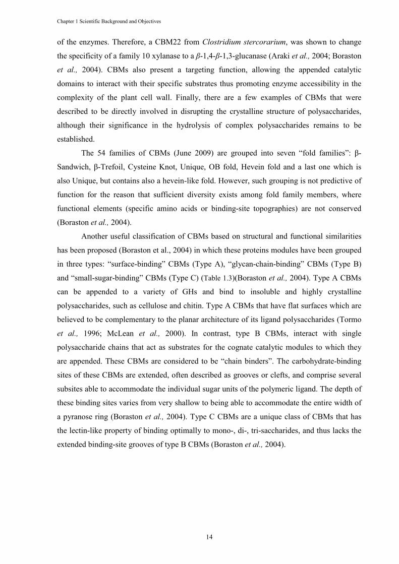
Table 1.3 Classification of CBMs into functional types and their relation with the family fold and family classification by Henrissat. ......................................................................... 15
Table 1.4 The modular structure of scaffoldins from various microorganisms. Adapted from Doi and Kosugi (2004) and Bayer et al. (1998). ........................................................... 18
Table 2.1 Primers used to obtain the genes encoding the cohesin and dockerin derivatives used in this work and for the mutagenesis of Doc-Cel44A. ......................................... 31
Table 2.2 Recognition of C. thermocellum and C. cellulolyticum cohesins by C. thermocellum dockerins as evaluated by ITC. ..................................................................................... 40
Table 2.3 Stability of previously assembled cohesin-dockerin complexes to the presence of newly introduced cohesins or dockerins. ...................................................................... 44
Table 2.4 Identification of preferred cohesin and dockerin partners as evaluated by native gel electrophoresis. ............................................................................................................. 45
Table 2.5 Thermodynamics of the binding between wild type dockerin Doc-Cel44A and its mutant derivatives and C. cellulolyticum (Coh-Ccel) and C. thermocellum (Coh-CipA2) cohesins. ........................................................................................................... 46
Table 3.1 Primers used to clone the dockerin module of the GH5 C. cellulolyticum cellulase CcCel5A together with the first cohesin module of C. cellulolyticum scaffoldin CipC in the same plasmid and mutate the dockerin module. ........................................ 53
Table 3.2 Data collection, phasing, and refinement statistics of C. cellulolyticum CohDocA16S/L17T and CohDocA47S/F48T .............................................................. 58
Table 3.3 Thermodynamics of the binding between C. cellulolyticum dockerin and cobesin variants All ITC experiments were carried out at 308 K. ............................................. 62
Table 3.4 Polar interactions between the cohesin and dockerins in the CohDocA16S/L17T and Coh-DocA16S/L17T complexes ................................................................................... 63
Table 4.1 Primers used to amplify the gene coding CBM22-1-GH10 and obtain the mutagenic CBM22-1-GH10 E337A. .............................................................................................. 73
Table 4.2 Matthews Coefficient Calculations. .............................................................................. 75
Table 4.3 X-ray crystallography data-collection statistics. ........................................................... 76
Table 5.1 Primers used for cloning ctCE2 from Clostridium thermocellum. Engineered restriction sites and mutation points are depicted in bold. ............................................ 81
Table 5.2 Data collection and refinement statistics. ...................................................................... 84
Table 5.3 Catalytic activity of CE2 enzymes against 4-nitrophenyl acetate (4-NPAc). ............... 86
Table 5.4 Activity of CE2 esterases against acetylated polysaccharides ...................................... 86
Table 5.5 The bindinga of CE2 esterases to cellohexaose and β-glucan. ..................................... 90
Table 5.6 ITC analysis of CtCE2 wild-type and mutants binding to cellooligosaccharides and β-glucan ......................................................................................................................... 93
FIGURES
xvi
FIGURES
Figure 1.1 Model of the primary plant cell wall, showing major structural polymers and their likely arrangement in the wall. ..................................................................................... 3
Figure 1.2 Fragment (repeating unit) of a cellulose chain. Adapted from O’Sullivan (1997). ....... 4
Figure 1.3 Clostridium thermocellum cellulosomes. ....................................................................... 7
Figure 1.4 The mechanism of action of carbohydrate esterases. Carbohydrate esterases perform the de-O and de-N acetylation of acetylated sugars ..................................... 12
Figure 1.5 Schematic representation of the supramolecular architecture, and disposition on the bacterial cell surface, of representative cellulosomes. ................................................ 16
Figure 1.6 Structure of the type I cohesin-dockerin complex. ...................................................... 23
Figure 1.7 The dual binding mode of the Xyn10B dockerin. ........................................................ 24
Figure 1.8 Structure of the type II Coh–XDoc complex. .............................................................. 25
Figure 2.1 Simplified representation of Clostridium thermocellum cellulosome. ........................ 34
Figure 2.2 Structural alignments of C. thermocellum and C. cellulolyticum representative type I cohesins and dockerins. .................................................................................... 36
Figure 2.3 OlpC is a cell surface protein. Adsorption of CtUnk1, the N-terminal domain of OlpC, to C. thermocellum and E. coli cellular preparations. ...................................... 38
Figure 2.4 Examples of the isothermal titration calorimetry (ITC) experiments with selected cohesin and dockerin domains expressing different affinities. ................................... 39
Figure 2.5 The cohesin-dockerin interaction as evaluated by non-denaturing gel electrophoresis. ........................................................................................................... 41
Figure 2.6 Doc-Cel44A cannot bind C. thermocellum and C. cellulolyticum cohesins simultaneously. ........................................................................................................... 47
Figure 3.1 The influence of temperature on the thermodynamics of cohesin dockerin binding. .. 55
Figure 3.2 The three-dimensional structures of the C. cellulolyticum cohesin-dockerin complexes. a depicts the structure of Coh-DocA16S/L17T with the dockerin color-ramped from N terminus (blue) to C terminus (red) and the cohesin in pale brown. ......................................................................................................................... 57
Figure 3.3 The cohesin-dockerin interface in the two C. cellulolyticum cohesin-dockerin complexes. a shows the Coh-DocA16S/L17T complex with the dockerin in pale green and the cohesin in blue...................................................................................... 60
Figure 3.4 Overlap of the C. cellulolyticum and C. thermocellum cohesin-dockerin complexes in both binding modes highlighting the key recognition signature. ........................... 65
Figure 4.1 A Coomasie Brilliant Blue-stained 10% SDS-PAGE gel evaluation of protein purity during purification. ........................................................................................... 72
Figure 4.2 Crystals of CBM22-1-GH10 E337A obtained by hanging-drop vapour diffusion in the presence of 1M Na Acetate, 0.1M HEPES, pH 7.5 and 0.05M CdSO4. .............. 73
Figure 4.3 Plot of a 180º section of a self-rotation function from POLARRFN. .......................... 75
Figure 5.1 Schematic of the molecular architecture of the enzymes investigated in this study. ... 79
Figure 5.2 A. 3-D structure of CjCE2B with the catalytic domain in green and the β-sheet domain in magenta. ..................................................................................................... 87
Figure 5.3 Binding of cellooligosaccharides through the esterase active centre of CtCE2. .......... 89
Figure 5.4 Binding of CE2 esterases to cellulose. ......................................................................... 90
Figure 5.5 Examples of isothermal titration calorimetry of wild type and mutants of CtCE2. ..... 94
Figure 5.6 Inhibition of wild type CtCE2 by cellohexaose. .......................................................... 95
ABBREVIATIONS AND SYMBOLS
xvii
ABBREVIATIO�S A�D SYMBOLS
AGE Affinity gel electrophoresis Ala/A Alanine Arg/R Arginine Asn/� Asparagine Asp/D Aspartic acid/Aspartate B. Bacteroides
BSA Bovine serum albumin C. Clostridium
CAZy Carbohydrate-active enzyme CBD Cellulose-binding domain CBM Carbohydrate-binding module CCP4 Collaborative computational project number 4 CE Carbohydrate esterase Cel Cellulase CESA Plant cellulose synthase cipA C. thermocellum cellulosome-integrating protein Cj Clostridium japonicas
Coh Cohesin Ct Clostridium thermocellum
Cthe Clostridium thermocellum
Cys/C Cysteine ∆G Binding Gibbs energy ∆H Binding enthalpy ∆S Binding entropy ∆Cp Heat capacity Doc Dockerin D�A Deoxyribonucleic acid DTT Dithiothreitol DP Degree of polymerization E. Escherichia EC Enzyme commission ESRF European Synchrotron Radiation facility FPLC Fast protein liquid chromatography G Unsubstituted glucose GH Glycoside hydrolase Glc β-1,4 D-Glucan Gln/Q Glutamine Glu/E Glutamic acid/Glutamate Gly Glycine HEC Hydroxyethylcellulose HEPES 4-(2-Hydroxyethyl)-1-piperazine-ethanesulfonic acid HF-EPCS Hydrofluoric acid-extracted cell wall polymer His/H Histidine Ile/I Isoleucine IMAC Immobilized metal ion affinity chromatography IPTG Isopropyl 1-thio-β-D-galactopyranoside ITC Isothermal titration calorimetry IU International units IUBMB International Union of Biochemistry and Molecular Biology
ABBREVIATIONS AND SYMBOLS
xviii
K Kelvin degree Ka Equilibrium affinity constant kcat Catalytic efficiency KD Dissociation constant Ki Inibition constant KM Michaelis constant LB Luria-Bertani broth medium Leu/L Leucine Ln Natural logarithm Lnk Linker Lys/K Lysine MS Mass spectrometry Met/M Methionine Mw Molecular weight �CS Non crystallographic symmetry �PCS Native peptidoglycan-containing sacculi OD Optical density OlpA C.thermocellum outer layer protein A OlpB C.thermocellum outer layer protein B OlpC C. thermocellum outer layer protein C PASC Phosphoric acid swollen cellulose PCR Polymerase chain reaction PDB Protein data bank PEG Polyethyleneglycol Phe/F Phenylalanine PKD Polycystic kidney disease PL Polysaccharide lyases Pro/P Proline R Universal gas constant R. Ruminococcus
RMSD/r.m.s.d. Root mean square deviation SAXS Small angle x-ray scattering S/Ser Serine SCWP Secondary cell wall polymers SdbA Scaffoldin dockerin binding protein A SDS-PAGE Sodium dodecyl sulfate-polyacrylamide gel electrophoresis SEM Standard error mean SeMet Seleno-methionine Ser/S Serine SLH S-layer homology module Sp Species SRF Self rotation function Thr/T Threonine Tris 2-Amino-2-hydroxymethyl-1,3-propanediol Trp/W Tryptophan Tyr/Y Tyrosine Unk Unknown V Volt Val/V Valine v/v Volume/volume X Glucose decorated α-1,6 with xylose X-gal 5-Bromo-4-chloro-3-indolyl β-D-galactoside
ABBREVIATIONS AND SYMBOLS
xix
Xyl α-1,6 D-Xylosyl Xyn10B Xylanase G Terrestrial gravity constant (9,80665 m s-2) Zn Zinc

ABBREVIATIONS AND SYMBOLS
xx
Chapter 1 Scientific Background and Objectives
1
Chapter 1 Scientific Background and Objectives
1.1 I�TRODUCTIO�
Cellulose, the main structural component of plant cell walls, is the most abundant
carbohydrate polymer in nature. Recycling of the carbon and energy stored in structural
polysaccharides is orchestrated by a large repertoire of cellulases and hemicellulases. The
energetic constrains posed by anaerobic ecosystems lead to the evolution of a remarkably
efficient plant cell wall hydrolytic system. This system is formed by a large extracellular
enzymatic complex designated as cellulosome, which consists of a scaffoldin protein to which
a variety of glycoside hydrolases (GH) and carbohydrate esterases (CE) are bound.
Cellulosomes have many biotechnological applications since the conversion of biomass into
sugars results in the production of value products, such as butanol and amino acids, and
utilizable forms of energy, such as ethanol and methane. Indeed the ethanol produced from
cellulosic biomass, designated as cellulosic ethanol, is considered more ecological than other
types of biofuels since it is more efficient in reducing greenhouse gas emissions.
In this general introduction, the plant cell wall structure will be summarily reviewed,
with special attention to the role of the polysaccharide constituents. Subsequently, attention is
driven into the cellulosome, and its complexity and functionality will be described.
Cellulosome complexity will be analyzed by reviewing our current knowledge on the
structure and function of a variety of cellulosomal components, particularly GHs, CEs and
carbohydrate binding modules (CBM). Finally, a detailed description is made of the
mechanisms of cellulosome assembly, with a special focus on the cohesin-dockerin
interaction, which is the driving force of cellulosome assembly.
1.2 PLA�T CELL WALL
The plant cell wall is a complex and dynamic structure composed mostly of
polysaccharides, highly glycosylated proteins and lignin (Somerville et al., 2004). Plants
present two types of cell walls that differ in function and composition: the primary and
secondary cell walls. Primary walls surround growing and dividing plant cells, providing
mechanical strength but allowing the cells to expand. The primary cell wall is deposited and
continues to be deposited through cell growth and expansion while the secondary cell wall is
deposited internally to the wall of some cell types, only at the onset of differentiation, once
Chapter 1 Scientific Background and Objectives
2
cell growth has ceased. The majority of research regarding cell wall composition in diverse
plant groups has concentrated on the primary cell owing to the existence of cell type specific
variation between secondary cell walls (Popper, 2008).
The description of the primary wall is of a composite polymeric structure in which
crystalline cellulose microfibrils are embedded in a complex, highly hydrated, and less
ordered polysaccharide matrix, with smaller quantities of structural protein intercalated in the
matrix (Figure 1.1). Cellulose plays a major role in determining cell wall strength and
structure. Hemicelluloses, which are major components of the cell wall matrix, bind cellulose
microfibrils together or act as a lubricating coat, preventing direct microfibril-microfibril
contact. Pectin, the other major polysaccharide present in the cell wall matrix, forms a gel
phase, acting possibly as a hydrophilic filler to prevent the aggregation and collapse of the
cellulose network (Jarvis 1992) while modulating the porosity of the cell wall to
macromolecules (Baron-Epel et al., 1988). The precise role of the structural proteins in cell
wall complexity and structure remains a matter of speculation (Cosgrove, 1997).
Several models, which reflect the interactions established between its components,
have been proposed to explain the organization of plant cell walls. The first to be presented
was the “Albersheim model” by Keegstra et al. (1973) which proposed that matrix polymers
(xyloglucan, pectic polysaccharides and structural proteins) were covalently linked to form a
giant macromolecular network. In this model, cellulose is bonded to the matrix via H-bonding
to xyloglucans (Cosgrove, 2001). As the pectin-xyloglucan linkage could not be confirmed,
an alternative model was proposed by Hayashi (1989) and Fry (1989) which assume that
cellulose microfibrils are tethered together directly via long xyloglucan chains. Pectic
polysaccharides and structural proteins are imagined as co-extensive, but independent,
networks that physically entangle the cellulose-xyloglucan network, but are not covalently
bonded to it. Although this is the most popular model explaining plant cell wall organization,
several variations have been proposed. Talbott and Ray (1992) suggested a “multicoat” model
in which each microfibril is coated by a series of progressive less-tightly bound
polysaccharide layers and that the linkage between microfibrils is made indirectly via non-
covalent associations between the distinctive polysaccharide layers. More recently, Ha et al.
(1997) conceived a more stratified wall in which pectic layers serve as spacers between
cellulose-hemicellulose layers, allowing them to slide during wall expansion and also
controlling cell wall thickness. All these models have in common the concept that cellulose
microfibrils are coated with xyloglucan (Cosgrove, 2001).
Chapter 1 Scientific Background and Objectives
3
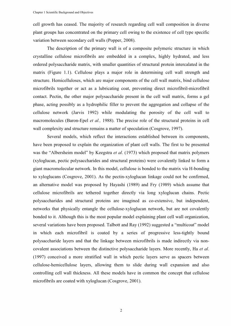
Figure 1.1 Model of the primary plant cell wall, showing major structural polymers and their likely arrangement in the wall. Cellulose microfibrils contain non crystalline regions that may be formed by entrapment of hemicelluloses such as xyloglucan. Xyloglucan can also bond to the surface of cellulose and may link two microfibrils together. Although the side chains of xyloglucan interfere with bonding of the glucan backbone to other glucans, they may twist such that short regions of the backbone form a planar configuration suitable for bonding to cellulose. Pectins form a space-filled hydrophilic gel between microfibrils. Adapted from Cosgrove (1997).
1.2.1 Cellulose
Cellulose is the most abundant bio-polymer on the earth (O’Sullivan, 1997) and the
main constituent of higher plant cell walls (Kobayashi and Ohmae, 2006). It is present in
bacteria, fungi, algae and even in animals (O’Sullivan, 1997, Capadona, 2008).
The remarkable properties of cellulose (high tensile strength, insolubility, chemical
stability and immunity to most of enzymatic attacks) are a result of its unique structure
(Cosgrove, 1997). Cellulose is an unbranched homopolysaccharide composed of β-D-
glucopyranose units linked by (1 → 4) glycosidic bonds (Purves, 1954; Marchessault and
Sundararajan, 1983)(Figure 1.2). In nature, cellulose chains have a degree of polymerization
(DP) of approximately 10,000 glucopyranose units in wood cellulose and 15 000
glucopyranose units in native cotton cellulose (Sjoström, 1981).
Chapter 1 Scientific Background and Objectives
4
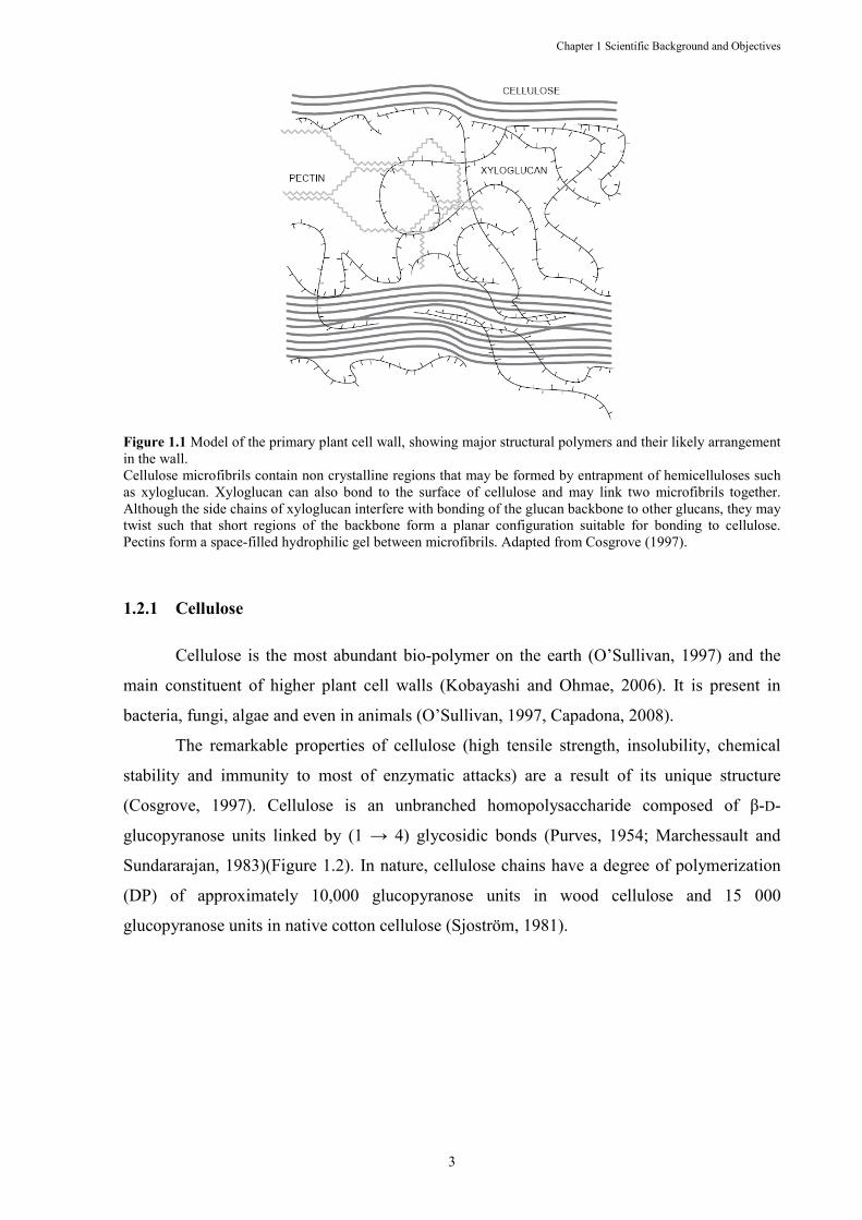
Figure 1.2 Fragment (repeating unit) of a cellulose chain. Adapted from O’Sullivan (1997).
Cellulose has polymorphs (I, II, III1, III11, IV1 and IV11) that can be interconverted
(Marchessault & Sarko, 1967; Walton & Blackwell, 1973; Marchessault & Sundararajan,
1983), with cellulose I being the form found in nature (native form). These polymorphs may
be divided in two groups; those with a unit cell similar to that of native cellulose (I, IIII and
IVI) and those with a cellulose II type arrangement (II, IIIII and IVII). Cellulose I is a mixture
of two polymorphs, Iα and Iβ (Atalla & Vanderhart, 1989), which were found to have the
same conformation in the heavy atom skeleton, but to differ in their hydrogen patterns. The
first polymorph (Iα) is the dominant component in celluloses produced by primitive
organisms while those produced by the higher plants have the second polymorph (Iβ)
dominant. The existence of these two polymorphs may affect the reactivity of native cellulose
as Iα is meta-stable, thus more reactive than Iβ, and probably the site of initial reaction in a
microfibril (O’ Sullivan, 1997). Although cellulose I is the most studied cellulose polymorph,
cellulose II has become an important focus of study because of its greater stability, being
actually the second most investigated form of cellulose (O’Sullivan, 1997).
Cellulose synthesis is catalysed by plant cellulose synthase (CESA), which are
proteins embedded in the plasma membrane in hexameric arrays called particle rosettes.
These membrane complexes probably contain other proteins that aid microfibril formation
and link the complexes to nearby microtubules for guidance along the membrane. Each
cellulose microfibril is formed from the spontaneous ‘bundling’ and crystallization of the
glucans chains (Cosgrove et al., 2005).
There is a controversy about the supermolecular structure of cellulose and various
models have been proposed to explain the molecular mechanisms of cellulose organization. In
the fifties, Frey-Wyssling (1953-1954) proposed that the microfibril is an aggregate of several
elementary fibrils (units of about 36 cellulose chains) which are embedded in paracrystalline
cellulose, i.e. unordered crystallized cellulose (Kratky & Mark, 1938; Nickerson, 1941).
However, the existence of these elementary fibrils was questioned. In contrast, the fringe
Chapter 1 Scientific Background and Objectives
5
micellar model (Astbury, 1933) suggests that, completely ordered or crystalline regions,
without any distinctive boundary, change into disordered or amorphous regions. Thus, a
single molecule was thought to pass from one crystalline region to another through an
amorphous area.
Whether native cellulose consists of elementary fibrils or not, its ultrastructure is
largely due to the presence of covalent bonds, hydrogen bonds and van der Waals forces.
Hydrogen bonding within cellulose chains may act to determine the “straightness” of the
chain. Interchain hydrogen bonds might introduce order or disorder into the system,
depending on its regularity (O’Sullivan, 1997).
1.2.2 Matrix polysaccharides
The crystalline cellulose microfibrils are embedded in a matrix of complex
polysaccharides, which are divided in two classes: pectins and hemicelluloses. These
polysaccharides, in contrast to cellulose, are synthesized in the Golgi apparatus and packed
into tiny vesicles that fuse with the plasma membrane and thereby deliver their cargo to the
wall. The matrix polysaccharides then become integrated into the wall network by physical
interactions, enzymatic ligations and crosslinking reactions (Cosgrove, 2005).
Hemicelluloses are branched polysaccharides containing backbones of neutral sugars
that can form hydrogen bonds with the surface of cellulose fibrils (Somerville et al., 2004).
The most important hemicelluloses are xyloglucans, galactoglucomannans and
glucuronoarabinoxylans (Gilbert et al., 2008).
The most abundant and best studied hemicellulose is xyloglucan, a branched polymer
consisting of a backbone of 1→4 linked β-D-glucopyranose residues with short side chains
containing xylose, galactose and, often, a terminal fucose (McNeil et al., 1984; Fry, 1989).
Xyloglucans are believed to cross-link cellulose microfibrils and confer plasticity to the cell
wall (Gilbert et al., 2008). Another hemicellulose widely distributed in the cell walls of land
plants, especially in secondary walls of woody tissues, is mannan. The principal structure of
this polysaccharide is a mixed β(1→4)-linked mannose/glucose backbone on which α(1→6)-
linked galactose and Ο-2/ Ο-3 acetyl groups may be appended to the mannose residues
(Gilbert et al., 2008).
Xylan (glucuronoarabino) is the most abundant hemicellulose in dicotyledonous
plants. These polymers consist of a β(1→4) xylose residues decorated with acetyl and
arabinofuranose, at Ο-2 and/or Ο-3, and 4-methylglucuronic acid at Ο-2. The arabinofuranose
residues can be further derivatised with ferulic acid that can cross-link xylan chains to each
Chapter 1 Scientific Background and Objectives
6
other, to pectins or lignin (Gilbert et al., 2008). Xylan can also be acetylated, at positions O-2
or O-3, which confer additional protection for the polysaccharide backbone to enzymatic
attack.
Pectins, the second cell wall matrix constituent, are the most soluble of the structural
polysaccharides. Like hemicelluloses, pectins also constitute a heterogeneous group of
polysaccharides, characteristically containing acidic sugars such as glucuronic acid and
galacturonic acid. Some pectins such as homogalacturonan, have a relatively simple primary
structure consisting of a linear polymer of (1→4) β galacturonic acid, with occasional
rhamnosyl residues that make a twist in the chain. Rhamnogalacturonan I has repeating
subunits of (1→2) α-L-rhamnosyl-(1→2) α-D-galacturonyl disaccharides, with long side
chains of arabinans and arabinogalactans (Cosgrove, 1997).
1.2.3 Plant cell wall hydrolysis
The polysaccharide molecules found in plant cell walls are structural components that
provide form, support and protection. These three functions are maximized by the inherent
recalcitrance of these structural carbohydrates to enzymatic attack. The major limiting factor
in the hydrolysis of such bio-polymers is probably the sequestration and lack of accessibility
to substrates by the plant cell wall degrading enzymes. In addition, enzyme processivity is
affected by the extensive interactions established by plant cell wall polysaccharides. Despite
these difficulties, many microorganisms degrade crystalline cellulose after an initial attack to
the amorphous, less-crystalline, regions of cellulose, suggesting that they can disrupt the
crystalline regions (Warren, 1996). As a consequence of the different sugars and linkages
present in plant cell walls, the systems hydrolyzing cellulose comprise a diversity of exo- and
endo-acting enzymes appended to substrate binding domains (Warren, 1996).
In nature two basic enzymatic systems were evolved for the hydrolysis of plant cell
wall polysaccharides: in anaerobes cell wall biocatalysts are organized in high molecular
weight multi-enzyme complexes while in aerobes plant cell wall hydrolases act individually
during carbohydrate degradation (Warren, 1996). Plant cell wall degrading enzymes display
modular architectures. Thus, cellulases and hemicellulases contain one catalytic domain
linked with non-catalytic CBMs that are responsible for targeting their appended catalytic
domains to the enzyme substrates. In anaerobic ecosystems enzymes are also organized in
supramolecular complexes with a quaternary structure that further potentiate enzyme-
substrate targeting and enzyme stability. These extracellular multi-enzyme complexes were
termed cellulosomes, due to their enhanced activity on cellulose hydrolysis (Schwarz, 2001).
1.3 THE CELLULOSOME ARCH
The cellulosome is a multi
hemicellulases produced by several anaer
Bacteroides, and Ruminococcus
structural carbohydrates such as cellulose, hemicelluloses and pectin. These microorganisms
are found, primarily, in soil, wood chip piles, sewage and rumens (
may be the largest extracellular enzyme complexes found in nature, since polycel
have been reported to be as large as 100 MDa, although individual cellulosomes r
about 650,000 Da to 2.5 MDa (Doi
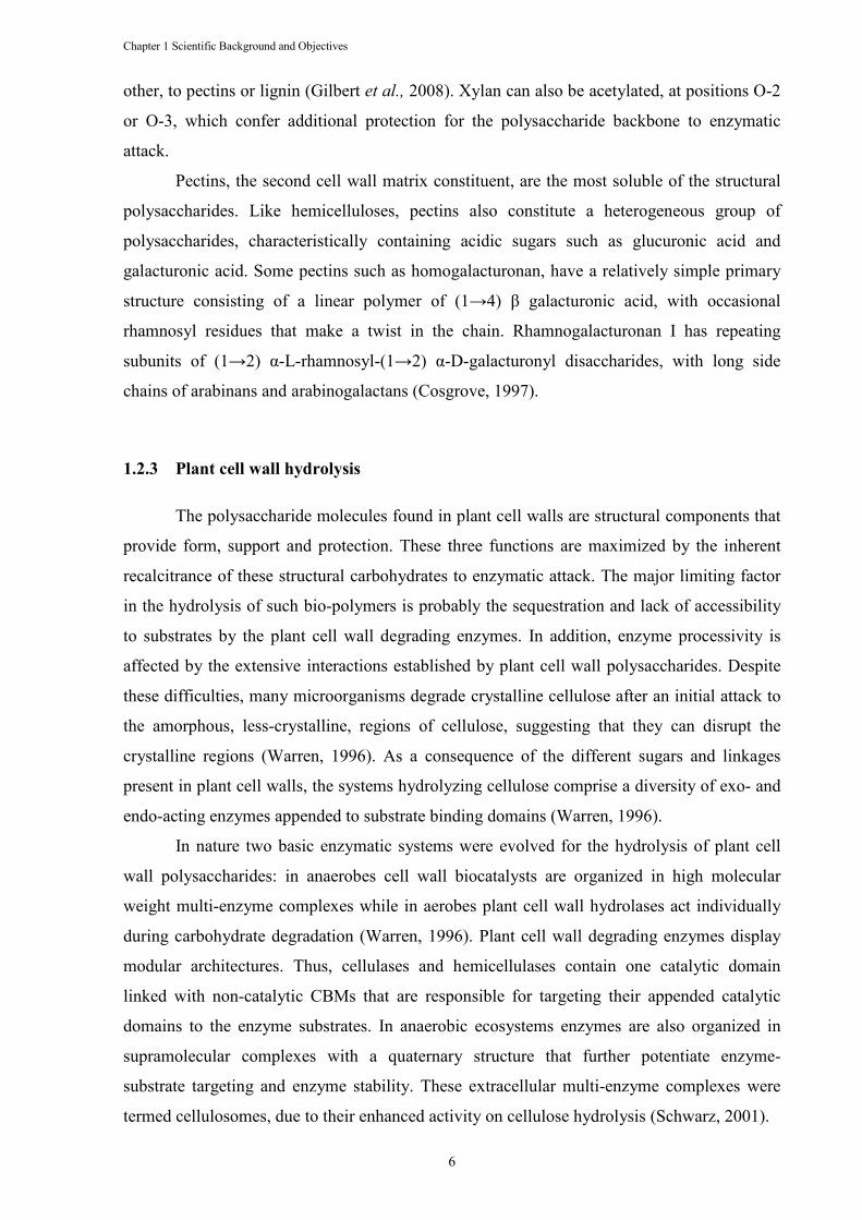
Figure 1.3 Clostridium thermocellum
(A) Transmission electron microscopy image showing protuberant structures at the bacteria surfaces; bar, 250nm, adapted from Bayer Scanning electron microscopy images depicting mutant AD2 with inability to adhere to cellulose (C); the protuberances at cell surface in the wildcellulosomes and do not appear in the mutants; bar, 200nm; adapted from Bayer and Lamed (1
Cellulosomes were first observed as large protuberances on the surface of
thermocellum (Figure 1.3)(Lamed
to consist of a non-enzymatic scaffolding protein that bound the cellulosomal catalytic
components (Bayer et al., 1998). Subsequently, new discoveries of cellulosomes, first in other
clostridial species (Belaich
al., 1994) and, more recently, in other, evolutionary more distant, bacteria and fungi (Bayer
al., 1998; Chen et al., 1998; Fanutti
molecular organization and functional importance of these highly complex nanomachines.
Chapter 1 Scientific Background and Objectives
7
THE CELLULOSOME ARCHITECTURE A�D FU�CTIO
The cellulosome is a multi-enzyme extracellular complex of cellulases and
hemicellulases produced by several anaerobic bacteria of the Clostridium
Ruminococcus genera, which is highly efficient in the degradation of
structural carbohydrates such as cellulose, hemicelluloses and pectin. These microorganisms
in soil, wood chip piles, sewage and rumens (Table
may be the largest extracellular enzyme complexes found in nature, since polycel
have been reported to be as large as 100 MDa, although individual cellulosomes r
5 MDa (Doi et al., 2003).
Clostridium thermocellum cellulosomes. nsmission electron microscopy image showing C. thermocellum ultrastructure; cellulosomes are the
protuberant structures at the bacteria surfaces; bar, 250nm, adapted from Bayer et al
Scanning electron microscopy images depicting C. thermocellum wild-type strain YS (B) and mutant AD2 with inability to adhere to cellulose (C); the protuberances at cell surface in the wildcellulosomes and do not appear in the mutants; bar, 200nm; adapted from Bayer and Lamed (1
Cellulosomes were first observed as large protuberances on the surface of
)(Lamed et al., 1983; Bayer et al., 1985) and the complex was found
enzymatic scaffolding protein that bound the cellulosomal catalytic
1998). Subsequently, new discoveries of cellulosomes, first in other
clostridial species (Belaich et al., 1997; Doi et al., 1994; Karita et al.,
1994) and, more recently, in other, evolutionary more distant, bacteria and fungi (Bayer
1998; Fanutti et al., 1995), improved our understanding on the
n and functional importance of these highly complex nanomachines.
Chapter 1 Scientific Background and Objectives
ITECTURE A�D FU�CTIO�
enzyme extracellular complex of cellulases and
Clostridium, Acetivibrio,
genera, which is highly efficient in the degradation of
structural carbohydrates such as cellulose, hemicelluloses and pectin. These microorganisms
Table 1.1). Cellulosomes
may be the largest extracellular enzyme complexes found in nature, since polycellulosomes
have been reported to be as large as 100 MDa, although individual cellulosomes range from
ultrastructure; cellulosomes are the et al. (1985). (B) and (C)
type strain YS (B) and C. thermocellum mutant AD2 with inability to adhere to cellulose (C); the protuberances at cell surface in the wild-type are cellulosomes and do not appear in the mutants; bar, 200nm; adapted from Bayer and Lamed (1987).
Cellulosomes were first observed as large protuberances on the surface of C.
and the complex was found
enzymatic scaffolding protein that bound the cellulosomal catalytic
1998). Subsequently, new discoveries of cellulosomes, first in other
1997; Pohlschroder et
1994) and, more recently, in other, evolutionary more distant, bacteria and fungi (Bayer et
1995), improved our understanding on the
n and functional importance of these highly complex nanomachines.
Chapter 1 Scientific Background and Objectives
8
Table 1.1 Anaerobic bacteria producing cellulosomes: environmental niches and optimal growth temperatures. Adapted from Doi et al. (2003).
Bacteria species Source Optimal growth
temperatures References
Acetivibrio cellulolyticus sewage mesophilic Ding et al. (1999) Bacteroides cellulosolvens sewage mesophilic Ding et al. (2000) Butyrivibrio fibrisolvens rumen mesophilic Hespell and O’Bryan (1992)
Clostridium acetobutylicum soil mesophilic Weyer and Rettger (1927) Clostridium cellulovorans wood fermenter mesophilic Sleat et al. (1984) Clostridium cellobioparum rumen mesophilic Lamed et al. (1987) Clostridium cellulolyticum compost mesophilic Pagès et al. (1997)
Clostridium josui compost mesophilic Kakiuchi et al. (1998) Clostridium papyrosolvens paper mill mesophilic Pohlschröder et al. (1995) Clostridium thermocellum sewage soil thermophilic Bayer et al. (1985)
Ruminococcus albus rumen mesophilic Ohara et al. (2000) Ruminococcus flavefaciens rumen mesophilic Ding et al. (2001)
Ruminococcus succinogenes rumen mesophilic Miron et al. (2001)
The cellulosome was initially defined as “a discrete, cellulose-binding, multienzyme
complex for the degradation of cellulosic substrates” (Lamed et al., 1983). Before the
cellulosome had been discovered, cellulase systems of cellulolytic organisms were considered
as a mixture of different enzymes in the free state and the original definition wasmainly based
on the cellulase system of Trichoderma reesei (Warren, 1996; Doi and Kosugi, 2004). With
the discovery of cellulosomes in C. thermocellum and in other cellulolytic bacteria, the
concept of this multi-enzyme complex has changed to include the degradation of other
polysaccharides, since cellulosomes also degrade hemicelluloses, chitin and pectin (Bayer et
al., 2004). The “discrete” defining quality of the cellulosome results of its multi-component
nature, where intercalating cellulosomal modules are functionally independent and seem to be
mixed and matched to serve the apparent molecular needs of a given microorganism (Bayer et
al., 2004).
1.3.1 Cellulosomal proteins
The cellulosomal enzymes include cellulases, hemicellulases, pectinase, chitinase and
many ancillary enzymes that can degrade plant cell wall materials (Doi and Kosugi, 2004).
Both free and cellulosomal enzymes have a modular nature and comprise catalytic domains
fused through linker sequences to a variety of non-catalytic domains the most predominant of
which are CBMs. In anaerobic systems, cellulosomal enzymes also contain a distinctive and
signature module that was termed the dockerin (see below). Dockerins are non-catalytic
domains that bind cohesin domains located in a scaffolding protein thus contributing for
cellulosome assembly. Therefore, cohesins and their dockerin counterparts represent the
Chapter 1 Scientific Background and Objectives
9
definitive “signature” components that dictate cellulosome assembly and architecture (Bayer
et al., 1998).
A given bacteria produces an extraordinary number and variety of enzymes, that
match the chemical and structural intricacy of plant cell walls. The presence of enzymes that
degrade polysaccharides other than cellulose in exclusive cellulolytic bacteria may be
essential in exposing the preferred substrate, cellulose, from the intricacy of the plant cell wall
matrix, thereby facilitating its subsequent hydrolysis and assimilation (Bayer et al., 1994 and
Bayer et al., 2004). The cellulosomal enzymes exhibit enhanced synergy on crystalline, not on
amorphous, cellulosic substrates when compared to free cellulase systems of both bacteria and
fungi. Fierobe et al. (2001 and 2002) obtained an experimental insight into the mechanisms
responsible for the observed enhanced activity after the construction of artificial mini-
cellulosomes of defined enzyme composition. The enhancement in synergistic action can be
attributed to the two different but complementary phenomena: targeting via the scaffoldin-
borne CBM and a spatial proximity of the resident cellulosomal enzymes (Fierobe et al.,
2002). The genome sequence of C. thermocellum and C. cellulolyticum revealed that
cellulosomes contain not only GHs, but also CEs and polysaccharide lyases (PL). PL will not
be reviewed here since they are not on the scope of this project.
1.3.1.1 Glycoside hydrolases
Several systems of classification exist for GHs, including those based on their
substrate or product specificities, mode of attack (exo versus endo enzymes), stereochemical
mechanism and, more recently, on amino acid sequence similarities (Henrissat, 1991;
Henrissat & Davies, 1997). The latest classification provides now 115 families (June 2009).
The classification recommended by the International Union of Biochemistry and
Molecular Biology (IUBMB) is that based on enzyme substrate specificities, being expressed
in the EC number for a given enzyme. The major problem associated with this classification is
that it does not appropriately accommodate enzymes which act on several substrates. An
example is the endoglucanases, which can be active to various degrees on cellulose, xylan,
xyloglucan, β-glucan, and various artificial substrates. In addition, many structurally unrelated
enzymes display similar substrate specificities and hence have identical IUBMB
classification. The opposite also happens as well (Henrissat & Davies, 1997).
GHs may also be classified according to their mechanism of action. These enzymes
can act via two basic mechanisms which result in a net retention or inversion of the anomeric
configuration (Koshland, 1953; Sinnott, 1990; McCarter & Withers, 1994). The disadvantage
Chapter 1 Scientific Background and Objectives
10
of this classification is that it cannot be used in general terms and it contains a very small
determinating power. Another classification for GHs is based on the enzyme mode of action,
where “exo” or “endo” relates to the capacity of the enzyme to attack one of the termini of the
polysaccharide or the carbohydrate backbone, respectively. Structural work revealed that the
enzyme mode of action is a reflection of the shape of the active-site cleft (Henrissat &
Bairoch, 1996). The exo versus endo terminology provides additional complementary
information, but it is often difficult to determine, and can be confusing if applied to enzymes
that display intermediate behavior or if determined with inappropriate substrates (Henrissat &
Davies, 1997).
In contrast with the previous methods used to classify GHs, the classification based on
amino acid sequence similarities can originate useful structural and mechanistic information
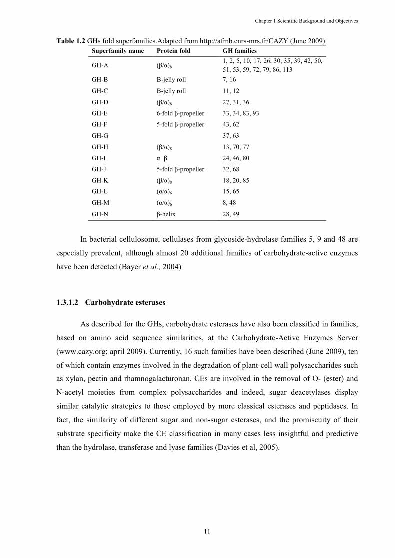
from an amino acid sequence alone, since sequence and structure are related (Table 1.2)
(Henrissat, 1991). As the catalytic mechanism is dictated by fine structure, the family
classification directly reflects the known stereochemistry of each family (Gebler et al., 1992).
In addition, members of a sequence-related family will have similar folds, and this can direct
the choice of appropriate search models for molecular replacement (Turkenburg & Dodson,
1996) opening up the potential for homology modeling of related sequences (Henrissat &
Davies, 1997). Interestingly, many of sequence-based families are polyspecific, that is, they
contain enzymes of different substrate specificities suggesting an evolutionary divergence.
Simultaneously, enzymes with similar specificities are sometimes found in different families,
which also raise the possibility of convergent evolution. GH families were further grouped in
family “clans” since some sequenced-based families displayed related structures. Compared
with the classifications described above, this classification has to be considered as a
complementary but much more powerful analytical and predictive tool (Henrissat & Davies,
1997).
Chapter 1 Scientific Background and Objectives
11
Table 1.2 GHs fold superfamilies.Adapted from http://afmb.cnrs-mrs.fr/CAZY (June 2009). Superfamily name Protein fold GH families
GH-A (β/α)8 1, 2, 5, 10, 17, 26, 30, 35, 39, 42, 50, 51, 53, 59, 72, 79, 86, 113
GH-B Β-jelly roll 7, 16
GH-C Β-jelly roll 11, 12
GH-D (β/α)8 27, 31, 36
GH-E 6-fold β-propeller 33, 34, 83, 93
GH-F 5-fold β-propeller 43, 62
GH-G 37, 63
GH-H (β/α)8 13, 70, 77
GH-I α+β 24, 46, 80
GH-J 5-fold β-propeller 32, 68
GH-K (β/α)8 18, 20, 85
GH-L (α/α)6 15, 65
GH-M (α/α)6 8, 48
GH-N β-helix 28, 49
In bacterial cellulosome, cellulases from glycoside-hydrolase families 5, 9 and 48 are
especially prevalent, although almost 20 additional families of carbohydrate-active enzymes
have been detected (Bayer et al., 2004)
1.3.1.2 Carbohydrate esterases
As described for the GHs, carbohydrate esterases have also been classified in families,
based on amino acid sequence similarities, at the Carbohydrate-Active Enzymes Server
(www.cazy.org; april 2009). Currently, 16 such families have been described (June 2009), ten
of which contain enzymes involved in the degradation of plant-cell wall polysaccharides such
as xylan, pectin and rhamnogalacturonan. CEs are involved in the removal of O- (ester) and
N-acetyl moieties from complex polysaccharides and indeed, sugar deacetylases display
similar catalytic strategies to those employed by more classical esterases and peptidases. In
fact, the similarity of different sugar and non-sugar esterases, and the promiscuity of their
substrate specificity make the CE classification in many cases less insightful and predictive
than the hydrolase, transferase and lyase families (Davies et al, 2005).
Chapter 1 Scientific Background and Objectives
12
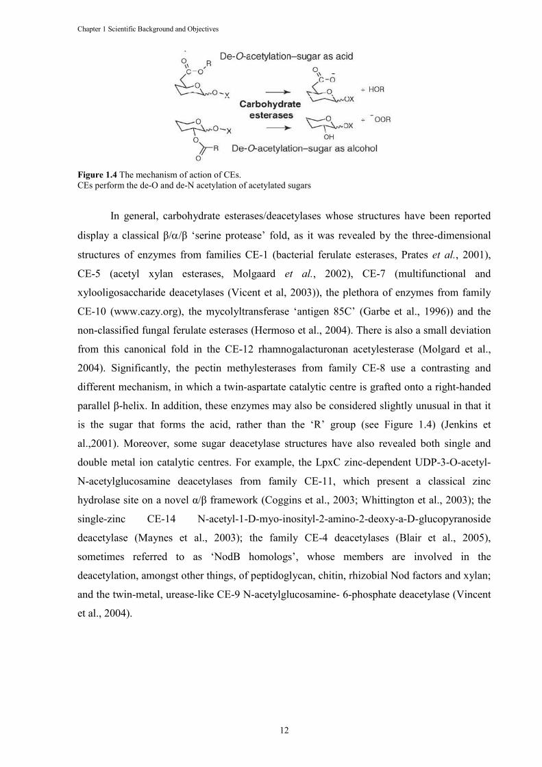
Figure 1.4 The mechanism of action of CEs. CEs perform the de-O and de-N acetylation of acetylated sugars
In general, carbohydrate esterases/deacetylases whose structures have been reported
display a classical β/α/β ‘serine protease’ fold, as it was revealed by the three-dimensional
structures of enzymes from families CE-1 (bacterial ferulate esterases, Prates et al., 2001),
CE-5 (acetyl xylan esterases, Molgaard et al., 2002), CE-7 (multifunctional and
xylooligosaccharide deacetylases (Vicent et al, 2003)), the plethora of enzymes from family
CE-10 (www.cazy.org), the mycolyltransferase ‘antigen 85C’ (Garbe et al., 1996)) and the
non-classified fungal ferulate esterases (Hermoso et al., 2004). There is also a small deviation
from this canonical fold in the CE-12 rhamnogalacturonan acetylesterase (Molgard et al.,
2004). Significantly, the pectin methylesterases from family CE-8 use a contrasting and
different mechanism, in which a twin-aspartate catalytic centre is grafted onto a right-handed
parallel β-helix. In addition, these enzymes may also be considered slightly unusual in that it
is the sugar that forms the acid, rather than the ‘R’ group (see Figure 1.4) (Jenkins et
al.,2001). Moreover, some sugar deacetylase structures have also revealed both single and
double metal ion catalytic centres. For example, the LpxC zinc-dependent UDP-3-O-acetyl-
N-acetylglucosamine deacetylases from family CE-11, which present a classical zinc
hydrolase site on a novel α/β framework (Coggins et al., 2003; Whittington et al., 2003); the
single-zinc CE-14 N-acetyl-1-D-myo-inosityl-2-amino-2-deoxy-a-D-glucopyranoside
deacetylase (Maynes et al., 2003); the family CE-4 deacetylases (Blair et al., 2005),
sometimes referred to as ‘NodB homologs’, whose members are involved in the
deacetylation, amongst other things, of peptidoglycan, chitin, rhizobial Nod factors and xylan;
and the twin-metal, urease-like CE-9 N-acetylglucosamine- 6-phosphate deacetylase (Vincent
et al., 2004).
Chapter 1 Scientific Background and Objectives
13
1.3.1.3 Carbohydrate-binding modules
Many GHs have carbohydrate-binding domains that function independently of the
catalytic domains (Warren, 1996). First known as cellulose-binding domains (CBDs), since
the first examples of these proteins were found to bind crystalline cellulose as their primary
ligand (Van Tilbeurgh et al., 1986; Tomme et al., 1988; Gilkes et al., 1988), these modules
were later named as CBMs in order to reflect their diverse ligand specificities (Boraston et al.,
1999). Many CBMs have now been identified experimentally, and several hundred putative
CBMs can be further identified on the basis of amino acid similarity. CBMs are divided into
families taking into account their amino acid similarity. There are currently (June 2009) 54
defined families of CBMs and these CBMs display substantial variation in ligand specificity
(Coutinho & Henrissat, 1999, Boraston et al., 2004).
CBMs are appended to GHs, CEs and polysaccharide lyases that degrade a variety of
polysaccharides. Although many of these modules target components of the plant cell wall,
several CBM families contain proteins that bind to insoluble storage polysaccharides such as
starch and glycogen. In some CBM families, typically those that recognize crystalline
polysaccharides, ligand specificity is invariant, while other families contain proteins that bind
a range of different carbohydrates (Boraston et al., 2004).
The CBMs are classified keeping the systematic nomenclature adopted for GHs
(Henrissat et al., 1998). A CBM is named by its family, but it may also include the organism
and even the enzyme from which it is derived to improve clarity. If the GHs contain tandem
CBMs belonging to the same family, a number corresponding to the position of the CBM in
the enzyme relative to the N-terminus is included (Boraston et al., 2004).
CBMs have three general roles with respect to their cognate catalytic modules: (i) a
proximity effect, (ii) a targeting function and (iii) a disruptive function (Boraston et al., 2004).
Through their sugar-binding activity, CBMs concentrate enzymes on to the polysaccharide
substrates. Through their carbohydrate-binding activity, CBMs promote the association of the
enzyme with its target substrate. It is believed that maintaining the enzyme in the proximity of
its substrate leads to more rapid degradation of the polysaccharide (Bolam et al., 1998),
therefore resulting in improved enzyme efficiency. There are numerous examples where
proteolytic excision or genetic truncation of CBMs from the catalytic modules significantly
decreases the activity of the enzyme against insoluble substrates (Gilkes et al., 1988; Tomme
et al., 1998; Bolam et al., 1998; Charnock et al., 2000; Ali et al., 2001; Zverlov et al., 2001;
Boraston et al., 2003a). In addition, there are CBMs that have become components of the
substrate-binding sites of GHs, and are pivotal to the substrate specificity and mode of action
Chapter 1 Scientific Background and Objectives
14
of the enzymes. Therefore, a CBM22 from Clostridium stercorarium, was shown to change
the specificity of a family 10 xylanase to a β-1,4-β-1,3-glucanase (Araki et al., 2004; Boraston
et al., 2004). CBMs also present a targeting function, allowing the appended catalytic
domains to interact with their specific substrates thus promoting enzyme accessibility in the
complexity of the plant cell wall. Finally, there are a few examples of CBMs that were
described to be directly involved in disrupting the crystalline structure of polysaccharides,
although their significance in the hydrolysis of complex polysaccharides remains to be
established.
The 54 families of CBMs (June 2009) are grouped into seven “fold families”: β-
Sandwich, β-Trefoil, Cysteine Knot, Unique, OB fold, Hevein fold and a last one which is
also Unique, but contains also a hevein-like fold. However, such grouping is not predictive of
function for the reason that sufficient diversity exists among fold family members, where
functional elements (specific amino acids or binding-site topographies) are not conserved
(Boraston et al., 2004).
Another useful classification of CBMs based on structural and functional similarities
has been proposed (Boraston et al., 2004) in which these proteins modules have been grouped
in three types: “surface-binding” CBMs (Type A), “glycan-chain-binding” CBMs (Type B)
and “small-sugar-binding” CBMs (Type C) (Table 1.3)(Boraston et al., 2004). Type A CBMs
can be appended to a variety of GHs and bind to insoluble and highly crystalline
polysaccharides, such as cellulose and chitin. Type A CBMs that have flat surfaces which are
believed to be complementary to the planar architecture of its ligand polysaccharides (Tormo
et al., 1996; McLean et al., 2000). In contrast, type B CBMs, interact with single
polysaccharide chains that act as substrates for the cognate catalytic modules to which they
are appended. These CBMs are considered to be “chain binders”. The carbohydrate-binding
sites of these CBMs are extended, often described as grooves or clefts, and comprise several
subsites able to accommodate the individual sugar units of the polymeric ligand. The depth of
these binding sites varies from very shallow to being able to accommodate the entire width of
a pyranose ring (Boraston et al., 2004). Type C CBMs are a unique class of CBMs that has
the lectin-like property of binding optimally to mono-, di-, tri-saccharides, and thus lacks the
extended binding-site grooves of type B CBMs (Boraston et al., 2004).
Chapter 1 Scientific Background and Objectives
15
Table 1.3 Classification of CBMs into functional types and their relation with the family fold and family classification by Henrissat.
CBM Type Fold family CBM families
A 1, 3, 4, 5 1, 2a, 3, 5, 10
B 1 2b, 4, 6, 11, 15, 17, 20, 22, 25, 26, 27, 28, 29, 30, 31, 33, 34, 35, 36, 41, 44, 47
C 1, 2, 6, 7 9, 12, 13, 14, 18, 32, 40, 42, 43, 50
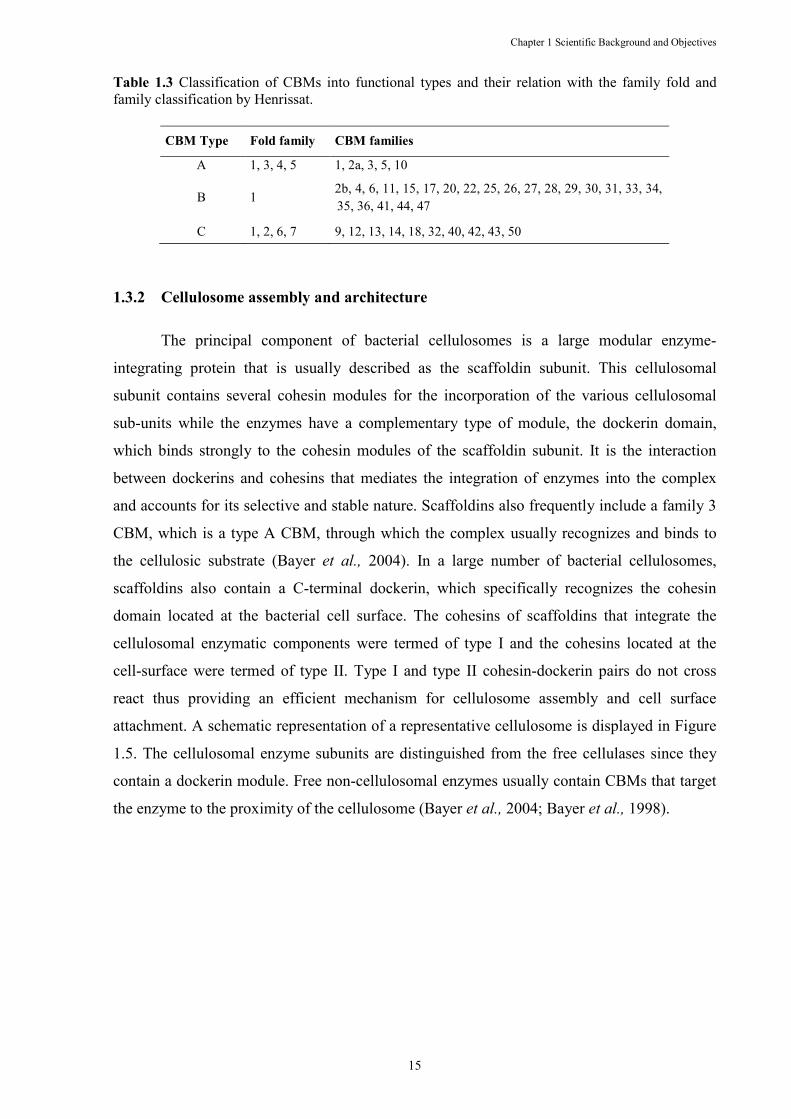
1.3.2 Cellulosome assembly and architecture
The principal component of bacterial cellulosomes is a large modular enzyme-
integrating protein that is usually described as the scaffoldin subunit. This cellulosomal
subunit contains several cohesin modules for the incorporation of the various cellulosomal
sub-units while the enzymes have a complementary type of module, the dockerin domain,
which binds strongly to the cohesin modules of the scaffoldin subunit. It is the interaction
between dockerins and cohesins that mediates the integration of enzymes into the complex
and accounts for its selective and stable nature. Scaffoldins also frequently include a family 3
CBM, which is a type A CBM, through which the complex usually recognizes and binds to
the cellulosic substrate (Bayer et al., 2004). In a large number of bacterial cellulosomes,
scaffoldins also contain a C-terminal dockerin, which specifically recognizes the cohesin
domain located at the bacterial cell surface. The cohesins of scaffoldins that integrate the
cellulosomal enzymatic components were termed of type I and the cohesins located at the
cell-surface were termed of type II. Type I and type II cohesin-dockerin pairs do not cross
react thus providing an efficient mechanism for cellulosome assembly and cell surface
attachment. A schematic representation of a representative cellulosome is displayed in Figure
1.5. The cellulosomal enzyme subunits are distinguished from the free cellulases since they
contain a dockerin module. Free non-cellulosomal enzymes usually contain CBMs that target
the enzyme to the proximity of the cellulosome (Bayer et al., 2004; Bayer et al., 1998).
Chapter 1 Scientific Background and Objectives
Figure 1.5 Schematic representation of the supramolecular architecturesurface, of representative cellulosomes. (A) Simplification of the cellulosome system. Dockerinscaffoldin by virtue of their interaction with the resident cohesins. An adaptor scaffoldinmicroorganisms, mediates the bindinganchoring scaffoldin, whose SLH module attaches the entire numerous cohesins on the scaffoldins result in an amplification of the number of enzymes incorporated per unit cellulosome. The integrity of the system is maintained by the respective specificities of the differedockerin pairs, which are color-targets the complex and the entire cell to the cellulosic substrate. The figure is representative of cellulosomes that contain a primary bearingscaffoldins that lack a dockerin (adapted from Bayer thermocellum cellulosome. Dockerin(enumerated) of the primary CipA scaffoldin. In turn, the terminal Xto the type-II cohesins of anchoring scaffoldins SdbA, Orf2p, or OlpB, each of which is connected to the csurface via an S-layer homology module
Chapter 1 Scientific Background and Objectives
16
representation of the supramolecular architecture, and disposition on the bacterial cell of representative cellulosomes.
(A) Simplification of the cellulosome system. Dockerin-containing enzymes are incorporated into a primary e of their interaction with the resident cohesins. An adaptor scaffoldin
the binding between the dockerin of the primary scaffoldin and the cohesins of an anchoring scaffoldin, whose SLH module attaches the entire cellulosome system to the cell surface. The numerous cohesins on the scaffoldins result in an amplification of the number of enzymes incorporated per unit cellulosome. The integrity of the system is maintained by the respective specificities of the differe
-coded in the figure. A single cellulose-binding CBM in the primary scaffoldin targets the complex and the entire cell to the cellulosic substrate. The figure is representative of cellulosomes
y bearing-dockerin scaffoldin and not for mesophilic clostridia that contain primary scaffoldins that lack a dockerin (adapted from Bayer et al. 2004). (B) A representation of
cellulosome. Dockerin-containing enzymes bind selectively to any of the nine type(enumerated) of the primary CipA scaffoldin. In turn, the terminal X–dockerin dyad of the CipA scaffoldin binds
II cohesins of anchoring scaffoldins SdbA, Orf2p, or OlpB, each of which is connected to the clayer homology module. Adapted from Bayer et al. (2008).
and disposition on the bacterial cell
containing enzymes are incorporated into a primary e of their interaction with the resident cohesins. An adaptor scaffoldin, present in some
between the dockerin of the primary scaffoldin and the cohesins of an cellulosome system to the cell surface. The
numerous cohesins on the scaffoldins result in an amplification of the number of enzymes incorporated per unit cellulosome. The integrity of the system is maintained by the respective specificities of the different cohesin
binding CBM in the primary scaffoldin targets the complex and the entire cell to the cellulosic substrate. The figure is representative of cellulosomes
dockerin scaffoldin and not for mesophilic clostridia that contain primary A representation of Clostridium
ly to any of the nine type-I cohesins dockerin dyad of the CipA scaffoldin binds
II cohesins of anchoring scaffoldins SdbA, Orf2p, or OlpB, each of which is connected to the cell
Chapter 1 Scientific Background and Objectives
17
1.3.2.1 The scaffoldin subunit
The scaffoldin is a major component of any cellulosome, as its ternary function
includes binding the cellulosomal enzymes, binding to the primary cellulosomal substrate,
cellulose, and binding cell-surface-associated proteins (Doi and Kosugi, 2004). Thus, each
scaffoldin usually contains several cohesin domains and a CBD or CBM (Boraston et al.,
1999). However, there are significant variations among the various scaffoldins that have been
characterized: scaffoldins can contain different types of cohesin, a dockerin, hydrophilic
domains of unknown function, GH catalytic domains, various uncharacterized domains and
even lack CBMs (Doi and Kosugi, 2004).
The first scaffoldin to be sequenced was from Clostridium cellulovorans and, as a
consequence, the cellulose binding function was recognized (Shoseyov et al., 1992).
However, the significance of the repeating elements and its relationship with the duplicated
sequences of cellulosomal enzymes was only discovered with the sequencing of a second
scaffoldin derived from C. thermocellum (Salamitou et al., 1992; Tokatlidis et al., 1991).
Shortly thereafter, several other cohesin-containing proteins were discovered downstream of
the C. thermocellum scaffoldin (Fujino et al., 1993). The scaffoldins that were sequenced
subsequently from C. cellulolyticum (Pagés et al., 1999) and C. josui (Kakiuchi et al., 1998)
resembled more the scaffoldin of C. cellulovorans rather than that of C. thermocellum, since
the latter contained a C-terminal dockerin that was absent in the others (Bayer et al., 2004).
The cellulosomal scaffoldin CBM is from family 3a, which recognizes and binds
strongly to crystalline cellulose (Morag et al., 1995). As a consequence, this CBM mediates
the primary recognition and binding of the scaffoldin subunit (along with its attached
cellulosomal enzymes) to the cellulosic substrate. In addition to the binding event, these
CBMs were suggested to play an active role in disrupting the crystalline structure of the
recalcitrant crystalline regions of cellulose (Din et al., 1991, 1994). In the C. thermocellum
cellulosome, the scaffoldin-borne dockerin interacts with the cohesins found in cell surface
proteins, which contain a C-terminal S-layer homology (SLH) module that mediates the
attachment of the protein to the cell surface (Lemaire et al., 1995). Proteins containing an
SLH module were then termed as anchoring proteins. C. thermocellum cellulosome
architecture is similar to the cellulosomes of Acetivibrio cellulolyticus and Bacteroides
cellulosolvens (Ding et al., 1999; Ding et al., 2000; Xu et al., 2003 and Xu et al., 2004). More
recently, it was realized that rumen bacteria, especially Ruminococcus flavefaciens, present a
highly complex multi-enzyme complex that only the complete sequence of this bacterium
genome will allow to elucidate (Ding et al., 2001; Rincon et al., 2003 and Rincon et al.,
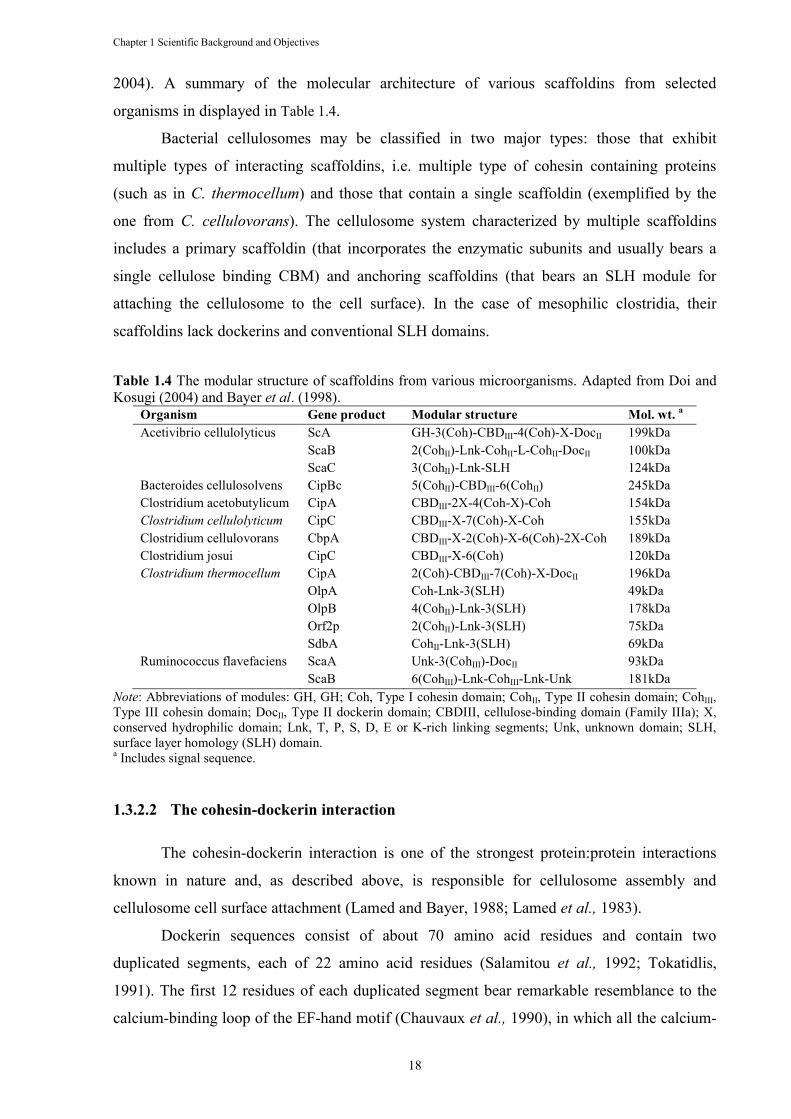
Chapter 1 Scientific Background and Objectives
18
2004). A summary of the molecular architecture of various scaffoldins from selected
organisms in displayed in Table 1.4.
Bacterial cellulosomes may be classified in two major types: those that exhibit
multiple types of interacting scaffoldins, i.e. multiple type of cohesin containing proteins
(such as in C. thermocellum) and those that contain a single scaffoldin (exemplified by the
one from C. cellulovorans). The cellulosome system characterized by multiple scaffoldins
includes a primary scaffoldin (that incorporates the enzymatic subunits and usually bears a
single cellulose binding CBM) and anchoring scaffoldins (that bears an SLH module for
attaching the cellulosome to the cell surface). In the case of mesophilic clostridia, their
scaffoldins lack dockerins and conventional SLH domains.
Table 1.4 The modular structure of scaffoldins from various microorganisms. Adapted from Doi and Kosugi (2004) and Bayer et al. (1998).
Organism Gene product Modular structure Mol. wt. a
Acetivibrio cellulolyticus ScA GH-3(Coh)-CBDIII-4(Coh)-X-DocII 199kDa ScaB 2(CohII)-Lnk-CohII-L-CohII-DocII 100kDa ScaC 3(CohII)-Lnk-SLH 124kDa Bacteroides cellulosolvens CipBc 5(CohII)-CBDIII-6(CohII) 245kDa Clostridium acetobutylicum CipA CBDIII-2X-4(Coh-X)-Coh 154kDa Clostridium cellulolyticum CipC CBDIII-X-7(Coh)-X-Coh 155kDa Clostridium cellulovorans CbpA CBDIII-X-2(Coh)-X-6(Coh)-2X-Coh 189kDa Clostridium josui CipC CBDIII-X-6(Coh) 120kDa Clostridium thermocellum CipA 2(Coh)-CBDIII-7(Coh)-X-DocII 196kDa OlpA Coh-Lnk-3(SLH) 49kDa OlpB 4(CohII)-Lnk-3(SLH) 178kDa Orf2p 2(CohII)-Lnk-3(SLH) 75kDa SdbA CohII-Lnk-3(SLH) 69kDa Ruminococcus flavefaciens ScaA Unk-3(CohIII)-DocII 93kDa ScaB 6(CohIII)-Lnk-CohIII-Lnk-Unk 181kDa
,ote: Abbreviations of modules: GH, GH; Coh, Type I cohesin domain; CohII, Type II cohesin domain; CohIII, Type III cohesin domain; DocII, Type II dockerin domain; CBDIII, cellulose-binding domain (Family IIIa); X, conserved hydrophilic domain; Lnk, T, P, S, D, E or K-rich linking segments; Unk, unknown domain; SLH, surface layer homology (SLH) domain. a Includes signal sequence.
1.3.2.2 The cohesin-dockerin interaction
The cohesin-dockerin interaction is one of the strongest protein:protein interactions
known in nature and, as described above, is responsible for cellulosome assembly and
cellulosome cell surface attachment (Lamed and Bayer, 1988; Lamed et al., 1983).
Dockerin sequences consist of about 70 amino acid residues and contain two
duplicated segments, each of 22 amino acid residues (Salamitou et al., 1992; Tokatidlis,
1991). The first 12 residues of each duplicated segment bear remarkable resemblance to the
calcium-binding loop of the EF-hand motif (Chauvaux et al., 1990), in which all the calcium-
Chapter 1 Scientific Background and Objectives
19
binding residues (i.e. aspartic acid and asparagines) are highly conserved. However, the
resemblance to the EF-hand motif was restricted to the calcium-binding loop and the F-helix
alone, thereby suggesting an “F-hand motif” (Noach et al., 2003) and the calcium dependence
of functional dockerins was demonstrated (Choi & Ljungdahl, 1996; Lytle et al., 2001; Pagès
et al., 1997; Yaron et al., 1995). The presence of the duplicated segment suggested that the
final structural model would exhibit a two-fold symmetry (Bayer at al., 2004).The cohesin
module is an elongated, conical molecule comprising a jellyroll topology that folds into a
nine-stranded β-sandwich. The two sheets of the sandwich are composed of strands 8,3,6,5
(which interacts with the dockerin) and 9,1,2,7,4, respectively, in which the C-terminal β-
strand 9 and N-termini β-strand 1 run parallel and the remaining strands are antiparallel.
Initially, it was noticed that the cohesins from the primary scaffoldins of C.
thermocellum were similar to those of other species, but differed from those of anchoring
scaffoldins, which also share similarity (Leibovitz & Béguin, 1996; Salamitou et al., 1992).
The previous were named type I cohesins and the latter were classified as type II, as it was
described above. This classification was originally linked to dockerin preference and identity
of the parent protein and later to sequence homology. With the discovery of the R.
flavefaciens cohesins, this classification was altered with the addition of a distinct class,
designated as type III (Ding et al., 2001). It seemed that there was a connection between the
cohesin type and parent protein. However, with the discovery of the reversed situation in B.
cellulosolvens, wherein the primary scaffoldin bears type II cohesins and the anchoring
scaffoldin bears those of type I, this relation was severed (Ding et al., 2000; Xu et al., 2004).
Since then, several cohesins have been described which can’t be classified in none of the
groups because their classification is located along the route connecting two of the three type
groups (Bayer et al., 2004).
1.3.2.3 Cohesin-dockerin specificity
Pagés and colleagues (1997) proposed that there is a correlation with defined positions
within the dockerin subunit that appear to represent specificity determinants of the interaction.
The dockerin sequences were compared using the observed intraspecies fidelity versus the
interspecies variance. The observed recognition codes were considered to comprise only four
residues (i.e., two tandem pairs at position 10 and 11 of the duplicated dockerin sequence) of
the dockerin domains. In the two species examined at the time (C. thermocellum and C.
cellulolyticum), these positions were essentially invariant within each species but divergent
between them. The same interspecies effect also exists between the cohesin-dockerin
Chapter 1 Scientific Background and Objectives
20
interaction of C. thermocellum and C. josui, owing to the intimate relationship and near
identity of the component sequences from the latter strain with those of C. cellulolyticum
(Jindou et al., 2004).
In order to confirm the previous hypothesis, Mechaly et al. (2000) and Schaeffer et al.
(2002) tried to convert the specificity of one dockerin species to that of the other. Thus, site-
directed mutagenesis of the suspected residues led to a creation of a new specificity, with the
mutated dockerins recognizing the cohesins from the rival species. However, these dockerins
still recognized cognate cohesins. Subsequent mutagenesis studies suggested that additional
dockerin residues (namely, positions 17 and 22 of the duplicated region) also played a role in
the exclusivity of the interaction. Position 18 was also implicated in that study, although its
requirement could not be examined by this method, because the dockerins of both species
usually bear the same residue (lysine) at this position (Mechaly et al., 2001).
The same approach could not be applied with identical success to the cohesin because
of its larger size (when comparing with the dockerin) and the numerous divergent residues
identified through sequence alignments of dockerins sequences. Mutagenesis revealed several
amino acids suspected of being involved in recognition, with the mutations resulting in the
loss of binding activity without, however, having interspecies conversion (Kakiushi et al.,
1998 and Miras et al., 2002).
1.3.2.4 The C. thermocellum and C. cellulolyticum cellulolytic system
Cellulosomes from C. thermocellum and C. cellulolyticum have differences in their
gene clusters, modular organization of the scaffoldin and in the mechanisms of cell surface
attachment. In addition, as described above, there are also differences in cohesin-dockerin
specificity that preclude cohesins and dockerins to bind to their non-cognate counterparts.
One major difference between C. thermocellum and C. cellulolyticum is the
organization of cellulase and hemicellulase encoding genes in the bacterial genomes. In C.
thermocellum, the cellulosomal genes seem to be scattered on the chromosome (Guglielmi &
Béguin, 1998), some of which are situated in relatively small clusters (Fernandes et al., 1999;
Hayashi et al., 1999; Zverlov et al., 1999, 2003). The gene products can be subgrouped
functionally into the primary scaffoldin, the cell surface-anchoring scaffoldins that mediate
the attachment of the cellulosome to the cell surface, and the catalytic subunits with their
cellulolytic and/or related activities (Bayer et al., 2004). Opposite to C. thermocellum, in C.
cellulolyticum, the cellulosomal genes are clustered on the chromosome (Bagnara-Tardif et
al., 1992; Doi et al., 2003 and Tamaru et al., 2000).
Chapter 1 Scientific Background and Objectives
21
The primary scaffoldin (cohesin integrating protein, cipA) of C. thermocellum is
located upstream of three other scaffoldin genes. All three contain cohesins and SLH
domains, indicating that the proteins bind dockerin-containing cellulosome components and
are cell surface associated. Subsequent biochemical work indicated that two of the proteins—
OlpB and Orf2p, containing seven and two cohesins, respectively— bind to the C-terminal
dockerin of the CipA primary scaffoldin (Lemaire et al., 1995; Salamitou et al., 1994;
Salamitou et al., 1992). In contrast, the single cohesin of the third scaffoldin, OlpA, was
specific for the enzyme-borne dockerin. Another gene for an additional SLH-bearing
scaffoldin, termed SdbA, whose single cohesin binds to the CipA dockerin, was located
elsewhere on the C. thermocellum chromosome (Leibovitz and Béguin, 1996). The sequences
of the nine CipA cohesins and the single OlpA cohesin are all of type I, whereas those of
OlpB, Orf2p, and SdbA are type-II cohesins. In contrast to what was observed for C.
thermocellum, the scaffoldin of C. cellulolyticum (Bagnara-Tardif et al., 1992, Belaich et al.,
1999), as the ones from C. acetobutylicum (Nolling et al., 2001), C. cellulovorans and C.
josui (Tamaru et al., 2000), lacks C-terminal dockerins but contains multiple (between six and
nine) cohesins interspersed with one or more X2 domains of currently unverified function.
The four clusters contain numerous genes enconding family-9 cellulases (Bayer et al., 2004).
Another difference between the cellulosomes of these two organisms seems to be the
way their cellulosome attaches to the cell surface. The C. thermocellum cellulosome binds
envelop proteins through a type II dockerin domain present in its scaffoldin (CipA); this type
II dockerin interacts with the type II cohesin domain present in its three cell surface proteins
(SdbA, OlpB and Orf2p) which permit the non-covalent attachment of the cellulosome on the
bacterial cell surface. The observation that some of these proteins contain more than one copy
of type II cohesins explains how cellulosomes can be organized in polycellulosomes. These
cell surface proteins seem not to be present in the C. cellulolyticum, so the mechanism for
cellulosome cell surface attachment remains unknown (Desvaux, 2005).
1.3.2.5 Cohesin-dockerin structural models
For a long time the determination of the cohesin-dockerin complex structure has been
elusive even if considered as one of the most important goals in this field. For this reason,
crystals structures were initially only reported for various individual cohesins from the
cellulosomal scaffoldin subunits of C. thermocellum and C. cellulolyticum (Shimon et al.,
1997; Spinelli et al., 2000; Tavares et al., 1997). The dockerin domain also remained difficult
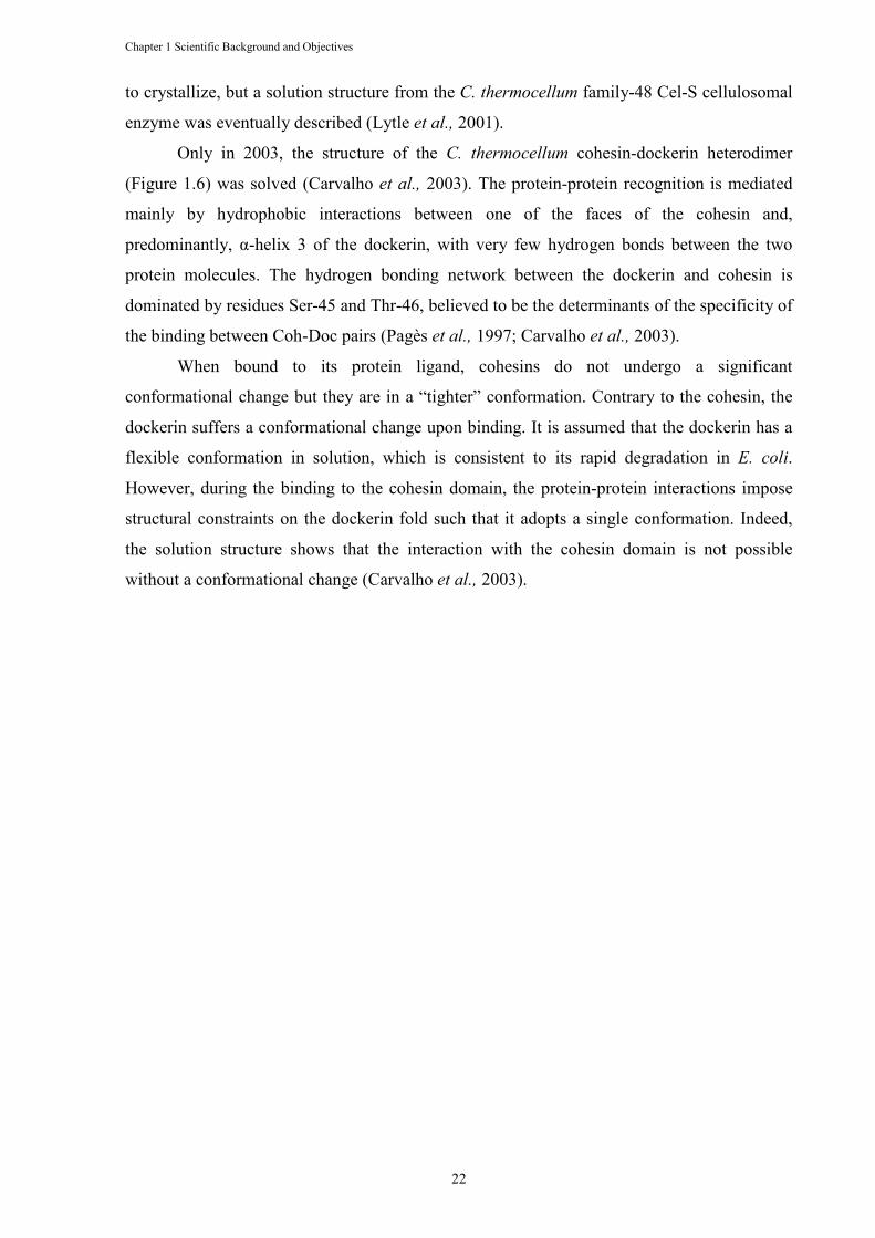
Chapter 1 Scientific Background and Objectives
22
to crystallize, but a solution structure from the C. thermocellum family-48 Cel-S cellulosomal
enzyme was eventually described (Lytle et al., 2001).
Only in 2003, the structure of the C. thermocellum cohesin-dockerin heterodimer
(Figure 1.6) was solved (Carvalho et al., 2003). The protein-protein recognition is mediated
mainly by hydrophobic interactions between one of the faces of the cohesin and,
predominantly, α-helix 3 of the dockerin, with very few hydrogen bonds between the two
protein molecules. The hydrogen bonding network between the dockerin and cohesin is
dominated by residues Ser-45 and Thr-46, believed to be the determinants of the specificity of
the binding between Coh-Doc pairs (Pagès et al., 1997; Carvalho et al., 2003).
When bound to its protein ligand, cohesins do not undergo a significant
conformational change but they are in a “tighter” conformation. Contrary to the cohesin, the
dockerin suffers a conformational change upon binding. It is assumed that the dockerin has a
flexible conformation in solution, which is consistent to its rapid degradation in E. coli.
However, during the binding to the cohesin domain, the protein-protein interactions impose
structural constraints on the dockerin fold such that it adopts a single conformation. Indeed,
the solution structure shows that the interaction with the cohesin domain is not possible
without a conformational change (Carvalho et al., 2003).
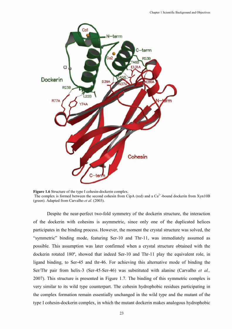
Chapter 1 Scientific Background and Objectives
23
Figure 1.6 Structure of the type I cohesin-dockerin complex. The complex is formed between the second cohesin from CipA (red) and a Ca2+-bound dockerin from Xyn10B (green). Adapted from Carvalho et al. (2003).
Despite the near-perfect two-fold symmetry of the dockerin structure, the interaction
of the dockerin with cohesins is asymmetric, since only one of the duplicated helices
participates in the binding process. However, the moment the crystal structure was solved, the
“symmetric” binding mode, featuring Ser-10 and Thr-11, was immediately assumed as
possible. This assumption was later confirmed when a crystal structure obtained with the
dockerin rotated 180º, showed that indeed Ser-10 and Thr-11 play the equivalent role, in
ligand binding, to Ser-45 and thr-46. For achieving this alternative mode of binding the
Ser/Thr pair from helix-3 (Ser-45-Ser-46) was substituted with alanine (Carvalho et al.,
2007). This structure is presented in Figure 1.7. The binding of this symmetric complex is
very similar to its wild type counterpart. The cohesin hydrophobic residues participating in
the complex formation remain essentially unchanged in the wild type and the mutant of the
type I cohesin-dockerin complex, in which the mutant dockerin makes analogous hydrophobic
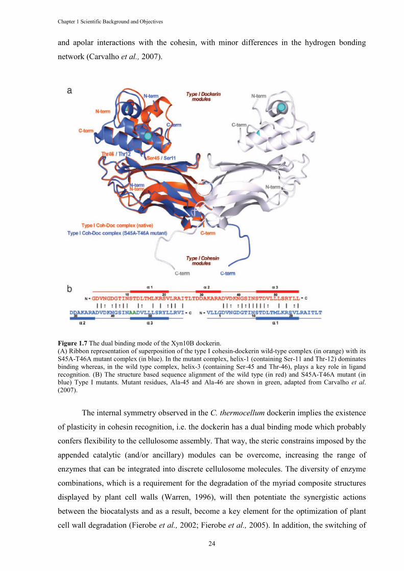
Chapter 1 Scientific Background and Objectives
24
and apolar interactions with the cohesin, with minor differences in the hydrogen bonding
network (Carvalho et al., 2007).
Figure 1.7 The dual binding mode of the Xyn10B dockerin. (A) Ribbon representation of superposition of the type I cohesin-dockerin wild-type complex (in orange) with its S45A-T46A mutant complex (in blue). In the mutant complex, helix-1 (containing Ser-11 and Thr-12) dominates binding whereas, in the wild type complex, helix-3 (containing Ser-45 and Thr-46), plays a key role in ligand recognition. (B) The structure based sequence alignment of the wild type (in red) and S45A-T46A mutant (in blue) Type I mutants. Mutant residues, Ala-45 and Ala-46 are shown in green, adapted from Carvalho et al. (2007).
The internal symmetry observed in the C. thermocellum dockerin implies the existence
of plasticity in cohesin recognition, i.e. the dockerin has a dual binding mode which probably
confers flexibility to the cellulosome assembly. That way, the steric constrains imposed by the
appended catalytic (and/or ancillary) modules can be overcome, increasing the range of
enzymes that can be integrated into discrete cellulosome molecules. The diversity of enzyme
combinations, which is a requirement for the degradation of the myriad composite structures
displayed by plant cell walls (Warren, 1996), will then potentiate the synergistic actions
between the biocatalysts and as a result, become a key element for the optimization of plant
cell wall degradation (Fierobe et al., 2002; Fierobe et al., 2005). In addition, the switching of
Chapter 1 Scientific Background and Objectives
25
the mode of binding from one site to another can introduce quaternary flexibility into the
multienzyme complex and enhance substrate targeting and hydrolysis (Carvalho et al., 2007).
Figure 1.8 Structure of the type II Coh–XDoc complex. Ribbon representation of the complex with the type II Coh module in blue, type II Doc in green, and X module in magenta. The β-strands of the X module and type II Coh are numbered in yellow. The N- and C-termini are labeled accordingly, and the Ca2+ ions are depicted as orange spheres. Adapted from Adams et al. (2006).
More recently, the crystal structure of a type II cohesin-dockerin complex was
obtained almost three years after the first type I complex had been solved (Figure 1.8). As it
was observed with the type I interactions, the first type II crystal structures to be solved were
from individual type II cohesins from various microorganisms. The structures of type II
cohesins obtained from C. thermocellum (Carvalho et al., 2005), Acetivibrio cellulolyticus
(Noach et al., 2003) and Bacteroide cellulosolvens (Noach et al., 2005), suggest a β-jellyroll
topology common to the type I cohesin modules. However, the type II cohesin had a unique
crowning α-helix, first identified by solution NMR studies (Smith et al., 2002), and two
regions disrupting strands 4 and 8, termed β-flaps, which have been proposed to play roles in
the type II interaction and specificity (Carvalho et al., 2005, Noach et al., 2005). The reason
for the delay in solving the type II complex was due, primarily, to the fact that the role of the
module appended to the type II dockerin was unknown. This module, named as module X, is
now known to be important for dockerin stability and enhancement of cohesin recognition.
The tight interaction involves one face of the cohesin and, in contrast to the type I complex,
Chapter 1 Scientific Background and Objectives
26
both helixes of the dockerin. As the type I complex, it comprises a significant hydrophobic
character and a complementary extensive hydrogen-bond network. However, the interacting
surfaces in the type II complex are less charged than the analogous surfaces from the type I
complex, with more bulky aliphatic side-chain groups (Adams et al, 2006).
1.4 OBJECTIVES
Overall, this study aims to elucidate several unresolved questions concerning the
importance of the cohesin-dockerin interaction in the assembly of C. thermocellum and C.
cellulolyticum cellulosomes. In addition, there are several cellulosomal catalytic components
whose function remains unknown, thus limiting our understanding of the full enzymatic
capabilities of this highly efficient nanomachine. The specific objectives of this thesis can,
therefore, be resumed to the following points:
• To explore the differences in specificity and selectivity established between
various C. thermocellum type I cohesins (Coh-CipA2, Coh-OlpC and Coh-
OlpA) and dockerins (Doc-Xyn10B, Doc-435, Doc-918, Doc-624 and Doc-
Cel44). To probe the capacity of the mentioned dockerins to interact with C.
cellulolyticum partners (Chapter 2).
• To obtain the structure of C. cellulolyticum cohesin-dockerin complexes and
probe the existence of a dual binding mode in the dockerins produced by this
bacterium (Chapter 3).
• To obtain the crystal structure of the N-terminal CBM22-GH10 domain of
Xyn10B (Chapter 4). The CBM22 module of Xyn10B does not present a CBM
or thermostabilizing function and thus structural information may help
clarifying the role of Xyn10B in plant cell wall degradation.
• To study the biochemical and structural properties of CtCE2, a protein that
displays divergent catalytic esterase and non-catalytic carbohydrate-binding
functions and which is an important component of C. thermocellum
cellulosomes (Chapter 5).
Chapter 2 Novel type I cohesins and dockerin domains
27
Chapter 2 Functional insights into the role of novel type I cohesin
and dockerin domains from Clostridium thermocellum
Benedita A. Pinheiro1, Harry J. Gilbert2, Kazutaka Sakka3, Kazuo Sakka3, Vânia O. Fernandes1, José A. M.
Prates1, Victor D. Alves1, David N. Bolam2, Luís M. A. Ferreira1 and Carlos M. G. A Fontes1
1 CIISA – Faculdade de Medicina Veterinária, Pólo Universitário do Alto da Ajuda, Avenida da Universidade
Técnica, 1300-477 Lisboa, Portugal; 2 Institute for Cell and Molecular Biosciences, Newcastle University, The
Medical School, Newcastle upon Tyne NE2 4HH, United Kingdom; 3 Applied Microbiology Laboratory,
Graduate School of Bioresources, Mie University, Tsu, Japan
Submited to Biochemical Journal
Abstract
Cellulosomes, synthesized by anaerobic microorganisms such as Clostridium thermocellum, are highly elaborate
and remarkably complex nanomachines that efficiently degrade plant cell wall polysaccharides, particularly
cellulose. Cellulosome assembly results from the interaction of type I dockerin domains, present on the catalytic
subunits, and the cohesin domains of a large non-catalytic integrating protein that acts as a molecular scaffold. In
general type I dockerins contain two distinct cohesin binding interfaces that appear to display identical ligand
specificities. Inspection of the Clostridium thermocellum genome reveals 72 dockerin-containing proteins. In
four of these proteins, Cthe_0258, Cthe_0435, Cthe_0624 and Cthe_0918, there are significant differences in the
residues that comprise the two ligand binding sites of their type I dockerins. In addition, a protein of unknown
function (Cthe_0452), containing a C-terminal cohesin highly similar to the equivalent domains present in C.
thermocellum CipA, was also identified. Here the ligand specificities of the newly identified cohesin and
dockerin domains are described. The results revealed that Cthe_0452 is located at the C. thermocellum cell
surface and thus the protein was renamed as OlpC. The dockerins of proteins Cthe_0258 and Cthe_0435
recognize, preferentially, the OlpC cohesin and thus these enzymes are believed to be predominantly located at
the surface of the bacterium. By contrast, the dockerin domains of Cthe_0624 and Cthe_0918 are primarily
cellulosomal since they bind preferentially to the cohesins of CipA. OlpC, which is relatively abundant protein,
may also adopt a “warehouse” function by transiently retaining cellulosomal enzymes at the cell surface before
they are assembled onto the multi-enzyme complex.
Chapter 2 Novel type I cohesins and dockerin domains
28
2.1 I�TRODUCTIO�
The plant cell wall represents the most abundant source of terrestrial organic carbon.
The degradation of this composite structure by microbial enzymes is central to the carbon
cycle, while there is currently considerable interest in the application of these biocatalysts in
the conversion of lignocellulosic biomass into biofuels such as ethanol and butanol. The
complex chemical and physical structure of plant cell walls restrict enzyme access to the
interlocking polysaccharides, primarily cellulose and hemicellulose, thus limiting carbon
turnover. A common feature of anaerobic plant cell wall degrading microorganisms is that
extracellular cellulases and hemicellulases are organized into high molecular weight multi-
enzyme complexes, which are referred to as cellulosomes (Bayer, et al., 1994, Beguin &
Alzari, 1998, Doi & Tamaru, 2001). Cellulosomes enhance the hydrolytic activity of the
assembled enzymes by i) potentiating the synergistic interactions between the biocatalysts
and, ii) through a central cellulose-specific carbohydrate binding module, bring the catalytic
subunits into intimate contact with their target substrates, thereby reducing the enzyme
accessibility problem (Fierobe, et al., 2001, Fierobe, et al., 2002).
Clostridium thermocellum displays one of the fastest known growth rates on cellulose
and its cellulosome is the paradigm for plant cell wall degrading enzyme complexes. The
protein that mediates the assembly of the C. thermocellum cellulosome is the scaffoldin
subunit termed CipA – a 1853 amino acid non-catalytic polypeptide that contains nine highly
conserved modules, known as type I cohesins, a family 3 CBM that attaches the cellulosome
onto crystalline cellulose, and a type II dockerin (Gerngross, et al., 1993). Type I dockerins,
located in cellulosomal enzymes, primarily GHs, but also CEs and polysaccharide lyases, bind
extremely tightly to CipA cohesins thus anchoring the enzymes onto the macromolecular
scaffold (Bayer, et al., 1994, Beguin & Alzari, 1998). The C-terminal type II dockerin of
CipA, which recognizes, specifically, type II cohesins located in proteins bound to the
bacterial peptidoglycan layer, maintains the cellulosome on the bacterial cell surface
(Lemaire, et al., 1995). There is no cross specificity between type I and type II cohesin-
dockerin partners. Significantly, a type I cohesin domain was also identified in a cell bound
protein, OlpA, suggesting that cellulosomal enzymes can also adhere directly, and
individually, onto the bacterial surface (Salamitou, et al., 1994).
Crystal structures of type I cohesin-dockerin complexes have provided important
insights into the mechanism of cellulosome assembly. Dockerins contain a tandem duplication
of a 22-residue sequence that are organized into two F-hand calcium binding motif and an α-
helix that also display remarkable structural conservation – the N and C-terminal α-helices
Chapter 2 Novel type I cohesins and dockerin domains
29
overlay with minor structural deviations (Carvalho, et al., 2003, Adams, et al., 2006,
Carvalho, et al., 2007). Internal two-fold symmetry of the dockerin molecule has a profound
influence on the mechanism of cohesin recognition. Structural and mutagenesis data revealed
that type I dockerins contain two, essentially identical, cohesin-binding interfaces. Residues
participating in cohesin recognition at the two binding interfaces, particularly a Ser-Thr motif
at positions 11 and 12 and a Lys-Arg motif at positions 18 and 19, are highly conserved in the
two segments of the majority of C. thermocellum dockerins (see below), suggesting that, in
general, both binding interfaces display similar protein specificities(Carvalho, et al., 2007,
Karpol, et al., 2008). The dockerin dual binding mode may reduce the steric constraints that
are likely to be imposed by assembling a large number of different catalytic modules into a
single cellulosome. In addition, the switching of the binding mode between two
conformations may also introduce quaternary flexibility into multi-enzyme complexes thus
enhancing substrate targeting and the synergistic interactions between some enzymes,
particularly exo- and endo-acting cellulases (Carvalho, et al., 2003, Carvalho, et al., 2007).
Similar structural observations were made for C. cellulolyticum dockerins, within cohesin-
dockerin complexes, suggesting an evolutionary pressure for the retention of a dual binding
mode in, at least, Clostridial dockerins (Pinheiro, et al., 2008). Despite the structural similarity
between the C. thermocellum and C. cellulolyticum cohesins and dockerins, there is no cross
specificity between the protein partners of the two organisms (Pages, et al., 1997). Residues at
dockerin positions 11 and 12 were shown to play a critical role in organism-specific
recognition (Mechaly, et al., 2000, Mechaly, et al., 2001), with C. thermocellum dockerins
presenting, typically, a pair of hydroxy residues, while the corresponding amino acids in C.
cellulolyticum dockerins are alanine and a second, larger, hydrophobic amino acid.
The genome of C. thermocellum ATCC 27405 encodes 72 polypeptides containing
type I dockerin sequences. Inspection of dockerin sequences at the two ligand binding sites
revealed a strong conservation of the amino acids that mediate cohesin recognition. There are,
however, at least four dockerins that are components of the proteins Cthe_0435, Cthe_0918,
Cthe_0258 and Cel9D-Cel44A (accession number Cthe_0624), which deviate from the
canonical C. thermocellum motifs in one of the ligand binding interfaces. In these dockerins
the usually conserved Ser-Thr pair, which dominates the hydrogen bond network with the
cohesin, is replaced in one of the duplicated segments by non-hydroxy residues. The
implications of these amino acid substitutions for cohesin recognition remain to be
investigated. Here we have established the ligand specificity of C. thermocellum dockerins
which display divergent cohesin binding interfaces.

Chapter 2 Novel type I cohesins and dockerin domains
30
2.2 EXPERIME�TAL PROCEDURES
2.2.1 Cloning, expression and purification
Genes encoding dockerin domains were amplified from C. thermocellum genomic
DNA using the thermostable DNA polymerase NZYPremium (NZYTech Ltd). Amplified
DNA was directly cloned into pNZY28 (NZYTech Ltd) and sequenced to ensure that no
mutations were accumulated during the amplification. Genes encoding the dockerin domains
of Cthe_0435, termed Doc-435 (residues 32-112), Cthe_0918, termed Doc-918 (residues
1146-1209) and Cthe_0258, termed Doc-258 (residues 33-105), were subcloned into pET32a
(Novagen) restricted with EcoR1 and XhoI (see Table 2.1). Recombinant Doc-435, Doc-918
and Doc-258 were expressed in fusion with thioredoxin, to improve dockerin solubility and
stability. Similarly, to improve protein stability, the dockerin domain of Xyn10B was
produced in fusion with the N-terminal CBM22 xylan-binding domain and the Cel9D-Cel44A
dockerin was expressed with the GH44 catalytic domain at the N-terminus and the polycystic
kidney-disease like module (PKD) followed by the family 44 CBM (CBM44) at the C-
terminus. The genes encoding the Xyn10B derivative, termed Doc-Xyn10B (residues 558-
799, Cthe_0912) and Cel9D-Cel44A derivative, termed Doc-Cel44A (residues 774-1601,
Cthe_0624), were subcloned into pET21a using a primer pair containing ,heI and XhoI
restriction sites, respectively. The recombinant plasmid encoding Doc-Cel44A was termed
pD44-21a. Genes encoding the second cohesin of CipA, termed Coh-CipA2 (residues 182-
328, Cthe_3077), the type I cohesin of OlpA, termed Coh-OlpA (residues 30-175,
Cthe_3080), and type I cohesin of OlpC, termed Coh-OlpC (residues 100-258, Cthe_0452)
were amplified as described above, cloned into pNZY28 (NZYTech Ltd.) and sequenced.
Coh-OlpA and Coh-OlpC genes were subcloned into BglII/EcoRI and BamHI/EcoRI digested
pRSETa, respectively (Invitrogen). The gene encoding Coh-CipA2 was sub-cloned into
pET21a (Novagen). The DNA sequences encoding the N-terminal domain of Cthe_0452
(residues 25-112), termed CtUnk1, and the type II dockerin of CipA fused with the
endogenous N-terminal X module (residues 1691-1853, Cthe_3077) were amplified using the
procedures described above and the primers presented in Table 1. Both genes were
subsequently subcloned into pET32a (Novagen). Clostridium cellulolyticum type I cohesin
was produced using plasmid pHF1, as described by Pinheiro et al. (Pinheiro, et al., 2008). All
recombinant proteins contained an internal or a C-terminal His6-tag.
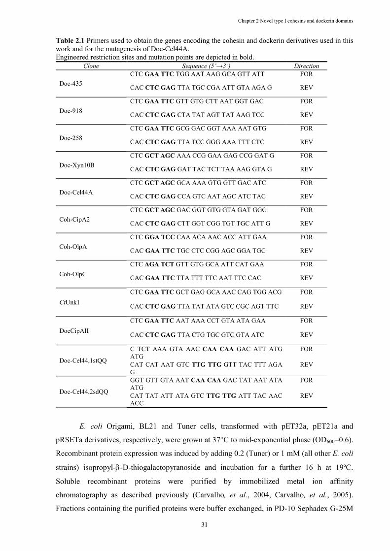
Chapter 2 Novel type I cohesins and dockerin domains
31
Table 2.1 Primers used to obtain the genes encoding the cohesin and dockerin derivatives used in this work and for the mutagenesis of Doc-Cel44A. Engineered restriction sites and mutation points are depicted in bold.
Clone Sequence (5’→3’) Direction
Doc-435 CTC GAA TTC TGG AAT AAG GCA GTT ATT FOR
CAC CTC GAG TTA TGC CGA ATT GTA AGA G REV
Doc-918 CTC GAA TTC GTT GTG CTT AAT GGT GAC FOR
CAC CTC GAG CTA TAT AGT TAT AAG TCC REV
Doc-258 CTC GAA TTC GCG GAC GGT AAA AAT GTG FOR
CAC CTC GAG TTA TCC GGG AAA TTT CTC REV
Doc-Xyn10B CTC GCT AGC AAA CCG GAA GAG CCG GAT G FOR
CAC CTC GAG GAT TAC TCT TAA AAG GTA G REV
Doc-Cel44A CTC GCT AGC GCA AAA GTG GTT GAC ATC FOR
CAC CTC GAG CCA GTC AAT AGC ATC TAC REV
Coh-CipA2 CTC GCT AGC GAC GGT GTG GTA GAT GGC FOR
CAC CTC GAG CTT GGT CGG TGT TGC ATT G REV
Coh-OlpA CTC GGA TCC CAA ACA AAC ACC ATT GAA FOR
CAC GAA TTC TGC CTC CGG AGC GGA TGC REV
Coh-OlpC CTC AGA TCT GTT GTG GCA ATT CAT GAA FOR
CAC GAA TTC TTA TTT TTC AAT TTC CAC REV
CtUnk1
CTC GAA TTC GCT GAG GCA AAC CAG TGG ACG FOR
CAC CTC GAG TTA TAT ATA GTC CGC AGT TTC REV
DocCipAII
CTC GAA TTC AAT AAA CCT GTA ATA GAA FOR
CAC CTC GAG TTA CTG TGC GTC GTA ATC REV
Doc-Cel44,1stQQ
C TCT AAA GTA AAC CAA CAA GAC ATT ATG ATG
FOR
CAT CAT AAT GTC TTG TTG GTT TAC TTT AGA G
REV
Doc-Cel44,2sdQQ
GGT GTT GTA AAT CAA CAA GAC TAT AAT ATA ATG
FOR
CAT TAT ATT ATA GTC TTG TTG ATT TAC AAC ACC
REV
E. coli Origami, BL21 and Tuner cells, transformed with pET32a, pET21a and
pRSETa derivatives, respectively, were grown at 37°C to mid-exponential phase (OD600=0.6).
Recombinant protein expression was induced by adding 0.2 (Tuner) or 1 mM (all other E. coli
strains) isopropyl-β-D-thiogalactopyranoside and incubation for a further 16 h at 19ºC.
Soluble recombinant proteins were purified by immobilized metal ion affinity
chromatography as described previously (Carvalho, et al., 2004, Carvalho, et al., 2005).
Fractions containing the purified proteins were buffer exchanged, in PD-10 Sephadex G-25M
Chapter 2 Novel type I cohesins and dockerin domains
32
gel filtration columns (GE Healthcare), into 50 mM Hepes, pH 7.5, containing 100 mM NaCL
and 5 mM CaCl2. For isothermal calorimetry, a further purification step by gel exclusion
chromatography was performed and the proteins were maintained in the same buffer
excluding the NaCl (GE Healthcare). SDS/PAGE showed that all the recombinant proteins
were more than 99 % pure.
2.2.2 Mutagenesis
Site-directed mutagenesis was carried out using the PCR-based NZYMutagenesis site-
directed mutagenesis kit (NZYTech Ltd) according to the manufacturer’s instructions, using
plasmid pD44-21a as template. The sequences of the primers used to generate these mutants
are displayed in Table 2.1. The mutated DNA sequences were sequenced to ensure that only
the appropriate mutations had been incorporated into the nucleic acid.
2.2.3 Interaction of CtUnk1 with bacterial cell wall preparations
Native peptidoglycan-containing sacculi (NPCS) and hydrofluoric acid-extracted cell-
wall polymer (HF-EPCS) were prepared from E. coli and C. thermocellum cells as described
previously (Zhao, et al., 2006). Recombinant CtUnk1 (10 µg) was separately incubated with
NPCS and HF-EPCS (80 µg), in 50 µl of 20 mM sodium phosphate buffer (pH 7.4), at 4ºC for
1 h with occasional shaking. Following incubation, the insoluble fractions were pelleted by
centrifugation and the supernatant and pellet fractions were analyzed by SDS-PAGE.
2.2.4 Complex Formation in Solution
As an initial approach to test cohesin-dockerin affinity, the two proteins were
combined in 50 Hepes buffer (pH 7.5), containing 100 mM NaCl and 5 mM CaCl2 for 1 hour
at room temperature and the presence of complexes was evaluated by non-denaturing
polyacrylamide gel electrophoresis. For lower affinity interactions, complexes were detected
by increasing concentrations of a dockerin against a fixed concentration of cohesin. New
bands appearing in the native gel were used as an indication of complex formation. The
differences in the affinities of the complexes were assessed by combining, in the same
solution, equimolar concentrations of two dockerins and one cohesin (or two cohesins and one
dockerin) and analyzing the gel for complex formation. To determine the stability of cohesin-
dockerin complexes, a cohesin-dockerin pair was assembled for an hour. After this incubation
Chapter 2 Novel type I cohesins and dockerin domains
33
period a second cohesin or dockerin was added to the mixture and incubated for another hour.
Complex stability was evaluated by non-denaturing gels as explained above.
2.2.5 Isothermal Calorimetry of Coh-Doc Binding
Isothermal titration calorimetry was carried essentially as described previously
(Carvalho, et al., 2007, Pinheiro, et al., 2008), except that measurements were made at 55°C,
and proteins were dialyzed into 50 mM NaHepes, pH 7.5, containing 2 mM CaCl2. During
titration, the dockerin (15 µM) was stirred at 300 rpm in the reaction cell, which was injected
with 29 or 58 successive 5-µl aliquots of ligand comprising cohesin (180 µM) at 200-s
intervals. Integrated heat effects, after correction for heats of dilution, were analyzed by
nonlinear regression by using a single site-binding model (Microcal ORIGIN, Ver. 5.0,
Microcal Software, Northampton, MA). The fitted data yield the association constant (KA) and
the enthalpy of binding (∆H). Other thermodynamic parameters were calculated by using the
standard thermodynamic equation: ∆RTlnKA = -∆G = ∆H-T∆S. The c values (product of the
molar concentration of binding sites × the association constant) were ≈100.
2.3 RESULTS A�D DISCUSSIO�
2.3.1 �ovel type I cohesin and dockerin domains in C. thermocellum proteins
The C. thermocellum proteome was searched using Xyn10B (Fontes, et al., 1995b)
and Cel9D-Cel44A (Ahsan, et al., 1996, Ahsan, et al., 1997, Najmudin, et al., 2006) dockerin
sequences through BLAST (http://blast.ncbi.nlm.nih.gov) to identify the complete repertoire
of cellulosomal proteins. The data revealed that 72 C. thermocellum proteins contain type I
dockerins. Alignment of all the identified dockerins revealed that at least four of these
domains display a lack of conservation in the residues that interact with the type I cohesin
partners present in CipA (data not shown). These domains belong to proteins Cthe_0258,
Cthe_0435, Cthe_0624 and Cthe_0918. Cthe_0624 is the bifunctional cellulase Cel9D-
Cel44A (initially referred to as CelJ (Ahsan, et al., 1997)) where the dockerin is located
internally between the two catalytic modules (GH9 and GH44) and a C-terminal CBM44
(Najmudin, et al., 2006). The other three proteins do not have an assigned function and the
dockerin is positioned either at the N- (Cthe_0258 and Cthe_0435) or C-terminus
(Cthe_0918) (Figure 2.1). Modules of unknown function in Cthe_0258, Cthe_0435 and
Cthe_0918 have, respectively, 365, 218 and 1136 residues and may comprise catalytic
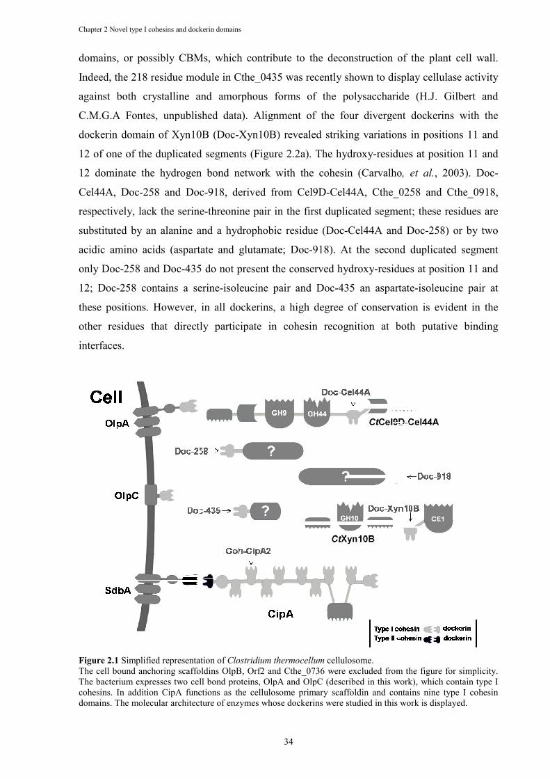
Chapter 2 Novel type I cohesins and dockerin domains
domains, or possibly CBMs, which contribute to the deconstruction of
Indeed, the 218 residue module in Cthe_0435 was recently shown to display cellulase activity
against both crystalline and amorphous forms of the polysaccharide (H.J. Gilbert and
C.M.G.A Fontes, unpublished data). Alignment of the four
dockerin domain of Xyn10B (Doc
12 of one of the duplicated segments (
12 dominate the hydrogen bond network with the cohesin
Cel44A, Doc-258 and Doc
respectively, lack the serine
substituted by an alanine and a hydrophobic residue (Doc
acidic amino acids (aspartate and glutamate; Doc
only Doc-258 and Doc-435 do not present the conserved hydroxy
12; Doc-258 contains a serine
these positions. However, in all dockerins, a high degree of conservation is evident in the
other residues that directly participate in cohesin recognition at both put
interfaces.
Figure 2.1 Simplified representation of The cell bound anchoring scaffoldins OlpB, Orf2 and Cthe_0736 were excluded from the figure for The bacterium expresses two cell bond proteins, OlpA and OlpC (described in this work), which contain type I cohesins. In addition CipA functions as the cellulosome primary scaffoldin and contains nine type I cohesin domains. The molecular architecture of enzymes whose dockerins were studied in this work is displayed
Chapter 2 Novel type I cohesins and dockerin domains
34
domains, or possibly CBMs, which contribute to the deconstruction of
Indeed, the 218 residue module in Cthe_0435 was recently shown to display cellulase activity
against both crystalline and amorphous forms of the polysaccharide (H.J. Gilbert and
C.M.G.A Fontes, unpublished data). Alignment of the four divergent dockerins with the
dockerin domain of Xyn10B (Doc-Xyn10B) revealed striking variations in positions 11 and
12 of one of the duplicated segments (Figure 2.2a). The hydroxy-residues at position 11 and
12 dominate the hydrogen bond network with the cohesin (Carvalho
258 and Doc-918, derived from Cel9D-Cel44A, Cthe_0258 and
respectively, lack the serine-threonine pair in the first duplicated segment; these residues are
d by an alanine and a hydrophobic residue (Doc-Cel44A and Doc
acidic amino acids (aspartate and glutamate; Doc-918). At the second duplicated segment
435 do not present the conserved hydroxy-residues at position 11 and
258 contains a serine-isoleucine pair and Doc-435 an aspartate
these positions. However, in all dockerins, a high degree of conservation is evident in the
other residues that directly participate in cohesin recognition at both put
Simplified representation of Clostridium thermocellum cellulosome. The cell bound anchoring scaffoldins OlpB, Orf2 and Cthe_0736 were excluded from the figure for The bacterium expresses two cell bond proteins, OlpA and OlpC (described in this work), which contain type I cohesins. In addition CipA functions as the cellulosome primary scaffoldin and contains nine type I cohesin
itecture of enzymes whose dockerins were studied in this work is displayed
domains, or possibly CBMs, which contribute to the deconstruction of the plant cell wall.
Indeed, the 218 residue module in Cthe_0435 was recently shown to display cellulase activity
against both crystalline and amorphous forms of the polysaccharide (H.J. Gilbert and
divergent dockerins with the
Xyn10B) revealed striking variations in positions 11 and
residues at position 11 and
(Carvalho, et al., 2003). Doc-
Cel44A, Cthe_0258 and Cthe_0918,
threonine pair in the first duplicated segment; these residues are
Cel44A and Doc-258) or by two
918). At the second duplicated segment
residues at position 11 and
435 an aspartate-isoleucine pair at
these positions. However, in all dockerins, a high degree of conservation is evident in the
other residues that directly participate in cohesin recognition at both putative binding
The cell bound anchoring scaffoldins OlpB, Orf2 and Cthe_0736 were excluded from the figure for simplicity. The bacterium expresses two cell bond proteins, OlpA and OlpC (described in this work), which contain type I cohesins. In addition CipA functions as the cellulosome primary scaffoldin and contains nine type I cohesin
itecture of enzymes whose dockerins were studied in this work is displayed.
Chapter 2 Novel type I cohesins and dockerin domains
35
A similar bioinformatic strategy was employed to identify the complete array of type I
cohesin domains encoded by the C. thermocellum genome. Three proteins were shown to
contain type I cohesins: the scaffolding protein CipA (Cthe_3077), which contains nine
cohesin modules, OlpA, with one cohesin module (Cthe_3080) and a previously unknown
protein, Cthe_0452, which also contains a single type I cohesin domain (Figure 2.1). OlpA is
a cell surface bi-modular protein presenting an N-terminal type I cohesin domain fused to
three SLH repeats (Salamitou, et al., 1994). The SLH repeats were previously shown to
interact with the bacterial surface (Salamitou, et al., 1994, Zhao, et al., 2006, Zhao, et al.,
2006) and, therefore, cellulosomal enzymes may also be directed to the bacterial surface
through the binding to the exposed type I cohesin domain of OlpA. In contrast, Cthe_0452
contains a C-terminal type I cohesin domain and an N-terminal module of unknown function.
Alignment of the eleven C. thermocellum type I cohesin domains (Figure 2.2b) reveals that
the dockerin-interacting residues of all nine CipA cohesins are highly conserved, suggesting
that CipA cohesins cannot discriminate between dockerin modules, as indicated previously
(Carvalho, et al., 2003, Carvalho, et al., 2007). In contrast, both the OlpA and Cthe_0452
cohesins reveal several substitutions in key residues involved in dockerin recognition. For
example, Asn37, which dominates the hydrogen bond network with dockerins, is replaced by
a serine in both proteins (this substitution also occurs in the ninth cohesin of CipA). In
addition, the other two residues that make polar interactions with the dockerin, Asp39 and
Glu131, are also replaced by an Asn and a Pro, respectively, in Cthe_0452 cohesin. The
significance of these amino acid substitutions in cohesin-ligand specificity is explored below.
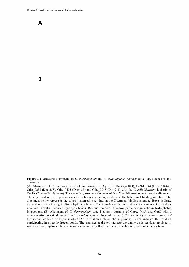
Chapter 2 Novel type I cohesins and dockerin domains
Figure 2.2 Structural alignments of dockerins. (A) Alignment of C. thermocellum
Cthe_0258 (Doc-258), Cthe_0435 (DocCel5A (Doc- cellulolyticum). The secondary structure elements of DocThe alignment on the top represents the cohesin interacting residues at the Nalignment below represents the cohesin interacting residues at the Cthe residues participating in direct hydrogen bonds. The triangles at the top indicate the amino acids residues involved in water mediated hydrogen bonds. Residues colored in yellow participate in cohesin hydrophobic interactions. (B) Alignment of representative cohesin domain from the second cohesin of CipA (Cohparticipating in direct hydrogen bonds. The triangles at the top indicate the amino acids residues involved in water mediated hydrogen bonds. Residues colored in yellow participate in cohesin hydrophobic interactions.
Chapter 2 Novel type I cohesins and dockerin domains
36
nts of C. thermocellum and C. cellulolyticum representative type I cohesins and
C. thermocellum dockerin domains of Xyn10B (Doc-Xyn10B), Cel9258), Cthe_0435 (Doc-435) and Cthe_0918 (Doc-918) with the C. cellulolyticum
cellulolyticum). The secondary structure elements of Doc-Xyn10B are shown The alignment on the top represents the cohesin interacting residues at the N-terminal binding interface. The alignment below represents the cohesin interacting residues at the C-terminal binding interface. Boxes indicate
participating in direct hydrogen bonds. The triangles at the top indicate the amino acids residues involved in water mediated hydrogen bonds. Residues colored in yellow participate in cohesin hydrophobic interactions. (B) Alignment of C. thermocellum type I cohesin domains of CipA, OlpA and OlpC with a representative cohesin domain from C. cellulolyticum (Coh-cellulolyticum). The secondary structure elements of the second cohesin of CipA (Coh-CipA2) are shown above the alignment. Boxes indicate the residuesparticipating in direct hydrogen bonds. The triangles at the top indicate the amino acids residues involved in water mediated hydrogen bonds. Residues colored in yellow participate in cohesin hydrophobic interactions.
representative type I cohesins and
Xyn10B), Cel9-GH44 (Doc-Cel44A), C. cellulolyticum dockerin of
Xyn10B are shown above the alignment. terminal binding interface. The
terminal binding interface. Boxes indicate participating in direct hydrogen bonds. The triangles at the top indicate the amino acids residues
involved in water mediated hydrogen bonds. Residues colored in yellow participate in cohesin hydrophobic I cohesin domains of CipA, OlpA and OlpC with a
cellulolyticum). The secondary structure elements of CipA2) are shown above the alignment. Boxes indicate the residues
participating in direct hydrogen bonds. The triangles at the top indicate the amino acids residues involved in water mediated hydrogen bonds. Residues colored in yellow participate in cohesin hydrophobic interactions.
Chapter 2 Novel type I cohesins and dockerin domains
37
2.3.2 C. thermocellum Cthe_0452 is a cell-surface protein
Cthe_0452 was shown to contain, in addition to a C-terminal type I cohesin, an 88
residue N-terminal domain of unknown function, designated CtUnk1. To determine the role
of CtUnk1, in carbohydrate metabolism or cellulosomal function the module was purified to
electrophoretic homogeneity and its biochemical properties evaluated. Initially we explored
the hypothesis that CtUnk1 constituted a CBM. However, affinity gel electrophoresis analysis
revealed that CtUnk1 displays no significant affinity for xyloglucan, the β1,4-β1,3-mixed
glucans barley β-glucan and lichenan, the β1,4-glucan hydroxyethylcellulose (HEC), konjac
glucomannan, oat spelt xylan, the β1,3-glucans laminarin and curdlan, carob galactomannan,
potato galactan, pullulan or pustulan (data not shown). In addition, the domain was unable to
depolymerase these polysaccharides indicating that Cthe_0452 is not a plant cell wall
degrading enzyme (data not shown).
Similar to OlpA, it is possible that Cthe_0452 constitutes a cell surface protein based
on the assumption that CtUnk1 binds to the bacterial cell envelope. To explore this hypothesis
the capacity of CtUnk1 to interact with C. thermocellum cell-wall fractions was investigated.
Proteins located at the outermost cell envelop of Gram-positive bacteria, known as the
bacterial S-layer, bind secondary cell wall polymers (SCWP) rather than the peptidoglycan
cell wall matrix. The data from pull-down experiments, displayed in Figure 2.3, revealed that
CtUnk1 bound to the insoluble C. thermocellum peptidoglycan-containing sacculi (NPCS);
only a small amount of the protein was detected in the unbound soluble fraction. In contrast,
CtUnk1, was unable to interact with C. thermocellum hydrofluoric acid-extracted cell wall
polymer (HF-EPCS). Hydrofluoric extraction removes SCWP from NPCS (Zhao, et al., 2006,
Zhao, et al., 2006) and, therefore, HF-EPCS consists mainly of peptidoglycan. Therefore, the
data suggest that CtUnk1 displays affinity for C. thermocellum SCWP. To further confirm
that CtUnk1 was unable to interact with the peptidoglycan fraction of the cell wall, an HF-
EPCS fraction was prepared from Escherichia coli, which is free of Gram-positive SCWP.
The data (Figure 2.3) revealed that CtUnk1 was unable to interact with the E. coli HF-EPCS
preparation, strongly suggesting that CtUnk1 specifically interacts with SCWP present in C.
thermocellum, or at least in Gram-positive bacterial cell walls. Since it appears that CtUnk1
directs Cthe_0452 onto the bacterial envelop we propose defining Cthe_0452 as OlpC (Outer
Layer Protein analogous to OlpA). Interestingly, CtUnk1 displays a specificity that is similar
to the OlpA SLH domain, and other SLH domains, in recognizing SCWP (Zhao, et al., 2006,
Zhao, et al., 2006). In contrast, the SLH domains of SdbA, OlpB and Orf2, unusually for such
modules, bind to the peptidoglycan layer (Zhao, et al., 2006, Zhao, et al., 2006). Therefore,
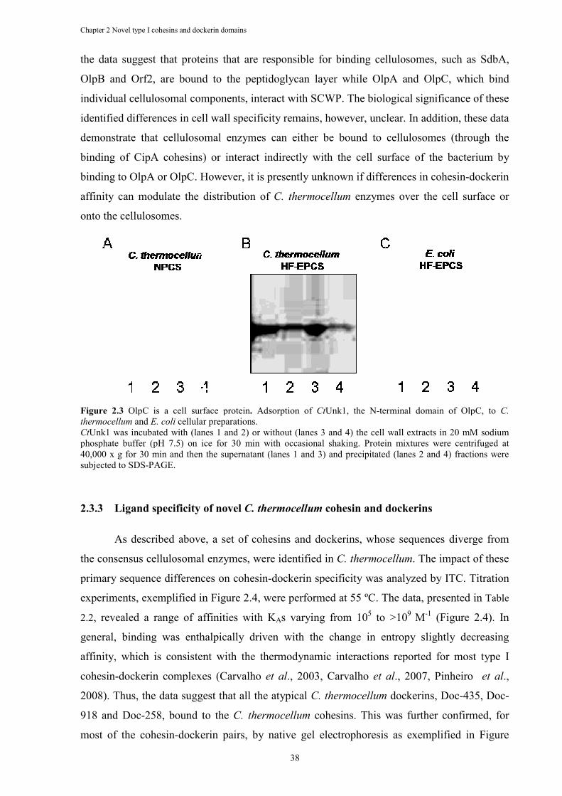
Chapter 2 Novel type I cohesins and dockerin domains
the data suggest that proteins that are responsible for binding cellulosomes, such as SdbA,
OlpB and Orf2, are bound to the peptidoglycan layer while OlpA and OlpC, which bind
individual cellulosomal components, interact with SCWP. The biological significance of these
identified differences in cell wall specificity remains, however, unclear. In addition, these data
demonstrate that cellulosomal enzymes can either be bound to cellulosomes (thro
binding of CipA cohesins) or interact indirectly with the cell surface of the bacterium by
binding to OlpA or OlpC. However, it is presently unknown if differences in cohesin
affinity can modulate the distribution of
onto the cellulosomes.
Figure 2.3 OlpC is a cell surface proteinthermocellum and E. coli cellular preparations.CtUnk1 was incubated with (lanes 1 and 2) or without (lanes 3 and 4) the cell wall extracts in 20 mM sodium phosphate buffer (pH 7.5) on ice for 30 min with occasional shaking. Protein mixtures were centrifuged at 40,000 x g for 30 min and then the supernatant (subjected to SDS-PAGE.
2.3.3 Ligand specificity of novel
As described above, a set of cohesins and dockerins, whose sequences diverge from
the consensus cellulosomal enzymes, were identified in
primary sequence differences on cohesin
experiments, exemplified in
2.2, revealed a range of affinities with K
general, binding was enthalpically driven with the change in entropy slightly decreasing
affinity, which is consistent with the thermodynamic interactions reported for most type I
cohesin-dockerin complexes (Carvalho
2008). Thus, the data suggest that all the atypical
918 and Doc-258, bound to the
most of the cohesin-dockerin pairs, by native gel electrophoresis as exempli
Chapter 2 Novel type I cohesins and dockerin domains
38
the data suggest that proteins that are responsible for binding cellulosomes, such as SdbA,
OlpB and Orf2, are bound to the peptidoglycan layer while OlpA and OlpC, which bind
ulosomal components, interact with SCWP. The biological significance of these
identified differences in cell wall specificity remains, however, unclear. In addition, these data
demonstrate that cellulosomal enzymes can either be bound to cellulosomes (thro
binding of CipA cohesins) or interact indirectly with the cell surface of the bacterium by
binding to OlpA or OlpC. However, it is presently unknown if differences in cohesin
affinity can modulate the distribution of C. thermocellum enzymes over the cell surface or
OlpC is a cell surface protein. Adsorption of CtUnk1, the N-terminal domain of OlpC, to cellular preparations.
was incubated with (lanes 1 and 2) or without (lanes 3 and 4) the cell wall extracts in 20 mM sodium phosphate buffer (pH 7.5) on ice for 30 min with occasional shaking. Protein mixtures were centrifuged at 40,000 x g for 30 min and then the supernatant (lanes 1 and 3) and precipitated (lanes 2 and 4) fractions were
Ligand specificity of novel C. thermocellum cohesin and dockerins
As described above, a set of cohesins and dockerins, whose sequences diverge from
the consensus cellulosomal enzymes, were identified in C. thermocellum
primary sequence differences on cohesin-dockerin specificity was analyzed by ITC.
exemplified in Figure 2.4, were performed at 55 ºC. The data, presented in
, revealed a range of affinities with KAs varying from 105 to >10
was enthalpically driven with the change in entropy slightly decreasing
affinity, which is consistent with the thermodynamic interactions reported for most type I
dockerin complexes (Carvalho et al., 2003, Carvalho et al., 2007, Pinheiro
uggest that all the atypical C. thermocellum dockerins, Doc
258, bound to the C. thermocellum cohesins. This was further confirmed, for
dockerin pairs, by native gel electrophoresis as exempli
the data suggest that proteins that are responsible for binding cellulosomes, such as SdbA,
OlpB and Orf2, are bound to the peptidoglycan layer while OlpA and OlpC, which bind
ulosomal components, interact with SCWP. The biological significance of these
identified differences in cell wall specificity remains, however, unclear. In addition, these data
demonstrate that cellulosomal enzymes can either be bound to cellulosomes (through the
binding of CipA cohesins) or interact indirectly with the cell surface of the bacterium by
binding to OlpA or OlpC. However, it is presently unknown if differences in cohesin-dockerin
s over the cell surface or
terminal domain of OlpC, to C.
was incubated with (lanes 1 and 2) or without (lanes 3 and 4) the cell wall extracts in 20 mM sodium phosphate buffer (pH 7.5) on ice for 30 min with occasional shaking. Protein mixtures were centrifuged at
lanes 1 and 3) and precipitated (lanes 2 and 4) fractions were
cohesin and dockerins
As described above, a set of cohesins and dockerins, whose sequences diverge from
C. thermocellum. The impact of these
dockerin specificity was analyzed by ITC. Titration
performed at 55 ºC. The data, presented in Table
to >109 M-1 (Figure 2.4). In
was enthalpically driven with the change in entropy slightly decreasing
affinity, which is consistent with the thermodynamic interactions reported for most type I
., 2007, Pinheiro et al.,
dockerins, Doc-435, Doc-
cohesins. This was further confirmed, for
dockerin pairs, by native gel electrophoresis as exemplified in Figure
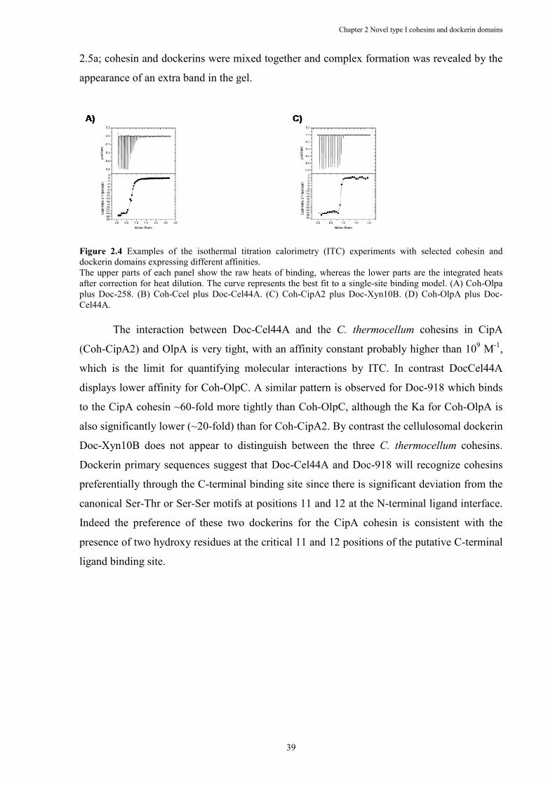
2.5a; cohesin and dockerins were mixed together and complex formation was revealed by the
appearance of an extra band in the gel.
Figure 2.4 Examples of the isothermal titration calorimetry (ITC) experiments with selected cohesin and dockerin domains expressing different affinities.The upper parts of each panel show the raw heats of binding, whereas the lower parts are the integrated heats after correction for heat dilution. The curve represents the best fit to a singleplus Doc-258. (B) Coh-Ccel plusCel44A.
The interaction between Doc
(Coh-CipA2) and OlpA is very tight, with an affinity constant probably higher than 10
which is the limit for quantifying molecular interactions by ITC. In contrast DocCel44A
displays lower affinity for Coh
to the CipA cohesin ~60-fold more tightly than Coh
also significantly lower (~20
Doc-Xyn10B does not appear to distinguish between the three
Dockerin primary sequences suggest that Doc
preferentially through the C
canonical Ser-Thr or Ser-Ser motifs at positions 11 and 12 at the N
Indeed the preference of these two dockerins for the
presence of two hydroxy residues at the critical 11 and 12 positions of the putative C
ligand binding site.
Chapter 2 Novel type I cohesins and dockerin
39
a; cohesin and dockerins were mixed together and complex formation was revealed by the
appearance of an extra band in the gel.
Examples of the isothermal titration calorimetry (ITC) experiments with selected cohesin and dockerin domains expressing different affinities. The upper parts of each panel show the raw heats of binding, whereas the lower parts are the integrated heats after correction for heat dilution. The curve represents the best fit to a single-site binding model.
Ccel plus Doc-Cel44A. (C) Coh-CipA2 plus Doc-Xyn10B. (D) Coh
The interaction between Doc-Cel44A and the C. thermocellum
CipA2) and OlpA is very tight, with an affinity constant probably higher than 10
limit for quantifying molecular interactions by ITC. In contrast DocCel44A
displays lower affinity for Coh-OlpC. A similar pattern is observed for Doc
fold more tightly than Coh-OlpC, although the Ka for Coh
also significantly lower (~20-fold) than for Coh-CipA2. By contrast the cellulosomal dockerin
Xyn10B does not appear to distinguish between the three C. thermocellum
Dockerin primary sequences suggest that Doc-Cel44A and Doc-918 will recogniz
preferentially through the C-terminal binding site since there is significant deviation from the
Ser motifs at positions 11 and 12 at the N-terminal ligand interface.
Indeed the preference of these two dockerins for the CipA cohesin is consistent with the
presence of two hydroxy residues at the critical 11 and 12 positions of the putative C
Chapter 2 Novel type I cohesins and dockerin domains
a; cohesin and dockerins were mixed together and complex formation was revealed by the
Examples of the isothermal titration calorimetry (ITC) experiments with selected cohesin and
The upper parts of each panel show the raw heats of binding, whereas the lower parts are the integrated heats site binding model. (A) Coh-Olpa
Xyn10B. (D) Coh-OlpA plus Doc-
C. thermocellum cohesins in CipA
CipA2) and OlpA is very tight, with an affinity constant probably higher than 109 M-1,
limit for quantifying molecular interactions by ITC. In contrast DocCel44A
OlpC. A similar pattern is observed for Doc-918 which binds
OlpC, although the Ka for Coh-OlpA is
CipA2. By contrast the cellulosomal dockerin
C. thermocellum cohesins.
918 will recognize cohesins
terminal binding site since there is significant deviation from the
terminal ligand interface.
CipA cohesin is consistent with the
presence of two hydroxy residues at the critical 11 and 12 positions of the putative C-terminal
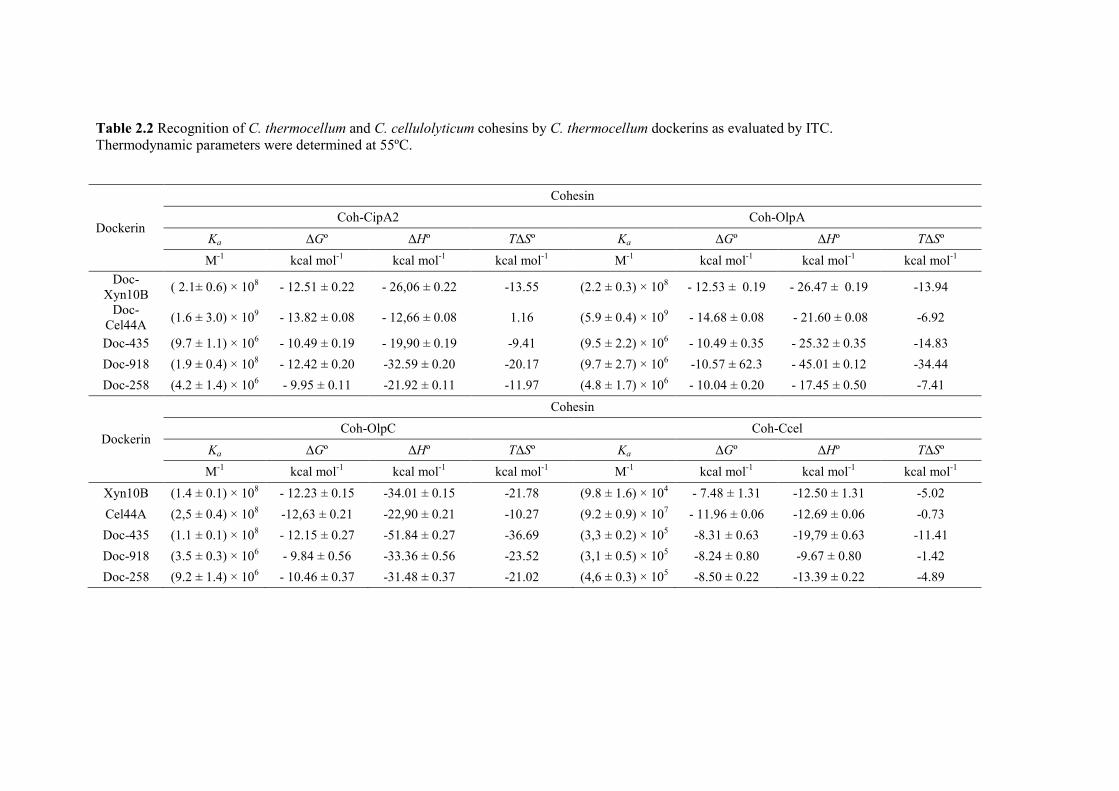
40
Table 2.2 Recognition of C. thermocellum and C. cellulolyticum cohesins by C. thermocellum dockerins as evaluated by ITC. Thermodynamic parameters were determined at 55ºC.
Dockerin
Cohesin
Coh-CipA2 Coh-OlpA
Ka ∆Gº ∆Hº T∆Sº Ka ∆Gº ∆Hº T∆Sº
M-1 kcal mol-1 kcal mol-1 kcal mol-1 M-1 kcal mol-1 kcal mol-1 kcal mol-1 Doc-
Xyn10B ( 2.1± 0.6) × 108 - 12.51 ± 0.22 - 26,06 ± 0.22 -13.55 (2.2 ± 0.3) × 108 - 12.53 ± 0.19 - 26.47 ± 0.19 -13.94
Doc-Cel44A (1.6 ± 3.0) × 109 - 13.82 ± 0.08 - 12,66 ± 0.08 1.16 (5.9 ± 0.4) × 109 - 14.68 ± 0.08 - 21.60 ± 0.08 -6.92
Doc-435 (9.7 ± 1.1) × 106 - 10.49 ± 0.19 - 19,90 ± 0.19 -9.41 (9.5 ± 2.2) × 106 - 10.49 ± 0.35 - 25.32 ± 0.35 -14.83
Doc-918 (1.9 ± 0.4) × 108 - 12.42 ± 0.20 -32.59 ± 0.20 -20.17 (9.7 ± 2.7) × 106 -10.57 ± 62.3 - 45.01 ± 0.12 -34.44
Doc-258 (4.2 ± 1.4) × 106 - 9.95 ± 0.11 -21.92 ± 0.11 -11.97 (4.8 ± 1.7) × 106 - 10.04 ± 0.20 - 17.45 ± 0.50 -7.41
Dockerin
Cohesin
Coh-OlpC Coh-Ccel
Ka ∆Gº ∆Hº T∆Sº Ka ∆Gº ∆Hº T∆Sº
M-1 kcal mol-1 kcal mol-1 kcal mol-1 M-1 kcal mol-1 kcal mol-1 kcal mol-1
Xyn10B (1.4 ± 0.1) × 108 - 12.23 ± 0.15 -34.01 ± 0.15 -21.78 (9.8 ± 1.6) × 104 - 7.48 ± 1.31 -12.50 ± 1.31 -5.02
Cel44A (2,5 ± 0.4) × 108 -12,63 ± 0.21 -22,90 ± 0.21 -10.27 (9.2 ± 0.9) × 107 - 11.96 ± 0.06 -12.69 ± 0.06 -0.73
Doc-435 (1.1 ± 0.1) × 108 - 12.15 ± 0.27 -51.84 ± 0.27 -36.69 (3,3 ± 0.2) × 105 -8.31 ± 0.63 -19,79 ± 0.63 -11.41
Doc-918 (3.5 ± 0.3) × 106 - 9.84 ± 0.56 -33.36 ± 0.56 -23.52 (3,1 ± 0.5) × 105 -8.24 ± 0.80 -9.67 ± 0.80 -1.42
Doc-258 (9.2 ± 1.4) × 106 - 10.46 ± 0.37 -31.48 ± 0.37 -21.02 (4,6 ± 0.3) × 105 -8.50 ± 0.22 -13.39 ± 0.22 -4.89
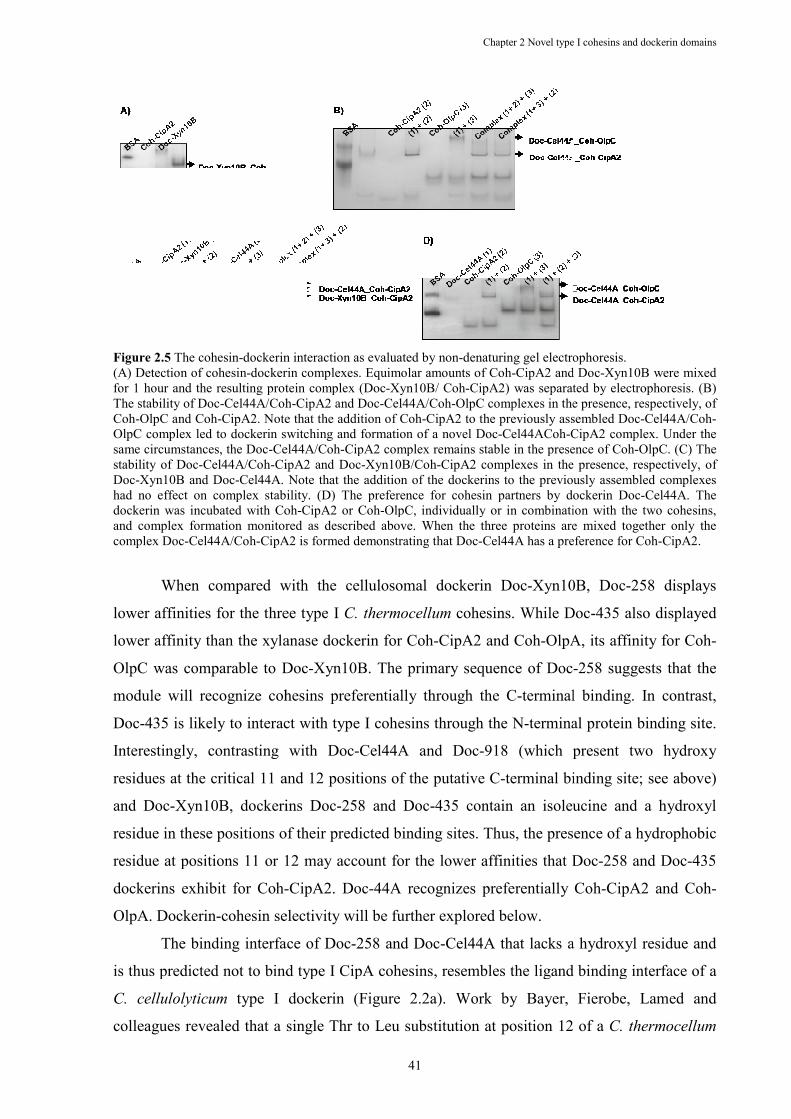
Figure 2.5 The cohesin-dockerin interaction as evaluated by non(A) Detection of cohesin-dockerin complexes. for 1 hour and the resulting protein complex (DocThe stability of Doc-Cel44A/CohCoh-OlpC and Coh-CipA2. Note that the addition of CohOlpC complex led to dockerin switching and formation of a novel Docsame circumstances, the Doc-Cel44A/Cstability of Doc-Cel44A/Coh-CipA2 and DocDoc-Xyn10B and Doc-Cel44A. Note that the addition of the dockerins to the previouslyhad no effect on complex stability. (D) The preference for cohesin partners by dockerin Docdockerin was incubated with Cohand complex formation monitored as described above. When the three proteins are mixed together only the complex Doc-Cel44A/Coh-CipA2 is formed demonstrating that Doc
When compared with the cellulosomal dockerin Doc
lower affinities for the three type I
lower affinity than the xylanase dockerin for Coh
OlpC was comparable to Doc
module will recognize cohesins preferentially through the C
Doc-435 is likely to interact with type I cohesins through the N
Interestingly, contrasting with Doc
residues at the critical 11 and 12 positions of the putative C
and Doc-Xyn10B, dockerins Doc
residue in these positions of their predicted bindi
residue at positions 11 or 12 may account for the lower affinities that Doc
dockerins exhibit for Coh-
OlpA. Dockerin-cohesin selectivit
The binding interface of Doc
is thus predicted not to bind type I CipA cohesins, resembles the ligand binding interface of a
C. cellulolyticum type I
colleagues revealed that a single Thr to Leu substitution at position 12 of a
Chapter 2 Novel type
41
dockerin interaction as evaluated by non-denaturing gel electrophoresis.dockerin complexes. Equimolar amounts of Coh-CipA2 and Doc
for 1 hour and the resulting protein complex (Doc-Xyn10B/ Coh-CipA2) was separated by electrophoresis. (B) Cel44A/Coh-CipA2 and Doc-Cel44A/Coh-OlpC complexes in the presence, resCipA2. Note that the addition of Coh-CipA2 to the previously assembled Doc
OlpC complex led to dockerin switching and formation of a novel Doc-Cel44ACoh-CipA2 complex. Under the Cel44A/Coh-CipA2 complex remains stable in the presence of CohCipA2 and Doc-Xyn10B/Coh-CipA2 complexes in the presence, respectively, of
Cel44A. Note that the addition of the dockerins to the previouslyhad no effect on complex stability. (D) The preference for cohesin partners by dockerin Docdockerin was incubated with Coh-CipA2 or Coh-OlpC, individually or in combination with the two cohesins,
tored as described above. When the three proteins are mixed together only the CipA2 is formed demonstrating that Doc-Cel44A has a preference for Coh
When compared with the cellulosomal dockerin Doc-Xyn10B, Doc
ower affinities for the three type I C. thermocellum cohesins. While Doc
lower affinity than the xylanase dockerin for Coh-CipA2 and Coh-OlpA, its affinity for Coh
OlpC was comparable to Doc-Xyn10B. The primary sequence of Doc
module will recognize cohesins preferentially through the C-terminal binding. In contrast,
435 is likely to interact with type I cohesins through the N-terminal protein binding site.
Interestingly, contrasting with Doc-Cel44A and Doc-918 (which present two hydroxy
residues at the critical 11 and 12 positions of the putative C-terminal binding site; see above)
Xyn10B, dockerins Doc-258 and Doc-435 contain an isoleucine and a hydroxyl
residue in these positions of their predicted binding sites. Thus, the presence of a hydrophobic
residue at positions 11 or 12 may account for the lower affinities that Doc
-CipA2. Doc-44A recognizes preferentially Coh
cohesin selectivity will be further explored below.
The binding interface of Doc-258 and Doc-Cel44A that lacks a hydroxyl residue and
is thus predicted not to bind type I CipA cohesins, resembles the ligand binding interface of a
type I dockerin (Figure 2.2a). Work by Bayer, Fierobe, Lamed and
colleagues revealed that a single Thr to Leu substitution at position 12 of a
Chapter 2 Novel type I cohesins and dockerin domains
denaturing gel electrophoresis.
CipA2 and Doc-Xyn10B were mixed CipA2) was separated by electrophoresis. (B)
OlpC complexes in the presence, respectively, of CipA2 to the previously assembled Doc-Cel44A/Coh-
CipA2 complex. Under the CipA2 complex remains stable in the presence of Coh-OlpC. (C) The
CipA2 complexes in the presence, respectively, of Cel44A. Note that the addition of the dockerins to the previously assembled complexes
had no effect on complex stability. (D) The preference for cohesin partners by dockerin Doc-Cel44A. The OlpC, individually or in combination with the two cohesins,
tored as described above. When the three proteins are mixed together only the Cel44A has a preference for Coh-CipA2.
Xyn10B, Doc-258 displays
cohesins. While Doc-435 also displayed
OlpA, its affinity for Coh-
-258 suggests that the
terminal binding. In contrast,
terminal protein binding site.
hich present two hydroxy
terminal binding site; see above)
435 contain an isoleucine and a hydroxyl
ng sites. Thus, the presence of a hydrophobic
residue at positions 11 or 12 may account for the lower affinities that Doc-258 and Doc-435
44A recognizes preferentially Coh-CipA2 and Coh-
Cel44A that lacks a hydroxyl residue and
is thus predicted not to bind type I CipA cohesins, resembles the ligand binding interface of a
, Fierobe, Lamed and
colleagues revealed that a single Thr to Leu substitution at position 12 of a C. thermocellum
Chapter 2 Novel type I cohesins and dockerin domains
42
dockerin allows the protein to bind C. cellulolyticum cohesins (Mechaly, et al., 2001).
Therefore, the C. thermocellum dockerins were also probed against a type I cohesin from C.
cellulolyticum, termed Coh-Ccel. The data (Table 2.2), revealed that Doc-Cel44A presented a
high affinity for the C. cellulolyticum cohesin Coh-Ccel (Ka 108 M-1), while all other C.
thermocellum dockerins bound much more weakly (100-1000 lower affinities) to the non-
cognate cohesin. Overall the data suggest that apart from Doc-Cel44A, C. thermocellum
dockerins displayed species restricted ligand specificity. However, there are no obvious amino
acid substitutions in the Doc-258 primary sequence, when compared with Doc-Cel44A
(Figure 2.2a), which can explain its inability to recognize the C. cellulolyticum cohesin.
2.3.4 Complex stability and cohesin-dockerin selectivity
In order to gain further insights into the mechanism of cohesin-dockerin recognition,
the capacity of individual cohesins or dockerins to destabilize previously assembled
complexes was evaluated. Thus, individual cohesin or dockerin domains were added to an
assembled complex and the stability of the original assembly, or the formation of a new
complex, was evaluated by native gel electrophoresis (examples in Figure 2.5 and 2.5c). If a
previously formed complex was reconfigured, a new band would appear on the gel. Since
detection of several complexes was not straightforward through gel electrophoresis, due to
low cohesin-dockerin affinity, some protein domains were excluded from the analysis. The
data, displayed in Table 2.3, revealed that cohesin exchange occurred readily, although a
dockerin bound to its cohesin partner was more rarely displaced by the addition of an
unliganded dockerin. For example, addition of Coh-OlpA to a Coh-CipA2/Doc-Xyn10B
complex led to the formation of a Coh-OlpA/Doc-Xyn10B complex, while no dockerin was
able to destabilize the Coh-CipA2/Doc-Xyn10B or the Coh-CipA2/Doc-Cel44A complexes.
The existence of two cohesin binding interfaces in dockerins may explain this observation as
they may allow these domains to switch more easily from one cohesin to another and yet
maintain a high affinity protein:protein interaction. Significantly, dockerin switching seems to
occur only towards higher affinity cohesins. Since cohesins contain only one binding
interface, their switching from one dockerin to another is more difficult considering the high
affinity interaction established in cohesin-dockerin complexes. Therefore, taken together the
data suggest that free dockerins cannot easily displace already bound dockerins. However,
bound dockerins can easily bind to an unliganded cohesin if this is energetically favored.
Thus, dockerin cohesin switching may reflect the existence of two cohesin-binding interfaces
in dockerins. Significantly, complexes containing Doc-258 were dissociated in the presence of
Chapter 2 Novel type I cohesins and dockerin domains
43
Doc-Xyn10B or Doc-918, which likely reflects their substantially higher affinity for cohesins,
compared to Doc-258.
In addition, dockerin and cohesin preference for protein partners was analyzed by
mixing one dockerin with two cohesins (or one cohesin with two dockerins when cohesin
selectivity was evaluated) and assessing complex formation by native gel electrophoresis (see
Figure 2.6d for an example). Each cohesin and dockerin was mixed with the combination of
all possible protein partners. The resulting data were integrated and allowed, for each cohesin
or dockerin domain, ordering their protein partners by binding preference. The data, presented
in Table 2.4, revealed that subtle differences in the affinity of the various cohesin-dockerin
pairs, as evaluated by ITC, can have a significant effect in cohesin-dockerin preference. For
example, dockerins Doc_Xyn10B and Doc_Cel44A, which were shown to bind cohesins Coh-
CipA2 and Coh-OlpA with similar thermodynamic parameters and affinities, bind
preferentially Coh-OlpA rather than Coh-CipA2. In addition, the two dockerins displayed a
lower preference for Coh-OlpC as a protein partner. When compared with Doc_Xyn10B and
Doc_Cel44A, Dockerin Doc-918 displayed similar cohesin selectivity, although it does not
display a preference for either Coh-CipA2 or Coh-OlpA. In contrast to the cohesin
preferences expressed by dockerins Doc-918, Doc_Xyn10B and Doc_Cel44A, dockerins
Doc-435 and Doc-258 bound, preferentially, Coh-OlpC. In addition, these dockerins revealed
the lowest affinities for cohesin Coh-OlpA. Thus, the data suggest that substitution of one of
the hydroxyl residues of the conserved Ser-Thr pair of the putative cohesin binding interface
with an isoleucine (Figure Figure 2.2a) leads to a switch in cohesin preference in dockerins
Doc-435 and Doc-258 from cohesins Coh-CipA2 or Coh-OlpA to cohesin Coh-OlpC. In the
cohesin side, Coh-CipA2 and Coh-OlpA displayed identical dockerin preferences and bound,
with similar affinities, dockerins Doc-Cel44A, Doc-Xyn10B and Doc-918. These two
cohesins display a lower selectivity for Doc-258 and particularly for Doc-435. Thus, amino
acid substitutions in Coh-OlpA, and most probably in the ninth cohesin of CipA (see above),
do not seem to affect the capacity of OlpA cohesin to interact with cellulosomal dockerin
partners such as Doc_Xyn10B. In contrast, cohesin Coh-OlpC interacts, preferentially, with
dockerin Doc-435, suggesting that Coh-OlpC amino acid substitutions at the dockerin binding
platform (such as D39N and E131P) are particularly suited for the recognition of dockerin
Doc-435. Surprisingly, Coh-OlpC still recognizes dockerins Doc-Cel44A and Doc-Xyn10B
with considerable affinity.
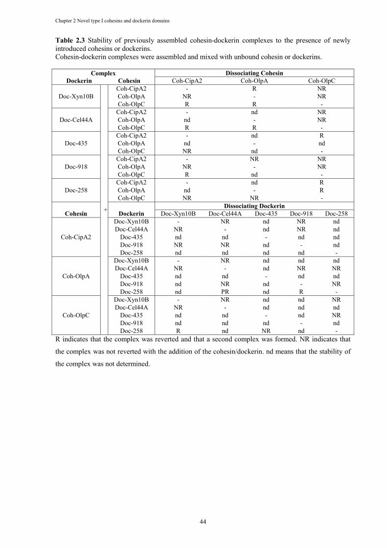
Chapter 2 Novel type I cohesins and dockerin domains
44
Table 2.3 Stability of previously assembled cohesin-dockerin complexes to the presence of newly introduced cohesins or dockerins. Cohesin-dockerin complexes were assembled and mixed with unbound cohesin or dockerins.
R indicates that the complex was reverted and that a second complex was formed. NR indicates that
the complex was not reverted with the addition of the cohesin/dockerin. nd means that the stability of
the complex was not determined.
Complex
Dockerin Cohesin Dissociating Cohesin
Coh-CipA2 Coh-OlpA Coh-OlpC
Doc-Xyn10B
+
Coh-CipA2 - R NR Coh-OlpA NR - NR Coh-OlpC R R -
Doc-Cel44A Coh-CipA2 - nd NR Coh-OlpA nd - NR Coh-OlpC R R -
Doc-435 Coh-CipA2 - nd R Coh-OlpA nd - nd Coh-OlpC NR nd -
Doc-918 Coh-CipA2 - NR NR Coh-OlpA NR - NR Coh-OlpC R nd -
Doc-258 Coh-CipA2 - nd R Coh-OlpA nd - R Coh-OlpC NR NR -
Cohesin
Dockerin Dissociating Dockerin
Doc-Xyn10B Doc-Cel44A Doc-435 Doc-918 Doc-258
Coh-CipA2
Doc-Xyn10B - NR nd NR nd Doc-Cel44A NR - nd NR nd
Doc-435 nd nd - nd nd Doc-918 NR NR nd - nd Doc-258 nd nd nd nd -
Coh-OlpA
Doc-Xyn10B - NR nd nd nd Doc-Cel44A NR - nd NR NR
Doc-435 nd nd - nd nd Doc-918 nd NR nd - NR Doc-258 nd PR nd R -
Coh-OlpC
Doc-Xyn10B - NR nd nd NR Doc-Cel44A NR - nd nd nd
Doc-435 nd nd - nd NR Doc-918 nd nd nd - nd Doc-258 R nd NR nd -
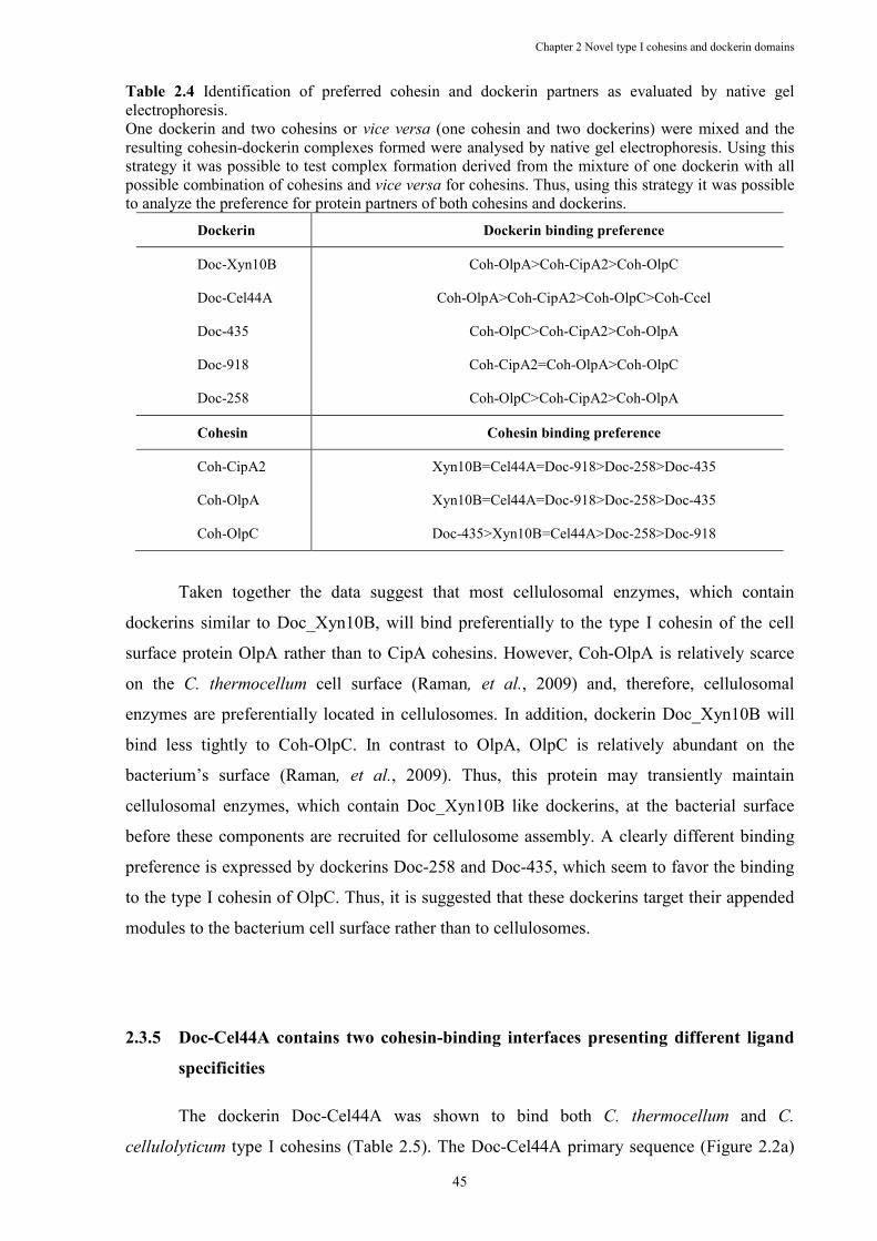
Chapter 2 Novel type I cohesins and dockerin domains
45
Table 2.4 Identification of preferred cohesin and dockerin partners as evaluated by native gel electrophoresis. One dockerin and two cohesins or vice versa (one cohesin and two dockerins) were mixed and the resulting cohesin-dockerin complexes formed were analysed by native gel electrophoresis. Using this strategy it was possible to test complex formation derived from the mixture of one dockerin with all possible combination of cohesins and vice versa for cohesins. Thus, using this strategy it was possible to analyze the preference for protein partners of both cohesins and dockerins.
Dockerin Dockerin binding preference
Doc-Xyn10B Coh-OlpA>Coh-CipA2>Coh-OlpC
Doc-Cel44A Coh-OlpA>Coh-CipA2>Coh-OlpC>Coh-Ccel
Doc-435 Coh-OlpC>Coh-CipA2>Coh-OlpA
Doc-918 Coh-CipA2=Coh-OlpA>Coh-OlpC
Doc-258 Coh-OlpC>Coh-CipA2>Coh-OlpA
Cohesin Cohesin binding preference
Coh-CipA2 Xyn10B=Cel44A=Doc-918>Doc-258>Doc-435
Coh-OlpA Xyn10B=Cel44A=Doc-918>Doc-258>Doc-435
Coh-OlpC Doc-435>Xyn10B=Cel44A>Doc-258>Doc-918
Taken together the data suggest that most cellulosomal enzymes, which contain
dockerins similar to Doc_Xyn10B, will bind preferentially to the type I cohesin of the cell
surface protein OlpA rather than to CipA cohesins. However, Coh-OlpA is relatively scarce
on the C. thermocellum cell surface (Raman, et al., 2009) and, therefore, cellulosomal
enzymes are preferentially located in cellulosomes. In addition, dockerin Doc_Xyn10B will
bind less tightly to Coh-OlpC. In contrast to OlpA, OlpC is relatively abundant on the
bacterium’s surface (Raman, et al., 2009). Thus, this protein may transiently maintain
cellulosomal enzymes, which contain Doc_Xyn10B like dockerins, at the bacterial surface
before these components are recruited for cellulosome assembly. A clearly different binding
preference is expressed by dockerins Doc-258 and Doc-435, which seem to favor the binding
to the type I cohesin of OlpC. Thus, it is suggested that these dockerins target their appended
modules to the bacterium cell surface rather than to cellulosomes.
2.3.5 Doc-Cel44A contains two cohesin-binding interfaces presenting different ligand
specificities
The dockerin Doc-Cel44A was shown to bind both C. thermocellum and C.
cellulolyticum type I cohesins (Table 2.5). The Doc-Cel44A primary sequence (Figure 2.2a)
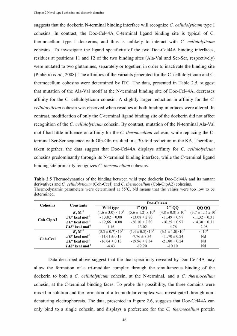
Chapter 2 Novel type I cohesins and dockerin domains
46
suggests that the dockerin N-terminal binding interface will recognize C. cellulolyticum type I
cohesins. In contrast, the Doc-Cel44A C-terminal ligand binding site is typical of C.
thermocellum type I dockerins, and thus is unlikely to interact with C. cellulolyticum
cohesins. To investigate the ligand specificity of the two Doc-Cel44A binding interfaces,
residues at positions 11 and 12 of the two binding sites (Ala-Val and Ser-Ser, respectively)
were mutated to two glutamines, separately or together, in order to inactivate the binding site
(Pinheiro et al., 2008). The affinities of the variants generated for the C. cellulolyticum and C.
thermocellum cohesins were determined by ITC. The data, presented in Table 2.5, suggest
that mutation of the Ala-Val motif at the N-terminal binding site of Doc-Cel44A, decreases
affinity for the C. cellulolyticum cohesin. A slightly larger reduction in affinity for the C.
cellulolyticum cohesin was observed when residues at both binding interfaces were altered. In
contrast, modification of only the C-terminal ligand binding site of the dockerin did not affect
recognition of the C. cellulolyticum cohesin. By contrast, mutation of the N-terminal Ala-Val
motif had little influence on affinity for the C. thermocellum cohesin, while replacing the C-
terminal Ser-Ser sequence with Gln-Gln resulted in a 30-fold reduction in the KA. Therefore,
taken together, the data suggest that Doc-Cel44A displays affinity for C. cellulolyticum
cohesins predominantly through its N-terminal binding interface, while the C-terminal ligand
binding site primarily recognizes C. thermocellum cohesins.
Table 2.5 Thermodynamics of the binding between wild type dockerin Doc-Cel44A and its mutant derivatives and C. cellulolyticum (Coh-Ccel) and C. thermocellum (Coh-CipA2) cohesins. Thermodynamic parameters were determined at 55ºC. Nd means that the values were too low to be determined.
Data described above suggest that the dual specificity revealed by Doc-Cel44A may
allow the formation of a tri-modular complex through the simultaneous binding of the
dockerin to both a C. cellulolyticum cohesin, at the N-terminal, and a C. thermocellum
cohesin, at the C-terminal binding faces. To probe this possibility, the three domains were
mixed in solution and the formation of a tri-modular complex was investigated through non-
denaturing electrophoresis. The data, presented in Figure 2.6, suggests that Doc-Cel44A can
only bind to a single cohesin, and displays a preference for the C. thermocellum protein
Cohesins Constants Doc-Cel44A
Wild type 1st QQ 2
nd QQ QQ QQ
Coh-CipA2
Ka M-1
(1.6 ± 3.0) × 109 (5.6 ± 1.2) x 108 (4.8 ± 0.8) x 107 (3.7 ± 1.1) x 107
∆Gº kcal mol-1
- 13.82 ± 0.08 -13.08 ± 2.80 -11.49 ± 0.97 -11.32 ± 0.31 ∆Hº kcal mol
-1 - 12,66 ± 0.08 -26.10 ± 2.80 -16.25 ± 0.97 -14.30 ± 0.31
T∆Sº kcal mol-1
1.16 -13.02 -4.76 -2.98
Coh-Ccel
Ka M-1
(5.3 ± 0.7)×107 (1.4 ± 0.3)×105 (6.1 ± 1.0)×107 < 104 ∆Gº kcal mol
-1 -11.61 ± 0.13 -7.76 ± 8.34 -11.70 ± 0.24 Nd
∆Hº kcal mol-1
-16.04 ± 0.13 -19.96 ± 8.34 -21.80 ± 0.24 Nd T∆Sº kcal mol
-1 -4.43 -12.20 -10.10 Nd
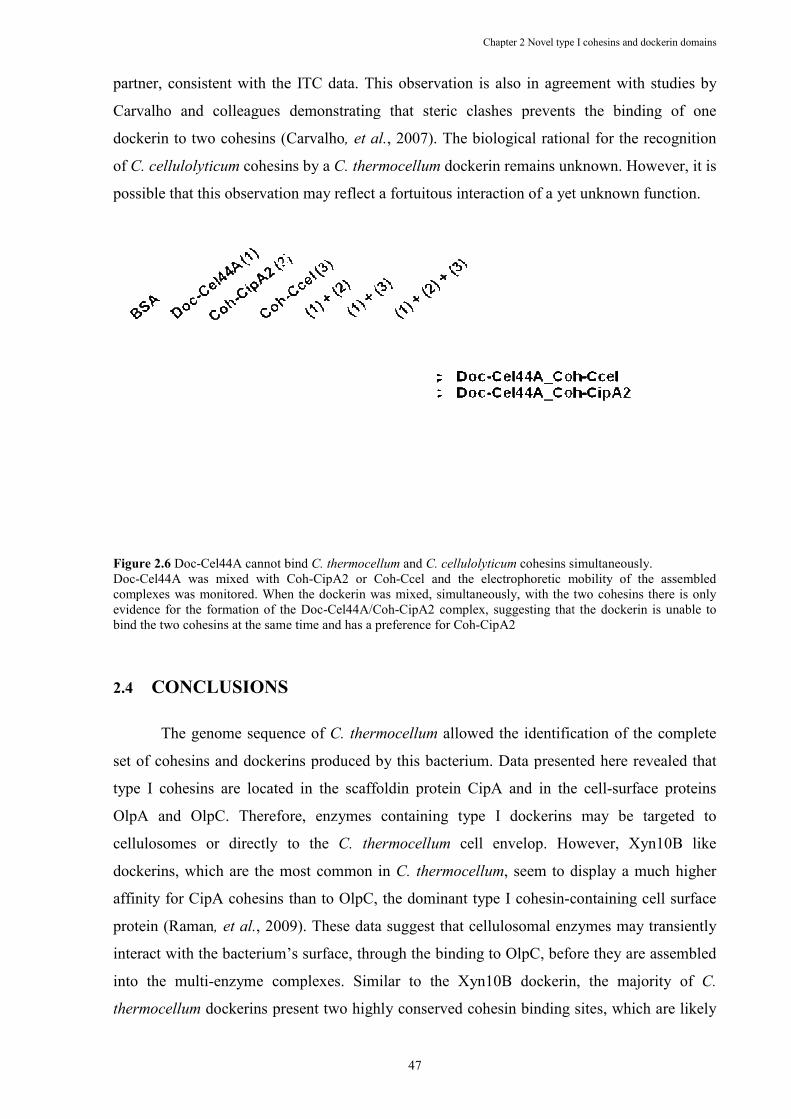
partner, consistent with the ITC data. This observation is also in agreement with studies by
Carvalho and colleagues demonstrating that steric clashes prevents the binding of one
dockerin to two cohesins (Carvalho
of C. cellulolyticum cohesins by a
possible that this observation may reflect a fortuitous interaction of
Figure 2.6 Doc-Cel44A cannot bind Doc-Cel44A was mixed with Cohcomplexes was monitored. When the dockevidence for the formation of the Docbind the two cohesins at the same time and has a preference for Coh
2.4 CO�CLUSIO�S
The genome sequence of
set of cohesins and dockerins produced by this bacterium. Data presented here revealed that
type I cohesins are located in the scaffoldin protein CipA and in the
OlpA and OlpC. Therefore, enzymes containing type I dockerins may be targeted to
cellulosomes or directly to the
dockerins, which are the most common in
affinity for CipA cohesins than to OlpC, the dominant type I cohesin
protein (Raman, et al., 2009)
interact with the bacterium’s surface, through the binding to OlpC, before they are assembled
into the multi-enzyme complexes. Similar to the Xyn10B dockerin, the majority of
thermocellum dockerins present two highly conserved cohesin binding sites, which are
Chapter 2 Novel type
47
partner, consistent with the ITC data. This observation is also in agreement with studies by
Carvalho and colleagues demonstrating that steric clashes prevents the binding of one
(Carvalho, et al., 2007). The biological rational for the recognition
cohesins by a C. thermocellum dockerin remains unknown. However, it is
possible that this observation may reflect a fortuitous interaction of a yet unknown function.
Cel44A cannot bind C. thermocellum and C. cellulolyticum cohesins simultaneouslyCel44A was mixed with Coh-CipA2 or Coh-Ccel and the electrophoretic mobility of the assembled
complexes was monitored. When the dockerin was mixed, simultaneously, with the two cohesins there is only evidence for the formation of the Doc-Cel44A/Coh-CipA2 complex, suggesting that the dockerin is unable to bind the two cohesins at the same time and has a preference for Coh-CipA2
CO�CLUSIO�S
The genome sequence of C. thermocellum allowed the identification of the complete
set of cohesins and dockerins produced by this bacterium. Data presented here revealed that
type I cohesins are located in the scaffoldin protein CipA and in the
OlpA and OlpC. Therefore, enzymes containing type I dockerins may be targeted to
cellulosomes or directly to the C. thermocellum cell envelop. However, Xyn10B like
dockerins, which are the most common in C. thermocellum, seem to displ
affinity for CipA cohesins than to OlpC, the dominant type I cohesin-containing cell surface
, 2009). These data suggest that cellulosomal enzymes may transiently
ct with the bacterium’s surface, through the binding to OlpC, before they are assembled
enzyme complexes. Similar to the Xyn10B dockerin, the majority of
dockerins present two highly conserved cohesin binding sites, which are
Chapter 2 Novel type I cohesins and dockerin domains
partner, consistent with the ITC data. This observation is also in agreement with studies by
Carvalho and colleagues demonstrating that steric clashes prevents the binding of one
e biological rational for the recognition
dockerin remains unknown. However, it is
yet unknown function.
cohesins simultaneously.
Ccel and the electrophoretic mobility of the assembled erin was mixed, simultaneously, with the two cohesins there is only
CipA2 complex, suggesting that the dockerin is unable to
allowed the identification of the complete
set of cohesins and dockerins produced by this bacterium. Data presented here revealed that
type I cohesins are located in the scaffoldin protein CipA and in the cell-surface proteins
OlpA and OlpC. Therefore, enzymes containing type I dockerins may be targeted to
cell envelop. However, Xyn10B like
, seem to display a much higher
containing cell surface
. These data suggest that cellulosomal enzymes may transiently
ct with the bacterium’s surface, through the binding to OlpC, before they are assembled
enzyme complexes. Similar to the Xyn10B dockerin, the majority of C.
dockerins present two highly conserved cohesin binding sites, which are likely
Chapter 2 Novel type I cohesins and dockerin domains
48
to display identical ligand affinities (Carvalho, et al., 2007). The dockerin dual binding mode
may facilitate dockerin switching, through the recognition of unbound cohesins leading to a
continuous reorganization of the cellulosome. The potential switching of dockerins between
various protein partners within the cellulosome may introduce the required conformational
flexibility in the quaternary structure of the multi-enzyme complex. Nevertheless, C.
thermocellum expresses a set of dockerins that have not retained this molecular conservation
at the two binding sites. Significantly, two of these dockerins, Doc-258 and Doc-435, appear
to bind preferentially to the type I cohesin of OlpC rather than to CipA cohesins. Thus, the
data suggest that a particular set of enzymes might preferentially bind directly to the
bacterium cell surface rather than the cellulosome. One of these enzymes, Cthe_0435, was
recently shown to display cellulase activity. The biological significance of C. thermocellum
targeting a set of enzymes to the cell envelop instead of the cellulosome remains unknown.
However, it should be noted that the cellulosome, when appended to the surface of C.
thermocellum, is a more efficient cellulose degrading nanomachine than when the complex is
released into the culture media (Lu, et al., 2006). It is possible that the increased activity
reflects synergistic interactions between catalytic components of the cellulosome and
enzymes directly appended to the surface of the bacterium.
ACK�OWLEDGEME�TS
This work was supported by grants PPCDT/BIA-PRO/59118/2004 and
PTDC/BIAPRO/69732/2006 from the Fundação para a Ciência e a Tecnologia, Portugal, and
by the individual grant SFRH/BD/25439/05 (to B.A.P.).
Chapter 3 C. cellulolyticum dockerin dual binding mode
49
Chapter 3 The Clostridium cellulolyticum Dockerin Displays a
Dual Binding Mode for Its Cohesin Partner
Benedita A. Pinheiro1, Mark R. Proctor2, Carlos Martinez-Fleites3, Jose´ A. M. Prates1, Victoria A. Money3, Gideon J. Davies3, Edward A. Bayer5, Carlos M. G. A. Fontes1, Henri-Pierre Fierobe4, and Harry J. Gilbert2
1 Centro Interdisciplinar de Investigação em Sanidade Animal, Faculdade de Medicina Veterinária, Universidade Técnica de Lisboa, Avenida da Universidade Técnica, 1300-477 Lisboa, Portugal, 2 Institute for Cell and Molecular Biosciences, The Medical School, University of Newcastle upon Tyne, Framlington Place, Newcastle upon Tyne, NE2 4HH, United Kingdom, 3 Structural Biology Laboratory, Department of Chemistry, University of York, Heslington, York, YO10 5YW, United Kingdom, 4 Laboratoire de Chimie Bactérienne (UPR9043), CNRS, IBSM, 13402 Marseille, France, and 5 Department of Biological Chemistry, The Weizmann Institute of Science, Rehovot 76100, Israel
Adapted from Journal of Biological Chemistry (2008) 283, 18422–18430
Abstract
The plant cell wall degrading apparatus of anaerobic bacteria includes a large multienzyme complex termed the
“cellulosome.” The complex assembles through the interaction of enzyme-derived dockerin modules with the multiple
cohesin modules of the non-catalytic scaffolding protein. Here we report the crystal structure of the Clostridium
cellulolyticum cohesin-dockerin complex in two distinct orientations. The data show that the dockerin displays structural
symmetry reflected by the presence of two essentially identical cohesin binding surfaces. In one binding mode, visualized
through the A16S/L17T dockerin mutant, the C-terminal helix makes extensive interactions with its cohesin partner. In the
other binding mode observed through the A47S/F48T dockerin variant, the dockerin is reoriented by 180° and interacts with
the cohesin primarily through the N-terminal helix. Apolar interactions dominate cohesin-dockerin recognition that is
centered around a hydrophobic pocket on the surface of the cohesin, formed by Leu-87 and Leu-89, which is occupied, in the
two binding modes, by the dockerin residues Phe-19 and Leu-50, respectively. Despite the structural similarity between the
C. cellulolyticum and Clostridium thermocellum cohesins and dockerins, there is no cross-specificity between the protein
partners from the two organisms. The crystal structure of the C. cellulolyticum complex shows that organism-specific
recognition between the protomers is dictated by apolar interactions primarily between only two residues, Leu-17 in the
dockerin and the cohesin amino acid Ala-129. The biological significance of the plasticity in dockerin-cohesin recognition,
observed here in C. cellulolyticum and reported previously in C. thermocellum, is discussed.
Chapter 3 C. cellulolyticum dockerin dual binding mode
50
3.1 I�TRODUCTIO�
The microbial degradation of the plant cell wall is an important biological process that
is central to the cycling of carbon between microorganisms, plants, and herbivores.
Furthermore, the enzymes that catalyze the hydrolyses of plant structural polysaccharides are
deployed in several biotechnological processes (Bhat, 2000), although there is currently
considerable interest in the application of these biocatalysts in the conversion of
lignocellulose, an abundant renewable source of organic carbon, into biofuels such as ethanol
and butanol (Boudet et al., 2003, Ragauskas et al., 2006). The plant cell wall is an insoluble
highly recalcitrant macromolecule consisting mainly of interlocking polysaccharides.
Saprophytic microorganisms that utilize the plant cell wall as a major nutrient synthesize
enzyme consortia in which the biocatalysts act in synergy to degrade the composite substrate.
A common feature of the plant cell wall apparatus synthesized by both eukaryotic and
prokaryotic anaerobic microorganisms is that the component enzymes assemble into large
complexes, which are referred to as cellulosomes (for review see Bayer et al., 2004). The
cellulosome is assembled by the binding of the catalytic subunits, comprising GHs, esterases,
and lyases, to a noncatalytic protein scaffold (hereafter referred to as Cip) (Gerngross et al.,
1993). The integration of the plant cell wall hydrolases into the cellulosome has been
proposed to potentiate the synergistic interactions between the enzymes and contributes to
substrate targeting through the cellulose binding capacity of most Cip molecules (Fierobe et
al., 2002, Fierobe et al., 2005). The Cip molecules of Clostridium cellulolyticum and
Clostridium thermocellum (designated CipC and CipA, respectively) can bind eight and nine
enzymes, respectively (Handelsman et al., 2004), although the cellulosomes of other
anaerobic bacteria deploy multiple Cip and adapter molecules in assembling as many as 96
catalytic subunits into a single complex (Xu et al., 2003). In Clostridia the Cip contains
multiple type I cohesin modules that bind tightly to the type I dockerins present on the
catalytic subunits and thus assemble these enzymes into the cellulosome (Salamitou et al.,
1994). In general, within a single organism, cohesin modules of the Cip display a very high
level of sequence identity, and the type I dockerins appear to display little if any
discrimination between their receptors in the cellulosome scaffold (Ciruela et al., 1998, Yaron
et al., 1995). Similarly, the type I dockerin modules also display extensive sequence identity
consistent with the lack of specificity for the type I cohesins (Bayer et al., 2004, Pages et al.,
1997).
The crystal structures of type I C. thermocellum cohesin-dockerin complexes have
provided insight into the mechanism of cellulosome assembly (Carvalho et al., 2003,
Chapter 3 C. cellulolyticum dockerin dual binding mode
51
Carvalho et al., 2005). Within the dockerin there is a tandem duplication of a 22-residue
sequence that contributes an α-helix and an EF-hand calcium-binding motif and displays
remarkable structural conservation. Indeed the structure of the first duplicated segment, which
contains the N-terminal helix, can be superimposed precisely over the structure containing the
second segment containing the C-terminal helix (helix-3). This symmetry, coupled with
several mutagenesis studies (Mechaly et al., 2001, Schaeffer et al., 2002), indicates that the C.
thermocellum type I dockerin contains two equivalent ligand-binding sites, which have been
maintained during evolution. This view is entirely consistent with the crystal structure of the
complexes. Thus in one complex helix-3 dominates cohesin recognition with Ser-45 and Thr-
46 playing a central role in the polar interactions between the two protein partners (Carvalho
et al., 2003). In the second crystal structure the dockerin is rotated 180° relative to the
cohesin, and helix-1, rather than helix-3, plays a central role in complex formation (Carvalho
et al., 2005). Thus, the equivalent residues to Ser-45 and Thr-46 in the N-terminal helix, Ser-
11 and Thr-12, dominate the hydrogen bonding interactions between the dockerin and its
cohesin partner in this second binding mode.
Although the sequences of C. cellulolyticum type I cohesins and dockerins are very
similar to the corresponding C. thermocellum modules, there is no cross-specificity between
the proteins derived from the two organisms (Handelsman et al., 2004, Pages et al., 1997,
Mechaly et al., 2001). Significantly the Ser-Thr dyad in the C. thermocellum dockerins is
replaced with hydrophobic residues in the corresponding C. cellulolyticum protein modules.
Mutagenesis studies have shown that replacing the Ser-Thr dyad with hydrophobic residues in
the C. thermocellum dockerin, and similarly substituting the Ala-Leu and Ala-Phe motifs with
hydroxyl amino acids in the C. cellulolyticum dockerin, extends ligand specificity (Mechaly et
al., 2001). Thus the mutant C. cellulolyticum and C. thermocellum dockerins gain the capacity
to recognize the C. thermocellum and C. cellulolyticum cohesins, respectively, while retaining
a diminished affinity for their original protein target (Mechaly et al., 2001).
Currently it is unclear whether the dual binding mode displayed by the C.
thermocellum type I dockerins is a generic feature of cellulosome assembly, whereas the
mechanistic basis for the organism-based specificity displayed by cohesin-dockerin partners
remains essentially unknown. Here we report the crystal structure of the C. cellulolyticum
dockerin-cohesin complex. The data show that the dockerin displays dyad symmetry and is
able to interact with its cognate cohesin through a dual binding mode. Cohesin-dockerin
recognition is dominated by hydrophobic interactions, which are centered around a
hydrophobic pocket on the surface of the cohesin, formed by Leu-87 and Leu-89, which is
occupied in the two binding modes by the dockerin residues Phe-19 and Leu-50, respectively.
Chapter 3 C. cellulolyticum dockerin dual binding mode
52
Intriguingly, organism specificity is dictated primarily by apolar interactions between only
two residues, Leu-17 in the dockerin and the cohesin amino acid Ala-129.
3.2 EXPERIME�TAL PROCEDURES
3.2.1 Cloning and Expression
DNA encoding the dockerin module of the GH5 C. cellulolyticum cellulase CcCel5A
(residues 410–473; note that the proline at position 473 in the published sequence is a
glutamate) from C. cellulolyticum was amplified by PCR from pJFAc (Fierobe et al., 1991),
and the resulting product was ligated into ,deI/XhoI-digested pET22b (Novagen), to generate
pHF1. The dockerin gene under the control of the pET22b T7 promoter and terminator was
amplified by PCR from pHF1 using the forward primer T7f and the reverse primer T7br that
incorporated 5’ and 3’ BglII sites. To express C. cellulolyticum dockerin and cohesin genes in
the same plasmid, the resulting PCR product was digested with BglII and subcloned into the
BglII site of plasmid pET-coh1B, which encodes the first cohesin module (residues 277–439)
of C. cellulolyticum scaffoldin CipC (Fierobe et al., 1999). The resulting recombinant
plasmid, termed pHF2, contained both genes organized in tandem and was sequenced to
ensure that no mutations had occurred during PCR. In the wild type cohesin-dockerin
complex, derived from the co-expression of the cohesin and dockerin genes from pHF2, only
the cohesin contained a C-terminal His6 tag. The construction of the plasmid encoding the
mutated dockerins of Cel5A and the first cohesin from CipC was performed by the overlap-
extension PCR method. The regions encoding the N-terminal and C-terminal parts of the
dockerin were amplified by PCR from pHF2 using mutagenic primer pairs. The resulting
overlapping fragments were mixed, and a combined fragment was synthesized using the
external primers. The fragment was subsequently cloned into BglII linearized pHF2, thereby
generating pHF3. Positive clones were verified by DNA sequencing. To study cohesin-
dockerin binding, the dockerin was also expressed independent of its cohesin partner but
fused to thioredoxin, encoded by pET32a, to ensure higher levels of protein expression. DNA
encoding the dockerin of the cellulase Cel5A (residues 410–475) was amplified by PCR from
pHF2 and cloned into EcoRI- and XhoI-restricted pET32a to generate pHF5.
Chapter 3 C. cellulolyticum dockerin dual binding mode
53
Table 3.1 Primers used to clone the dockerin module of the GH5 C. cellulolyticum cellulase CcCel5A together with the first cohesin module of C. cellulolyticum scaffoldin CipC in the same plasmid and mutate the dockerin module. Engineered restriction sites and mutation points are depicted in bold.
Clone Sequence (5’→3’) Direction
pHF1 CTC CAT ATG ATG GTA ATT GTA TAT GGA FOR CAC CTC GAG TTC TAG CTT ACT TAC CAT REV
pHF2 (T7) CACGATGCGTCCGGCGTAGAGGAT FOR
GGGGGGAGATCTATCCGGATATAGTTCCTCCTTTCA REV pHF3 1st helix TGC AAA ATC AGT TGA ATC AAC ATT REV
pHF3 2nd helix GAA GTG AAT TCT ACC GAC CTT GCT FOR
pHF5 CTC GAA TTC ATG GTA ATT GTA TAT GGA FOR
3.2.2 Protein Expression and Purification
The pET22b derivatives encoding the cohesin-dockerin complexes and discrete
cohesins and the pET32a plasmids encoding C. cellulolyticum dockerins were transformed
into Escherichia coli strains BL21 (DE3) and Origami, respectively. To express the clostridial
proteins, recombinant E. coli strains harboring the appropriate recombinant plasmids were
cultured in LB containing 100 µg/ml ampicillin at 37 °C to mid-exponential phase (A550 0.6).
Isopropyl β-D-thiogalactopyranoside was then added to a final concentration of 1 mM, and
the cultures were incubated for a further 16 h at 19 °C. The cohesin-dockerin A16S-L17T and
cohesin-dockerin A47S-F48T complexes were purified by metal- ion affinity
chromatography, buffer exchanged into 20 mM Tris/HCl, pH 8.0, containing 2 mM CaCl2,
and then further purified by anion exchange chromatography using a Source 30Q column and
a gradient elution of 0–1 M NaCl (Amersham Biosciences) to separate the complexes from
unbound cohesin. Fractions containing the protein complexes were buffer exchanged and then
concentrated in 2mM CaCl2 to a final concentration of 20 and 10 g/liter for the A16S-L17T
and A47SF48T complexes, respectively. Discrete His-tagged cohesins and dockerins were
purified by metal-ion affinity chromatography as described previously (Carvalho et al., 2003).
3.2.3 Isothermal Titration Calorimetry
Isothermal titration calorimetry (ITC) was deployed to measure the affinity of native
and mutant forms of the C. cellulolyticum cohesin for its dockerin partner essentially as
described previously (Carvalho et al., 2003). Briefly the wild type and mutant forms of the
dockerin (7–119 µM), fused to thioredoxin, were stirred at 300 rpm in the reaction cell, which
was injected with 25 × 10 or 48 × 5-µl aliquots of a 70–1715 µM solution of cohesin at 300-s
Chapter 3 C. cellulolyticum dockerin dual binding mode
54
intervals. The buffer consists of 50 mM NaHepes, pH 7.5, containing 2 mM CaCl2, and
titrations were carried out at 35ºC unless otherwise stated. Integrated heat effects, after
correction for heats of dilution, were analyzed by nonlinear regressing using a single site
model (Microcal ORIGIN version 7.0, Microcal Software, Northampton, MA). The fitted data
yield the association constant (KA) and the change in enthalpy associated with binding (∆H).
Other thermodynamic parameters were calculated using the standard equation - RTlnKA = ∆G
= ∆H - T∆S. The c values (product of the molar concentration of binding sites × KA) were
>2.6.
3.2.4 Crystallization of the C. cellulolyticum Cohesin-Dockerin Complexes and
Structure Resolution
Crystals of the cohesin-dockerin A16S-L17T complex grew over a period of 10–12
days, in 0.2 M potassium sulfate and 20% w/v polyethylene glycol 3350, and were
cryoprotected with 20% (v/v) of glycerol. Crystals of the cohesin-dockerin A47S-F48T
complex grew over a period of 4–5 days, in 0.2 M lithium sulfate and 25% w/v polyethylene
glycol monomethyl ether 2000, and were cryoprotected with 20% (v/v) of glycerol. The
crystals were harvested in rayon fiber loops and frozen in liquid nitrogen. Data were
collected, for both constructs, using single crystals at the European Synchrotron Radiation
Facility on station ID14-2 at 100 K using an ADSC Q4 charged-coupled device detector and
at a wavelength of 0.9330 Å. All diffraction data were indexed and integrated in MOSFLM
(CCP4) or DENZO/ SCALEPACK (Otwinowski and Minor, 1997). All other computing was
carried out using the CCP4 suite unless otherwise stated. Crystals of the A16SL17T complex
belong to the space group P212121 with cell dimensions a = 39.3 Å, b = 60.5 Å, and c = 100.7
Å and one complex in the asymmetric unit. In contrast, crystals of the cohesin-dockerin
A47S-F48T complex belong to the space group P3221 with approximate cell dimensions of a
= b = 76.42 Å and c = 111.09 Å and two independent complexes in the asymmetric unit. Both
cohesin-dockerin complex structures were solved by molecular replacement using PHASER
(McCoy et al., 2007) with the search model being the structure of the previously reported apo-
form of the type I C. cellulolyticum cohesin module (PDB accession code 1g1k (Spinelli, et
al., 2000)). Initial building of the dockerin subunits into the electron density was performed
using ARP/wARP (23), and the remaining residues were built by hand using COOT (Emsley
and Cowtan, 2004). Refinement was carried out with REFMAC (Murshudov et al., 1997) with
5% of the data set aside for cross-validation purposes. A summary of the refinement statistics
is shown in Table 3.2. Coordinates and observed structure factor amplitudes have been
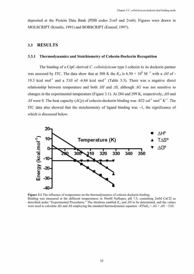
Chapter 3 C. cellulolyticum dockerin dual binding mode
55
deposited at the Protein Data Bank (PDB codes 2vn5 and 2vn6). Figures were drawn in
MOLSCRIPT (Kraulis, 1991) and BOBSCRIPT (Esnouf, 1997).
3.3 RESULTS
3.3.1 Thermodynamics and Stoichiometry of Cohesin-Dockerin Recognition
The binding of a CipC-derived C. cellulolyticum type I cohesin to its dockerin partner
was assessed by ITC. The data show that at 308 K the KA is 6.50 × 10⁸ M -1 with a ∆H of -
19.3 kcal mol-1 and a T∆S of -6.84 kcal mol-1 (Table 3.3). There was a negative direct
relationship between temperature and both ∆H and ∆S, although ∆G was not sensitive to
changes in the experimental temperature (Figure 3.1). At 284 and 299 K, respectively, ∆H and
∆S were 0. The heat capacity (∆Cp) of cohesin-dockerin binding was -822 cal-1 mol-1 K-1. The
ITC data also showed that the stoichiometry of ligand binding was ~1, the significance of
which is discussed below.
Figure 3.1 The influence of temperature on the thermodynamics of cohesin dockerin binding. Binding was measured at the different temperatures in 50mM NaHepes, pH 7.5, containing 2mM CaCl2 as described under “Experimental Procedures.” The titrations enabled KA and ∆H to be determined, and the values were used to calculate ∆G and ∆S employing the standard thermodynamic equation −RTlnKA = ∆G = ∆H − T∆S.
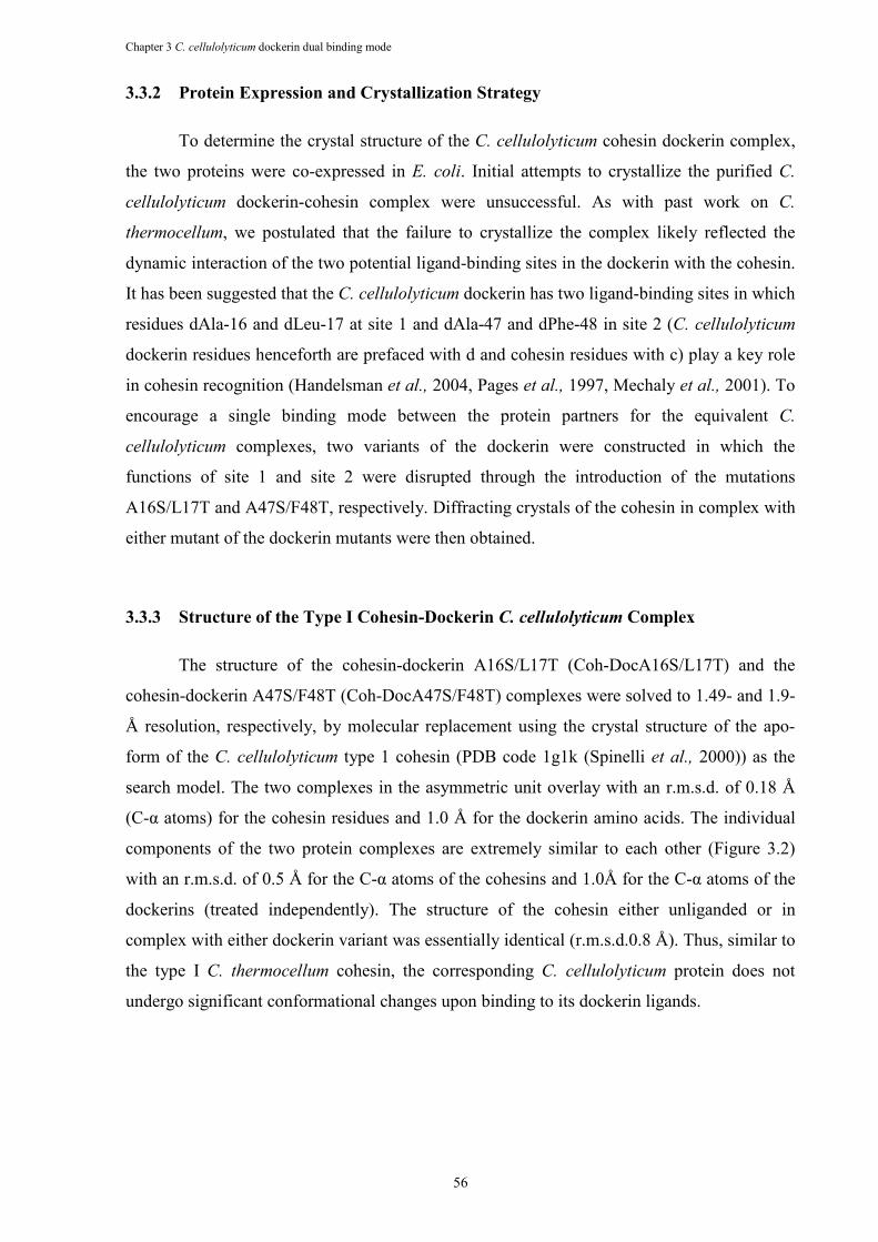
Chapter 3 C. cellulolyticum dockerin dual binding mode
56
3.3.2 Protein Expression and Crystallization Strategy
To determine the crystal structure of the C. cellulolyticum cohesin dockerin complex,
the two proteins were co-expressed in E. coli. Initial attempts to crystallize the purified C.
cellulolyticum dockerin-cohesin complex were unsuccessful. As with past work on C.
thermocellum, we postulated that the failure to crystallize the complex likely reflected the
dynamic interaction of the two potential ligand-binding sites in the dockerin with the cohesin.
It has been suggested that the C. cellulolyticum dockerin has two ligand-binding sites in which
residues dAla-16 and dLeu-17 at site 1 and dAla-47 and dPhe-48 in site 2 (C. cellulolyticum
dockerin residues henceforth are prefaced with d and cohesin residues with c) play a key role
in cohesin recognition (Handelsman et al., 2004, Pages et al., 1997, Mechaly et al., 2001). To
encourage a single binding mode between the protein partners for the equivalent C.
cellulolyticum complexes, two variants of the dockerin were constructed in which the
functions of site 1 and site 2 were disrupted through the introduction of the mutations
A16S/L17T and A47S/F48T, respectively. Diffracting crystals of the cohesin in complex with
either mutant of the dockerin mutants were then obtained.
3.3.3 Structure of the Type I Cohesin-Dockerin C. cellulolyticum Complex
The structure of the cohesin-dockerin A16S/L17T (Coh-DocA16S/L17T) and the
cohesin-dockerin A47S/F48T (Coh-DocA47S/F48T) complexes were solved to 1.49- and 1.9-
Å resolution, respectively, by molecular replacement using the crystal structure of the apo-
form of the C. cellulolyticum type 1 cohesin (PDB code 1g1k (Spinelli et al., 2000)) as the
search model. The two complexes in the asymmetric unit overlay with an r.m.s.d. of 0.18 Å
(C-α atoms) for the cohesin residues and 1.0 Å for the dockerin amino acids. The individual
components of the two protein complexes are extremely similar to each other (Figure 3.2)
with an r.m.s.d. of 0.5 Å for the C-α atoms of the cohesins and 1.0Å for the C-α atoms of the
dockerins (treated independently). The structure of the cohesin either unliganded or in
complex with either dockerin variant was essentially identical (r.m.s.d.0.8 Å). Thus, similar to
the type I C. thermocellum cohesin, the corresponding C. cellulolyticum protein does not
undergo significant conformational changes upon binding to its dockerin ligands.
Chapter 3 C. cellulolyticum dockerin dual binding mode
57
Figure 3.2 The three-dimensional structures of the C. cellulolyticum cohesin-dockerin complexes. a depicts the structure of Coh-DocA16S/L17T with the dockerin color-ramped from N terminus (blue) to C terminus (red) and the cohesin in pale brown. In this complex the hydrophobic residues Phe-48 and Ala-47 dominate the hydrophobic contribution of the dockerin, and these residues are shown as ball-and-stick. Ca2+ ions are shown as shaded spheres. b shows the CohDocA47S/F48T complex, similarly colored. Here Leu-17 and Ala-16 of the dockerin form the basis of the hydrophobic surface of the dockerin. c depicts an overlap of the two binding modes showing the very high degree of overall similarity reflecting the internal 2-fold symmetry of the dockerin itself (see text). A47S/F48T is shown in blue and A16S/L17T in yellow. Figure was drawn in MOLSCRIPT and BOBSCRIPT.
3.3.4 Structure of the C. cellulolyticum Type I Cohesin in Complex with Its Cognate
Dockerin.
The type I C. cellulolyticum cohesin in complex with its cognate dockerin has an
elliptical structure comprising a 4-residue α-helix and 10 β-strands, which forms 2 β-sheets
aligned in an elongated β-sandwich and displays a classical jelly roll fold (Figure 3.2). The
two sheets include β-strands 10, 1, 2, and 7 on one face (sheet A) and β-strands 5, 6, 3, and 9
on the other face (sheet B). The two sheets are connected by β-strand 8 that makes hydrogen
bonds with both β-strand 9 on sheet A and β-strand 3 on sheet B. The cohesin also displays
striking similarity to type I C. thermocellum cohesins (r.m.s.d. 0.8 Å).
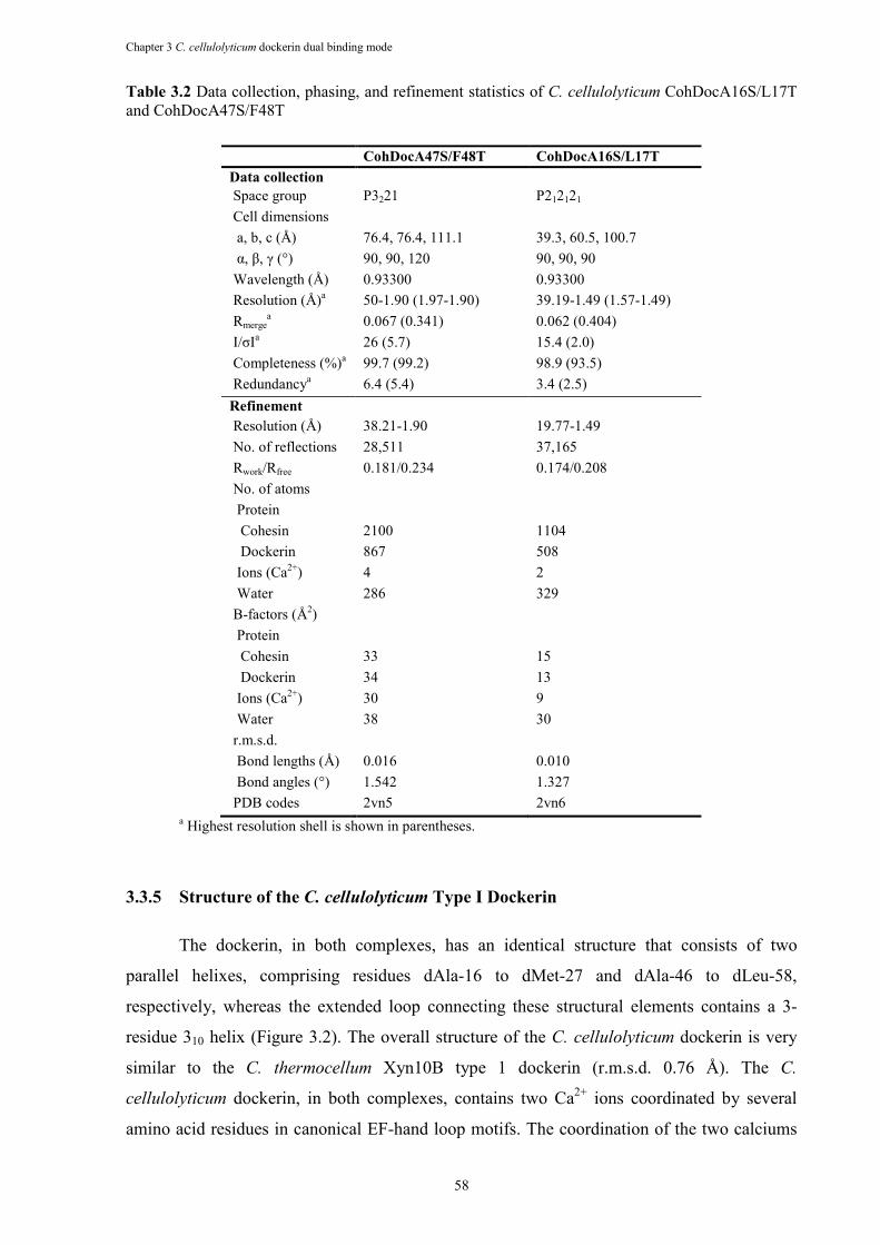
Chapter 3 C. cellulolyticum dockerin dual binding mode
58
Table 3.2 Data collection, phasing, and refinement statistics of C. cellulolyticum CohDocA16S/L17T and CohDocA47S/F48T
CohDocA47S/F48T CohDocA16S/L17T
Data collection Space group P3221 P212121 Cell dimensions a, b, c (Å) 76.4, 76.4, 111.1 39.3, 60.5, 100.7 α, β, γ (°) 90, 90, 120 90, 90, 90 Wavelength (Å) 0.93300 0.93300 Resolution (Å)a 50-1.90 (1.97-1.90) 39.19-1.49 (1.57-1.49) Rmerge
a 0.067 (0.341) 0.062 (0.404) I/σIa 26 (5.7) 15.4 (2.0) Completeness (%)a 99.7 (99.2) 98.9 (93.5) Redundancya 6.4 (5.4) 3.4 (2.5) Refinement Resolution (Å) 38.21-1.90 19.77-1.49 No. of reflections 28,511 37,165 Rwork/Rfree 0.181/0.234 0.174/0.208 No. of atoms Protein Cohesin 2100 1104 Dockerin 867 508 Ions (Ca2+) 4 2 Water 286 329 B-factors (Å2) Protein Cohesin 33 15 Dockerin 34 13 Ions (Ca2+) 30 9 Water 38 30 r.m.s.d. Bond lengths (Å) 0.016 0.010 Bond angles (°) 1.542 1.327 PDB codes 2vn5 2vn6
a Highest resolution shell is shown in parentheses.
3.3.5 Structure of the C. cellulolyticum Type I Dockerin
The dockerin, in both complexes, has an identical structure that consists of two
parallel helixes, comprising residues dAla-16 to dMet-27 and dAla-46 to dLeu-58,
respectively, whereas the extended loop connecting these structural elements contains a 3-
residue 310 helix (Figure 3.2). The overall structure of the C. cellulolyticum dockerin is very
similar to the C. thermocellum Xyn10B type 1 dockerin (r.m.s.d. 0.76 Å). The C.
cellulolyticum dockerin, in both complexes, contains two Ca2+ ions coordinated by several
amino acid residues in canonical EF-hand loop motifs. The coordination of the two calciums
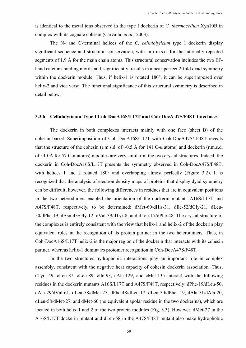
Chapter 3 C. cellulolyticum dockerin dual binding mode
59
is identical to the metal ions observed in the type I dockerin of C. thermocellum Xyn10B in
complex with its cognate cohesin (Carvalho et al., 2003).
The N- and C-terminal helices of the C. cellulolyticum type I dockerin display
significant sequence and structural conservation, with an r.m.s.d. for the internally repeated
segments of 1.9 Å for the main chain atoms. This structural conservation includes the two EF-
hand calcium-binding motifs and, significantly, results in a near-perfect 2-fold dyad symmetry
within the dockerin module. Thus, if helix-1 is rotated 180°, it can be superimposed over
helix-2 and vice versa. The functional significance of this structural symmetry is described in
detail below.
3.3.6 Cellulolyticum Type I Coh-DocA16S/L17T and Coh-DocA 47S/F48T Interfaces
The dockerin in both complexes interacts mainly with one face (sheet B) of the
cohesin barrel. Superimposition of Coh-DocA16S/L17T with Coh-DocA47S/ F48T reveals
that the structure of the cohesin (r.m.s.d. of ~0.5 Å for 141 C-α atoms) and dockerin (r.m.s.d.
of ~1.0Å for 57 C-α atoms) modules are very similar in the two crystal structures. Indeed, the
dockerin in Coh-DocA16S/L17T presents the symmetry observed in Coh-DocA47S/F48T,
with helices 1 and 2 rotated 180° and overlapping almost perfectly (Figure 3.2). It is
recognized that the analysis of electron density maps of proteins that display dyad symmetry
can be difficult; however, the following differences in residues that are in equivalent positions
in the two heterodimers enabled the orientation of the dockerin mutants A16S/L17T and
A47S/F48T, respectively, to be determined: dMet-60/dHis-31, dIle-52/dGly-21, dLeu-
50/dPhe-19, dAsn-43/Gly-12, dVal-39/dTyr-8, and dLeu-17/dPhe-48. The crystal structure of
the complexes is entirely consistent with the view that helix-1 and helix-2 of the dockerin play
equivalent roles in the recognition of its protein partner in the two heterodimers. Thus, in
Coh-DocA16S/L17T helix-2 is the major region of the dockerin that interacts with its cohesin
partner, whereas helix-1 dominates protomer recognition in Coh-DocA47S/F48T.
In the two structures hydrophobic interactions play an important role in complex
assembly, consistent with the negative heat capacity of cohesin dockerin association. Thus,
cTyr- 49, cLeu-87, cLeu-89, cIle-93, cAla-129, and cMet-135 interact with the following
residues in the dockerin mutants A16S/L17T and A47S/F48T, respectively: dPhe-19/dLeu-50,
dAla-29/dVal-61, dLeu-58/dMet-27, dPhe-48/dLeu-17, dLeu-50/dPhe- 19, dAla-51/dAla-20,
dLeu-58/dMet-27, and dMet-60 (no equivalent apolar residue in the two dockerins), which are
located in both helix-1 and 2 of the two protein modules (Fig. 3.3). However, dMet-27 in the
A16S/L17T dockerin mutant and dLeu-58 in the A47S/F48T mutant also make hydrophobic
Chapter 3 C. cellulolyticum dockerin dual binding mode
60
contacts with a shallow pocket formed by the β-carbons of cSer-76, cSer-78, and cSer-85, and
these interactions may also contribute to overall affinity. The heterodimer also associates
through four direct and numerous solvent-mediated polar interactions, which are described in
detail in Table 3.4. The direct hydrogen bonds are between O-δ1 of cAsn-47 and N of dAla-
16/dAla-47, γ-O of cThr-45 and O of dAla-16/dAla-47, OH of cTyr-49 and both O of dLeu-
57/dIle-26 and dN-2 of His-31 (the side chain of the equivalent residue in the dockerin mutant
A16S/L17T, dMet-60, is nonpolar), γ-O of cSer-75 and O of dLeu-58/ dMet-27 (Figure 3.3).
Although only the C-terminal region of dockerin helix-2 interacts with the cohesin, the whole
length of dockerin helix-1 contacts its protein partner in Coh-DocA47S/F48T. Conversely, in
Coh-DocA16S/L17T only the N-terminal region of helix-1 contacts the cohesin, whereas all
of helix-2 interacts with its protein partner.
Figure 3.3 The cohesin-dockerin interface in the two C. cellulolyticum cohesin-dockerin complexes. a shows the Coh-DocA16S/L17T complex with the dockerin in pale green and the cohesin in blue. Residues at the interface are shown as ball-and-stick. b includes the CohDocA47S/F48T complex with the dockerin in yellow and the cohesin in blue. Residues at the interface are shown as ball-and-stick. Both figures are in divergent (wall-eyed) stereo.
Site-directed mutagenesis data (Table 3.3) show that alanine substitution of the three
hydrophobic cohesin residues cLeu- 87, cLeu-89, and cMet-135 results in a substantial
decrease in affinity (9–130-fold), whereas the cI93A mutation reduces the KA 3-fold.
Chapter 3 C. cellulolyticum dockerin dual binding mode
61
Although the cN47A and cY49A mutations also cause an 5-fold decrease in the KA, the amino
acid substitutions cT45A, cS76A, cS85A, cN91A, and cK137G had little influence on the
affinity of the cohesin for its C. cellulolyticum dockerin partner. Thus, the only significant
polar interactions between the protomers in Coh-DocA16S/L17T are the hydrogen bonds
between the side chain carbonyl of cAsn-47 and the main chain amide of dAla-47 and
between the phenolic is therefore apparent that hydrophobic interactions play a dominant role
in the assembly of this complex.
The most significant hydrophobic interaction at the protomer interface is between the
apolar pocket, formed by cLeu-87 and cLeu-89, which is occupied by the aromatic side chain
of dLeu-50 in Coh-DocA16S/L17T and dPhe-19 in Coh-DocA47S/F48T. Indeed, the affinity
of the cL87A/cL89A cohesin mutant for its cognate dockerin is ~104-fold less than the wild
type protein. The reciprocal dockerin mutations, dF19A/dL50A, also caused a significant
reduction in KA (3000-fold), although this loss in affinity was less dramatic than in the
corresponding cohesin mutant. This is consistent with the observation that cLeu-87 and cLeu-
89, in addition to interacting with dPhe- 19/dLeu-50, also make van der Waals contacts with
dAla-20/ dAla-51 and dAla16/dAla-47. The modest decrease in affinity displayed by the I93A
mutant is rather surprising as C-δ1 and C-γ1 make hydrophobic contacts with dMet- 27/dLeu-
58, whereas C-γ2 interacts with dMet-60. The contribution of cMet-135 to dockerin
recognition is through weak hydrophobic interactions with the aliphatic side chains of dLys-
24/dLys-55, and strong van der Waals contacts with the C-β of dAla20/dAla51. The
methionine may also contribute, indirectly, to dockerin recognition by making strong
hydrophobic interactions with cLeu-89 and cIle-93 and thus play a role in optimizing the
position of these residues for dockerin recognition. The hydrophobic interaction that appears
to play a central role in the specificity of the C. cellulolyticum cohesin-dockerin interaction is
between dLeu-17 and a shallow hydrophobic pocket of which cAla-129 is the major
contributor (see below).
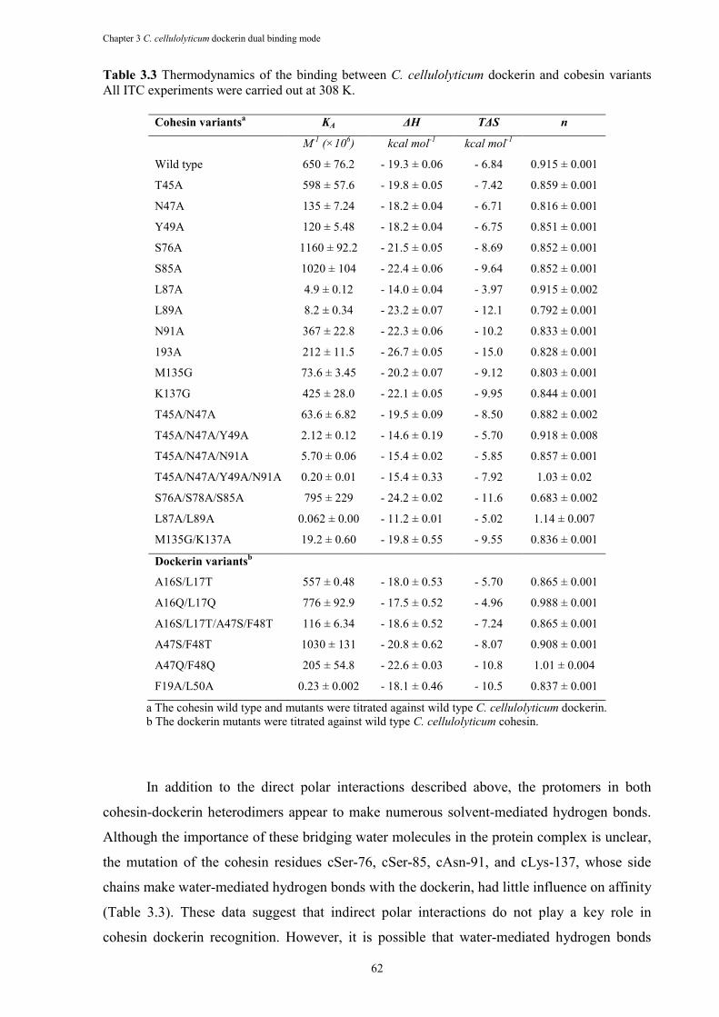
Chapter 3 C. cellulolyticum dockerin dual binding mode
62
Table 3.3 Thermodynamics of the binding between C. cellulolyticum dockerin and cobesin variants All ITC experiments were carried out at 308 K.
Cohesin variantsa KA ∆H T∆S n
M-1
(×106) kcal mol
-1 kcal mol
-1
Wild type 650 ± 76.2 - 19.3 ± 0.06 - 6.84 0.915 ± 0.001
T45A 598 ± 57.6 - 19.8 ± 0.05 - 7.42 0.859 ± 0.001
N47A 135 ± 7.24 - 18.2 ± 0.04 - 6.71 0.816 ± 0.001
Y49A 120 ± 5.48 - 18.2 ± 0.04 - 6.75 0.851 ± 0.001
S76A 1160 ± 92.2 - 21.5 ± 0.05 - 8.69 0.852 ± 0.001
S85A 1020 ± 104 - 22.4 ± 0.06 - 9.64 0.852 ± 0.001
L87A 4.9 ± 0.12 - 14.0 ± 0.04 - 3.97 0.915 ± 0.002
L89A 8.2 ± 0.34 - 23.2 ± 0.07 - 12.1 0.792 ± 0.001
N91A 367 ± 22.8 - 22.3 ± 0.06 - 10.2 0.833 ± 0.001
193A 212 ± 11.5 - 26.7 ± 0.05 - 15.0 0.828 ± 0.001
M135G 73.6 ± 3.45 - 20.2 ± 0.07 - 9.12 0.803 ± 0.001
K137G 425 ± 28.0 - 22.1 ± 0.05 - 9.95 0.844 ± 0.001
T45A/N47A 63.6 ± 6.82 - 19.5 ± 0.09 - 8.50 0.882 ± 0.002
T45A/N47A/Y49A 2.12 ± 0.12 - 14.6 ± 0.19 - 5.70 0.918 ± 0.008
T45A/N47A/N91A 5.70 ± 0.06 - 15.4 ± 0.02 - 5.85 0.857 ± 0.001
T45A/N47A/Y49A/N91A 0.20 ± 0.01 - 15.4 ± 0.33 - 7.92 1.03 ± 0.02
S76A/S78A/S85A 795 ± 229 - 24.2 ± 0.02 - 11.6 0.683 ± 0.002
L87A/L89A 0.062 ± 0.00 - 11.2 ± 0.01 - 5.02 1.14 ± 0.007
M135G/K137A 19.2 ± 0.60 - 19.8 ± 0.55 - 9.55 0.836 ± 0.001
Dockerin variantsb
A16S/L17T 557 ± 0.48 - 18.0 ± 0.53 - 5.70 0.865 ± 0.001
A16Q/L17Q 776 ± 92.9 - 17.5 ± 0.52 - 4.96 0.988 ± 0.001
A16S/L17T/A47S/F48T 116 ± 6.34 - 18.6 ± 0.52 - 7.24 0.865 ± 0.001
A47S/F48T 1030 ± 131 - 20.8 ± 0.62 - 8.07 0.908 ± 0.001
A47Q/F48Q 205 ± 54.8 - 22.6 ± 0.03 - 10.8 1.01 ± 0.004
F19A/L50A 0.23 ± 0.002 - 18.1 ± 0.46 - 10.5 0.837 ± 0.001
a The cohesin wild type and mutants were titrated against wild type C. cellulolyticum dockerin. b The dockerin mutants were titrated against wild type C. cellulolyticum cohesin.
In addition to the direct polar interactions described above, the protomers in both
cohesin-dockerin heterodimers appear to make numerous solvent-mediated hydrogen bonds.
Although the importance of these bridging water molecules in the protein complex is unclear,
the mutation of the cohesin residues cSer-76, cSer-85, cAsn-91, and cLys-137, whose side
chains make water-mediated hydrogen bonds with the dockerin, had little influence on affinity
(Table 3.3). These data suggest that indirect polar interactions do not play a key role in
cohesin dockerin recognition. However, it is possible that water-mediated hydrogen bonds
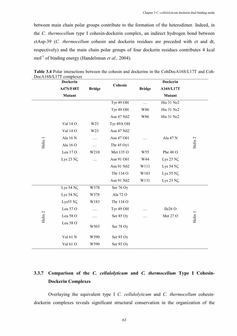
Chapter 3 C. cellulolyticum dockerin dual binding mode
63
between main chain polar groups contribute to the formation of the heterodimer. Indeed, in
the C. thermocellum type I cohesin-dockerin complex, an indirect hydrogen bond between
ctAsp-39 (C. thermocellum cohesin and dockerin residues are preceded with ct and dt,
respectively) and the main chain polar groups of four dockerin residues contributes 4 kcal
mol-1 of binding energy (Handelsman et al., 2004).
Table 3.4 Polar interactions between the cohesin and dockerins in the CohDocA16S/L17T and Coh-DocA16S/L17T complexes Dockerin
A47S/F48T
Mutant
Bridge Cohesin
Bridge
Dockerin
A16S/L17T
Mutant
Hel
ix 1
Tyr 49 OH … His 31 Nε2
Hel
ix 2
Tyr 49 OH W86 His 31 Nε2
Asn 47 Nδ2 W86 His 31 Nε2
Val 14 O W23 Tyr 49A OH
Val 14 O W23 Asn 47 Nδ2
Ala 16 N … Asn 47 Oδ1 … Ala 47 N
Ala 16 O … Thr 45 Oγ1
Leu 17 O W210 Met 135 O W55 Phe 48 O
Lys 23 Nζ … Asn 91 Oδ1 W44 Lys 23 Nζ
Asn 91 Nδ2 W111 Lys 54 Nζ
Thr 134 O W183 Lys 55 Nζ
Asn 91 Nδ2 W131 Lys 23 Nζ
Hel
ix 2
Lys 54 Nζ W378 Ser 76 Oγ
Hel
ix 1
Lys 54 Nζ W378 Ala 72 O
Lys55 Nζ W183 Thr 134 O
Leu 57 O … Tyr 49 OH … Ile26 O
Leu 58 O … Ser 85 Oγ … Met 27 O
Leu 58 O
W503 Ser 78 Oγ
Val 61 N W590 Ser 85 Oγ
Val 61 O W590 Ser 85 Oγ
3.3.7 Comparison of the C. cellulolyticum and C. thermocellum Type I Cohesin-
Dockerin Complexes
Overlaying the equivalent type I C. cellulolyticum and C. thermocellum cohesin-
dockerin complexes reveals significant structural conservation in the organization of the
Chapter 3 C. cellulolyticum dockerin dual binding mode
64
complexes. Thus, comparison of C. cellulolyticum Coh-DocA16S/L17T with the native C.
thermocellum Coh-Doc complex yields an overall r.m.s.d of 1.36 Å for 180 eq C-α atoms (of
both cohesin and dockerin), whereas comparison of the C. cellulolyticum Coh-
DocA47A/F48T heterodimer with C. thermocellum Coh-DocS45A/T46A yields an r.m.s.d of
1.2 Å for 183 equivalent C-α atoms. The location of the interface between the two protomers
within the heterodimer is identical (Figure 3.4), although there is little cross-specificity
between the two complexes (Handelsman et al., 2004, Pages et al., 1997, Mechaly et al.,
2001). Inspection of the cohesin-dockerin interactions in the C. thermocellum and C.
cellulolyticum complexes provides insight into the molecular basis for the distinct specificities
displayed by these protein pairs. Thus, the hydrophobic residues of the C. cellulolyticum
cohesin that interact with the dockerin are clustered toward the C-terminal region of β-strand
6, whereas in the C. thermocellum protein the ligand binding apolar residues extend across the
full length of β-strand 3 and also include amino acids in β-strands 5 and 6 (Carvalho et al.,
2003, Carvalho et al., 2005). There is, however, some conservation in these key aliphatic
residues. Thus, the following pairs of C. cellulolyticum and C. thermocellum cohesin residues
occupy equivalent positions in their respective complexes: cLeu-87/ctLeu-83, cLeu89/ctAla-
85, and cTyr-49/ctVal-41. Several C. cellulolyticum and C. thermocellum hydrophobic
dockerin residues that interact with the cognate cohesin are also highly conserved and include
the following pairs: dMet-27/dtLeu-56, dIle-26/dtLeu-55, dLeu-50/dtLeu14, dPhe-19/dtLeu-
48, dLeu- 58/dtLeu-22, and d-Met-60/dtAla-24. It is therefore not surprising that many of the
hydrophobic interactions at the protomer interface are conserved in the two protein
complexes. For example, the key hydrophobic pocket in the C. cellulolyticum cohesin, formed
by cLeu-87 and cLeu-89, would be occupied by the C. thermocellum dockerin amino acid
dtLeu-14. Conversely, the hydrophobic pocket in the C. thermocellum cohesin, formed by
ctAla-72, ctVal-81, and ctLeu-83, would be occupied by the C. cellulolyticum dockerin
residue dMet-27.
In contrast to the hydrophobic interactions, there are significant differences in the
polar interactions between the protomers in the C. cellulolyticum and C. thermocellum
heterodimers. The only similar polar cohesin residues that interact with the cognate dockerins
are cAsn-47/ctAsp-39 and cAsp-87/ ctAsn-91, although mutagenesis studies have shown that
the D39N mutation in a type I C. thermocellum cohesin reduces affinity for the C.
thermocellum dockerin 1000-fold indicating significant differences in the role of similar
residues in the two complexes (Handelsman et al., 2004). It has been suggested that the
dramatic effect of the D39N reflects the capacity of both O-δ1 and O-δ2 to act as hydrogen
bond acceptors in polar interactions with the dockerin. The lack of crossspecies recognition
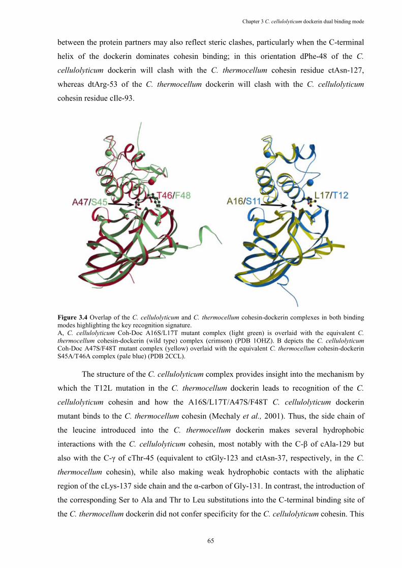
Chapter 3 C. cellulolyticum dockerin dual binding mode
65
between the protein partners may also reflect steric clashes, particularly when the C-terminal
helix of the dockerin dominates cohesin binding; in this orientation dPhe-48 of the C.
cellulolyticum dockerin will clash with the C. thermocellum cohesin residue ctAsn-127,
whereas dtArg-53 of the C. thermocellum dockerin will clash with the C. cellulolyticum
cohesin residue cIle-93.
Figure 3.4 Overlap of the C. cellulolyticum and C. thermocellum cohesin-dockerin complexes in both binding modes highlighting the key recognition signature. A, C. cellulolyticum Coh-Doc A16S/L17T mutant complex (light green) is overlaid with the equivalent C.
thermocellum cohesin-dockerin (wild type) complex (crimson) (PDB 1OHZ). B depicts the C. cellulolyticum Coh-Doc A47S/F48T mutant complex (yellow) overlaid with the equivalent C. thermocellum cohesin-dockerin S45A/T46A complex (pale blue) (PDB 2CCL).
The structure of the C. cellulolyticum complex provides insight into the mechanism by
which the T12L mutation in the C. thermocellum dockerin leads to recognition of the C.
cellulolyticum cohesin and how the A16S/L17T/A47S/F48T C. cellulolyticum dockerin
mutant binds to the C. thermocellum cohesin (Mechaly et al., 2001). Thus, the side chain of
the leucine introduced into the C. thermocellum dockerin makes several hydrophobic
interactions with the C. cellulolyticum cohesin, most notably with the C-β of cAla-129 but
also with the C-γ of cThr-45 (equivalent to ctGly-123 and ctAsn-37, respectively, in the C.
thermocellum cohesin), while also making weak hydrophobic contacts with the aliphatic
region of the cLys-137 side chain and the α-carbon of Gly-131. In contrast, the introduction of
the corresponding Ser to Ala and Thr to Leu substitutions into the C-terminal binding site of
the C. thermocellum dockerin did not confer specificity for the C. cellulolyticum cohesin. This
Chapter 3 C. cellulolyticum dockerin dual binding mode
66
may reflect the energetic penalty imposed by the predicted steric clashes discussed above. The
capacity of the A16S/L17T/ A47S/F48T C. cellulolyticum dockerin mutant to bind the C.
thermocellum cohesin likely reflects the capacity of the introduced polar residues to make
hydrogen bonds with ctAsn-37, ctAsp-39, and ctGlu-131. An intriguing feature of these
“change in specificity” mutations (or substituting these amino acids with alanine) (Table 3.3)
is that the engineered dockerins retain significant affinity for their original cohesin partners.
This likely reflects the retention of the extensive hydrophobic interactions between the
engineered dockerins and their native protein partners, exemplified by the functional
importance of the hydrophobic pocket formed by cLeu-87 and cLeu-89 in the C.
cellulolyticum cohesin (see above).
3.4 DISCUSSIO�
Previous studies showed that the type I C. thermocellum dockerin domains contain
two near identical type I cohesin-binding sites (Carvalho et al., 2003, Carvalho et al., 2005
and Mechaly et al., 2001). The observation that the corresponding C. cellulolyticum system
displays a similar binding mode, even though there is little cross-specificity between
clostridial heterodimers, indicates that there is a strong evolutionary driver for the retention of
two identical cohesin-binding sites in the dockerin domains. The reasons for the retention of
two, apparently equivalent, binding modes is unclear. It could be argued that the two binding
modes may enable the catalytic subunits to bind to multiple cohesins leading to
polycellulosome assembly, as observed in C. thermocellum (Bayer et al., 1985). The observed
stoichiometry of one, where measured, however, indicates that both ligand-binding sites on
the dockerin cannot be occupied simultaneously. Indeed, overlaying the two C. cellulolyticum
complexes shows that the cohesin α-helix, extending from cAsn-70 to cAsn-74, will make
steric clashes precluding the formation of a trimolecular complex.
The high degree of sequence conservation in the two cohesin- and calcium-binding
sites of the C. cellulolyticum dockerins (Pages et al., 1997), leading to the retention of dyad
symmetry, indicates that the observed dual binding mode is a general feature of the
cellulosome of the bacterium. Indeed, the lack of sequence conservation in the regions linking
the two helices/EF-hand motifs again points to how retention of both cohesin-binding sites
represents a strong evolutionary driver. Furthermore, the structures of the protein complex, in
combination with mutagenesis data, show a high degree of sequence conservation of the
dockerin recognition residues displayed by the cohesin domains of C. cellulolyticum
(Handelsman et al., 2004), indicating that there is little, if any, specificity between specific
Chapter 3 C. cellulolyticum dockerin dual binding mode
67
cohesin-dockerin partners in the cellulosome of the bacterium. Thus, the biological
significance of the dual binding mode displayed by C. cellulolyticum and C. thermocellum
dockerins is an intriguing question. One possibility, which may merit further exploration, is
that dual binding confers flexibility in cellulosome assembly (for review see Bayer et al.,
2004 and Gilbert et al., 2007). It could be argued that steric constraints imposed by the
appended catalytic modules might restrict the combination of enzymes that can be assembled
into a cellulosome complex. The dual binding mode displayed by dockerins could overcome
these spatial limitations and thus increase the range of enzymes that can be integrated into
discrete cellulosome molecules. There is a very large number of possibilities for cellulosomal
organization, and the flexibility provided by dual binding may contribute to the plasticity
required for correct incorporation of different enzyme combinations. Indeed the different
binding modes displayed by the dockerins may allow optimal placement of the enzymes on
the complex substrate, which is more easily accomplished in the plant cell wall degrading
apparatus of aerobic microorganisms as the free enzymes of these organisms do not physically
associate (Hazlewood et al., 1992). It should be emphasized, however, that the linker
sequences that join the dockerin and catalytic modules may contribute significant
conformational flexibility to the cellulosome quaternary structure, questioning the proposed
requirement for the dual binding mode reported here for C. cellulolyticum dockerins.
However, small angle x-ray scattering (SAXS) experiments have demonstrated that when
Cel48F binds to its cognate cohesin, the rigidity of the linker that connects the catalytic
module of the enzyme to its dockerin increases and the cellulase becomes more compact
(Hammel et al., 2004). The SAXS data also showed that the possible motions of the catalytic
module of Cel48F with respect to the cohesin/dockerin interface are highly restricted,
compared with the free enzyme. Thus, the enzyme linker is not likely to generate the
flexibility, which may be required for plant cell wall degradation, once the enzyme is bound
within the cellulosome (Hammel et al., 2004). It should also be acknowledged that another
source of flexibility may stem from the linkers that connect the cohesin modules in the
scaffoldins. Similar SAXS studies have indeed shown that the linker joining the two cohesins
of divergent species in the hybrid scaffoldin Scaf4 remains flexible, even when large enzymes
(Cel48F) are anchored to both cohesin modules (Hammel et al., 2005). It is apparent,
however, that the 49 residue hybrid linker is longer than typical C. cellulolyticum and C.
thermocellum scaffoldin linkers, comprising 10 and 39 residues, respectively. Indeed short
linkers is a generic feature of mesophilic clostridial scaffoldins exemplified by the
corresponding proteins produced by Clostridium cellulovorans and Clostridium josui
(Kakiuchi et al., 1998, Shoseyov et al., 1992). It is unclear why nature should select a dual
Chapter 3 C. cellulolyticum dockerin dual binding mode
68
binding mode between cohesins and dockerins, in at least some species. Indeed if, as we
propose, it is to confer conformational flexibility, it would seem facile for evolution to impose
a selection for longer linker sequences. It is possible that extended flexible linkers may be
more prone to proteolytic attack than short inter-module sequences, and thus evolution may
have selected a dual binding mode to introduce structural plasticity. In any event, the
biological rationale for the dual binding mode remains opaque, and there is clearly an urgent
need to investigate its biological significance.
This study, in conjunction with previous studies on the C. thermocellum cellulosome
(Carvalho et al., 2003, Carvalho et al., 2005), shows that the flexibility in cohesin recognition
appears to be a general feature of the type I dockerin modules that recruit the catalytic
subunits into the clostridial cellulosome. In nonclostridial cellulosomes there are examples of
sequence divergence in the duplicated regions of dockerins exemplified by the Ruminoccocus
flavefaciens system, suggesting that the dual binding mode reported here may not be a
universal feature of cellulosomes. This study also provides a mechanistic understanding of
how the C. cellulolyticum cohesins recognize their cognate dockerins, and how subtle changes
to the structure at the interface leads to significant changes in specificity, providing a template
for engineering novel specificities into these highly efficient nanomachines.
Chapter 4 C. thermocellum endo-1,4-β-D-xylanase 10B
69
Chapter 4 Purification, crystallization and crystallographic
analysis of Clostridium thermocellum endo-1,4-β-D-xylanase
10B in complex with xylohexaose
Shabir Najmudin1, Benedita A. Pinheiro2, Maria J. Romão1, José A. M. Prates2 and Carlos M. G. A. Fontes2
1 REQUIMTE, Departamento de Química, FCT-UNL, 2829-516 Caparica, Portugal, and 2 CIISA – Faculdade de
Medicina Veterinária, Universidade Técnica de Lisboa, Avenida da Universidade Técnica, 1300-477 Lisboa,
Portugal
Adapted from Acta Crystallographica F (2008) 64, 715–718
Abstract
The cellulosome of Clostridium thermocellum is a highly organized multienzyme complex of
cellulases and hemicellulases involved in the hydrolysis of plant cell-wall polysaccharides.
The bifunctional multi-modular xylanase Xyn10B is one of the hemicellulase components of
the C. thermocellum cellulosome. The enzyme contains an internal GH family 10 catalytic
domain (GH10) and a C-terminal family 1 CE domain (CE1). The N-terminal moiety of
Xyn10B (residues 32–551), comprising a CBM (CBM22-1) and the GH10 E337A mutant,
was crystallized in complex with xylohexaose. The crystals belong to the trigonal space group
P3221 and contain a dimer in the asymmetric unit. The crystals diffracted to beyond 2.0 Å
resolution.
Chapter 4 C. thermocellum endo-1,4-β-D-xylanase 10B
70
4.1 I�TRODUCTIO�
Cellulose and hemicelluloses are major constituents of plant cell walls (Bayer et al.,
2004) (see also the CAZy - Carbohydrate-Active enzymes - website http://www.cazy.org/).
Xylanase Xyn10B (formally XynY, EC 3.2.1.8) from Clostridium thermocellum is an endo-
1,4-β-D-xylan hydrolase involved in the degradation of xylan, one of the most abundant
hemicelluloses. Xyn10B comprises two family 22 CBMs (CBM22-1 and CBM22-2, formerly
X6a and X6b, respectively), flanking the GH GH10 catalytic module, a dockerin sequence
and a C-terminal family 1 CE catalytic module (CE1) (Fontes et al., 1995a). Structures of the
CBM22-2 (Charnock et al., 2000), the dockerin module in complex with a cognate cohesin
(Carvalho et al., 2003), and CE1 (Prates et al., 2001) have been elucidated, providing
important insights into the molecular role of Xyn10B in hemicellulose hydrolysis. CE1
(residues 792-1077) displays the α/β-hydrolase fold with a classical Ser-His-Asp catalytic
triad. CE1 was shown to cleave the ferulate groups involved in the cross-linking of
arabinoxylans to lignin (Tarbouriech et al., 2005, Prates et al., 2001). The dockerin module
(residues 730-791), which is a repeat of approximately 24 amino acids, was solved in
complex with the second cohesin module of C. thermocellum cellulosomal scaffolding
protein, termed CipA. Both helices of the dockerin helix-turn-helix motif may interact with
the cohesin β-sheet revealing a general dual binding mode of dockerins to cohesins (Carvalho
et al., 2003, Carvalho et al., 2007, Carvalho et al., 2005). Cellulosome assembly results from
the flexible interaction of dockerin modules located on the enzymes to one of the nine cohesin
domains of CipA scaffold (Bayer et al., 2004). Therefore, the cellulosome of C. thermocellum
comprises nine enzymatic subunits in addition to CipA. CBM22-2 (residues 560-720) has a
classic β-jelly roll fold with a cleft containing three aromatic and two polar conserved
residues (Charnock et al., 2000). Trp 53, Tyr 103 and Glu 138 play a crucial role in xylan
recognition, while Tyr 136 and Arg 25, although involved in ligand binding, also maintain the
structural integrity of the cleft (Xie et al., 2001). The catalytic site of the GH10 module
comprises Glu 337 (proton donor) and Glu 480 (nucleophile) (Fontes et al., 1995b). A mutant
construct (CBM22-1-GH10 E337A) was made to capture the substrate xylohexaose in the
inactivated site. Here we report the preliminary crystal structural characterization of the N-
terminus of Xyn10B (residues 32-551), comprising the modules CBM22-1 and GH10 E337A,
in complex with xylohexaose.
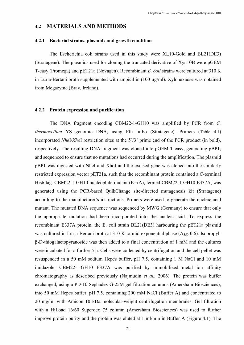
Chapter 4 C. thermocellum endo-1,4-β-D-xylanase 10B
71
4.2 MATERIALS A�D METHODS
4.2.1 Bacterial strains, plasmids and growth condition
The Escherichia coli strains used in this study were XL10-Gold and BL21(DE3)
(Stratagene). The plasmids used for cloning the truncated derivative of Xyn10B were pGEM
T-easy (Promega) and pET21a (Novagen). Recombinant E. coli strains were cultured at 310 K
in Luria-Bertani broth supplemented with ampicillin (100 µg/ml). Xylohexaose was obtained
from Megazyme (Bray, Ireland).
4.2.2 Protein expression and purification
The DNA fragment encoding CBM22-1-GH10 was amplified by PCR from C.
thermocellum YS genomic DNA, using Pfu turbo (Stratagene). Primers (Table 4.1)
incorporated ,heI/XhoI restriction sites at the 5´/3´ prime end of the PCR product (in bold),
respectively. The resulting DNA fragment was cloned into pGEM T-easy, generating pBP1,
and sequenced to ensure that no mutations had occurred during the amplification. The plasmid
pBP1 was digested with NheI and XhoI and the excised gene was cloned into the similarly
restricted expression vector pET21a, such that the recombinant protein contained a C-terminal
His6 tag. CBM22-1-GH10 nucleophile mutant (E→A), termed CBM22-1-GH10 E337A, was
generated using the PCR-based QuikChange site-directed mutagenesis kit (Stratagene)
according to the manufacturer’s instructions. Primers were used to generate the nucleic acid
mutant. The mutated DNA sequence was sequenced by MWG (Germany) to ensure that only
the appropriate mutation had been incorporated into the nucleic acid. To express the
recombinant E337A protein, the E. coli strain BL21(DE3) harbouring the pET21a plasmid
was cultured in Luria-Bertani broth at 310 K to mid-exponential phase (A550 0.6). Isopropyl-
β-D-thiogalactopyranoside was then added to a final concentration of 1 mM and the cultures
were incubated for a further 5 h. Cells were collected by centrifugation and the cell pellet was
resuspended in a 50 mM sodium Hepes buffer, pH 7.5, containing 1 M NaCl and 10 mM
imidazole. CBM22-1-GH10 E337A was purified by immobilized metal ion affinity
chromatography as described previously (Najmudin et al., 2006). The protein was buffer
exchanged, using a PD-10 Sephadex G-25M gel filtration columns (Amersham Biosciences),
into 50 mM Hepes buffer, pH 7.5, containing 200 mM NaCl (Buffer A) and concentrated to
20 mg/ml with Amicon 10 kDa molecular-weight centrifugation membranes. Gel filtration
with a HiLoad 16/60 Superdex 75 column (Amersham Biosciences) was used to further
improve protein purity and the protein was eluted at 1 ml/min in Buffer A (Figure 4.1). The
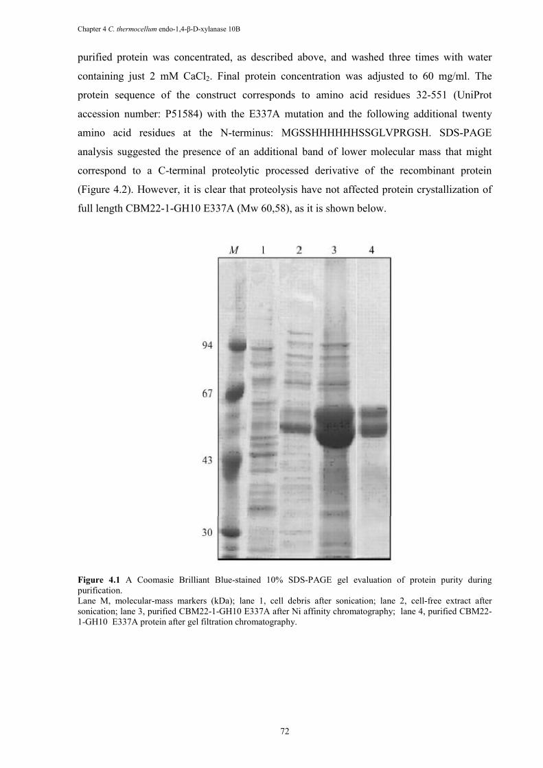
Chapter 4 C. thermocellum endo-1,4-β-D-xylanase 10B
72
purified protein was concentrated, as described above, and washed three times with water
containing just 2 mM CaCl2. Final protein concentration was adjusted to 60 mg/ml. The
protein sequence of the construct corresponds to amino acid residues 32-551 (UniProt
accession number: P51584) with the E337A mutation and the following additional twenty
amino acid residues at the N-terminus: MGSSHHHHHHSSGLVPRGSH. SDS-PAGE
analysis suggested the presence of an additional band of lower molecular mass that might
correspond to a C-terminal proteolytic processed derivative of the recombinant protein
(Figure 4.2). However, it is clear that proteolysis have not affected protein crystallization of
full length CBM22-1-GH10 E337A (Mw 60,58), as it is shown below.
Figure 4.1 A Coomasie Brilliant Blue-stained 10% SDS-PAGE gel evaluation of protein purity during purification. Lane M, molecular-mass markers (kDa); lane 1, cell debris after sonication; lane 2, cell-free extract after sonication; lane 3, purified CBM22-1-GH10 E337A after Ni affinity chromatography; lane 4, purified CBM22-1-GH10 E337A protein after gel filtration chromatography.
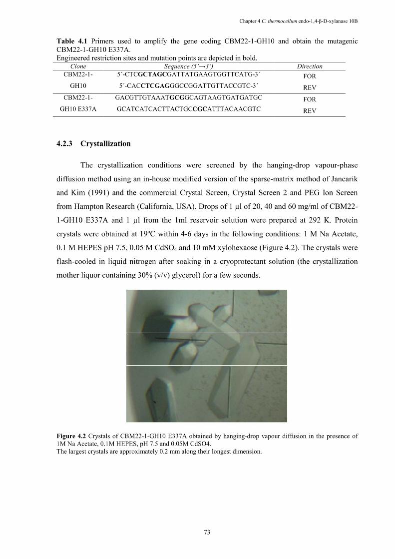
Chapter 4 C. thermocellum endo-1,4-β-D-xylanase 10B
73
Table 4.1 Primers used to amplify the gene coding CBM22-1-GH10 and obtain the mutagenic CBM22-1-GH10 E337A. Engineered restriction sites and mutation points are depicted in bold.
Clone Sequence (5’→3’) Direction
CBM22-1-
GH10
5´-CTCGCTAGCGATTATGAAGTGGTTCATG-3´ FOR 5´-CACCTCGAGGGCCGGATTGTTACCGTC-3´ REV
CBM22-1-
GH10 E337A
GACGTTGTAAATGCGGCAGTAAGTGATGATGC FOR GCATCATCACTTACTGCCGCATTTACAACGTC REV
4.2.3 Crystallization
The crystallization conditions were screened by the hanging-drop vapour-phase
diffusion method using an in-house modified version of the sparse-matrix method of Jancarik
and Kim (1991) and the commercial Crystal Screen, Crystal Screen 2 and PEG Ion Screen
from Hampton Research (California, USA). Drops of 1 µl of 20, 40 and 60 mg/ml of CBM22-
1-GH10 E337A and 1 µl from the 1ml reservoir solution were prepared at 292 K. Protein
crystals were obtained at 19ºC within 4-6 days in the following conditions: 1 M Na Acetate,
0.1 M HEPES pH 7.5, 0.05 M CdSO4 and 10 mM xylohexaose (Figure 4.2). The crystals were
flash-cooled in liquid nitrogen after soaking in a cryoprotectant solution (the crystallization
mother liquor containing 30% (v/v) glycerol) for a few seconds.
Figure 4.2 Crystals of CBM22-1-GH10 E337A obtained by hanging-drop vapour diffusion in the presence of 1M Na Acetate, 0.1M HEPES, pH 7.5 and 0.05M CdSO4. The largest crystals are approximately 0.2 mm along their longest dimension.
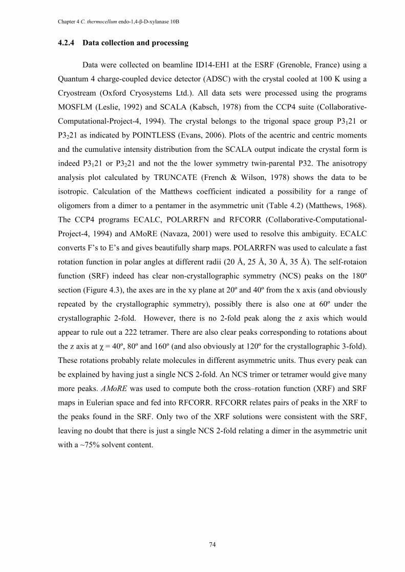
Chapter 4 C. thermocellum endo-1,4-β-D-xylanase 10B
74
4.2.4 Data collection and processing
Data were collected on beamline ID14-EH1 at the ESRF (Grenoble, France) using a
Quantum 4 charge-coupled device detector (ADSC) with the crystal cooled at 100 K using a
Cryostream (Oxford Cryosystems Ltd.). All data sets were processed using the programs
MOSFLM (Leslie, 1992) and SCALA (Kabsch, 1978) from the CCP4 suite (Collaborative-
Computational-Project-4, 1994). The crystal belongs to the trigonal space group P3121 or
P3221 as indicated by POINTLESS (Evans, 2006). Plots of the acentric and centric moments
and the cumulative intensity distribution from the SCALA output indicate the crystal form is
indeed P3121 or P3221 and not the the lower symmetry twin-parental P32. The anisotropy
analysis plot calculated by TRUNCATE (French & Wilson, 1978) shows the data to be
isotropic. Calculation of the Matthews coefficient indicated a possibility for a range of
oligomers from a dimer to a pentamer in the asymmetric unit (Table 4.2) (Matthews, 1968).
The CCP4 programs ECALC, POLARRFN and RFCORR (Collaborative-Computational-
Project-4, 1994) and AMoRE (Navaza, 2001) were used to resolve this ambiguity. ECALC
converts F’s to E’s and gives beautifully sharp maps. POLARRFN was used to calculate a fast
rotation function in polar angles at different radii (20 Å, 25 Å, 30 Å, 35 Å). The self-rotaion
function (SRF) indeed has clear non-crystallographic symmetry (NCS) peaks on the 180º
section (Figure 4.3), the axes are in the xy plane at 20º and 40º from the x axis (and obviously
repeated by the crystallographic symmetry), possibly there is also one at 60º under the
crystallographic 2-fold. However, there is no 2-fold peak along the z axis which would
appear to rule out a 222 tetramer. There are also clear peaks corresponding to rotations about
the z axis at χ = 40º, 80º and 160º (and also obviously at 120º for the crystallographic 3-fold).
These rotations probably relate molecules in different asymmetric units. Thus every peak can
be explained by having just a single NCS 2-fold. An NCS trimer or tetramer would give many
more peaks. AMoRE was used to compute both the cross–rotation function (XRF) and SRF
maps in Eulerian space and fed into RFCORR. RFCORR relates pairs of peaks in the XRF to
the peaks found in the SRF. Only two of the XRF solutions were consistent with the SRF,
leaving no doubt that there is just a single NCS 2-fold relating a dimer in the asymmetric unit
with a ~75% solvent content.
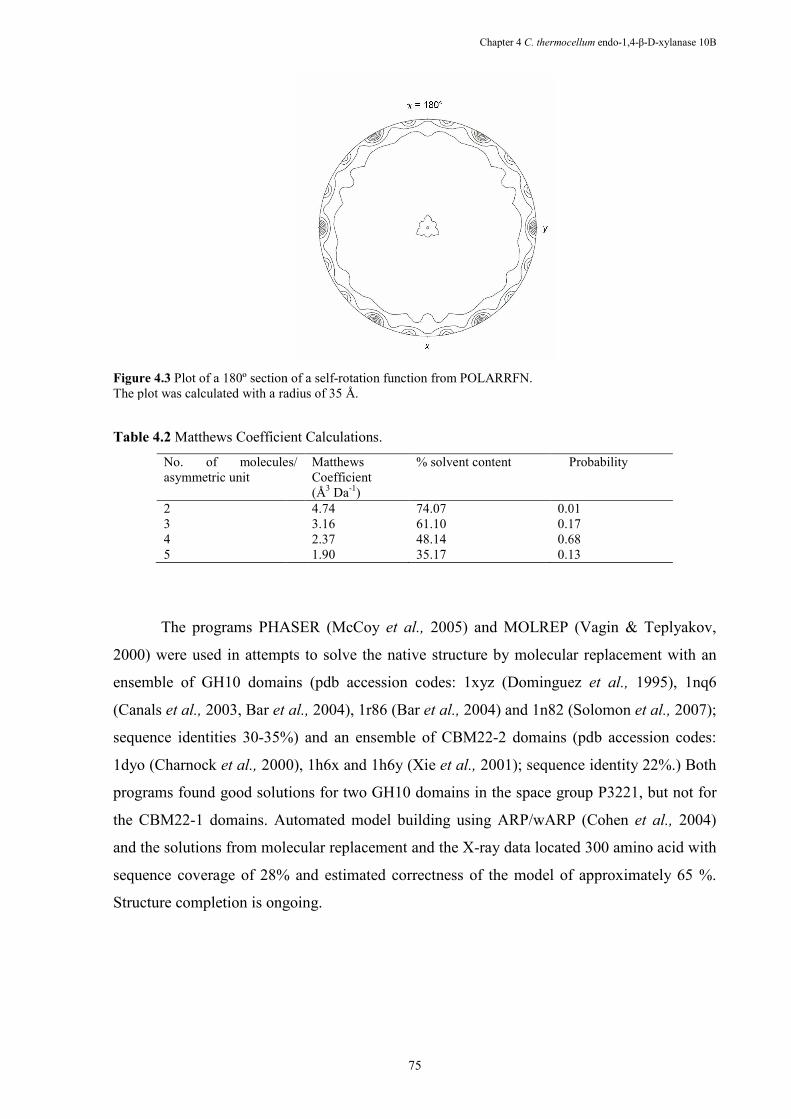
Figure 4.3 Plot of a 180º section of a selfThe plot was calculated with a radius of 35 Å.
Table 4.2 Matthews Coefficient Calculations.
No. of molecules/ asymmetric unit
2 3 4 5
The programs PHASER
2000) were used in attempts to solve the native structure by molecular replacement with an
ensemble of GH10 domains (pdb accession codes: 1xyz (Dominguez
(Canals et al., 2003, Bar et al.,
sequence identities 30-35%) and an ensemble of CBM22
1dyo (Charnock et al., 2000), 1h6x and 1h6y (Xie
programs found good solutions for two GH10 domains in the space group P3221, but not for
the CBM22-1 domains. Automated model building using ARP/wARP (Cohen
and the solutions from molecular
sequence coverage of 28% and estimated correctness of the model
Structure completion is ongoing.
Chapter 4 C. thermocellum
75
ot of a 180º section of a self-rotation function from POLARRFN. The plot was calculated with a radius of 35 Å.
Matthews Coefficient Calculations.
No. of molecules/ Matthews Coefficient (Å3 Da-1)
% solvent content Probability
4.74 74.07 0.013.16 61.10 0.172.37 48.14 0.681.90 35.17 0.13
PHASER (McCoy et al., 2005) and MOLREP
2000) were used in attempts to solve the native structure by molecular replacement with an
ensemble of GH10 domains (pdb accession codes: 1xyz (Dominguez
et al., 2004), 1r86 (Bar et al., 2004) and 1n82 (Solomon
35%) and an ensemble of CBM22-2 domains (pdb accession codes:
2000), 1h6x and 1h6y (Xie et al., 2001); sequence identity 22%.) Both
programs found good solutions for two GH10 domains in the space group P3221, but not for
1 domains. Automated model building using ARP/wARP (Cohen
and the solutions from molecular replacement and the X-ray data located 300 amino
sequence coverage of 28% and estimated correctness of the model of approximately
Structure completion is ongoing.
C. thermocellum endo-1,4-β-D-xylanase 10B
Probability
0.01 0.17 0.68 0.13
(Vagin & Teplyakov,
2000) were used in attempts to solve the native structure by molecular replacement with an
ensemble of GH10 domains (pdb accession codes: 1xyz (Dominguez et al., 1995), 1nq6
2004) and 1n82 (Solomon et al., 2007);
2 domains (pdb accession codes:
2001); sequence identity 22%.) Both
programs found good solutions for two GH10 domains in the space group P3221, but not for
1 domains. Automated model building using ARP/wARP (Cohen et al., 2004)
ray data located 300 amino acid with
of approximately 65 %.
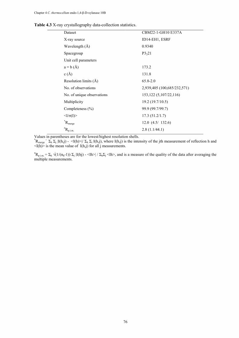
Chapter 4 C. thermocellum endo-1,4-β-D-xylanase 10B
76
Table 4.3 X-ray crystallography data-collection statistics.
Dataset CBM22-1-GH10 E337A
X-ray source ID14-EH1, ESRF
Wavelength (Å) 0.9340
Spacegroup P3221
Unit cell parameters
a = b (Å) 173.2
c (Å) 131.8
Resolution limits (Å) 65.8-2.0
No. of observations 2,939,405 (100,685/232,571)
No. of unique observations 153,122 (5,107/22,116)
Multiplicity 19.2 (19.7/10.5)
Completeness (%) 99.9 (99.7/99.7)
<I/σ(I)> 17.3 (51.2/1.7) ٩Rmerge
12.0 (4.5/ 132.6) #Rp.i.m.
2.8 (1.1/44.1)
Values in parentheses are for the lowest/highest resolution shells. ٩Rmerge
= Σh Σj |I(h,j) - <I(h)>|/ Σh Σi I(h,j), where I(h,j) is the intensity of the jth measurement of reflection h and <I(h)> is the mean value of I(h,j) for all j measurements.
#Rp.i.m. = Σh √(1/(nh-1)) Σj |I(hj) - <Ih>| / ΣhΣj <Ih>, and is a measure of the quality of the data after averaging the multiple measurements.
Chapter 5 C. thermocellum CE2 divergent functions
77
Chapter 5 The active site of a carbohydrate esterase displays
divergent catalytic and non-catalytic binding functions
Cedric Montanier1, Victoria A. Money2, Virginia M.R Pires3, James E. Flint1, Benedita A. Pinheiro3, Arun Goyal3, José A.M. Prates3, Atsushi Izumi2, Henrik Stålbrand4, Carl Morland1, Alan Cartmell1, Katarina Kolenova4, Evangelos Topakas1, Eleanor J Dodson2, David N. Bolam1, Gideon J. Davies2
, Carlos M. G. A.
Fontes3 and Harry J. Gilbert1.
1Institute for Cell and Molecular Biosciences, Newcastle University, The Medical School, Newcastle upon Tyne NE2 4HH, UK, 2York Structural Biology laboratory, Department of Chemistry, University of York, Heslington, York YO10 5YW UK, 3CIISA - Faculdade de Medicina Veterinária, Universidade Técnica de Lisboa, Avenida da Universidade Técnica, 1300-477 Lisboa, Portugal, 4Department of Biochemistry, Center for Chemistry and Chemical Engineering, Lund University, P.O. Box 124, S-22100 Lund, Sweden. Adapted from PLoS Biology (2009) 7(3), e1000071.
Abstract
Multi-functional proteins, which play a critical role in many biological processes, have typically
evolved through the recruitment of different domains that possess the required functional diversity.
Thus the different activities displayed by these proteins are mediated by spatially distinct domains,
consistent with the specific chemical requirements of each activity. Indeed current evolutionary
theory argues that the co-localization of diverse activities within an enzyme is likely to be a rare event
as it would compromise the existing activity of the protein. In contrast to this view a potential example
of multifunctional recruitment into a single protein domain is provided by CtCel5C-CE2, which
contains an N-terminal module that displays cellulase activity and a C-terminal module, CtCE2, which
exhibits a non-catalytic cellulose-binding function but also shares sequence identity with the CE2
family of esterases. Here we show that, unlike other CE2 members, the CtCE2 domain displays
divergent catalytic esterase and non-catalytic carbohydrate binding functions. Significantly these
diverse activities are housed within the same site on the protein. Thus, a critical component of the
active site of CtCE2, the catalytic Ser-His dyad, in harness with inserted aromatic residues, confers
non-catalytic binding to cellulose whilst the active site of the domain retains its esterase activity.
CtCE2 catalyses deacetylation of non-cellulosic plant structural polysaccharides to deprotect these
substrates for attack by other enzymes. Yet it also acts as a cellulose-binding domain which promotes
the activity of the appended cellulase on recalcitrant substrates. The CE2 family encapsulates the
requirement for multiple activities by biocatalysts that attack challenging macromolecular substrates,
including the grafting of a second, powerful and discrete non-catalytic binding functionality into the
active site of an enzyme. This report provides a rare example of “gene sharing” where the introduction
of a second functionality into the active site of an enzyme does not compromise the original activity of
the biocatalyst.
Chapter 5 C. thermocellum CE2 divergent functions
78
5.1 I�TRODUCTIO�
Multi-functional proteins, which play a critical role in many biological processes,
typically contain discrete domains that possess the required functional diversity. Thus the
different activities displayed by these proteins are mediated by spatially distinct regions of the
macromolecule driven by the chemical requirements of each activity. A natural enzyme
system that contains large numbers of proteins with complex molecular architectures is
presented by the plant cell wall degrading apparatus from a range of microbial species. Plant
cell wall degradation, now of great environmental significance, particularly with respect to the
generation of renewable and sustainable biofuels (Ragauskas et al., 2006; Sticklen 2006), is a
challenging process that requires a large consortium of different enzyme activities. The plant
cell wall consists primarily of an array of interlocking polysaccharides. While cellulose,
which forms crystalline microfibrils, has a simple chemical structure consisting of β-1,4-
linked glucopyranoside moieties, the matrix polysaccharides are chemically complex
molecules in which the backbone polymers are protected with both sugars and organic esters
(Brett and Waldren 1996). Plant cell wall degrading systems thus feature GHs, which cleave
the glycosidic bonds that link the sugars, and esterases that remove the diverse acylations
(Tomme et al., 1995; Warren 1996). In addition to their chemical complexity the physical
structure of plant cell walls presents a physical barrier to enzyme attack. To compensate for
the accessibility problem, plant cell wall degrading enzymes generally contain a non-catalytic
carbohydrate binding function that, by bringing the biocatalyst into prolonged and intimate
contact with its substrate increases the rate of catalysis (Hall et al., 1995). In general these
diverse catalytic and non-catalytic carbohydrate-binding activities are housed in discrete
modules within the same protein (Coutinho and Henrissat 1999).
A family of CEs (family CE2 in the CAZy classification (http://www.cazy.org/
(Henrissat 1991; Coutinho and Henrissat 1999)) are especially intriguing with respect to the
multiple functions required of plant cell wall degrading systems. CE2 enzymes are acetyl
esterases (that are generally not appended to other catalytic modules), which are reported to
be active on synthetic aryl-esters and acetylated xylan (Dalrymple et al., 1997). The
bacterium Clostridium thermocellum contains a single CE2 member, designated CtCE2,
which is linked to the cellulase, CtCel5C, within the modular protein designated CtCel5C-
CE2. The enzyme also contains a type I dockerin module that, by binding to cohesin modules
in the scaffoldin protein, incorporates CtCel5C-CE2 into the multienzyme plant cell wall
degrading complex known as the cellulosome (Bayer et al., 2004; Durrant et al., 1991).
Intriguingly, CtCE2 was previously characterized as a CBM by virtue of its cellulose-binding
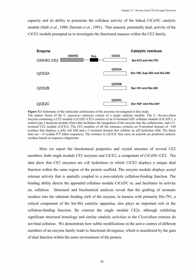
Chapter 5 C. thermocellum CE2 divergent functions
79
capacity and its ability to potentiate the cellulase activity of the linked CtCel5C catalytic
module (Hall et al., 1988; Durrant et al., 1991). This unusual, potentially dual, activity of the
CtCE2 module prompted us to investigate the functional nuances within the CE2 family.
Figure 5.1 Schematic of the molecular architecture of the enzymes investigated in this study. The mature forms of the C. japonicus esterases consist of a single catalytic module. The C. thermocellum
enzyme containing a CE2 module (CtCel5C-CE2) consists of an N-terminal GH5 cellulase module (CtCel5C), a central type I dockerin module (Doc) that facilitates the integration of the enzyme into the cellulosome, and a C-terminal CE2 module (CtCE2). The CE2 modules of all the esterases contains an N-terminal domain of ~160 residues that displays a jelly roll fold and a C-terminal domain that exhibits an α/β hydrolase fold. The black lines are ~15 residue P/T linker sequences. The residues in CjCE2C that carry an asterisk are predicted catalytic residues based on sequence alignments.
Here we report the biochemical properties and crystal structure of several CE2
members, both single module CE2 enzymes and CtCE2, a component of CtCel5C-CE2. The
data show that CE2 enzymes are α/β hydrolases in which CtCE2 displays a unique dual
function within the same region of the protein scaffold. The enzyme module displays acetyl
esterase activity that is spatially coupled to a non-catalytic cellulose-binding function. The
binding ability directs the appended cellulase module CtCel5C to, and facilitates its activity
on, cellulose. Structural and biochemical analyses reveal that the grafting of aromatic
residues into the substrate binding cleft of the enzyme, in harness with primarily His-791, a
critical component of the Ser-His catalytic apparatus, also plays an important role in the
cellulose-binding function. By contrast the single module CE2s, although exhibiting
significant structural homology and similar catalytic activities to the Clostridium esterase do
not bind cellulose. We demonstrate how subtle modifications in the active centres of different
members of an enzyme family leads to functional divergence, which is manifested by the gain
of dual function within the same environment of the protein.
Chapter 5 C. thermocellum CE2 divergent functions
80
5.2 EXPERIME�TAL PROCEDURES
5.2.1 Gene cloning and protein expression
The region of the C. thermocellum cellulase-esterase gene, cel5C-ce2, encoding the
C-terminal CE2 esterase module, CtCE2 (residues 482 to 814 of the full-length enzyme), was
amplified by PCR from genomic DNA (strain ATCC 27405) using the thermostable DNA
polymerase Pfu Turbo (Stratagene) and primers (Table 4.1) that contain ,deI and XhoI
restriction sites, respectively, depicted in bold. The DNA product was cloned into the ,deI
and XhoI sites of the Escherichia coli expression vector pET22b (Novagen) to generate
pCtCE2. CtCE2 encoded by pCtCE2 contains a C-terminal His6-tag. The region of the genes
encoding the mature C. japonicus and B. thetaiotaomicron VPI-5482 CE2 esterases were
amplified by PCR from genomic DNA, using primers listed in Table 5.1, and cloned into ,deI
and XhoI restricted pET22b such that the proteins encoded by the recombinant plasmids
contained a C-terminal His6-tag.
5.2.2 Protein expression and purification
E. coli BL21 cells harbouring the CE2 esterase-encoding recombinant expression
vectors were cultured in Luria-Bertani broth at 37 °C to mid-exponential phase (A600nm ~0.6-
1.0) and recombinant protein expression was induced by the addition of 1 mM isopropyl 1-
thio-β-D-galactopyranoside (IPTG) and incubation for a further five hours at 37 °C. The CE2
esterases were purified by immobilized metal ion affinity chromatography (IMAC) using
TalonTM resin (Clontech) and elution in 20 mM Tris/HCl buffer containing 300 mM NaCl and
100 mM imidazole. The eluted esterase was then dialyzed against 20 mM Tris/HCl buffer pH
8.0 (Buffer A) and applied to a Bio-Rad Q12 column. The esterases were eluted with a 400 ml
0-500 mM NaCl gradient in Buffer A. The fractions containing esterase activity were
concentrated using a 10kDa MWCO Vivaspin 20 centrifugal concentrator and applied to a
SuperdexTM 200 26/60 HiloadTM column (Amersham) equilibrated in 10 mM Tris-HCl buffer,
pH 8.0, containing 150 mM NaCl. Protein was eluted at a flow rate of 1 ml/min. Both
chromatography steps employed a Bio-Rad FPLC system. Purified enzymes were adjudged
homogenous by SDS-PAGE. To produce seleno-methionine-containing proteins the same
protocol was employed except that the enzyme was expressed in E. coli B834 (Novagen)
using growth conditions described previously (Charnock et al., 2000) and 2 mM 2-
mercaptoethanol was included in all buffers during purification up to the point of IMAC and
Chapter 5 C. thermocellum CE2 divergent functions
81
10 mM for all subsequent steps. The purified seleno-methionine enzyme eluted from the gel
filtration column was exchanged into ddH2O containing 10 mM DTT using Amersham PD10
column.
5.2.3 Mutagenesis
Site-directed mutagenesis was carried out using the PCR-based QuikChange site-
directed mutagenesis kit (Stratagene) according to the manufacturer’s instructions, using
pCtCE2 or pCjCE2A as the template and primers pairs that are listed in Table 5.1.
Table 5.1 Primers used for cloning ctCE2 from Clostridium thermocellum.Engineered restriction sites and mutation points are depicted in bold.
Construct Primers Sequence (5’- 3’)
CtCE2 Forward CTC CAT ATG CCT GAT GAA GAC AAT CCG Reverse CAC CTC GAG CCA TCC AAG CTT GTT TTT
CjCE2A Forward CAGCTCCAGCATATGAATACGCAATCACTTATGTCATCCAC Reverse CTCCAGCTCGAGCCCATTGCGATAGTTACCCAGGAGCCGA
CjCE2B Forward CAGCTCCAGCATATGGCCGACTCAACCAAGCCGC Reverse CTCCAGCTCGAGCCAGATACCTTTTTTCTGCTGCAAATGG
CjCE2C Forward CAGCTCCAGCATATGGCCCAGGCGGAGCCGGC Reverse CTCCAGCTCGAGCCAACGCATTTTTTCCCGGATAAATGC
BtCE2 Forward CTC CAT ATG ATG TGG AAA AAT GTG TTA Reverse CAC CTC GAG ATA AAA CGA TTC CTT TTT
D57A Forward CAG GCT ATT GTA GCC GGC AAT CCT CTT Reverse AAG AGG ATT GCC GGC TAC AAT AGC CTG
H84A Forward GCA GAA GGA GCT GCT CAT CTT GTA TTG Reverse CAA TAC AAG ATG AGC AGC TCC TTC TGC
Y133A Forward ATC ACA TGT GCA GCC GGA AAT GAA GGA Reverse TCC TTC ATT TCC GGC TGC ACA TGT GAT
Y193A Forward ATA ATG GAC CGT GCT CCT TAT ACC CTT Reverse AAG GGT ATA AGG AGC ACG GTC CAT TAT
W204A Forward AGC GGA GTC AGA GCG GAT TTT AGC AAA Reverse TTT GCT AAA ATC CGC TCT GAC TCC GCT
5.2.4 Enzyme assays
Substrates used in the enzyme assays described below were purchased from Sigma
except acetylated Konjac glucomannan, which was purchased from Megazyme International
(Bray, Ireland); acetylated xylan, which was prepared from birchwood xylan (Sigma) using
acetic anhydride following the method of Johnson et al (Johnson et al., 1988), and O-acetyl
galactoglucomannan that was extracted from spruce (Picea abies) (Lundqvist et al., 2002). To
determine activity against 4-NPAc 1 ml enzyme reactions were carried out in 50 mM sodium
phosphate buffer, pH 7.0, containing 1 mg/ml of BSA and substrate concentrations up to 10
Chapter 5 C. thermocellum CE2 divergent functions
82
mM. The reaction was initiated by the addition of an appropriate concentration of enzyme; 10
nM for wild type and 10 µM for the most inactive mutants, and the release of 4-nitrophenolate
was monitored at 400 nm. To determine the rate of deacetylation of acetylated
polysaccharides the release of acetate was determined using an acetic acid detection kit
(Megazyme International) following the manufacturer’s recommendation except that the
product was measured continuously rather that deploying a stopped reaction. Pre-steady-state
kinetics were performed using a stopped-flow apparatus (Applied Photophysics Model SX-
17MV), with the flow path thermostatically controlled at 15 oC, and a 2-mm light path to
monitor the formation of the product 4-nitrophenolate at A400. Equal volumes (50 µl) of
solutions mixed with a dead time of 1.5 ms, contained, respectively, 10 µM CtCE2 and 2 mM
4-NPAc in 50 mM sodium phosphate buffer, pH 7.0. These solutions were supplemented with
0-100 µM cellohexaose.
5.2.5 Carbohydrate binding studies
The capacity of the CE2 enzymes to bind to soluble saccharides was determined by
affinity gel electrophoresis (AGE) or by ITC. AGE was performed as described previously
(Charnock et al., 2000) with the polysaccharide ligand included in the polyacrylamide gel at
0.1 %. ITC (Bolam et al., 2004) was carried out in 50 mM HEPES-Na buffer, pH 8.0,
containing 300 mM NaCl at 25 °C. Data collected for titrations with 10 mg/ml of β-glucan
were fitted with a molar concentration of 5 mM, the value at which n = 1. The concentrations
of cellooligosaccharides in the syringe ranged from 0.7-5 mM and the CE2 enzymes in the
reaction cell were at 80-100 µM. The determined KA and ∆H values were used to calculate ∆S
from the standard thermodynamic equation. Binding to insoluble cellulose was determined by
pull-down assay using phosphoric acid swollen cellulose (PASC). Briefly 1mg of washed
PASC in 50mM Tris-HCl, pH 8.0, containing 300 mM NaCl (Buffer B), was mixed with 50
µg protein in a total volume of 100 µl and incubated on ice for 30 min. This was then
centrifuged for 1 min at 13,000 × g and the supernatant containing the unbound protein
removed. The cellulose was then washed 3 times with 100 µl ice-cold Buffer B and each wash
discarded, before 50 µl SDS-PAGE loading buffer (contains 10 % SDS) was added and the
bound protein eluted from the cellulose by boiling for 5 min. Approximately equal volumes of
starting protein and material eluted from the cellulose were then subjected to SDS-PAGE to
assess binding.
Chapter 5 C. thermocellum CE2 divergent functions
83
5.2.6 Crystallisation and structure solution
CjCE2A: Native and seleno-methionine crystals of CjCE2A were grown by hanging
drop vapour-phase diffusion in 1 M diammonium hydrogen phosphate/0.1 M sodium acetate
pH 5.0 for 1-2 days at 20 ˚C (30 % v/v glycerol was added as the cryoprotectant). Data from
seleno-methionine derivatised CjCE2A were collected on ID23-1 at a wavelength of 0.97880
Å using an ADSC Q315R CCD (charge coupled device) detector the wavelength was
optimised for the f’’ component of the anomalous signal using a fluorescence scan. Data from
crystals of native CjCE2A were collected on ID14.2 using an ADSC Q4 CCD detector at a
wavelength of 0.9330 Å. Data for all CE2 enzymes were processed with either DENZO
(Otwinowski and Minor 1997) or MOSFLM from the CCP4 suite (Collaborative
Computational Project Number 4 1994). The structure of CjCE2A was solved using the
single-wavelength anomalous dispersion (SAD) method. Selenium positions were determined
using SHELXD (Schneider and Sheldrick 2002) and the phases were subsequently calculated
using SHELXE. 5% of the data were set aside for cross validation analysis and the behaviour
of Rfree was used to monitor and guide the refinement protocols. ARP/wARP (Perrakis et al.,
1999) in conjunction with REFMAC (Murshudov et al., 1997) was used to automatically
build the sequence into the electron density. Refinement was undertaken in REFMAC with
manual correction to the model using COOT (Emsley and Cowtan 2004).
CtCE2: Crystals of both wild-type and S612A CtCE2 in complex with cellohexaose
were grown by hanging drop vapour-phase diffusion from 20% PEG3350, and 0.2-0.3M
ammonium iodide, with protein at ~8mg ml-1 and cellohexaose at approx. 1mM. Crystals
were harvested in rayon fibre loops before being bathed in cryo-protectant solution
(crystallistaion coditions augmented with 25% v/v glycerol) and flash frozen in liquid
nitrogen. Data were collected at the ESRF from single crystals at 100 K for cellohexaose
complexes of both wild-type and S612A mutant forms with data processed as previously. The
structure of CtCE2 was solved by molecular replacement using PHASER with the search
model being the CjCE2A structure. ARP/wARP was used to build the initial model and
refinement was undertaken as described above.
CjCE2B: Native and SeMet crystals of CjCE2B were grown by hanging drop vapor
diffusion in 0.1 M imidazole (pH 8.0), 10 % PEG 8,000 for 3 days at 20˚C (25 % v/v glycerol
was added as the cryoprotectant). Native data were collected at ESRF on ID14.2 using ADSC
Q4 CCD detector at a wave length of 0.933 Å. The structure was solved using a combined
approach in which molecular replacement using a combined CtCE2 / CjCE2A model was
used in PHASER (McCoy et al., 2007) was used in conjunction with the Se-Met derived SAD
Chapter 5 C. thermocellum CE2 divergent functions
84
phases for map calculation and verification of methionine positions. The final model was
build using COOT and refined using REFMAC. CjCE2B crystals contains two molecules in
the asymmetric unit. The final models of CjCE2B in chain A was ordered from 5 to 341
containing five missing regions between amino acids 21-24, 41-43, 61-64, 78-88, and 107-
117, while chain B was ordered from 6 to 341 and contained one missing region between
residues 79-87. Coordinates and observed structure factor amplitudes have been deposited at
the wwPDB and the crystal structure statistics are in Table 5.2.
Table 5.2 Data collection and refinement statistics.
a The S619A mutant is a derivative of CtCE2
CtCE2-cellohexaose
S612Aa- cellohexaose
CjCE2A CjCE2B
Data collection Space group P21 P21212 P41212 P43 Cell dimensions a, b, c (Å) 41.6, 60.1,
69.0 41.2, 141.2, 58.2
132.2, 132.2, 45.8
75.3, 75.3,
141.6 α, β, γ (°) 90, 78.01, 90 90,90,90 90,90,90 90,90,90 Resolution (Å) 67.60-1.80
(1.90-1.80) 70.53-1.90 (2.00-1.90)
46.73-1.80 (1.90-1.80)
53.23 (2.10-2.00)
Rmerge 0.08 (0.30) 0.10 (0.35) 0.12 (0.58) 0. 06 (0.62)
I / σI 12.7 (3.9) 10.8 (3.9) 15.6 (3.3) 15.5 (3.0) Completeness (%) 99.3 (99.3) 99.8 (99.8) 100 (100) 100 (100) Redundancy 4.0 (4.1) 4.7 (4.8) 8.0 (8.1) 5.5 (5.4) Refinement Resolution (Å) 1.80 1.90 1.80 2.0 No. reflections 31011 27641 38157 49944 Rwork / Rfree 0.14/0.19 0.15/0.20 0.15/0.20 0.19/0.23 No. atoms Protein 2569 2574 2725 5141 Ligand/ion 56 114 30 42 Water 430 375 445 420 B-factors Protein main chain/side chain
8/11 13/15 11/13 41/41
Ligand/ion 25 24 30 51 Water 25 27 28 42 R.m.s. deviations Bond lengths (Å)
0.019 0.018 0.019 0.012
Bond angles (°) 1.7 1.6 1.7 1.3
Chapter 5 C. thermocellum CE2 divergent functions
85
5.3 RESULTS
5.3.1 CE2 is a large family of diverse esterases displaying the α/βα/βα/βα/β hydrolase fold with a
serine nucleophile.
In order to probe the function and role of the diverse members of the CE2 esterase
family, which currently contains 31 members (http://www.cazy.org/fam/CE2.html) (March
2009), the biochemical properties of five CE2 enzymes were assessed. In addition to CtCE2,
which comprises the C-terminal region of C. thermocellum CtCel5C-CE2 (formerly EGE;
(Hall et al., 1988; Durrant et al., 1991)) we also characterised three Cellvibrio japonicus CE2
members, CjCE2A, B and C and the Bacteroides thetaiotaomicron esterase BtCE2, identified
from the genome sequence of the two bacteria (Xu et al., 2003; DeBoy et al., 2008); these
latter four enzymes are not appended to other enzyme modules, Figure 5.1. All these CE2
enzymes act as acetyl esterases, releasing acetate from activated artificial substrates such as 4-
nitrophenyl acetate (4-NPAc; Table 5.3) and, to different extents, the acetylated plant
polysaccharides xylan and glucomannan Table 5.4 (Note: Only very small amounts of the
Bacteroides CE2 could be produced and so only initial qualitative assays could be carried out
with this enzyme). Based on their catalytic efficiencies, CtCE2 and CjCE2B exhibit a
significant preference for acetylated glucomannan over xylan, while CjCE2A and CjCE2C do
not distinguish between the two polysaccharides. It should be noted that the specificity of
CtCE2 for glucomannan reflects an extremely low KM. The esterases appear specific for
acetyl groups and do not hydrolyse aryl-ferulates or aryl-coumarates (data not shown). The
only other reported analysis of the catalytic activity of CE2 enzymes are the esterases from
,eocallimastix patriciarum (Dalrymple et al., 1997) that display xylan esterase activity and
hydrolyse 4-NPAc, but their activity on other substrates were not assessed. The activities
reported here extend the substrates deacetylated by CE2 enzymes.
Chapter 5 C. thermocellum CE2 divergent functions
86
Table 5.3 Catalytic activity of CE2 enzymes against 4-nitrophenyl acetate (4-NPAc).
a Mutants of CtCE2 b NA: No activity detected Table 5.4 Activity of CE2 esterases against acetylated polysaccharides.
aThe KM values relates to the concentration of acetate moieties present in the polysaccharides.
b Mutants of CtCE2.
c NA: No activity detected. d ND: The KM and kcat values could not be determined as there was a
linear relationship between rate and substrate concentration up to 0.6 mM.
e Mutants of CjCE2A
Enzyme 4-�PAc
kcat
(min-1
)
KM
(µM)
kcat/KM
(min-1
µM-1
) CtCE2 7032 165 43
D789Aa 2512 75 33
D789Na 2976 186 16
S612Aa NAb NA NA
H791Aa NA NA NA
CjCE2A 7717 339 23 CjCE2B 49820 262 190 CjCE2C 7251 45 161
Birchwood xylan Glucomannan
kcat
(min-1
)
KMa
(mM)
kcat/KM
(min-1
mM-1
) kcat
(min-1
)
KMa
(mM)
kcat/KM
(min-1
mM-1
)
CtCE2 12 2.7 4.4 1.1 0.019 58 D789Ad
4.5 3.1 1.5 0.23 0.025 9.2 D789Nd
6.5 2.9 2.2 0.71 0.031 23 S612Ad
- - NAb - - NAe H791Ad
- - NA - - NA Y665Ad
15 7.7 1.9 NDc ND 32 Y683Ad
10 2.4 4.2 0.56 0.026 21 W746Ad
13 3.3 4.1 10 0.14 71 Q780Ad
8.5 2.2 3.9 2.0 0.046 43 W790Ad
14 2.5 5.6 7.9 0.088 89 Y683A/W746Ad
13 2.9 4.3 12 0.13 89 W746A/Q780Ad
17 3.1 5.5 9.4 0.12 78 Y683A/W746/Q780Ad
22 4.2 5.1 ND ND 31 CjCE2A 69 1.5 46 163 4.2 39 W127Ae 65 10 6.5 217 87 2.5 W212Ae 55 6.2 8.9 468 58 8.1 W127A/W212Ae 12 14 0.85 4.6 37 0.12 CjCE2B 66 4.6 14 1348 0.84 1605 CjCE2C 119 3.2 37 1212 14 87
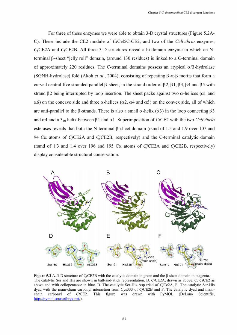
Chapter 5 C. thermocellum CE2 divergent functions
87
For three of these enzymes we were able to obtain 3-D crystal structures (Figure 5.2A-
C). These include the CE2 module of CtCel5C-CE2, and two of the Cellvibrio enzymes,
CjCE2A and CjCE2B. All three 3-D structures reveal a bi-domain enzyme in which an N-
terminal β-sheet “jelly roll” domain, (around 130 residues) is linked to a C-terminal domain
of approximately 220 residues. The C-terminal domains possess an atypical α/β-hydrolase
(SGNH-hydrolase) fold (Akoh et al., 2004), consisting of repeating β-α-β motifs that form a
curved central five stranded parallel β-sheet, in the strand order of β2, β1, β3, β4 and β5 with
strand β2 being interrupted by loop insertion. The sheet packs against two α-helices (α1 and
α6) on the concave side and three α-helices (α2, α4 and α5) on the convex side, all of which
are anti-parallel to the β-strands. There is also a small α-helix (α3) in the loop connecting β3
and α4 and a 310 helix between β1 and α1. Superimposition of CtCE2 with the two Cellvibrio
esterases reveals that both the N-terminal β-sheet domain (rsmd of 1.5 and 1.9 over 107 and
94 Cα atoms of CjCE2A and CjCE2B, respectively) and the C-terminal catalytic domain
(rsmd of 1.3 and 1.4 over 196 and 195 Cα atoms of CjCE2A and CjCE2B, respectively)
display considerable structural conservation.
Figure 5.2 A. 3-D structure of CjCE2B with the catalytic domain in green and the β-sheet domain in magenta. The catalytic Ser and His are shown in ball-and-atick representation. B. CjCE2A, drawn as above. C. CtCE2 as above and with cellopentaose in blue. D. The catalytic Ser-His-Asp triad of CjCe2A, E. The catalytic Ser-His dyad with the main-chain carbonyl interaction from Cys333 of CjCE2B and F. The catalytic dyad and main-chain carbonyl of CtCE2. This figure was drawn with PyMOL (DeLano Scientific, http://pymol.sourceforge.net/).
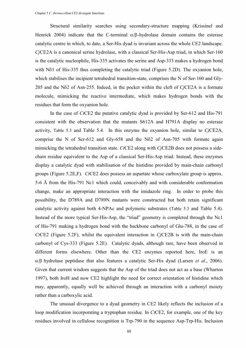
Chapter 5 C. thermocellum CE2 divergent functions
88
Structural similarity searches using secondary-structure mapping (Krissinel and
Henrick 2004) indicate that the C-terminal α/β-hydrolase domain contains the esterase
catalytic centre in which, to date, a Ser-His dyad is invariant across the whole CE2 landscape.
CjCE2A is a canonical serine hydrolase, with a classical Ser-His-Asp triad, in which Ser-160
is the catalytic nucleophile, His-335 activates the serine and Asp-333 makes a hydrogen bond
with Nδ1 of His-335 thus completing the catalytic triad (Figure 5.2D). The oxyanion hole,
which stabilises the incipient tetrahedral transition-state, comprises the N of Ser-160 and Gly-
205 and the Nδ2 of Asn-255. Indeed, in the pocket within the cleft of CjCE2A is a formate
molecule, mimicking the reactive intermediate, which makes hydrogen bonds with the
residues that form the oxyanion hole.
In the case of CtCE2 the putative catalytic dyad is provided by Ser-612 and His-791
consistent with the observation that the mutants S612A and H791A display no esterase
activity, Table 5.3 and Table 5.4. In this enzyme the oxyanion hole, similar to CjCE2A,
comprise the N of Ser-612 and Gly-658 and the Nδ2 of Asn-705 with formate again
mimicking the tetrahedral transition state. CtCE2 along with CjCE2B does not possess a side-
chain residue equivalent to the Asp of a classical Ser-His-Asp triad. Instead, these enzymes
display a catalytic dyad with stabilisation of the histidine provided by main-chain carbonyl
groups (Figure 5.2E,F). CtCE2 does possess an aspartate whose carboxylate group is approx.
5-6 Å from the His-791 Nε1 which could, conceivably and with considerable conformation
change, make an appropriate interaction with the imidazole ring. In order to probe this
possibility, the D789A and D789N mutants were constructed but both retain significant
catalytic activity against both 4-NPAc and polymeric substrates (Table 5.3 and Table 5.4).
Instead of the more typical Ser-His-Asp, the “triad” geometry is completed through the Nε1
of His-791 making a hydrogen bond with the backbone carbonyl of Glu-788, in the case of
CtCE2 (Figure 5.2F), whilst the equivalent interaction in CjCE2B is with the main-chain
carbonyl of Cys-333 (Figure 5.2E). Catalytic dyads, although rare, have been observed in
different forms elsewhere. Other than the CE2 enzymes reported here, IroE is an
α/β hydrolase peptidase that also features a catalytic Ser-His dyad (Larsen et al., 2006).
Given that current wisdom suggests that the Asp of the triad does not act as a base (Wharton
1997), both IroH and now CE2 highlight the need for correct orientation of histidine which
may, apparently, equally well be achieved through an interaction with a carbonyl moiety
rather than a carboxylic acid.
The unusual divergence to a dyad geometry in CE2 likely reflects the inclusion of a
loop modification incorporating a tryptophan residue. In CtCE2, for example, one of the key
residues involved in cellulose recognition is Trp-790 in the sequence Asp-Trp-His. Inclusion
Chapter 5 C. thermocellum CE2 divergent functions
89
of the Trp forces the adjacent aspartate into a conformation that prevents it hydrogen-bonding
with His-791 (Figure 5.3). The dyad geometry and the topology of the active-centre binding
surfaces are described below in light of the dissection of polysaccharide recognition in CE2
enzymes. Sequence analysis suggests that the dyad geometry is not unique to these two
enzymes, with 14 of the 26 CE2 members lacking the putative catalytic aspartate.
Furthermore, of the remaining 12 CE2 enzymes that appear to have a canonical catalytic triad,
10 have a tryptophan between the His and Asp, as observed with CtCE2, suggesting that in
these enzymes the aromatic ring may also place the Asp into an orientation that prevents it
forming a classical triad.
Figure 5.3 Binding of cellooligosaccharides through the esterase active centre of CtCE2. Observed electron density (maximum-likelihood weighted 2Fobs-Fcalc contoured at 1σ) for cellohexaose (in which cellopentaose is ordered) bound to wild-type CtCE2. Ser-612 and His-791 form the catalytic dyad, with Trp-790 causing both a change in position of Asp-789 \and also forming the binding-platform for the second glucose. Other interactions with aromatic residues discussed in the text are shown. The figure is in divergent (“wall-eyed”) stereo and was drawn with PyMOL.
5.3.2 The esterase and cellulose binding functions of CtCE2 are in close proximity.
The most intriguing CE2 member is CtCE2, derived from the multi-domain
cellulase/esterase, CtCel5C-CE2. Historical work has shown that the C-terminal region of
this enzyme, CtCE2, not only bound cellulose but also potentiated the activity of the cellulase
catalytic module on insoluble substrates (Durrant et al., 1991): the CE2 module thus behaved
as a classical CBM (Boraston et al., 2004). In this report we show that CtCE2 binds to
insoluble cellulose Figure 5.4. Affinity gel electrophoresis also demonstrated that the protein
interacts with β-glucan (β-1,3:β-1,4 mixed linked glucan) and soluble derivatized forms of
cellulose (carboxymethylcellulose and hydroxyethylcellulose, Figure 5.4), but displays no
affinity for laminarin (β-1,3-linked glucan), α-linked glucans or the β-1,4-linked xylose
polymer, xylan (data not shown). Isothermal titration calorimetry (ITC) revealed that CtCE2
binds to cellooligosaccharides with a KD for cellohexaose of 33 µM, Table 5.5 and Table 5.6
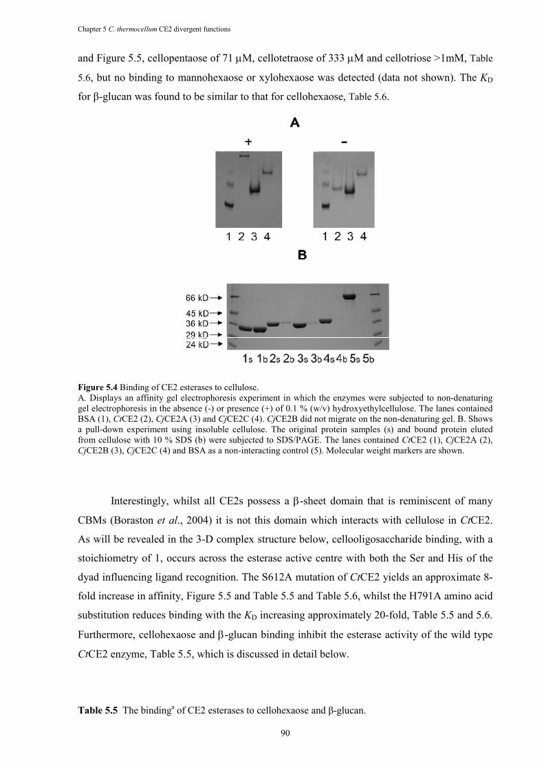
Chapter 5 C. thermocellum CE2 divergent functions
90
and Figure 5.5, cellopentaose of 71 µM, cellotetraose of 333 µM and cellotriose >1mM, Table
5.6, but no binding to mannohexaose or xylohexaose was detected (data not shown). The KD
for β-glucan was found to be similar to that for cellohexaose, Table 5.6.
Figure 5.4 Binding of CE2 esterases to cellulose. A. Displays an affinity gel electrophoresis experiment in which the enzymes were subjected to non-denaturing gel electrophoresis in the absence (-) or presence (+) of 0.1 % (w/v) hydroxyethylcellulose. The lanes contained BSA (1), CtCE2 (2), CjCE2A (3) and CjCE2C (4). CjCE2B did not migrate on the non-denaturing gel. B. Shows
a pull-down experiment using insoluble cellulose. The original protein samples (s) and bound protein eluted from cellulose with 10 % SDS (b) were subjected to SDS/PAGE. The lanes contained CtCE2 (1), CjCE2A (2), CjCE2B (3), CjCE2C (4) and BSA as a non-interacting control (5). Molecular weight markers are shown.
Interestingly, whilst all CE2s possess a β-sheet domain that is reminiscent of many
CBMs (Boraston et al., 2004) it is not this domain which interacts with cellulose in CtCE2.
As will be revealed in the 3-D complex structure below, cellooligosaccharide binding, with a
stoichiometry of 1, occurs across the esterase active centre with both the Ser and His of the
dyad influencing ligand recognition. The S612A mutation of CtCE2 yields an approximate 8-
fold increase in affinity, Figure 5.5 and Table 5.5 and Table 5.6, whilst the H791A amino acid
substitution reduces binding with the KD increasing approximately 20-fold, Table 5.5 and 5.6.
Furthermore, cellohexaose and β-glucan binding inhibit the esterase activity of the wild type
CtCE2 enzyme, Table 5.5, which is discussed in detail below.
Table 5.5 The bindinga of CE2 esterases to cellohexaose and β-glucan.
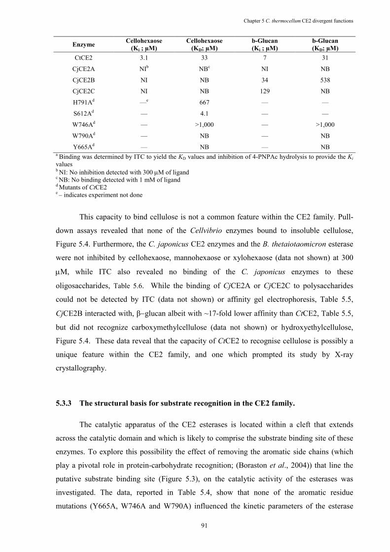
Chapter 5 C. thermocellum CE2 divergent functions
91
Enzyme Cellohexaose
(Ki ; µM)
Cellohexaose
(KD; µM)
b-Glucan
(Ki ; µM)
b-Glucan
(KD; µM)
CtCE2 3.1 33 7 31
CjCE2A NIb NBc NI NB
CjCE2B NI NB 34 538
CjCE2C NI NB 129 NB
H791Ad —e 667 — —
S612Ad — 4.1 — —
W746Ad — >1,000 — >1,000
W790Ad — NB — NB
Y665Ad — NB — NB a Binding was determined by ITC to yield the KD values and inhibition of 4-PNPAc hydrolysis to provide the Ki values b NI: No inhibition detected with 300 µM of ligand c NB: No binding detected with 1 mM of ligand d Mutants of CtCE2 e – indicates experiment not done
This capacity to bind cellulose is not a common feature within the CE2 family. Pull-
down assays revealed that none of the Cellvibrio enzymes bound to insoluble cellulose,
Figure 5.4. Furthermore, the C. japonicus CE2 enzymes and the B. thetaiotaomicron esterase
were not inhibited by cellohexaose, mannohexaose or xylohexaose (data not shown) at 300
µM, while ITC also revealed no binding of the C. japonicus enzymes to these
oligosaccharides, Table 5.6. While the binding of CjCE2A or CjCE2C to polysaccharides
could not be detected by ITC (data not shown) or affinity gel electrophoresis, Table 5.5,
CjCE2B interacted with, β−glucan albeit with ~17-fold lower affinity than CtCE2, Table 5.5,
but did not recognize carboxymethylcellulose (data not shown) or hydroxyethylcellulose,
Figure 5.4. These data reveal that the capacity of CtCE2 to recognise cellulose is possibly a
unique feature within the CE2 family, and one which prompted its study by X-ray
crystallography.
5.3.3 The structural basis for substrate recognition in the CE2 family.
The catalytic apparatus of the CE2 esterases is located within a cleft that extends
across the catalytic domain and which is likely to comprise the substrate binding site of these
enzymes. To explore this possibility the effect of removing the aromatic side chains (which
play a pivotal role in protein-carbohydrate recognition; (Boraston et al., 2004)) that line the
putative substrate binding site (Figure 5.3), on the catalytic activity of the esterases was
investigated. The data, reported in Table 5.4, show that none of the aromatic residue
mutations (Y665A, W746A and W790A) influenced the kinetic parameters of the esterase
Chapter 5 C. thermocellum CE2 divergent functions
92
against xylan. While the catalytic efficiencies of the CtCE2 variants against glucomannan
were similar to the wild type enzyme, there was a significant increase in KM, exemplified by
the Y665A mutant (KM >0.6 mM), which was mirrored by a similar increase in kcat. It should
also be noted that the activity of the Tyr-665, Trp-790 and Trp-746 mutants of CtCE2 against
acetylated glucomannan was subject to substrate inhibition. Thus, these aromatic residues not
only bind glucomannan but also guide the polysaccharide into the substrate binding cleft such
that the acetyl groups are presented at the active site. These mutational studies indicate that
the aromatic residues lining the substrate binding cleft of CtCE2 not only contribute to the
tight binding of acetylated glucomannan, reflected by the very low KM, but also limit product
release, which appears to be the rate determining step in catalysis. Mutation of the single
aromatic residue in the substrate binding cleft of CjCE2A, Trp-212, although not effecting
kcat, leads to a significant increase in KM for both glucomannan and xylan, Table 5.4. These
data suggest that Trp-212 contributes to substrate binding but, in contrast to CtCE2, product
departure is not the rate limiting step and thus mutation of the tryptophan does not lead to an
increase in kcat.
In addition to the catalytic α/β hydrolase domain, the CE2 members also contain an all
β-sheet domain, somewhat reminiscent of a CBM. In CtCE2 this jelly-roll domain appears to
extend the substrate/cellulose binding cleft of the catalytic domain. To address this issue Tyr-
683 and Trp-127, which are located in. or at the interface, of the CBM-like domains of CtCE2
and CjCE2A, respectively, were mutated and the activities of the two esterase variants were
assessed. The data, reported in Table 5.4, show that the W127A CjCE2A mutation caused a
significant increase in KM but not kcat for glucomannan and xylan, while the catalytic
properties of the CtCE2 mutant Y683A were similar to the wild type enzyme. It would
appear, therefore, that the CBM-like domain contributes to substrate recognition in CjCE2A
but its role in the catalytic activity of CtCE2 is less evident.
5.3.4 Structural characterisation of cellooligosaccharide binding to CtCE2.
The structure of the α/β hydrolase domain of CtCE2 revealed a deep cleft. One wall of
the cleft is formed by the loops derived from β2, β3 and β4, while the other face contains the
extended loop that links β5 to α5. Both wild-type CtCE2 and S612A mutant structures were
determined in the presence of cellohexaose and clear electron density for five β-1,4-linked D-
glucose molecules was observed in the cleft (Figure 5.3), consistent with the affinities
revealed by ITC, Table 5.6. The interaction of the oligosaccharide with CtCE2 is dominated by
planar hydrophobic contacts between the pyranose rings of Glc-1 with Trp-746, Glc-2 and
Trp-790 and Glc-4 and Tyr-665 (Figure 5.3). Hydrophobic interactions represent the major
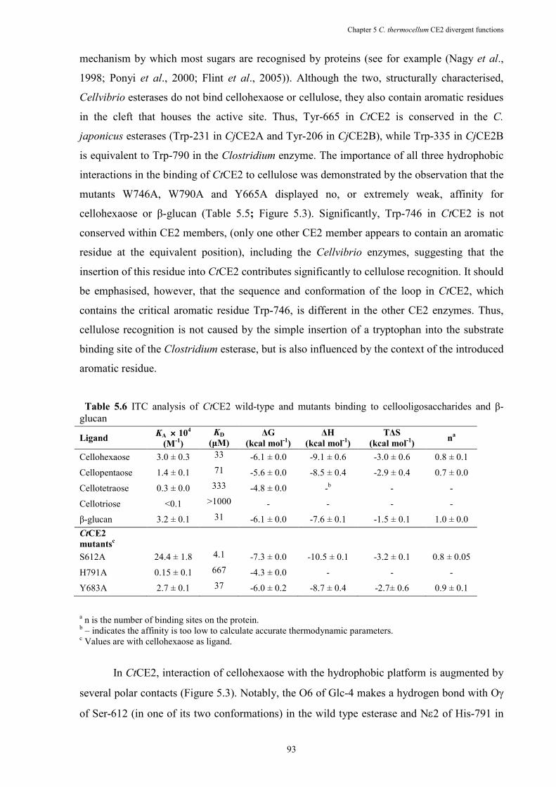
Chapter 5 C. thermocellum CE2 divergent functions
93
mechanism by which most sugars are recognised by proteins (see for example (Nagy et al.,
1998; Ponyi et al., 2000; Flint et al., 2005)). Although the two, structurally characterised,
Cellvibrio esterases do not bind cellohexaose or cellulose, they also contain aromatic residues
in the cleft that houses the active site. Thus, Tyr-665 in CtCE2 is conserved in the C.
japonicus esterases (Trp-231 in CjCE2A and Tyr-206 in CjCE2B), while Trp-335 in CjCE2B
is equivalent to Trp-790 in the Clostridium enzyme. The importance of all three hydrophobic
interactions in the binding of CtCE2 to cellulose was demonstrated by the observation that the
mutants W746A, W790A and Y665A displayed no, or extremely weak, affinity for
cellohexaose or β-glucan (Table 5.5; Figure 5.3). Significantly, Trp-746 in CtCE2 is not
conserved within CE2 members, (only one other CE2 member appears to contain an aromatic
residue at the equivalent position), including the Cellvibrio enzymes, suggesting that the
insertion of this residue into CtCE2 contributes significantly to cellulose recognition. It should
be emphasised, however, that the sequence and conformation of the loop in CtCE2, which
contains the critical aromatic residue Trp-746, is different in the other CE2 enzymes. Thus,
cellulose recognition is not caused by the simple insertion of a tryptophan into the substrate
binding site of the Clostridium esterase, but is also influenced by the context of the introduced
aromatic residue.
Table 5.6 ITC analysis of CtCE2 wild-type and mutants binding to cellooligosaccharides and β-glucan
a n is the number of binding sites on the protein. b – indicates the affinity is too low to calculate accurate thermodynamic parameters. c Values are with cellohexaose as ligand.
In CtCE2, interaction of cellohexaose with the hydrophobic platform is augmented by
several polar contacts (Figure 5.3). Notably, the O6 of Glc-4 makes a hydrogen bond with Oγ
of Ser-612 (in one of its two conformations) in the wild type esterase and Nε2 of His-791 in
Ligand KA ×××× 10
4
(M-1
)
KD
(µM) ∆G
(kcal mol-1
)
∆H
(kcal mol-1
)
T∆S
(kcal mol-1
) n
a
Cellohexaose 3.0 ± 0.3 33 -6.1 ± 0.0 -9.1 ± 0.6 -3.0 ± 0.6 0.8 ± 0.1
Cellopentaose 1.4 ± 0.1 71 -5.6 ± 0.0 -8.5 ± 0.4 -2.9 ± 0.4 0.7 ± 0.0
Cellotetraose 0.3 ± 0.0 333 -4.8 ± 0.0 -b - -
Cellotriose <0.1 >1000 - - - -
β-glucan 3.2 ± 0.1 31 -6.1 ± 0.0 -7.6 ± 0.1 -1.5 ± 0.1 1.0 ± 0.0 CtCE2
mutantsc
S612A 24.4 ± 1.8 4.1 -7.3 ± 0.0 -10.5 ± 0.1 -3.2 ± 0.1 0.8 ± 0.05
H791A 0.15 ± 0.1 667 -4.3 ± 0.0 - - -
Y683A 2.7 ± 0.1 37 -6.0 ± 0.2 -8.7 ± 0.4 -2.7± 0.6 0.9 ± 0.1
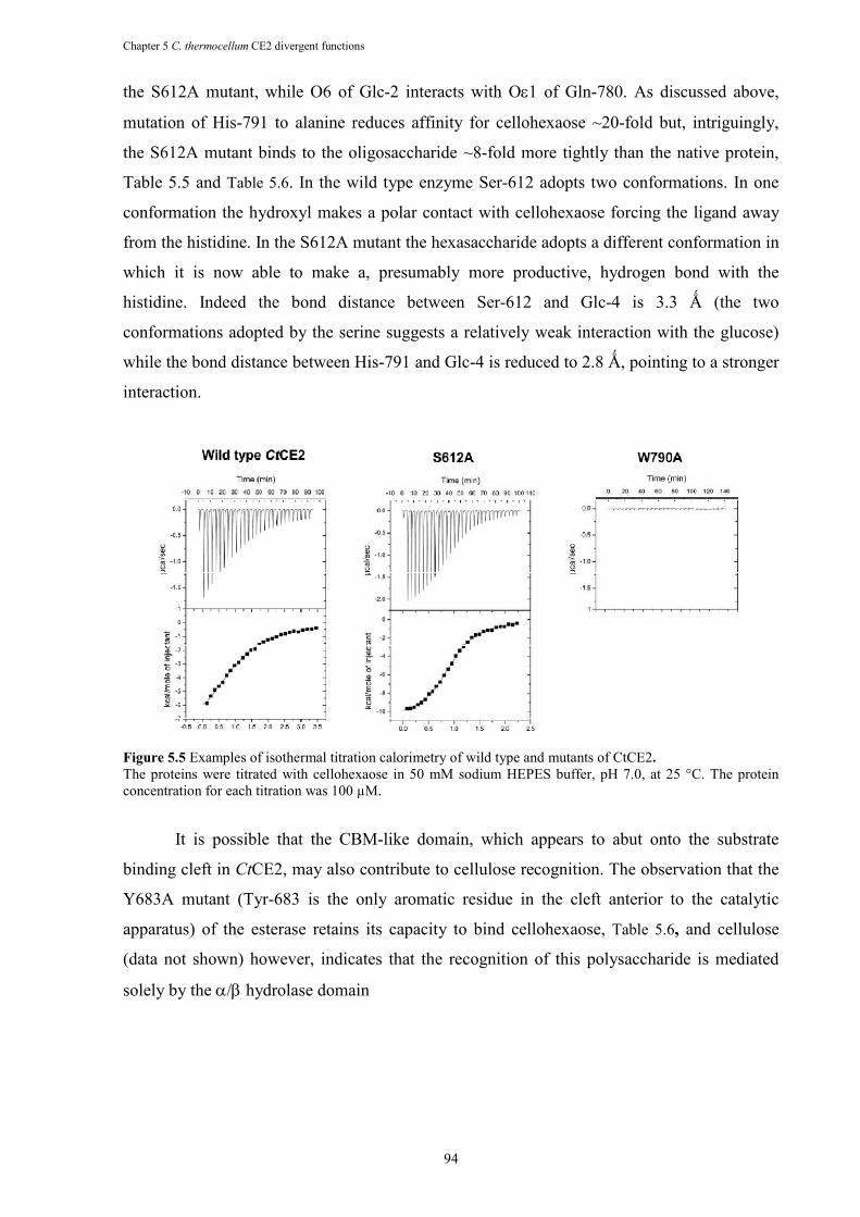
Chapter 5 C. thermocellum CE2 divergent functions
94
the S612A mutant, while O6 of Glc-2 interacts with Oε1 of Gln-780. As discussed above,
mutation of His-791 to alanine reduces affinity for cellohexaose ~20-fold but, intriguingly,
the S612A mutant binds to the oligosaccharide ~8-fold more tightly than the native protein,
Table 5.5 and Table 5.6. In the wild type enzyme Ser-612 adopts two conformations. In one
conformation the hydroxyl makes a polar contact with cellohexaose forcing the ligand away
from the histidine. In the S612A mutant the hexasaccharide adopts a different conformation in
which it is now able to make a, presumably more productive, hydrogen bond with the
histidine. Indeed the bond distance between Ser-612 and Glc-4 is 3.3 Ǻ (the two
conformations adopted by the serine suggests a relatively weak interaction with the glucose)
while the bond distance between His-791 and Glc-4 is reduced to 2.8 Ǻ, pointing to a stronger
interaction.
Figure 5.5 Examples of isothermal titration calorimetry of wild type and mutants of CtCE2.
The proteins were titrated with cellohexaose in 50 mM sodium HEPES buffer, pH 7.0, at 25 °C. The protein concentration for each titration was 100 µM.
It is possible that the CBM-like domain, which appears to abut onto the substrate
binding cleft in CtCE2, may also contribute to cellulose recognition. The observation that the
Y683A mutant (Tyr-683 is the only aromatic residue in the cleft anterior to the catalytic
apparatus) of the esterase retains its capacity to bind cellohexaose, Table 5.6, and cellulose
(data not shown) however, indicates that the recognition of this polysaccharide is mediated
solely by the α/β hydrolase domain
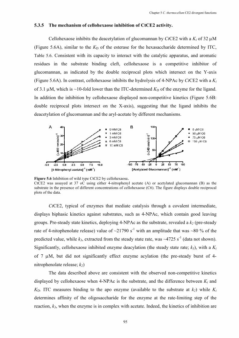
Chapter 5 C. thermocellum CE2 divergent functions
95
5.3.5 The mechanism of cellohexaose inhibition of CtCE2 activity.
Cellohexaose inhibits the deacetylation of glucomannan by CtCE2 with a Ki of 32 µM
(Figure 5.6A), similar to the KD of the esterase for the hexasaccharide determined by ITC,
Table 5.6. Consistent with its capacity to interact with the catalytic apparatus, and aromatic
residues in the substrate binding cleft, cellohexaose is a competitive inhibitor of
glucomannan, as indicated by the double reciprocal plots which intersect on the Y-axis
(Figure 5.6A). In contrast, cellohexaose inhibits the hydrolysis of 4-NPAc by CtCE2 with a Ki
of 3.1 µM, which is ~10-fold lower than the ITC-determined KD of the enzyme for the ligand.
In addition the inhibition by cellohexaose displayed non-competitive kinetics (Figure 5.6B:
double reciprocal plots intersect on the X-axis), suggesting that the ligand inhibits the
deacetylation of glucomannan and the aryl-acetate by different mechanisms.
Figure 5.6 Inhibition of wild type CtCE2 by cellohexaose.
CtCE2 was assayed at 37 oC using either 4-nitrophenyl acetate (A) or acetylated glucomannan (B) as the substrate in the presence of different concentrations of cellohexaose (C6). The figure displays double reciprocal plots of the data.
CtCE2, typical of enzymes that mediate catalysis through a covalent intermediate,
displays biphasic kinetics against substrates, such as 4-NPAc, which contain good leaving
groups. Pre-steady state kinetics, deploying 4-NPAc as the substrate, revealed a k2 (pre-steady
rate of 4-nitophenolate release) value of ~21790 s-1 with an amplitude that was ~80 % of the
predicted value, while k3, extracted from the steady state rate, was ~4725 s-1 (data not shown).
Significantly, cellohexaose inhibited enzyme deacylation (the steady state rate; k3), with a Ki
of 7 µM, but did not significantly effect enzyme acylation (the pre-steady burst of 4-
nitrophenolate release; k2)
The data described above are consistent with the observed non-competitive kinetics
displayed by cellohexaose when 4-NPAc is the substrate, and the difference between Ki and
KD. ITC measures binding to the apo enzyme (available to the substrate at k2) while Ki
determines affinity of the oligosaccharide for the enzyme at the rate-limiting step of the
reaction, k3, when the enzyme is in complex with acetate. Indeed, the kinetics of inhibition are
Chapter 5 C. thermocellum CE2 divergent functions
96
consistent with the observation that cellohexaose binds more tightly to the enzyme when it
makes a polar contact with His-791 rather than Ser-612; when the serine is in complex with
acetate (or formate in the crystal structure) it is not available to hydrogen bond with the
hexasaccharide which is now able to interact with His-791. By contrast, k2 is the rate limiting
step when glucomannan is the substrate and thus cellohexaose must now bind to the apo form
of the enzyme to inhibit the reaction leading to a lower affinity (as it will make a polar contact
with Ser-612) and competitive kinetics.
5.4 DISCUSSIO�
Family CE2 esterases raise intriguing questions about the optimization of plant cell-
wall degrading systems. The most interesting feature of the divergence of function within the
CE2 family is that CtCE2, derived from the CtCel5C-CE2 multi-modular enzyme, is uniquely
able to act as a CBM, with structural and biochemical studies highlighting Trp-746 as a key
recognition element; a residue observed in only one other CE2 enzyme. It is likely highly
relevant that CtCE2 is the only member of CE2 that is a component of a modular enzyme.
Thus, CtCE2 is an example of an emerging biochemical theme that protein scaffolds possess a
latent potential to acquire new, orthogonal, functionality (O'Brien and Herschlag 1999;
Aharoni et al., 2005).
This report is in contrast to the Ohno model of protein evolution which proposes that
mutations that provide new functionalities are introduced into a redundant copy of a
duplicated gene (Ohno 1970; Kimura and Ota 1974). A central component of the Ohno
model is that the introduction of new functions into a protein compromises its original
activity, hence the requirement for gene duplication subsequent to mutation. The observation
that the chicken structural proteins δ-crystallin and a metabolic enzyme are encoded by the
same gene led to the “gene sharing” hypothesis, which states that a gene can be recruited for a
novel function without significant changes to its protein coding sequence (Piatigorsky et al.,
1988). Bergthorsson et al. (Bergthorsson et al., 2007) and others argue that Ohno’s model
poses a dilemma as it requires that the duplicated copy of the gene, which incurs an energetic
cost, must be retained within a population prior to acquiring the mutations that confer the new
activity. These concerns led to the proposal that gene products display secondary activities
prior to duplication of the encoding gene, and that there is a selection for both these activities
subsequent to amplification (these models are reviewed in (Bershtein and Tawfik 2008). A
major caveat with models based on “gene sharing”, however, is that amino acid changes that
introduce new functions often compromise the stability or original activity of the protein
Chapter 5 C. thermocellum CE2 divergent functions
97
(Wang et al., 2002). Thus, in general, the introduced functionality replaces the endogenous
activity of the enzyme. Here we show that the active site of CtCE2 displays dual activities; it
catalyses the deacetylation of plant polysaccharides and also potentiates the activity of its
appended cellulase catalytic module through its non-catalytic cellulose binding function. As
such CtCE2 provides an example of gene sharing in which the new function (cellulose
binding) has developed without having a significant deleterious effect on the existing esterase
activity of the enzyme. It would appear, therefore, that there are different ways to evolve new
enzyme functions without compromising the original activity displayed by the protein. Which
of these pathways a protein follows depends on i) what the new function is, ii) whether it is
advantageous to retain both activities and iii) does the chemistry or steric constraints of the
two divergent functions preclude the housing of these activities in the same active site.
Throughout plant-cell wall degrading systems, non-catalytic binding to
polysaccharides by enzymes that attack insoluble substrates is primarily conferred by CBMs
that are linked to, but spatially distinct from, the catalytic module (Boraston et al., 2004). In
general these targeting modules are joined by flexible linker sequences, although non-
catalytic carbohydrate binding regions, while spatially distinct from the active site, can
occasionally be distal components of the catalytic module itself (Robert et al., 2003; Allouch
et al., 2004). This report shows that the presentation of an additional tryptophan residue
within the substrate binding cleft of the esterase enables the enzyme to acquire cellulose-
binding capacity – potentiating the activity of the appended cellulase catalytic module –
whilst still retaining its original catalytic activity. Indeed the importance of cellulose
recognition by CtCE2 in the function of the appended catalytic module CtCel5C is supported
by the observation that mutation of Trp-790, Tyr-665 or Trp-746 in the full length enzyme
(CtCel5C-CE2) reduces the activity of the cellulase against insoluble substrates by 3-5 fold.
Hence the CE2 family displays a spectrum of activities reflecting the grafting of new
functionality upon the α/β framework. This manifests itself, most powerfully, in the
acquisition of cellulose binding by CtCE2, demonstrating how nature has exploited latent
carbohydrate recognition to introduce additional “non-catalytic” polysaccharide binding
features that are complementary to the activities displayed by a complex modular enzyme.
The composite structure of the plant cell wall, which requires the synergistic interactions of
multiple catalytic and binding functions to elicit its degradation, exerts selection pressures that
lead to the generation of single proteins with multiple activities. The CE2 family, and CtCE2
in particular, reveal an extreme example of this functional complexity and provides a platform
for the future directed engineering of plant cell wall degrading enzyme systems, which is one
of the key environmental goals of the twenty-first century.
Chapter 5 C. thermocellum CE2 divergent functions
98
Acknowledgments
B. A. Pinheiro performed the crystallization and mutagenic work of CtCE2. In
addition, B.A. Pinheiro performed part of the biochemical characterization of CtCE2.
Remaining experiments, particurlary the characterization of other CE2s, were performed by
other authors.
Chapter 6 General discussion and future perspectives
99
Chapter 6 General Discussion and Future Perspectives
The plant cell wall is composed of an intricate network of polysaccharides joined
together by several molecular forces, including covalent and non-covalent bonds. Its
degradation is a complex process and, as a consequence, complex plant cell degrading
systems have evolved in various microorganisms. In anaerobic bacteria, the system is
constituted by various modular enzymes appended to a scaffoldin protein, leading to the
organization of a multienzyme of cellulases and hemicellulases termed the cellulosome.
Cellulosome assembly requires that the dockerin module present in the various catalytic
subunit bind to one of the various cohesin modules of the scaffolding protein. In C.
thermocellum, the cellulosome is connected to the bacterial cell wall through the interaction
of a C-terminal dockerin located on the scaffolding subunit and a cohesin module of a
bacterial cell surface protein. Attachment of proteins to the cell envelop of Gram-positive
bacteria is mediated by SLH domains and a novel family of anchoring domains that was
discovered in this work (Chapter 2). Cohesin-dockerin complexes involved in cellulosome
assembly were termed of type I and in cell-surface attachment of type II. Lack of cross
reaction between type I and type II Coh-Doc pairs allows a correct assembly and cell
anchoring of the multi-enzyme complexes.
In Chapter 2, the search of the complete genome sequence of C. thermocellum allowed
the identification of the complete set of cohesin and dockerin domains present in this
bacterium. Four of these dockerins do not present conservation in the residues that interact
with cohesins at one of their two binding interfaces. In addition, two cohesins reveal several
substitutions in key residues involved in dockerin recognition. Furthermore, it was found that
type I cohesins can be both found in the scaffoldin protein CipA or in cell surface proteins
like OlpA and OlpC. This last protein, OlpC, was found to be a cell wall protein that binds
specifically only to C. thermocellum SCWP rather than the peptidoglycan matrix itself. In
addition, the dockerins of proteins Cthe_0258 and Cthe_0435 recognize, preferentially, OlpC
cohesin and thus these enzymes are believed to be predominantly located at the bacterium cell
surface. All remaining dockerin domains are suggested to be primarily cellulosomal since
they bind preferentially CipA cohesins. OlpC, which is relatively abundant at the bacterium
surface, might also function by transiently retaining cellulosomal enzymes at the cell surface
before they are assembled into the multi-enzyme complex. After evaluating the binding
specificity of the various C. thermocellum dockerins and cohesins, it was observed that apart

Chapter 6 General discussion and future perspectives
100
Doc-Cel44A, C. thermocellum dockerins display restricted ligand specificity. The dockerin
Doc-Cel44A has a high affinity to C. thermocellum cohesins. However, besides its typical C.
thermocellum specific binding interface, Doc-Cel44A has also a mesophile cohesin binding
interface in its N-terminal. For this reason, mutagenic studies combined with ITC
measurements were performed with Doc-Cel44A, which confirmed that, indeed, it is the
dockerin N-terminal binding interface that displays affinity for the cellulolyticum cohesins.
In Chapter 2 it was also studied the complex stability and cohesin-dockerin selectivity.
The data revealed that cohesins display a more destabilizing effect in previously formed
cohesin-dockerin complexes than dockerins do. The reason for this greater destabilizing effect
of the cohesins could be due to the dockerin structure. The existence of the two binding
interfaces in dockerin may allow the switching of these domains from one cohesin to another
without compromising the high affinity of this interaction. This dockerin dual binding mode
may be required for the conformational flexibility in the quaternary structure of the
multienzyme complex. Finally, the data revealed that complexes containing Coh-OlpC were
most prone to dissociation. Since this cohesin is relatively abundant on the bacterium surface,
it may fulfill an important role in maintaining dockerin enzymes at the bacterial surface
before these components are recruited for cellulosomes.
The cohesin-dockerin interaction is essential for cellulosome assembly and its
recruitment by the plant cell wall. In Chapter 3, the crystal structure of C. cellulolyticum
cohesin-dockerin complex was described. Due to the dynamic interaction between the two
heterodimers and to the fact that the N- and C-terminal helices from the dockerin display
significant sequence and structural correlation, only through the introduction of the mutations
A16S/L17T and A47S/F48T in the dockerin it was possible to obtain the described structures.
This way, the A16S/L17T dockerin mutant makes interactions to its cohesin partner through
its C-terminal helix while A47S/F48T variant partner, reoriented by 180º, interacts with the
cohesin primarily through the N-terminal helix. Apolar interactions dominate cohesin-
dockerin recognition that is centered around a hydrophobic pocket on the surface of the
cohesin, formed by Leu-87 and Leu-89, which is occupied in the two binding modes by the
dockerin residues Phe-19 and Leu-50, respectively. Additionally, the specific recognition
between the protomers is dictated also by apolar interaction, primarily between the amino
acids Leu-17 in the dockerin and Ala-129 in the cohesin.
In Chapter 3, the cohesin-dockerin complexes from C. cellulolyticum and C.
thermocellum were compared revealing significant structural conservation in their
organization. Although there is little cross-specificity between the two complexes, the
location of the interface between the two protomers within the heterodimer is identical and the
Chapter 6 General discussion and future perspectives
101
hydrophobic interactions are very similar. Differences are only observed in the polar
interactions between the protomers in the C. cellulolyticum and C. thermocellum
heterodimers. Finally, it was made the attempt to answer the question about the significance
of the dual binding mode. Since both ligand-binding sites on the dockerin cannot be occupied
simultaneously due to steric clashes of the cohesin α-helix, it was excluded the hypothesis of
the trimolecular complex formation. The dual binding may be necessary to confer flexibility
in the cellulosome assembly and, as a consequence, to contribute to the plasticity required for
correct incorporation of different enzyme combinations.
Xylan is one of the most abundant hemicellulose in plant cell walls and xylanase
Xyn10B is an important component of C. thermocellum cellulosomes. Xyn10B is a modular
xylanase that contains an N-terminal family 22 CBM (CBM22-1) followed by a GH10
catalytic domain and a second CBM22 (CBM22-2). At the C-termini, after the internal
CBM22, there is an internal dockerin and the enzyme contains a C-terminal family 1 CE
(CtCE1). In previous studies, work developed at the Faculdade de Medicina Veterinária
laboratory (Universidade Técnica de Lisboa) revealed that the internal CBM22 of Xyn10B
binds xylan polysaccharides, although the N-terminal CBM22 presented no apparent
biological function (Dias et al., 2004). In Chapter 4, the N-terminal moiety of this enzyme,
comprising the enzyme CBM22-1 and the GH10 E337A mutant was crystallized in complex
with xylohexaose. The GH10 E337A mutant was made in order to capture the substrate
xylohexaose in the inactive site. The data was processed using the programs MOSFL and
SCALA but the completion of the structure is still ongoing. It is anticipated that the structure
of the bi-modular truncated derivative of Xyn10B will provide important insights into the role
of the cellulosomal enzyme in xylan hydrolysis.
Matrix polysaccharides are chemically complex molecules in which the backbone
polymers are protected with both sugars and organic esters. Cellulosomal components like
esterases and noncatalytical binding modules remove the diverse acylations and bring the
catalytic unit into contact with its substrate, respectively. In Chapter 5 we present the structure
and function of CtCE2, an esterase derived from the CtCel5C-CE2 multimodular enzyme,
which has the unique ability to act as a CBM. CtCE2 was a previously characterized binding
module with a binding capacity to cellulose and an ability to potentiate the cellulase activity
of the linked CtCel5C catalytic module. The biochemical properties of five CE2 enzymes
were assessed: the C-terminal region of C. thermocellum CtCel5C-CE2, three Celvibrio
japonicus CE2 members, CjCE2A, B and C and the Bacteroides thetaiotaomicron esterase
BtCE2. Furthermore, in this Chapter, the crystal structures of three of these proteins were
obtained: the CE2 module of CtCel5C-CE2 and two of the Celvibrio enzymes, CjCE2A and
Chapter 6 General discussion and future perspectives
102
CjCE2B. The three 3D structures revealed a bi-domain enzyme in which an N-terminal β-
sheet “jelly roll” domain (around 130 residues) is linked to a C-terminal of approximately 220
residues that possesses an atypical α/β-hydrolase (SGNH-hydrolase) fold. The C-terminal
α/β-hydrolase domain contains the esterase catalytic centre. In CtCE2, the catalytic centre is
responsible for the cellooligosaccharide binding, with both Ser and His of the dyad
influencing ligand recognition. The interaction of the oligosaccharide with CtCE2 is
dominated by planar hydrophobic contacts between the polysaccharide and Trp-746, Trp-790
and Try-665. The data showed that the amino acid residue Trp-746 in CtCE2, which is not
conserved within CE2 members, enables the enzyme to acquire cellulose binding capacity.
Finally, enzymatic assays demonstrated that CtCE2 displays biphasic kinetics against
substrates that contain good leaving groups.
References
103
References
Adams, J. J., Pal, G., Jia, Z., & Smith, S. P. (2006). Mechanism of bacterial cell-surface attachment revealed by the structure of cellulosomal type II cohesin-dockerin complex. Proceedings of the ,ational Academy of Sciences of the United States of America, 103, 305-310.
Aharoni, A., Gaidukov, L., Khersonsky, O., Mc, Q. G. S., Roodveldt, C., & Tawfik, D. S. (2005). The 'evolvability' of promiscuous protein functions. ,ature genetics, 37, 73-76.
Ahsan, M., Matsumoto, M., Karita, S., Kimura, T., Sakka, K., & Ohmiya, K. (1997). Purification and characterization of the family J catalytic domain derived from the Clostridium thermocellum endoglucanase CelJ. Bioscience, biotechnology, and biochemistry., 61, 427-431.
Akoh, C. C., Lee, G. C., Liaw, Y. C., Huang, T. H., & Shaw, J. F. (2004). GDSL family of serine esterases/lipases. Progress in lipid research, 43, 534-552.
Ali, M. K., Hayashi, H., Karita, S., Goto, M., Kimura, T., Sakka, K., & Ohmiya, K. (2001). Importance of the carbohydrate-binding module of Clostridium stercorarium Xyn10B to xylan hydrolysis. Bioscience, biotechnology, and biochemistry., 65, 41-47.
Allouch, J., Helbert, W., Henrissat, B., & Czjzek, M. (2004). Parallel substrate binding sites in a beta-agarase suggest a novel mode of action on double-helical agarose. Structure, 12, 623-632.
Atalla, R. H., Hackney, J., & Vanderhart, D. L. (1989). Studies on the structure of native celluloses using raman-spectroscopy and solid-state c-13 nmr. Abstracts of Papers of
the American Chemical Society, 197, 31.
Bagnara-Tardif, C., Gaudin, C., Belaich, A., Hoest, P., Citard, T., & Belaich, J. P. (1992). Sequence analysis of a gene cluster encoding cellulases from Clostridium cellulolyticum. Gene, 119, 17-28.
Bar, M., Golan, G., Nechama, M., Zolotnitsky, G., Shoham, Y., & Shoham, G. (2004). A new crystal form of XT6 enables a significant improvement of its diffraction quality and resolution. Acta crystallographica, 60, 545-549.
Baronepel, O., Gharyal, P. K., & Schindler, M. (1988). Pectins as mediators of wall porosity in soybean cells. Planta, 175, 389-395.
Bayer, E. A., Setter, E., & Lamed, R. (1985). Organization and distribution of the cellulosome in Clostridium thermocellum. Journal of bacteriology, 163, 552-559.
Bayer, E. A., Morag, E., & Lamed, R. (1994). The cellulosome--a treasure-trove for biotechnology. Trends in biotechnology, 12, 379-386.
References
104
Bayer, E. A., Chanzy, H., Lamed, R., & Shoham, Y. (1998). Cellulose, cellulases and cellulosomes. Current opinion in structural biology, 8, 548-557.
Bayer, E. A., Belaich, J. P., Shoham, Y., & Lamed, R. (2004). The cellulosomes: multienzyme machines for degradation of plant cell wall polysaccharides. Annual review of
microbiology, 58, 521-554.
Beguin, P., & Alzari, P. M. (1997, Dec 15-17). The cellulosome of Clostridium
thermocellum. Paper presented at the 664th Meeting of the Biochemical-Society, Reading, England.
Belaich, J. P., Tardif, C., Belaich, A., & Gaudin, C. (1997). The cellulolytic system of Clostridium cellulolyticum. Journal of Biotechnology, 57, 3-14.
Bergthorsson, U., Andersson, D. I., & Roth, J. R. (2007). Ohno's dilemma: evolution of new genes under continuous selection. Proceedings of the ,ational Academy of Sciences of
the United States of America, 104(43), 17004-17009.
Bershtein, S., & Tawfik, D. S. (2008). Ohno's model revisited: measuring the frequency of potentially adaptive mutations under various mutational drifts. Molecular
biology and evolution, 25, 2311-2318.
Bhat, M. K. (2000). Cellulases and related enzymes in biotechnology. Biotechnology
advances, 18, 355-383.
Blair, D. E., Schuttelkopf, A. W., MacRae, J. I., & van Aalten, D. M. (2005). Structure and metal-dependent mechanism of peptidoglycan deacetylase, a streptococcal virulence factor. Proceedings of the ,ational Academy of Sciences of the United States of America, 102, 15429-15434.
Bolam, D. N., Ciruela, A., McQueen-Mason, S., Simpson, P., Williamson, M. P., Rixon, J. E., Boraston, A., Hazlewood, G. P. & Gilbert, H. J. (1998). Pseudomonas cellulose-binding domains mediate their effects by increasing enzyme substrate proximity. The
Biochemical journal, 331, 775-781.
Bolam, D. N., Xie, H., Pell, G., Hogg, D., Galbraith, G., Henrissat, B. & Gilbert, H. J. (2004). X4 modules represent a new family of carbohydrate-binding modules that display novel properties. The Journal of biological chemistry, 279, 22953-22963.
Boraston, A. B., McLean, B. W., Kormos, J. M., Alam, M., Gilkes, N. R., Haynes, C. A., Tomme, P., Kilburn, D. G. & Warren, R. A. J. (1999, Apr 11-14). Carbohydrate-binding modules: diversity of structure and function. Paper presented at the 3rd Carbohydrate
Bioengineering Meeting, Newcastle Tyne, England.
Boraston, A. B., Kwan, E., Chiu, P., Warren, R. A., & Kilburn, D. G. (2003). Recognition and hydrolysis of noncrystalline cellulose. The Journal of biological chemistry, 278, 6120-6127.
References
105
Boraston, A. B., Bolam, D. N., Gilbert, H. J., & Davies, G. J. (2004). Carbohydrate-binding modules: fine-tuning polysaccharide recognition. The Biochemical journal, 382, 769-781.
Boudet, A. M., Kajita, S., Grima-Pettenati, J., & Goffner, D. (2003). Lignins and lignocellulosics: a better control of synthesis for new and improved uses. Trends in plant
science, 8, 576-581.
Canals, A., Vega, M. C., Gomis-Ruth, F. X., Diaz, M., Santamaria, R. R., & Coll, M. (2003). Structure of xylanase Xys1delta from Streptomyces halstedii. Acta crystallographica, 59, 1447-1453.
Capadona, J. R., Shanmuganathan, K., Tyler, D. J., Rowan, S. J., & Weder, C. (2008). Stimuli-responsive polymer nanocomposites inspired by the sea cucumber dermis. Science, 319, 1370-1374.
Carvalho, A. L., Dias, F. M., Prates, J. A., Nagy, T., Gilbert, H. J., Davies, G. J., Ferreira, L. M., Romao, M. J. & Fontes, C. M. (2003). Cellulosome assembly revealed by the crystal structure of the cohesin-dockerin complex. Proceedings of the ,ational Academy of
Sciences of the United States of America, 100, 13809-13814.
Carvalho, A. L., Goyal, A., Prates, J. A. M., Bolam, D. N., Gilbert, H. J., Pires, V. M. R., et al. (2004). The family 11 carbohydrate-binding module of Clostridium thermocellum Lic26A-Cel5E accommodates beta-1,4- and beta-1,3-1,4-mixed linked glucans at a single binding site. Journal of Biological Chemistry, 279, 34785-34793.
Carvalho, A. L., Pires, V. M., Gloster, T. M., Turkenburg, J. P., Prates, J. A., Ferreira, L. M., Romao, M. J., Davies, G. J., Fontes, C. M., & Gilbert, H. J. (2005). Insights into the structural determinants of cohesin-dockerin specificity revealed by the crystal structure of the type II cohesin from Clostridium thermocellum SdbA. Journal of molecular biology, 349, 909-915.
Carvalho, A. L., Dias, F. M., Nagy, T., Prates, J. A., Proctor, M. R., Smith, N., Bayer, E. A., Davies, G. J., Ferreira, L. M., Romao, M. J., Fontes, C. M., & Gilbert, H. J. (2007). Evidence for a dual binding mode of dockerin modules to cohesins. Proceedings of the
,ational Academy of Sciences of the United States of America, 104, 3089-3094.
Charnock, S. J., Bolam, D. N., Turkenburg, J. P., Gilbert, H. J., Ferreira, L. M., Davies, G. J., & Fontes, C. M. (2000). The X6 "thermostabilizing" domains of xylanases are carbohydrate-binding modules: structure and biochemistry of the Clostridium thermocellum X6b domain. Biochemistry, 39, 5013-5021.
Chauvaux, S., Beguin, P., Aubert, J. P., Bhat, K. M., Gow, L. A., Wood, T. M., & Bairoch, A. (1990). Calcium-binding affinity and calcium-enhanced activity of Clostridium
thermocellum endoglucanase D. The Biochemical journal, 265, 261-265.
Chen, H., Li, X., Blum, D., and Ljungdahl, L. (1998) Two genes ofthe anaerobic fungus Orpinomyces sp. strain PC-2 encoding cellulases with endoglucanase activities may have arisen by gene duplication, FEMS Microbiology Letters 159, 63–68.
References
106
Choi, S. K., & Ljungdahl, L. G. (1996). Structural role of calcium for the organization of the cellulosome of Clostridium thermocellum. Biochemistry, 35, 4906-4910.
Ciruela, A., Gilbert, H. J., Ali, B. R., & Hazlewood, G. P. (1998). Synergistic interaction of the cellulosome integrating protein (CipA) from Clostridium thermocellum with a cellulosomal endoglucanase. FEBS letters, 422, 221-224.
Coggins, B. E., Li, X., McClerren, A. L., Hindsgaul, O., Raetz, C. R., & Zhou, P. (2003). Structure of the LpxC deacetylase with a bound substrate-analog inhibitor. ,ature
Structural Biology, 10, 645-651.
Cohen, S. X., Morris, R. J., Fernandez, F. J., Ben Jelloul, M., Kakaris, M., Parthasarathy, V., Lamzin, V. S., Kleywegt, G. J., & Perrakis, A. (2004). Towards complete validated models in the next generation of ARP/wARP. Acta crystallographica, 60, 2222-2229.
Cosgrove, D. J. (1997). Assembly and enlargement of the primary cell wall in plants. Annual review of cell and developmental biology, 13, 171-201.
Cosgrove, D. J. (2001). Wall structure and wall loosening. A look backwards and forwards. Plant physiology, 125(1), 131-134.
Cosgrove, D. J. (2005). Growth of the plant cell wall. ,ature reviews, 6, 850-861.
Coutinho, P. M., & Henrissat, B. (1999). Life with no sugars? Journal of molecular
microbiology and biotechnology, 1, 307-308.
Dalrymple, B. P., Cybinski, D. H., Layton, I., McSweeney, C. S., Xue, G. P., Swadling, Y. J., & Lowry, J. B. (1997). Three ,eocallimastix patriciarum esterases associated with the degradation of complex polysaccharides are members of a new family of hydrolases. Microbiology, 143, 2605-2614.
Davies, G. J., Gloster, T. M., & Henrissat, B. (2005). Recent structural insights into the expanding world of carbohydrate-active enzymes. Current opinion in structural biology, 15, 637-645.
DeBoy, R. T., Mongodin, E. F., Fouts, D. E., Tailford, L. E., Khouri, H., Emerson, J. B., Mohamoud, Y., Watkins, K., Henrissat, B., Gilbert, H. J., & Nelson, K. E. (2008). Insights into plant cell wall degradation from the genome sequence of the soil bacterium Cellvibrio
japonicus. Journal of bacteriology, 190, 5455-5463.
Desvaux, M. (2005). Clostridium cellulolyticum: model organism of mesophilic cellulolytic clostridia. FEMS microbiology reviews, 29, 741-764.
Din, N., Gilkes, N. R., Tekant, B., Miller, R. C., Warren, A. J., & Kilburn, D. G. (1991). Non-hydrolytic disruption of cellulose fibers by the binding domain of a bacterial cellulase. Bio-Technology, 9, 1096-1099.
References
107
Din, N., Damude, H. G., Gilkes, N. R., Miller, R. C., Warren, R. A. J., & Kilburn, D. G. (1994). C-1-C-X revisited - intramolecular synergism in a cellulase. Proceedings of the
,ational Academy of Sciences of the United States of America, 91, 11383-11387.
Ding, S. Y., Bayer, E. A., Steiner, D., Shoham, Y., & Lamed, R. (1999). A novel cellulosomal scaffoldin from Acetivibrio cellulolyticus that contains a family 9 glycosyl hydrolase. Journal of bacteriology, 181, 6720-6729.
Ding, S. Y., Bayer, E. A., Steiner, D., Shoham, Y., & Lamed, R. (2000). A scaffoldin of the Bacteroides cellulosolvens cellulosome that contains 11 type II cohesins. Journal of
bacteriology, 182, 4915-4925.
Ding, S. Y., Rincon, M. T., Lamed, R., Martin, J. C., McCrae, S. I., Aurilia, V., Shoham, Y., Bayer, E. A., & Flint, H. J. (2001). Cellulosomal scaffoldin-like proteins from Ruminococcus flavefaciens. Journal of bacteriology, 183, 1945-1953.
Doi, R. H., Goldstein, M., Hashida, S., Park, J. S., & Takagi, M. (1994). The Clostridium cellulovorans cellulosome. Critical reviews in microbiology, 20, 87-93.
Doi, R. H., & Tamaru, Y. (2001). The Clostridium cellulovorans cellulosome: An enzyme complex with plant cell wall degrading activity. Chemical Record, 1, 24-32.
Doi, R. H., Kosugi, A., Murashima, K., Tamaru, Y., & Han, S. O. (2003). Cellulosomes from mesophilic bacteria. Journal of bacteriology, 185, 5907-5914.
Doi, R. H., & Kosugi, A. (2004). Cellulosomes: plant-cell-wall-degrading enzyme complexes. ,ature Reviews Microbiology, 2, 541-551.
Dominguez, R., Souchon, H., Spinelli, S., Dauter, Z., Wilson, K. S., Chauvaux, S., Beguin, P., & Alzari, P. M. (1995). A common protein fold and similar active-site in 2 distinct families of beta-glycanases. ,ature Structural Biology, 2, 569-576.
Durrant, A. J., Hall, J., Hazlewood, G. P., & Gilbert, H. J. (1991). The non-catalytic C-terminal region of endoglucanase E from Clostridium thermocellum contains a cellulose-binding domain. The Biochemical journal, 273, 289-293.
Emsley, P., & Cowtan, K. (2004). Coot: model-building tools for molecular graphics. Acta crystallographica, 60, 2126-2132.
Esnouf, R. M. (1997). An extensively modified version of MolScript that includes greatly enhanced coloring capabilities. Journal of molecular graphics & modelling, 15, 132-134, 112-133.
Evans, P. (2006). Scaling and assessment of data quality. Acta Crystallographica, 62, 72-82.
References
108
Fanutti, C., Ponyi, T., Black, G. W., Hazlewood, G. P., & Gilbert, H. J. (1995). The conserved noncatalytic 40-residue sequence in cellulases and hemicellulases from anaerobic fungi functions as a protein docking domain. The Journal of biological chemistry, 270, 29314-29322.
Fernandes, A. C., Fontes, C. M., Gilbert, H. J., Hazlewood, G. P., Fernandes, T. H., & Ferreira, L. M. (1999). Homologous xylanases from Clostridium thermocellum: evidence for bi-functional activity, synergism between xylanase catalytic modules and the presence of xylan-binding domains in enzyme complexes. The Biochemical journal, 342, 105-110.
Fierobe, H. P., Gaudin, C., Belaich, A., Loutfi, M., Faure, E., Bagnara, C., Baty, D., & Belaich, J. P. (1991). Characterization of endoglucanase A from Clostridium cellulolyticum. Journal of bacteriology, 173, 7956-7962.
Fierobe, H. P., Pages, S., Belaich, A., Champ, S., Lexa, D., & Belaich, J. P. (1999). Cellulosome from Clostridium cellulolyticum: molecular study of the Dockerin/Cohesin interaction. Biochemistry, 38, 12822-12832.
Fierobe, H. P., Mechaly, A., Tardif, C., Belaich, A., Lamed, R., Shoham, Y., Belaich, J. P., & Bayer, E. A. (2001). Design and production of active cellulosome chimeras. Selective incorporation of dockerin-containing enzymes into defined functional complexes. The Journal
of biological chemistry, 276, 21257-21261.
Fierobe, H. P., Bayer, E. A., Tardif, C., Czjzek, M., Mechaly, A., Belaich, A., Lamed, R., Shoham, Y., & Belaich, J. P. (2002). Degradation of cellulose substrates by cellulosome chimeras. Substrate targeting versus proximity of enzyme components. The Journal of
biological chemistry, 277, 49621-49630.
Fierobe, H. P., Mingardon, F., Mechaly, A., Belaich, A., Rincon, M. T., Pages, S., Lamed, R., Tardif, C., Belaich, J. P., & Bayer, E. A. (2005). Action of designer cellulosomes on homogeneous versus complex substrates: controlled incorporation of three distinct enzymes into a defined trifunctional scaffoldin. The Journal of biological chemistry, 280, 16325-16334.
Flint, J., Bolam, D. N., Nurizzo, D., Taylor, E. J., Williamson, M. P., Walters, C., Davies, G. J., & Gilbert, H. J. (2005). Probing the mechanism of ligand recognition in family 29 carbohydrate-binding modules. The Journal of biological chemistry, 280, 23718-23726.
Fontes, C. M., Hall, J., Hirst, B. H., Hazlewood, G. P., & Gilbert, H. J. (1995). The resistance of cellulases and xylanases to proteolytic inactivation. Applied microbiology and
biotechnology, 43, 52-57.
Fontes, C. M., Hazlewood, G. P., Morag, E., Hall, J., Hirst, B. H., & Gilbert, H. J. (1995). Evidence for a general role for non-catalytic thermostabilizing domains in xylanases from thermophilic bacteria. The Biochemical journal, 307, 151-158.
French, S., & Wilson, K. (1978). Treatment of Negative Intensity Observations. Acta
Crystallographica, 34, 517-525.
References
109
Frey-Wyssling, A. (1953) Submicroscopic structure of the elementary fibers of cellulose. Experintia 9,181-183.
Frey-Wyssling, A. (1954) The fine structure of cellulose microfibrils. Science 119, 80-82.
Fry, S.C. (1989). The structure and functions of xyloglucan. Journal of Experimental
Botany, 40, 1–12
Fujino, T., Beguin, P., & Aubert, J. P. (1993). Organization of a Clostridium
thermocellum gene cluster encoding the cellulosomal scaffolding protein CipA and a protein possibly involved in attachment of the cellulosome to the cell surface. Journal of
bacteriology, 175, 1891-1899.
Garbe, T. R., Hibler, N. S., & Deretic, V. (1996). Isoniazid induces expression of the antigen 85 complex in Mycobacterium tuberculosis. Antimicrobial agents and chemotherapy, 40, 1754-1756.
Gebler, J., Gilkes, N. R., Claeyssens, M., Wilson, D. B., Beguin, P., Wakarchuk, W. W., Kilburn, D. G., Miller, R. C., Jr., Warren, R. A., & Withers, S. G. (1992). Stereoselective hydrolysis catalyzed by related beta-1,4-glucanases and beta-1,4-xylanases. The Journal of
biological chemistry, 267, 12559-12561.
Gerngross, U. T., Romaniec, M. P., Kobayashi, T., Huskisson, N. S., & Demain, A. L. (1993). Sequencing of a Clostridium thermocellum gene (cipA) encoding the cellulosomal SL-protein reveals an unusual degree of internal homology. Molecular microbiology, 8, 325-334.
Gilbert, H. J. (2007). Cellulosomes: microbial nanomachines that display plasticity in quaternary structure. Molecular microbiology, 63, 1568-1576.
Gilbert, H. J., Stalbrand, H., & Brumer, H. (2008). How the walls come crumbling down: recent structural biochemistry of plant polysaccharide degradation. Current opinion in
plant biology, 11, 338-348.
Gilkes, N. R., Warren, R. A., Miller, R. C., Jr., & Kilburn, D. G. (1988). Precise excision of the cellulose binding domains from two Cellulomonas fimi cellulases by a homologous protease and the effect on catalysis. The Journal of biological chemistry, 263, 10401-10407.
Guglielmi, G., & Beguin, P. (1998). Cellulase and hemicellulase genes of Clostridium
thermocellum from five independent collections contain few overlaps and are widely scattered across the chromosome. FEMS microbiology letters, 161, 209-215.
Ha, M. A., Apperley, D. C., & Jarvis, M. C. (1997). Molecular Rigidity in Dry and Hydrated Onion Cell Walls. Plant physiology, 115, 593-598.
References
110
Hall, J., Hazlewood, G. P., Barker, P. J., & Gilbert, H. J. (1988). Conserved reiterated domains in Clostridium thermocellum endoglucanases are not essential for catalytic activity. Gene, 69, 29-38.
Hall, J., Black, G. W., Ferreira, L. M., Millward-Sadler, S. J., Ali, B. R., Hazlewood, G. P., et al. (1995). The non-catalytic cellulose-binding domain of a novel cellulase from Pseudomonas fluorescens subsp. cellulosa is important for the efficient hydrolysis of Avicel. The Biochemical journal, 309, 749-756.
Hammel, M., Fierobe, H. P., Czjzek, M., Finet, S., & Receveur-Brechot, V. (2004). Structural insights into the mechanism of formation of cellulosomes probed by small angle X-ray scattering. The Journal of biological chemistry, 279, 55985-55994.
Hammel, M., Fierobe, H. P., Czjzek, M., Kurkal, V., Smith, J. C., Bayer, E. A., Finet, S., & Receveur-Brechot, V. (2005). Structural basis of cellulosome efficiency explored by small angle X-ray scattering. The Journal of biological chemistry, 280, 38562-38568.
Handelsman, T., Barak, Y., Nakar, D., Mechaly, A., Lamed, R., Shoham, Y., & Bayer, E. A. (2004). Cohesin-dockerin interaction in cellulosome assembly: a single Asp-to-Asn mutation disrupts high-affinity cohesin-dockerin binding. FEBS letters, 572, 195-200.
Hayashi, T. (1989). Xyloglucans in the primary-cell wall. Annual Review of Plant
physiologyogy and Plant Molecular Biology, 40, 139-168.
Hayashi, H., Takehara, M., Hattori, T., Kimura, T., Karita, S., Sakka, K., & Ohmiya, K. (1999). Nucleotide sequences of two contiguous and highly homologous xylanase genes xynA and xynB and characterization of XynA from Clostridium thermocellum. Applied
microbiology and biotechnology, 51, 348-357.
Hazlewood, G. P., Laurie, J. I., Ferreira, L. M., & Gilbert, H. J. (1992). Pseudomonas
fluorescens subsp. cellulosa: an alternative model for bacterial cellulase. The Journal of
applied bacteriology, 72, 244-251.
Henrissat, B. (1991). A classification of glycosyl hydrolases based on amino acid sequence similarities. The Biochemical journal, 280, 309-316.
Henrissat, B., & Bairoch, A. (1996). Updating the sequence-based classification of glycosyl hydrolases. The Biochemical journal, 316, 695-696.
Henrissat, B., & Davies, G. (1997). Structural and sequence-based classification of glycoside hydrolases. Current opinion in structural biology, 7, 637-644.
Henrissat, B. (1998). Glycosidase families. Biochemical Society transactions, 26, 153-156.
Hermoso, J. A., Sanz-Aparicio, J., Molina, R., Juge, N., Gonzalez, R., & Faulds, C. B. (2004). The crystal structure of feruloyl esterase A from Aspergillus niger suggests evolutive functional convergence in feruloyl esterase family. Journal of molecular biology, 338, 495-506.

References
111
Jancarik, J., Scott, W. G., Milligan, D. L., Koshland, D. E., Jr., & Kim, S. H. (1991). Crystallization and preliminary X-ray diffraction study of the ligand-binding domain of the bacterial chemotaxis-mediating aspartate receptor of Salmonella typhimurium. Journal of
molecular biology, 221, 31-34.
Jarvis, M. C. (1992). Control of thickness of collenchyma cell-walls by pectins. Planta, 187, 218-220.
Jenkins, J., Mayans, O., Smith, D., Worboys, K., & Pickersgill, R. W. (2001). Three-dimensional structure of Erwinia chrysanthemi pectin methylesterase reveals a novel esterase active site. Journal of molecular biology, 305, 951-960.
Jindou, S., Soda, A., Karita, S., Kajino, T., Beguin, P., Wu, J. H., Inagaki, M., Kimura, T., Sakka, K., & Ohmiya, K. (2004). Cohesin-dockerin interactions within and between Clostridium josui and Clostridium thermocellum: binding selectivity between cognate dockerin and cohesin domains and species specificity. The Journal of biological chemistry, 279, 9867-9874.
Kabsch, W. (1978). Discussion of solution for best rotation to relate 2 sets of vectors. Acta Crystallographica, 34, 827-828.
Kakiuchi, M., Isui, A., Suzuki, K., Fujino, T., Fujino, E., Kimura, T., Karita, S., Sakka, K., & Ohmiya, K. (1998). Cloning and DNA sequencing of the genes encoding Clostridium josui scaffolding protein CipA and cellulase CelD and identification of their gene products as major components of the cellulosome. Journal of bacteriology, 180, 4303-4308.
Karita, S., Kimura, T., Sakka, K., & Ohmiya, K. (1997). Purification of the Ruminococcus albus endoglucanase IV using a cellulose-binding domain as an affinity tag. Journal of Fermentation and Bioengineering, 84, 354-357.
Karpol, A., Barak, Y., Lamed, R., Shoham, Y., & Bayer, E. A. (2008). Functional asymmetry in cohesin binding belies inherent symmetry of the dockerin module: insight into cellulosome assembly revealed by systematic mutagenesis. Biochemical Journal, 410, 331-338.
Keegstra, K., Talmadge, K. W., Bauer, W. D., & Albersheim, P. (1973). The Structure of Plant Cell Walls: III. A Model of the Walls of Suspension-cultured Sycamore Cells Based on the Interconnections of the Macromolecular Components. Plant physiology, 51, 188-197.
Kimura, M., & Ota, T. (1974). On some principles governing molecular evolution. Proceedings of the ,ational Academy of Sciences of the United States of America, 71, 2848-2852.
Kobayashi, S., & Ohmae, M. (2006). Enzymatic polymerization to polysaccharides. In Enzyme-Catalyzed Synthesis of Polymers (Vol. 194, pp. 159-210). Berlin: Springer-Verlag Berlin.
References
112
Koshland, D. E. (1953). Stereochemistry and the mechanism of enzymatic reactions. Biological Reviews, 28, 416-436.
Kratky, O. and Mark, H. (1938) Supermolecular structure of fibers. Papier-Fabr, 36, 345-348.
Kraulis, P. J. (1991). Molscript - a program to produce both detailed and schematic plots of protein structures. Journal of Applied Crystallography, 24, 946-950.
Lamed, R., Setter, E., & Bayer, E. A. (1983). Characterization of a cellulose-binding, cellulase-containing complex in Clostridium thermocellum. Journal of bacteriology, 156, 828-836.
Lamed, R., & Bayer, E. A. (1988). The cellulosome of Clostridium thermocellum. Advances in Applied Microbiology, 33, 1–46
Larsen, N. A., Lin, H., Wei, R., Fischbach, M. A., & Walsh, C. T. (2006). Structural characterization of enterobactin hydrolase IroE. Biochemistry, 45, 10184-10190.
Leibovitz, E., & Beguin, P. (1996). A new type of cohesin domain that specifically binds the dockerin domain of the Clostridium thermocellum cellulosome-integrating protein CipA. Journal of bacteriology, 178, 3077-3084.
Lemaire, M., Ohayon, H., Gounon, P., Fujino, T., & Beguin, P. (1995). OlpB, a new outer layer protein of Clostridium thermocellum, and binding of its S-layer-like domains to components of the cell envelope. Journal of bacteriology, 177, 2451-2459.
Leslie, A. G. W. (1992). Joint CCP4 and ESF-EACBM Newsletters on Protein Crystallography. 26
Lu, Y., Zhang, Y. H., & Lynd, L. R. (2006). Enzyme-microbe synergy during cellulose hydrolysis by Clostridium thermocellum. Proceedings of the ,ational Academy of
Sciences of the United States of America, 103, 16165-16169.
Lundqvist, J., Teleman, A., Junel, L., Zacchi, G., Dahlman, O., Tjerneld, F., & Stalbrand, H. (2002). Isolation and characterization of galactoglucomannan from spruce (Picea abies). Carbohydrate Polymers, 48, 29-39.
Lytle, B. L., Volkman, B. F., Westler, W. M., Heckman, M. P., & Wu, J. H. (2001). Solution structure of a type I dockerin domain, a novel prokaryotic, extracellular calcium-binding domain. Journal of molecular biology, 307, 745-753.
Marchessault, R. H. and Sarko, A. (1967) X-ray structure of polysaccharides. In M. L. Wolfrom (Eds.), Advanced Carbohydrate Chemistry (22, pp. 421-483). New York: Academic Press.
Marchessault, R. H. and Sundararajan, P. R. (1983) In Cellulose, in the
Polysaccharides. New York: Academic Press, p. 11.
References
113
Maynes, J. T., Garen, C., Cherney, M. M., Newton, G., Arad, D., Av-Gay, Y., Fahey, R. C., & James, M. N. (2003). The crystal structure of 1-D-myo-inosityl 2-acetamido-2-deoxy-alpha-D-glucopyranoside deacetylase (MshB) from Mycobacterium tuberculosis reveals a zinc hydrolase with a lactate dehydrogenase fold. The Journal of biological
chemistry, 278, 47166-47170.
McCarter, J. D., & Withers, S. G. (1994). Mechanisms of enzymatic glycoside hydrolysis. Current opinion in structural biology, 4, 885-892.
McCoy, A. J., Grosse-Kunstleve, R. W., Adams, P. D., Winn, M. D., Storoni, L. C., & Read, R. J. (2007). Phaser crystallographic software. Journal of applied crystallography, 40, 658-674.
McLean, B. W., Bray, M. R., Boraston, A. B., Gilkes, N. R., Haynes, C. A., & Kilburn, D. G. (2000). Analysis of binding of the family 2a carbohydrate-binding module from Cellulomonas fimi xylanase 10A to cellulose: specificity and identification of functionally important amino acid residues. Protein engineering, 13, 801-809.
McNeil, M., Darvill, A.G., Fry, S.C., & Albersheim, P. (1984). Structure and function of the primary cell walls of plants. Annu Rev Biochem, 53, 625–663.
Mechaly, A., Yaron, S., Lamed, R., Fierobe, H. P., Belaich, A., Belaich, J. P., Shoham, Y., & Bayer, E. A. (2000). Cohesin-dockerin recognition in cellulosome assembly: experiment versus hypothesis. Proteins, 39, 170-177.
Mechaly, A., Fierobe, H. P., Belaich, A., Belaich, J. P., Lamed, R., Shoham, Y., & Bayer, E. A. (2001). Cohesin-dockerin interaction in cellulosome assembly: a single hydroxyl group of a dockerin domain distinguishes between nonrecognition and high affinity recognition. The Journal of biological chemistry, 276, 9883-9888.
Miras, I., Schaeffer, F., Beguin, P., & Alzari, P. M. (2002). Mapping by site-directed mutagenesis of the region responsible for cohesin-dockerin interaction on the surface of the seventh cohesin domain of Clostridium thermocellum CipA. Biochemistry, 41, 2115-2119.
Molgaard, A., & Larsen, S. (2002). A branched N-linked glycan at atomic resolution in the 1.12 A structure of rhamnogalacturonan acetylesterase. Acta crystallographica, 58, 111-119.
Molgaard, A., & Larsen, S. (2004). Crystal packing in two pH-dependent crystal forms of rhamnogalacturonan acetylesterase. Acta crystallographica, 60, 472-478.
Morag, E., Lapidot, A., Govorko, D., Lamed, R., Wilchek, M., Bayer, E. A., et al. (1995). Expression, purification, and characterization of the cellulose-binding domain of the scaffoldin subunit from the cellulosome of Clostridium thermocellum. Applied and
environmental microbiology, 61, 1980-1986.
Murshudov, G. N., Vagin, A. A., & Dodson, E. J. (1997). Refinement of macromolecular structures by the maximum-likelihood method. Acta crystallographica, 53, 240-255.
References
114
Nagy, T., Simpson, P., Williamson, M. P., Hazlewood, G. P., Gilbert, H. J., & Orosz, L. (1998). All three surface tryptophans in Type IIa cellulose binding domains play a pivotal role in binding both soluble and insoluble ligands. FEBS letters, 429, 312-316.
Najmudin, S., Guerreiro, C. I., Carvalho, A. L., Prates, J. A., Correia, M. A., Alves, V. D., Ferreira, L. M., Romao, M. J., Gilbert, H. J., Bolam, D. N., & Fontes, C. M. (2006). Xyloglucan is recognized by carbohydrate-binding modules that interact with beta-glucan chains. The Journal of biological chemistry, 281, 8815-8828.
Navaza, J. (2001). Implementation of molecular replacement in AMoRe. Acta
crystallographica, 57, 1367-1372.
Nickerson, R. F. (1941) Hydrolysis and catalytic oxidation of cellulosic materials. Hydrolysis of natural regenerated and substituted celluloses. Industrial Engineering
Chemistry, 33, 1022-1027.
Noach, I., Lamed, R., Xu, Q., Rosenheck, S., Shimon, L. J., Bayer, E. A., & Frolow, F. (2003). Preliminary X-ray characterization and phasing of a type II cohesin domain from the cellulosome of Acetivibrio cellulolyticus. Acta crystallographica, 59, 1670-1673.
Noach, I., Frolow, F., Jakoby, H., Rosenheck, S., Shimon, L. W., Lamed, R., & Bayer, E. A. (2005). Crystal structure of a type-II cohesin module from the Bacteroides
cellulosolvens cellulosome reveals novel and distinctive secondary structural elements. Journal of molecular biology, 348, 1-12.
Nolling, J., Breton, G., Omelchenko, M. V., Makarova, K. S., Zeng, Q., Gibson, R., Lee, H. M., Dubois, J., Qiu, D., Hitti, J., Wolf, Y. I., Tatusov, R. L., Sabathe, F., Doucette-Stamm, L., Soucaille, P., Daly, M. J., Bennett, G. N., Koonin, E. V., & Smith, D. R. (2001). Genome sequence and comparative analysis of the solvent-producing bacterium Clostridium
acetobutylicum. Journal of bacteriology, 183, 4823-4838.
O'Brien, P. J., & Herschlag, D. (1999). Catalytic promiscuity and the evolution of new enzymatic activities. Chemistry & biology, 6, R91-R105.
Ohno, S. (1970) Evolution by gene duplication. New York: Springer.
Osullivan, A. C. (1997). Cellulose: the structure slowly unravels. Cellulose, 4, 173-207.
Otwinowski, Z., & Minor, W. (1997). Processing of X-ray diffraction data collected in oscillation mode. In Macromolecular Crystallography, Pt A (Vol. 276, pp. 307-326). San Diego: Academic Press Inc.
Pages, S., Belaich, A., Belaich, J. P., Morag, E., Lamed, R., Shoham, Y., & Bayer, E. A. (1997). Species-specificity of the cohesin-dockerin interaction between Clostridium
thermocellum and Clostridium cellulolyticum: prediction of specificity determinants of the dockerin domain. Proteins, 29, 517-527.
References
115
Pages, S., Belaich, A., Fierobe, H. P., Tardif, C., Gaudin, C., & Belaich, J. P. (1999). Sequence analysis of scaffolding protein CipC and ORFXp, a new cohesin-containing protein in Clostridium cellulolyticum: comparison of various cohesin domains and subcellular localization of ORFXp. Journal of bacteriology, 181, 1801-1810.
Perrakis, A., Morris, R., & Lamzin, V. S. (1999). Automated protein model building combined with iterative structure refinement. ,ature Structural Biology, 6, 458-463.
Piatigorsky, J., O'Brien, W. E., Norman, B. L., Kalumuck, K., Wistow, G. J., Borras, T., Nickerson, J. M., & Wawrousek, E. F. (1988). Gene sharing by delta-crystallin and argininosuccinate lyase. Proceedings of the ,ational Academy of Sciences of the United
States of America, 85, 3479-3483.
Pinheiro, B. A., Proctor, M. R., Martinez-Fleites, C., Prates, J. A. M., Money, V. A., Davies, G. J., Bayer, E. A., Fontes, C., Fierobe, H. P., & Gilbert, H. J. (2008). The Clostridium cellulolyticum dockerin displays a dual binding mode for its cohesin partner. Journal of Biological Chemistry, 283, 18422-18430.
Pohlschroder, M., Leschine, S. B., & Canale-Parola, E. (1994). Multicomplex cellulase-xylanase system of Clostridium papyrosolvens C7. Journal of bacteriology, 176, 70-76.
Ponyi, T., Szabo, L., Nagy, T., Orosz, L., Simpson, P. J., Williamson, M. P., & Gilbert, H. J. (2000). Trp22, Trp24, and Tyr8 play a pivotal role in the binding of the family 10 cellulose-binding module from Pseudomonas xylanase A to insoluble ligands. Biochemistry, 39, 985-991.
Popper, Z. A. (2008). Evolution and diversity of green plant cell walls. Current
opinion in plant biology, 11, 286-292.
Prates, J. A., Tarbouriech, N., Charnock, S. J., Fontes, C. M., Ferreira, L. M., & Davies, G. J. (2001). The structure of the feruloyl esterase module of xylanase 10B from Clostridium thermocellum provides insights into substrate recognition. Structure, 9, 1183-1190.
Purves, C. B. (1954). Einwirkung von chlordioxyd auf lignin und modellsubstanzen. Angewandte Chemie, 66, 615-615.
Ragauskas, A. J., Williams, C. K., Davison, B. H., Britovsek, G., Cairney, J., Eckert, C. A., Frederick, W. J., Jr., Hallett, J. P., Leak, D. J., Liotta, C. L., Mielenz, J. R., Murphy, R., Templer, R., & Tschaplinski, T. (2006). The path forward for biofuels and biomaterials. Science, 311, 484-489.
Raman, B., Pan, C., Hurst, G. B., Rodriguez, M., Jr., McKeown, C. K., Lankford, P. K., Samatova, N. F., & Mielenz, J. R. (2009). Impact of pretreated Switchgrass and biomass carbohydrates on Clostridium thermocellum ATCC 27405 cellulosome composition: a quantitative proteomic analysis. PLoS One, 4, e5271.
References
116
Rincon, M. T., Ding, S. Y., McCrae, S. I., Martin, J. C., Aurilia, V., Lamed, R., Shoham, Y., Bayer, E. A., & Flint, H. J. (2003). Novel organization and divergent dockerin specificities in the cellulosome system of Ruminococcus flavefaciens. Journal of bacteriology, 185, 703-713.
Rincon, M. T., Martin, J. C., Aurilia, V., McCrae, S. I., Rucklidge, G. J., Reid, M. D., Bayer, E. A., Lamed, R., & Flint, H. J. (2004). ScaC, an adaptor protein carrying a novel cohesin that expands the dockerin-binding repertoire of the Ruminococcus flavefaciens 17 cellulosome. Journal of bacteriology, 186, 2576-2585.
Robert, X., Haser, R., Gottschalk, T. E., Ratajczak, F., Driguez, H., Svensson, B., & Aghajari, N. (2003). The structure of barley alpha-amylase isozyme 1 reveals a novel role of domain C in substrate recognition and binding: a pair of sugar tongs. Structure, 11, 973-984.
Salamitou, S., Tokatlidis, K., Beguin, P., & Aubert, J. P. (1992). Involvement of separate domains of the cellulosomal protein S1 of Clostridium thermocellum in binding to cellulose and in anchoring of catalytic subunits to the cellulosome. FEBS letters, 304, 89-92.
Salamitou, S., Lemaire, M., Fujino, T., Ohayon, H., Gounon, P., Beguin, P., & Aubert, J. P. (1994). Subcellular localization of Clostridium thermocellum ORF3p, a protein carrying a receptor for the docking sequence borne by the catalytic components of the cellulosome. Journal of bacteriology, 176, 2828-2834.
Salamitou, S., Raynaud, O., Lemaire, M., Coughlan, M., Beguin, P., & Aubert, J. P. (1994). Recognition specificity of the duplicated segments present in Clostridium
thermocellum endoglucanase CelD and in the cellulosome-integrating protein CipA. Journal
of bacteriology, 176, 2822-2827.
Schaeffer, F., Matuschek, M., Guglielmi, G., Miras, I., Alzari, P. M., & Beguin, P. (2002). Duplicated dockerin subdomains of Clostridium thermocellum endoglucanase CelD bind to a cohesin domain of the scaffolding protein CipA with distinct thermodynamic parameters and a negative cooperativity. Biochemistry, 41, 2106-2114.
Schneider, T. R., & Sheldrick, G. M. (2002). Substructure solution with SHELXD. Acta crystallographica, 58, 1772-1779.
Schwarz, W. H. (2001). The cellulosome and cellulose degradation by anaerobic bacteria. Applied microbiology and biotechnology, 56, 634-649.
Shimon, L. J., Bayer, E. A., Morag, E., Lamed, R., Yaron, S., Shoham, Y., & Frolow, F. (1997). A cohesin domain from Clostridium thermocellum: the crystal structure provides new insights into cellulosome assembly. Structure, 5, 381-390.
Shoseyov, O., Takagi, M., Goldstein, M. A., & Doi, R. H. (1992). Primary sequence analysis of Clostridium cellulovorans cellulose binding protein A. Proceedings of the
,ational Academy of Sciences of the United States of America, 89, 3483-3487.
Sjostrom, E. (1981). The chemistry of oxygen delignification. Paperi Ja Puu-Paper
and Timber, 63, 438-442.
References
117
Smith, S. P., Beguin, P., Alzari, P. M., & Gehring, K. (2002). 1H, 13C, 15N NMR sequence-specific resonance assignment of a Clostridium thermocellum type II cohesin module. Journal of Biomolecular ,MR, 23(1), 73-74.
Solomon, V., Teplitsky, A., Shulami, S., Zolotnitsky, G., Shoham, Y., & Shoham, G. (2007). Structure-specificity relationships of an intracellular xylanase from Geobacillus
stearothermophilus. Acta crystallographica, 63(Pt 8), 845-859.
Somerville, C., Bauer, S., Brininstool, G., Facette, M., Hamann, T., Milne, J., Osborne, E., Paredez, A., Persson, S., Raab, T., Vorwerk, S., & Youngs, H. (2004). Toward a systems approach to understanding plant cell walls. Science, 306(5705), 2206-2211.
Spinelli, S., Fierobe, H. P., Belaich, A., Belaich, J. P., Henrissat, B., & Cambillau, C. (2000). Crystal structure of a cohesin module from Clostridium cellulolyticum: implications for dockerin recognition. Journal of molecular biology, 304(2), 189-200.
Sticklen, M. (2006). Plant genetic engineering to improve biomass characteristics for biofuels. Current Opinion in Biotechnology, 17, 315-319.
Talbott, L. D., & Ray, P. M. (1992). Molecular-size and separability features of pea cell-wall polysaccharides - implications for models of primary wall structure. Plant
physiologyogy, 98, 357-368.
Tamaru, Y., Karita, S., Ibrahim, A., Chan, H., & Doi, R. H. (2000). A large gene cluster for the Clostridium cellulovorans cellulosome. Journal of bacteriology, 182, 5906-5910.
Tarbouriech, N., Prates, J. A. M., Fontes, C., & Davies, G. J. (2005). Molecular determinants of substrate specificity in the feruloyl esterase module of xylanase 10B from Clostridium thermocellum. Acta Crystallographica, 61, 194-197.
Tavares, G. A., Beguin, P., & Alzari, P. M. (1997). The crystal structure of a type I cohesin domain at 1.7 A resolution. Journal of molecular biology, 273, 701-713.
Tokatlidis, K., Salamitou, S., Beguin, P., Dhurjati, P., & Aubert, J. P. (1991). Interaction of the duplicated segment carried by Clostridium thermocellum cellulases with cellulosome components. FEBS letters, 291, 185-188.
Tomme, P., Van Tilbeurgh, H., Pettersson, G., Van Damme, J., Vandekerckhove, J., Knowles, J., Teeri, T., & Claeyssens, M. (1988). Studies of the cellulolytic system of Trichoderma reesei QM 9414. Analysis of domain function in two cellobiohydrolases by limited proteolysis. European journal of biochemistry, 170, 575-581.
Tomme, P., Warren, R. A., & Gilkes, N. R. (1995). Cellulose hydrolysis by bacteria and fungi. Advances in microbial physiology, 37, 1-81.
Tomme, P., Boraston, A., McLean, B., Kormos, J., Creagh, A. L., Sturch, K., et al. (1998). Characterization and affinity applications of cellulose-binding domains. Journal of
Chromatography B: Biomedical Sciences and Applications, 715, 283-296.
References
118
Tormo, J., Lamed, R., Chirino, A. J., Morag, E., Bayer, E. A., Shoham, Y., & Steitz, T. A. (1996). Crystal structure of a bacterial family-III cellulose-binding domain: a general mechanism for attachment to cellulose. The EMBO journal, 15, 5739-5751.
Turkenburg, J. P., & Dodson, E. J. (1996). Modern developments in molecular replacement. Current opinion in structural biology, 6, 604-610.
Vagin, A., & Teplyakov, A. (2000). An approach to multi-copy search in molecular replacement. Acta crystallographica, 56, 1622-1624.
Van Tilbeurgh, H., Tomme, P., Claeyssens, M., Bhikhabhai, R.L., & Pettersson, G. (1986) Limited proteolysis of the cellobiohydrolase I from Trichoderma reesei. FEBS letters. 204, 223–227
Vincent, F., Charnock, S. J., Verschueren, K. H., Turkenburg, J. P., Scott, D. J., Offen, W. A., Roberts, S., Pell, G., Gilbert, H. J., Davies, G. J., & Brannigan, J. A. (2003). Multifunctional xylooligosaccharide/cephalosporin C deacetylase revealed by the hexameric structure of the Bacillus subtilis enzyme at 1.9A resolution. Journal of molecular biology, 330, 593-606.
Vincent, F., Yates, D., Garman, E., Davies, G. J., & Brannigan, J. A. (2004). The three-dimensional structure of the N-acetylglucosamine-6-phosphate deacetylase, NagA, from Bacillus subtilis: a member of the urease superfamily. The Journal of biological chemistry, 279, 2809-2816.
Walton, A. G. and Blackwell, J. (1973) In Biopolymers (Vol 22) (p. 468) New York: Academic Press.
Wang, X., Minasov, G., & Shoichet, B. K. (2002). Evolution of an antibiotic resistance enzyme constrained by stability and activity trade-offs. Journal of molecular
biology, 320, 85-95.
Warren, R. A. (1996). Microbial hydrolysis of polysaccharides. Annual review of
microbiology, 50, 183-212.
Wharton, C. W. (1997) The Serine Proteinases. In: M. L. Sinnott (Ed.). Comprehensive Biological Catalysis. (pp. 345–379). London: Academic Press.
Whittington, D. A., Rusche, K. M., Shin, H., Fierke, C. A., & Christianson, D. W. (2003). Crystal structure of LpxC, a zinc-dependent deacetylase essential for endotoxin biosynthesis. Proceedings of the ,ational Academy of Sciences of the United States of
America, 100, 8146-8150.
Xie, H., Gilbert, H. J., Charnock, S. J., Davies, G. J., Williamson, M. P., Simpson, P. J., Raghothama, S., Fontes, C. M., Dias, F. M., Ferreira, L. M., & Bolam, D. N. (2001). Clostridium thermocellum Xyn10B carbohydrate-binding module 22-2: the role of conserved amino acids in ligand binding. Biochemistry, 40, 9167-9176.
References
119
Xu, Q., Gao, W., Ding, S. Y., Kenig, R., Shoham, Y., Bayer, E. A., & Lamed, R. (2003). The cellulosome system of Acetivibrio cellulolyticus includes a novel type of adaptor protein and a cell surface anchoring protein. Journal of bacteriology, 185, 4548-4557.
Xu, Q., Bayer, E. A., Goldman, M., Kenig, R., Shoham, Y., & Lamed, R. (2004). Architecture of the Bacteroides cellulosolvens cellulosome: description of a cell surface-anchoring scaffoldin and a family 48 cellulase. Journal of bacteriology, 186, 968-977
Yaron, S., Morag, E., Bayer, E. A., Lamed, R., & Shoham, Y. (1995). Expression, purification and subunit-binding properties of cohesins 2 and 3 of the Clostridium
thermocellum cellulosome. FEBS letters, 360, 121-124.
Zhao, G., Li, H., Wamalwa, B., Sakka, M., Kimura, T., & Sakka, K. (2006). Different binding specificities of S-layer homology modules from Clostridium thermocellum AncA, Slp1, and Slp2. Bioscience, biotechnology, and biochemistry., 70, 1636-1641.
Zhao, G. S., Ali, E., Sakka, M., Kimura, T., & Sakka, K. (2006). Binding of S-layer homology modules from Clostridium thermocellum SdbA to peptidoglycans. Applied
Microbiology and Biotechnology, 70, 464-469.
Zverlov, V. V., Velikodvorskaya, G. A., Schwarz, W. H., Kellermann, J., & Staudenbauer, W. L. (1999). Duplicated Clostridium thermocellum cellobiohydrolase gene encoding cellulosomal subunits S3 and S5. Applied microbiology and biotechnology, 51, 852-859.
Zverlov, V. V., Volkov, I. Y., Velikodvorskaya, G. A., & Schwarz, W. H. (2001). The binding pattern of two carbohydrate-binding modules of laminarinase Lam16A from Thermotoga neapolitana: differences in beta-glucan binding within family CBM4. Microbiology, 147, 621-629.
Zverlov, V. V., Velikodvorskaya, G. A., & Schwarz, W. H. (2003). Two new cellulosome components encoded downstream of celI in the genome of Clostridium
thermocellum: the non-processive endoglucanase CelN and the possibly structural protein CseP. Microbiology, 149, 515-524.