Gold nanorod-mediated hyperthermia enhances the efficacy of HPMA copolymer-90Y conjugates in...
8
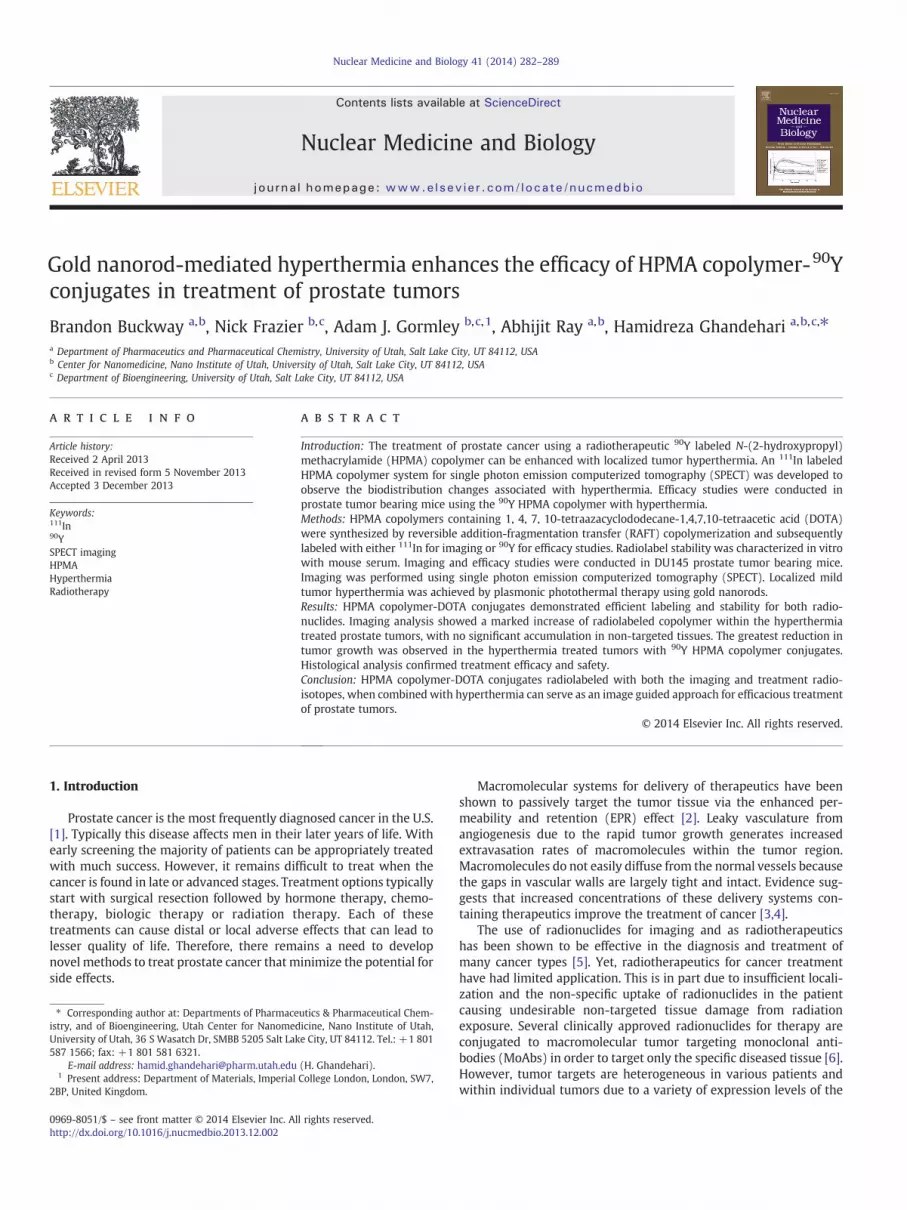
Gold nanorod-mediated hyperthermia enhances the efficacy of HPMA copolymer- 90 Y conjugates in treatment of prostate tumors Brandon Buckway a, b , Nick Frazier b, c , Adam J. Gormley b, c, 1 , Abhijit Ray a, b , Hamidreza Ghandehari a, b, c, ⁎ a Department of Pharmaceutics and Pharmaceutical Chemistry, University of Utah, Salt Lake City, UT 84112, USA b Center for Nanomedicine, Nano Institute of Utah, University of Utah, Salt Lake City, UT 84112, USA c Department of Bioengineering, University of Utah, Salt Lake City, UT 84112, USA abstract article info Article history: Received 2 April 2013 Received in revised form 5 November 2013 Accepted 3 December 2013 Keywords: 111 In 90 Y SPECT imaging HPMA Hyperthermia Radiotherapy Introduction: The treatment of prostate cancer using a radiotherapeutic 90 Y labeled N-(2-hydroxypropyl) methacrylamide (HPMA) copolymer can be enhanced with localized tumor hyperthermia. An 111 In labeled HPMA copolymer system for single photon emission computerized tomography (SPECT) was developed to observe the biodistribution changes associated with hyperthermia. Efficacy studies were conducted in prostate tumor bearing mice using the 90 Y HPMA copolymer with hyperthermia. Methods: HPMA copolymers containing 1, 4, 7, 10-tetraazacyclododecane-1,4,7,10-tetraacetic acid (DOTA) were synthesized by reversible addition-fragmentation transfer (RAFT) copolymerization and subsequently labeled with either 111 In for imaging or 90 Y for efficacy studies. Radiolabel stability was characterized in vitro with mouse serum. Imaging and efficacy studies were conducted in DU145 prostate tumor bearing mice. Imaging was performed using single photon emission computerized tomography (SPECT). Localized mild tumor hyperthermia was achieved by plasmonic photothermal therapy using gold nanorods. Results: HPMA copolymer-DOTA conjugates demonstrated efficient labeling and stability for both radio- nuclides. Imaging analysis showed a marked increase of radiolabeled copolymer within the hyperthermia treated prostate tumors, with no significant accumulation in non-targeted tissues. The greatest reduction in tumor growth was observed in the hyperthermia treated tumors with 90 Y HPMA copolymer conjugates. Histological analysis confirmed treatment efficacy and safety. Conclusion: HPMA copolymer-DOTA conjugates radiolabeled with both the imaging and treatment radio- isotopes, when combined with hyperthermia can serve as an image guided approach for efficacious treatment of prostate tumors. © 2014 Elsevier Inc. All rights reserved. 1. Introduction Prostate cancer is the most frequently diagnosed cancer in the U.S. [1]. Typically this disease affects men in their later years of life. With early screening the majority of patients can be appropriately treated with much success. However, it remains difficult to treat when the cancer is found in late or advanced stages. Treatment options typically start with surgical resection followed by hormone therapy, chemo- therapy, biologic therapy or radiation therapy. Each of these treatments can cause distal or local adverse effects that can lead to lesser quality of life. Therefore, there remains a need to develop novel methods to treat prostate cancer that minimize the potential for side effects. Macromolecular systems for delivery of therapeutics have been shown to passively target the tumor tissue via the enhanced per- meability and retention (EPR) effect [2]. Leaky vasculature from angiogenesis due to the rapid tumor growth generates increased extravasation rates of macromolecules within the tumor region. Macromolecules do not easily diffuse from the normal vessels because the gaps in vascular walls are largely tight and intact. Evidence sug- gests that increased concentrations of these delivery systems con- taining therapeutics improve the treatment of cancer [3,4]. The use of radionuclides for imaging and as radiotherapeutics has been shown to be effective in the diagnosis and treatment of many cancer types [5]. Yet, radiotherapeutics for cancer treatment have had limited application. This is in part due to insufficient locali- zation and the non-specific uptake of radionuclides in the patient causing undesirable non-targeted tissue damage from radiation exposure. Several clinically approved radionuclides for therapy are conjugated to macromolecular tumor targeting monoclonal anti- bodies (MoAbs) in order to target only the specific diseased tissue [6]. However, tumor targets are heterogeneous in various patients and within individual tumors due to a variety of expression levels of the Nuclear Medicine and Biology 41 (2014) 282–289 ⁎ Corresponding author at: Departments of Pharmaceutics & Pharmaceutical Chem- istry, and of Bioengineering, Utah Center for Nanomedicine, Nano Institute of Utah, University of Utah, 36 S Wasatch Dr, SMBB 5205 Salt Lake City, UT 84112. Tel.: +1 801 587 1566; fax: +1 801 581 6321. E-mail address: [email protected] (H. Ghandehari). 1 Present address: Department of Materials, Imperial College London, London, SW7, 2BP, United Kingdom. 0969-8051/$ – see front matter © 2014 Elsevier Inc. All rights reserved. http://dx.doi.org/10.1016/j.nucmedbio.2013.12.002 Contents lists available at ScienceDirect Nuclear Medicine and Biology journal homepage: www.elsevier.com/locate/nucmedbio
-
Upload
independent -
Category
Documents
-
view
4 -
download
0
Transcript of Gold nanorod-mediated hyperthermia enhances the efficacy of HPMA copolymer-90Y conjugates in...
Nuclear Medicine and Biology 41 (2014) 282–289
Contents lists available at ScienceDirect
Nuclear Medicine and Biology
j ourna l homepage: www.e lsev ie r .com/ locate /nucmedb io
Gold nanorod-mediated hyperthermia enhances the efficacy of HPMA copolymer-90Yconjugates in treatment of prostate tumors
Brandon Buckway a,b, Nick Frazier b,c, Adam J. Gormley b,c,1, Abhijit Ray a,b, Hamidreza Ghandehari a,b,c,⁎a Department of Pharmaceutics and Pharmaceutical Chemistry, University of Utah, Salt Lake City, UT 84112, USAb Center for Nanomedicine, Nano Institute of Utah, University of Utah, Salt Lake City, UT 84112, USAc Department of Bioengineering, University of Utah, Salt Lake City, UT 84112, USA
a b s t r a c ta r t i c l e i n f o
⁎ Corresponding author at: Departments of Pharmaceistry, and of Bioengineering, Utah Center for NanomedUniversity of Utah, 36 S Wasatch Dr, SMBB 5205 Salt Lak587 1566; fax: +1 801 581 6321.
E-mail address: [email protected] Present address: Department of Materials, Imperial
2BP, United Kingdom.
0969-8051/$ – see front matter © 2014 Elsevier Inc. Alhttp://dx.doi.org/10.1016/j.nucmedbio.2013.12.002
Article history:
Received 2 April 2013Received in revised form 5 November 2013Accepted 3 December 2013Keywords:111In90YSPECT imagingHPMAHyperthermiaRadiotherapy
Introduction: The treatment of prostate cancer using a radiotherapeutic 90Y labeled N-(2-hydroxypropyl)methacrylamide (HPMA) copolymer can be enhanced with localized tumor hyperthermia. An 111In labeledHPMA copolymer system for single photon emission computerized tomography (SPECT) was developed toobserve the biodistribution changes associated with hyperthermia. Efficacy studies were conducted inprostate tumor bearing mice using the 90Y HPMA copolymer with hyperthermia.Methods: HPMA copolymers containing 1, 4, 7, 10-tetraazacyclododecane-1,4,7,10-tetraacetic acid (DOTA)were synthesized by reversible addition-fragmentation transfer (RAFT) copolymerization and subsequentlylabeled with either 111In for imaging or 90Y for efficacy studies. Radiolabel stability was characterized in vitrowith mouse serum. Imaging and efficacy studies were conducted in DU145 prostate tumor bearing mice.Imaging was performed using single photon emission computerized tomography (SPECT). Localized mildtumor hyperthermia was achieved by plasmonic photothermal therapy using gold nanorods.
Results: HPMA copolymer-DOTA conjugates demonstrated efficient labeling and stability for both radio-nuclides. Imaging analysis showed a marked increase of radiolabeled copolymer within the hyperthermiatreated prostate tumors, with no significant accumulation in non-targeted tissues. The greatest reduction intumor growth was observed in the hyperthermia treated tumors with 90Y HPMA copolymer conjugates.Histological analysis confirmed treatment efficacy and safety.Conclusion: HPMA copolymer-DOTA conjugates radiolabeled with both the imaging and treatment radio-isotopes, when combined with hyperthermia can serve as an image guided approach for efficacious treatmentof prostate tumors.© 2014 Elsevier Inc. All rights reserved.
1. Introduction
Prostate cancer is the most frequently diagnosed cancer in the U.S.[1]. Typically this disease affects men in their later years of life. Withearly screening the majority of patients can be appropriately treatedwith much success. However, it remains difficult to treat when thecancer is found in late or advanced stages. Treatment options typicallystart with surgical resection followed by hormone therapy, chemo-therapy, biologic therapy or radiation therapy. Each of thesetreatments can cause distal or local adverse effects that can lead tolesser quality of life. Therefore, there remains a need to developnovel methods to treat prostate cancer that minimize the potential forside effects.
utics & Pharmaceutical Chem-icine, Nano Institute of Utah,e City, UT 84112. Tel.: +1 801
(H. Ghandehari).College London, London, SW7,
l rights reserved.
Macromolecular systems for delivery of therapeutics have beenshown to passively target the tumor tissue via the enhanced per-meability and retention (EPR) effect [2]. Leaky vasculature fromangiogenesis due to the rapid tumor growth generates increasedextravasation rates of macromolecules within the tumor region.Macromolecules do not easily diffuse from the normal vessels becausethe gaps in vascular walls are largely tight and intact. Evidence sug-gests that increased concentrations of these delivery systems con-taining therapeutics improve the treatment of cancer [3,4].
The use of radionuclides for imaging and as radiotherapeuticshas been shown to be effective in the diagnosis and treatment ofmany cancer types [5]. Yet, radiotherapeutics for cancer treatmenthave had limited application. This is in part due to insufficient locali-zation and the non-specific uptake of radionuclides in the patientcausing undesirable non-targeted tissue damage from radiationexposure. Several clinically approved radionuclides for therapy areconjugated to macromolecular tumor targeting monoclonal anti-bodies (MoAbs) in order to target only the specific diseased tissue [6].However, tumor targets are heterogeneous in various patients andwithin individual tumors due to a variety of expression levels of the
283B. Buckway et al. / Nuclear Medicine and Biology 41 (2014) 282–289
targeted antigen. One other shortfall of targeted delivery usingMoAbs is that the target receptor is rarely only expressed on thetargeted disease tissue which may lead to increased uptake in non-specific tissues thereby increasing the chance of treatment relatedtoxicity. There remains a need to target tumors using other macro-molecular systems.
Use of water-soluble polymers based on N-(2-hydroxypropyl)methacrylamide (HPMA) is one potential method to increase radio-therapeutic accumulation in the tumor [7–9]. HPMA copolymersare ideal macromolecular carriers for radionuclide delivery becauseof their ability to be synthesized in a size controlled manner andpresence of a variety of comonomers available to incorporate drugs,imaging agents or tumor targeting ligands [9–12]. Because of theirmacromolecular nature they are also able to passively target tumorsvia the enhanced permeability and retention (EPR) effect [2]. How-ever, the delivery of HPMA copolymers and other macromoleculesvia the EPR effect has been variable from patient to patient [13].Therefore, other methods must be considered to increase localizationwithin the tumor.
Previous studies have shown the advantage of localized hyper-thermia to increase HPMA copolymer conjugate localization andefficacy in treating prostate tumors [14–16]. Hyperthermia can beeasily controlled and localized using plasmonic photothermal ther-apy (PPTT) [17]. PPTT uses the surface plasmon resonance of goldnanorods (GNR) when activated by the appropriate wavelength oflight for controlled activation of heat [17]. Delivery of GNRs to thetumor is also based on passive accumulation and once localized totumors can be irradiated by laser to augment the localization ofsubsequently injected polymer therapeutics [15].
The central hypothesis of this work is that by using localizedhyperthermia with gold-nanorod-mediated plasmonic photothermaltherapy, it is possible to enhance the delivery of HPMA copolymer-yttrium 90 conjugates to prostate tumors and improve radiother-apeutic efficacy. The overall design of the copolymer system describedin this work includes side chain conjugated 1, 4, 7, 10-tetraaza-cyclododecane-1,4,7,10-tetraacetic acid (DOTA) for chelation ofeither 111In for imaging of the biodistribution of the HPMA copo-lymers after hyperthermia treatment, or 90Y for radiotherapeutictreatment of the tumor. 90Y is a pure beta emitting isotope which isnot an ideal imaging agent for γ-ray detection. Nuclear medicinetechniques such as single photon emission computerized tomography(SPECT) offer relatively high resolution and quantitative im-ages [18,19]. Therefore imaging of HPMA copolymer 111In usingSPECT should provide more detailed information as to the quantityand kinetics of tumor localization and enable correlation of suchlocalization with therapy. Correlation performed in this study
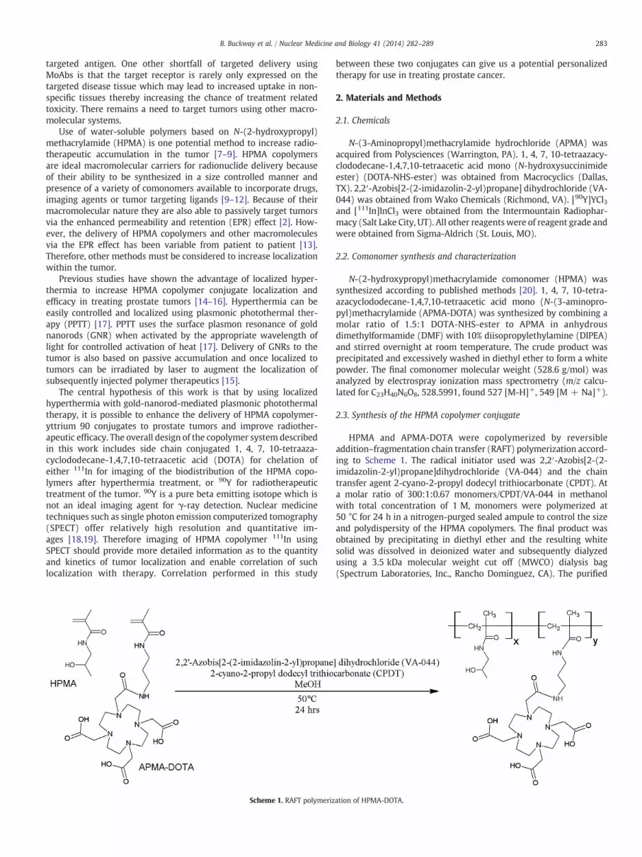
Scheme 1. RAFT polymeriz
between these two conjugates can give us a potential personalizedtherapy for use in treating prostate cancer.
2. Materials and Methods
2.1. Chemicals
N-(3-Aminopropyl)methacrylamide hydrochloride (APMA) wasacquired from Polysciences (Warrington, PA). 1, 4, 7, 10-tetraazacy-clododecane-1,4,7,10-tetraacetic acid mono (N-hydroxysuccinimideester) (DOTA-NHS-ester) was obtained from Macrocyclics (Dallas,TX). 2,2′-Azobis[2-(2-imidazolin-2-yl)propane] dihydrochloride (VA-044) was obtained from Wako Chemicals (Richmond, VA). [90Y]YCl3and [111In]InCl3 were obtained from the Intermountain Radiophar-macy (Salt Lake City, UT). All other reagentswere of reagent grade andwere obtained from Sigma-Aldrich (St. Louis, MO).
2.2. Comonomer synthesis and characterization
N-(2-hydroxypropyl)methacrylamide comonomer (HPMA) wassynthesized according to published methods [20]. 1, 4, 7, 10-tetra-azacyclododecane-1,4,7,10-tetraacetic acid mono (N-(3-aminopro-pyl)methacrylamide (APMA-DOTA) was synthesized by combining amolar ratio of 1.5:1 DOTA-NHS-ester to APMA in anhydrousdimethylformamide (DMF) with 10% diisopropylethylamine (DIPEA)and stirred overnight at room temperature. The crude product wasprecipitated and excessively washed in diethyl ether to form a whitepowder. The final comonomer molecular weight (528.6 g/mol) wasanalyzed by electrospray ionization mass spectrometry (m/z calcu-lated for C23H40N6O8, 528.5991, found 527 [M-H]+, 549 [M + Na]+).
2.3. Synthesis of the HPMA copolymer conjugate
HPMA and APMA-DOTA were copolymerized by reversibleaddition–fragmentation chain transfer (RAFT) polymerization accord-ing to Scheme 1. The radical initiator used was 2,2′-Azobis[2-(2-imidazolin-2-yl)propane]dihydrochloride (VA-044) and the chaintransfer agent 2-cyano-2-propyl dodecyl trithiocarbonate (CPDT). Ata molar ratio of 300:1:0.67 monomers/CPDT/VA-044 in methanolwith total concentration of 1 M, monomers were polymerized at50 °C for 24 h in a nitrogen-purged sealed ampule to control the sizeand polydispersity of the HPMA copolymers. The final product wasobtained by precipitating in diethyl ether and the resulting whitesolid was dissolved in deionized water and subsequently dialyzedusing a 3.5 kDa molecular weight cut off (MWCO) dialysis bag(Spectrum Laboratories, Inc., Rancho Dominguez, CA). The purified
ation of HPMA-DOTA.
284 B. Buckway et al. / Nuclear Medicine and Biology 41 (2014) 282–289
copolymer was obtained by lyophilization and analyzed by FastProtein Liquid Chromatography (FPLC) system (GE Healthcare,Piscataway, NJ) equipped with a multi-angle light scattering (MALS)detector (Wyatt Technologies, Santa Barbara, CA). DOTA content wasdetermined by analyzing gadolinium content (assuming a 1:1 ratio)after chelation according to previously described methods [21].
2.4. Radiolabeling with 111In and 90Y
HPMA copolymer-DOTA conjugate was labeled with radioisotopesaccording to previously published methods [22,23]. 10 mg of HPMAcopolymer-DOTA was dissolved in 250 μl of 1.0 M sodium acetatebuffer pH 5.0. 10 mCi of [111In]InCl3 or [90Y]YCl3 was also treated with0.25 ml of 1.0 M sodium acetate buffer pH 5.0. Radioactive com-pounds were added to the HPMA copolymer-DOTA solution andincubated at 50 °C for 1.0 h with mixing under nitrogen. The solutionwas allowed to cool to room temperature, and then treated with100 μl of 0.05 M ethylenediaminetetraacetic acid (EDTA) for about10 min in order to remove free or loosely bound 111In+3 or 90Y+3
ions. Radioactive polymers were purified by Sephadex G25 PD-10columns (GE Life Sciences, Piscataway, NJ). Radioactivity wasmeasured using a CAPTUS 3000 multichannel analyzer (CanberraIndustries, Inc., Meriden, CT). Radiostability was determined by incu-bating radiolabeled copolymers at 37 °C in the presence of mouseserum. Samples were collected at 24, 48 and 72 h and subjected toPD-10 column separation to determine the free radiolabel content.
2.5. Synthesis of PEGylated gold nanorods
Gold nanorods (GNRs) were synthesized using the seed-mediatedgrowth method with an aspect ratio that correlates to a surfaceplasmon resonance (SPR) peak between 800 and 810 nm [24]. Thelight absorption profile was measured by UV spectrometry. The GNRswere then centrifuged and washed three times with deionized waterto remove excess hexadecyltrimethylammonium bromide (CTAB).After washing, poly(ethylene glycol) (PEG) (methoxy-PEG-thiol 5 kD,Creative PEGWorks, Winston Salem, NC) was added to the GNR
Fig. 1. Methodology for combination radiotherapy and hyp
suspension and stirred for 1 h to allow for sufficient coating. The PEG-GNRs were then dialyzed (10 k MWCO, Spectrum labs), centrifuged,washed, and concentrated to remove any excess, unbound PEG. Thefinal concentration of the PEG-GNRs was 1.2 mg/ml (OD = 120) andwere stored at 4 °C. Finally, the PEG-GNR solution was sterile filteredprior to use in vivo.
2.6. Animal tumor model
DU-145 prostate tumor cells (ATCC, Manassas, VA) were culturedin Eagle’s Minimum Essential Medium (EMEM) (ATCC, Manassas, VA)supplemented with 10% (v/v) fetal bovine serum (FBS) at 37 ºC in ahumidified atmosphere of 5% CO2 (v/v). Cell cultures were harvestedat approximately 80% confluence by treatment with TrypLE™ Ex-press (Invitrogen, Grand Island, NY) and subsequent dilution inphosphate buffered saline (PBS). Athymic Nu/Nu female mice wereinoculated with 1 × 10⁷ cells on both the left and right lower flanks ofeach mouse. Experiments were initiated after tumor diameters hadreached 5–7 mm in diameter by external caliper measurement. Allanimal experiments were conducted under an approved protocolfrom the Institutional Animal Care and Use Committee at the Uni-versity of Utah (Salt Lake City, UT).
2.7. Biodistribution of 111In HPMA copolymer-DOTA conjugates
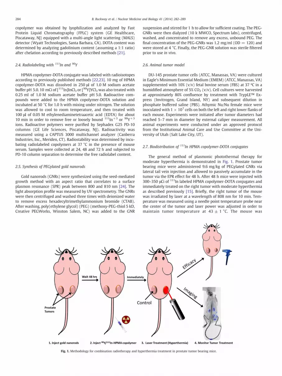
The general method of plasmonic photothermal therapy formoderate hyperthermia is demonstrated in Fig. 1. Prostate tumorbearing mice were administered 9.6 mg/kg of PEGylated GNRs vialateral tail vein injection and allowed to passively accumulate in thetumor via the EPR effect for 48 h. After 48 h mice were injected with300–350 μCi of 111In labeled HPMA copolymer-DOTA conjugates andimmediately treated on the right tumor with moderate hyperthermiaas described previously [15]. Briefly, the right tumor of the mousewas irradiated by laser at a wavelength of 808 nm for 10 min. Tem-perature was measured using a needle point temperature probe nearthe center of the tumor and laser power was adjusted in order tomaintain tumor temperature at 43 ± 1 °C. The mouse was
erthermia treatment in prostate tumor bearing mice.
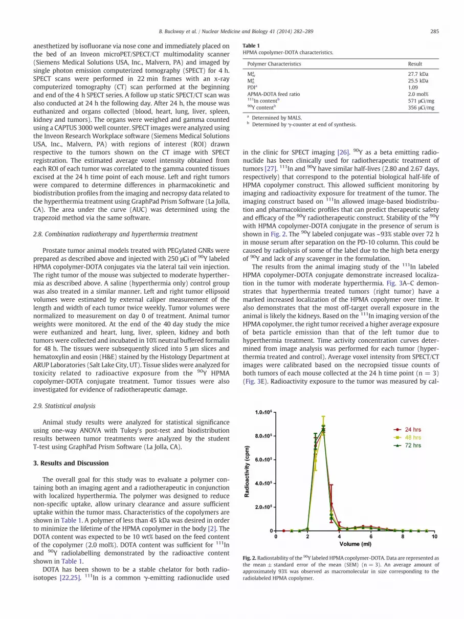
Table 1HPMA copolymer-DOTA characteristics.
Polymer Characteristics Result
Mwa 27.7 kDa
Mna 25.5 kDa
PDIa 1.09APMA-DOTA feed ratio 2.0 mol%111In contentb 571 μCi/mg90Y contentb 356 μCi/mg
a Determined by MALS.b Determined by γ-counter at end of synthesis.
Fig. 2. Radiostability of the 90Y labeled HPMA copolymer-DOTA. Data are represented asthe mean ± standard error of the mean (SEM) (n = 3). An average amount oapproximately 93% was observed as macromolecular in size corresponding to theradiolabeled HPMA copolymer.
285B. Buckway et al. / Nuclear Medicine and Biology 41 (2014) 282–289
anesthetized by isofluorane via nose cone and immediately placed onthe bed of an Inveon microPET/SPECT/CT multimodality scanner(Siemens Medical Solutions USA, Inc., Malvern, PA) and imaged bysingle photon emission computerized tomography (SPECT) for 4 h.SPECT scans were performed in 22 min frames with an x-raycomputerized tomography (CT) scan performed at the beginningand end of the 4 h SPECT series. A follow up static SPECT/CT scan wasalso conducted at 24 h the following day. After 24 h, the mouse waseuthanized and organs collected (blood, heart, lung, liver, spleen,kidney and tumors). The organs were weighed and gamma countedusing a CAPTUS 3000 well counter. SPECT images were analyzed usingthe Inveon Research Workplace software (Siemens Medical SolutionsUSA, Inc., Malvern, PA) with regions of interest (ROI) drawnrespective to the tumors shown on the CT image with SPECTregistration. The estimated average voxel intensity obtained fromeach ROI of each tumor was correlated to the gamma counted tissuesexcised at the 24 h time point of each mouse. Left and right tumorswere compared to determine differences in pharmacokinetic andbiodistribution profiles from the imaging and necropsy data related tothe hyperthermia treatment using GraphPad Prism Software (La Jolla,CA). The area under the curve (AUC) was determined using thetrapezoid method via the same software.
2.8. Combination radiotherapy and hyperthermia treatment
Prostate tumor animal models treated with PEGylated GNRs wereprepared as described above and injected with 250 μCi of 90Y labeledHPMA copolymer-DOTA conjugates via the lateral tail vein injection.The right tumor of the mouse was subjected to moderate hyperther-mia as described above. A saline (hyperthermia only) control groupwas also treated in a similar manner. Left and right tumor ellipsoidvolumes were estimated by external caliper measurement of thelength and width of each tumor twice weekly. Tumor volumes werenormalized to measurement on day 0 of treatment. Animal tumorweights were monitored. At the end of the 40 day study the micewere euthanized and heart, lung, liver, spleen, kidney and bothtumors were collected and incubated in 10% neutral buffered formalinfor 48 h. The tissues were subsequently sliced into 5 μm slices andhematoxylin and eosin (H&E) stained by the Histology Department atARUP Laboratories (Salt Lake City, UT). Tissue slides were analyzed fortoxicity related to radioactive exposure from the 90Y HPMAcopolymer-DOTA conjugate treatment. Tumor tissues were alsoinvestigated for evidence of radiotherapeutic damage.
2.9. Statistical analysis
Animal study results were analyzed for statistical significanceusing one-way ANOVA with Tukey’s post-test and biodistributionresults between tumor treatments were analyzed by the studentT-test using GraphPad Prism Software (La Jolla, CA).
3. Results and Discussion
The overall goal for this study was to evaluate a polymer con-taining both an imaging agent and a radiotherapeutic in conjunctionwith localized hyperthermia. The polymer was designed to reducenon-specific uptake, allow urinary clearance and assure sufficientuptake within the tumor mass. Characteristics of the copolymers areshown in Table 1. A polymer of less than 45 kDa was desired in orderto minimize the lifetime of the HPMA copolymer in the body [2]. TheDOTA content was expected to be 10 wt% based on the feed contentof the copolymer (2.0 mol%). DOTA content was sufficient for 111Inand 90Y radiolabelling demonstrated by the radioactive contentshown in Table 1.
DOTA has been shown to be a stable chelator for both radio-isotopes [22,25]. 111In is a common γ-emitting radionuclide used
in the clinic for SPECT imaging [26]. 90Y as a beta emitting radio-nuclide has been clinically used for radiotherapeutic treatment oftumors [27]. 111In and 90Y have similar half-lives (2.80 and 2.67 days,respectively) that correspond to the potential biological half-life ofHPMA copolymer construct. This allowed sufficient monitoring byimaging and radioactivity exposure for treatment of the tumor. Theimaging construct based on 111In allowed image-based biodistribu-tion and pharmacokinetic profiles that can predict therapeutic safetyand efficacy of the 90Y radiotherapeutic construct. Stability of the 90Ywith HPMA copolymer-DOTA conjugate in the presence of serum isshown in Fig. 2. The 90Y labeled conjugate was ~93% stable over 72 hin mouse serum after separation on the PD-10 column. This could becaused by radiolysis of some of the label due to the high beta energyof 90Y and lack of any scavenger in the formulation.
The results from the animal imaging study of the 111In labeledHPMA copolymer-DOTA conjugate demonstrate increased localiza-tion in the tumor with moderate hyperthermia. Fig. 3A–C demon-strates that hyperthermia treated tumors (right tumor) have amarked increased localization of the HPMA copolymer over time. Italso demonstrates that the most off-target overall exposure in theanimal is likely the kidneys. Based on the 111In imaging version of theHPMA copolymer, the right tumor received a higher average exposureof beta particle emission than that of the left tumor due tohyperthermia treatment. Time activity concentration curves deter-mined from image analysis was performed for each tumor (hyper-thermia treated and control). Average voxel intensity from SPECT/CTimages were calibrated based on the necropsied tissue counts ofboth tumors of each mouse collected at the 24 h time point (n = 3)(Fig. 3E). Radioactivity exposure to the tumor was measured by cal-
f
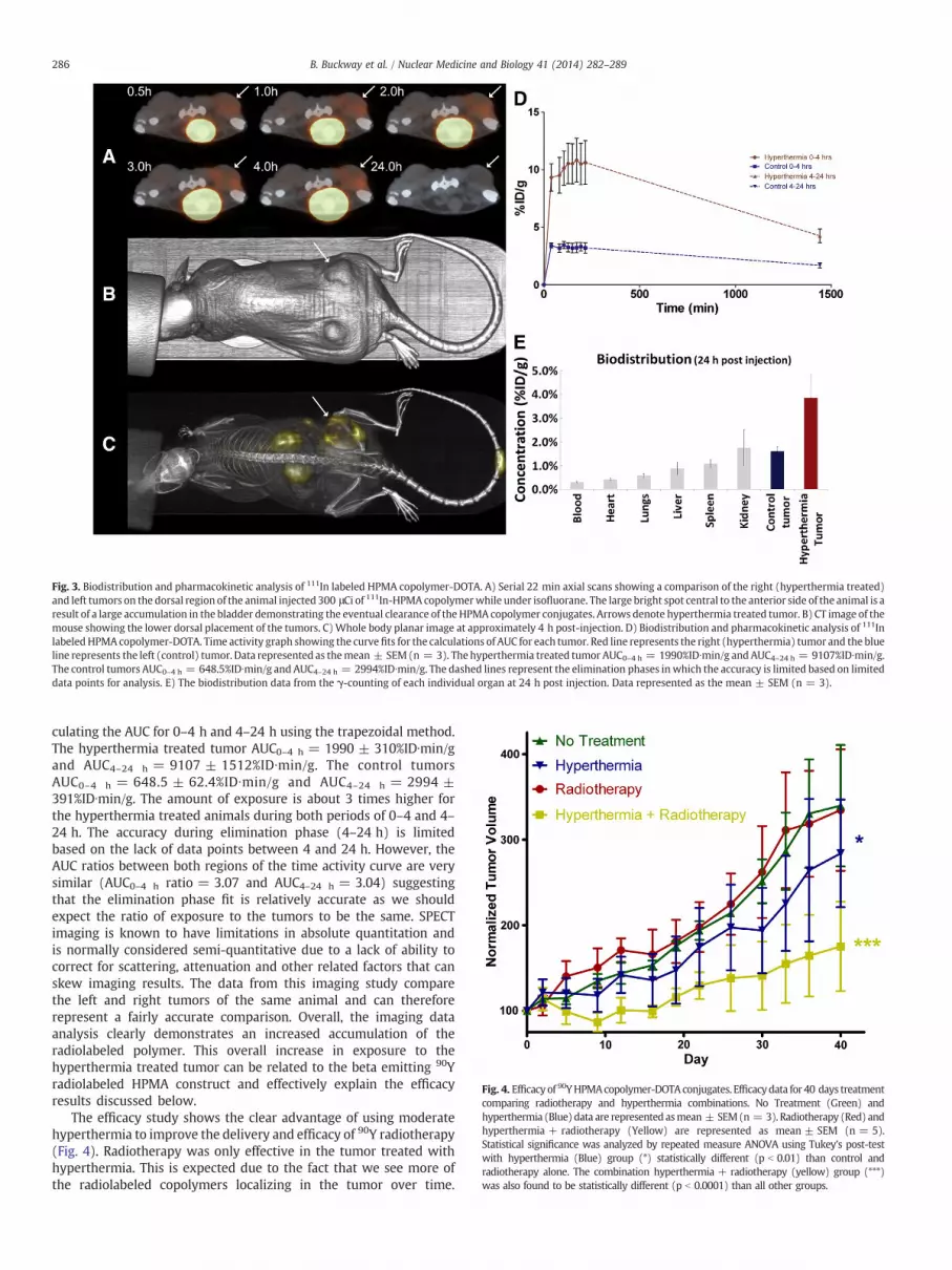
Fig. 3. Biodistribution and pharmacokinetic analysis of 111In labeled HPMA copolymer-DOTA. A) Serial 22 min axial scans showing a comparison of the right (hyperthermia treated)and left tumors on the dorsal region of the animal injected 300 μCi of 111In-HPMA copolymerwhile under isofluorane. The large bright spot central to the anterior side of the animal is aresult of a large accumulation in the bladder demonstrating the eventual clearance of theHPMA copolymer conjugates. Arrows denote hyperthermia treated tumor. B) CT image of themouse showing the lower dorsal placement of the tumors. C)Whole body planar image at approximately 4 h post-injection. D) Biodistribution and pharmacokinetic analysis of 111InlabeledHPMAcopolymer-DOTA. Time activity graph showing the curvefits for the calculations of AUC for each tumor. Red line represents the right (hyperthermia) tumor and the blueline represents the left (control) tumor. Data represented as themean ± SEM(n = 3). The hyperthermia treated tumor AUC0–4 h = 1990%ID∙min/g andAUC4–24 h = 9107%ID∙min/g.The control tumors AUC0–4 h = 648.5%ID∙min/g and AUC4–24 h = 2994%ID∙min/g. The dashed lines represent the elimination phases inwhich the accuracy is limited based on limiteddata points for analysis. E) The biodistribution data from the γ-counting of each individual organ at 24 h post injection. Data represented as the mean ± SEM (n = 3).
Fig. 4. Efficacy of 90YHPMAcopolymer-DOTAconjugates. Efficacydata for 40 days treatmentcomparing radiotherapy and hyperthermia combinations. No Treatment (Green) andhyperthermia (Blue) data are represented asmean ± SEM(n = 3). Radiotherapy (Red) andhyperthermia + radiotherapy (Yellow) are represented as mean ± SEM (n = 5).Statistical significance was analyzed by repeated measure ANOVA using Tukey’s post-testwith hyperthermia (Blue) group (*) statistically different (p b 0.01) than control andradiotherapy alone. The combination hyperthermia + radiotherapy (yellow) group (***)was also found to be statistically different (p b 0.0001) than all other groups.
286 B. Buckway et al. / Nuclear Medicine and Biology 41 (2014) 282–289
culating the AUC for 0–4 h and 4–24 h using the trapezoidal method.The hyperthermia treated tumor AUC0–4 h = 1990 ± 310%ID∙min/gand AUC4–24 h = 9107 ± 1512%ID∙min/g. The control tumorsAUC0–4 h = 648.5 ± 62.4%ID∙min/g and AUC4–24 h = 2994 ±391%ID∙min/g. The amount of exposure is about 3 times higher forthe hyperthermia treated animals during both periods of 0–4 and 4–24 h. The accuracy during elimination phase (4–24 h) is limitedbased on the lack of data points between 4 and 24 h. However, theAUC ratios between both regions of the time activity curve are verysimilar (AUC0–4 h ratio = 3.07 and AUC4–24 h = 3.04) suggestingthat the elimination phase fit is relatively accurate as we shouldexpect the ratio of exposure to the tumors to be the same. SPECTimaging is known to have limitations in absolute quantitation andis normally considered semi-quantitative due to a lack of ability tocorrect for scattering, attenuation and other related factors that canskew imaging results. The data from this imaging study comparethe left and right tumors of the same animal and can thereforerepresent a fairly accurate comparison. Overall, the imaging dataanalysis clearly demonstrates an increased accumulation of theradiolabeled polymer. This overall increase in exposure to thehyperthermia treated tumor can be related to the beta emitting 90Yradiolabeled HPMA construct and effectively explain the efficacyresults discussed below.
The efficacy study shows the clear advantage of using moderatehyperthermia to improve the delivery and efficacy of 90Y radiotherapy(Fig. 4). Radiotherapy was only effective in the tumor treated withhyperthermia. This is expected due to the fact that we see more ofthe radiolabeled copolymers localizing in the tumor over time.
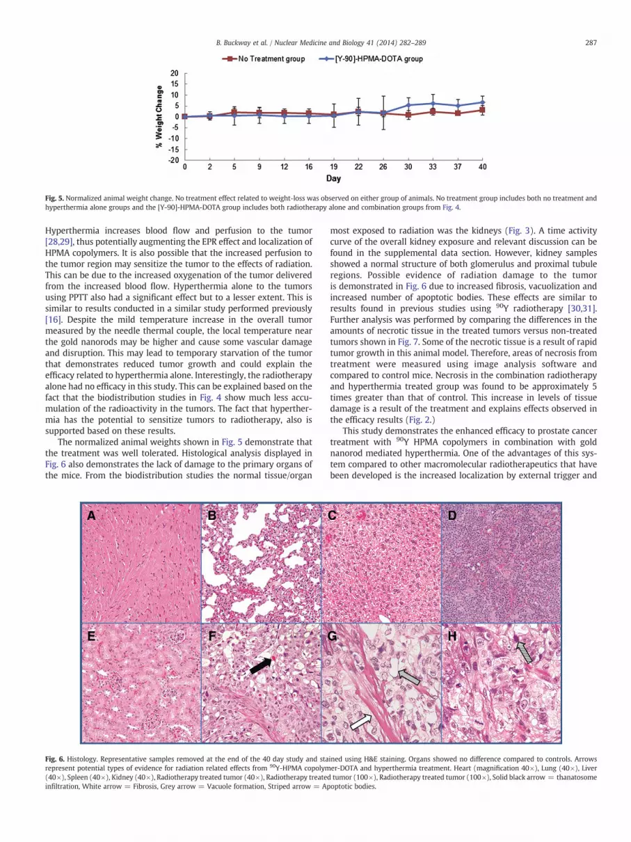
Fig. 5. Normalized animal weight change. No treatment effect related to weight-loss was observed on either group of animals. No treatment group includes both no treatment andhyperthermia alone groups and the [Y-90]-HPMA-DOTA group includes both radiotherapy alone and combination groups from Fig. 4.
287B. Buckway et al. / Nuclear Medicine and Biology 41 (2014) 282–289
Hyperthermia increases blood flow and perfusion to the tumor[28,29], thus potentially augmenting the EPR effect and localization ofHPMA copolymers. It is also possible that the increased perfusion tothe tumor region may sensitize the tumor to the effects of radiation.This can be due to the increased oxygenation of the tumor deliveredfrom the increased blood flow. Hyperthermia alone to the tumorsusing PPTT also had a significant effect but to a lesser extent. This issimilar to results conducted in a similar study performed previously[16]. Despite the mild temperature increase in the overall tumormeasured by the needle thermal couple, the local temperature nearthe gold nanorods may be higher and cause some vascular damageand disruption. This may lead to temporary starvation of the tumorthat demonstrates reduced tumor growth and could explain theefficacy related to hyperthermia alone. Interestingly, the radiotherapyalone had no efficacy in this study. This can be explained based on thefact that the biodistribution studies in Fig. 4 show much less accu-mulation of the radioactivity in the tumors. The fact that hyperther-mia has the potential to sensitize tumors to radiotherapy, also issupported based on these results.
The normalized animal weights shown in Fig. 5 demonstrate thatthe treatment was well tolerated. Histological analysis displayed inFig. 6 also demonstrates the lack of damage to the primary organs ofthe mice. From the biodistribution studies the normal tissue/organ
Fig. 6. Histology. Representative samples removed at the end of the 40 day study and starepresent potential types of evidence for radiation related effects from 90Y-HPMA copolym(40×), Spleen (40×), Kidney (40×), Radiotherapy treated tumor (40×), Radiotherapy treatedinfiltration, White arrow = Fibrosis, Grey arrow = Vacuole formation, Striped arrow = Ap
most exposed to radiation was the kidneys (Fig. 3). A time activitycurve of the overall kidney exposure and relevant discussion can befound in the supplemental data section. However, kidney samplesshowed a normal structure of both glomerulus and proximal tubuleregions. Possible evidence of radiation damage to the tumoris demonstrated in Fig. 6 due to increased fibrosis, vacuolization andincreased number of apoptotic bodies. These effects are similar toresults found in previous studies using 90Y radiotherapy [30,31].Further analysis was performed by comparing the differences in theamounts of necrotic tissue in the treated tumors versus non-treatedtumors shown in Fig. 7. Some of the necrotic tissue is a result of rapidtumor growth in this animal model. Therefore, areas of necrosis fromtreatment were measured using image analysis software andcompared to control mice. Necrosis in the combination radiotherapyand hyperthermia treated group was found to be approximately 5times greater than that of control. This increase in levels of tissuedamage is a result of the treatment and explains effects observed inthe efficacy results (Fig. 2.)
This study demonstrates the enhanced efficacy to prostate cancertreatment with 90Y HPMA copolymers in combination with goldnanorod mediated hyperthermia. One of the advantages of this sys-tem compared to other macromolecular radiotherapeutics that havebeen developed is the increased localization by external trigger and
ined using H&E staining. Organs showed no difference compared to controls. Arrowser-DOTA and hyperthermia treatment. Heart (magnification 40×), Lung (40×), Livertumor (100×), Radiotherapy treated tumor (100×), Solid black arrow = thanatosomeoptotic bodies.
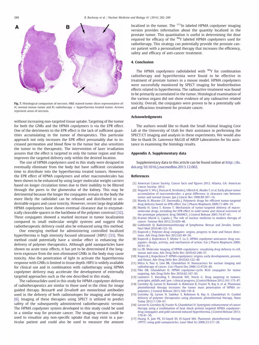
Fig. 7. Histological comparison of necrosis. H&E stained tumor slices representative of:A) normal mouse tumor and B) radiotherapy + hyperthermia treated tumor. Arrowsrepresent areas of necrosis.
288 B. Buckway et al. / Nuclear Medicine and Biology 41 (2014) 282–289
without increasing non-targeted tissue uptake. Targeting of the tumorfor both the GNRs and the HPMA copolymers is via the EPR effect.One of the detriments to the EPR effect is the lack of sufficient quan-tities accumulating in the tumor of therapeutics. This particularapproach not only increases the EPR effect presumably due to in-creased permeation and blood flow to the tumor but also sensitizesthe tumor to the therapeutic. The intervention of laser irradiationassures that the effect is targeted to only the tumor region and thusimproves the targeted delivery only within the desired location.
The size of HPMA copolymers used in this study were designed toeventually eliminate from the body but have sufficient circulationtime to distribute into the hyperthermia treated tumors. However,the EPR effect of HPMA copolymers and other macromolecules hasbeen shown to be enhanced by using larger molecular weight carriersbased on longer circulation times due to their inability to be filteredthrough the pores in the glomerulus of the kidney. This may bedetrimental because the longer the conjugates remain in the body themore likely the radiolabel can be released and distributed to un-desirable organs and cause toxicity. However, recent large degradableHPMA copolymers have shown eventual clearance due to enzymat-ically cleavable spacers in the backbone of the polymer construct [32].These conjugates showed a marked increase in tumor localizationcompared to small molecular weight systems and suggest thatradiotherapeutic delivery could also be enhanced using this method.
One emerging method for administering controlled localizedhyperthermia is high intensity focused ultrasound (HIFU) [33]. Thismethod could potentially have a similar effect in enhancing thedelivery of polymer therapeutics. Although gold nanoparticles haveshown no acute toxic effects, it has yet to be determined if the long-term exposure from the non-eliminated GNRs in the body may causetoxicity. Also the penetration of light to activate the hyperthermiaresponse with GNRs is limited in tissue depth. HIFU is widely availablefor clinical use and in combination with radiotherapy using HPMAcopolymer delivery may accelerate the development of externallytargeted approaches such as the one described in this study.
The radionuclides used in this study for HPMA copolymer deliveryof radiotherapeutics are similar to those used in the clinic for imageguided therapy. Bexxar® and Zevalin® are monoclonal antibodiesused in the delivery of both radioisotopes for imaging and therapy[6]. Imaging of these therapies using SPECT is utilized to predictsafety of the subsequently administered radiotherapeutic version.The HPMA copolymer system developed in this study could be usedin a similar way for prostate cancer. The imaging version could beused to visualize any non-specific uptake that may exist in a par-ticular patient and could also be used to measure the amount
localized in the tumor. The 111In labeled HPMA copolymer imagingversion provides information about the quantity localized in theprostate tumor. This quantitation is useful in determining the doserequired for efficacy of the 90Y labeled HPMA copolymers used forradiotherapy. This strategy can potentially provide the prostate can-cer patient with a personalized therapy that increases the efficiency,safety and efficacy of anti-cancer treatment.
4. Conclusion
The HPMA copolymers radiolabeled with 90Y for combinationradiotherapy and hyperthermia were found to be effective intreatment of prostate tumors in a mouse model. HPMA copolymerswere successfully monitored by SPECT imaging for biodistributioneffects related to hyperthermia. The radioactive treatment was foundto be primarily accumulated in the tumor. Histological examination ofthe various organs did not show evidence of any radioactive relatedtoxicity. Overall, the conjugates were proven to be a potentially safeand efficacious treatment for prostate cancer.
Acknowledgments
The authors would like to thank the Small Animal Imaging CoreLab at the University of Utah for their assistance in performing theSPECT/CT imaging and analysis in these experiments. We would alsolike to thank Dr. Lawrence McGill of ARUP Laboratories for his assis-tance in examining the histology results.
Appendix A. Supplementary data
Supplementary data to this article can be found online at http://dx.doi.org/10.1016/j.nucmedbio.2013.12.002.
References
[1] American Cancer Society. Cancer facts and figures 2012. Atlanta, GA: AmericanCancer Society; 2012.
[2] Noguchi Y, Wu J, Duncan R, Strohalm J, Ulbrich K, Akaike T, et al. Early phase tumoraccumulation of macromolecules: a great difference in clearance rate betweentumor and normal tissues. Jpn J Cancer Res 1998;89:307–14.
[3] Maeda H, Bharate GY, Daruwalla J. Polymeric drugs for efficient tumor-targeteddrug delivery based on EPR-effect. Eur J Pharm Biopharm 2009;71:409–19.
[4] Maeda H, Sawa T, Konno T. Mechanism of tumor-targeted delivery of macro-molecular drugs, including the EPR effect in solid tumor and clinical overview ofthe prototype polymeric drug SMANCS. J Control Release 2001;74:47–61.
[5] Kramer-Marek G, Capala J. The role of nuclear medicine in modern therapy ofcancer. Tumour Biol 2012;33:629–40.
[6] Goldsmith SJ. Radioimmunotherapy of lymphoma: Bexxar and Zevalin. SeminNucl Med 2010;40:122–35.
[7] Kopecek J. Polymer-drug conjugates: origins, progress to date and future direc-tions. Adv Drug Deliv Rev 2013;65:49–59.
[8] Kopecek J, Kopeckova P, Minko T, Lu Z. HPMA copolymer-anticancer drug con-jugates: design, activity, and mechanism of action. Eur J Pharm Biopharm 2000;50:61–81.
[9] Lu ZR. Molecular imaging of HPMA copolymers: visualizing drug delivery in cell,mouse and man. Adv Drug Deliv Rev 2010;62:246–57.
[10] Kopecek J, Kopeckova P. HPMA copolymers: origins, early developments, present,and future. Adv Drug Deliv Rev 2010;62:122–49.
[11] Mitra A, Nan A, Line BR, Ghandehari H. Nanocarriers for nuclear imaging andradiotherapy of cancer. Curr Pharm Des 2006;12:4729–49.
[12] Pike DB, Ghandehari H. HPMA copolymer-cyclic RGD conjugates for tumortargeting. Adv Drug Deliv Rev 2010;62:167–83.
[13] Lammers T, Kiessling F, Hennink WE, Storm G. Drug targeting to tumors:principles, pitfalls and (pre-) clinical progress. J Control Release 2012;161:175–87.
[14] Gormley AJ, Larson N, Banisadr A, Robinson R, Frazier N, Ray A, et al. Plasmonicphotothermal therapy increases the tumor mass penetration of HPMA co-polymers. J Control Release 2013;166:130–8.
[15] Gormley AJ, Larson N, Sadekar S, Robinson R, Ray A, Ghandehari H. Guideddelivery of polymer therapeutics using plasmonic photothermal therapy. NanoToday 2012;7:158–67.
[16] Larson N, Gormley AJ, Frazier N, Ghandehari H. Synergistic enhancement of cancertherapy using a combination of heat shock protein targeted HPMA copolymer-drug conjugates and gold nanorod induced hyperthermia. J Control Release 2013;170:41–50.
[17] Huang X, Jain PK, El-Sayed IH, El-Sayed MA. Plasmonic photothermal therapy(PPTT) using gold nanoparticles. Laser Med Sci 2008;23:217–28.
289B. Buckway et al. / Nuclear Medicine and Biology 41 (2014) 282–289
[18] Alberini JL, Edeline V, Giraudet AL, Champion L, Paulmier B, Madar O, et al. Singlephoton emission tomography/computed tomography (SPET/CT) and positronemission tomography/computed tomography (PET/CT) to image cancer. J SurgOncol 2011;103:602–6.
[19] Weissleder R. Molecular imaging: principles and practice. People's Medical Pub.House-USA: Shelton, CT; 2010.
[20] Strohalm J, Kopecek J. Poly N-(2-hydroxypropyl) methacrylamide: 4. Heteroge-nous polymerization. Angew Makromol Chem 1978;70:109–18.
[21] Zarabi B, BorgmanMP, Zhuo J, Gullapalli R, Ghandehari H. Noninvasivemonitoringof HPMA copolymer-RGDfK conjugates by magnetic resonance imaging. PharmRes 2009;26:1121–9.
[22] KukisDL,DeNardoSJ,DeNardoGL,O'Donnell RT,MearesCF. Optimizedconditions forchelation of yttrium-90-DOTA immunoconjugates. J Nucl Med 1998;39:2105–10.
[23] MitraA,NanA, Papadimitriou JC, GhandehariH, LineBR. Polymer-peptide conjugatesfor angiogenesis targeted tumor radiotherapy. Nucl Med Biol 2006;33:43–52.
[24] Nikoobakht B, Burda C, Braun M, Hun M, El-Sayed MA. The quenching of CdSequantum dots photoluminescence by gold nanoparticles in solution. PhotochemPhotobiol 2002;75:591–7.
[25] Liu S, Pietryka J, Ellars CE, Edwards DS. Comparison of yttrium and indiumcomplexes of DOTA-BA and DOTA-MBA: models for (90)Y- and (111)In-labeledDOTA-biomolecule conjugates. Bioconjug Chem 2002;13:902–13.
[26] Biersack HJ. Clinical nuclear medicine. Berlin; New York: Springer; 2007.
[27] Goffredo V, Paradiso A, Ranieri G, Gadaleta CD. Yttrium-90 (90Y) in the principalradionuclide therapies: an efficacy correlation between peptide receptorradionuclide therapy, radioimmunotherapy and transarterial radioembolizationtherapy. Ten years of experience (1999–2009). Crit Rev Oncol Hematol 2011;80:393–410.
[28] Griffin RJ, Dings RP, Jamshidi-Parsian A, Song CW. Mild temperature hyperthermiaand radiation therapy: role of tumour vascular thermotolerance and relevantphysiological factors. Int J Hyperthermia 2010;26:256–63.
[29] Song CW, Park HJ, Lee CK, Griffin R. Implications of increased tumor blood flowand oxygenation caused by mild temperature hyperthermia in tumor treatment.Int J Hyperthermia 2005;21:761–7.
[30] Huang J, Chunta JL, Amin M, Lee DY, Grills IS, Wong CY, et al. Detailedcharacterization of the early response of head-neck cancer xenografts toirradiation using (18)F-FDG-PET imaging. Int J Radiat Oncol Biol Phys 2012;84:485–91.
[31] Stone HB, Coleman CN, Anscher MS, McBride WH. Effects of radiation on normaltissue: consequences and mechanisms. Lancet Oncol 2003;4:529–36.
[32] Zhang R, Luo K, Yang J, Sima M, Sun Y, Janat-Amsbury MM, et al. Synthesis andevaluation of a backbone biodegradable multiblock HPMA copolymer nanocarrierfor the systemic delivery of paclitaxel. J Control Release 2013;166:66–74.
[33] Sanches PG, Grull H, Steinbach OC. See, reach, treat: ultrasound-triggered image-guided drug delivery. Ther Deliv 2011;2:919–34.