Comparative evaluation of heating ability and biocompatibility of different ferrite‐based magnetic...
11
Comparative Evaluation of Heating Ability and Biocompatibility of Different Ferrite-Based Magnetic Fluids for Hyperthermia Application Pallab Pradhan, 1 Jyotsnendu Giri, 2 Gopal Samanta, 3 Haladhar Dev Sarma, 4 Kaushala Prasad Mishra, 4 Jayesh Bellare, 1 Rinti Banerjee, 1 Dhirendra Bahadur 2 1 School of Biosciences and Bioengineering, Indian Institute of Technology, Mumbai 400076, India 2 Department of Metallurgical Engineering and Materials Science, Indian Institute of Technology, Mumbai 400076, India 3 Department of Veterinary Surgery and Radiology, West Bengal University of Animal and Fishery Sciences, Kolkata 700037, India 4 Radiation Biology and Health Science Division, Bhabha Atomic Research Centre, Mumbai 400085, India Received 12 November 2005; revised 31 March 2006; accepted 10 April 2006 Published online 21 August 2006 in Wiley InterScience (www.interscience.wiley.com). DOI: 10.1002/jbm.b.30630 Abstract: In this study, lauric acid-coated, superparamagnetic, nanoparticle-based magnetic fluids of different ferrites (Fe 3 O 4 , MnFe 2 O 4 , and CoFe 2 O 4 ) were prepared and compared in terms of heating ability and biocompatibility to evaluate the feasibility of use in hyperthermia treatment of cancer. All the magnetic fluids prepared had particles of average sizes 9–11 nm. Heating ability of these magnetic fluids was evaluated by calorimetric measurement of specific absorption rate (SAR) at 300 kHz frequency and 15 kA/m field. Fe 3 O 4 and MnFe 2 O 4 showed higher SAR (120 and 97 W/g of ferrite, respectively) than CoFe 2 O 4 (37 W/g of ferrite) . In vitro study on BHK 21 cell lines showed dose-dependent cell viability for all the magnetic fluids. Threshold-biocompatible ferrite concentration for all the magnetic fluids was 0.1 mg/mL. Above 0.2 mg/mL, CoFe 2 O 4 was more toxic than the other magnetic fluids. On intravenous injection of different doses (50, 200, and 400 mg/kg body weight) of magnetic fluids in mice, no significant changes in hematological and biochemical parameters were observed for Fe 3 O 4 and MnFe 2 O 4 . With CoFe 2 O 4 , an increase in SGPT levels at a dose rate of 400 mg/kg body weight was observed, indicating its mild hepatotoxic effect. However, histology of different vital organs showed no pathological changes for all the three magnetic fluids. Further, long term in vivo evaluation of biocompatibility of the lauric acid-coated ferrites is warranted. This study shows that lauric acid-coated, superparamagnetic Fe 3 O 4 and MnFe 2 O 4 may be used for hyperthermia treatment and are to be preferred over CoFe 2 O 4. ' 2006 Wiley Periodicals, Inc. J Biomed Mater Res Part B: Appl Biomater 81B: 12–22, 2007 Keywords: magnetic nanoparticle; magnetic fluid; biocompatibility; specific absorption rate; hyperthermia INTRODUCTION In recent years, magnetic particles have generated a lot of in- terest in the biomedical applications, like MRI contrast enhancement, magnetic separation, drug delivery, and hyper- thermia. 1–3 One of the prospective applications of magnetic nanoparticles is hyperthermia treatment of cancer. 4,5 In clini- cal hyperthermia, efforts are made to optimize the thermal ho- mogeneity at 42–468C in the tumor tissue by advanced therapy and thermometry systems. Magnetic materials with Curie temperature (T c ) between 42 and 608C are the best can- didates for effective treatment, since they act as in vivo tem- perature control switches, preventing over-heating. 6 For hyperthermia, superparamagnetic as well as ferrimagnetic par- ticles, such as Fe 3 O 4 or c-Fe 2 O 3 , have been explored exten- sively. 4,7,8 The systems M 1x Zn x Fe 2 O 4 (M ¼ Mn, Co), which have versatile magnetic properties and Curie temperature (T c ) obtained by substitution of nonmagnetic cations, have not yet been explored. 9,10 The heating ability and biocompatibility of these magnetic fluids need to be investigated. Heating ability of magnetic fluid which is measured by specific absorption rate (SAR) depends on physical (particle size, shape, and dis- tribution) as well as magnetic properties of particles. 11 There Correspondence to: D. Bahadur (e-mail: [email protected]) Contract grant sponsor: Department of Biotechnology (DBT) and Department of Science and Technology (DST), India ' 2006 Wiley Periodicals, Inc. 12
-
Upload
independent -
Category
Documents
-
view
3 -
download
0
Transcript of Comparative evaluation of heating ability and biocompatibility of different ferrite‐based magnetic...

Comparative Evaluation of Heating Ability and Biocompatibilityof Different Ferrite-Based Magnetic Fluids for HyperthermiaApplication
Pallab Pradhan,1 Jyotsnendu Giri,2 Gopal Samanta,3 Haladhar Dev Sarma,4 Kaushala Prasad Mishra,4
Jayesh Bellare,1 Rinti Banerjee,1 Dhirendra Bahadur2
1 School of Biosciences and Bioengineering, Indian Institute of Technology, Mumbai 400076, India
2 Department of Metallurgical Engineering and Materials Science, Indian Institute of Technology, Mumbai 400076, India
3 Department of Veterinary Surgery and Radiology, West Bengal University of Animal and Fishery Sciences,Kolkata 700037, India
4 Radiation Biology and Health Science Division, Bhabha Atomic Research Centre, Mumbai 400085, India
Received 12 November 2005; revised 31 March 2006; accepted 10 April 2006Published online 21 August 2006 in Wiley InterScience (www.interscience.wiley.com). DOI: 10.1002/jbm.b.30630
Abstract: In this study, lauric acid-coated, superparamagnetic, nanoparticle-based magnetic
fluids of different ferrites (Fe3O4, MnFe2O4, and CoFe2O4) were prepared and compared in
terms of heating ability and biocompatibility to evaluate the feasibility of use in hyperthermia
treatment of cancer. All the magnetic fluids prepared had particles of average sizes 9–11 nm.
Heating ability of these magnetic fluids was evaluated by calorimetric measurement of specific
absorption rate (SAR) at 300 kHz frequency and 15 kA/m field. Fe3O4 and MnFe2O4 showed
higher SAR (120 and 97 W/g of ferrite, respectively) than CoFe2O4 (37 W/g of ferrite). In vitrostudy on BHK 21 cell lines showed dose-dependent cell viability for all the magnetic fluids.
Threshold-biocompatible ferrite concentration for all the magnetic fluids was 0.1 mg/mL.
Above 0.2 mg/mL, CoFe2O4 was more toxic than the other magnetic fluids. On intravenous
injection of different doses (50, 200, and 400 mg/kg body weight) of magnetic fluids in mice, no
significant changes in hematological and biochemical parameters were observed for Fe3O4 and
MnFe2O4. With CoFe2O4, an increase in SGPT levels at a dose rate of 400 mg/kg body weight
was observed, indicating its mild hepatotoxic effect. However, histology of different vital
organs showed no pathological changes for all the three magnetic fluids. Further, long term invivo evaluation of biocompatibility of the lauric acid-coated ferrites is warranted. This study
shows that lauric acid-coated, superparamagnetic Fe3O4 and MnFe2O4 may be used for
hyperthermia treatment and are to be preferred over CoFe2O4. ' 2006 Wiley Periodicals, Inc.
J Biomed Mater Res Part B: Appl Biomater 81B: 12–22, 2007
Keywords: magnetic nanoparticle; magnetic fluid; biocompatibility; specific absorption rate;
hyperthermia
INTRODUCTION
In recent years, magnetic particles have generated a lot of in-
terest in the biomedical applications, like MRI contrast
enhancement, magnetic separation, drug delivery, and hyper-
thermia.1–3 One of the prospective applications of magnetic
nanoparticles is hyperthermia treatment of cancer.4,5 In clini-
cal hyperthermia, efforts are made to optimize the thermal ho-
mogeneity at 42–468C in the tumor tissue by advanced
therapy and thermometry systems. Magnetic materials with
Curie temperature (Tc) between 42 and 608C are the best can-
didates for effective treatment, since they act as in vivo tem-
perature control switches, preventing over-heating.6 For
hyperthermia, superparamagnetic as well as ferrimagnetic par-
ticles, such as Fe3O4 or c-Fe2O3, have been explored exten-
sively.4,7,8 The systems M1�xZnxFe2O4 (M ¼ Mn, Co), which
have versatile magnetic properties and Curie temperature (Tc)obtained by substitution of nonmagnetic cations, have not yet
been explored.9,10 The heating ability and biocompatibility of
these magnetic fluids need to be investigated. Heating ability
of magnetic fluid which is measured by specific absorption
rate (SAR) depends on physical (particle size, shape, and dis-
tribution) as well as magnetic properties of particles.11 There
Correspondence to: D. Bahadur (e-mail: [email protected])
Contract grant sponsor: Department of Biotechnology (DBT) and Department ofScience and Technology (DST), India
' 2006 Wiley Periodicals, Inc.
12

are several research papers on SAR of superparamagnetic as
well as ferrimagnetic Fe3O4 and c-Fe2O3 particles, but suffi-
cient data is not available on other ferrites. In case of other fer-
rites, magnetic anisotropy energy plays an important role. A
high value of SAR for hyperthermia treatment of cancer is
beneficial, as the required dose can be minimized and thereby
toxicity due to overdoses can be avoided.
Biocompatibility of magnetic fluid is also an important
issue for suitability of the magnetic fluids for hyperthermia
treatment. Magnetic fluids are stable colloidal suspensions of
magnetic nanoparticles in carrier liquid. For suspension sta-
bility, magnetic nanoparticles are usually coated with sub-
stances, like surfactants or long chain polymers. Hence, the
biocompatibility of magnetic fluids is determined by both the
core ferrite material and the coatings.12 Fe3O4 has been
reported to be biocompatible.13 The other substituted ferrites
(M1�xZnxFe2O4 and Fe1�xMxFe2O4, M ¼ Co, Mn), where
the Fe is substituted by Mn, Co, and Zn ions, having versatile
magnetic properties could have superior magnetic properties,
but there is scanty data about their biocompatibility. In this
study, we have chosen two substituted ferrites (MnFe2O4,
CoFe2O4), where the maximum substitution of Fe ion for
Fe3O4 has been done by two metal ions (Mn and Co) for
evaluation of properties of relevance to hyperthermia. Coat-
ing materials also influence the biocompatibility of core
magnetic particles. The coating of long chain polymer mole-
cules, phospholipids, and other surfactants on ferrite particles
serves as protective layer on the nanoparticles and minimizes
the direct exposure of the ferrite surface to the biological
environment. Several surfactant and polymer-based coating
materials, like citrate, polyaspartic acid, dextran, for mainly
Fe3O4-based magnetic fluids, have been tested for biocom-
patibility.13–17 The dextran coating has been found to be
promising as far as biocompatibility is concerned.4,13,18 But
the use of dextran coating has been restricted for hyperther-
mia treatment of cancer because of loss of dextran shell by
enzymatic degradation in vivo.4 However, we have chosen
lauric acid (dodecanoic acid) for the stabilization of magnetic
particles in aqueous-based magnetic fluids. It is a fatty acid
having twelve (C-12) carbon chains, which gives stable and
dilution-insensitive, water-based magnetic fluid.19 Further,
C-12 is a natural medium chain fatty acid and is already
approved for use in pharmaceuticals and food industry.20
Therefore, the objective of this study was to develop and
characterize lauric acid-coated aqueous-based magnetic flu-
ids of different ferrites (Fe3O4, MnFe2O4, and CoFe2O4) and
compare their heating ability and biocompatibility for feasi-
bility of use in hyperthermia treatment of cancer.
MATERIALS AND METHODS
Materials
The salts used for magnetic fluid synthesis were manga-
nese(II) chloride (MnCl2�H2O), iron(III) chloride (FeCl3�H2O),
iron(II) chloride (FeCl2), cobalt chloride (CoCl2), tetrame-
thylammonium hydroxide, and ammonium hydroxide (spe-
cific gravity, 0.96). All the chemicals were from Aldrich
Chemie, USA, except bases (Thomas Baker, India). All
chemicals were analytical grades with 99.99% purity and
were used without further purification.
Synthesis of Magnetic Fluids
Magnetic fluids of different substituted ferrites were prepared
by coprecipitation technique in N2 atmosphere. A typical
detailed synthesis process was discussed elsewhere.19 In brief,
stoichiometric quantities of metal chlorides were dissolved in
milli-Q water. Metal ion solution (0.05M) was quickly added
into the base solution (NH4OH/tetramethyl ammonium hy-
droxide) with vigorous stirring, and the pH was adjusted to
11–12. The slurry was aged between 90 and 958C for 15–
20 min. The beaker containing the as-prepared slurry was
kept for 5 min on a permanent magnet (0.2 T surface fields)
to accelerate the settling. Then the salt solution was decanted
and the precipitate was repeatedly washed to remove any im-
purity ions. Lauric acid (30–40 wt % of ferrite) was added to
the precipitate. This mixture containing lauric acid and water
was heated (�1008C) with stirring till magnetic fluid forma-
tion. The magnetic fluid was subjected to purification for fur-
ther use. A portion of the wet particles was washed with
acetone and dried at room temperature for characterization,
while the rest was used for magnetic fluid preparation.
Purification of Magnetic Fluids
For purification, magnetic fluids were fast filtered through
Whatman 2 (qualitative) paper. Then excess lauric acid and
NH4+ ions were removed by dialysis against deionized water
using a cellulose membrane (molecular cut off, 12.4 kDa) for
72 h. The resultant magnetic fluids were centrifuged at
3500g for 15 min at room temperature, and purified magnetic
fluids were used for further characterization.
Determination of Ferrite Concentration in Fluid
Known volumes of magnetic fluid were weighed and kept in
an oven at 100–1208C for 3 h for drying. The dried coated
ferrites were weighed. These coated ferrites were subjected
to thermogravimetric analysis (TGA) up to 6008C. From
these data, weight fraction of ferrite, lauric acid, and water
was calculated. The dried coated particles were used for
magnetization measurement.
Characterizations
Determination of phase purity and identification was done by
X-ray diffraction (XRD) studies, using Philips powder dif-
fractometer PW1710, with Cu Ka radiation. The crystallite
size was determined from the X-ray line broadening using
Scherrer’s formula,
D ¼ 0:9k=b cos h ð1Þ
13COMPARATIVE EVALUATION OF DIFFERENT FERRITE-BASED MAGNETIC FLUIDS
Journal of Biomedical Materials Research Part B: Applied BiomaterialsDOI 10.1002/jbmb

where D is the average crystalline size, k is the X-ray wave-
length used, b is the angular line width of half maximum in-
tensity, and h is the Bragg’s angle in degrees. The size and
morphology of the coated particles in the ferrofluid were
observed using a transmission electron microscope (TEM)
(CM 200, Philips). For TEM observation, magnetic fluids
were diluted 10 times, and the samples were deposited drop-
wise onto a copper grid coated with carbon film and dried.
The hydrodynamic diameter of magnetic particles was deter-
mined using photon correlation spectroscopy (PCS) by Zeta
Plus (Brookhaven Instrument, USA). TGA of the uncoated
and coated magnetic particles was done using a Thermowage
L 81 (Germany) TGA unit with an incremental rate of tem-
perature of 78C/min.
Magnetic measurement of samples (powder and liquid) at
room temperature was done by VSM (Vibratory Sample
Magnetometer, Lake Shore, model-7410).
Measurement of Heating Ability by SAR
The heating ability of the different magnetic fluids was mea-
sured from the time-dependent calorimetric measurements.
The magnetic fluids of different concentrations were diluted,
so that resultant fluid contained 20 mg ferrite per milliliter.
Three milliliters of the sample was taken in a glass sample
holder, with suitable arrangements to minimize the heat loss.
The RF generator used for this experiment has a variable
field up to 20 kA/m with a fixed frequency of 300 kHz.
In this study, an AC magnetic field of 15 kA/m field and
300 kHz frequency was used. The SAR was calculated using
following equation:
SAR ¼ CDTDt
1
mFe
ð2Þ
where C is the specific heat of sample and sample holder,
DT/Dt is the slope of the time-dependent temperature curve
and mFe is mass of ferrite in grams.
In Vitro Cytocompatibility Study
In vitro biocompatibility study of different magnetic fluids
was done using BHK 21 (Syrian Baby Hamster Kidney) cell
lines purchased from National Centre for Cell Science, Pune,
India. The cells were grown in basal medium eagle (Sigma,
USA) supplemented with 10% fetal bovine serum, (Sigma),
10% tryptose phosphate broth (Becton Dickinson, USA), and
1% antibiotic antimycotic solutions (Himedia, India) and
incubated at 378C temperature under 5% CO2 under satu-
rated humid environment. Nearly confluent cells in 25-cm2
tissue culture flask (Nunc, USA) were trypsinized by trypsin-
EDTA solution and centrifuged at 1000g for 10 min. The cell
pellet was resuspended in fresh media. 1 3 104 cells were
added to each well of a 96-well tissue culture plate (Nunc)
and incubated in a CO2 incubator (Jouan IG150, France). Af-
ter 24 h, the magnetic fluid samples were added in five differ-
ent ferrite concentrations ranging from 0.05 to 0.6 mg/mL of
culture medium.
After 48 h, sulphorhodamine B (Sigma-Aldrich Chemie,
USA) assay was conducted similar to the procedure by Skehan
et al., with slight modifications.21 In brief, cells were fixed by
adding 50 mL of ice-cold 50% trichloroacetic acid (Loba
Chemie, India) slowly to the medium and incubated at 48C for
1 h. Then, the plates were washed five times with deionized
water and dried in air. Hundred microliters of 0.4% sulforhod-
amine B (Sigma) dissolved in 1% acetic acid was added to the
fixed cells and kept at room temperature for 20 min, after
which they were washed with 1% acetic acid to remove the
unbound dye. The plates were dried and 100 mL of 10 mM Tris
base (Sigma) was added to each well and kept for 20 min to
solubilize the dye. Thereafter, the plates were placed on a
shaker to allow mixing, and the absorbance (OD) of each well
was read in a plate reader (Thermo Electron Corporation,
USA) at 560 nm. Cell viability was measured as follows:
% viability ¼ Absorbance of sample/absorbance of control
3 100
In Vivo Biocompatibility Study
Animal Model and Experimental Design. For in vivobiocompatibility study, 84 Swiss female mice, weighing
about 20–25 g within an age group of 6–8 weeks, were taken.
They were maintained under standard laboratory conditions,
with supplementation of food and water ad libitum. The ani-mal experiment protocol was approved by the Institutional
Animal Ethics Committee, and all the experiments were done
in accordance with the guidelines of the Committee for the
Purpose of Control and Supervision of Experiments on
Animals, India. For this study, all the mice were divided into
different groups, having three animals in each group. Groups
consisted of control (common for all the three magnetic
Figure 1. XRD patterns of Fe3O4, MnFe2O4, and CoFe2O4 fattyacid-coated nanoparticles.
14 PRADHAN ET AL.
Journal of Biomedical Materials Research Part B: Applied BiomaterialsDOI 10.1002/jbmb

fluids), different magnetic fluids in three different doses
killed at three different time points.
The magnetic fluids were sterilized by autoclaving at 1218Cat 15 lb pressure for 30 min before in vivo application. These
magnetic fluids were injected intravenously through the tail
vein at three different dosages (50 mg, 200 mg, and 400 mg/kg
body weight). After 1, 6, and 24 h, animals were killed under
chloroform anesthesia. Blood samples were collected from the
heart in two different vials (one with EDTA for whole blood
and other without any anticoagulant for serum). Tissue samples
were collected from liver, lung, spleen, heart, and kidney, and
preserved in 10% formalin.
Hematological Parameters. Whole blood samples were
analyzed for total red blood cells (RBC), white blood cells
(WBC), hematocrit (HCT), hemoglobin (Hb), mean corpus-
cular Hb (MCH), MCH concentration (MCHC), and mean
corpuscular volume (MCV) in an automated hematology an-
alyzer (Sysmex, KX-21), and serum samples were analyzed
for blood urea nitrogen (BUN) by diacetylmonoxime method
and for serum glutamate pyruvate transaminase (SGPT) by
2,4-dinitrophenyl hydrazine colorimetric method, using com-
mercial kits (Span Diagnostic, India).
Histological Studies. Small fragments of each organ
were fixed in 10% formalin solution, dehydrated in series
dilution of ethanol, embedded in paraffin, and sections of
�3–5 mm were made with a microtome. Thereafter sections
were stained with hematoxylene–eosin solution. Observations
were made under an Olympus BX 51 microscope and photo-
graphs were taken in differential interference contrast (DIC)
mode.
Prussian Blue Staining. Qualitative study of magnetic
nanoparticle distribution in different organs was done by
Prussian blue staining of the organs. Iron deposition in differ-
ent organs was detected by the characteristic blue color of
Prussian blue. In brief, deparaffinized and hydrated sections
were immersed in a mixture containing equal volume of 20%
aqueous solution of HCl and 10% aqueous solution of potas-
sium ferrocyanide (K4Fe(CN)6,�3H2O) for 20 min, and coun-
terstaining was done by nuclear fast red for 5 min.
Photographs were taken in DIC mode using Olympus BX 51
microscope.
Statistical Analysis
Statistical analysis was done by analysis of variance
(ANOVA) and Newman–Keul’s test using Microsoft Excel
2000 software. A p value < 0.05 was taken as the cut off for
significance.
RESULTS
Characterization
Figure 1 shows the typical XRD pattern of the Fe3O4,
MnFe2O4, and CoFe2O4 nanoparticles. All the peaks corre-
spond to the cubic ferrite. The crystallite size of these ferrites
was measured from XRD line (311) broadening, using Scher-
rer’s formula. The average crystalline size is 9–11 nm. Fig-
ure 2 shows the TGA curve of the fatty acid-coated dried
sample of MnFe2O4 as a typical example. The mass profile
shows two distinct weight losses at different temperatures.
The dw/dt versus temperature curve shows that the major
weight loss temperatures are around 260 and 3408C. Similar
weight loss profile with temperature (TGA) was also
Figure 2. TGA curve and dw/dt versus temperature plot of fatty
acid-coated MnFe2O4 particles, showing different percentage of
weight loss and maximum temperature of decomposition.
Figure 3. Typical TEM photographs of lauric acid-coated (a) Fe3O4, (b) MnFe2O4, and (c) CoFe2O4
magnetic fluids. Bar ¼ 100 nm.
15COMPARATIVE EVALUATION OF DIFFERENT FERRITE-BASED MAGNETIC FLUIDS
Journal of Biomedical Materials Research Part B: Applied BiomaterialsDOI 10.1002/jbmb

observed for other ferrites (data not shown). The morphology
of the fatty acid-coated magnetic particles of the three mag-
netic fluids is shown in the Figure 3(a–c). The particles are
spherical in shape and well dispersed. The sizes of the par-
ticles in the three ferrofluids vary from 8 to 16 nm, and the
mean size is about 9–11 nm. Table I shows ferrite concentra-
tion, lauric acid content, average physical diameters, and
average hydrodynamic diameters of three magnetic fluids
and magnetization at 18 kOe field for three different mag-
netic fluids.
The particle size distribution pattern as obtained from the
PCS measurement is unimodal for all the magnetic fluids
(figure not shown). The magnetization loop of MnFe2O4
nanoparticles is depicted in Figure 4 as a typical example.
The result shows zero coercivity and zero remanence, and
the magnetization does not saturate at 20 kOe (see inset
of Figure 4). The magnetization at 200 Oe field is shown in
Table II.
Measurement of Heating Ability by SAR
Figure 5 shows time-dependent temperature rise of magnetic
fluids under AC magnetic field of 15 kA/m field and 300 kHz
frequency. SAR was calculated from the initial slope of the
time–temperature curve of different fluids, using Eq. (2).
Taking the value of K1 from literature,22 superparamagnetic
critical size (DP), Neel relaxation time (sN) of three ferrite
particles, and optimum particles size (DI) for maximum SAR
value, the product of x and sN with the ferrite composition
were calculated and summarized in Table II.
The superparamagnetic critical sizes (DP) are estimated
from the equation KV ¼ 25kT (where k ¼ Boltzmann’s con-
stant, V ¼ particle volume, K ¼ anisotropy constant, and T ¼absolute temperature). Since cobalt ferrite has higher mag-
netic crystalline anisotropy constant (1.8 3 106 erg/cm3)
than does the manganese ferrite/magnetite (1.4 3 105erg/
cm3 for Fe3O4 and 3.3 3 104 erg/cm3 for MnFe2O4), the esti-
mated superparamagnetic critical size for CoFe2O4, Fe3O4,
and MnFe2O4 are 14, 25, and 50 nm, respectively. For the
entire calculation, we have assumed that the particles are
spherical, and the average particle size was considered as the
magnetic core diameter. Anisotropy (K) of these spherical
particles is considered to be equal to magnetocrystalline ani-
sotropy (K1). It is important to note that the magnetic core di-
ameter for nanoparticles is always less than that of physical
diameter (obtained from TEM). The SAR values for different
magnetic fluids are given in Table II. Fe3O4 and MnFe2O4
showed SAR values of 120 and 97 W/g of ferrite, respec-
tively, which were much higher than CoFe2O4 (37 W/g
of ferrite). Figure 6 shows the variation of specific magnet-
ization at 200 Oe and SAR for the three magnetic fluids.
SAR of the Fe3O4 and c-Fe2O3 is reported with unit per gram
of iron in literature.23 Since we have substituted ferrite
particles (Fe2+ substituted by Co2+/Mn2+) to compare our
SAR values with the reported values, we have expressed
SAR per gram of metal ions, where the molecular weight of
metal ions is considered as the weight average (56.57) of
three metal ions. Thus the conversion factor between two
units will be 1.40 [SAR (W/g) of metal ions ¼ 1.40 3 SAR
(W/g) of ferrite)].
In Vitro Cytocompatibility Studies
In vitro cytocompatibility study showed dose-dependent cell
viability for all the magnetic fluids. Up to 0.1 mg/mL con-
centration, cell viability percentage was more than 90% for
all the magnetic fluids (Figure 7). At 0.2 mg/mL of ferrite
concentration, decrease in cell viability resulted with more
than 30% loss of viability for Fe3O4 and MnFe2O4 and more
than 50% loss of viability for CoFe2O4. CoFe2O4 showed
higher loss of cell viability than do Fe3O4 and MnFe2O4
TABLE I. Concentration of Ferrite, Lauric Acid Content, Physical Diameter, Hydrodynamic Diameter, and Magnetizationof Different Magnetic Fluid Samples
Magnetic Fluid
Composition
Ferrite
Concentration
(mg/mL)
Lauric Acid
Per Gram of
Ferrite
Avg. Physical
Diameter by
TEM (nm)
Hydrodynamic
Diameter by
PCS (nm)
Magnetization
at 18 kOe
(emu/g)
Fe3O4 27.12 0.100 9–10 100 64.54
MnFe2O4 32.20 0.115 10–11 70 58.18
CoFe2O4 27.74 0.110 9–10 90 59.56
Figure 4. Plot of magnetization versus magnetic field of MnFe2O4
nanoparticles. (Inset shows expanded scale.)
16 PRADHAN ET AL.
Journal of Biomedical Materials Research Part B: Applied BiomaterialsDOI 10.1002/jbmb

above 0.2 mg/mL ferrite concentration. But MnFe2O4
showed comparable cell viability as Fe3O4, for all the con-
centrations tested.
In Vivo Biocompatibility Studies
General Examination. The animals injected with mag-
netic fluids were active. No clinical signs of discomfort/dis-
tress were observed. No adverse reactions occurred during or
after the injection of magnetic fluids.
Hematological Parameters. There was no significant
change in hematological parameters, like total RBC, WBC,
HCT, Hb, MCH, MCHC, and MCV, in mice treated with any
of the magnetic fluids as compared to control group (p >
0.05; ANOVA, data not shown). None of the magnetic fluid-
treated mice showed any significant changes in BUN levels
as compared to control group up to the dose rate of 400 mg/
kg body weight (p > 0.05; ANOVA) (Figure 8). Though
SGPT levels increased with increased doses and intervals for
CoFe2O4, the results were not statistically significant (p >
0.05; ANOVA and Newman–Keul’s test) (Figure 9). How-
ever, no such increasing trend in SGPT levels was observed
for Fe3O4- and MnFe2O4-treated mice.
Histological Studies. There were no pathological
changes in organs like liver, lung, spleen, heart, and kidney
because of the administration of magnetic fluids upto 400
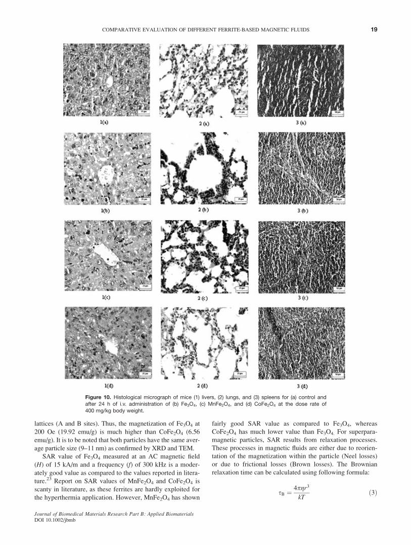
mg/kg body weight intravenously after 24 h. Figure 10 shows
the histological photomicrographs of liver [Figures 10 and
1(a–d)], lung, [Figures 10 and 2(a–d)] and spleen [Figures 10
and 3(a–d)] for 400 mg/kg body weight dose group at 24-h
intervals. In magnetic fluid-treated organs, tissue architecture
remained unchanged and no inflammatory or pathological
changes were detected even after 24 h of administration of
400 mg/kg body weight of ferrites intravenously. Numerous
Kupffer cells with engulfed particles were seen in the liver.
Clusters of particles were found in the liver, lung, and spleen
of magnetic fluid-treated mice. No pathological changes or
clusters of particles were observed in the heart and kidney.
Prussian Blue Staining. Micrographs from Prussian blue-
stained sections of Fe3O4-treated mice [Figure 11(a–e)] show
that there was extensive accumulation of magnetic nanopar-
ticles particularly in liver, lung, and spleen, which is evident
by the presence of blue color. But there was no accumulation
of magnetic nanoparticles in the heart and kidney. In spleen,
the particles were mostly accumulated in the red pulp area.
Similar distribution pattern was also observed for other mag-
netic fluids (figures not shown). As expected, Prussian blue
coloration was not observed for the control group (figures
not shown).
TABLE II. Variation of Magnetocrystalline Anisotropy Constant (K1) and Estimated Particles Size (DI) for Maximizationof SAR for the Different Magnetic Fluids
Ferrite
Composition
K1a
(joule/m3)
DP
(nm)
DI at 300
kHz (nm)
Dexp
(nm)
m at 200
Oe [emu/g]
SAR
(W/g ferrite)
SAR
(W/g metal)
xsNValueb
Fe3O4 1.4 3 104 25 15 10 19.92 120 168 0.0197
MnFe2O4 3.3 3 103 50 24 10 18.58 97 135.8 0.00327
CoFe2O4 1.8 3 105 14 7 10 6.56 37 51.8 1.33 3 107
DP, superparamagnetic critical size; DI, calculated particle size to satisfy the maximum SAR condition (xsN ¼ 1); Dexp, particle size of ferrofluid samples; m, particle magnetic
moment at 200 Oe.a K1 taken from reported value22.b Considered value of x ¼ 1.88 3 106 s�1 (i.e. 300 kHz) and the particle size Dexp.
Figure 5. Time–temperature curve of lauric acid-coated different
ferrite-based magnetic fluids.
Figure 6. Variation of specific magnetization (at 200 Oe) and SAR(at 15 kA/m) for three magnetic fluid samples.
17COMPARATIVE EVALUATION OF DIFFERENT FERRITE-BASED MAGNETIC FLUIDS
Journal of Biomedical Materials Research Part B: Applied BiomaterialsDOI 10.1002/jbmb

DISCUSSION
Magnetic fluids of different compositions (Fe3O4, MnFe2O4,
and CoFe2O4) were synthesized and characterized to evalu-
ate the possibilities of their successful use in hyperthermia
treatment. From XRD pattern of the Fe3O4, MnFe2O4, and
CoFe2O4 nanoparticles, it is evident that single phases were
formed in each of these ferrites. The broadness of the peak in
XRD of these ferrites is the signature of nanocrystallinity.
The arrangement of fatty acid molecules on the magnetic
nanoparticle in the stable water-based magnetic fluid may be
in bilayers with primary and secondary layers.24 In the pri-
mary layer, the carboxylic acid groups of the fatty acid are
chemisorbed to the ferrite surface and thus help in stabilizing
the nanoparticles. A secondary fatty acid layer is present on
the primary fatty acid molecules, with the interpenetration of
two layers equivalent to four carbon–carbon bonds. The sec-
ondary fatty acid layer provides a hydrophilic outer shell
exposed to the surrounding polar solvent, resulting in stable
water-based magnetic fluid. From the TGA curve, two dis-
tinct weight losses at different temperatures were found. dw/dt versus temperature curve reveals that the major weight
loss temperatures are around 260 and 3408C. These weight
losses may be due to desorption and evaporation of second-
ary (outer) and primary (inner) layers of fatty acid molecules.
The broad weight loss at higher temperature at around 3408Ccorresponds to the inner layer (primary) fatty acid molecule,
which is chemisorbed on the particle surface having higher
bond energy. The relatively sharp weight loss at 2608C is
due to the outer layer (secondary) fatty acid molecules with
lower bond energy. It is to be noted that three different fatty
acid-coated ferrite nanoparticles (Fe3O4, MnFe2O4, and
CoFe2O4) show the same weight loss profile with tempera-
ture (TGA), indicating presence of double layer of lauric acid
on nanoparticles for all the three magnetic fluids.
From the TEM micrographs, it can be seen that the par-
ticles are spherical in shape. The mean particles sizes are
about 9–11 nm. This is in good agreement with the size
measured form the X-ray line (311) broadening using Scher-
rer’s formula.
Magnetization loops of ferrite nanoparticles show zero
coercivity and zero remanence, and the magnetization does
not saturate at 20 kOe, indicating superparamagnetic nature
of ferrite particles. All the ferrite nanoparticles have room
temperature-specific magnetization values less than that of
their corresponding bulk state. The reduction of magnetic
properties of nanosized particles may be due to several addi-
tional factors, like formation of magnetic dead layer on the
surface, existence of random canting of particle surface spin,
nonsaturation effects due to random distribution of particle
size, deviation from the normal cation distribution, absorbed
water etc.25–27 It is interesting to note that the specific
magnetization at 1.8 kOe for CoFe2O4 is higher than for
MnFe2O4 whereas at lower field (200 Oe) it reverses. The
mean particles size of all the ferrites are nearly same; thus,
one can expect that the same degree of reduction of magnet-
ization may be due to reduction of size. Sato et al. observed
that the extent of reduction of saturation magnetization for
different ferrite nanoparticles (having same particle size)
largely depends on the crystalline magnetic anisotropy con-
stant K1.28 Among the three ferrites studied, CoFe2O4 has the
highest K1 value whereas MnFe2O4 has the lowest K1 value
(Table II). The higher specific magnetization (1.8 kOe) of
CoFe2O4 nanoparticles than of MnFe2O4 nanoparticles may
be taken into account by K1. The magnetization at lower field
(200 Oe), that is, the initial susceptibility, depends on several
parameters like particle size and anisotropy. The initial sus-
ceptibility also depends on the composition of ferrite par-
ticles, since the magnetocrystalline anisotropy (K1) depends
on the presence and types of cations at the different sub-
Figure 8. Effect of different magnetic fluids on BUN levels following
i.v. administration at the dose rate of 400 mg/kg body weight for differ-ent intervals in mice. Results are expressed as mean6 SD; n¼ 3.
Figure 9. Effect of different magnetic fluids on SGPT levels following
i.v. administration at the dose rate of 400 mg/kg body weight for differ-
ent intervals in mice. Results are expressed as mean6 SD; n¼ 3.Figure 7. Percent viability of BHK 21 cells 48 h after incubation of
different magnetic fluids at different ferrite concentrations. Results
are expressed as mean 6 SD; n ¼ 3.
18 PRADHAN ET AL.
Journal of Biomedical Materials Research Part B: Applied BiomaterialsDOI 10.1002/jbmb
lattices (A and B sites). Thus, the magnetization of Fe3O4 at
200 Oe (19.92 emu/g) is much higher than CoFe2O4 (6.56
emu/g). It is to be noted that both particles have the same aver-
age particle size (9–11 nm) as confirmed by XRD and TEM.
SAR value of Fe3O4 measured at an AC magnetic field
(H) of 15 kA/m and a frequency (f) of 300 kHz is a moder-
ately good value as compared to the values reported in litera-
ture.23 Report on SAR values of MnFe2O4 and CoFe2O4 is
scanty in literature, as these ferrites are hardly exploited for
the hyperthermia application. However, MnFe2O4 has shown
fairly good SAR value as compared to Fe3O4, whereas
CoFe2O4 has much lower value than Fe3O4. For superpara-
magnetic particles, SAR results from relaxation processes.
These processes in magnetic fluids are either due to reorien-
tation of the magnetization within the particle (Neel losses)
or due to frictional losses (Brown losses). The Brownian
relaxation time can be calculated using following formula:
sB ¼ 4pgr3
kTð3Þ
Figure 10. Histological micrograph of mice (1) livers, (2) lungs, and (3) spleens for (a) control and
after 24 h of i.v. administration of (b) Fe3O4, (c) MnFe2O4, and (d) CoFe2O4 at the dose rate of400 mg/kg body weight.
19COMPARATIVE EVALUATION OF DIFFERENT FERRITE-BASED MAGNETIC FLUIDS
Journal of Biomedical Materials Research Part B: Applied BiomaterialsDOI 10.1002/jbmb

and the Neel relaxation time is
sN ¼ s0 expKV
kTð4Þ
where g is the viscosity of carrier liquid, r the hydrodynamic
radius of particles, k the Boltzmann’s constant, s0 the time
constant (�10�9 s), V the particle volume, and K is the ani-
sotropy constant. Thus the power loss corresponds to Neel
and/or Brownian relaxation and is calculated approximately
from the following equation:
P ¼ ðmHxsÞ2=½2skT qVð1þ x2s2Þ� ð5Þ
where m is the particle magnetic moment, x the angular fre-
quency, H the AC field amplitude, and q is the density of fer-
rite. From Eq. (5), it is clear that the power loss (P) dependson the (a) particle magnetic moment (m), (b) AC magnetic
field amplitude (H), and (c) AC magnetic field frequency
(x). Thus the SAR is proportional to the square of magnetic
moment. The variation of SAR for different magnetic fluids
follows the same pattern as the specific magnetizations vary
at lower field (200 Oe), that is, 16 kA/m. On the other hand,
at a particular value of m and H, P will reach a maximum
value when xs ¼ 1.29 In our systems (dh ¼ 70–100 nm, f ¼300 kHz), contribution due to Brownian losses is very small,
since the product of x and sB is far apart from 1. This was
also confirmed by a polyacrylic gel experiment in which the
magnetic fluids were immobilized in polyacrylic gel and
thereby the viscosity of magnetic particles becomes too high
to show the Brownian loss (result not shown here). If we con-
sider the SAR due to Neel losses, the relaxation time depends
on V and K. For magnetic particles of particular composition,
Neel relaxation time (sN) depends on the particle volume
(i.e. size). To maximize P (SAR) of a particular ferrite parti-
cle at any AC field frequency (f or x), the particle volume
should be such that the product of x and sN becomes 1. With
varying composition of the ferrite particles, the K value will
change and it will decide sN. With the knowledge of K, theoptimum particle size could be calculated for maximum P(SAR) and is summarized in Table II. The optimum particle
sizes (DI) to maximize SAR at 300 kHz AC frequency for
Fe3O4, MnFe2O4, and CoFe2O4 are 15, 25, and 7 nm, respec-
tively. The average particle size of our ferrite nanoparticles
is in between 9 and 11 nm (measured from XRD and TEM)
which are bigger/smaller than their optimum sizes (DI). The
effect of particle size on sN, as well as on SAR, reflects on
the value of xsN and hence it influences its degree of devia-
tion from the maximum power loss condition (i.e. xsN ¼ 1).
From the table it is found that though there is not much dif-
ference in low field specific magnetization for MnFe2O4 and
Fe3O4, the SAR is relatively higher for Fe3O4. This could be
due to the deviation of particle sizes from their optimum
sizes. Thus, the value of xsN for MnFe2O4 (0.00327) is
smaller than that of Fe3O4 (0.0197) and much away from 1
(maximum power loss condition).
From the earlier discussion it is clear that the SAR of the
Fe3O4 and MnFe2O4 can be enhanced to higher value by sim-
Figure 11. Prussian blue-stained sections of mice (a) liver, (b) lung, (c) spleen, (d) heart, and (e) kid-
ney after 24 h of i.v. administration of Fe3O4 magnetic fluid at the dose rate of 400 mg/kg bodyweight. [Color figure can be viewed in the online issue, which is available at www.interscience.
wiley.com.]
20 PRADHAN ET AL.
Journal of Biomedical Materials Research Part B: Applied BiomaterialsDOI 10.1002/jbmb

ply increasing the particle size to their optimum size (DI)
(15 nm for Fe3O4 and 25 nm for MnFe2O4, respectively). In
case of CoFe2O4, SAR could be enhanced further by reduc-
ing the particle size to 7 nm (DI). But, initial susceptibility
(magnetization at 200 Oe) of CoFe2O4 will be further
reduced because of size reduction and thereby lower value of
SAR will be observed. Thus, even with optimum particle size
(DI), one can expect very low SAR value for CoFe2O4
because of high anisotropy value.
From in vitro biocompatibility study, it is evident that the
cytotoxicity of magnetic fluids depends on the concentration of
different ferrites. For all the magnetic fluids, 0.1 mg/mL is the
threshold concentration beyond which decrease in cell viability
is observed. Threshold concentrations for dextran-coated Fe3O4
has been reported to be in the range of 1–2 mg/mL,4 which is
higher than lauric acid-coated Fe3O4 reported here. This differ-
ence in threshold concentration might be due to the use of dif-
ferent coatings (i.e. lauric acid) in this study. Moreover, reports
on cytotoxicity of other ferrites, like MnFe2O4 and CoFe2O4,
having lauric acid coating are scanty in literature. When lauric
acid-coated MnFe2O4 is compared with lauric acid-coated
Fe3O4, no significant difference in viability is found upto
0.6 mg/mL (highest concentration tested) (p > 0.05; ANOVA).
But CoFe2O4 has shown comparatively higher loss of cell via-
bility (>10% viability) at higher doses (>0.2 mg/mL), indicating
more cytotoxicity as compared to Fe3O4 and MnFe2O4.
For in vivo evaluation of biocompatibility, different hemato-
logical parameters and histology of different organs were eval-
uated. Effects of different magnetic fluids on renal and liver
functions were evaluated by changes in BUN and SGPT levels,
respectively, for different doses and intervals. All the magnetic
fluids have been found to be nonnephrotoxic, as revealed by no
significant change in BUN levels for magnetic fluid-treated
groups as compared to control group (p > 0.05; ANOVA).
Fe3O4 and MnFe2O4 did not cause any significant change in
SGPT levels as compared to control groups (p > 0.05; ANOVA
and Newman–Keul’s test), indicating their nontoxicity to liver.
But for CoFe2O4, an increasing trend (nonsignificant, p > 0.05;
ANOVA and Newman–Keul’s test) in SGPT levels could be
due to the mild hepatotoxic effect of CoFe2O4. However, histol-
ogy of organs did not reveal any pathological changes for all
the three magnetic fluids, indicating their biocompatibility.
Prussian blue staining shows extensive accumulation of
magnetic particles mainly in liver, lung, and spleen, which is
attributed to the phagocytosis of magnetic particles by the
phagocytic cells of these organs. In spleen, accumulation of
particles was mainly in the red pulp, which is in agreement
with other reports.13,18 As spleen white pulp does not have
mononuclear phagocytes, no accumulation of particles was
found in this region. However, unlike spleen, clusters of par-
ticles are distributed homogenously throughout the liver and
lung. On the contrary, kidney and heart did not show any
accumulation of magnetic particles. However, no pathologi-
cal changes were detected even after massive accumulation
of magnetic particles in liver, lung, and spleen, indicating
their biocompatibility.
To the best of our knowledge, very few groups have eval-
uated the biocompatibility of MnFe2O4- and CoFe2O4-based
magnetic fluid. Lacava et al.30,31 reported that ionic-, citrate-,
and tartarate-coated manganese ferrite-based magnetic fluid
were not biocompatible. However, in our study lauric acid-
coated manganese ferrite-based magnetic fluids did not show
any toxic effects at 400 mg/kg body weight up to 24 h. This
difference might be due to the different coating materials
used (i.e. lauric acid) in this study. Kuckelhaus et al.12,32 did
not find any morphological alteration following intravenous
injection of citrate-coated cobalt ferrite-based magnetic fluid.
In our study, though no morphological changes in vital
organs have been detected after magnetic fluid injection, an
increase in SGPT levels has been found after CoFe2O4-based
magnetic fluid injection, which indicates that this composi-
tion is mildly hepatotoxic. But Fe3O4- and MnFe2O4-based
magnetic fluid has been found to be nontoxic from both the
morphological and hematological point of view.
SUMMARY
In this study, lauric acid-coated different ferrites (Fe3O4,
MnFe2O4, and CoFe2O4) based magnetic fluids have been
successfully prepared and characterized, and their heating
ability and biocompatibility have been comparatively eval-
uated. CoFe2O4-based magnetic fluid has lower SAR
(thereby heating ability) than Fe3O4- and MnFe2O4-based
magnetic fluids. However, Fe3O4- and MnFe2O4-based mag-
netic fluids have comparable SAR values. The SAR of the
Fe3O4 and MnFe2O4 systems can be further enhanced to a
maximum value by increasing the particles size to the opti-
mum size (DI). But even after optimizing particle size, higher
SAR values for CoFe2O4 system could not be achieved
because of its high anisotropy energy. All the magnetic fluids
have dose-dependent effect on cell viability. Threshold cyto-
compatible concentration of all the magnetic fluids is 0.1 mg/
mL. However, CoFe2O4-based magnetic fluid is more cyto-
toxic to BHK 21 cells than the others at higher concentra-
tions (0.2 mg/mL). Lauric acid-coated Fe3O4- and MnFe2O4-
based magnetic fluids are biocompatible in vivo whereas
CoFe2O4-based magnetic fluid is mildly hepatotoxic at
higher doses. However, in vivo biocompatibility study was
evaluated only for a short term of 24 h in the present study.
Further, longer term in vivo biocompatibility needs to be
evaluated for the evidence of inertness of the surfactant-
coated magnetic particles prior to use in hyperthermia.
From this study, it can be concluded that lauric acid-
coated, superparamagnetic, MnFe2O4-based magnetic fluid is
comparable to Fe3O4-based magnetic fluid as far as biocom-
patibility and SAR are concerned. But CoFe2O4-based mag-
netic fluid has lesser SAR and biocompatibility as compared
to Fe3O4 and MnFe2O4. Hence Fe3O4- and MnFe2O4-based
magnetic fluids are suitable for hyperthermia application,
whereas CoFe2O4 is not suitable for this purpose. However,
further optimization of particle size to maximize SAR, effi-
cacy of hyperthermia treatment, and longer term in vivo bio-
21COMPARATIVE EVALUATION OF DIFFERENT FERRITE-BASED MAGNETIC FLUIDS
Journal of Biomedical Materials Research Part B: Applied BiomaterialsDOI 10.1002/jbmb

compatibility study are required prior to their use in hyper-
thermia application (work in progress).
REFERENCES
1. Bahadur D, Giri J. Biomaterials and magnetism. Sadhana2003;28:639–656.
2. Pankhurst QA, Connolly J, Jones SK, Dobson J. Applicationsof magnetic nanoparticles in biomedicine. J Phys D: ApplPhys 2003;36:167–181.
3. Gupta AK, Gupta M. Synthesis and surface engineering ofiron oxide nanoparticles for biomedical applications. Biomate-rials 2005;26:3995–4021.
4. Jordan A, Wust P, Scholz R, Tesche B, Fahling H, MitrovicsT, Vogl T, Cervos-Navarro J, Felix R. Cellular uptake ofmagnetic fluid particles and their effects on human adenocar-cinoma cells exposed to AC magnetic fields in vitro. Int JHyperthermia 1996;12:705–722.
5. Jordan A, Scholz R, Wust P, Fahling H, Felix R. Magneticfluid hyperthermia (MFH): Cancer treatment with AC mag-netic field induced excitation of biocompatible superparamag-netic nanoparticles. J Magn Magn Mater 1999;201:413–419.
6. Giri J, Ray A, Dasgupta S, Datta D, Bahadur D. Investigationon Tc tuned nano particles of magnetic oxides for hyperther-mia applications. Biomed Mater Eng 2003;13:387–399.
7. Kawashita M, Tanaka M, Kokubo T, Inoue Y, Yao T, Ham-ada S, Shinjo T. Preparation of ferrimagnetic magnetitemicrospheres for in situ hyperthermic treatment of cancer.Biomaterials 2005;26:2231–2238.
8. Hergt R, Hiergeist R, Zeisberger M, Glockl G, Weitschies W,Ramirez LP, Hilger I, Kaiser WA. Enhancement of AC-lossesof magnetic nanoparticles for heating applications. J MagnMagn Mater 2004;280:358–368.
9. Grasset F, Mornet S, Demourgues A, Portier J, Bonnet J,Vekris A, Duguet E. Synthesis, magnetic properties, surfacemodification and cytotoxicity evaluation of Y3Fe5-xAlxO12 (0� x � 2) garnet submicron particles for biomedical applica-tions. J Magn Magn Mater 2001;234:409–418.
10. Giri J, Sriharsha T, Bahadur D. Optimization of parametersfor the synthesis of nano-sized Co1–xZnxFe2O4, (0 � x � 0.8)by microwave refluxing. J Mater Chem 2004;14:875–880.
11. Hergt R, Hiergeist R, Hilger I, Kaiser WA, Lapatnikov Y, Mar-gel S, Richter U. Maghemite nanoparticles with very high AC-losses for application in RF-magnetic hyperthermia. J MagnMagn Mater 2004;270:345–357.
12. Kuckelhaus S, Reis SC, Carneiro MF, Tedesco AC, OliveiraDM, Lima ECD, Morais PC, Azevedo RB, Lacava ZGM.In vivo investigation of cobalt ferrite-based magnetic fluid andmagnetoliposomes using morphological tests. J Magn MagnMater 2004;272:2402,2403.
13. Lacava LM, Garcia VAP, Kuckelhaus S, Azevedo RB, Sade-ghiani N, Buske N, Morais PC, Lacava ZGM. Long-termretention of dextran-coated magnetite nanoparticles in theliver and spleen. J Magn Magn Mater 2004;272:2434, 2435.
14. Lacava LM, Lacava ZGM, Azevedo RB, Chaves SB, GarciaVAP, Silva O, Pelegrini F, Buske N, Gansau C, Da Silva MF,Morais PC. Use of magnetic resonance to study biodistribu-tion of dextran-coated magnetic fluid intravenously adminis-tered in mice. J Magn Magn Mater 2002;252:367–369.
15. Freitas MLL, Silva LP, Azevedo RB, Garcia VAP, LacavaLM, Grisolia CK, Lucci CM, Morais PC, Da Silva MF, Buske
N, Curi R, Lacava ZGM. A double-coated magnetite-basedmagnetic fluid evaluation by cytometry and genetic tests.J Magn Magn Mater 2002;252:396–398.
16. Sadeghiani N, Barbosa LS, Silva LP, Azevedo RB, MoraisPC, Lacava ZGM. Genotoxicity and inflammatory investiga-tion in mice treated with magnetite nanoparticles surfacecoated with polyaspartic acid. J Magn Magn Mater 2005;28:466–468.
17. Petri-Fink A, Chastellain M, Juillerat-Jeanneret L, Ferrari A,Hofmann H. Development of functionalized superparamag-netic iron oxide nanoparticles for interaction with human can-cer cells. Biomaterials 2005;26:2685–2694.
18. Lacava LM, Garcia VAP, Kuckelhaus S, Azevedo RB,Lacava ZGM, Silva O, Pelegrini F, Gansau C, Buske N, Mor-ais PC. Magnetic resonance and light microscopy investiga-tion of a dextran coated magnetic fluid. J Appl Phys 2003;93:7563–7565.
19. Giri J, Pradhan P, Banerjee R, Datta D, Bahadur D. Synthesis,characterization and in vitro evaluation of water based tempera-ture sensitive ferrofluids for hyperthermia applications. Pre-sented at fifth International conference on the scientific andclinical applications of magnetic carriers, Lyon, May 20–22,2004.
20. Kinderlerer JL. Degradation of the lauric acid oils. Int Bio-deter Biodegrad 1994;33:345–354.
21. Skehan P, Storeng R, Scudiero D, Monks A, McMahon J,Vistica D, Warren JT, Bokesch H, Kenney S, Boyd MR. Newcolorimetric cytotoxicity assay for anticancer-drug screening.J Natl Cancer Inst 1990;82:1107–1112.
22. Bozorth RM, Tilden EF, Williams AJ. Anisotropy and magne-tostriction of some ferrites. Phys Rev 1955;99:1788–1798.
23. Andra W. Magnetic hyperthermia. In: Andra W, Nowak H,editor. Magnetism inMedicine. Berlin: Wiley; 1998. p 450–470.
24. Shen L, Laibinis PE, Hatton TA. Aqueous magnetic fluids sta-bilized by surfactant bilayers. J Magn Magn Mater 1999;194:37–44.
25. Coey JMD. Noncollinear spin arrangement in ultrafine ferri-magnetic crystallites Phys Rev Lett 1971;27:1140–1142.
26. Pankhurst QA, Polland RJ. Origin of the spin-canting anom-aly in small ferrimagnetic particles. Phys Rev Lett 1991;67:248–250.
27. Chen JP, Sorensen CM, Klabunde KJ, Hadjipanayis GC,Delvin E, Kostikas A. Size-dependent magnetic properties ofMnFe2O4 fine particles synthesized by coprecipitation. PhysRev B: Condens Matter 1996;54:9288–9296.
28. Sato T, Iijima T, Seki M, Inagaki N. Magnetic properties ofultrafine ferrite particles. J Magn Magn Mater 1987;65:252–256.
29. Debye P. Polar Molecules. New York: The Chemical CatalogCompany; 1929.
30. Lacava ZGM, Azevedo RB, Martins EV, Lacava LM, FreitasMLL, Garcia VAP, Rebula CA, Lemos APC, Sousa MH,Tourinho FA, Da Silva MF, Morais PC. Biological effects ofmagnetic fluids: Toxicity studies. J Magn Magn Mater 1999;201:431–434.
31. Lacava ZGM, Azevedo RB, Lacava LM, Martins EV, GarciaVAP, Rebula CA, Lemos APC, Sousa MH, Tourinho FA,Morais PC, Da Silva MF. Toxic effects of ionic magnetic flu-ids in mice. J Magn Magn Mater 1999;194:90–95.
32. Kuckelhaus S, Garcia VAP, Lacava LM, Azevedo RB,Lacava ZGM, Lima ECD, Figueiredo F, Tedesco AC, MoraisPC. Biological investigation of a citrate-coated cobalt–ferrite-based magnetic fluid. J Appl Physics 2003;93:6707,6708.
22 PRADHAN ET AL.
Journal of Biomedical Materials Research Part B: Applied BiomaterialsDOI 10.1002/jbmb