Theoretical study of convergent ultrasound hyperthermia for treating bone tumors
11
Medical Engineering & Physics 22 (2000) 253–263 www.elsevier.com/locate/medengphy Theoretical study of convergent ultrasound hyperthermia for treating bone tumors Bing-Yuh Lu a, b , Rong-Sen Yang c , Win-Li Lin d,* , Kung-Shan Cheng d , Cheng-Yi Wang d , Te-Son Kuo a, d a Department of Electrical Engineering, National Taiwan University, Taipei, Taiwan, ROC b Department of Electronic Engineering, Tung-Nan Junior College of Technology, Taipei, Taiwan, ROC c Department of Orthopedics, National Taiwan University, Taipei, Taiwan, ROC d Institute of Biomedical Engineering, National Taiwan University, # 1, Section 1, Jen-Ai Road, Taipei, Taiwan, ROC Received 10 May 1999; received in revised form 9 May 2000; accepted 6 June 2000 Abstract This study investigates the optimal external parameters for using an ultrasound applicator for treating bone tumors. This system utilized spherically arranged applicators such as scanned focused ultrasound, and spherically focused multielement applicators. The power deposition pattern is modeled as geometric gain with exponential attenuation. The specific absorption rate ratio (SARR) criteria have been used to determine the proper heating domain of ultrasound driving frequency and therapeutic tumor diameter. The results demonstrate that the optimal driving frequency depends on tumor depth, ultrasound absorption of bone marrow, and diameter of bone, but it is independent of the acoustic window area and SARR. The treatable diameter of bone tumor increased when the absorption ratio of bone marrow to tumor, acoustic window of surface skin, and diameter of bone were elevated. However, the treatable diameter of bone tumor decreased when muscle thickness, SARR of bone tumor site to the surface skin, bone marrow, and bone declined. To deliver the ultrasound energy into the tumor site and to avoid the potential damage to the normal tissue as much as possible, the specific absorption rate (SAR) in the bone tumor site has to be three times higher than that in the surface skin, tumor/marrow, and marrow/bone interfaces. The temperature distributions can verify the SARR criteria in this model. This study provides the information for choosing the optimal operating frequency of the ultrasound transducer and the acoustic window on the skin surface, and for designing the ultrasound applicator for clinical implementation. 2000 IPEM. Published by Elsevier Science Ltd. All rights reserved. Keywords: Therapeutic domain; Ultrasound hyperthermia; Bone tumor; Optimal driving frequency 1. Introduction Hyperthermia, i.e. heating tumor cells, is a kind of adjuvant cancer therapy. In the previous studies, heating tissues to temperatures above 42°C has been shown to kill cells as well as to augment the effects of conven- tional cancer therapy including radiotherapy and chemo- therapy [1–3]. Various kinds of hyperthermia have been developed. Fan et al. [4] reported the preliminary data on treatment of bone tumors with microwave-induced hyperthermia. The bone tumor was dissected from the * Corresponding author. Tel.: + 886-2-23970800x1445; fax: + 886- 2-23940049. E-mail address: [email protected] (W.-L. Lin). 1350-4533/00/$ - see front matter 2000 IPEM. Published by Elsevier Science Ltd. All rights reserved. PII:S1350-4533(00)00031-X surrounding normal tissues in situ by surgery, and inter- stitial microwave hyperthermia was employed to destroy the tumor cells. In most of the 62 evaluated cases, the knee joints functioned well, were stable and painless, and retained almost full range of motion. Ikenaga et al. [5] carried out localized hyperthermic treatment of experimental bone tumors with ferromagnetic ceramics in rabbit. The area of tumor necrosis in the hyperthermic therapy group was significantly larger than that in the control group. These studies have shown that hyperther- mia is an efficient therapy for treating bone tumors, but complex surgical procedures are involved in the invasive treatment. Charny et al. [6] used a mini-annular phased array to heat human leg by emitting radio frequency energy. They pointed out that the specific absorption rate (SAR) in bone may not be neglected and the specific
-
Upload
independent -
Category
Documents
-
view
3 -
download
0
Transcript of Theoretical study of convergent ultrasound hyperthermia for treating bone tumors
Medical Engineering & Physics 22 (2000) 253–263www.elsevier.com/locate/medengphy
Theoretical study of convergent ultrasound hyperthermia fortreating bone tumors
Bing-Yuh Lu a, b, Rong-Sen Yangc, Win-Li Lin d,*, Kung-Shan Chengd,Cheng-Yi Wangd, Te-Son Kuoa, d
a Department of Electrical Engineering, National Taiwan University, Taipei, Taiwan, ROCb Department of Electronic Engineering, Tung-Nan Junior College of Technology, Taipei, Taiwan, ROC
c Department of Orthopedics, National Taiwan University, Taipei, Taiwan, ROCd Institute of Biomedical Engineering, National Taiwan University, # 1, Section 1, Jen-Ai Road, Taipei, Taiwan, ROC
Received 10 May 1999; received in revised form 9 May 2000; accepted 6 June 2000
Abstract
This study investigates the optimal external parameters for using an ultrasound applicator for treating bone tumors. This systemutilized spherically arranged applicators such as scanned focused ultrasound, and spherically focused multielement applicators. Thepower deposition pattern is modeled as geometric gain with exponential attenuation. The specific absorption rate ratio (SARR)criteria have been used to determine the proper heating domain of ultrasound driving frequency and therapeutic tumor diameter.The results demonstrate that the optimal driving frequency depends on tumor depth, ultrasound absorption of bone marrow, anddiameter of bone, but it is independent of the acoustic window area andSARR. The treatable diameter of bone tumor increasedwhen the absorption ratio of bone marrow to tumor, acoustic window of surface skin, and diameter of bone were elevated. However,the treatable diameter of bone tumor decreased when muscle thickness,SARRof bone tumor site to the surface skin, bone marrow,and bone declined. To deliver the ultrasound energy into the tumor site and to avoid the potential damage to the normal tissue asmuch as possible, the specific absorption rate (SAR) in the bone tumor site has to be three times higher than that in the surfaceskin, tumor/marrow, and marrow/bone interfaces. The temperature distributions can verify theSARRcriteria in this model. Thisstudy provides the information for choosing the optimal operating frequency of the ultrasound transducer and the acoustic windowon the skin surface, and for designing the ultrasound applicator for clinical implementation. 2000 IPEM. Published by ElsevierScience Ltd. All rights reserved.
Keywords:Therapeutic domain; Ultrasound hyperthermia; Bone tumor; Optimal driving frequency
1. Introduction
Hyperthermia, i.e. heating tumor cells, is a kind ofadjuvant cancer therapy. In the previous studies, heatingtissues to temperatures above 42°C has been shown tokill cells as well as to augment the effects of conven-tional cancer therapy including radiotherapy and chemo-therapy [1–3]. Various kinds of hyperthermia have beendeveloped. Fan et al. [4] reported the preliminary dataon treatment of bone tumors with microwave-inducedhyperthermia. The bone tumor was dissected from the
* Corresponding author. Tel.:+886-2-23970800x1445; fax:+886-2-23940049.
E-mail address:[email protected] (W.-L. Lin).
1350-4533/00/$ - see front matter 2000 IPEM. Published by Elsevier Science Ltd. All rights reserved.PII: S1350-4533 (00)00031-X
surrounding normal tissues in situ by surgery, and inter-stitial microwave hyperthermia was employed to destroythe tumor cells. In most of the 62 evaluated cases, theknee joints functioned well, were stable and painless,and retained almost full range of motion. Ikenaga et al.[5] carried out localized hyperthermic treatment ofexperimental bone tumors with ferromagnetic ceramicsin rabbit. The area of tumor necrosis in the hyperthermictherapy group was significantly larger than that in thecontrol group. These studies have shown that hyperther-mia is an efficient therapy for treating bone tumors, butcomplex surgical procedures are involved in the invasivetreatment. Charny et al. [6] used a mini-annular phasedarray to heat human leg by emitting radio frequencyenergy. They pointed out that the specific absorption rate(SAR) in bone may not be neglected and the specific

254 B.-Y. Lu et al. / Medical Engineering & Physics 22 (2000) 253–263
absorption rate ratio (SARR) of bone to muscle at inter-face was computed in different angles from the interfacein muscle. Oglilvie et al. [7] indicated that the presenttechniques and equipment utilizing microwave and radiofrequency energies appeared to produce largely inad-equate heating patterns in superficial or accessibletumors and normal tissues. The advantages of ultrasoundtechniques have shown promise for delivering controlledheating of both superficial and deep lesions. Althoughthe application of ultrasound energy for hyperthermia issomewhat limited, because it cannot propagate throughair cavities and is excessively reflected and absorbed bybone, this modality holds great promise for the con-trolled heating of superficial and deep lesions.
Bone pain associated with ultrasound hyperthermiatreatment for bone tumors is probably caused by extremetemperatures in bone. This can be avoided by sonicatingthe tumor in such a manner that the intensities at thebone surface are low enough to avoid large temperatureelevations. Hynynen et al. [8] pointed out that selectionof the proper driving frequency in ultrasound hyperther-mia provides the major possible solution to the appli-cation of Scanned Focused Ultrasound System (SFUS)treatment in many sites. Therefore, location-orientedtreatment planning is necessary for successful clinicalutilization of scanned focused ultrasound hyperthermia[9,10]. Fujii et al. [11] have studied the reason behindthe temperature rise at the muscle–bone interface duringultrasound hyperthermia. They have focused ultrasoundenergy on the muscle–bone interface to study the inci-dent angle of ultrasound vs. temperature rise and con-cluded that the bone pain could be reduced if the incidentangle can be adjusted to about 15° or larger than 60°.In addition, Hill [12] has developed a spherical bowlmodel to optimize acoustic frequency for focused ultra-sound surgery. Fry et al. [13] pointed out that similartriangles can be used to determine minimum distancefrom the focus where spherical theory is applicable. Car-stensen et al. [14] have proposed a cone shape model todescribe the intensity distribution in an essentially lossmedium. They defined geometric gain as (r0/r)2a wherer0 is the radius of curvature of the focused source,r isthe distance from the center of radius curvature to thefield point, anda models the convergence of the fielddue to focusing (a=0 for plane wave,a=1 for sphericalwave). According to these studies, we adopt a coneshape convergent/divergent model (Fig. 1) for thespherically arranged applicators, but not only for a singlespherical transducer. The power deposition pattern ismodeled as geometric gain with exponential attenuation.
The purpose of this study is to investigate the optimaltreatment parameters using a simplified power depo-sition model [15] of spherically arranged ultrasoundapplicators to deliver the ultrasound energy into the bonetumor site and to avoid the potential overheating in thenormal tissues. The diameter of the acoustic window,
Fig. 1. The hypothetical model of a bone tumor.
tumor size, tumor depth, ultrasound attenuation in thebone tumor tissue, the absorption on the bone, and thedriving frequency of transducer have been investigatedto determine the appropriate driving frequency for agiven tumor depth and tumor size, and to estimate thetherapeutic domain for a given acoustic window on theskin surface of the limbs. The results are helpful for thetreatment planning and the designing of ultrasound hyp-erthermic applicators in bone tumor treatment.
2. Methods
2.1. Ultrasound power deposition model
The anatomic relationship shown in Fig. 1 is the hypo-thetically spherical tumor that breaks out from the innerside of a long bone.z is defined as the depth from theskin surface whiled(z) is the diameter of acoustic win-dow at positionz. Therefore,d(0) is the diameter of theacoustic window on the skin surface of the limbs andthe d(zt) is the diameter of the focal zone that meets thebone tumor diameter.zma andzb are defined as the depthof the tumor/marrow and the marrow/bone interfacesfrom the skin surface, respectively. The deposition of theultrasound energy is modeled as a cone ofconvergent/divergent shape [12–14]. The ultrasoundenergy absorbed by the tissues follows the exponentiallaw for attenuation and uniform ideal power depositionwithin the cone-shape model. In this model, the acousticpower is assumed equal at the same depth within thecone shape region [15]. Then, we have:
Q(z)=Q(0)·expH−2FO4i51
mi−1(zi−1−zi−2)+mi(z−zi−1)GJm0=0, m1=mm, m2=mt, m3=mma, m4=mb
z0=0, z1=zm, z2=zma, z3=zb
(1)
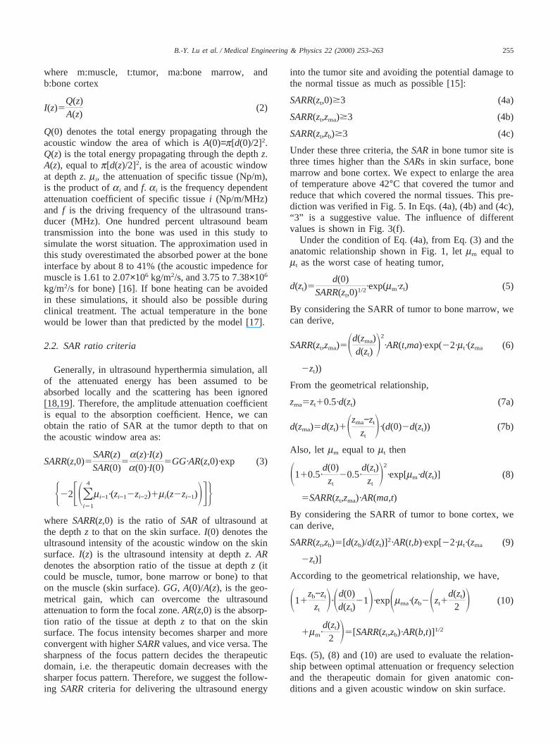
255B.-Y. Lu et al. / Medical Engineering & Physics 22 (2000) 253–263
where m:muscle, t:tumor, ma:bone marrow, andb:bone cortex
I(z)5Q(z)A(z)
(2)
Q(0) denotes the total energy propagating through theacoustic window the area of which isA(0)=p[d(0)/2]2.Q(z) is the total energy propagating through the depthz.A(z), equal top[d(z)/2]2, is the area of acoustic windowat depthz. mi, the attenuation of specific tissue (Np/m),is the product ofai and f. ai is the frequency dependentattenuation coefficient of specific tissuei (Np/m/MHz)and f is the driving frequency of the ultrasound trans-ducer (MHz). One hundred percent ultrasound beamtransmission into the bone was used in this study tosimulate the worst situation. The approximation used inthis study overestimated the absorbed power at the boneinterface by about 8 to 41% (the acoustic impedence formuscle is 1.61 to 2.07×10⁶ kg/m2/s, and 3.75 to 7.38×10⁶
kg/m2/s for bone) [16]. If bone heating can be avoidedin these simulations, it should also be possible duringclinical treatment. The actual temperature in the bonewould be lower than that predicted by the model [17].
2.2. SAR ratio criteria
Generally, in ultrasound hyperthermia simulation, allof the attenuated energy has been assumed to beabsorbed locally and the scattering has been ignored[18,19]. Therefore, the amplitude attenuation coefficientis equal to the absorption coefficient. Hence, we canobtain the ratio of SAR at the tumor depth to that onthe acoustic window area as:
SARR(z,0)5SAR(z)SAR(0)
5a(z)·I(z)a(0)·I(0)
5GG·AR(z,0)·exp (3)
H22FSO4i51
mi−1·(zi−12zi−2)1mi(z2zi−1)DGJwhere SARR(z,0) is the ratio ofSARof ultrasound atthe depthz to that on the skin surface.I(0) denotes theultrasound intensity of the acoustic window on the skinsurface.I(z) is the ultrasound intensity at depthz. ARdenotes the absorption ratio of the tissue at depthz (itcould be muscle, tumor, bone marrow or bone) to thaton the muscle (skin surface).GG, A(0)/A(z), is the geo-metrical gain, which can overcome the ultrasoundattenuation to form the focal zone.AR(z,0) is the absorp-tion ratio of the tissue at depthz to that on the skinsurface. The focus intensity becomes sharper and moreconvergent with higherSARRvalues, and vice versa. Thesharpness of the focus pattern decides the therapeuticdomain, i.e. the therapeutic domain decreases with thesharper focus pattern. Therefore, we suggest the follow-ing SARRcriteria for delivering the ultrasound energy
into the tumor site and avoiding the potential damage tothe normal tissue as much as possible [15]:
SARR(zt,0)$3 (4a)
SARR(zt,zma)$3 (4b)
SARR(zt,zb)$3 (4c)
Under these three criteria, theSARin bone tumor site isthree times higher than theSARs in skin surface, bonemarrow and bone cortex. We expect to enlarge the areaof temperature above 42°C that covered the tumor andreduce that which covered the normal tissues. This pre-diction was verified in Fig. 5. In Eqs. (4a), (4b) and (4c),“3” is a suggestive value. The influence of differentvalues is shown in Fig. 3(f).
Under the condition of Eq. (4a), from Eq. (3) and theanatomic relationship shown in Fig. 1, letmm equal tomt as the worst case of heating tumor,
d(zt)5d(0)
SARR(zt,0)1/2·exp(mm·zt) (5)
By considering the SARR of tumor to bone marrow, wecan derive,
SARR(zt,zma)5Sd(zma)d(zt)
D2
·AR(t,ma)·exp(22·mt·(zma (6)
2zt))
From the geometrical relationship,
zma5zt10.5·d(zt) (7a)
d(zma)5d(zt)1Szma−zt
ztD·(d(0)2d(zt)) (7b)
Also, let mm equal tomt then
S110.5·d(0)zt
20.5·d(zt)
ztD2
·exp[mm·d(zt)] (8)
5SARR(zt,zma)·AR(ma,t)
By considering the SARR of tumor to bone cortex, wecan derive,
SARR(zt,zb)5[d(zb)/d(zt)]2·AR(t,b)·exp[22·mt·(zma (9)
2zt)]
According to the geometrical relationship, we have,
S11zb−zt
ztD·Sd(0)
d(zt)21D·expSmma·(zb2Szt1
d(zt)2 D (10)
1mm·d(zt)
2 D5[SARR(zt,zb)·AR(b,t)]1/2
Eqs. (5), (8) and (10) are used to evaluate the relation-ship between optimal attenuation or frequency selectionand the therapeutic domain for given anatomic con-ditions and a given acoustic window on skin surface.
256 B.-Y. Lu et al. / Medical Engineering & Physics 22 (2000) 253–263
2.3. Temperature solver
The temperature distributions are simulated by finitedifference technique for calculating the steady-state bio-heat transfer equation (BHTE) [20]:
2=·(k=T)1WCb(T2Tar)5Q (11)
whereT is the tissue temperature, in°C. k is the tissuethermal conductivity, in W/(m°C). It is 0.55 W/(m°C)in soft tissue, and is 1.16 W/(m°C) in bone cortex [21–23]. W is the blood perfusion rate, in kg/(m3 s), and isassumed to be 0 kg/(m3 s) in bone cortex.Cb is the bloodspecific heat, in J/(kg°C), is 3770 J/(kg°C) in softtissue.Tar is the arterial blood temperature, in°C; hereit is 37°C. Q is the power applied via ultrasound, inW/m3. All boundaries are treated as constant temperature37°C boundary. The outer dimensions of this simulationare 100 mm acoustic window, 50 mm muscle depth, 30mm bone diameter, and 2 mm bone cortex thickness and30 mm muscle depth behind bone.
3. Results
3.1. Proper heating domain
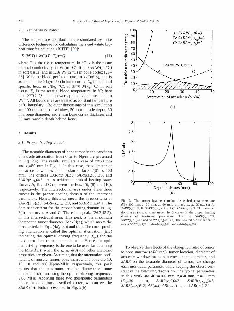
The treatable diameters of bone tumor in the conditionof muscle attenuation from 0 to 50 Np/m are presentedin Fig. 2(a). The results simulate a case ofzt=50 mmand zb=80 mm in Fig. 1. In this case, the diameter ofthe acoustic window on the skin surface,d(0), is 100mm. The criteriaSARR(zt,0)̂ 3, SARR(zt,zma)^3, andSARR(zt,zb)^3 are to achieve a critical heating state.Curves A, B and C represent the Eqs. (5), (8) and (10),respectively. The intersectional area under these threecurves is the proper heating domain of the treatmentparameters. Hence, this area meets the three criteria ofSARR(zt,0)̂ 3, SARR(zt,zma)^3, andSARR(zt,zb)$3. Thedominant criteria for the proper heating domain in Fig.2(a) are curves A and C. There is a peak, (26.3,15.5),in this intersectional area. This peak is the maximumtherapeutic tumor diameter (Max(d(zt)) which meets thethree criteria in Eqs. (4a), (4b) and (4c). The correspond-ing attenuation is called the optimal attenuation (mopt)indicating the optimal driving frequency (fopt) for themaximum therapeutic tumor diameter. Hence, the opti-mal driving frequency is the one to be used for obtainingthe Max(d(zt)) when thezt, zb, d(0) and other anatomicproperties are given. Assuming that the attenuation coef-ficients of muscle, tumor, bone marrow and bone are 10,10, 10 and 300 Np/m/MHz, respectively, this peakmeans that the maximum treatable diameter of bonetumor is 15.5 mm using the optimal driving frequency,2.63 MHz. Applying these two therapeutic parametersunder the conditions described above, we can get theSARRdistribution presented in Fig. 2(b).
Fig. 2. The proper heating domain: the typical parameters ared(0)=100 mm, zt=50 mm, zb=80 mm, mma=mm=mt, mb=30·mm, (a) A:SARR(zt,0)=3, B: SARR(zt,zma)=3 and C:SARR(zt,zb)=3. The intersec-tional area (shaded area) under the 3 curves is the proper heatingdomain of treatment parameters. That isSARR(zt,0)̂ 3,SARR(zt,zma)^3 andSARR(zt,zb)^3; (b) TheSARratio distribution: itmeetsSARR(zt,0)=3, SARR(zt,zma)^3 andSARR(zt,zb)=3.
To observe the effects of the absorption ratio of tumorto bone marrow (AR(ma,t)), tumor location, diameter ofacoustic window on skin surface, bone diameter, andSARRon the treatable diameter of tumor, we changeeach individual parameter while keeping the others con-stant in the following discussion. The typical parametersin this work ared(0)=100 mm, zt=50 mm, zb=80 mm(Db=30 mm), SARR(zt,0)̂ 3, SARR(zt,zma)^3,SARR(zt,zb)^3, AR(m,t) AR(ma,t)=1, andAR(b,t)=30.

257B.-Y. Lu et al. / Medical Engineering & Physics 22 (2000) 253–263
3.2. Absorption ratio of tumor to bone marrow
Owing to the uncertain percentage of the bone trabec-uli in bone marrow resulting in the uncertain attenuationcoefficient of bone marrow, it is necessary to estimatethe different absorption ratio of tumor to bone marrow.Let AR(ma,t) be in the range of 0.5 to 2.0 and others bethe typical parameters, we can investigate the effects ofAR(ma,t) to Max(d(zt)). The peaks in Fig. 3(a) are (31.0,11.9), (26.3, 15.5), (23.1, 18.0), and (0,0) for theAR(ma,t)=0.5, 1.0, 1.5, and 2.0, respectively. IfAR(ma,t)^2.0, it cannot meet theSARR(zt,zma)^3 cri-terion. Letd(0)=100 mm,zt=50 mm,zb=80 mm (Db=30mm), AR(m,t)=1, andAR(b,t)=30, we can investigate theeffects of AR(ma,t) to Max(d(zt)). Here, we skip theSARR(zt,zma)^3 criterion and obtain the results in Fig.3(b). The peaks are (26.3,15.5), (21.0,20.1), (18.0,23.5),(16.1,26.0), and (14.0,28.0) for theAR(ma,t)=1, 2, 3, 4,and 5, respectively.
3.3. Tumor location
Setting the parameters for typical conditions, we varythe tumor depth (zt) to investigate the effects of tumordepth (zt) on Max(d(zt)) and mopt. The relationshipbetween the therapeutic tumor diameter (d(zt)) and theattenuation (m) for the tumor located at different depthis depicted in Fig. 3(c). The peaks, represented by (mopt,Max(d(zt))), are (27.2,19.0), (26.3,15.5), (24.9, 12.7) and(24.0, 10.6) forzt=40, 50, 60 and 70 mm, respectively.The aforementioned curves demonstrate that the attenu-ation for those peaks,mopt and Max(d(zt)), decreases withthe tumor depth (zt). The areas under the curves A to Ein Fig. 3(c) meet the three criteria. In curve F, the attenu-ation of muscle%12 Np/m cannot meet the criterionSARR(zt,zma)^3. When the attenuation%12 Np/m, thearea between F1 and F2 meet the threeSARRcriteria.
3.4. Diameter of acoustic window on skin surface
Figure 3(d) provides the simulation results revealingthat the optimal attenuation is independent of the acous-tic window on skin surface with the typical parametersin this work. As seen in this figure, whenmopt is main-tained at 26.3 Np/m, Max(d(0))s are 0, 0, 11.4, 15.5 and19.0 mm for d(0)s being 25, 50, 75, 100, and 125mm, respectively.
3.5. Bone diameter
We have observed the effects of bone diameter (Db)to Max(d(zt)) andmopt by changing theDb value. In Fig.3(e),mopt decreases with theDb, but Max(d(zt)) increaseswith Db. The peaks are (50.0,4.0), (35.3,9.7), (26.3,15.5),and (20.9,20.4) for theDb=10, 20, 30, and 40 mm,respectively. The largerDb causes more absorption in
bone marrow to reduce power delivery to bone, so thecriteria, SARR(zt,zb)^3, are loosened to increase thed(zt). This indicates that a lower driving frequencyshould be used for largerDb. In addition, Max(d(zt))becomes larger asDb increases.
3.6. SARR
To examine the effects ofAR(ma,t) on Max(d(zt)), wehave changed theSARRvalue while keeping the otherparameters at typical values. As seen in Fig. 3(f), whenmopt is maintained at 26.3 Np/m, Max(d(zt)) is 27.7, 18.9,15.5, 13.1, and 11.8 mm forSARR(t, m)=SARR(t,ma)=SARR(t, b)=1, 2, 3, 4, and 5, respectively. Thisreveals thatmopt is independent ofSARR. If we setSARR.4 as the effective heating, the optimal frequencyfor therapy will not be changed.
3.7. Temperature distributions
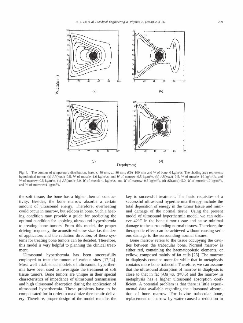
Under the three SARR criteria, SARR(zt,0)̂ 3,SARR(zt,zma)^3 andSARR(zt,zb)^3, there is a low levelof SARRdistribution in the tumor/marrow interface. Inother words, tumor cells near the tumor/marrow interfacecannot receive adequate ultrasound energy to achievehyperthermic therapeutic effect. However, the highesttemperature point in the focal zone transfers heat to thetumor/marrow interface. The temperature contour distri-butions of different blood perfusions in tissues andAR(ma, t)s are displayed in Fig. 4. The area of whichtemperature is higher than 42°C covers not only thewhole tumor but also part of muscle and bone marrow.The temperature distribution depends on theSARRdistri-bution, blood perfusion in tissues, and thermal conduc-tivity of tissues. Assuming thatmb is 30 timesmm, mma
is 0.5, 1, 3, and 5 timesmm, we specify the blood per-fusion in muscle and blood perfusion in bone marrowas shown in Fig. 5. To find theSARRdistributions inthe four differentmma, the temperature distributions weresimulated in 12 cases by BHTE to make all the highesttemperature in tumor be 45°C. The percentages oftumor.42°C/tumor, muscle.42°C/muscle, bonemarrow.42°C/bone marrow, and bone.42°C/bone inthe cone are shown in Fig. 5. We can find that higherattenuation and lower blood perfusion in bone marrowmay achieve 100%.42°C of tumor.
4. Discussion
In this study, we utilized a model to investigate theoptimal frequency and therapeutic domain for appli-cation of ultrasound hyperthermia to bone tumor treat-ment. The results have demonstrated that in spite of thehigh ultrasound absorption of bone, the temperature inbone cortex is not high (Figs. 4 and 5). As compared to

258 B.-Y. Lu et al. / Medical Engineering & Physics 22 (2000) 253–263
Fig. 3. The effect of parameters (AR(ma,t),zt, d(0), SARR, andzb) on therapeutic tumor diameter and optimal attenuation: (a)AR(ma,t),2.0; (b)AR(ma,t)̂ 2.0; (c)zt; (d) zb; (f) SARR. (The typical parameters in this work ared(0)=100 mm,zt=50 mm,zb=80 mm (Db=30 mm),SARR(zt,0)̂ 3,SARR(zt,zma)^3, SARR(zt,zb)^3, AR(m,t) AR(ma,t)=1, andAR(b,t)=30).
259B.-Y. Lu et al. / Medical Engineering & Physics 22 (2000) 253–263
Fig. 4. The contour of temperature distribution, here,zt=50 mm,zb=80 mm,d(0)=100 mm andW of bone=0 kg/m3/s. The shading area representshypothetical tumor: (a)AR(ma,t)=0.5, W of muscle=1.0 kg/m3/s, andW of marrow=0.5 kg/m3/s, (b) AR(ma,t)=0.5, W of muscle=10 kg/m3/s, andW of marrow=0.5 kg/m3/s, (c) AR(ma,t)=5.0, W of muscle=1 kg/m3/s, andW of marrow=0.5 kg/m3/s, (d) AR(ma,t)=5.0, W of muscle=10 kg/m3/s,andW of marrow=1 kg/m3/s.
the soft tissue, the bone has a higher thermal conduc-tivity. Besides, the bone marrow absorbs a certainamount of ultrasound energy. Therefore, overheatingcould occur in marrow, but seldom in bone. Such a heat-ing condition may provide a guide for predicting theoptimal condition for applying ultrasound hyperthermiato treating bone tumors. From this model, the properdriving frequency, the acoustic window size, i.e. the sizeof applicators and the radiation direction, of these sys-tems for treating bone tumors can be decided. Therefore,this model is very helpful to planning the clinical treat-ment.
Ultrasound hyperthermia has been successfullyemployed to treat the tumors of various sites [17,24].Most well established models of ultrasound hyperther-mia have been used to investigate the treatment of softtissue tumors. Bone tumors are unique in their specialcharacteristics of impedance of ultrasound transmissionand high ultrasound absorption during the application ofultrasound hyperthermia. These problems have to becompensated for in order to maximize therapeutic deliv-ery. Therefore, proper design of the model remains the
key to successful treatment. The basic requisites of asuccessful ultrasound hyperthermia therapy include thetotal deposition of energy in the tumor tissue and mini-mal damage of the normal tissue. Using the presentmodel of ultrasound hyperthermia model, we can achi-eve 42°C in the bone tumor tissue and cause minimaldamage to the surrounding normal tissues. Therefore, thetherapeutic effect can be achieved without causing seri-ous damage to the surrounding normal tissues.
Bone marrow refers to the tissue occupying the cavi-ties between the trabecular bone. Normal marrow iseither red, containing the haematopoietic elements, oryellow, composed mainly of fat cells [25]. The marrowin diaphysis contains more fat while that in metaphysiscontains more bone trabeculi. Therefore, we can assumethat the ultrasound absorption of marrow in diaphysis isclose to that in fat (AR(ma, t)=0.5) and the marrow inmetaphysis has a higher ultrasound absorption coef-ficient. A potential problem is that there is little experi-mental data available regarding the ultrasound absorp-tion of bone marrow. For bovine trabecular bone,replacement of marrow by water caused a reduction in

260 B.-Y. Lu et al. / Medical Engineering & Physics 22 (2000) 253–263
Fig. 5. The percentages of tumor.42°C/tumor, muscle.42°C/muscle, bone marrow.42°C/bone marrow, and bone.42°C/bone in the cone(assuming the blood perfusion of bone=0): (a) AR(ma,t)=0.5, (b)AR(ma,t)=1.0, (c)AR(ma,t)=3.0, and (d)AR(ma,t)=5.0.
the mean specific differential attenuation (SDA) of 2.0dB/cm/MHz and 5.0 dB/cm/MHz for the 500 kHz and1 MHz transducer pairs, respectively [26]. We estimateAR(ma, t)=0.5 to 5.0 in Fig. 3(a) and (b) showing thatmopt decreases with theAR(ma, b), while Max(d(zt))increases withAR(ma, t). Therefore, the treatable diam-
eter of tumor in the metaphysis is greater than that inthe diaphysis. From another point of view, the diameterof the metaphysis is greater than that of the diaphysis inthe long bone. As shown in Fig. 3(e), the treatable diam-eter of bone tumor increased with a larger bone diameter.Thus, the treatable diameter of tumor in the metaphysis
261B.-Y. Lu et al. / Medical Engineering & Physics 22 (2000) 253–263
is greater than that in diaphysis. According to these tworesults, we can conclude that the treatable diameter oftumor in the metaphysis is greater than that in thediaphysis.
The blood perfusion of bone marrow was assumed tobe 0 to 5 kg/m3/s in this study [16]. As aforementionedin the long bone, the blood perfusion in the metaphysiscould be higher than that in the diaphysis. Therefore, inthe metaphysis, bone marrow has higherAR(ma, t) andblood perfusion; while in the diaphysis, bone marrowhas lowerAR(ma, t) and blood perfusion. In Fig. 5, wefound that the lower blood perfusion ratio of bone mar-row to muscle and the higherAR(ma, t) can easily achi-eve 100% tumor therapeutic percentage. From this pointof view, the blood perfusion andAR(ma, t) are in thetrade-off state for ultrasound hyperthermia application inthe diaphysis and metaphysis.
The therapeutic parameters in this work are drivingfrequency of ultrasonic applicators and acoustic windowon the surface skin (Fig. 3). The variations in blood per-fusion and attenuation coefficient in Fig. 5 are used toverify the SARRcriteria, and not as controllable para-meters to be optimized. In fact, blood perfusion can bea controllable parameter in hyperthermia. For example,Vertrees et al. had developed the venovenous perfusionand temperature monitoring system. Blood was drainedfrom the jugular vein, pumped through a servo-regulatedheat exchange system, with controlled flow, and rein-fused into the femoral vein [27].
For a planar 1D array,GG=1 [13]. The effect of powerconvergence is worse than that for a 2-D convergent sys-tem. Hence, for a spherical 2-D phased array, if thepower is converged by the in-phase method, the wav-eform is similar to that of the spherical transducer. Sothe GG equals (d(0)/d(z))2. In this viewpoint, the modelin this work can be approached to the spherical 2-Dphased array. Moreover, it is useful to employ the 0.5to 1.0 MHz 2-D phased arrays for treating the deep-seated tumor in homogeneous tissue. However, in ourstudy, the aforementioned driving frequencies are rela-tively low. Overheating in bone cortex cannot be avo-ided because of the low attenuation in soft tissue byadjusting the amplitude and relative phase of theelements in these arrays. One of the better ways for opti-mization is to select the higher resonant frequencyelements in the phased array. Therefore, this study sup-ports an inverse method for finding the optimal fre-quency range for treating bone tumor treatment.
Fujii et al. [11] have indicated that at incident angleslarger than the critical angle of the transversal wave, notemperature rise results from the ultrasound absorptionbone; the incident longitudinal wave, strengthened by thereflected wave, is absorbed in the muscle and is trans-ported to the interface, so that the temperature of theinterface and bone increases slightly. They havedesigned the experiment to focus on the ultrasound
power at the muscle–bone interface. The experimentalresults show that when the incident angle=40°, the tem-perature rises about 14°C at the interface. In our study,the ultrasound power is focused on the tumor site. Thepower at the muscle–bone interface is relatively smallas compared to that in the focal zone. Therefore, if theincident angle is also 40°, the temperature rise shouldbe much smaller than 14°C. Furthermore, the model inthis work covered the various incident angles within theacoustic window, and the approximation used in thisstudy overestimated the absorbed power at the boneinterface by about 8 to 41%. If bone heating can be avo-ided in these simulations, it should also be possible dur-ing clinical treatment. The actual temperature in the bonewould be lower than predicted by the model [17].
In this study, we use theSARRcriteria to estimate thetherapeutic parameters, but not temperature distributions.However, McGough et al. pointed out that the BHTEhas the property of the spatial low-pass filter effect [28].Although the temperature is not exactly proportional toQ(SAR), the temperature distribution can be estimatedfrom the SARdistribution. Charny et al. [6] applied amini-annular phased array to treating human leg withemitting radio frequency energy. The design of thisphased array tries to maximize theSARi.e. energy depo-sition, in the central area. The heating results werepresented inSAR distribution. Ebbini and Cain havepresented a field optimization method which concen-trates power at a focal point and reduces interferencepatterns (hot spot) elsewhere to treat deep-seated tumorin homogeneous tissue [29]. Therefore, we can use theSARdistribution to estimate the temperature distribution.Fig. 5 verifies that theSARRcriteria support the propertemperature distributions.
However, the model in this work cannot calculate theprecise ultrasound intensity field as the Rayleigh–Somm-erfield diffraction integral method [24] or source pointmethod [30]. But, it is helpful to estimate the operatingfrequency and the size of ultrasound transducers quickly,especially in the multilayer tissue cases. Further investi-gation is needed for the evaluation of its potential clini-cal applications. For the moving energy of focus scan-ning ultrasound hyperthermic system, it is reasonable toaverage temporally the absorption energy in tissue toneglect roughly the interference phenomenon in a steadystate. Therefore, this model can represent geometric con-vergent focused and spherically arranged hyperthermicapplicators such as the SFUS, and spherically segmentedarrays [31–33], but not necessarily a single focused appl-icator. The simulations of temperature distribution inthese systems are very complex. For example, the powerarray in SFUS has to be tilted, rotated, translated andconvoluted to obtain the power distributions [18]. If theresonant frequency is changed, the procedures describedabove have to restart. Therefore, it is time-consuming,especially in the multi-layered tissues, to find the optimal
262 B.-Y. Lu et al. / Medical Engineering & Physics 22 (2000) 253–263
driving frequency by changing the control parameters inthese systems. The present model supports an easy andrapid theoretical estimation for the range of optimal driv-ing frequency in these systems. The results in Fig. 3 arehelpful for estimating quickly whether the bone tumoris treatable in the multilayer tissue condition, to choosethe optimal operating frequency of ultrasound transducerand the acoustic window on the skin surface when thetumor size, tumor depth, and bone diameter are known,and to design an optimal ultrasound applicator forbone tumors.
5. Conclusion
This study employs a model with a simplified ultra-sound beam form model to examine the optimal drivingfrequency and treatable domain formed by the tumor sizeand tumor depth and to simulate theSARdistribution forthe hyperthermic treatment of a bone tumor. The resultsdemonstrate that the treatable diameter of a bone tumorincreases with the absorption ratio of bone marrow totumor, acoustic window of surface skin, and diameter ofbone, but decreases with muscle depth, andSARratio ofbone tumor site to the surface skin, bone marrow, andbone. Besides, the optimal driving frequency depends onthe tumor depth, ultrasound absorption of bone marrow,and bone diameter, but is independent on the acousticwindow area andSARratio under the threeSARRcri-teria. The results are helpful for estimating rapidlywhether the bone tumor is treatable in the multilayertissue condition, to choose the optimal operating fre-quency of the ultrasound transducer and the acousticwindow on the skin surface when the tumor size, tumordepth, and bone diameter of bone are known, and todesign ultrasound applicators, such as SFUS, and spheri-cally segmented arrays for treating bone tumors. There-fore, this model is very helpful for planning the clinicaltreatment with these systems.
Acknowledgements
The authors thank the National Science Council andthe Department of Health of the Republic of China forpartially supporting this research, NSC 87-2213-E-002-075 and DOH 87-HR-635.
References
[1] Seegenschmiedt MH, Feldmann HJ. Clinical rationale for thermo-radiotherapy. In Seegenschmiedt MH, Feldmann HJ, Vernon CC,editors. Thermoradiotherapy and Thermochemotherapy, vol. 2.Berlin/Heidelberg/New York: Springer-Verlag, 1995:3–23.
[2] Overgaard J. The current and potential role of hyperthermia inradiotherapy. Int J Radiat Oncol Biol Phy 1989;16:535–49.
[3] Vernon CC, Hand JW, Field SB, Machin D, Whaley JB, van derZee J, van Putten WL, van Rhoon GC, van Dijk JDP, GonzalezDG, Liu FF, Goodman P, Sherar M. Radiotherapy with or withouthyperthermia in the treatment of superficial localized breast can-cer: results from five randomized controlled trials. Int J RadiatOncol Biol Phys 1996;35:731–44.
[4] Fan QY, Ma BA, Qiu XC, Li YL, Ye J, Zhou Y. Preliminaryreport on treatment of bone tumors with microwave-induced hyp-erthermia. Bioelectromagnetics 1996;17:218–22.
[5] Ikenaga M, Ohura K, Yamamuro T, Kotoura Y, Oka M, KokuboT. Localized hyperthermic treatment of experimental bone tumorswith ferromagnetic ceramics. J Orthop Res 1993;11:849–55.
[6] Charny CK, Guerquin-Kern J, Hagmann MJ, Levin SW, LackEE, Sindelar WF, Zabell A, Glatstein E, Levin RL. Human legheating using a mini-annular phased array. Med Phy1986;13:449–56.
[7] Ogilvie GK, Reynolds HA, Richardson BC, Badger CW, GossSA, Burdette EC. Performance of a multi-sector ultrasound hyp-erthermia applicator and control system: in vivo studies. Int JHyperthermia 1990;6:697–705.
[8] Hynynen K, Shimm D, Anhalt D, Stea B, Sykes H, Cassady JR,Roemer RB. Temperature distributions during clinical scanned,focused ultrasound hyperthermia treatments. Int J Hyperthermia1990;6:891–908.
[9] Hynynen K, DeYoung D. Temperature elevation at muscle–boneinterface during scanned, focused ultrasound hyperthermia. Int JHyperthermia 1988;4:267–79.
[10] Corry PM, Barlogie B, Tilchen EJ, Armour EP. Ultrasound-induced hyperthermia for the treatment of human superficialtumors. Int J Radiat Oncol Biol Phys 1982;8:1225–9.
[11] Fujii M, Sakamoto K, Toda Y, Negishi A, Kanai H. Study of thecause of the temperature rise at the muscle–bone interface duringultrasound hyperthermia. IEEE Trans Biomed Eng1999;46:494–504.
[12] Hill C. Optimum acoustic frequency for focused ultrasound sur-gery. Ultrasound Med Biol 1994;20:271–7.
[13] Fry FJ, Dines KA, Reilly CR, Goss SA. Losses in tissue associa-ted with finite amplitude ultrasound transmission. UltrasoundMed Biol 1989;15:481–97.
[14] Carstensen EL, McKay ND, Dalecki D, Muir TG. Absorption offinite amplitude ultrasound in tissues. Acoustica 1982;51:116–23.
[15] Lin WL, Yen JY, Chen YY, Jin KW, Shieh MJ. Relationshipbetween aperture size and tumor conditions for external ultra-sound hyperthermia. Med Phy 1999;26:818–24.
[16] Hynynen K. Biophysics and technology of ultrasound hyperther-mia. In: Gautherie M, editor. Method of External HyperthermicHeating. Berlin/Heidelberg/New York: Springer-Verlag,1990:61–115.
[17] Tu SJ, Hynynen K, Roemer RB. Simulation of bidirectional ultra-sound hyperthermia treatments of neck tumours. Int J Hyperther-mia 1994;10:707–22.
[18] Moros EG. Simulations of scanned focused ultrasound hyperther-mia: The effect of scanning speed, scanning pattern and multipletitled transducers. Master Thesis, Univ. of Arizona, Tucson, Ari-zona, USA, 1987.
[19] Moros EG, Roemer RB, Hynynen K. Pre-focal plane high tem-perature regions induced by scanning focus ultrasound beams. IntJ Hyperthermia 1990;6:351–66.
[20] Pennes HH. Analysis of tissue and arterial blood temperatures inthe resting human forearm. J Appl Physiol 1948;1:93–122.
[21] Chivers RC, Parry RJ. Ultrasonic velocity and attenuation inmammalian tissues. J Acoustic Society Am 1978;63:940–53.
[22] Goss SA, Johnston RL, Dunn F. Comprehensive compilation ofempirical ultrasonic properties of mammalian tissues. J AcousticSociety Am 1978;64(2):423–57.
[23] Goss SA, Johnston RL, Dunn F. Comprehensive compilation of
263B.-Y. Lu et al. / Medical Engineering & Physics 22 (2000) 253–263
empirical ultrasonic properties of mammalian tissues, II. J Acous-tic Society Am 1982;68(1):93–108.
[24] Hynynen K. Ultrasound heating techonology. In: SeegenschmiedtMH, Feldmann HJ, Vernon CC, editors. Thermoradiotherapy andThermochemotherapy, vol. 1. Berlin/Heidelberg/New York:Springer-Verlag, 1995:253–79.
[25] Frisch B, Lewis SM, Burkhardt R, Bartl R. Biopsy pathology ofbone and bone marrow. London: Chapman and Hall, 1985.
[26] Alves JM, Ryaby JT, Kaufman JJ, Magee PF, Siffert RS. Influ-ence of marrow on ultrasonic velocity and attenuation in bovinetrabecular bone. Calcif Tissue Int 1996;58:362–7.
[27] Vertrees RA, Tao W, Pencil SD, Sites JP, Althoff DP, Zwi-schenberger JB. Induction of whole body hyperthermia withvenovenous perfusion. ASAIO J 1996;42:250–4.
[28] McGough R, Ebbini E, Cain C. Direct computation of ultrasoundphased-array driving signals from a specified temperature distri-bution for hyperthermia. IEEE Trans Biomed Eng1992;39:825–35.
[29] Ebbini E, Cain C. Multiple-focus ultrasound phased-array patternsynthesis: Optimal driving signal distributions for hyperthermia.IEEE Trans Ultrason Ferroelect Freq Contr 1989;36:540–8.
[30] Goss SA, Frizzell LA, Kouzmanoff JT, Barich JM, Yang JM.Sparse random ultrasound phased array for local surgery. IEEETrans Ultrason Ferroelec Freq Contr 1996;43:1111–21.
[31] Ebbini ES, Cain CA. A spherical-section ultrasound phased arrayapplicator for deep localized hyperthermia. IEEE Trans onBiomed Eng 1991;38:634–43.
[32] Ibbini MS, Ebbini ES, Cain CA. N×N square-element ultrasoundphased-array applicator simulated temperature distributions asso-ciated with directly synthesized heating patterns. IEEE TransUltrason Ferroelec Freq Contr 1990;37:491–500.
[33] Fan X, Moros EG, Straube WL. A concentric-ring equivalentphased array method to model fields of large axisymmetric ultra-sound transducers. IEEE Trans Ultrason Ferroelec Freq Contr1999;37:830–41.


![[Treating frostbite injuries]](https://static.fdokumen.com/doc/165x107/633ff39332b09e4bae09a1b5/treating-frostbite-injuries.jpg)