In vivo biocompatibility of n-CDHA/PAA biomaterials
14
© 2015 Dai et al. This work is published by Dove Medical Press Limited, and licensed under Creative Commons Attribution – Non Commercial (unported, v3.0) License. The full terms of the License are available at http://creativecommons.org/licenses/by-nc/3.0/. Non-commercial uses of the work are permitted without any further permission from Dove Medical Press Limited, provided the work is properly attributed. Permissions beyond the scope of the License are administered by Dove Medical Press Limited. Information on how to request permission may be found at: http://www.dovepress.com/permissions.php International Journal of Nanomedicine 2015:10 6303–6316 International Journal of Nanomedicine Dovepress submit your manuscript | www.dovepress.com Dovepress 6303 ORIGINAL RESEARCH open access to scientific and medical research Open Access Full Text Article http://dx.doi.org/10.2147/IJN.S90273 In vivo biocompatibility of new nano-calcium- deficient hydroxyapatite/poly-amino acid complex biomaterials Zhenyu Dai 1,2, * Yue Li 3, * Weizhong Lu 2, * Dianming Jiang 4 Hong Li 1 Yonggang Yan 1 Guoyu Lv 1 Aiping Yang 1 1 College of Physical Science and Technology, Sichuan University, Chengdu, 2 Department of Orthopedics, Chongqing Hospital of Traditional Chinese Medicine, 3 Department of Clinical Laboratory, the Second Affiliated Hospital, 4 Department of Orthopedics, the First Affiliated Hospital, Chongqing Medical University, Chongqing, People’s Republic of China *These authors contributed equally to this work Objective: To evaluate the compatibility of novel nano-calcium-deficient hydroxyapatite/ poly-amino acid (n-CDHA/PAA) complex biomaterials with muscle and bone tissue in an in vivo model. Methods: Thirty-two New Zealand white rabbits were used in this study. Biomaterials were surgically implanted into each rabbit in the back erector spinae and in tibia with induced defect. Polyethylene was implanted into rabbits in the control group and n-CDHA/PAA into those of the experimental group. Animals were examined at four different points in time: 2 weeks, 4 weeks, 12 weeks, and 24 weeks after surgery. They were euthanized after embolization. Back erector spinae muscles with the surgical implants were examined after hematoxylin and eosin (HE) staining at these points in time. Tibia bones with the surgical implants were examined by X-ray and scanning electron microscopy (SEM) at these points in time to evaluate the interface of the bone with the implanted biomaterials. Bone tissues were sectioned and subjected to HE, Masson, and toluidine blue staining. Results: HE staining of back erector spinae muscles at 4 weeks, 12 weeks, and 24 weeks after implantation of either n-CDHA/PAA or polyethylene showed disappearance of inflammation and normal arrangement in the peripheral tissue of implant biomaterials; no abnormal staining was observed. At 2 weeks after implantation, X-ray imaging of bone tissue samples in both experimental and control groups showed that the peripheral tissues of the implanted biomaterials were continuous and lacked bone osteolysis, absorption, necrosis, or osteomyelitis. The con- nection between implanted biomaterials and bone tissue was tight. The results of HE, Masson, toluidine blue staining and SEM confirmed that the implanted biomaterials were closely con- nected to the bone defect and that no rejection had taken place. The n-CDHA/PAA biomaterials induced differentiation of a large number of chondrocytes. New bone trabecula began to form at 4 weeks after implanting n-CDHA/PAA biomaterials, and lamellar bone gradually formed at 12 weeks and 24 weeks after implantation. Routine blood and kidney function tests showed no significant changes at 2 weeks and 24 weeks after implantation of both biomaterials. Conclusion: n-CDHA/PAA composites showed good compatibility in in vivo model. In this study, n-CDHA/PAA were found to be safe, nontoxic, and biologically active in bone repair. Keywords: in vivo implantation, histological evaluation, n-CDHA/PAA, bioactive composite Introduction Trauma- and tumor-induced bone defects are problems that can emerge after surgery. 1 Although bone autograft and allograft implants have been used clinically to address bone defects, multiple bone autografts can burden patients with numerous adverse effects and so cause considerable suffering; in addition, implantation of allografts Correspondence: Hong Li; Yonggang Yan College of Physical Science and Technology, Sichuan University, Chengdu 610064, People’s Republic of China Tel +86 028 8512 7592 Fax +86 028 8518 7573 Email [email protected]m; [email protected]m
Transcript of In vivo biocompatibility of n-CDHA/PAA biomaterials
© 2015 Dai et al. This work is published by Dove Medical Press Limited, and licensed under Creative Commons Attribution – Non Commercial (unported, v3.0) License. The full terms of the License are available at http://creativecommons.org/licenses/by-nc/3.0/. Non-commercial uses of the work are permitted without any further
permission from Dove Medical Press Limited, provided the work is properly attributed. Permissions beyond the scope of the License are administered by Dove Medical Press Limited. Information on how to request permission may be found at: http://www.dovepress.com/permissions.php
International Journal of Nanomedicine 2015:10 6303–6316
International Journal of Nanomedicine Dovepress
submit your manuscript | www.dovepress.com
Dovepress 6303
O r I g I N a l r e s e a r c h
open access to scientific and medical research
Open access Full Text article
http://dx.doi.org/10.2147/IJN.S90273
In vivo biocompatibility of new nano-calcium-deficient hydroxyapatite/poly-amino acid complex biomaterials
Zhenyu Dai1,2,*Yue li3,*Weizhong Lu2,*Dianming Jiang4
Hong Li1
Yonggang Yan1
guoyu lv1
Aiping Yang1
1College of Physical Science and Technology, Sichuan University, Chengdu, 2Department of Orthopedics, Chongqing Hospital of Traditional Chinese Medicine, 3Department of Clinical Laboratory, the Second Affiliated Hospital, 4Department of Orthopedics, the First Affiliated Hospital, Chongqing Medical University, Chongqing, People’s Republic of China
*These authors contributed equally to this work
Objective: To evaluate the compatibility of novel nano-calcium-deficient hydroxyapatite/
poly-amino acid (n-CDHA/PAA) complex biomaterials with muscle and bone tissue in an
in vivo model.
Methods: Thirty-two New Zealand white rabbits were used in this study. Biomaterials were
surgically implanted into each rabbit in the back erector spinae and in tibia with induced defect.
Polyethylene was implanted into rabbits in the control group and n-CDHA/PAA into those
of the experimental group. Animals were examined at four different points in time: 2 weeks,
4 weeks, 12 weeks, and 24 weeks after surgery. They were euthanized after embolization. Back
erector spinae muscles with the surgical implants were examined after hematoxylin and eosin
(HE) staining at these points in time. Tibia bones with the surgical implants were examined by
X-ray and scanning electron microscopy (SEM) at these points in time to evaluate the interface
of the bone with the implanted biomaterials. Bone tissues were sectioned and subjected to HE,
Masson, and toluidine blue staining.
Results: HE staining of back erector spinae muscles at 4 weeks, 12 weeks, and 24 weeks after
implantation of either n-CDHA/PAA or polyethylene showed disappearance of inflammation
and normal arrangement in the peripheral tissue of implant biomaterials; no abnormal staining
was observed. At 2 weeks after implantation, X-ray imaging of bone tissue samples in both
experimental and control groups showed that the peripheral tissues of the implanted biomaterials
were continuous and lacked bone osteolysis, absorption, necrosis, or osteomyelitis. The con-
nection between implanted biomaterials and bone tissue was tight. The results of HE, Masson,
toluidine blue staining and SEM confirmed that the implanted biomaterials were closely con-
nected to the bone defect and that no rejection had taken place. The n-CDHA/PAA biomaterials
induced differentiation of a large number of chondrocytes. New bone trabecula began to form
at 4 weeks after implanting n-CDHA/PAA biomaterials, and lamellar bone gradually formed
at 12 weeks and 24 weeks after implantation. Routine blood and kidney function tests showed
no significant changes at 2 weeks and 24 weeks after implantation of both biomaterials.
Conclusion: n-CDHA/PAA composites showed good compatibility in in vivo model. In this
study, n-CDHA/PAA were found to be safe, nontoxic, and biologically active in bone repair.
Keywords: in vivo implantation, histological evaluation, n-CDHA/PAA, bioactive
composite
IntroductionTrauma- and tumor-induced bone defects are problems that can emerge after surgery.1
Although bone autograft and allograft implants have been used clinically to address
bone defects, multiple bone autografts can burden patients with numerous adverse
effects and so cause considerable suffering; in addition, implantation of allografts
Correspondence: Hong Li; Yonggang YanCollege of Physical Science and Technology, Sichuan University, Chengdu 610064, People’s Republic of ChinaTel +86 028 8512 7592 Fax +86 028 8518 7573 email [email protected]; [email protected]
Journal name: International Journal of NanomedicineArticle Designation: Original ResearchYear: 2015Volume: 10Running head verso: Dai et alRunning head recto: In vivo biocompatibility of n-CDHA/PAA biomaterials DOI: http://dx.doi.org/10.2147/IJN.S90273
International Journal of Nanomedicine 2015:10submit your manuscript | www.dovepress.com
Dovepress
Dovepress
6304
Dai et al
may induce rejection and other problems.2–4 The develop-
ment of an ideal artificial restoration material has become an
important topic in bone tissue engineering. Nano-calcium-
deficient hydroxyapatite (n-CDHA) and poly-amino acid
(PAA) undergo in situ polymerization to form n-CDHA/
PAA composites. These composites were recently produced
in the current laboratory for the first time and may serve as
bone repair biomaterials.5 The copolymers of these amino
acids were based on 6-aminocaproic acid as a main chain and
α-amino acids of human body as copolymerized units. The
6-aminocaproic acid gives the copolymer good mechanical
properties and process abilities to polymers. Copolymeriza-
tion of other natural amino acid monomers can produce
polymers with different physical and chemical properties
(eg, affinity, hydrophobic properties, degradation rates, deg-
radation product, and pH).6 n-CDHA bases on the particle
structure (particle diameter of 80–100 nm) cause the particle
to disperse in PAA matrix biomaterial uniformly, allowing
two types of chemical bonds on the interface of composites
between n-CDHA and PAA: -COO- ions of PAA replace the
anions (OH- and Po34
-) of n-CDHA and form a strong chemi-
cal bond with Ca2+ ions; O–H of n-CDHA forms hydrogen
bonds with the amide bond in the polymer chain. These two
types of combinations allow very good transmission of force
and dispersion of stress, improving the mechanical properties
of the biomaterials.6 A previous study showed that n-CDHA/
PAA composites had good mechanical properties and cell
compatibility.5 To further explore the possibility of apply-
ing n-CDHA/PAA in the repair of human bone tissue and
their biocompatibility in in vivo tissues, this study implanted
n-CDHA/PAA composites into erector spinae and tibia bone
of experimental animals.
Materials and methodsMaterialsφ6×2 mm polyethylene (PE) and n-CDHA/PAA composites
were provided by Sichuan International Nano Co., Ltd.
(Sichuan, People’s Republic of China). n-CDHA/PAA com-
posites were legally prepared by in situ polymerization.7,8
n-CDHA mass fraction of n-CDHA/PAA was 30 wt%.
PAA contains six kinds of amino acids: 6-aminocaproic
acid, glycine, l-alanine, l-phenylalanine, l-proline, and
l-lysine. All engineered implants were sterilized using
ethylene oxide.
Experimental animals and groupingA total of 32 New Zealand white rabbits were provided by
Laboratory Animal Center, Chongqing Medical University
(Chongqing, People’s Republic of China), of both sexes and
body weight ranging from 2.0 kg to 2.5 kg. For all animals,
breeding conditions were kept the same before and during the
study. Animals were divided into n-CDHA/PAA experimen-
tal group and PE control group. All animals received humane
care in compliance with the Public Health Service Policy
on Humane Care and Use of Laboratory Animals. Ethical
approval was obtained from the Animal Care and Ethics
Committee of Chongqing Medical University of China.
MethodsAll 32 animals were anesthetized with 3% sodium pentobar-
bital (1 mL/kg) intravenously via the ear. Each animal was
fixed in a supine position. The skin was prepared in the con-
ventional manner, including disinfection and placement of
surgical drapes. A longitudinal skin incision ~2 cm in length
was made along the lateral tibia of the upper two-thirds of
rabbit calf, followed by separation of the subcutaneous tissue,
fascia, and muscle to expose the proximal tibia. Upper and
lower holes were drilled outward from the lateral side of tibia
to the contralateral cortex, with ~10 mm spacing and 2 mm
diameter. n-CDHA/PAA and PE biomaterials were implanted
into animals in the experimental and the control groups, fol-
lowed by suturing and topical skin closure, disinfection, and
application of erythromycin ointment to the incisions. Each
animal that received an intramuscular implant was fixed in
a prone position, followed by conventional skin preparation,
disinfection, and placement of surgical drapes. Longitudinal
incision along the back of the skin was performed, followed
by separation of the subcutaneous tissue and superficial fas-
cia to expose the erector spinae muscle on both sides. This
muscle was blunt dissection. In the experimental group, two
pieces of n-CDHA/PAA biomaterials were longitudinally
implanted in each side of the erector muscle (with spacing
of 2 cm). A total of four pieces of n-CDHA/PAA biomate-
rial were implanted into each animal. In the control group,
PE biomaterials were implanted into each animal as in the
experimental group. All animals were sutured to close the
skin. This was followed by disinfection and application of
erythromycin ointment to the incisions. Each animal received
intramuscular injection of penicillin (200,000 units daily) in
the first 3 days after the surgery.
extraction and observation index of implant biomaterialsPostoperative conditions, general food intake, and incision
sites of each animal were examined at 2 weeks, 4 weeks,
12 weeks, and 24 weeks, followed by embolization to eutha-
nize the animals in both groups (four animals per point in time;
16 animals per group). Approximately 5–10 mm of muscle
International Journal of Nanomedicine 2015:10 submit your manuscript | www.dovepress.com
Dovepress
Dovepress
6305
In vivo biocompatibility of n-CDHA/PAA biomaterials
surrounding the intramuscular implanted biomaterials was
dissected. General conditions of the peripheral muscle of
the implant biomaterials were observed with the naked eye.
Three samples were randomly selected from each group
at each point in time for the hematoxylin and eosin (HE)
staining. Areas of tibia bones with implanted biomaterials
underwent frontal and lateral X-ray imaging at the specified
points in time to show the interface of the implant biomaterials
with the animal’s own tissue. Animals in both groups were
euthanized after embolism at the specified points in time.
Approximately 10 mm width of the bone tissue surrounding
the implanted biomaterials were extracted along the original
incision. General conditions of the tissue surrounding the
implanted biomaterials were observed with the naked eye.
Two samples were randomly selected from both groups at
each point in time, and the soft tissue was carefully removed
from the surface of implanted biomaterials. Samples were
then fixed in 2% glutaraldehyde for scanning electron micros-
copy (SEM) of the implant-bone interface. Three samples
were randomly selected from both groups at each point in
time and fixed in formalin buffer. This was followed by
hard-tissue sectioning and HE, Masson, and toluidine blue
staining to assess inflammation, foreign object rejection,
immune response, and osteogenesis in the bone tissue around
the implanted biomaterials. Three animals from each group
were examined 2 weeks before and after the surgery and
24 weeks after the surgery. Blood was extracted for routine
testing and analysis of biochemical markers for study of the
impacts of implant biomaterials on animal blood, liver, and
kidney functions.
statistical analysisSPSS16.0 software (SPSS Inc., Chicago, IL, USA) were
used for statistical analysis. Results are presented as
mean ± standard deviation (x– ± s). Measurement data were
analyzed by t-test. P,0.05 was considered indicative of
statistically significant differences.
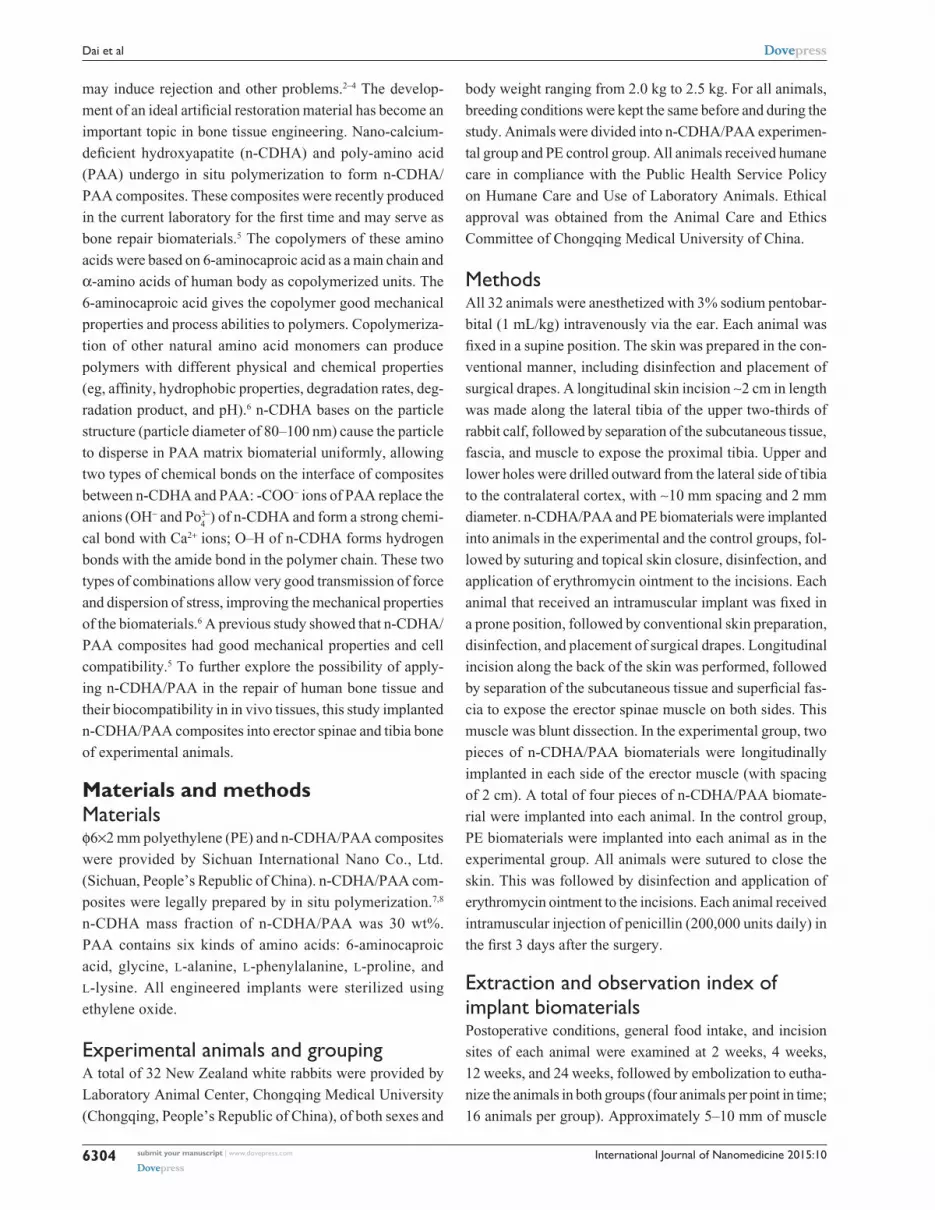
Resultsgeneral condition of in vivo implantationThe general condition of all animals was good after the surgery
and showed no particular abnormalities, incision infection,
or dehiscence of incision. Intramuscular embedded regions
were examined at different points in time after n-CDHA/PAA
implantation. The muscles had engulfed around the bioma-
terials, and no significant edema, infection, or necrosis had
occurred in the peripheral muscles (Figure 1A and C); similar
results were also observed in the control group (Figure 1B
and D). The tibia bone area with implant biomaterials was
collected at postoperative 2 weeks and showed that the
peripheral bones of both implant biomaterials were closely
connected. No significant necrosis, osteolysis, or bleaching
of materials was observed in the bone tissues. In addition,
no significant inflammatory reactions, such as swelling or
fluid accumulation, were observed in the peripheral bone
tissues (Figure 1E and F). Over time, both implant materials
and bone tissues became closely connected. The interface
between bone and implant materials showed no abnormal
reaction. Neither type of implant biomaterials showed any
bleaching. At postoperative 24 weeks, only n-CDHA/PAA in
the experimental group was covered by a thin layer of newly
formed bone tissue (Figure 1G and H).
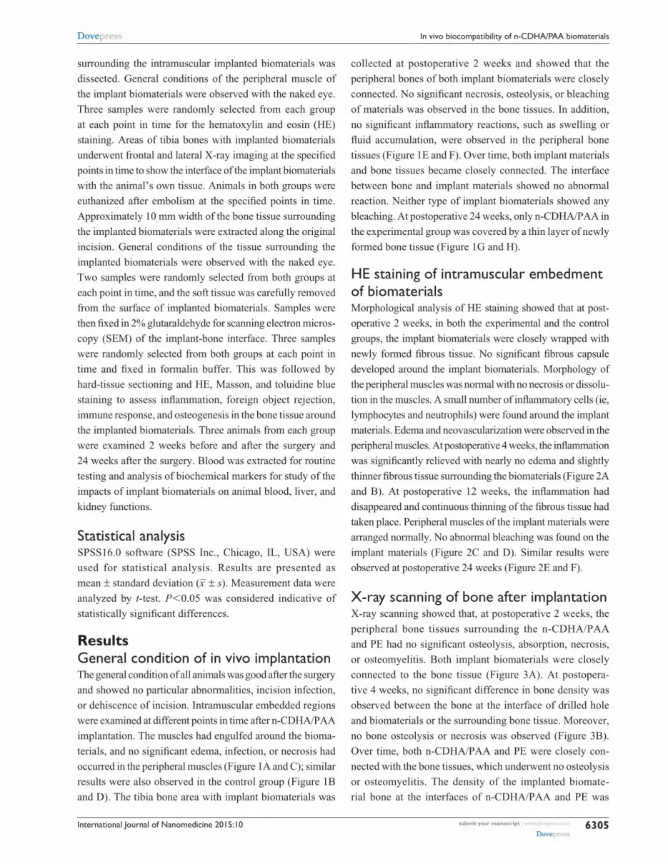
HE staining of intramuscular embedment of biomaterialsMorphological analysis of HE staining showed that at post-
operative 2 weeks, in both the experimental and the control
groups, the implant biomaterials were closely wrapped with
newly formed fibrous tissue. No significant fibrous capsule
developed around the implant biomaterials. Morphology of
the peripheral muscles was normal with no necrosis or dissolu-
tion in the muscles. A small number of inflammatory cells (ie,
lymphocytes and neutrophils) were found around the implant
materials. Edema and neovascularization were observed in the
peripheral muscles. At postoperative 4 weeks, the inflammation
was significantly relieved with nearly no edema and slightly
thinner fibrous tissue surrounding the biomaterials (Figure 2A
and B). At postoperative 12 weeks, the inflammation had
disappeared and continuous thinning of the fibrous tissue had
taken place. Peripheral muscles of the implant materials were
arranged normally. No abnormal bleaching was found on the
implant materials (Figure 2C and D). Similar results were
observed at postoperative 24 weeks (Figure 2E and F).
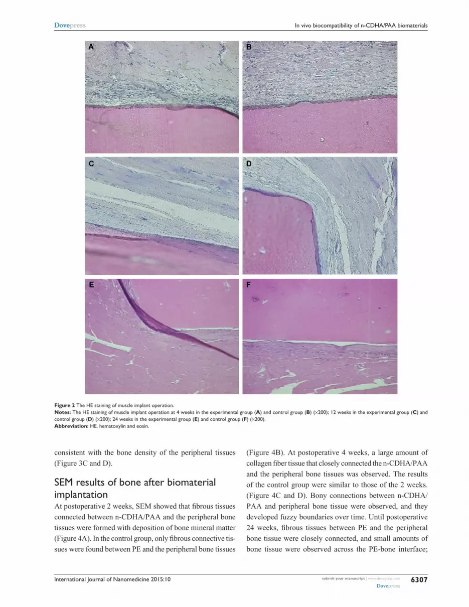
X-ray scanning of bone after implantationX-ray scanning showed that, at postoperative 2 weeks, the
peripheral bone tissues surrounding the n-CDHA/PAA
and PE had no significant osteolysis, absorption, necrosis,
or osteomyelitis. Both implant biomaterials were closely
connected to the bone tissue (Figure 3A). At postopera-
tive 4 weeks, no significant difference in bone density was
observed between the bone at the interface of drilled hole
and biomaterials or the surrounding bone tissue. Moreover,
no bone osteolysis or necrosis was observed (Figure 3B).
Over time, both n-CDHA/PAA and PE were closely con-
nected with the bone tissues, which underwent no osteolysis
or osteomyelitis. The density of the implanted biomate-
rial bone at the interfaces of n-CDHA/PAA and PE was

International Journal of Nanomedicine 2015:10submit your manuscript | www.dovepress.com
Dovepress
Dovepress
6306
Dai et al
Figure 1 The results of the muscle and bone implants observed by naked eye in the different groups.Notes: The specimen of muscle implant operation at 4 weeks in the experimental group (A) and control group (B); 24 weeks in the experimental group (C) and control group (D). The specimen of bone implant operation at 4 weeks in the experimental group (E) and control group (F); 24 weeks in the experimental group (G) and control group (H).
International Journal of Nanomedicine 2015:10 submit your manuscript | www.dovepress.com
Dovepress
Dovepress
6307
In vivo biocompatibility of n-CDHA/PAA biomaterials
consistent with the bone density of the peripheral tissues
(Figure 3C and D).
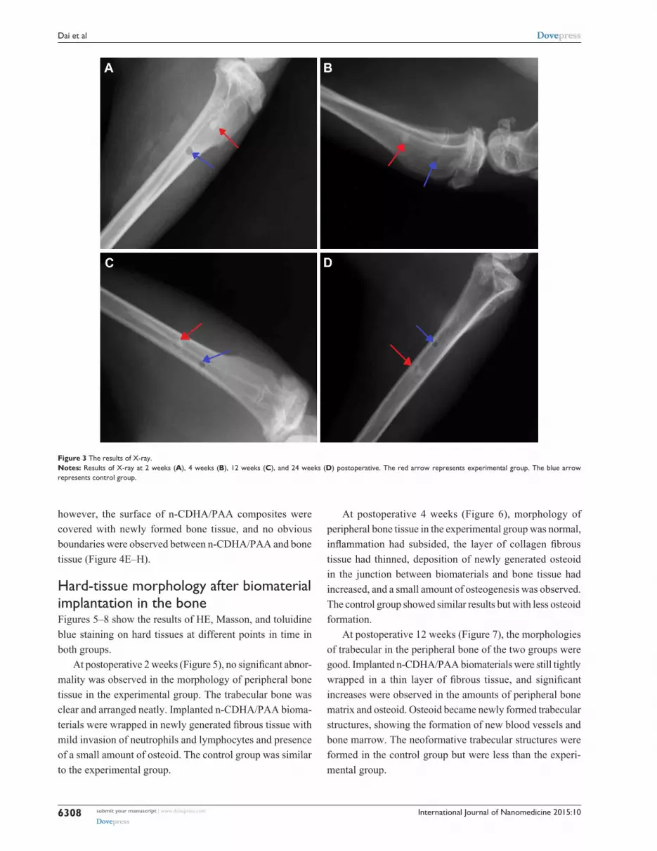
SEM results of bone after biomaterial implantationAt postoperative 2 weeks, SEM showed that fibrous tissues
connected between n-CDHA/PAA and the peripheral bone
tissues were formed with deposition of bone mineral matter
(Figure 4A). In the control group, only fibrous connective tis-
sues were found between PE and the peripheral bone tissues
(Figure 4B). At postoperative 4 weeks, a large amount of
collagen fiber tissue that closely connected the n-CDHA/PAA
and the peripheral bone tissues was observed. The results
of the control group were similar to those of the 2 weeks.
(Figure 4C and D). Bony connections between n-CDHA/
PAA and peripheral bone tissue were observed, and they
developed fuzzy boundaries over time. Until postoperative
24 weeks, fibrous tissues between PE and the peripheral
bone tissue were closely connected, and small amounts of
bone tissue were observed across the PE-bone interface;
Figure 2 The HE staining of muscle implant operation.Notes: The HE staining of muscle implant operation at 4 weeks in the experimental group (A) and control group (B) (×200); 12 weeks in the experimental group (C) and control group (D) (×200); 24 weeks in the experimental group (E) and control group (F) (×200).Abbreviation: HE, hematoxylin and eosin.
International Journal of Nanomedicine 2015:10submit your manuscript | www.dovepress.com
Dovepress
Dovepress
6308
Dai et al
however, the surface of n-CDHA/PAA composites were
covered with newly formed bone tissue, and no obvious
boundaries were observed between n-CDHA/PAA and bone
tissue (Figure 4E–H).
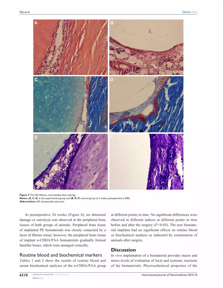
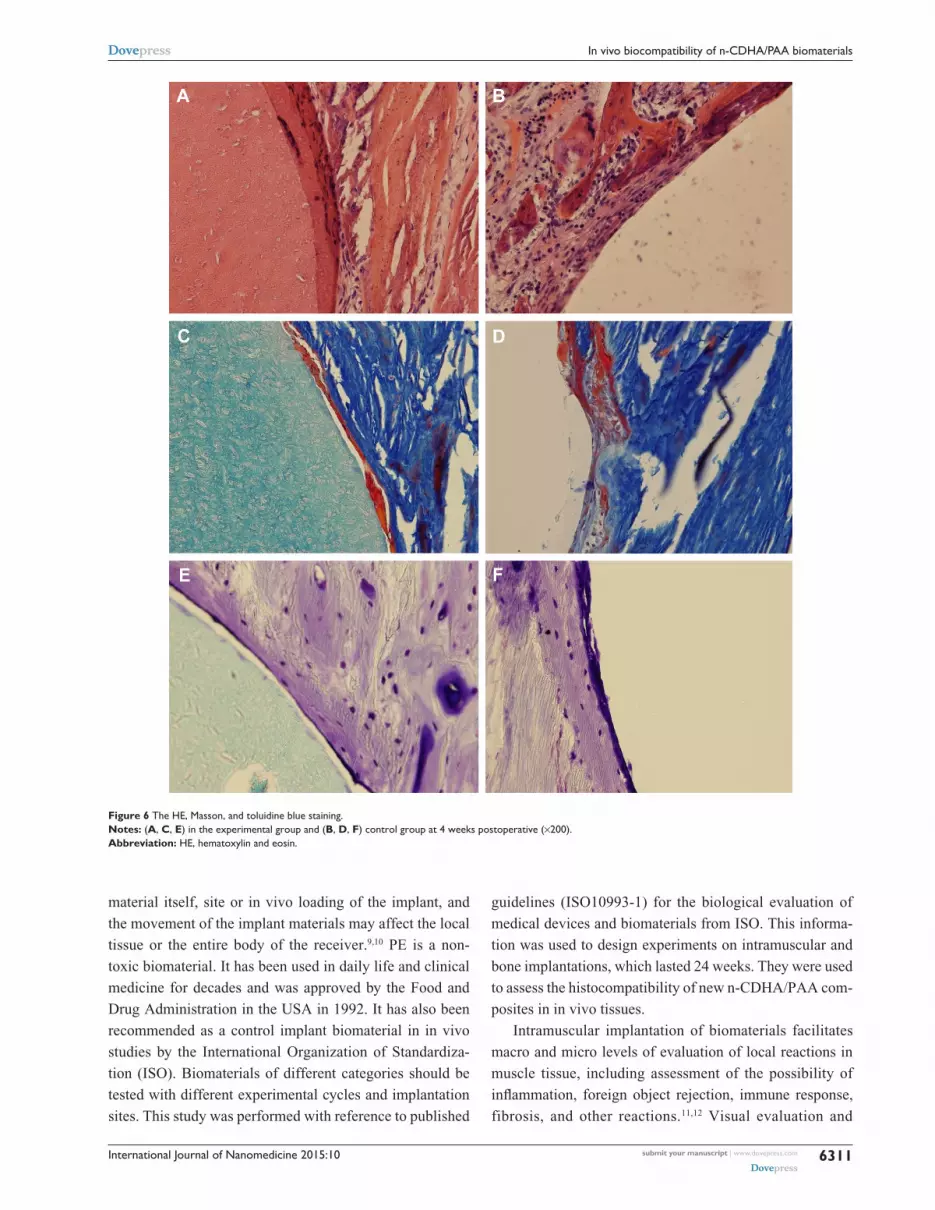
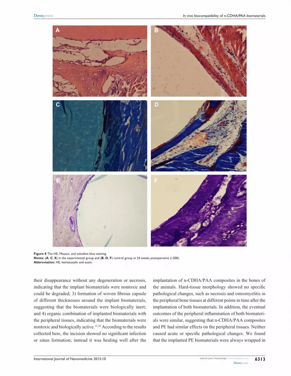
Hard-tissue morphology after biomaterial implantation in the boneFigures 5–8 show the results of HE, Masson, and toluidine
blue staining on hard tissues at different points in time in
both groups.
At postoperative 2 weeks (Figure 5), no significant abnor-
mality was observed in the morphology of peripheral bone
tissue in the experimental group. The trabecular bone was
clear and arranged neatly. Implanted n-CDHA/PAA bioma-
terials were wrapped in newly generated fibrous tissue with
mild invasion of neutrophils and lymphocytes and presence
of a small amount of osteoid. The control group was similar
to the experimental group.
At postoperative 4 weeks (Figure 6), morphology of
peripheral bone tissue in the experimental group was normal,
inflammation had subsided, the layer of collagen fibrous
tissue had thinned, deposition of newly generated osteoid
in the junction between biomaterials and bone tissue had
increased, and a small amount of osteogenesis was observed.
The control group showed similar results but with less osteoid
formation.
At postoperative 12 weeks (Figure 7), the morphologies
of trabecular in the peripheral bone of the two groups were
good. Implanted n-CDHA/PAA biomaterials were still tightly
wrapped in a thin layer of fibrous tissue, and significant
increases were observed in the amounts of peripheral bone
matrix and osteoid. Osteoid became newly formed trabecular
structures, showing the formation of new blood vessels and
bone marrow. The neoformative trabecular structures were
formed in the control group but were less than the experi-
mental group.
Figure 3 The results of X-ray.Notes: Results of X-ray at 2 weeks (A), 4 weeks (B), 12 weeks (C), and 24 weeks (D) postoperative. The red arrow represents experimental group. The blue arrow represents control group.

International Journal of Nanomedicine 2015:10 submit your manuscript | www.dovepress.com
Dovepress
Dovepress
6309
In vivo biocompatibility of n-CDHA/PAA biomaterials
Figure 4 The results of SEM.Notes: The results of SEM at 2 weeks postoperative in the experimental group (A, ×90) and control group (B, ×180); 4 weeks postoperative in the experimental group (C, ×200) and control group (D, ×80); 12 weeks postoperative in the experimental group (E, ×50) and control group (F, ×300); 24 weeks postoperative in the experimental group (G, ×350) and control group (H, ×35). The red arrow represents biomaterials. The blue arrow represents the interface between biomaterial and bone.Abbreviation: SEM, scanning electron microscopy.
International Journal of Nanomedicine 2015:10submit your manuscript | www.dovepress.com
Dovepress
Dovepress
6310
Dai et al
At postoperative 24 weeks (Figure 8), no abnormal
damage or osteolysis was observed in the peripheral bone
tissues of both groups of animals. Peripheral bone tissue
of implanted PE biomaterials was closely connected by a
layer of fibrous tissue; however, the peripheral bone tissue
of implant n-CDHA/PAA biomaterials gradually formed
lamellar bones, which were arranged vertically.
Routine blood and biochemical markersTables 1 and 2 show the results of routine blood and
serum biochemical analyses of the n-CDHA/PAA group
at different points in time. No significant differences were
observed in different indices at different points in time
before and after the surgery (P.0.05). The new biomate-
rial implants had no significant effects on routine blood
or biochemical markers as indicated by examination of
animals after surgery.
DiscussionIn vivo implantation of a biomaterial provides macro and
micro levels of evaluation of local and systemic reactions
of the biomaterials. Physicochemical properties of the
Figure 5 The HE, Masson, and toluidine blue staining.Notes: (A, C, E) in the experimental group and (B, D, F) control group at 2 weeks postoperative (×200).Abbreviation: HE, hematoxylin and eosin.
International Journal of Nanomedicine 2015:10 submit your manuscript | www.dovepress.com
Dovepress
Dovepress
6311
In vivo biocompatibility of n-CDHA/PAA biomaterials
material itself, site or in vivo loading of the implant, and
the movement of the implant materials may affect the local
tissue or the entire body of the receiver.9,10 PE is a non-
toxic biomaterial. It has been used in daily life and clinical
medicine for decades and was approved by the Food and
Drug Administration in the USA in 1992. It has also been
recommended as a control implant biomaterial in in vivo
studies by the International Organization of Standardiza-
tion (ISO). Biomaterials of different categories should be
tested with different experimental cycles and implantation
sites. This study was performed with reference to published
guidelines (ISO10993-1) for the biological evaluation of
medical devices and biomaterials from ISO. This informa-
tion was used to design experiments on intramuscular and
bone implantations, which lasted 24 weeks. They were used
to assess the histocompatibility of new n-CDHA/PAA com-
posites in in vivo tissues.
Intramuscular implantation of biomaterials facilitates
macro and micro levels of evaluation of local reactions in
muscle tissue, including assessment of the possibility of
inflammation, foreign object rejection, immune response,
fibrosis, and other reactions.11,12 Visual evaluation and
Figure 6 The HE, Masson, and toluidine blue staining.Notes: (A, C, E) in the experimental group and (B, D, F) control group at 4 weeks postoperative (×200).Abbreviation: HE, hematoxylin and eosin.

International Journal of Nanomedicine 2015:10submit your manuscript | www.dovepress.com
Dovepress
Dovepress
6312
Dai et al
HE staining were carried out and compared between
intramuscular implantation of PE and n-CDHA/PAA. No sig-
nificant necrosis, abnormal inflammation, immune responses,
or heterotopic ossification was observed in the peripheral
muscle tissue at 24 weeks after implantation. Acute nonspe-
cific inflammation was induced by a small amount of lympho-
cytes and neutrophils at 2 weeks after implantation. This was
presumably caused by surgical trauma, secondary microbial
invasion, or implantation-induced local immune response.13,14
At 4 weeks after the implantation, this acute inflammation
had disappeared from both the experimental and control
groups. No inflammation was observed at 12 weeks after the
intramuscular implantation of both biomaterials. Implanted
biomaterials required only a small amount of fibrous tissue to
connect closely to muscle tissues. Implantation of biomateri-
als in the bone had a very complicated effect on the receivers.
In general, four major reactions took place after implanta-
tion: 1) necrosis in the peripheral bone tissues, indicating
the toxicity of the implanted biomaterials; 2) absorption of
implanted biomaterials by the peripheral tissues, causing
Figure 7 The HE, Masson, and toluidine blue staining.Notes: (A, C, E) in the experimental group and (B, D, F) control group at 12 weeks postoperative (×200).Abbreviation: HE, hematoxylin and eosin.
International Journal of Nanomedicine 2015:10 submit your manuscript | www.dovepress.com
Dovepress
Dovepress
6313
In vivo biocompatibility of n-CDHA/PAA biomaterials
their disappearance without any degeneration or necrosis,
indicating that the implant biomaterials were nontoxic and
could be degraded; 3) formation of woven fibrous capsule
of different thicknesses around the implant biomaterials,
suggesting that the biomaterials were biologically inert;
and 4) organic combination of implanted biomaterials with
the peripheral tissues, indicating that the biomaterials were
nontoxic and biologically active.15,16 According to the results
collected here, the incision showed no significant infection
or sinus formation; instead it was healing well after the
implantation of n-CDHA/PAA composites in the bones of
the animals. Hard-tissue morphology showed no specific
pathological changes, such as necrosis and osteomyelitis in
the peripheral bone tissues at different points in time after the
implantation of both biomaterials. In addition, the eventual
outcomes of the peripheral inflammation of both biomateri-
als were similar, suggesting that n-CDHA/PAA composites
and PE had similar effects on the peripheral tissues. Neither
caused acute or specific pathological changes. We found
that the implanted PE biomaterials were always wrapped in
Figure 8 The HE, Masson, and toluidine blue staining.Notes: (A, C, E) in the experimental group and (B, D, F) control group at 24 weeks postoperative (×200).Abbreviation: HE, hematoxylin and eosin.

International Journal of Nanomedicine 2015:10submit your manuscript | www.dovepress.com
Dovepress
Dovepress
6314
Dai et al
fibrous connective tissue. In contrast, the areas surrounding
the n-CDHA/PAA composites gradually showed increased
bone mass, newly formed trabecular bone, neovasculature,
and formation of lamellar bone. These results indicated that
n-CDHA/PAA composites were nontoxic and bioactive.
Interaction between foreign biomaterials and bone tissues,
specifically the healing mechanism of material-bone inter-
face, had a profound effect on the functions of biomaterial
implants and on prognosis. In general, after the in vivo
implantation of biomaterials into the bone, biomaterials
had direct contact with a variety of matrix components and
proteins of body tissue fluid and blood. They also adsorbed
the surrounding biological macromolecules from tissue and
blood, such as bone laminin, fibronectin, fibrinogen, various
bone morphogenetic proteins, and other cytokines, to form
a layer of biological macromolecules, thereby causing a
Table 1 The results of blood routine in the n-CDHA/PAA group at different time points (x– ± s, n=3)
Examination item Preoperative 2 weeks 24 weeks
WBC (×109/L) 4.85±0.95 4.75±1.035a 4.82±0.85b
Lymphocyte (%) 52.25±8.32 53.02±10.05a 51.00±12.36b
Monocyte (%) 5.68±1.85 5.88±1.58a 6.04±1.66b
Neutropenia (%) 42.56±11.35 46.53±13.25a 40.78±11.23b
Eosinophil (%) 0.38±0.25 0.42±0.22a 0.42±0.32b
Basophil (%) 2.55±1.00 2.62±0.84a 2.60±0.75b
RBC (×1012/L) 6.32±0.52 5.88±0.34a 6.10±0.53b
HGB (g/L) 112.20±7.68 114.31±4.25a 110.45±5.32b
hcT 50.62±6.32 52.41±5.33a 50.12±4.98b
MCV (fl) 82.25±4.52 77.80±6.32a 85.40±5.55b
MCH (pg) 20.15±0.47 21.65±0.54a 20.47±0.46b
MCHC (g/L) 220.35±6.58 219.87±4.25a 230.47±7.46b
RDW (%) 14.85±2.35 13.74±3.65a 15.26±1.58b
PLT (×109/L) 254.36±35.246 269.24±26.591a 259.54±30.684b
MPV (fl) 6.88±0.25 7.03±0.33a 6.80±0.36b
PCT 0.16±0.02 0.18±0.04a 0.16±0.05b
Notes: aP.0.05, compared with the preoperative; bP.0.05, compared with the preoperative.Abbreviations: n-CDHA/PAA, nano-calcium-deficient hydroxyapatite/poly-amino acid; WBC, white blood cell; RBC, red blood cell; HGB, hemoglobin; HCT, hematocrit; MCV, mean corpuscular volume; MCH, mean corpuscular hemoglobin; MCHC, mean corpuscular hemoglobin concentration; RDW, red cell distribution width; PLT, platelet; MPV, mean platelet volume; PCT, procalcitonin.
Table 2 The results of biochemical indicator in the n-CDHA/PAA group at different time points (x– ± s, n=3)
Examination item Preoperative 2 weeks 24 weeks
ALT (U/L) 2.36±0.56 2.61±0.23a 2.71±0.51b
AST (U/L) 1.44±0.35 1.35±0.28a 1.39±0.45b
TP (g/L) 67.88±6.52 65.28±5.7a 66.59±7.24b
Alb (g/L) 53.59±4.68 56.32±3.4a 54.68±4.21b
TBIL (µmol/L) 4.61±0.55 4.92±0.4a 4.90±0.44b
Glu (mmol/L) 7.56±1.23 7.85±0.58a 7.45±0.86b
BUN (mmol/L) 10.23±1.58 11.35±1.23a 11.32±0.99b
CREA (µmol/L) 156.25±16.35 163.49±15.42a 160.87±18.42b
UA (µmol/L) 7.23±2.36 7.56±4.23a 7.44±2.14b
TC (µmol/L) 1.35±0.33 1.34±0.26a 1.30±0.16b
TG (µmol/L) 0.77±0.32 0.80±0.23a 0.82±0.19b
HDL (µmol/L) 23.65±5.42 22.69±3.48a 22.86±4.26b
LDL (µmol/L) 12.30±4.68 13.00±3.56a 12.82±4.36b
LDH (U/L) 5.55±2.32 5.34±2.64a 5.32±2.14b
A/G 6.66±2.15 6.92±2.36a 6.89±2.85b
GGT (U/L) 86.49±26.48 87.64±24.69a 88.64±26.14b
ALP (U/L) 0.69±0.46 0.85±0.36a 0.79±0.42b
Notes: aP.0.05, compared with the preoperative; bP.0.05, compared with the preoperative.Abbreviations: n-CDHA/PAA, nano-calcium-deficient hydroxyapatite/poly-amino acid; ALT, alanine transaminase; AST, aspartate aminotransferase; TP, total protein; Alb, albumin; TBIL, increased total bilirubin; Glu, glucose; BUN, blood urea nitrogen; CREA, creatinine; UA, uric acid; TC, total count; TG, triglycerides; HDL, high-density lipoprotein; LDL, Low-density lipoprotein; LDH, lactate dehydrogenase; A/G, albumin/globulin; GGT, gamma-glutamyl transpeptidase; ALP, alkaline phosphatase.
International Journal of Nanomedicine 2015:10 submit your manuscript | www.dovepress.com
Dovepress
Dovepress
6315
In vivo biocompatibility of n-CDHA/PAA biomaterials
series of cytological changes. Cellular transformation factors
induce the undifferentiated osteoblasts, mesenchymal cells,
and osteoblasts to migrate and adhere to the surface of the
biomaterials through cellular adhesion. Undifferentiated
mesenchymal cells begin to proliferate and differentiate
through the reactions of growth factors in the cells.17–19 The
initial properties of the surfaces of the biomaterials deter-
mined the type and the quantity of protein adsorption, thereby
affecting the binding status of the host cells and the surface
of the materials.20,21 Compared with the n-HA, n-CDHA has
higher solubility (ie, degradability) at a lower Ca/P ratio
(1.5–1.67), as well as being more similar in composition and
crystal structure to the mineral of natural bone.22,23 Thus, it
is envisaged that n-CDHA can be fabricated as a novel bone
regeneration material in order to get better bioperformance
of apatite biomaterial.24,25 The PAA copolymer could be
degraded in HCL-Tris solution with weight loss of ~30 wt%
after 12 weeks of soaking, and it had no significant effect
on the pH value of the ambient environment during the
degradation period.26 If the pH value in the ambient solution
was decreased, it was believed to induce the inflammatory
reaction in vivo.27 Cell culture experiments and in vivo
implantation results showed that the PAA also had good
biocompatibility.26 When implanted in cortical bone of the
dogs, the PAA copolymer implants were directly connected
with the host bone tissue without obvious intervening con-
nective layer, and some new bone tissues were found to
extend along the copolymer surface, which was known as
bone-bonding.26 Bone-bonding could ensure that the implant
integrated with natural bone through biochemical reaction at
the interface between biomaterials and bone tissue, which was
in favor of implant fixation in host bone.28 Therefore, these
previous experiments suggested that the combination of PAA
with n-CDHA would not have apparent adverse reaction with
the bone tissue and surrounding biological macromolecules
from tissue and blood, and it may have excellent biocompat-
ibility and osteoconductivity as potential implants in ortho-
pedic surgery. Histological and X-ray scanning results in this
study indicated that the majority of the biomaterials could
connect closely to the peripheral bone tissues 2 weeks after
the implantation into the bone. Light microscopy and Masson
and toluidine blue stained tissues showed osteoid deposition
on the material-bone interface 4 weeks after the implanta-
tion. The amount of bone tissue formed increased over time.
Mineralization and reconstruction increased the strength of
the new bone, and the trabecular structure had formed by
12 weeks after implantation. Results of SEM showed that
new bone tissue had crossed the material-bone interface and
had grown on the surface of the biomaterials by 12 weeks
after the implantation. By 24 weeks, the implant biomaterials
had become completely covered by new bone tissues. The
results of histological staining showed that the n-CDHA/
PAA composites did not have a stationary relationship but
rather a strong biological bond with the bone-tissue interface.
Further analyses of the specific mechanisms underlying this
bond will be necessary in further studies.
ConclusionThe compatibility of n-CDHA/PAA biocomposite was evalu-
ated with muscle and bone tissue in an in vivo model. The
results showed that no manifest inflammation was observed
after the implantation of n-CDHA/PAA in vivo and a strong
biological bond formed between implant and the bone tis-
sue, indicating that the composites were safe, nontoxic, and
biologically active in bone repair.
AcknowledgmentsThis work was supported by international S&T cooperation
“Cooperative research on advanced composite biological
materials and clinical products” Program (Grant No
2013DFB50280) and Key Technologies R&D Program of
Sichuan province (Grant No 2014GZX0010).
DisclosureThe authors report no conflicts of interest in this work.
References1. Aurégan JC, Bégué T. Induced membrane for treatment of critical sized
bone defect: a review of experimental and clinical experiences. Int Orthop. 2014;38(9):1971–1978.
2. Kleinschmidt K, Wagner-Ecker M, Bartek B, Holschbach J, Richter W. Superior angiogenic potential of GDF-5 and GDF-5(V453/V456) com-pared with BMP-2 in a rabbit long-bone defect model. J Bone Joint Surg Am. 2014;96(20):1699–1707.
3. Reichert JC, Saifzadeh S, Wullschleger ME, et al. The challenge of establishing preclinical models for segmental bone defect research. Biomaterials. 2009;30(12):2149–2163.
4. Hesse E, Kluge G, Atfi A, et al. Repair of a segmental long bone defect in human by implantation of a novel multiple disc graft. Bone. 2010;46(5): 1457–1463.
5. Li H, Gong M, Yang A, Ma J, Li X, Yan Y. Degradable biocomposite of nano calcium-deficient hydroxyapatite-multi(amino acid) copolymer. Int J Nanomedicine. 2012;7:1287–1295.
6. Li H, Yang L, Dong X, Gu Y, Lv G, Yan Y. Composite scaffolds of nano calcium deficient hydroxyapatite/multi-(amino acid)copolymer for bone tissue regeneration. J Mater Sci Mater Med. 2014;25(5):1257–1265.
7. Bogdanović U, Vodnik V, Mitrić M, et al. Nanomaterial with high anti-microbial efficacy-copper/polyaniline nanocomposite. ACS Appl Mater Interfaces. 2015;7(3):1955–1966.
8. Yao Q, Ye J, Xu Q, Mo A, Gong P. Composite scaffolds of dicalcium phosphate anhydrate/multi-(amino acid) copolymer: in vitro degradability and osteoblast biocompatibility. J Biomater Sci Polym Ed. 2015;26(4): 211–223.
9. Sancey L, Kotb S, Truillet C, et al. Long-term in vivo clearance of gadolinium-based AGuIX nanoparticles and their biocompatibility after systemic injection. ACS Nano. 2015;9(3):2477–2488.

International Journal of Nanomedicine
Publish your work in this journal
Submit your manuscript here: http://www.dovepress.com/international-journal-of-nanomedicine-journal
The International Journal of Nanomedicine is an international, peer-reviewed journal focusing on the application of nanotechnology in diagnostics, therapeutics, and drug delivery systems throughout the biomedical field. This journal is indexed on PubMed Central, MedLine, CAS, SciSearch®, Current Contents®/Clinical Medicine,
Journal Citation Reports/Science Edition, EMBase, Scopus and the Elsevier Bibliographic databases. The manuscript management system is completely online and includes a very quick and fair peer-review system, which is all easy to use. Visit http://www.dovepress.com/testimonials.php to read real quotes from published authors.
International Journal of Nanomedicine 2015:10submit your manuscript | www.dovepress.com
Dovepress
Dovepress
Dovepress
6316
Dai et al
10. Watari F, Yokoyama A, Omori M, et al. Biocompatibility of materi-als and development to functionally graded implant for bio-medical application. Compos Sci Technol. 2004;64(6):893–908.
11. Walker J, Shadanbaz S, Woodfield TB, Staiger MP, Dias GJ. The in vitro and in vivo evaluation of the biocompatibility of Mg alloys. Biomed Mater. 2014;9(1):015006.
12. Wang H, Zhi W, Lu X, et al. Comparative studies on ectopic bone formation in porous hydroxyapatite scaffolds with complementary pore structures. Acta Biomater. 2013;9(9):8413–8421.
13. Kamelger FS, Marksteiner R, Margreiter E, et al. A comparative study of three different biomaterials in the engineering of skeletal muscle using a rat animal model. Biomaterials. 2004;25(9):1649–1655.
14. Srivastava S, Gorham SD, French DA, Shivas AA, Courtney JM. In vivo evaluation and comparison of collagen, acetylated collagen and collagen/glycosaminoglycan composite films and sponges as candidate biomaterials. Biomaterials. 1990;11(3):155–161.
15. Gauthier O, Müller R, von Stechow D, et al. In vivo bone regeneration with injectable calcium phosphate biomaterial: a three-dimensional micro-computed tomographic, biomechanical and SEM study. Bioma-terials. 2005;26(27):5444–5453.
16. Chu CL, Xue XY, Zhu JC, Yin ZD. In vivo study on biocompatibility and bonding strength of Ti/Ti–20 vol.% HA/Ti–40 vol.% HA function-ally graded biomaterial with bone tissues in the rabbit. Mat Sci Eng A Struct. 2006;429(1–2):18–24.
17. Roessler S, Born R, Scharnweber D, Worch H, Sewing A, Dard M. Biomimetic coatings functionalized with adhesion peptides for dental implants. J Mater Sci Mater Med. 2001;12(10–12):871–877.
18. Chehroudi B, Gould TR, Brunette DM. A light and electron microscopic study of the effects of surface topography on the behavior of cells attached to titanium-coated percutaneous implants. J Biomed Mater Res. 1991;25(3):387–405.
19. Kirkpatrick CJ, Wagner M, Köhler H, Bittinger F, Otto M, Klein CL. The cell and molecular biological approach to biomaterial research: a perspective. J Mater Sci Mater Med. 1997;8(3):131–141.
20. Ziats NP, Miller KM, Anderson JM. In vitro and in vivo interactions of cells with biomaterials. Biomaterials. 1988;9(11):5–13.
21. Wang H, Lee JK, Moursi AM, et al. Microstructural disassembly of calcium phosphates. J Biomed Mater Res A. 2004;68(1):61–70.
22. Guo H, Su J, Wei J, Kong H, Liu C. Biocompatibility and osteogenicity of degradable Ca-deficient hydroxyapatite scaffolds from calcium phos-phate cement for bone tissue engineering. Acta Biomater. 2009;5(1): 268–278.
23. Lin JH, Kuo KH, Ding SJ, Ju CP. Surface reaction of stoichiometric and calcium-deficient hydroxyapatite in simulated body fluid. J Mater Sci Mater Med. 2001;12(8):731–741.
24. Gustavsson J, Ginebra MP, Planell J, Engel E. Osteoblast-like cellular response to dynamic changes in the ionic extracellular environment produced by calcium-deficient hydroxyapatite. J Mater Sci Mater Med. 2012;23(10):2509–2520.
25. Akiyama N, Takemoto M, Fujibayashi S, Neo M, Hirano M, Nakamura T. Difference between dogs and rats with regard to osteoclast-like cells in calcium-deficient hydroxyapatite-induced osteoinduction. J Biomed Mater Res A. 2011;96(2):402–412.
26. Li H, Yan Y, Wei J, et al. Bone substitute biomedical material of multi-(amino acid) copolymer: in vitro degradation and biocompatibility. J Mater Sci Mater Med. 2011;22(11):2555–2563.
27. Ambrose CG, Clanton TO. Bioabsorbable implants: review of clini-cal experience in orthopedic surgery. Ann Biomed Eng. 2004;32(1): 171–177.
28. Davies JE. Bone bonding at natural and biomaterial surfaces. Biomateri-als. 2007;28(34):5058–5067.