Biocompatibility Assessment of Liquid Artificial Vitreous Replacements: Relevance of In Vitro...
11
CURRENT RESEARCH EDWARD COTLIER AND ROBERT WEINREB, EDITORS Biocompatibility Assessment of Liquid Artificial Vitreous Replacements: Relevance of In Vitro Studies Andrea Matteucci, BS, 1 Giuseppe Formisano, BS, 2 Silvia Paradisi, BS, 3 Giovanna Carnovale-Scalzo, MD, 4 Giovanni Scorcia, MD, 4 Salvatore Caiazza, BS, 2 Hans Hoerauf, MD, 5 and Fiorella Malchiodi-Albedi, MD 3 1 G.B. Bietti Foundation for Ophthalmology (I.R.C.C.S.), Rome, Italy; 2 Department of Technology and Health, Istituto Superiore di Sanita `, Rome, Italy; 3 Department of Cell Biology and Neuroscience, Istituto Superiore di Sanita `, Rome, Italy; 4 Eye Clinic, ‘‘Magna Graecia’’ University, Catanzaro, Italy; and 5 University Eye Hospital, Lubeck, Germany Abstract. The biocompatibility of liquid artificial vitreous replacements is generally assessed by performing tests in animal models before their clinical use, whereas in vitro experimentation is seldom carried out due to their physico-chemical characteristics. Since their introduction in vitreoretinal surgery, however, the use of some certified vitreous replacements has been discouraged after clinical trials, because of the occurrence of serious side effects. This observation suggests that the tests currently performed for biocompatibility assessment cannot fully guarantee their safety when they are used in humans. Here we review the available literature on in vitro biocompatibility testing of liquid artificial vitreous replacements and survey our own experience on the subject, obtained by using primary retinal cell cultures, seeded on micro-porous inserts. We suggest that in vitro biocompatibility assessment, conducted before experiments in animal models, could improve the required safety evaluation and decrease the risk of undesired side effects, as well as providing a beneficial reduction of animal experimentation. (Surv Ophthalmol 52:289--299, 2007. Ó 2007 Elsevier Inc. All rights reserved.) Key words. biocompatibility biomaterials ocular endotamponades partially fluorinated alkanes perfluorocarbon liquids retinal cell culture vitreous Temporary or permanent vitreous replacement is an essential part of modern complex vitreoretinal surgery. Since the introduction of vitrectomy, there is an ongoing search for an ideal substance. The traditional and widely used ocular endotamponades include air, synthetic expansive gases, and silicone oil, which are all lighter than water. 19,40,59,63 The introduction of heavy perfluorocarbon liquids (PFCLs), such as perfluorodecalin, perfluorooctane, and perfluoroperhydrophenanthrene, as an intra- operative tool represented a major progress for the management of complex retinal detach- ments. 10,11,21,23,52 Their higher specific gravity en- abled intraoperative stabilization and reattachment of the retina. 5,12 Unfortunately, due to retinal damage and inflammatory reactions in long-term 289 Ó 2007 by Elsevier Inc. All rights reserved. 0039-6257/07/$--see front matter doi:10.1016/j.survophthal.2007.02.004 SURVEY OF OPHTHALMOLOGY VOLUME 52 NUMBER 3 MAY–JUNE 2007
-
Upload
independent -
Category
Documents
-
view
0 -
download
0
Transcript of Biocompatibility Assessment of Liquid Artificial Vitreous Replacements: Relevance of In Vitro...
SURVEY OF OPHTHALMOLOGY VOLUME 52 � NUMBER 3 � MAY–JUNE 2007
CURRENT RESEARCHEDWARD COTLIER AND ROBERT WEINREB, EDITORS
Biocompatibility Assessment of Liquid ArtificialVitreous Replacements: Relevanceof In Vitro StudiesAndrea Matteucci, BS,1 Giuseppe Formisano, BS,2 Silvia Paradisi, BS,3
Giovanna Carnovale-Scalzo, MD,4 Giovanni Scorcia, MD,4
Salvatore Caiazza, BS,2 Hans Hoerauf, MD,5 and Fiorella Malchiodi-Albedi, MD3
1G.B. Bietti Foundation for Ophthalmology (I.R.C.C.S.), Rome, Italy; 2Department of Technology and Health, IstitutoSuperiore di Sanita, Rome, Italy; 3Department of Cell Biology and Neuroscience, Istituto Superiore di Sanita, Rome, Italy;4Eye Clinic, ‘‘Magna Graecia’’ University, Catanzaro, Italy; and 5University Eye Hospital, Lubeck, Germany
Abstract. The biocompatibility of liquid artificial vitreous replacements is generally assessed byperforming tests in animal models before their clinical use, whereas in vitro experimentation is seldomcarried out due to their physico-chemical characteristics. Since their introduction in vitreoretinalsurgery, however, the use of some certified vitreous replacements has been discouraged after clinicaltrials, because of the occurrence of serious side effects. This observation suggests that the testscurrently performed for biocompatibility assessment cannot fully guarantee their safety when they areused in humans. Here we review the available literature on in vitro biocompatibility testing of liquidartificial vitreous replacements and survey our own experience on the subject, obtained by usingprimary retinal cell cultures, seeded on micro-porous inserts. We suggest that in vitro biocompatibilityassessment, conducted before experiments in animal models, could improve the required safetyevaluation and decrease the risk of undesired side effects, as well as providing a beneficial reduction ofanimal experimentation. (Surv Ophthalmol 52:289--299, 2007. � 2007 Elsevier Inc. All rightsreserved.)
Key words. biocompatibility � biomaterials � ocular endotamponades � partially fluorinatedalkanes � perfluorocarbon liquids � retinal cell culture � vitreous
Temporary or permanent vitreous replacement is anessential part of modern complex vitreoretinalsurgery. Since the introduction of vitrectomy, thereis an ongoing search for an ideal substance. Thetraditional and widely used ocular endotamponadesinclude air, synthetic expansive gases, and siliconeoil, which are all lighter than water.19,40,59,63 Theintroduction of heavy perfluorocarbon liquids
28
� 2007 by Elsevier Inc.All rights reserved.
(PFCLs), such as perfluorodecalin, perfluorooctane,and perfluoroperhydrophenanthrene, as an intra-operative tool represented a major progress forthe management of complex retinal detach-ments.10,11,21,23,52 Their higher specific gravity en-abled intraoperative stabilization and reattachmentof the retina.5,12 Unfortunately, due to retinaldamage and inflammatory reactions in long-term
9
0039-6257/07/$--see front matterdoi:10.1016/j.survophthal.2007.02.004
290 Surv Ophthalmol 52 (3) May--June 2007 MATTEUCCI ET AL
animal studies13,15,71 and clinical case reports,16,61
their removal at the end of surgery is consideredmandatory. Natural, synthetic, or semi-syntheticpolymers, such as collagen, hyaluronic acid, hydrox-ypropylmethylcellulose, or poly (1-vinyl-2-pyrrolidi-none) hydrogels were investigated in the 1990s asliquid artificial vitreous replacements (LAVRs).29,31
Although these polymers have remarkable advan-tages (they are fully hydrated, clear, transparent,autoclavable and easily injected), they are quicklybiodegraded in vivo.6,7 Currently, they are beinginvestigated as biodegradable matrix materials forcontrolled drug delivery in the vitreous.9,22,33
In recent years, a new class of fluorocarbonliquids, partially fluorinated alkanes (PFA), withreduced specific gravity but still heavier than water,was introduced in the early 2000s to provideadequate postoperative tamponade in presence ofinferior retinal pathologies.48,76 Because of theirlower gravity, they were considered to produce lessretinal damage than PFCLs and showed promisingresults in animal experiments.34,77 Nonetheless, theinitial enthusiastic response to this new treatmentmodality was greatly tempered by a high incidenceof side effects in the human eye, such as in-flammatory reactions, macrophage reaction, disper-sion, and early emulsification.26,50,60,73 Recently, twoheavy silicone oils produced by a mixture of PFAand silicone oil were approved in Europe aspostoperative ocular endotamponade, but little hasbeen reported about their biocompatibility in cellcultures or in vivo experiments. Controversial re-ports about their biocompatibility in human eyeshave so far been published.54,69,75
From these considerations, it clearly appears thata satisfactory long-term replacement of the vitreoushas yet to be found. Besides several physicochemicaland optical properties, an ideal substance should beinert, unable to trigger inflammatory or foreignbody reactions and well tolerated by adjacent oculartissues with which the substance is in contact. Theseproperties are generally referred to as ‘‘biocompat-ibility.’’ Biocompatibiliy is generally assessed byperforming in vitro and in vivo tests.
Here we review the available literature on in vitrobiocompatibility tests on LAVRs and survey our ownexperience on the subject, obtained by growingprimary retinal cell cultures on micro-porous inserts.
In Vitro Biocompatibility Assessment:Not an Easy Task
LAVRs may be classified in three groups accordingto their physical properties: a) water-immisciblecompounds whose specific gravity is higher thanwater, such as PFCLs or PFAs; b) water-immiscible
compounds whose specific gravity is lower than water,such as silicone oil; and c) water-miscible compounds,such as the natural polymers collagen and hyaluronicacid, the semi-synthetic polymer hydroxypropylme-thylcellulose or the synthetic polymers poly(1-vinyl-2-pyrrolidinone), and polyvinylalcohol (Table 1).
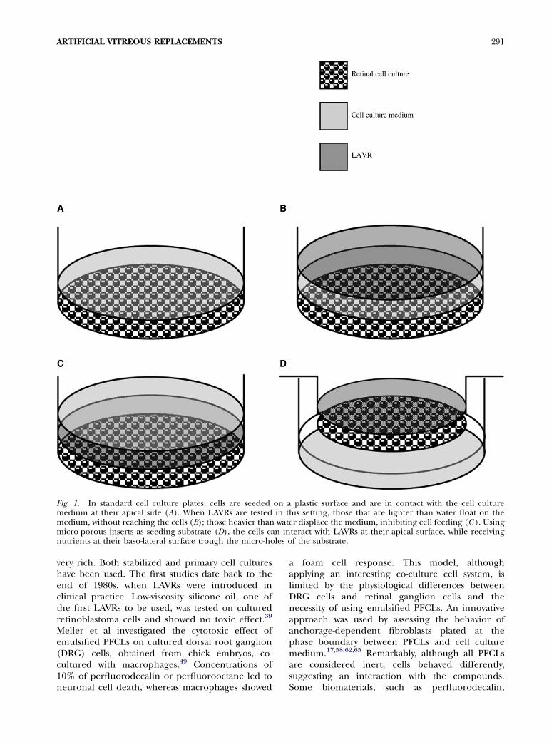
LAVRs, as any other biomaterial, should undergoa series of tests assessing their biocompatibilitybefore clinical use, with the aim of limiting possibleadverse reactions from human tissues. The testsrequired, according to the International Organiza-tion for Standardization Guidelines (ISO 10993 and7405), should evaluate cytotoxicity, hemocompati-bility, mutagenicity, and pyrogenicity. Some of theseprocedures, such as those used to test cytoxicity, areperformed in vitro. Unfortunately, most materialsthat are currently used as LAVRs are water-immisci-bile, an important requisite for their in situ use,because it prevents tissues re-absorption throughpynocitic mechanisms. This feature, however, ham-pers their testing in cell cultures, which need to bein contact with an aqueous medium for theirsurvival, providing nutrients and allowing gasexchanges (Fig. 1A). When a water-immisciblecompound lighter than water is tested in a cellculture seeded in a standard plate, it will float ontop of the medium, without reaching the cell layer(Fig. 1B). If cells are treated with a compoundheavier than water, it will displace the cell culturemedium, which will float on the surface of thecompounds and the normal exchange processesbetween cells and medium will be limited (Fig. 1C).Thus in vitro testing has only been an occasional wayof approaching LAVR safety assessment.
Review of the Literature
Due to these technical obstacles, the literature onin vitro biocompatibility assessment of LAVRs is not
TABLE 1
Liquid Artificial Vitreous Replacements
Perfluorocarbon liquids (PFCLs)PerfluorodecalinPerfluorooctanePerfluoroperhydrophenanthrene
Partially fluorinated alkanes (PFAs)Perfluorohexyloctane (F6H8)Perfluorohexylethan
Silicone oilNatural polymers
CollagenHyaluronic acid
Semi-synthetic polymersHydroxypropylmethylcellulose
Synthetic polymersPoly(1-vinyl-2-pyrrolidinone) hydrogelPolyvinylalcohol
ARTIFICIAL VITREOUS REPLACEMENTS 291
Fig. 1. In standard cell culture plates, cells are seeded on a plastic surface and are in contact with the cell culturemedium at their apical side (A). When LAVRs are tested in this setting, those that are lighter than water float on themedium, without reaching the cells (B); those heavier than water displace the medium, inhibiting cell feeding (C ). Usingmicro-porous inserts as seeding substrate (D), the cells can interact with LAVRs at their apical surface, while receivingnutrients at their baso-lateral surface trough the micro-holes of the substrate.
very rich. Both stabilized and primary cell cultureshave been used. The first studies date back to theend of 1980s, when LAVRs were introduced inclinical practice. Low-viscosity silicone oil, one ofthe first LAVRs to be used, was tested on culturedretinoblastoma cells and showed no toxic effect.39
Meller et al investigated the cytotoxic effect ofemulsified PFCLs on cultured dorsal root ganglion(DRG) cells, obtained from chick embryos, co-cultured with macrophages.49 Concentrations of10% of perfluorodecalin or perfluorooctane led toneuronal cell death, whereas macrophages showed
a foam cell response. This model, althoughapplying an interesting co-culture cell system, islimited by the physiological differences betweenDRG cells and retinal ganglion cells and thenecessity of using emulsified PFCLs. An innovativeapproach was used by assessing the behavior ofanchorage-dependent fibroblasts plated at thephase boundary between PFCLs and cell culturemedium.17,58,62,65 Remarkably, although all PFCLsare considered inert, cells behaved differently,suggesting an interaction with the compounds.Some biomaterials, such as perfluorodecalin,
292 Surv Ophthalmol 52 (3) May--June 2007 MATTEUCCI ET AL
perfluorooctane, and perfluoroperhydrophenan-threne, prevented cell attachment; the few that didattach fail to show a normal proliferative activity.Conversely,on perfluoromethyldecaline, perfluorofluor-ene, perfluorotributylamine, the perfluoropolyether K-6hexamer, trifluoropropylmethylsiloxane (fluorosili-cone), and diphenyldimethylsiloxane, some cells be-came fusiform-shaped and exhibited proliferation.65
Hydrogels with a high water content, such as thepolymer poly(1-vinyl-2-pyrrolidinone), were pro-duced and tested as a possible vitreous substitute.29
Cytotoxicity was evaluated in vitro, using culturedmouse (Balb/ c-3T3) fibroblasts.30 The polymers didnot show cytocidal effects. Some of them, forexample gels crosslinked with diethylene glycoldimethacrylate, induced a significant enhancementof cell proliferation, especially in serum-free cellculture medium. These results suggested the poten-tial use of the polymer hydrogel as vitreous sub-stitutes. However, in vivo experiments showed arelatively short retention of crosslinked hydrogelsin the eye. Histopathologic examination revealedinfiltration of mononuclear phagocytes in bothvitreous and retinal tissues, suggesting a phagocyte-mediated mechanism for the removal.31 Polymerssynthesized with the aim of reducing degradativereaction did not show analogous biocompatibility,although recent results are more promising. TheMTT cytotoxicity assay in fact revealed that thematerials were cytotoxic to chondrosarcoma cellsand human fibroblasts isolated from donor corneas.7
Recently, biopolymers based on gellan gums incombination with hyaluronic acid have been foundto have shorter degradation tendency and goodcytocompatibility. These compounds have been pro-posed as short term vitreous substitutes.47,68 Bio-degradable, biocompatible polymers are currentlystudied as drug-releasing devices.6 With the sameaim, silicone oil has been used in experimentalmodels of proliferative vitreoretinopathy.2 Morerecently, microspheres, obtained by dispersion ofdrugs in fluorosilicone oil and poly (DL-lactide-co-glycolide), have been object of investigation.18,24,25,46
PFAs were introduced in ophthalmic surgery aftersuccessful in vivo experimentation.34,77 The obser-vation of undesired side effects after the clinical useof F6H8 prompted new investigations at the cellculture level. Evidence of toxic side effects of F6H8
was observed in human corneal endothelial andretinal pigment epithelial cells.50 An in vitro studywas conducted to verify whether emulsification ofvitreous tamponades might trigger inflammatorycell activation, which could account for the epireti-nal membrane formation described in patients afteruse of F6H8.26 Data obtained by incubating humanmonocytes and neutrophils with silicone oil and
F6H8 vesicles did not support the hypothesis thatemulsification of the tamponades in the microenvi-ronment of the eye might easily activate neutrophilsor stimulate phagocytosis by monocytes.37
A more promising, tissue-oriented approach wasrecently applied to test LAVR biocompatibility.35
The use of retinal explants, in this model, allowedmaintaining the polar architecture of the tissue andconsidering the toxic effect that may arise byinteractions among different cell types. Explantswere mounted in tissue carriers to improve nutritionand handling of the sample.
Relevant End Points
In recent years, due to the growing concern overthe use of experimental animals for biologicalevaluation, in vitro methods have been stronglyencouraged for assessing material biocompatibility.To satisfactorily predict LAVR biocompatibility invitro, however, a test should be able to mimic in vivotissue response as closely as possible; this requiresthe identification of the pertinent toxicological end-points and the appropriate methodology for theirevaluation. Among the possible end-points to beconsidered, the following have a special relevancefor LAVR biocompatibility.
CYTOTOXICITY
Cytotoxicity is usually assessed by performing testssuch as LDH assay or vital stains in stabilized cellcultures, based on loss of plasma membrane in-tegrity. To correctly evaluate cell death, however,apoptosis should also be considered. In early phasesof apoptosis, the membrane integrity is typicallypreserved, so the mentioned cytotoxicity tests canseldom detect apoptosis. To avoid an underestima-tion of possible toxic effects induced by a biomate-rial, tests that specifically evaluate apoptosis shouldbe included. A number of procedures are availableto detect various markers of apoptosis, such aschromatin condensation, DNA fragmentation, acti-vation of caspase-3, cleavage of poly (ADP-ribose)-polymerase, release of cytochrome C, and detectionof the proapoptotic proteins Bax and BID.56
INTERFERENCE WITH PATTERN OF GENE
EXPRESSION
Replacement of the vitreous gel with syntheticsubstitutes may interfere with maintenance of thenormal differentiation of retinal cells resulting ina functional impairment.74 The use of primaryretinal cell cultures may be particularly useful tocheck LAVR effects on cell differentiation, sincemost of the different cell types present in the retinain situ are preserved in the culture. In addition, the
ARTIFICIAL VITREOUS REPLACEMENTS 293
neuronal cell population usually undergoes in vitroa sequential gene expression pattern leading toa maturational process similar to that in situ, asshown by the development of polarized neuritic treeand synapses. Modification of the synapse networkcan evidence possible interferences with the normalphysiology of neurons.
INFLAMMATORY RESPONSE
Some of the adverse reactions, described afterLAVR use in vitreoretinal surgery, are a consequenceof an inflammatory reaction.16,72,73 In addition, thepresence of mononuclear phagocytic cells, can beevidence of the tendency of a material to biodegra-dation, which is suggested in tissues by the presenceof vacuolized macrophages.31 The possibility of invitro monitoring of an inflammatory response tobiomaterials would be very advantageous. Microglialcells are the most important effectors of inflamma-tory response in the retina and their presence inculture may allow detecting their activation andmonitor the release of anti-inflammatory (transform-ing growth factor b, PGE2) or pro-inflammatory(interleukin 1b, tumor necrosis factor a) cytokines.32
Moreover, the production of potentially neurotoxicfactors, such as reactive nitrogen or oxygen interme-diates, or the development of a phagocytic phenotypemay be evaluated.
Microporous Inserts as Cell CultureSubstrates
To overcome the problem of LAVR--water immis-cibility and test the materials in vitro, we have usedspecial inserts, whose bottom is made of a micropo-rous membrane, as substrate for seeding the cellcultures. These inserts were introduced in the 1980sto study plasma membrane asymmetry of epithelialcells, because they provide access not only to theapical plasma membrane, as in standard plates, butalso to the baso-lateral plasma membrane throughthe micro-holes of the substrate.55 More recently,they have been used for co-culturing experiments,where different cell monolayers are seeded onseparate substrates but share the same cell culturemedium4 or to measure electrical resistance acrossmonolayers of endothelial cells.14 In our experi-ments, cells are seeded in the insert and the insert ispositioned in a standard cell culture plate, filledwith medium (Fig. 1D). In this setting, the cells arein contact with the cell culture medium not only attheir apical surface, but also at their baso-lateralside. The effects of cell--LAVR interaction can bestudied by substituting the cell medium at the apicalsurface of the cells with the material to be testedwhile cell feeding is maintained through the holes
of the micro-porous insert. The use of devices toimprove cell nutrition in tissue explants has alsobeen recently addressed by Kloth et al.35
In the effort to provide a more efficient LAVRbiocompatibility testing, as an in vitro model, we haveused primary retinal cultures, obtained from embry-onic Wistar rat tissues at day 18, as already describedin detail.41,42 Briefly, the eyes are removed understerile conditions and the entire retinas are isolatedusing a stereomicroscope. After treatment with 0.25%trypsin (15 min at 37�C), cells are mechanically disso-ciated with a fire-polished Pasteur pipette and seededat high density onto poly-L-lysine-coated cell Falconculture inserts with cyclopore bottoms (Becton Dick-inson, Franklin Lakes, NJ, USA) in MinimumEssential Medium (MEM, Invitrogen Inc., Milan,Italy), with 10% fetal calf serum. The inserts then areplaced in standard, 25-mm wells containing 2 ml ofthe same medium so that the Cell layers are fullyimmersed in the medium. At 9 day-in-vitro, themedium on top of the cells is replaced with LAVRs. Inthis way, the cells are in contact with the material to betested, at the apical side, and with the nutrientmedium, at the basal side. After treatment, cells arefixed in 4% paraformaldeyde in PBS, 0.12M insucrose, for 30 min and treated for immunocyto-chemistry, TUNEL assay, or Hoechst stain.42--45
In these culturing conditions, rat embryonicretinal cells grow as a mixed cell population,composed of both glial and neuronal cells (Fig. 2).They form cords composed of large, flattened glialcells at the bottom, and smaller and plump neuronalcells on top of them, as shown by scanning electronmicroscope (SEM) analysis. The glial population iscomposed of Muller’s glia, immunostained forS100B, astrocytes, positive for glial fibrillary acidicprotein (GFAP), and microglia, labeled with Griffoniasimplicifolia lectin. Neurons also form a heteroge-neous population; larger neurons, with a rich neu-ritic tree, are positive for the microtubule-associatedprotein 2 (MAP2) and the inhibitory neurotransmit-ter g-amino-butyric acid (GABA), which identifiesamacrine and horizontal cells; smaller neurons arepositive for neurofilaments (NF). Photoreceptors arealso present; they form rosette-like aggregations(Haemat/Eos), are labeled for rhodopsin andshow the presence of the characteristic ribbonsynapses observed at transmission electron micro-scope (TEM). Ganglion, Thy1.1-positive cells areonly occasionally found (data not shown).
Testing LAVR Biocompatibility on RetinalCell Cultures
In the past few years, we have had the opportunityof testing several LAVRs in our cell culture system.

294 Surv Ophthalmol 52 (3) May--June 2007 MATTEUCCI ET AL
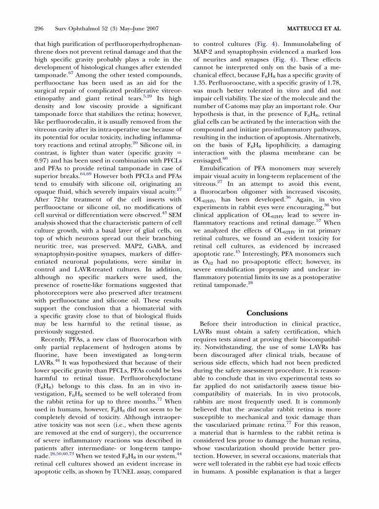
Fig. 2. Morphological and immunohistochemical characterization of primary rat retinal cell cultures. Theheterogeneous cell population of the cultures (SEM) is composed of Muller cells, labeled for S100B, GFAP-positiveastrocytes and microglia, labeled for Griffonia s. lectin. Some neurons are labeled for the microtubule-associated proteinMAP2 and the inhibitory neurotransmitter GABA (darker stain, arrows), whereas others only for neurofilaments (lighterstain, arrow heads). Rhodopsin-positive photoreceptors form rosette-like aggregations (Haemat/Eos). At theultrastructural level, they show the presence of the characteristic ribbon synapses (arrow).
Perfluorodecalin, initially studied as a potentialblood substitute, is one of the first perfluorocarbonsto be proposed as short-term LAVR.10 Its biocom-patibility, however, was questioned by the observa-tion of serious side effects, such as vacuolization inthe nerve fibers, progressive defects of the outerphotoreceptor segments, presence of pseudo-myelinic bodies in the inner plexiform layer, areasof necrosis, and inflammation in in vivo experi-ments.15,51 In addition, intravitreal inflammationhas also been reported in a patient.61 In our in vitro
experiments,42 a treatment with perfluorodecalininduced a disorganization of retinal cell growthpattern and a severe loss of neurites (Fig. 3). It hasbeen suggested that the adverse effects induced byperfluorodecalin in vivo are physical, more thantoxic.15 Due to its high specific gravity (1.94),perfluorodecalin was supposed to compress anddisorganize the retinal structure. Our results sup-ported this hypothesis. Under the weight ofperfluorodecalin, the cell clusters, which character-istically have a double layer-structure, appeared
ARTIFICIAL VITREOUS REPLACEMENTS 295
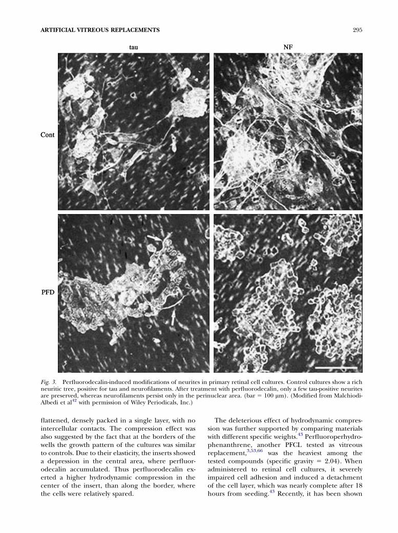
Fig. 3. Perfluorodecalin-induced modifications of neurites in primary retinal cell cultures. Control cultures show a richneuritic tree, positive for tau and neurofilaments. After treatment with perfluorodecalin, only a few tau-positive neuritesare preserved, whereas neurofilaments persist only in the perinuclear area. (bar 5 100 mm). (Modified from Malchiodi-Albedi et al42 with permission of Wiley Periodicals, Inc.)
flattened, densely packed in a single layer, with nointercellular contacts. The compression effect wasalso suggested by the fact that at the borders of thewells the growth pattern of the cultures was similarto controls. Due to their elasticity, the inserts showeda depression in the central area, where perfluor-odecalin accumulated. Thus perfluorodecalin ex-erted a higher hydrodynamic compression in thecenter of the insert, than along the border, wherethe cells were relatively spared.
The deleterious effect of hydrodynamic compres-sion was further supported by comparing materialswith different specific weights.43 Perfluoroperhydro-phenanthrene, another PFCL tested as vitreousreplacement,3,53,66 was the heaviest among thetested compounds (specific gravity 5 2.04). Whenadministered to retinal cell cultures, it severelyimpaired cell adhesion and induced a detachmentof the cell layer, which was nearly complete after 18hours from seeding.43 Recently, it has been shown
296 Surv Ophthalmol 52 (3) May--June 2007 MATTEUCCI ET AL
that high purification of perfluoroperhydrophenan-threne does not prevent retinal damage and that thehigh specific gravity probably plays a role in thedevelopment of histological changes after extendedtamponade.67 Among the other tested compounds,perfluooctane has been used as an aid for thesurgical repair of complicated proliferative vitreor-etinopathy and giant retinal tears.5,20 Its highdensity and low viscosity provide a significanttamponade force that stabilizes the retina; however,like perfluorodecalin, it is usually removed from thevitreous cavity after its intra-operative use because ofits potential for ocular toxicity, including inflamma-tory reactions and retinal atrophy.20 Silicone oil, incontrast, is lighter than water (specific gravity 5
0.97) and has been used in combination with PFCLsand PFAs to provide retinal tamponade in case ofsuperior breaks.64,69 However both PFCLs and PFAstend to emulsify with silicone oil, originating anopaque fluid, which severely impairs visual acuity.27
After 72-hr treatment of the cell inserts withperfluooctane or silicone oil, no modifications ofcell survival or differentiation were observed.43 SEManalysis showed that the characteristic pattern of cellculture growth, with a basal layer of glial cells, ontop of which neurons spread out their branchingneuritic tree, was preserved. MAP2, GABA, andsynaptophysin-positive synapses, markers of differ-entiated neuronal populations, were similar incontrol and LAVR-treated cultures. In addition,although no specific markers were used, thepresence of rosette-like formations suggested thatphotoreceptors were also preserved after treatmentwith perfluooctane and silicone oil. These resultssupport the conclusion that a biomaterial witha specific gravity close to that of biological fluidsmay be less harmful to the retinal tissue, aspreviously suggested.
Recently, PFAs, a new class of fluorocarbon withonly partial replacement of hydrogen atoms byfluorine, have been investigated as long-termLAVRs.48 It was hypothesized that because of theirlower specific gravity than PFCLs, PFAs could be lessharmful to retinal tissue. Perfluorohexyloctane(F6H8) belongs to this class. In an in vivo in-vestigation, F6H8 seemed to be well tolerated fromthe rabbit retina for up to three months.77 Whenused in humans, however, F6H8 did not seem to becompletely devoid of toxicity. Although intraoper-ative toxicity was not seen (i.e., when these agentsare removed at the end of surgery), the occurrenceof severe inflammatory reactions was described inpatients after intermediate- or long-term tampo-nade.26,50,60,73 When we tested F6H8 in our system,44
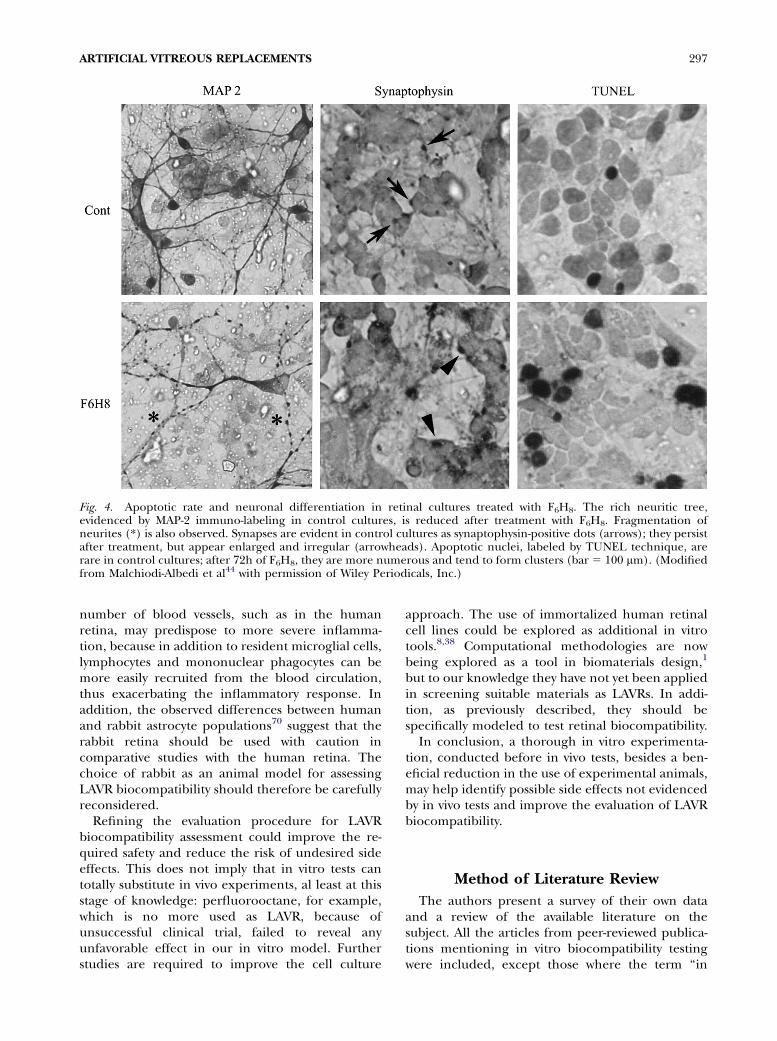
retinal cell cultures showed an evident increase inapoptotic cells, as shown by TUNEL assay, compared
to control cultures (Fig. 4). Immunolabeling ofMAP-2 and synaptophysin evidenced a marked lossof neurites and synapses (Fig. 4). These effectscannot be interpreted only on the basis of a me-chanical effect, because F6H8 has a specific gravity of1.35. Perfluorooctane, with a specific gravity of 1.78,was much better tolerated in vitro and did notimpair cell viability. The size of the molecule and thenumber of C-atoms may play an important role. Ourhypothesis is that, in the presence of F6H8, retinalglial cells can be activated by the interaction with thecompound and initiate pro-inflammatory pathways,resulting in the induction of apoptosis. Alternatively,on the basis of F6H8 lipophilicity, a damaginginteraction with the plasma membrane can beenvisaged.60
Emulsification of PFA monomers may severelyimpair visual acuity in long-term replacement of thevitreous.27 In an attempt to avoid this event,a fluorocarbon oligomer with increased viscosity,OL62HV, has been developed.36 Again, in vivoexperiments in rabbit eyes were encouraging,36 butclinical application of OL62HV lead to severe in-flammatory reactions and retinal damage.57 Whenwe analyzed the effects of OL62HV in rat primaryretinal cultures, we found an evident toxicity forretinal cell cultures, as evidenced by increasedapoptotic rate.45 Interestingly, PFA monomers suchas O62 had no pro-apoptotic effect; however, itssevere emulsification propensity and unclear in-flammatory potential limits its use as a postoperativeretinal tamponade.28
Conclusions
Before their introduction in clinical practice,LAVRs must obtain a safety certification, whichrequires tests aimed at proving their biocompatibil-ity. Notwithstanding, the use of some LAVRs hasbeen discouraged after clinical trials, because ofserious side effects, which had not been predictedduring the safety assessment procedure. It is reason-able to conclude that in vivo experimental tests sofar applied do not satisfactorily assess tissue bio-compatibility of materials. In in vivo protocols,rabbits are most frequently used. It is commonlybelieved that the avascular rabbit retina is moresusceptible to mechanical and toxic damage thanthe vascularized primate retina.77 For this reason,a material that is harmless to the rabbit retina isconsidered less prone to damage the human retina,whose vascularization should provide better pro-tection. However, in several occasions, materials thatwere well tolerated in the rabbit eye had toxic effectsin humans. A possible explanation is that a larger
ARTIFICIAL VITREOUS REPLACEMENTS 297
Fig. 4. Apoptotic rate and neuronal differentiation in retinal cultures treated with F6H8. The rich neuritic tree,evidenced by MAP-2 immuno-labeling in control cultures, is reduced after treatment with F6H8. Fragmentation ofneurites (*) is also observed. Synapses are evident in control cultures as synaptophysin-positive dots (arrows); they persistafter treatment, but appear enlarged and irregular (arrowheads). Apoptotic nuclei, labeled by TUNEL technique, arerare in control cultures; after 72h of F6H8, they are more numerous and tend to form clusters (bar 5 100 mm). (Modifiedfrom Malchiodi-Albedi et al44 with permission of Wiley Periodicals, Inc.)
number of blood vessels, such as in the humanretina, may predispose to more severe inflamma-tion, because in addition to resident microglial cells,lymphocytes and mononuclear phagocytes can bemore easily recruited from the blood circulation,thus exacerbating the inflammatory response. Inaddition, the observed differences between humanand rabbit astrocyte populations70 suggest that therabbit retina should be used with caution incomparative studies with the human retina. Thechoice of rabbit as an animal model for assessingLAVR biocompatibility should therefore be carefullyreconsidered.
Refining the evaluation procedure for LAVRbiocompatibility assessment could improve the re-quired safety and reduce the risk of undesired sideeffects. This does not imply that in vitro tests cantotally substitute in vivo experiments, al least at thisstage of knowledge: perfluorooctane, for example,which is no more used as LAVR, because ofunsuccessful clinical trial, failed to reveal anyunfavorable effect in our in vitro model. Furtherstudies are required to improve the cell culture
approach. The use of immortalized human retinalcell lines could be explored as additional in vitrotools.8,38 Computational methodologies are nowbeing explored as a tool in biomaterials design,1
but to our knowledge they have not yet been appliedin screening suitable materials as LAVRs. In addi-tion, as previously described, they should bespecifically modeled to test retinal biocompatibility.
In conclusion, a thorough in vitro experimenta-tion, conducted before in vivo tests, besides a ben-eficial reduction in the use of experimental animals,may help identify possible side effects not evidencedby in vivo tests and improve the evaluation of LAVRbiocompatibility.
Method of Literature Review
The authors present a survey of their own dataand a review of the available literature on thesubject. All the articles from peer-reviewed publica-tions mentioning in vitro biocompatibility testingwere included, except those where the term ‘‘in

298 Surv Ophthalmol 52 (3) May--June 2007 MATTEUCCI ET AL
vitro’’ refers to biophysical analysis of materials. Inaddition, articles reporting on LAVR in vivo bio-compatibility testing and clinical use were alsomentioned when they were considered relevant tothe main topic. The literature was searched using aninternational MEDLINE search from 1966 to 2006.Keywords included: in vitro, retinal cell culture, retina,biocompatibility, biomaterial, partially fluorinated alkane,semi-fluorinated alkane, perfluorocarbon liquid, perfluor-odecalin, perfluorooctane, perfluoroperhydrophenanthrene,silicone oil, perfluorohexyloctane, F6H8, vitreous sub-stitute, vitreous replacement, endoocular/intraocular tam-ponade, endotamponade, citotoxicity, or a combinationof these.
References
1. Anderson DG, Levenberg S, Langer R: Nanoliter-scalesynthesis of arrayed biomaterials and application to humanembryonic stem cells. Nat Biotechnol 22:863--6, 2004
2. Araiz JJ, Refojo MF, Arroyo MH, et al: Antiproliferative effectof retinoic acid in intravitreous silicone oil in an animalmodel of proliferative vitreoretinopathy. Invest OphthalmolVis Sci 34:522--30, 1993
3. Blinder KJ, Peyman GA, Paris CL, et al: Vitreon, a newperfluorocarbon. Br J Ophthalmol 75:240--4, 1991
4. Boveri M, Berezowski V, Price A, et al: Induction of blood-brain barrier properties in cultured brain capillary endo-thelial cells: comparison between primary glial cells and C6cell line. Glia 51:187--98, 2005
5. Brazitikos PD, Androudi S, D’Amico DJ, et al: Perfluor-ocarbon liquid utilization in primary vitrectomy repair ofretinal detachment with multiple breaks. Retina 23:615--21,2003
6. Bruining MJ, Edelbroek-Hoogendoorn PS, Blaauwgeers HG,et al: New biodegradable networks of poly(N-vinylpyrrolidi-none) designed for controlled nonburst degradation in thevitreous body. J Biomed Mater Res 47:189--97, 1999
7. Bruining MJ, Blaauwgeers HG, Kuijer R, et al: Biodegradablethree-dimensional networks of poly(dimethylamino ethylmethacrylate). Synthesis, characterization and in vitrostudies of structural degradation and cytotoxicity. Biomate-rials 21:595--604, 2000
8. Cai H, Del Priore LV: Gene expression profile of culturedadult compared to immortalized human RPE. Mol Vis 12:1--14, 2006
9. Cavalieri F, Miano F, D’Antona P, et al: Study of gellingbehavior of poly(vinyl alcohol)-methacrylate for potentialutilizations in tissue replacement and drug delivery. Bio-macromolecules 5:2439--46, 2004
10. Chang S, Ozmert E, Zimmerman NJ: Intraoperative per-fluorocarbon liquids in the management of proliferativevitreoretinopathy. Am J Ophthalmol 106:668--74, 1988
11. Chang S, Zimmerman NJ, Iwamoto T, et al: Experimentalvitreous replacement with perfluorotributylamine. Am JOphthalmol 103:29--37, 1987
12. Comaratta MR, Chang S: Perfluorocarbon liquids in themanagement of complicated retinal detachments. CurrOpin Ophthalmol 2:291--8, 1991
13. Conway MD, Peyman GA, Karacorlu M, et al: Perfluorooctyl-bromide (PFOB) as a vitreous substitute in non-humanprimates. Int Ophthalmol 17:259--64, 1993
14. Deli MA, Abraham CS, Kataoka Y, et al: Permeability studieson in vitro blood-brain barrier models: physiology, pathol-ogy, and pharmacology. Cell Mol Neurobiol 25:59--127, 2005
15. Devin F, Jourdan T, Saracco JB, et al: Experimental toleranceto perfluorodecalin used in prolonged intraocular tampo-nade. Ophthalmologica 209:306--14, 1995
16. Elsing SH, Fekrat S, Green WR, et al: Clinicopathologicfindings in eyes with retained perfluoro-n-octane liquid.Ophthalmology 108:45--8, 2001
17. Giaever I, Keese CR: Behavior of cells at fluid interfaces.Proc Natl Acad Sci U S A 80:219--22, 1983
18. Giordano GG, Refojo MF, Arroyo MH: Sustained delivery ofretinoic acid from microspheres of biodegradable polymerin PVR. Invest Ophthalmol Vis Sci 34:2743--51, 1993
19. Giordano GG, Refojo MF: Silicone oils as vitreous sub-stitutes. Prog Polym Sci 23:509--32, 1998
20. Glaser BM, Carter JB, Kuppermann BD, et al: Perfluoro-octane in the treatment of giant retinal tears with pro-liferative vitreoretinopathy. Ophthalmology 98:1613--21,1991
21. Hammer ME, Rinder DF, Hicks EL: Tolerance of perfluor-ocarbons, fluorosilicone, and silicone liquids in the vitreous,in Freeman HM, Tolentino FL (eds): Proliferative vitreor-etinopathy (PVR). New York, Springer Verlag, 1988, pp.156--61
22. Hashizoe M, Ogura Y, Takanashi T, et al: Biodegradablepolymeric device for sustained intravitreal release ofganciclovir in rabbits. Curr Eye Res 16:633--9, 1997
23. Hegazy HM, Peyman GA, Liang C, et al: Use of perfluor-ocarbon liquids, silicone oil, and 5-fluorouracil in themanagement of experimental PVR. Int Ophthalmol 22:239--46, 1998-99
24. Herrero-Vanrell R, Ramirez L, Fernandez-Carballido A, et al:Biodegradable PLGA microspheres loaded with ganciclovirfor intraocular administration. Encapsulation technique, invitro release profiles, and sterilization process. Pharm Res17:1323--8, 2000
25. Herrero-Vanrell R, Refojo MF: Biodegradable microspheresfor vitreoretinal drug delivery. Adv Drug Deliv Rev 52:5--16,2001
26. Hiscott P, Magee RM, Colthurst M, et al: Clinicopathologicalcorrelation of epiretinal membranes and posterior lensopacification following perfluorohexyloctane tamponade.Br J Ophthalmol 85:179--83, 2001
27. Hoerauf H, Laqua H: Severe emulsification after combineduse of a partially fluorinated alkane and silicone oil. GraefesArch Clin Exp Ophthalmol 240:131--6, 2002
28. Hoerauf H, Roider J, Kobuch K, et al: Perfluorohexylethan(O62) as ocular endotamponade in complex vitreoretinalsurgery. Retina 25:479--88, 2005
29. Hong Y, Chirila TV, Vijayasekaran S, et al: Crosslinkedpoly(1-vinyl-2-pyrrolidinone) as a vitreous substitute.J Biomed Mater Res 30:441--8, 1996
30. Hong Y, Chirila TV, Fitton JH, et al: Effect of crosslinkedpoly(1-vinyl-2-pyrrolidinone) gels on cell growth in static cellcultures. Biomed Mater Eng 7:35--47, 1997
31. Hong Y, Chirila TV, Vijayasekaran S, et al: Biodegradation invitro and retention in the rabbit eye of crosslinked poly(1-vinyl-2-pyrrolidinone) hydrogel as a vitreous substitute.J Biomed Mater Res 39:650--9, 1998
32. John GR, Lee SC, Brosnan CF: Cytokines: powerfulregulators of glial cell activation. Neuroscientist 9:10--22,2003
33. Kimura H, Ogura Y, Hashizoe M, et al: A new vitreal drugdelivery system using an implantable biodegradable poly-meric device. Invest Ophthalmol Vis Sci 35:2815--9, 1994
34. Kirchhof B, Wong D, Van Meurs J, et al: Use ofperfluorohexyloctane as a long-term internal tamponadeagent in complicated retinal detachment surgery. Am JOphthalmol 133:95--101, 2002
35. Kloth S, Kobuch K, Domokos J, et al: Polar application oftest substances in an organotypic environment and undercontinuous medium flow: a new tissue-based test concept fora broad range of applications in pharmacotoxicology.Toxicol In Vitro 14:265--74, 2000
36. Kobuch K, Menz IH, Hoerauf H, et al: New substances forintraocular tamponades: perfluorocarbon liquids, hydro-fluorocarbon liquids and hydrofluorocarbon-oligomers invitreoretinal surgery. Graefes Arch Clin Exp Ophthalmol239:635--42, 2001

ARTIFICIAL VITREOUS REPLACEMENTS 299
37. Kociok N, Gavranic C, Kirchhof B, et al: Influence onmembrane-mediated cell activation by vesicles of silicone oilor perfluorohexyloctane. Graefes Arch Clin Exp Ophthal-mol 243:345--58, 2005
38. Limb GA, Salt TE, Munro PM, et al: In vitro characterizationof a spontaneously immortalized human Muller cell line(MIO-M1). Invest Ophthalmol Vis Sci 43:864--9, 2002
39. Liu KR, Peyman GA, Miceli MV: Experimental evaluation oflow-viscosity fluorosilicone oil as a temporary vitreoussubstitute. Ophthalmic Surg 20:720--5, 1989
40. Maberley AL, Antworth MV: The use of silicone oil invitreoretinal surgery. Can J Ophthalmol 24:265--8, 1989
41. Malchiodi-Albedi F, Feher J, Caiazza S, et al: PEDF (pigmentepithelium-derived factor) promotes increase and matura-tion of pigment granules in pigment epithelial cells inneonatal albino rat retinal cultures. Int J Dev Neurosci 16:423--32, 1998
42. Malchiodi-Albedi F, Perilli R, Formisano G, et al: Perfluor-odecalin modifies the pattern of cell arrangement andinduces loss of neurites in rat retinal cultures. J BiomedMater Res 41:608--13, 1998
43. Malchiodi-Albedi F, Morgillo A, Formisano G, et al: Bio-compatibility assessment of silicone oil and perfluorocarbonliquids used in retinal reattachment surgery in rat retinalcultures. J Biomed Mater Res 60:548--55, 2002
44. Malchiodi-Albedi F, Matteucci A, Formisano G, et al:Perfluorohexyloctane (F6H8) induces structural modifica-tions and increases apoptosis in rat primary retinal cultures.J Biomed Mater Res B Appl Biomater 65:133--6, 2003
45. Malchiodi-Albedi F, Matteucci A, Formisano G, et al: In-duction of apoptosis in rat retinal cell cultures by partiallyfluorinated alkanes. Am J Ophthalmol 139:737--9, 2005
46. Martınez-Sancho C, Herrero-Vanrell R, Negro S: Poly (D,L-lactide-co-glycolide) microspheres for long-term intravitrealdelivery of aciclovir: influence of fatty and non-fattyadditives. J Microencapsul 20:799--810, 2003
47. Maruoka S, Matsuura T, Kawasaki K, et al: Biocompatibilityof polyvinylalcohol gel as a vitreous substitute. Curr Eye Res31:599--606, 2006
48. Meinert H, Roy T: Semifluorinated alkanes--a new class ofcompounds with outstanding properties for use in ophthal-mology. Eur J Ophthalmol 10:189--97, 2000
49. Meller D, Augustin AJ, Spitznas M, et al: Effects of differentperfluorochemicals on dorsal root ganglion cells in vitro.Graefes Arch Clin Exp Ophthalmol 236:182--7, 1998
50. Mertens S, Bednarz J, Engelmann K: Evidence of toxic sideeffects of perfluorohexyloctane after vitreoretinal surgery aswell as in previously established in vitro models with ocularcell types. Graefes Arch Clin Exp Ophthalmol 240:989--95,2002
51. Orzalesi N, Migliavacca L, Bottoni F, et al: Experimentalshort-term tolerance to perfluorodecalin in the rabbit eye:a histopathological study. Curr Eye Res 17:828--35, 1998
52. Peyman GA, Schulman JA, Sullivan B: Perfluorocarbonliquids in ophthalmology. Surv Ophthalmol 39:375--95, 1995
53. Ratiglia R, Berti E, Galimberti D, et al: Experimentalvitreous replacement with perfluorophenanthrene. Eur JOphthalmol 7:59--63, 1997
54. Rizzo S, Genovesi-Ebert F, Belting C, et al: A pilot study onthe use of silicone oil-RMN3 as heavier-than-water endo-tamponade agent. Graefes Arch Clin Exp Ophthalmol 28:1--5, 2005
55. Rodriguez Boulan E, Sabatini DD: Asymmetric budding ofviruses in epithelial monolayers: a model system for studyof epithelial polarity. Proc Natl Acad Sci USA 75:5071--5,1978
56. Rodriguez M, Schaper J: Apoptosis: measurement andtechnical issues. J Mol Cell Cardiol 38:15--20, 2005
57. Roider J, Hoerauf H, Kobuch K, et al: Clinical findings onthe use of long-term heavy tamponades (semifluorinatedalkanes and their oligomers) in complicated retinal de-
tachment surgery. Graefes Arch Clin Exp Ophthalmol 240:965--71, 2002
58. Rosemberg MD: The culture of cells and tissues at the saline-fluorocarbon interface, in Ramakrishnan CV (ed): BarodaTissue Culture Seminar. The Hague, Netherlands, Junk,1965, pp. 93--107
59. Saxena S, Gopal L: Fluid vitreous substitutes in vitreo retinalsurgery. Indian J Ophthalmol 44:191--206, 1996
60. Schatz B, El-Shabrawi Y, Haas A, et al: Adverse side effectswith perfluorohexyloctane as a long-term tamponade agentin complicated vitreoretinal surgery. Retina 24:567--73, 2004
61. Singh J, Ramaesh K, Wharton SB, et al: Perfluorodecalin-induced intravitreal inflammation. Retina 21:247--51, 2001
62. Sobel S, Rosenberg MD: Characterization of cellularattachment and spreading molecules at liquid-liquid in-terfaces. Anal Biochem 140:486--9, 1984
63. Soman N, Banerjee R: Artificial vitreous replacements.Biomed Mater Eng 13:59--74, 2003
64. Sparrow JR, Jayakumar A, Berrocal M, et al: Experimentalstudies of the combined use of vitreous substitutes of highand low specific gravity. Retina 12:134--40, 1992
65. Sparrow JR, Ortiz R, MacLeish PR, et al: Fibroblast behaviorat aqueous interfaces with perfluorocarbon, silicone, andfluorosilicone liquids. Invest Ophthalmol Vis Sci 31:638--46,1990
66. Stolba U, Krepler K, Pflug R, et al: Experimental vitreousand aqueous replacement with perfluorophenanthrene.Clinical, histologic, and electrophysiologic results. Retina17:146--53, 1997
67. Stolba U, Krepler K, Velikay-Parel M, et al: The effect ofspecific gravity of perfluorocarbon liquid on the retina afterexperimental vitreous substitution. Graefes Arch Clin ExpOphthalmol 242:931--6, 2004
68. Suri S, Banerjee R: In vitro evaluation of in situ gels as shortterm vitreous substitutes. J Biomed Mater Res 79A:650--64,2006
69. Theelen T, Tilanus MA, Klevering BJ: Intraocular inflam-mation following endotamponade with high-density siliconeoil. Graefes Arch Clin Exp Ophthalmol 242:617--20, 2004
70. Trivino A, Ramırez JM, Ramırez AI, et al: Comparative studyof astrocytes in human and rabbit retinae. Vision Res 37:1707--11, 1997
71. Velikay M, Stolba U, Wedrich A, et al: The effect of chemicalstability and purification of perfluorocarbon liquids inexperimental extended-term vitreous substitution. GraefesArch Clin Exp Ophthalmol 233:26--30, 1995
72. Versura P, Cellini M, Torreggiani A, et al: The biocompat-ibility of silicone, fluorosilicone and perfluorocarbon liquidsas vitreous tamponades. an ultrastructural and immunohis-tochemical study. Ophthalmologica 215:276--83, 2001
73. Vote B, Wheen L, Cluroe A, et al: Further evidence forproinflammatory nature of perfluorohexyloctane in the eye.Clin Experiment Ophthalmol 31:408--14, 2003
74. Winter M, Eberhardt W, Scholz C, Reichenbach A: Failure ofpotassium siphoning by Muller cells: a new hypothesis ofperfluorocarbon liquid-induced retinopathy. Invest Oph-thalmol Vis Sci 41:256--61, 2000
75. Wolf S, Schon V, Meier P, Wiedemann P: Silicone oil RMN3mixture (heavy silicone oil) as internal tamponade forcomplicated retinal detachment. Retina 23:335--42, 2003
76. Wong D, Lois N: Perfluorocarbons and semifluorinatedalkanes. Semin Ophthalmol 15:25--35, 2000
77. Zeana D, Becker J, Kuckelkorn R, Kirchhof B: Perfluor-ohexyloctane as a long-term vitreous tamponade in theexperimental animal. Experimental perfluorohexyloctanesubstitution. Int Ophthalmol 23:17--24, 1999
Reprint address: Fiorella Malchiodi-Albedi, Department of CellBiology and Neuroscience, Istituto Superiore di Sanita, VialeRegina Elena 299, 00161 Rome, Italy.