Evaluating Antibacterial Efficacy and Biocompatibility of PAN ...
13
polymers Article Evaluating Antibacterial Efficacy and Biocompatibility of PAN Nanofibers Loaded with Diclofenac Sodium Salt Muhammad Nauman Sarwar 1 , Azeem Ullah 1 , Md. Kaiser Haider 1 , Nadir Hussain 1 , Sana Ullah 1 , Motahira Hashmi 1 , Muhammad Qamar Khan 2 and Ick Soo Kim 1, * Citation: Sarwar, M.N.; Ullah, A.; Haider, M..K.; Hussain, N.; Ullah, S.; Hashmi, M.; Khan, M.Q.; Kim, I.S. Evaluating Antibacterial Efficacy and Biocompatibility of PAN Nanofibers Loaded with Diclofenac Sodium Salt. Polymers 2021, 13, 510. https:// doi.org/10.3390/polym13040510 Academic Editors: Nuno Alves, Juliana Dias and Antonio Gloria Received: 12 January 2021 Accepted: 4 February 2021 Published: 8 February 2021 Publisher’s Note: MDPI stays neutral with regard to jurisdictional claims in published maps and institutional affil- iations. Copyright: © 2021 by the authors. Licensee MDPI, Basel, Switzerland. This article is an open access article distributed under the terms and conditions of the Creative Commons Attribution (CC BY) license (https:// creativecommons.org/licenses/by/ 4.0/). 1 Nano Fusion Technology Research Group, Institute for Fiber Engineering (IFES), Interdisciplinary Cluster for Cutting Edge Research (ICCER), Shinshu University, Tokida 3-15-1, Ueda, Nagano 386-8567, Japan; [email protected] (M.N.S.); [email protected] (A.U.); [email protected] (M.K.H.); [email protected] (N.H.); [email protected] (S.U.); [email protected] (M.H.) 2 Nanotechnology Research Lab, Department of Textile & Clothing, Faculty of Engineering & Technology, National Textile University Karachi Campus, Karachi 74900, Pakistan; [email protected] * Correspondence: [email protected] Abstract: Side effects of the drugs’ oral administration led us to examine the possibility of using diclofenac sodium (DLF) in a polymeric drug delivery system based on electrospun polyacrylonitrile (PAN) nanofibers, which can be produced cost-effectively and with good applicability for transdermal treatments. The inclusion of DLF in PAN nanofibers increased the nanofiber diameter from ~200 nm to ~500 nm. This increase can be attributed to the increase in the spinning solution viscosity. FTIR spectra confirm the entrapment of the DLF into the PAN nanofibers. X-ray diffraction pattern showed that the inclusion of the DLF in the PAN nanofibers had caused the misalignment in the polymeric chains of the PAN, thus resulting in a decrease of the peak intensity at 2θ = 17 o . The DLF loaded PAN nanofibers were efficient against the gram-positive Staphylococcus aureus (S. aureus) and gram- negative Escherichia coli (E. coli), with maximum inhibition zone of 16 ± 0.46 mm for E. coli and 15.5 ± 0.28 mm for S. aureus. Good cell viability ~95% for L929 cells in more extended incubation periods was reported. A gradual release of DLF from the PAN nanofiber was observed and can be attributed to the stability of Pan in PBS medium. Cell adhesion micrographs show that cell-cell interaction is stronger than the cell-material interaction. This type of weak cell interaction with the wound dressing is particularly advantageous, as this will not disturb the wound surface during the nursing of the wound. Keywords: electrospinning; biocompatibility; antibacterial; wound care; nanofibers 1. Introduction Skin is the largest organ of our body. It is also the first defense line in protecting the body’s internal and most sensitive parts from the attack of bacteria, xenobiotics, and dehydration [1–5]. Any discontinuity in the structure of the skin may lead to a condition called a wound. A wound must be secured from external harms such as bacterial and fungal infections etc. Wound dressings serve as a barrier between the wound’s microenvironment and the external environment. For this purpose, wound dressings with antibacterial properties can provide an efficient barrier to pathogens. Nano-encapsulation technique for drug delivery systems has yielded numerous bene- fits, such as easy handling, stability, protection against oxidation, control over the release profile, multi-drug delivery, reduced toxicity, and side effects of the drugs [6,7]. Nanofiber based drug delivery systems for the control and site specific drug release were developed by encapsulating drugs into polymeric delivery system in order to provide a controllable release of certain ratio of drugs at a proper targeted site over a fairly long duration of time [8]. The application of micro and nanofiber is gaining interest in biomedical field due to the acquired advantage of improved therapeutic index, possibility for localized delivery and Polymers 2021, 13, 510. https://doi.org/10.3390/polym13040510 https://www.mdpi.com/journal/polymers
-
Upload
khangminh22 -
Category
Documents
-
view
2 -
download
0
Transcript of Evaluating Antibacterial Efficacy and Biocompatibility of PAN ...

polymers
Article
Evaluating Antibacterial Efficacy and Biocompatibility of PANNanofibers Loaded with Diclofenac Sodium Salt
Muhammad Nauman Sarwar 1, Azeem Ullah 1 , Md. Kaiser Haider 1, Nadir Hussain 1, Sana Ullah 1 ,Motahira Hashmi 1, Muhammad Qamar Khan 2 and Ick Soo Kim 1,*
�����������������
Citation: Sarwar, M.N.; Ullah, A.;
Haider, M..K.; Hussain, N.; Ullah, S.;
Hashmi, M.; Khan, M.Q.; Kim, I.S.
Evaluating Antibacterial Efficacy and
Biocompatibility of PAN Nanofibers
Loaded with Diclofenac Sodium Salt.
Polymers 2021, 13, 510. https://
doi.org/10.3390/polym13040510
Academic Editors: Nuno Alves,
Juliana Dias and Antonio Gloria
Received: 12 January 2021
Accepted: 4 February 2021
Published: 8 February 2021
Publisher’s Note: MDPI stays neutral
with regard to jurisdictional claims in
published maps and institutional affil-
iations.
Copyright: © 2021 by the authors.
Licensee MDPI, Basel, Switzerland.
This article is an open access article
distributed under the terms and
conditions of the Creative Commons
Attribution (CC BY) license (https://
creativecommons.org/licenses/by/
4.0/).
1 Nano Fusion Technology Research Group, Institute for Fiber Engineering (IFES), Interdisciplinary Cluster forCutting Edge Research (ICCER), Shinshu University, Tokida 3-15-1, Ueda, Nagano 386-8567, Japan;[email protected] (M.N.S.); [email protected] (A.U.); [email protected] (M.K.H.);[email protected] (N.H.); [email protected] (S.U.); [email protected] (M.H.)
2 Nanotechnology Research Lab, Department of Textile & Clothing, Faculty of Engineering & Technology,National Textile University Karachi Campus, Karachi 74900, Pakistan; [email protected]
* Correspondence: [email protected]
Abstract: Side effects of the drugs’ oral administration led us to examine the possibility of usingdiclofenac sodium (DLF) in a polymeric drug delivery system based on electrospun polyacrylonitrile(PAN) nanofibers, which can be produced cost-effectively and with good applicability for transdermaltreatments. The inclusion of DLF in PAN nanofibers increased the nanofiber diameter from ~200 nmto ~500 nm. This increase can be attributed to the increase in the spinning solution viscosity. FTIRspectra confirm the entrapment of the DLF into the PAN nanofibers. X-ray diffraction pattern showedthat the inclusion of the DLF in the PAN nanofibers had caused the misalignment in the polymericchains of the PAN, thus resulting in a decrease of the peak intensity at 2θ = 17o. The DLF loadedPAN nanofibers were efficient against the gram-positive Staphylococcus aureus (S. aureus) and gram-negative Escherichia coli (E. coli), with maximum inhibition zone of 16 ± 0.46 mm for E. coli and15.5 ± 0.28 mm for S. aureus. Good cell viability ~95% for L929 cells in more extended incubationperiods was reported. A gradual release of DLF from the PAN nanofiber was observed and canbe attributed to the stability of Pan in PBS medium. Cell adhesion micrographs show that cell-cellinteraction is stronger than the cell-material interaction. This type of weak cell interaction with thewound dressing is particularly advantageous, as this will not disturb the wound surface during thenursing of the wound.
Keywords: electrospinning; biocompatibility; antibacterial; wound care; nanofibers
1. Introduction
Skin is the largest organ of our body. It is also the first defense line in protectingthe body’s internal and most sensitive parts from the attack of bacteria, xenobiotics, anddehydration [1–5]. Any discontinuity in the structure of the skin may lead to a conditioncalled a wound. A wound must be secured from external harms such as bacterial and fungalinfections etc. Wound dressings serve as a barrier between the wound’s microenvironmentand the external environment. For this purpose, wound dressings with antibacterialproperties can provide an efficient barrier to pathogens.
Nano-encapsulation technique for drug delivery systems has yielded numerous bene-fits, such as easy handling, stability, protection against oxidation, control over the releaseprofile, multi-drug delivery, reduced toxicity, and side effects of the drugs [6,7]. Nanofiberbased drug delivery systems for the control and site specific drug release were developedby encapsulating drugs into polymeric delivery system in order to provide a controllablerelease of certain ratio of drugs at a proper targeted site over a fairly long duration oftime [8].
The application of micro and nanofiber is gaining interest in biomedical field due to theacquired advantage of improved therapeutic index, possibility for localized delivery and
Polymers 2021, 13, 510. https://doi.org/10.3390/polym13040510 https://www.mdpi.com/journal/polymers

Polymers 2021, 13, 510 2 of 13
reduced toxicity of drugs [9]. Nanotechnology can serve as a modest provision for avoidingthe high systemic and topical drug dose as a preventive medicine source. At present,electrospinning is the predominant technique to develop wound dressing because of itssimplicity, low cost, high production rate, and ability to create nanofibers having multiplemorphological structures [10,11]. Besides, their porous structure, ability to absorb woundexudates, high specific surface area, and structure similarity to the natural extracellularmatrix (ECM) nanofibers are widely used for tissue regeneration, wound dressing, and drugdelivery system [12–14]. Electrospun nanofibers have found their applications in numerousfields such as wound healing [15], antibacterial [16,17], sensors [18], filtration [19], andother environmental application. Various delivery systems based on (natural & synthetic)polymers, liposomes, inorganic nanoparticles, etc., are generally used for wound dressing.Among these systems, the polymer-based system is widely practiced clinically [20,21].A wide range of polymers (natural &synthetic) has been successfully electrospun formaking wound dressing. Among various polymers, polyacrylonitrile (PAN) is widely usedfor wound dressing because; it possesses outstanding features such as light resistance,chemical stability, non-toxicity, mechanical elasticity, and electrospinabilty [22,23]. PANor its composite with other polymers combined with different drugs or nanoparticleshave been used for medical applications. Many researchers have successfully synthesizeddifferent nanofiber composite and drug-loaded delivery systems by incorporating othermaterials such as acyclovir, TiO2, AgCl, and silica [24–26].
Diclofenac (DLF) is a non-steroidal anti-inflammatory drug (NSAID), which is abenzene-acetic acid derivative and considered one of the best NSAID’s medicine for thetreatment of acute and chronic Painful and inflammatory injuries [27,28]. Nano packagingof the drugs can enhance their medicinal efficacy, specificity, tolerability, therapeutic index,and polymer-based nano-carriers can further protect the drugs from degradation andreduce the toxicity or side effects. Though NSAID DLF has potent anti-inflammatory,analgesic, and antipyretic properties, but also is susceptible to risks of toxicity and otheradverse effects. Encapsulating DLF in Eudragit® L100-55 has been reported to reduce thesedrawbacks [29]. Huh, et al. proposed a surgical suture loaded with DLS for sustainablerelease as local pain relief of the surgical wound. DLF was loaded into the biodegradablepoly lactic-co-glycolic acid stand and then braided with a surgical suture VICRYL. In thisway, a sustained release for 10 days of DLF was recorded while maintaining the woundclosure’s mechanical strength [30]. In another work, DLF loaded zein nanoparticles wereprepared by the anti-solvent precipitation method and loaded into PVA nanofibers. Amore controlled release of DLF was achieved by embedding the zein nanoparticles in PVAnanofibers. The results suggested that embedding DLF in a polymeric system improvesits cell attachment and proliferation [31]. Nanonets of DLF loaded polycaprolactone andchitosan were prepared by electrospinning. Because of the nanofiber net’s slow degradation,the release was mostly governed by diffusion or permeation. The cell viability and imagingdata indicate that NIH 3T3 cells experienced no toxicity in the DLF- nanonets structures [32].Most of these studies carried in the near past are based on the release and toxicity study ofthe DLF loaded polymeric systems.
The side effects of oral administration of drugs have led us to investigate the possibilityof encapsulating DLF in a polymeric drug delivery system based on PAN nanofibers whichcan be produced cheaply and having high applicability to be employed for transdermaldelivery of drugs on the wound site. Besides, the polymer selection was based upon thehigh drug loading capacity, good mechanical strength, and prolong release characteris-tics [33]. The present work describes the preparation of antibacterial DLF loaded nanofibersusing PAN as the filament-forming matrix, aimed for transdermal delivery. The preparednanofibers were subjected to physicochemical, scanning electron microscopy, X-ray diffrac-tion, in vitro release, antibacterial efficacy, and in-vitro cell proliferation analysis.

Polymers 2021, 13, 510 3 of 13
2. Materials and Methods2.1. Materials
Polyacrylonitrile (PAN) (MW 150,000) procured from Sigma Aldrich (St. Louis, MO,USA). Diclofenac sodium salt (DLF) was purchased from Sigma Aldrich (St. Louis, MO, USA).N, N-Dimethylformamide (DMF) (99.8%) was obtained from Fujifilm Wako Chemicals Ltd.(Osaka, Japan). All chemicals were used as received without any further purification.
2.2. Preparation of PAN/Diclofenac Electrospinning Solution
The 10% w/w solution of polyacrylonitrile (PAN) was prepared in N, N- Dimethylfor-mamide (DMF) with continuous stirring. After stirring the PAN solution for 5 h at ambienttemperature, Diclofenac sodium salt (DLF) was added to the polymer solution with 3, 4,5, and 6% by weight of the PAN. Afterward, the stirring was continued for 2 h to preparethe final PAN/ DLF solutions for electrospinning. A brief overview of the electrospinningsolutions is given in Table 1.
Table 1. Composition of electrospinning solution.
Sample No.Weight Ratio (%)
PAN DLF
S1 100 0S2 97 3S3 96 4S4 95 5S5 94 6
2.3. Electrospinning Process
A horizontal electrospinning module was set up by using a high voltage power supply(Har-100*12, Matsusada Co., Tokyo, Japan) and a syringe pump (KDS-100, KD Scientific,Holliston, USA). The electrospinning solutions were fed in a 25 mL syringe equipped witha 22 gauge needle. A high voltage of 12.5 kV was applied across the needle and collectorcovered with butter paper. A fed rate adjusted at 0.7 mL/h, and a collector to needledistance was 130 mm. All samples were spun for 20 h to ensure a uniform thickness of thefibrous mats.
2.4. Surface Morphology Characterizations
The surface morphology of Nanofibers was evaluated by using scanning electronmicroscopy (SEM, JSM-5300, JEOL Ltd., Nagano, Japan) with an accelerating voltage of12 kV and TEM (JEM-2100 JEOL, Nagano, Japan) accelerated with 200 kV. The averagediameter distribution of nanofibers was calculated by image analysis software (ImageJ,version 1.49). The nanofiber specimen were sputter coated with platinum before subjectingto the SEM imagery.
2.5. Physicochemical Characterizations
An attenuated total reflectance Fourier transform infrared (ATR-FTIR) spectroscopy(Prestige-21, Shimadzu, Japan) was used to determine the nanofiber dressing chemicalcomposition. FTIR spectra were scanned over the wavelength range 4000–600 cm−1 atroom temperature, and a spectrum was accumulated over 20 scans.
2.6. XRD Characterizations
The X-ray diffractograms (XRD) of the neat and DLF loaded fibrous mats were deter-mined by an X-ray diffractometer (Miniflex 300, Rigaku Co., Ltd., Tokyo, Japan) operatingat 30 kV and 500 mA. Diffractions were detected in the 2θ = 5◦–80◦ range with a stepof 0.05◦.

Polymers 2021, 13, 510 4 of 13
2.7. The In-Vitro Release Behavior of DLF from the PAN Nanofibrous Mats
The in vitro release of DLF from the DLF/PAN nanofibrous mats was analyzed bythe total immersion method in phosphate buffer (pH 7.4) containing 0.5% Tween 80 and3% methanol (PTM buffer solution). The release medium was kept at 37 ◦C to mimic thebody core temperature. Each specimen was immersed in 30 mL of releasing medium, andthe dissolution time was 72 h. At a specific time interval, 1 mL of the release media wasextracted, and the bath was replenished with the fresh buffer solution to keep the bathconditions constant. The amount of DLF in the media was determined by UV-spectroscopyat the characteristic wavelength of 276 nm (λmax). The absorbance spectra of the DLFat certain time intervals were converted into concentration using the calibration curve;y = 0.5984x2 + 11.04x − 4.789.
2.8. Antibacterial Efficacy
The antibacterial characteristics of fabricated electrospun nanofibers dressing were as-sessed against gram-positive Staphylococcus aureus (S. aureus) and gram-negative Escherichiacoli (E. coli) bacteria by the qualitative method. S. aureus (ATCC 29213) and E. coli (ATCC25922) strains were provided by Gene Research Centre, Shinshu University. A 100 µL of10−5 dilution of overnight cultured E. coli and S. aureus were spread over the agar plates.The fiber mats were UV sterilized for 6 h before subjecting to an antibacterial test. The fibermat samples (10 mm, diameter) were gently placed over the agar plates and incubatedovernight. The agar plates were examined for bacterial growth interruption underneathand along the border for the inhibition zone.
2.9. Cell Viability and Adhesion
The L929 mouse normal fibroblast cell line mitochondrial activity was measuredby WST-1 colorimetric assay using ISO 10993-5 standard. L929 cell line was provideby Gene research Centre, Shinshu University. In toxicity studies, the use of continuouscell lines, such as L929 mouse fibroblast is favored because of their biological response,ease of production, and easy control of their culture [34]. L929 cells were cultured inDulbecco modified eagle medium (DMEM) accompanying 10% fetal bovine serum (FBS) ina humidified incubator at 37 ◦C and 5% CO2 environment. The fiber mats were sterilized in70% ethanol for 30 min and then washed with PBS to remove any impurity. The sterilizedmats were placed in 96 well glass culture plate. The culture plate was seeded with a densityof 1 × 10³ cells per well. At the predetermined times, 10 µL WST −1 was added to eachwell and incubated for two hours. Absorbance was measured at 440 nm using a microplatereader (Thermos Scientific, Multiskan FC instruments) to indicate proliferation. Readingswere measured in triplicate, and averages were recorded.
The cell-cell and cell-material interactions were observed with SEM micrographs.L929 cells were cultured with nanofiber mats for 72 h and were fixed on the mats with 4%paraformaldehyde at 4 ◦C. The mats were dehydrated using ethanol and dried in a vacuumoven. The nanofiber specimen were sputter coated with platinum before subjecting to theSEM imagery. The SEM micrographs were taken at a resolution of 300×.
2.10. Statistical Analysis
All statistical analysis regression equations and correlation coefficients were carriedout using MINITAB 17 ® statistical software [35].
3. Results and Discussion3.1. Morphology and Diameter Analysis
SEM micrographs (Figure 1) were evaluated to analyze any difference in the sur-face morphology and diameter of the DLF loaded PAN nanofibers compared to pris-tine PAN nanofibers. The SEM micrographs confirm that uniform, bead-free continuousnanofibers were obtained in all cases. The pure PAN fibers showed an average diameterof ~215 ± 50 nm. The inclusion of DLF in the PAN nanofibers increased the nanofiber

Polymers 2021, 13, 510 5 of 13
diameter. The increase in fiber diameter was in proportion to the amount of the DLFloaded into the fibers. The DLF loaded fibers showed an average diameter of ~279 ± 60,~432 ± 30, ~467 ± 40, and ~489 ± 50 nm for S2, S3, S4, and S5. Though DLF salt canincrease the electrical conductivity of the PAN solution and thereby should enhance theelectrical drawing of the spinning jet. But with the addition of the DLF to the PAN solutionthe viscosity of the solution increased, and this increase in the viscosity counteracted theinfluence of the electrical conductivity and gradually had a greater influence on the increasein fiber diameter. The increase in nanofiber diameter by the inclusion of DLF is in goodagreement with previously reported work [29].
Figure 1. SEM micrographs and diameter histograms of S1 (A,B), S2 (C,D), S3 (E,F), S4 (G,H), andS5 (I,J).

Polymers 2021, 13, 510 6 of 13
3.2. Study of XRD Spectra
Figure 2 represents the XRD diffractograms of the pristine and DLF loaded PANnanofibers. The XRD pattern of the virgin PAN fibers showed a characteristic peak at2θ = 17◦. The peak at 2θ = 17◦ was associated with the hexagonal crystal lattice of PAN.The extraneous substances in a pure material result in a change in its molecular packingand is a fact [36]. DLF loaded PAN nanofibers, a characteristic peak at a shorter angle2θ = 7.1◦ appeared, associated with the DLF and confirmed by the literature [37]. Inaddition to the appearance of a new peak at 2θ = 7.1◦, the intensity of the PAN peak at2θ = 17◦ in DLF loaded fibers also decreases. The addition of DLF could have causeda change in the molecular packing of the PAN, thus resulting in misalignment of thepolymer microstructure.
Figure 2. XRD diffractograms of neat (S1) and DLF loaded PAN nanofibers (S2, S3, S4, and S5).
3.3. Physicochemical Analysis
Figure 3 represents the ATR-FTIR spectrum of the pure PAN, DLF, and DLF loadedPAN nanofibers. In Figure 3a, we can observe the sharp peaks at 2931, 2276, and 1451 cm−1.These peaks are due to the stretching frequencies of methylene (-CH2), nitrile (-CN), andbending vibrations of the methylene groups, respectively present in the PAN polymerbackbone [38,39].
In DLF salt -NH and -NH-O stretching are visible at wavenumber 3407 and 3276 cm−1.The DLF also exhibited the two identifying bands of carboxylate stretching vibration at1567 cm−1 (asymmetric) and at 1401 cm−1 (symmetric). Phenyl rings stretching vibrationsare intense in the range of 1600–1450 cm−1 [40].
The spectra area between 1700–1000 cm−1 is quite similar to the ranges of purematerials. The only difference observed in the DLF loaded PAN nanofibers’ spectra fromthe Pure PAN nanofibers is in the region of 3500–3000 cm−1. The appearance of these peakscould be due to -NH and NH-O constituents of the DLF.

Polymers 2021, 13, 510 7 of 13
Figure 3. ATR-FTIR spectrum of (a) virgin PAN (S1), (b) Diclofenac sodium salt and (c) DLF loadedPAN nanofibers (S2, S3, S4, and S5).
3.4. Antibacterial Activity
The antibacterial efficiency of the pristine and DLF loaded PAN nanofiber was ana-lyzed against the gram-negative (E. coli)(A) and gram-positive (S. aureus)(B). The chosenbacterial strains are the most frequent infection-causing bacteria to the soft tissues and

Polymers 2021, 13, 510 8 of 13
results in the delayed wound healing. Most of the clinically acquired infections are resul-tant to Staphylococcus aureus. While Escherichia coli is considered as a primary source forinfection in the burn wounds [41]. The exact mechanism of the antibacterial activity ofdiclofenac is unclear. However studies have proposed that the NSAID inhibits the bacterialDNA synthesis [42] or impairment of the membrane activity [43]. Figure 4 shows the halocreated around the DLF loaded PAN nanofiber discs. These halos are the indication ofthe bacterial growth interruption. The halos were measured using ImageJ software andare recorded in millimeters and are given in Table 2. As the amount of DLF in the PANnanofiber increases, the halo diameter increases. The largest inhibition zone was recordedfor S5 with 16 ± 0.46 mm and 15.5 ± 0.28 mm for E. coli and S. aureus.
Figure 4. Bactericidal efficacy of DLF loaded PAN nanofiber (S2, S3, S4, and S5) using agar discdiffusion test (A) E. coli and (B) S. aureus.
Table 2. Bacterial growth inhibition (zone of inhibition) for DLF loaded PAN nanofibers.
Samples LabelsZone of Inhibition (mm)
E. coli S. aureus
S1(neat PAN) 0 0S2(3% DLF) 7 ± 0.15 8 ± 0.17S3(4% DLF) 10.5 ± 0.19 12 ± 0.27S4(5% DLF) 14 ± 0.38 15 ± 0.59S5(6% DLF) 16 ± 0.46 15.5 ± 0.28
3.5. Cell Viability and Cell Adhesion
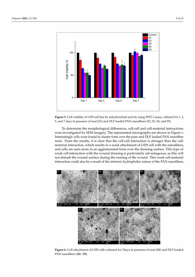
It is essential that the wound dressing should be biocompatible. Virgin and DLFloaded PAN nanofibers were subjected to the colorimetric WST-1 assay to determinethe mitochondrial activity of the L929 cells. The analysis was carried out according tothe standard ISO 10993-5 method. Figure 5 shows the viability of the L929 cell in thepresence of DLF loaded and pure PAN nanofibers at 1, 3, 5, and 7 days of incubation. Forthe shorter period of incubation, the samples showed moderate toxicity ranging between~50–70%. The DLF loaded mats showed a higher cell viability for more extended incubationperiods ranging between ~75–100%, indicating good biocompatibility of the compositedressing. Furthermore, it can be said that the addition of DLF did not adversely affect thebiocompatibility of the PAN nanofibers.
Polymers 2021, 13, 510 9 of 13
Figure 5. Cell viability of L929 cell line by mitochondrial activity using WST-1 assay, cultured for 1, 3,5, and 7 days in presence of neat (S1) and DLF loaded PAN nanofibers (S2, S3, S4, and S5).
To determine the morphological differences, cell-cell and cell-material interactionswere investigated by SEM imagery. The represented micrographs are shown in Figure 6.Interestingly cells were found in cluster form over the pure and DLF loaded PAN nanofibermats. From the results, it is clear that the cell-cell interaction is stronger than the cell-material interaction, which results in a week attachment of L929 cell with the nanofibers,and cells are seen more in an agglomerated form over the dressing surface. This type ofweak cell interaction with the wound dressing is particularly advantageous, as this willnot disturb the wound surface during the nursing of the wound. This week cell-materialinteraction could also be a result of the intrinsic hydrophobic nature of the PAN nanofibers.
Figure 6. Cell attachment of L929 cells cultured for 7days in presence of neat (S1) and DLF loadedPAN nanofibers (S2– S5).

Polymers 2021, 13, 510 10 of 13
3.6. The Release Profile of DLF in PTM
Drug release profile of the DLF from the PAN nanofibers was investigated by im-mersing PAN/DLF nanofibers in PTM buffer solution, pH (7.4), at 37 ◦C. The drug releaseprofile are demonstrated in Figure 7. It can be observed that DLF gradually released fromthe PAN nanofibers. At first the release rate was fast. This quick release is due to thediffusion of DLF from the surface of the PAN nanofibers. The gradual release of DLFcontinued until 48 h. After 48 h time interval the release almost become constant. It wasobserved that initial rapid release was dependent upon the amount of DLF loaded intothe PAN nanofibers. The gradual release of DLF from PAN nanofibers is attributed to thechemical stability of PAN in the PBS release media. Based on the drug release profile wecan say that the primary release of DLF from the PAN nanofiber is governed by diffusionand permeation from the surface of the nanofibers. This slow release is beneficial as PANnanofiber will retain the bioactive agent throughout their useful life. Similar results wereobtained with other drugs loaded into PAN nanofibers [33].
Figure 7. (a) In vitro release profile of DLF from PAN nanofibers (S2, S3, S4, and S5) in PBS, (b) cali-bration curve of the DLF in PBS showing regression model and coefficient of determination (R2).
3.7. Practical Implications and Future Perspective
The produced nanofibers were aimed for providing efficient antibacterial activityagainst both gram positive and gram negative bacterial strains, along with the addedadvantage of the anti-inflammatory and analgesic properties of the DLF. The release of thePAN/DLF nanofiber system was suitable for the acute open wounds. Where this nanofiberdressing can provide efficient barrier properties. Moreover, the drug release was gradualand can be site specific over a fairly long duration of time.
The solubility of the drug in the polymeric system is of utmost importance and de-termines the bioavailability and drug distribution through the delivery system. Drugsolubility has a major influence over the possible permeation across the biological mem-branes. The usage of the NSAID drugs as an effective antibacterial agent is relatively newand very few studies have been conducted for their effective encapsulation and targetedsite delivery. So, different polymeric systems can be examined and in vitro alongsidein vivo studies should be conducted to further validate the synergistic effect of NSAIDs aseffective antibacterial.
4. Conclusions
Uniform bead-free continuous DLF loaded PAN nanofibers were successfully pro-duced on horizontal electrospinning set up at a high voltage of 12.5 kV. FTIR spectrum andthe appearance of a new peak at 7.1o in XRD diffractogram indicates the entrapment of theDLF in the PAN nanofiber matrices after electrospinning. The PAN nanofiber diametersincreased proportionally to the increase in the amount of the DLF. The inclusion of DLFresults in the polymer chain misalignment in the PAN and can be seen with decreasedXRD peak intensity from the XRD diffractogram. DLF loaded nanofibers were efficient

Polymers 2021, 13, 510 11 of 13
in inhibiting the bacterial growth of both E. coli and S. aureus. The inclusion of DLF inthe PAN does not hinder the L929 cell proliferation. The SEM micrographs showed thatthe cell-cell interaction is stronger than the cell-material interaction, which is vital duringwound nursing. DLF loaded PAN nanofiber showed a slow release with a maximum of~40% release at 72 h time interval.
Based on the results it can be inferred that the produced nanofibers can provide anadequate antibacterial barrier for the acute wounds, along with a sustained release for longduration. Furthermore, the in vitro test revealed that the cell-cell interaction is strongerthan the cell-material interaction, specifically owing to the inherent hydrophobic nature ofthe PAN nanofibers. The future prospect holds to promulgate the efficacy of the PAN/DLFnanofibers by undergoing extensive in vivo analysis.
Author Contributions: Conceptualization, M.N.S.; methodology, M.N.S.; software, M.N.S.; valida-tion, I.S.K.; formal analysis, M.K.H., N.H., S.U. and M.H.; investigation, M.N.S.; resources, I.S.K.and M.Q.K.; data curation, M.N.S. and A.U.; writing—original draft preparation, M.N.S. and A.U.;writing—review and editing, M.N.S. and A.U.; visualization, M.N.S. and A.U.; supervision, I.S.K. Allauthors have read and agreed to the published version of the manuscript.
Funding: This research received no external funding. APC were funded by Global leader Programfor Fiber Renaissance, Shinshu University, Ueda Campus, Japan.
Institutional Review Board Statement: Not applicable.
Informed Consent Statement: Not applicable.
Data Availability Statement: The data can be requested from the corresponding author of the article.
Conflicts of Interest: The authors declare no conflict of interest.
References1. Hussain, T.; Masood, R.; Umar, M.; Areeb, T.; Ullah, A. Development and characterization of alginate-chitosan-hyaluronic acid
(ACH) composite fibers for medical applications. Fibers Polym. 2016, 17, 1749–1756. [CrossRef]2. Masood, R.; Hussain, T.; Umar, M.; Ullah, A.; Areeb, T.; Riaz, S. In situ development and application of natural coatings on non
absorbable sutures to reduce incision site infections. J. Wound Care 2017, 26, 115–120. [CrossRef] [PubMed]3. Masood, R.; Hussain, T.; Miraftab, M.; Ullah, A.; Raza, Z.A.; Areeb, T.; Umar, M. Novel alginate, chitosan, and psyllium composite
fiber for wound-care applications. J. Ind. Text. 2017, 47, 20–37. [CrossRef]4. Masood, R.; Hussain, T.; Miraftab, M.; Ali Raza, Z.; Ullah, A.; Areeb, T.; Umar, M.; Riaz, R. Development of tri-component
antibacterial hybrid fibres for potential use in wound care. J. Wound Care 2018, 27, 394–402. [CrossRef] [PubMed]5. Ullah, A.; Ullah, S.; Areeb, T.; Umar, M.; Nam, P.D.; Masood, R.; Park, S.; Kim, I.S. An Experimental Study on Modelling the
Physical Properties of Composite Psyllium, Alginate and Chitosan Fibers Using Box-Behnken Technique. Fibers Polym. 2020, 21,2494–2504. [CrossRef]
6. Maurer, N.; Fenske, D.B.; Cullis, P.R. Developments in liposomal drug delivery systems. Expert Opin. Biol. Ther. 2001, 1, 923–947.[CrossRef] [PubMed]
7. Woranuch, S.; Yoksan, R. Eugenol-loaded chitosan nanoparticles: I. Thermal stability improvement of eugenol through encapsu-lation. Carbohydr. Polym. 2013, 96, 578–585. [CrossRef] [PubMed]
8. Ko, J.A.; Park, H.J.; Hwang, S.J.; Park, J.B.; Lee, J.S. Preparation and characterization of chitosan microparticles intended forcontrolled drug delivery. Int. J. Pharm. 2002, 249, 165–174. [CrossRef]
9. Goonoo, N.; Bhaw-Luximon, A.; Jhurry, D. Drug loading and release from electrospun biodegradable nanofibers. J. Biomed.Nanotechnol. 2014, 10, 2173–2199. [CrossRef]
10. Phan, D.N.; Khan, M.Q.; Nguyen, N.T.; Phan, T.T.; Ullah, A.; Khatri, M.; Kien, N.N.; Kim, I.S. A review on the fabrication ofseveral carbohydrate polymers into nanofibrous structures using electrospinning for removal of metal ions and dyes. Carbohydr.Polym. 2021, 252, 117175. [CrossRef]
11. Umar, M.; Ullah, A.; Nawaz, H.; Areeb, T.; Hashmi, M.; Kharaghani, D.; Oh, K.; Soo, I. Wet-spun bi-component alginate basedhydrogel fi bers: Development and in-vitro evaluation as a potential moist wound care dressing. Int. J. Biol. Macromol. 2021, 168,601–610. [CrossRef]
12. Greiner, A.; Wendorff, J.H. Electrospinning: A fascinating method for the preparation of ultrathin fibers. Angew. Chem. Int. Ed.2007, 46, 5670–5703. [CrossRef]
13. Ahmadi Majd, S.; Rabbani Khorasgani, M.; Moshtaghian, S.J.; Talebi, A.; Khezri, M. Application of Chitosan/PVA Nano fiber as apotential wound dressing for streptozotocin-induced diabetic rats. Int. J. Biol. Macromol. 2016, 92, 1162–1168. [CrossRef]

Polymers 2021, 13, 510 12 of 13
14. Abdelgawad, A.M.; Hudson, S.M.; Rojas, O.J. Antimicrobial wound dressing nanofiber mats from multicomponent(chitosan/silver-NPs/polyvinyl alcohol) systems. Carbohydr. Polym. 2014, 100, 166–178. [CrossRef]
15. Haider, K.; Ullah, A.; Sarwar, M.N.; Saito, Y.; Sun, L.; Park, S.; Kim, I.S. Lignin-mediated in-situ synthesis of CuO nanoparticles oncellulose nanofibers: A potential wound dressing material. Int. J. Biol. Macromol. 2021. [CrossRef] [PubMed]
16. Phan, D.N.; Dorjjugder, N.; Saito, Y.; Khan, M.Q.; Ullah, A.; Bie, X.; Taguchi, G.; Kim, I.S. Antibacterial mechanisms of variouscopper species incorporated in polymeric nanofibers against bacteria. Mater. Today Commun. 2020, 25, 101377. [CrossRef]
17. Phan, D.N.; Dorjjugder, N.; Saito, Y.; Taguchi, G.; Ullah, A.; Kharaghani, D.; Kim, I.S. The synthesis of silver-nanoparticle-anchoredelectrospun polyacrylonitrile nanofibers and a comparison with as-spun silver/polyacrylonitrile nanocomposite membranesupon antibacterial activity. Polym. Bull. 2019. [CrossRef]
18. Kim, M.O.; Khan, M.Q.; Ullah, A.; Duy, N.P.; Zhu, C.; Lee, J.S.; Kim, I.S. Development of VOCs gas sensor with high sensitivityusing colorimetric polymer nanofiber: A unique sensing method. Mater. Res. Express 2019, 6. [CrossRef]
19. Nawaz, H.; Umar, M.; Ullah, A.; Razzaq, H.; Zia, K.M.; Liu, X. Polyvinylidene fluoride nanocomposite super hydrophilicmembrane integrated with Polyaniline-Graphene oxide nano fillers for treatment of textile effluents. J. Hazard. Mater. 2021, 403,123587. [CrossRef] [PubMed]
20. Ulbrich, K.; Holá, K.; Šubr, V.; Bakandritsos, A.; Tucek, J.; Zboril, R. Targeted Drug Delivery with Polymers and MagneticNanoparticles: Covalent and Noncovalent Approaches, Release Control, and Clinical Studies. Chem. Rev. 2016, 116, 5338–5431.[CrossRef] [PubMed]
21. Goyal, R.; Macri, L.K.; Kaplan, H.M.; Kohn, J. Nanoparticles and nanofibers for topical drug delivery. J. Control. Release 2016, 240,77–92. [CrossRef]
22. Pchelova, N.V.; Budkute, I.A.; Shcherbina, L.A.; Ustinov, K.Y. Polyacrylonitrile Fiber Modified by a Quaternary Ammonium Salt.Fibre Chem. 2014, 46, 97–100. [CrossRef]
23. Wu, Y.; Gao, R.; Gao, S.; Li, M. Poly(vinylidene fluoride)–polyacrylonitrile blend flat-sheet membranes reinforced with carbonnanotubes for wastewater treatment. J. Appl. Polym. Sci. 2018, 135. [CrossRef]
24. Yu, D.-G.; Branford-White, C.; Li, L.; Wu, X.-M.; Zhu, L.-M. The compatibility of acyclovir with polyacrylonitrile in the electrospundrug-loaded nanofibers. J. Appl. Polym. Sci. 2010. [CrossRef]
25. Kedem, S.; Schmidt, J.; Paz, Y.; Cohen, Y. Composite polymer nanofibers with carbon nanotubes and titanium dioxide particles.Langmuir 2005, 21, 5600–5604. [CrossRef] [PubMed]
26. Bai, J.; Li, Y.; Yang, S.; Du, J.; Wang, S.; Zhang, C.; Yang, Q.; Chen, X. Synthesis of AgCl/PAN composite nanofibres using anelectrospinning method. Nanotechnology 2007, 18. [CrossRef]
27. Pawar, H.V.; Tetteh, J.; Boateng, J.S. Preparation, optimisation and characterisation of novel wound healing film dressings loadedwith streptomycin and diclofenac. Colloids Surf. B Biointerfaces 2013, 102, 102–110. [CrossRef] [PubMed]
28. Lazarus, G.S.; Diane, M.; Knighton, D.R.; David, J.; Rodeheaver, G.; Robson, M.C. Definitions and Guidelines fro Assesment ofWounds ad Evaluation of Healing. Arch Dermatol. 1994, 130, 489–493. [CrossRef] [PubMed]
29. Shen, X.; Yu, D.; Zhu, L.; Branford-White, C.; White, K.; Chatterton, N.P. Electrospun diclofenac sodium loaded Eudragit® L100-55 nanofibers for colon-targeted drug delivery. Int. J. Pharm. 2011, 408, 200–207. [CrossRef] [PubMed]
30. Huh, B.K.; Kim, B.H.; Kim, S.N.; Park, C.G.; Lee, S.H.; Kim, K.R.; Heo, C.Y.; Choy, Y. Bin Surgical suture braided with adiclofenac-loaded strand of poly(lactic-co-glycolic acid) for local, sustained pain mitigation. Mater. Sci. Eng. C 2017, 79, 209–215.[CrossRef]
31. Ghalei, S.; Asadi, H.; Ghalei, B. Zein nanoparticle-embedded electrospun PVA nanofibers as wound dressing for topical deliveryof anti-inflammatory diclofenac. J. Appl. Polym. Sci. 2018, 135, 46643. [CrossRef]
32. Saudi, S.; Bhattarai, S.R.; Adhikari, U.; Khanal, S.; Sankar, J.; Aravamudhan, S.; Bhattarai, N. Nanonet-nano fiber electrospunmesh of PCL–chitosan for controlled and extended release of diclofenac sodium. Nanoscale 2020, 12. [CrossRef]
33. Semnani, K.; Shams-Ghahfarokhi, M.; Afrashi, M.; Fakhrali, A.; Semnani, D. Antifungal Activity of Eugenol Loaded ElectrospunPAN Nanofiber Mats Against Candida Albicans. Curr. Drug Deliv. 2018, 15, 860–866. [CrossRef]
34. Thonemann, B.; Schmalz, G.; Hiller, K.A.; Schweikl, H. Responses of L929 mouse fibroblasts, primary and immortalized bovinedental papilla-derived cell lines to dental resin components. Dent. Mater. 2002, 18, 318–323. [CrossRef]
35. Hussain, T.; Ullah, A.; Umar, M.; Areeb, T.; Zubair, Z.; Masood, R.; Zia, Q. Modelling the properties of pigment-printedpolypropylene nonwoven fabric using the Box-Behnken technique. Color. Technol. 2015, 131, 474–480. [CrossRef]
36. Ullah, A.; Saito, Y.; Ullah, S.; Haider, M.K.; Nawaz, H.; Duy-Nam, P.; Kharaghani, D.; Kim, I.S. Bioactive Sambong oil-loadedelectrospun cellulose acetate nanofibers: Preparation, characterization, and in-vitro biocompatibility. Int. J. Biol. Macromol. 2020,166, 1009–1021. [CrossRef] [PubMed]
37. Taha, E.I.; Shazly, G.A.G.; Harisa, G.I.; Barakat, N.S.; Al-Enazi, F.K.; Elbagory, I.M. Formulation and in vivo evaluation ofdiclofenac sodium sustained release matrix tablet: Effect of compression force. Pak. J. Pharm. Sci. 2015, 28, 573–579. [CrossRef][PubMed]
38. Jin, S.Y.; Kim, M.H.; Jeong, Y.G.; Yoon, Y. Il; Park, W.H. Effect of alkaline hydrolysis on cyclization reaction of PAN nanofibers.Mater. Des. 2017, 124, 69–77. [CrossRef]
39. Patel, S.; Konar, M.; Sahoo, H.K.; Hota, G. Surface functionalization of electrospun PAN nanofibers with ZnO-Ag heterostructurenanoparticles: Synthesis and antibacterial study. Nanotechnology 2019, 30, 205704. [CrossRef]

Polymers 2021, 13, 510 13 of 13
40. Fini, A.; Cavallari, C.; Ospitali, F. Diclofenac salts. V. examples of polymorphism among diclofenac Salts with Alkyl-hydroxyAmines Studied by DSC and HSM. Pharmaceutics 2010, 2, 136–158. [CrossRef]
41. Alavarse, A.C.; de Oliveira Silva, F.W.; Colque, J.T.; da Silva, V.M.; Prieto, T.; Venancio, E.C.; Bonvent, J.J. Tetracyclinehydrochloride-loaded electrospun nanofibers mats based on PVA and chitosan for wound dressing. Mater. Sci. Eng. C2017, 77, 271–281. [CrossRef] [PubMed]
42. Dastidar, S.G.; Ganguly, K.; Chaudhuri, K.; Chakrabarty, A.N. The anti-bacterial action of diclofenac shown by inhibition of DNAsynthesis. Int. J. Antimicrob. Agents 2000, 14, 249–251. [CrossRef]
43. Salem-Milani, A.; Balaei-Gajan, E.; Rahimi, S.; Moosavi, Z.; Abdollahi, A.; Zakeri-Milani, P.; Bolourian, M. Antibacterial Effect ofDiclofenac Sodium on Enterococcus faecalis. J. Dent. (Tehran) 2013, 10, 16–22.