In vitro–in vivo correlation from lactide-co-glycolide polymeric ...
Upload
khangminh22Category
view
2download
0
Heliyon 5 (2019) e01858
Contents lists available at ScienceDirect
Heliyon
journal homepage: www.heliyon.com
In vivo and in vitro biocompatibility study of novel microemulsionhybridized with bovine serum albumin as nanocarrier for drug delivery
Mahmoud Gharbavi a,e, Hamidreza Kheiri Manjili b,c, Jafar Amani f, Ali Sharafi c,d,e,*,Hossein Danafar a,**
a Department of Pharmaceutical Biomaterials, School of Pharmacy, Zanjan University of Medical Sciences, Zanjan, Iranb Pharmaceutical Nanotechnology Department, School of Pharmacy, Zanjan University of Medical Sciences, Zanjan, Iranc Zanjan Pharmaceutical Biotechnology Research Center, Zanjan University of Medical Sciences, Zanjan, Irand Cancer Gene Therapy Research Center, School of Pharmacy, Zanjan University of Medical Sciences, Zanjan, Irane Zanjan Applied Pharmacology Research Center, Zanjan University of Medical Sciences, Zanjan, Iranf Applied Microbiology Research Center, Systems Biology and Poisonings Institute, Baqiyatallah University of Medical Sciences, Tehran, Iran
A R T I C L E I N F O
Keywords:Pharmaceutical chemistryBovine serum albumin nanoparticles (BSA NPs)BiocompatibilityCo-deliveryMicroemulsionMultifunctionalT-BSA-ME
* Corresponding author.** Corresponding author.
E-mail addresses: [email protected] (A. Shara
https://doi.org/10.1016/j.heliyon.2019.e01858Received 28 January 2019; Received in revised for2405-8440/© 2019 The Authors. Published by Else
A B S T R A C T
The present study aimed to synthesize triacetin-microemulsion (T-ME) and T-ME hybridized with bovine serumalbumin nanoparticles (T-BSA-ME) having narrow particle size distribution and versatile carrier systems as anovel microemulsion system. The suggested ME system was characterized by Fourier Transform Infrared spec-troscopy (FTIR), Differential Scanning Calorimetry (DSC), and Atomic Force Microscopy (AFM). The physico-chemical properties of microemulsion system including particle size, PDI and ζ-potential, refractive index,Conductivity, %Transmittance, pH, and rheological behavior were also evaluated. In vivo biocompatibility wasdone using Median Lethal Dose (LD 50) calculated and trialed to evaluate the acute toxicity. In Addition, he-molysis and leukocyte proliferation assay were characterized to evaluate in-vitro biocompatibility of the sug-gested MEs systems. Moreover, cytotoxicity of MEs systems was also investigated on HFF-2 and HEK-293 cells.The presence of BSA NPs as a macromolecular biomaterial hybridized with T-ME reduced the cytotoxicity. Theproperties of the suggested MEs system proposed the T-ME hybridized with BSA-NPs as a promising candidate forco-delivery and multifunctional biomedicine applications.
1. Introduction
Microemulsions have many advantages such as, notably, opticaltransparency, low viscosity, single optically isotropic and thermody-namically stable isotropic systems, small particle size of <100, and theability to be formed spontaneously without any need to high-shearequipment [1].
Due to having many unique properties, they are used in several phar-maceutical applications such as parenteral delivery, oral drug delivery,topical drug delivery, ocular and pulmonary delivery and biotechnology[2]. Microemulsions are composed of four components: the surfactant,co-surfactant, oil and aqueous phase [3]. They are divided into mainlythree categories: water-in-oil (W/O), bicontinuous, and oil-in-water(O/W) microemulsions. The latest microemulsion type has been used ashydrophobic drug carrier, such as an anticancer drug. To fabricate of
fi), [email protected] (H. Dana
m 27 May 2019; Accepted 28 Mavier Ltd. This is an open access a
microemulsions, triacetin (glycerol triacetate) has been utilized as an oilphase (internal phase) [4]. It has several unique characteristics likenontoxicity, high hidrophobic drug solubilization capacity andSelf-Emulsifying formulations ability, so it can be used as a co-solvent aswell as an emulsification aid. Polyoxyethylene (20) sorbitan monooleate(Tween 80) and sorbitan monooleate (Span 80) are used as surfactant.Since Tween 80 have bulky polyoxyethylene groups, with a HLB of 15.0andmore solubility inwater, it tends to formO/W emulsions [5]. Span 80,with a HLB value of 4.3, is a viscous, lipophilic, emulsifying liquid agentwhich tends to formW/Oemulsions [6].Moreover, Tween 80 and Span 80are generally recognized as safe and are approved for use in a number ofpharmaceutical, cosmetic, and food products because they are nonirritantandhavea lowpotential for toxicity [6]. Themixture ofTween80andSpan80enhanced the long-termstability aswell as inclination to formexpandedfilms inmixedmonolayers, leading to an increase in hydrophobic area [7].
far).
y 2019rticle under the CC BY license (http://creativecommons.org/licenses/by/4.0/).
M. Gharbavi et al. Heliyon 5 (2019) e01858
Propylene glycol and glycerol have a salting-in effect, making itsuitable for being used as a co-surfactant [8, 9]. They are incorporatedinto the surfactant layer and thus, increases the interfacial fluidity.Furthermore, PG will decrease the polarity of the water because PG ismainly soluble in water, increasing the single-phase area. This phe-nomenon is very important in terms of application and economy. Itshould be noted that the main challenging factor in nanomedicine is howNPs can distinguish healthy tissue from damaged ones. For selectivetargeting of NPs into the pathogen sites, nanoparticle delivery systemrequired targeting. Surface functionalizing NPs is the widely-used tech-nique that allows conjugation with targeting ligands. It is inherently ableto direct selected binding to cell types or states and therefore, confer“smartness” to NPs. The NPs surface is stimulated to conjugate with oneor multiple cell ligands, resulting in an intelligent status. This techniquehas been widely used in various contexts. The major disadvantage ofmicroemulsions, especially oil in water (O/W) type, is the lack of suitablesurface that can accomplish this technique. To overcome the limitationsof microemulsions, microemulsions hybridized with BSA nanoparticlewere used as a novel strategy. BSA was widely used because of itsnon-toxicity, biodegradability, non-immunogenicity, water solubility,availability and its low cost, because large quantities of it can be readilypurified from bovine blood [10]. Bovine serum albumin (BSA) is animportant mode protein that has a molecular weight of 66 kDa andconsists of about 583 amino acids with a structure like heart-shapedglobular protein [11]. It has a variety of multiple functional groups atthe surface, such as thiol, amine and carboxyl groups. As a result, itssurface is activated by any functional groups (thiol, amine and carboxylgroups), as ligand, and can bind to significant amounts of the drug. Wealso PEGylated BSA NPs, to impart the in vivo longevity to drug carrierand reduce the protein binding (opsonization) [11, 12]. In this study,bovine serum albumin nanoparticles (BSA NPs) were used as excellentand powerful macromolecular biomaterials to activate microemulsionssurface. In this method, BSA-PEG NPs were utilized as fraction of themicroemulsions aqueous phase that activate microemulsions surface andstabilize the suggested microemulsions by the induced negative charge.The suggested microemulsions systems, triacetin in water (O/W) hy-bridized with BSA NPs (T-BSA-ME), were characterized by FT-IR, trans-mittance percentage, refractive index, pH analysis, conductivity, andthermal analysis. The cytotoxicity of MEs systems was evaluated as in vivoand in vitro. In vitro cell viability studies of microemulsions systems werecarried out using HFF-2 and HEK-293 cells by MTT assay at differentmicroemulsions concentrations for 48 h of incubation. In addition,biocompatibility of MEs systems was carried out using hemolysis andleukocyte proliferation assay.
2. Materials and methods
2.1. Materials
Triacetin (CAS.102-76-1), polyoxyethylene sorbitan monooleate(Tween 80- CAS.9005-65-6), propylene glycol (CAS.57-55-6), sorbitanmonooleate (span80-CAS.1338-43-8)), BSA (CAS.9048-46-8), PEG (Mn¼ 6000 Da) (CAS. 25322683) and MTT reagent were purchased fromSigma Aldrich Company in Germany. Other chemicals and solvents ofhigh purity were purchased from Emertat chimi (Tehran- Iran).
2.2. Synthesis BSA-PEG –NP
BSA-PEG-NP were prepared by desolation technique as reported byMerodio et al [13]. About 200 mg BSA and 20 mg PEG-6000 were dis-solved in 3m of deionized water under constant Stirring and then, kept atroom temperature for 15 min, followed by the adjustment of pH to 9.0with 0.2 M NaOH. Then, it was added to the above solution dropwiseunder constant stirring at room temperature with a constant addition rateof 2 ml/min. In order to stabilize particles, they were cross-linked with150 μl of 0.8% glutaraldehyde solution under stirring for 24 hours. The
2
obtained NPs were subjected to centrifugation at 10000 rpm for 10 minfor 3 cycles and the NPs were redispersed to the original volume indeionized water. The solution was lyophilized using freeze dryer andthen, the dried NPs were collected as BSA-PEG NPs.
2.3. Synthesis O/W microemulsions hybrid with BSA-PEG NPs
Modified O/W microemulsions were prepared by Tween80 andSpan80 as mixture surfactant and propylene glycol as co-surfactant thatwere hybridized with BSA-PEG NPs. According to optimum formulation,2.8% (0.28 g) BSA-PEG NPs was added to 9.72 g water; then, 17.48%(2.5g)W/W tween 80 and 2.97% (0.42g)W/W span 80 (as mixed sur-factant), 4.37% (0.62g)W/W PG (as co-surfactant) were added to theaqueous solution, respectively, at 35–40 �C under vigorous stirring for 15min. Then, 5.24% (0.75g) W/W triacetin (as oil phase) was addeddropwise to the aqueous phase to form O/W microemulsions hybridizedwith BSA-PEG NPs.
2.4. Physicochemical characterizations
2.4.1. FT-IR characterization of microemulsion hybrid with BSA-PEG NPsFourier transform infrared (FT-IR) spectroscopy of BSA, PEG, BSA-
PEG NPs, microemulsions free BSA-PEG NPs and microemulsions BSA-PEG NPs were performed on KBr pellets with an FT-IR spectrophotom-etry (Spectrum Two, USA). The spectra were scanned over the range400–4000 cm�1.
2.4.2. Particle size and ζ-potential analysis of samples perpetratedDynamic light scattering (DLS) was used to determine mean particle
size and size distribution (polydispersity index) of the modified O/Wmicroemulsions by employing the Zeta sizer Nanoseries instrument(Malvern Nano ZS®, Malvern Instruments Ltd, Worcestershire, UK). 0.5ml of microemulsions or modified microemulsions was diluted with 1.5ml of ultra-pure water in a cleanMalvern sample vial. The hydrodynamic,polydispersity index (PDI) and ζ-potential of all samples were analyzedby DLS.
2.4.3. Morphology analysisThe microemulsions morphology was studied by atomic force mi-
croscopy (AFM) (JPK, Nano Wizard 2, and Berlin Germany). For AFManalysis, samples were diluted by deionized water and then the cleanmica surface were smeared by a drop of the diluted sample, followed bybeing lyophilized to prepare a fixed sample. All AFM images were pro-cessed and analyzed using the JPK Data Processing software.
2.4.4. Differential scanning calorimetry (DSC)Differential scanning calorimetry (DSC) of Analysis (Mettler Toledo,
model Star SW 9.30, Schwerzenbach, Switzerland) was applied forthermal analysis of the microemulsions BSA-PEG NPs Each sample wasput inside the sealed aluminum-lead pans and run at a scanning rate of 15�C.min�1 from 0 �C to 300 �C.
2.4.5. Conductivity measurementsThe electrical conductivity of the prepared MEs samples were
measured by conductivity meter Metrohm Model 712. This experimentwas performed at room temperature (25� 1 �C). All measurements wererepeated three times.
2.4.6. pH determination of MEAfter calibration of pH meter with buffer solution of pH 4.0, 7.0, and
9.0, the pH values of MEs samples were measured at 25 �C by a Corning220 pH meter (Cole-Palmer, Teddington, UK). This experiment aimed toinvestigate the influence of ME type and presence or absence of BSA NPon pH and compare the results with those of blood plasma. The mea-surement of pH of MEs samples were done in triplicate and the meanvalues were calculated [14].

M. Gharbavi et al. Heliyon 5 (2019) e01858
2.4.7. Refractive index of MERefractive index for MEs samples was measured by refractometer
M46.17/63707 (Higler and Walts Ltd., England, UK) at 25 �C by pouringone drop of solution in the slide.
2.4.8. Limpidity test (percent transmittance)Transparency of the prepared MEs samples was determined by
measuring the amount of transmittance using spectrophotometry (Shi-madzu, UV-160, Japan) [15]. Transmittance of ME was measured at 633nmwith double distilled water taken as blank. It was done three times foreach sample [13].
2.4.9. Rheological behaviorThe rheological properties of microemulsion and microemulsion hy-
bridized with BSA NPs samples were measured using a viscometer(Brookfield Viscometer LTD) [16]. In all of the experiments, 800 μL of MEwas placed into the surface of the reading plate, and the excess samplewas removed. The data were collected using Brookfield Viscometer LTDSoftware.
2.5. Stability of ME
To confirm the physical stability of the preparedMEs, the particle sizedistribution was investigated every 15 days for 90 days at room tem-perature by DLS.
2.6. Cell culture and cell viability
For preliminary study of cell viability of the prepared MEs, the MTT(3-[4, 5-dimethylthiazol-2-yl]-2, 5-diphenyl tetrazolium bromide) assaytest was selected. HFF-2 and HEK-293 cells were cultured in Dulbecco'smodified eagle medium (DMEM) with 10% Penicillin-Streptomycin(10% Pen-Strep) and 10% heat-inactivated fetal bovine serum (FBS) inan incubator at 37 �C under an atmosphere of 5% CO2 and 80% relativehumidity. HFF-2 and HEK-293 cells were seeded at the density of 6�103
cell per well in 96-well plates for 48 h (80% confluency). Then the cellswere incubated for 48h with 200 μL of fresh culture medium containingsix various concentrations ranging from 0-50 μg/mL of T-ME and thesame serial concentration of T-BSA-ME and some wells without treat-ment as control. After the incubation time, the culture medium wasremoved and washed with PBS. Then, 20 μL of MTT (4 mg/mL) solutionwas added to each well and the plates were incubated in a humid envi-ronment containing 5%CO2 at 37 �C. After 4 h of incubation, the mediumwas removed, and then 100 μL of dimethyl sulfoxide (DMSO) was addedto each well for dissolving the violet formazan crystals. After shaking theplates for 15 min, cell viability was quantified by measuring the absor-bance of the formazan at the wavelength of 570 nm with a microplatereader (Spectra Max 190). The data were expressed as the percentage ofviable cells compared to the survival of the control group (untreated cellsas controls of 100% viability). The data were expressed as the mean �standard deviation (SD) of three individual measurements.
2.7. Hemolysis assay
Hemolysis refers to the damage of erythrocytes membrane, leading tothe leaking out of erythrocyte intracellular content (Hemoglobin) intoblood plasma. The occurrence of hemolysis inside the body can lead toanemia, jaundice and other pathological conditions, which may becomelife-threatening. Hemoglobin is one of the components of the red bloodcells, carrying efficiently oxygen from lungs to the tissues of the body. Italso plays an important role in transportation of carbon dioxide andhydrogen ions back to the lungs. However, extracellular hemoglobin istoxic and may affect vascular, myocardial, renal and central nervoussystem tissues. Since the microemulsion hybridized with BSA NPs is to beused for biomedical applications, hemolysis has to be addressed as animportant issue in this regard. The toxicity degree (hemolytic effect) of
3
MEs samples was evaluated by a previously reported method [17].Healthy human red blood cells (RBC) were taken from the blood bank ofMousavi Hospital (Zanjan, Iran), collected in tubes with heparin andcentrifuged at 4000 rpm for 5 minutes. The plasma was removed, andRBCs were washed three times with PBS. The purified RBC wasre-suspended in isotonic PBS until it was diluted to 10% of their initialconcentrations, i.e. six different mass concentrations of T-ME andT-BSA-ME (10, 40, 70, 100, 130 and 160 μg/ml). The%1 Sodium dodecylsulfate (SDS) and 500μl PBS was used as positive control (100% Hemo-lysis) and negative control (0% hemolysis), respectively. Aliquot of 200μl RBC were added to the tubs. Then, all samples (sample treatment,positive control and negative control) were added to different tubs andincubated for 4 h in a 37 �C water bath. After incubation, the tubes werecentrifuged for 10 min at 4000 g at room temperature. Finally, 200 μl ofthe hemoglobin released into the medium was transferred to a 96-wellplate. The absorbance was measured at 541 nm using a plate reader(Eppendorf Bio Photometer). This test was repeated three times. Theamount of hemolysis in percent was calculated as follows:
Hemolysisð%Þ¼ Asample � Anegative
Apositive � Anegative� 100
where Asample was represents mean absorbance of ME (test). Likewise,Anegative, and Apositive represent mean absorbance of negative and positivecontrol, respectively.
2.8. LD50 assay
Different chemicals, such as triacetin, Tween 80, Span 80, BSA, andPEG that are used for ME formulations have different toxic effects. LD50 isone of the methods of measuring the short-term toxic potency (acutetoxicity) of a prepared ME. In order to study the safety of the preparedME, an acute oral toxicity study was done in adult mic. 10 Adult mice ofan average weight of 30–35 g were taken in an ideal laboratory conditionas per OECD [18]. To carry out LD50 assay, T-ME and T-BSA-ME inconcentrations ranging from 175 to 5000 mg/kg were orally adminis-tered to each animal. All Animals were weighed before administrationand also, after one day and one week. Likewise, during the test, anyphysical activities and behavioral changes of animals were monitored. Ifall the animals survived after 24 h, two other animals were treated at thehighest does. If these animals survived too, the LD50 would be more thanthe dose limit and the test was stopped.
2.9. Leukocyte proliferation assay
MEs have been used in many areas including anticancer drug de-livery, local drug delivery, vaccine delivery, clinics. Hence, investigationof the effects of MEs on lymphocyte activation is the most importantissue. For this assay, in order to isolate lymphocytes, 4 mL of blood/PBSmixture were poured in the conical centrifuge tube and then, 3 mL ofFicoll-Paque solution was slowly added and then, centrifuged for 30 minat 900 x g, 19 �C, without brake. After the removal of supernatant, theremaining solution (mononuclear cell layer) was transferred into anothercentrifuge tube and cells ware washed by adding an excess of HBSS(Hank's balanced salt solution), and centrifuged for 10 min at 400 x g, 19�C. In the next step, mononuclear cells were cultured in RPMI-1640(Roswell Park Memorial Institute medium-1640) medium in an incu-bator at 37 �C under an atmosphere of 5% CO2 and 80% relative hu-midity. Monoclonal cells were seeded at the density of 2.5�106 cell perwell in 96-well plates for 48 h. Then, the cells were incubated for 48hwith fresh culture medium, making them ready for evaluation of thelymphocyte proliferation activity (or inhibition). Lymphocyte prolifera-tion activity was described with various concentrations of T-ME or T-BSA-ME, including 2.0. 0.2, 0.04, and 0.008 mg/ml. Also, some wellswithout treatment and some wells with 50 μl of 20 μg/ml of PHA-P(phytohemagglutinin) were used as negative and positive control,

M. Gharbavi et al. Heliyon 5 (2019) e01858
respectively. Likewise, lymphocyte proliferation inhibition was evalu-ated with various concentrations of T-ME or T-BSA-ME, including 4, 0.4,0.08, and 0.016 mg/ml, mixed with 50 μl of 20 μg/ml PHA-P. After beingincubated for 48 h, the culture medium was removed and washed withPBS. Then, 20 μL of MTT (4 mg/mL) solution was added to each well andthe plates were incubated in a humid environment containing 5% CO2 at37 �C. After 4 h of incubation, the medium was removed, and then, 100μL of dimethyl sulfoxide (DMSO) was added to each well for dissolvingthe violet formazan crystals. After shaking the plates for 15 min, cellviability was quantified by measuring the absorbance of the formazan atthe wavelength of 540 nm with a microplate reader. The data wereexpressed as the percentage of proliferation and proliferation inhibitionthat are calculated as follows:
%proliferation¼Mean ODtest sample �Mean ODuntreated cells
Mean ODuntreated cells� 100
%proliferation inhibition¼Mean ODPositive control �Mean ODPositive controlþME
Mean ODPositive control �Mean ODuntreatedcells
� 100
where ODuntreated cell represent optical density of plates without testsample (as negative control). Likewise, ODtest sample, ODpositive control, andODpositive controlþME were represents, optical density of plates in presenceof MEs (test samples), PHA-P (as positive control), and PHA-P with ME,respectively.
2.10. Statistical analysis
Statistical analysis of the data was carried up by the Graph Pad Prism 7software. The experiments were repeated at least three times, and thedata were expressed as means �SD. For normally distributed data, one-way ANOVA with repeated measures was used to compare within thesame group and Tukey's test for correction. Likewise, one-way ANOVAwith Dunnett's test was used to compare mean between different groups.For the non-normally distributed data, the Kruskal-Wallis test and
Table 1Composition and evaluation of triacetin MEs formulations.
Formulation Triacetin Tween 80 (%W/W) Span 80 (%W/W) PG (%W/W) Gl
1 8.33 20.83 - 4.16 -2 8.33 20.83 - - 4.3 6.78 21.18 - 4.23 -4 6.78 21.18 - - 4.5 5.17 21.55 - 4.31 -6 5.17 21.55 - - 4.7 8.44 16.86 2.87 4.22 -8 8.44 16.86 2.87 - 4.9 6.87 17.18 2.92 4.29 -10 6.87 17.18 2.92 - 4.11 5.24 17.48 2.97 4.37 -12 5.24 17.48 2.97 - 4.13 8.64 14.69 3.19 4.32 -14 8.64 14.69 3.19 - 4.15 7.05 14.95 3.25 4.39 -16 7.05 14.95 3.25 - 4.17 5.39 15.21 3.31 4.47 -18 5.39 15.21 3.31 - 4.19 8.42 18.53 1.43 4.23 -20 8.42 18.53 1.43 - 4.21 6.86 18.85 1.46 4.28 -22 6.86 18.85 1.46 - 4.23 5.23 19.18 1.48 4.36 -24 5.23 19.18 1.48 - 4.25 8.31 18.27 2.82 4.16 -26 8.31 18.27 2.82 - 4.27 6.76 18.58 2.87 4.22 -28 6.76 18.58 2.87 - 4.29 5.15 18.90 2.93 4.29 -30 5.15 18.90 2.93 - 4.
4
Fisher‘s exact test were employed for correction and categorical vari-ables, respectively. P value <0.05 and ns considered as significant andnot significant difference, respectively.
3. Results and discussion
3.1. Optimum formulation of T-ME
In order to fabricate ME systems, two phases were really needed: theaqueous phase that includes water, surfactant, and co-surfactant; the oilphase that includes oil composition. Propylene glycol (PG) and glycerolwere selected as co-surfactants for screening. Martino A. and Lwanaga Tshowed that PG and glycerol incorporated into the surfactant layer,leading to an increase in the interfacial fluidity, due to the great solubilityof PG and glycerol in water which in turn can decrease the polarity of thewater. Hence, PG and glycerol could increase the flexibility of surfactant,allowing water droplets to merge and incorporate more water. As aresult, PG and glycerol created a large single-phase region [9, 19]. Tween80 was selected as surfactant due to its low toxicity, biocompatibility andhigh HLB value (HLB ¼ 15). Therefore, it is suitable for O/W type of ME[20]. Furthermore, Span 80 (HLB ¼ 4.3) and Tween 80 were mixed assurfactant, characterized by a viscous, lipophilic, emulsifying liquidagent and low HLB value. The results showed that the mixture of Tween80 and Span 80 surfactants with significant different HLB values producestable formulations (Table 1 and Fig. 1). Span 80 with low HLB value andTween 80 with high HLB value were dissolved in oil phase and water,respectively, enabling them function together and leading to the stabilityof formulation [9]. Therefore, in ME composition, surfactant andco-surfactant type, as well as their concentrations are main parameters.
Hence, in order to obtain the best formulation of O/W ME, differentratio of triacetin, water, surfactant, and co-surfactant were evaluated(Table 1). In the first six formulations, MEs were prepared without Span80. The results showed that aqueous phase and organic phase wereseparated. The results indicated that Span 80 had an important role in thestability of formulation, which was confirmed with some previous study[21, 22]. In six formulation (13–18 formulations), although Span 80 was
ycerol (%W/W) Water (%W/W) State
66.68 Separation phase16 66.68 Separation phase
67.81 Separation phase23 67.81 Separation phase
68.97 Separation phase31 68.97 Separation phase
67.61 Slightly Cloudy- separation phases after week22 67.61 Slightly Cloudy- separation phases after 10 days
68.74 Slightly Cloudy and stable29 68.74 Slightly Cloudy and stable
69.94 Transparent37 69.94 Slightly Cloudy and stable
69.16 Cloudy32 69.16 Cloudy
70.36 Cloudy39 70.36 Cloudy
71.62 Cloudy47 71.62 Cloudy
67.39 Slightly Cloudy- separation phases after 2 week23 67.39 Slightly Cloudy- separation phases after 2 week
68.55 Slightly Cloudy and stable28 68.55 Slightly Cloudy- separation phases after 8 days
69.75 Slightly Cloudy- separation phases after 12 days36 69.75 Slightly Cloudy- separation phases after 10 days
66.44 Transparent16 66.44 Transparent
67.57 Transparent22 67.57 Transparent
68.73 Transparent29 68.73 Transparent
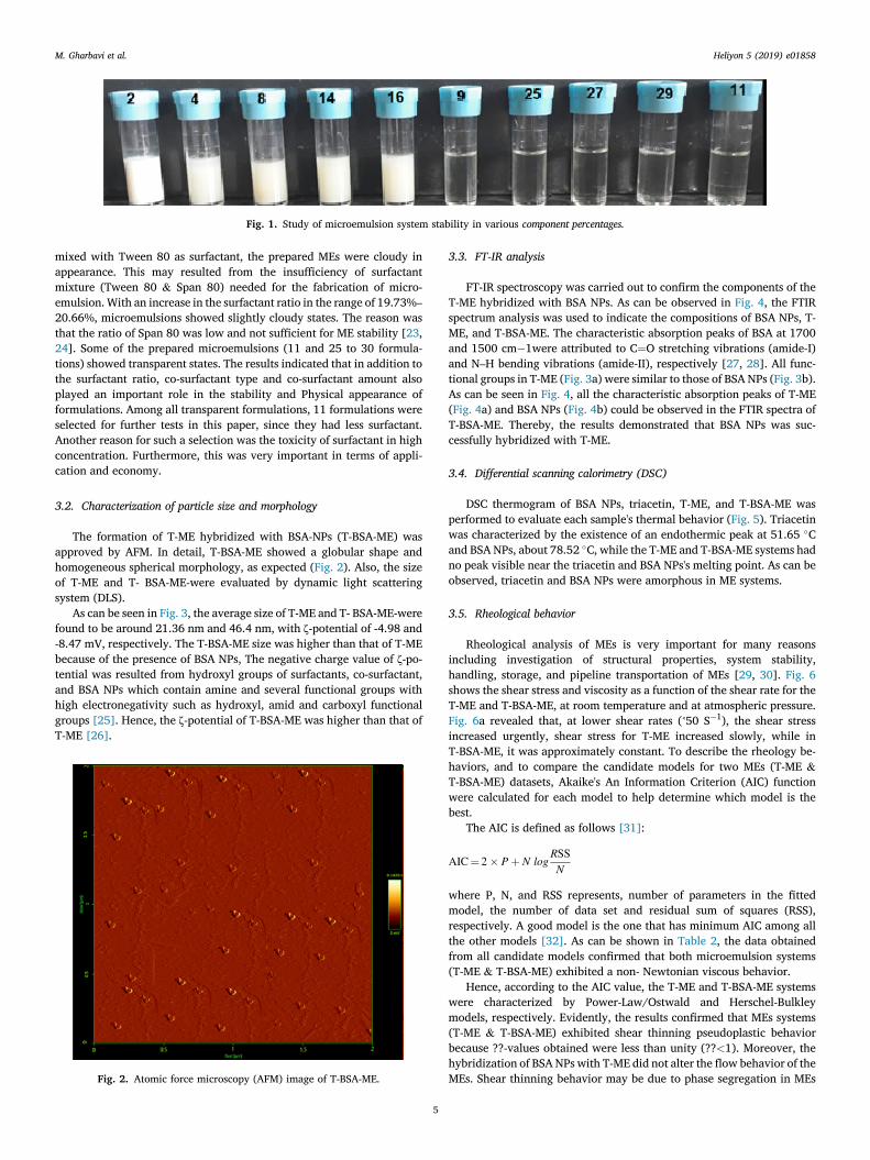
Fig. 1. Study of microemulsion system stability in various component percentages.
M. Gharbavi et al. Heliyon 5 (2019) e01858
mixed with Tween 80 as surfactant, the prepared MEs were cloudy inappearance. This may resulted from the insufficiency of surfactantmixture (Tween 80 & Span 80) needed for the fabrication of micro-emulsion. With an increase in the surfactant ratio in the range of 19.73%–
20.66%, microemulsions showed slightly cloudy states. The reason wasthat the ratio of Span 80 was low and not sufficient for ME stability [23,24]. Some of the prepared microemulsions (11 and 25 to 30 formula-tions) showed transparent states. The results indicated that in addition tothe surfactant ratio, co-surfactant type and co-surfactant amount alsoplayed an important role in the stability and Physical appearance offormulations. Among all transparent formulations, 11 formulations wereselected for further tests in this paper, since they had less surfactant.Another reason for such a selection was the toxicity of surfactant in highconcentration. Furthermore, this was very important in terms of appli-cation and economy.
3.2. Characterization of particle size and morphology
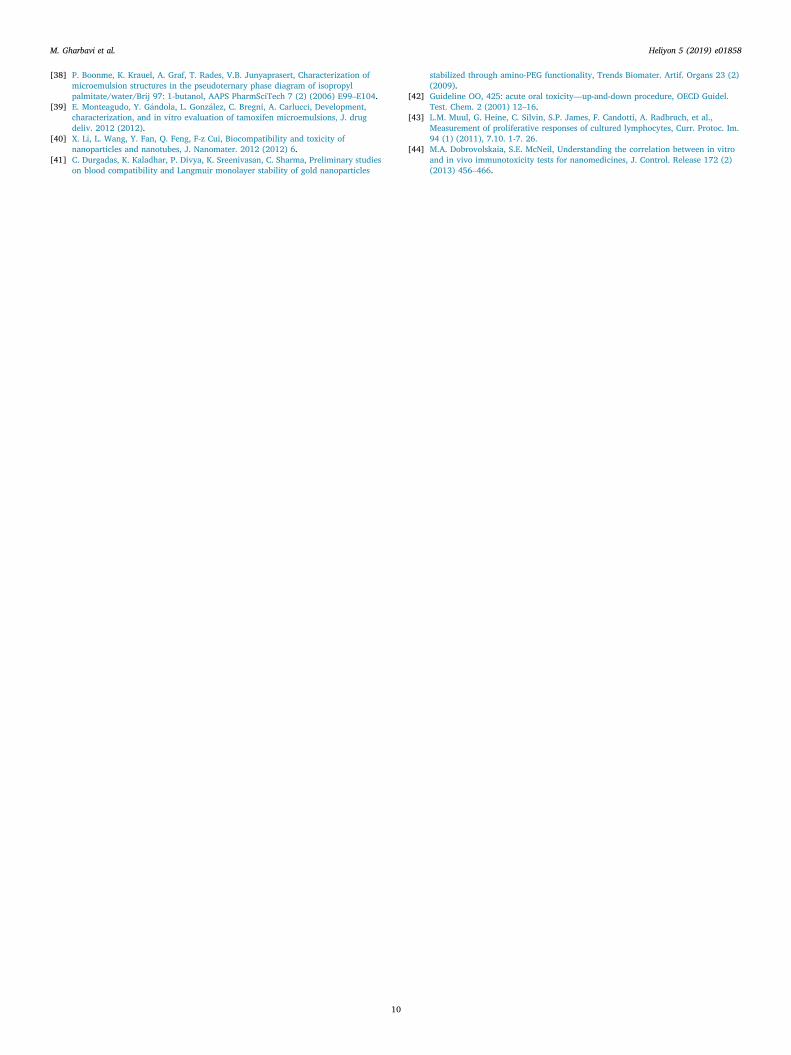
The formation of T-ME hybridized with BSA-NPs (T-BSA-ME) wasapproved by AFM. In detail, T-BSA-ME showed a globular shape andhomogeneous spherical morphology, as expected (Fig. 2). Also, the sizeof T-ME and T- BSA-ME-were evaluated by dynamic light scatteringsystem (DLS).
As can be seen in Fig. 3, the average size of T-ME and T- BSA-ME-werefound to be around 21.36 nm and 46.4 nm, with ζ-potential of -4.98 and-8.47 mV, respectively. The T-BSA-ME size was higher than that of T-MEbecause of the presence of BSA NPs, The negative charge value of ζ-po-tential was resulted from hydroxyl groups of surfactants, co-surfactant,and BSA NPs which contain amine and several functional groups withhigh electronegativity such as hydroxyl, amid and carboxyl functionalgroups [25]. Hence, the ζ-potential of T-BSA-ME was higher than that ofT-ME [26].
Fig. 2. Atomic force microscopy (AFM) image of T-BSA-ME.
5
3.3. FT-IR analysis
FT-IR spectroscopy was carried out to confirm the components of theT-ME hybridized with BSA NPs. As can be observed in Fig. 4, the FTIRspectrum analysis was used to indicate the compositions of BSA NPs, T-ME, and T-BSA-ME. The characteristic absorption peaks of BSA at 1700and 1500 cm�1were attributed to C¼O stretching vibrations (amide-I)and N–H bending vibrations (amide-II), respectively [27, 28]. All func-tional groups in T-ME (Fig. 3a) were similar to those of BSA NPs (Fig. 3b).As can be seen in Fig. 4, all the characteristic absorption peaks of T-ME(Fig. 4a) and BSA NPs (Fig. 4b) could be observed in the FTIR spectra ofT-BSA-ME. Thereby, the results demonstrated that BSA NPs was suc-cessfully hybridized with T-ME.
3.4. Differential scanning calorimetry (DSC)
DSC thermogram of BSA NPs, triacetin, T-ME, and T-BSA-ME wasperformed to evaluate each sample's thermal behavior (Fig. 5). Triacetinwas characterized by the existence of an endothermic peak at 51.65 �Cand BSA NPs, about 78.52 �C, while the T-ME and T-BSA-ME systems hadno peak visible near the triacetin and BSA NPs's melting point. As can beobserved, triacetin and BSA NPs were amorphous in ME systems.
3.5. Rheological behavior
Rheological analysis of MEs is very important for many reasonsincluding investigation of structural properties, system stability,handling, storage, and pipeline transportation of MEs [29, 30]. Fig. 6shows the shear stress and viscosity as a function of the shear rate for theT-ME and T-BSA-ME, at room temperature and at atmospheric pressure.Fig. 6a revealed that, at lower shear rates (˂50 S�1), the shear stressincreased urgently, shear stress for T-ME increased slowly, while inT-BSA-ME, it was approximately constant. To describe the rheology be-haviors, and to compare the candidate models for two MEs (T-ME &T-BSA-ME) datasets, Akaike's An Information Criterion (AIC) functionwere calculated for each model to help determine which model is thebest.
The AIC is defined as follows [31]:
AIC¼ 2� Pþ N logRSSN
where P, N, and RSS represents, number of parameters in the fittedmodel, the number of data set and residual sum of squares (RSS),respectively. A good model is the one that has minimum AIC among allthe other models [32]. As can be shown in Table 2, the data obtainedfrom all candidate models confirmed that both microemulsion systems(T-ME & T-BSA-ME) exhibited a non- Newtonian viscous behavior.
Hence, according to the AIC value, the T-ME and T-BSA-ME systemswere characterized by Power-Law/Ostwald and Herschel-Bulkleymodels, respectively. Evidently, the results confirmed that MEs systems(T-ME & T-BSA-ME) exhibited shear thinning pseudoplastic behaviorbecause ??-values obtained were less than unity (??<1). Moreover, thehybridization of BSA NPs with T-ME did not alter the flow behavior of theMEs. Shear thinning behavior may be due to phase segregation in MEs

Fig. 3. Size distribution by intensity of A) T-ME, B) T-BSA-ME and ζ-potential of C) T-ME, D) T-BSA-ME.
Fig. 4. FT-IR spectra of (a) T-ME, (b) BSA NPs, (c) T-BSA-ME.
Fig. 5. DSC thermograms of BSA NPs (a), triacetin (b), T-ME (c), and T-BSA-ME(d) in the temperature range of 30–340 �C.
M. Gharbavi et al. Heliyon 5 (2019) e01858
6
systems. Furthermore, ??-values decreased with the hybridization of BSANPs in MEs system, revealing an increasing trend in the shear thinningcharacteristic of the suggested system. As can be seen in Table 2, theconsistency index (??) increased with the hybridization of BSA NPs inMEs system, increasing the apparent viscosity of the T-BSA-ME that maybe due to different formation and more organized microstructure. Theviscous flow behavior of T-ME and T-BSA-ME were related to theapparent yield stress resulting from interactions between BSA NPs andthe continuous or external aqueous phase [33, 34]. Meanwhile, the in-teractions between the BSA NPs surface and the aqueous phase mightincrease the stiffness, as pointed out by Zhong [35] and Hato [36]. Thus,the hybridization of BSA NPs with T-ME leads to an increase in viscosity.Aqueous media (continuous phase) as a polar solvent in the presence ofBSA NPs resulted in the bulk viscosity and creation of a new internalphase. As a consequence, the presence of an internal aqueous phaseincreased dispersion viscosity, particularly in low shear rate range, asshown in Fig. 6b. Nonetheless, at high shear rates, the differences weresignificantly dampened.
3.6. Physicochemical characterizations
The physicochemical characterizations of the MEs were shown inTable 3. The conductivity was profoundly sensitive both to temperatureand composition. The composition of O/W ME system was fabricatedwith triacetin, Tween 80, Span 80, PG and water.
In the case of the O/WMEs systems when the content of water is over65 percent as the continuous phase, the conductivity of the systemsincreased. However, the W/O (water-in-oil) systems had low conduc-tivity. The electrical conductivity was used to determine the type ofmicroemulsions [37]. The electrical conductivity coefficient of T-ME andT-BSA-MEwas revealed in 1.47 ms/cm and 2.49ms/cm, respectively. thehigh value of conductivity coefficient is characteristic of oil-in-water(O/W) type of MEs (Table 3) [38, 39]. As can be seen, the conductivitycoefficient of T-ME was higher than that of T-BSA-ME which was relatedto the higher percentage of water in T-ME compared with that ofT-BSA-ME. The refractive index (RI) of T-ME and T-BSA-ME was found tobe around 1.38 and 1.2, respectively. Therefore, as we discussed before,

Fig. 6. Rheological behavior of T-ME and T-BSA-ME.
Table 2Description of different models for MEs systems.
Model Equation P T-BSA-ME T-ME
AIC n τ0 K AIC n τ0 K
Newtonian τ ¼ kƴ 1 72.14 0.20 52.55 0.08Herschel-Bulkley τ ¼ τ0 þ kƴn 3 6.08 0.20 9.91 10.83 -30.28 0.01 0 8.14Bingham τ ¼ τ0 þ kƴ 2 37.8 27.89 0.06 27.78 12.67 0.01Power-Law/Ostwald τ ¼ kƴn 2 22.79 0.14 19.31 -32.28 0.03 13.14
τ: Shear Stress, τ0: Yield Stress, ƴ: shear Rate, K: Consistency Index, n: Flow Index, P: Number of Parameters.
Table 3Physicochemical characterization of T-ME and T-BSA-ME.
Formulations Conductivity (ms/cm) pH RI %Transmittance
T-ME 2.49 5.23 1.38 93.9T-BSA-ME 1.47 6.16 1. 21 57.32
Fig. 7. Stability curves of T-ME and T-BSA-ME.
M. Gharbavi et al. Heliyon 5 (2019) e01858
the RI of the system became equal to the water's RI (1.33). Furthermore,the transmittance of T-ME and T-BSA-ME was found to be 93.9% and57.32%, respectively, indicating that the prepared ME were a trans-parent, single phase liquid, while the transmittance of T-BSA-ME waslower than that of T-ME that could be related to the presence of BSA NPsin the formulation. The pH value of T-ME and T-BSA-ME was in the rangeof 5.23–6.16. The pH value of the T-BSA-ME was less than that of T-ME,which could be associated to the presence of acidic groups at BSAstructure. The MEs samples (T-ME & T-BSA-ME) stability was approvedphysically. MEs systems were also of good thermodynamic stability, dueto ultralow interfacial tension between the oil and water phases. In orderto describe the ME stability, the particle structure integrity and thepossibility of aggregation were evaluated. Therefore, the particle size ofME as a function of incubation time was monitored for 3 months. Asshown in Fig. 7, Z-average of T-ME and T-BSA-ME did not increasesignificantly throughout the measurement period.
3.7. Cytotoxicity study
For clinical and biomedicine applications, toxicity is a critical factorthat should be considered. Biomedicine applications of MEs are inten-tionally designed for several clinical uses. It is essential to approve thatthis offer does not have any adverse effect. Besides T-ME, BSA NPs hy-bridized with T-ME should also be evaluated in terms of the cytotoxicityeffect on both HFF-2 and HEK-293 cell lines. The cytotoxicity effect of T-BSA-ME on both HFF-2 and HEK-293 cell lines were evaluated andmeasured by MTT assay. The cells were incubated with various con-centrations (50, 100, 200, 400, 600, 800 μg/ml) for 48 h in a 5 % CO2atmosphere. As can be seen, Fig. 8 compares the cells viability in differentT-ME and T-BSA-ME concentrations.
The cell viabilities of HFF-2 cells after being treated with variousconcentrations of T-ME and T-BSA-ME ranged from 78.99 � 3.18 to90.31 � 1.01% and 92.71 � 5.53 to 97.9 � 0.02% after 48 h,
7
respectively. At the same time, viability of HEK-293 cells, after treatmentwith various concentrations of T-ME and T-BSA-ME, were obtained to bein the range of 74.84 � 4.2 and 85.56 � 3.6% and 85.75 � 3.52 and90.28 � 3.04% after 48 h, respectively. Thereby, T-ME and T-BSA-MEwere not significantly toxic on HFF-2 and HEK-293 cell lines, suggestingthat MEs systems (T-BSA-ME especially) were of high-level biocompati-bility and likely to be used for further in vivo applications. Data wereanalyzed using ANOVA, *p < 0.05 and ns considered as significant andnot significant difference, respectively.
3.8. Hemolysis assay
For biomedicine uses, MEs systems enter the body and interact withtissues and cells directly. It is necessary for the evaluation of theirbiocompatibility [40]. To determine the biocompatibility of the T-MEand T-BSA-ME, the MEs systems must have a blood compatibility withminimal hemolytic effect on human red blood cells. As can be seen inFig. 9, the blood compatibility of T-ME and T-BSA-ME were evaluated at10–160 μg/ml concentration range. The hemolytic activity of the sampleswas quantitatively determined by the evaluation of the supernatantabsorbance at 540 nm (hemoglobin) with Eppendorf Bio Photometer

Fig. 8. The cell viabilities of the T-ME and T-BSA-ME on HEK-293 cell line (a), and HFF-2 cell line (b). Data are represented as mean � standard deviation (n ¼ 3) *p <
0.05 and ns considered as significant difference and not significant, respectively.
Fig. 9. Percentage of hemolysis induced by T-ME and T-BSA-ME at variousconcentration and 37 �C, conditions. Data are represented as mean � standarddeviation (n ¼ 3) *p < 0.05 considered as significant difference.
M. Gharbavi et al. Heliyon 5 (2019) e01858
[17]. The hemolytic activity of T-ME and T-BSA-ME in various massconcentrations was in the range of 6.32 � 0.02 to 13.00 � 0.12% and9.09 � 0.00 to 15.87 � 0.00%, respectively. The hemolytic activity ofMEs systems may be related to the presence of the surfactant that, whenincreases in the sample, causes an increase in the hemolytic rate [41].Likewise, the T-ME revealed significantly higher hemolytic ratescompared to T-BSA-ME in similar concentration that was due to thesurfactant used. Data were analyzed using ANOVA, *p < 0.05 wasconsidered as significant difference.
Fig. 10. The proliferation (a) and proliferation inhibition (b) rate of T-ME and T-Bconsidered as significant difference.
8
3.9. LD50 and acute toxicity
For Median Lethal Dose (LD 50) test, we treated mice with a series ofdoses. Mice were treated with T-ME and T-BSA-ME, ranging from 17.5 to5000 mg/kg. To find of out maximum safe dose, we used a control groupadministrated with saline and treatment groups were administrated with17.5, 175, 1750, and 5000mg/kg of T-ME and T-BSA-ME. The changes inthe mice weight after 24 h and 7 days were around 1.58 � 3.64% and5.42 � 1.78%. During the treatment, none of the mice died. So, usingOCED and Hodge and Sterner scale, we concluded that the MEs systemsintroduced were practically none toxic [42].
3.10. Leukocyte proliferation assay
Lymphocyte proliferation is one of the basic characteristic of theresponse of lymphocytes to antigenic stimulation. In order to measure theability of ME system to induce a proliferative response in humanLymphocyte, along with proliferation percentage, the percentage ofproliferation inhibition should be evaluated. For this Assay, Lymphocytewere isolated from human whole blood using Ficoll-Paque solution andthen, it was cultured. The amount of Proliferation showed MEs ability toinduce leukocytes proliferation compared with untreated sample. Theamount of proliferation inhibition also showed MEs inhibitory effects onPHA as a proliferative factor [43, 44].
As can be seen in Fig. 10, the proliferation rate of triacetin and T-BSA-ME in various concentrations ranged from-36.6 to -1.61% and -49.98 to-2.95%, respectively. These results revealed that the proliferation rate ofthe suggested MEs systems was lower than that of untreated samples(especially in high concentration). Likewise, the proliferation inhibitionrate of triacetin and T-BSA-ME in various concentrations ranged from47.15 to 1.93% and 52.7 to 2.99%, respectively. The results indicated
SA-ME. Data are represented as mean � standard deviation (n ¼ 5) *p < 0.05

M. Gharbavi et al. Heliyon 5 (2019) e01858
that the suggested MEs systems could act as a Lymphocyte proliferationinhibitory in presence of PHA as a proliferative factor (positive control).Data were analyzed using ANOVA, and*p < 0.05 was regarded as sig-nificant difference.
4. Conclusion
The present paper aimed to design and prepare T-BSA-ME, practicallynone toxic with potential applications such as co-delivery and multi-functional systems. In vitro and in vivo cytotoxicity evaluation revealedthat the suggested MEs systems were safe and none toxic. Likewise, theywere biocompatible, not active for Leukocyte Proliferation. The non-toxicity of the systems significantly increased their applicability in thedevelopment of in-vitro and in-vivo biomedicine fields such as brain co-delivery or multifunctional system in neurodegenerative and cancerdiseases. It is worth noting that this study established the foundation foreffective preparation of T-BSA-ME for further in vitro and in vivo studies(in progress) on conjugates of T-BSA-ME with drugs, drug candidates,targeting agent, and molecular probes.
Declarations
Author contribution statement
Mahmoud Gharbavi: Performed the experiments; Analyzed andinterpreted the data.
Hamidreza Kheiri Manjili: Conceived and designed the experiments.Jafar Amani: Contributed reagents, materials, analysis tools or data.Ali Sharafi: Conceived and designed the experiments; Performed the
experiments.Hossein Danafar: Conceived and designed the experiments; Wrote the
paper.
Funding statement
This research did not receive any specific grant from funding agenciesin the public, commercial, or not-for-profit sectors.
Competing interest statement
The authors declare no conflict of interest.
Additional information
No additional information is available for this paper.
Acknowledgements
The authors would like to thank the authority of the School ofPharmacy, Zanjan University of Medical Sciences, for their support.
References
[1] J. Rao, D.J. McClements, Formation of flavor oil microemulsions, nanoemulsionsand emulsions: influence of composition and preparation method, J. Agric. FoodChem. 59 (9) (2011) 5026–5035.
[2] M.J. Lawrence, G.D. Rees, Microemulsion-based media as novel drug deliverysystems, Adv. Drug Deliv. Rev. 64 (2012) 175–193.
[3] J.H. Fendler, Membrane Mimetic Chemistry: Characterizations and Applications ofMicelles, Microemulsions, Monolayers, Bilayers, Vesicles, Host-Guest Systems, andPolyions, John Wiley & Sons Inc, 1982.
[4] S.J. Choi, E.A. Decker, L. Henson, L.M. Popplewell, D.J. McClements, Stability ofcitral in oil-in-water emulsions prepared with medium-chain triacylglycerols andtriacetin, J. Agric. Food Chem. 57 (23) (2009) 11349–11353.
[5] M. Gharbavi, J. Amani, H. Kheiri-Manjili, H. Danafar, A. Sharafi, Niosome: apromising nanocarrier for natural drug delivery through blood-brain barrier, Adv.pharma. sci. 2018 (2018).
[6] J. Jiao, D.J. Burgess, Rheology and stability of water-in-oil-in-water multipleemulsions containing Span 83 and Tween 80, AAPS PharmSci 5 (1) (2003) 62–73.
9
[7] D. Lu, D.G. Rhodes, Mixed composition films of spans and tween 80 at the Air�water interface, Langmuir 16 (21) (2000) 8107–8112.
[8] L.B. Lopes, Overcoming the cutaneous barrier with microemulsions, Pharmaceutics6 (1) (2014) 52–77.
[9] T. Iwanaga, M. Suzuki, H. Kunieda, Effect of added salts or polyols on the liquidcrystalline structures of polyoxyethylene-type nonionic surfactants, Langmuir 14(20) (1998) 5775–5781.
[10] Y. Zhang, T. Sun, C. Jiang, Biomacromolecules as carriers in drug delivery andtissue engineering, Acta Pharm. Sin. B (2017).
[11] S. Ghosh, J. Dey, Binding of fatty acid amide amphiphiles to bovine serum albumin:role of amide hydrogen bonding, J. Phys. Chem. B 119 (25) (2015) 7804–7815.
[12] H.T. Phan, S. Bartelt-Hunt, K.B. Rodenhausen, M. Schubert, J.C. Bartz, Investigationof bovine serum albumin (BSA) attachment onto self-assembled monolayers (SAMs)using combinatorial quartz crystal microbalance with dissipation (QCM-D) andspectroscopic ellipsometry (SE), PLoS One 10 (10) (2015), e0141282.
[13] M. Merodio, A. Arnedo, M.J. Renedo, J.M. Irache, Ganciclovir-loaded albuminnanoparticles: characterization and in vitro release properties, Eur. J. Pharm. Sci.12 (3) (2001) 251–259.
[14] J. Patel, G. Kevin, A. Patel, M. Raval, N. Sheth, Design and development of a self-nanoemulsifying drug delivery system for telmisartan for oral drug delivery, Int. J.Pharma. Invest. 1 (2) (2011) 112.
[15] A.A. Badawi, S.A. Nour, W.S. Sakran, S.M.S. El-Mancy, Preparation and evaluationof microemulsion systems containing salicylic acid, AAPS PharmSciTech 10 (4)(2009) 1081–1084.
[16] E.L. França, E.B. Ribeiro, E.F. Scherer, D.G. Cantarini, R.S. Pessoa, F.L. França, et al.,Effects of Momordica charantia L. on the blood rheological properties in diabeticpatients, BioMed Res. Int. 2014 (2014).
[17] M.A. Dobrovolskaia, J.D. Clogston, B.W. Neun, J.B. Hall, A.K. Patri, S.E. McNeil,Method for analysis of nanoparticle hemolytic properties in vitro, Nano Lett. 8 (8)(2008) 2180–2187.
[18] Chemicals D, OECD Guideline for Testing of Chemicals, 2005.[19] A. Martino, E.W. Kaler, Phase behavior and microstructure of nonaqueous
microemulsions, J. Phys. Chem. 94 (4) (1990) 1627–1631.[20] H.K. Syed, K.K. Peh, Identification of phases of various oil, surfactant/co-surfactants
and water system by ternary phase diagram, Acta Pol. Pharm. 71 (2) (2014)301–309.
[21] M. Porras, C. Solans, C. Gonzalez, J. Gutierrez, Properties of water-in-oil (W/O)nano-emulsions prepared by a low-energy emulsification method, Colloid. Surf.Physicochem. Eng. Asp. 324 (1-3) (2008) 181–188.
[22] J. Liang, Y. Qian, X. Yuan, L. Leng, G. Zeng, L. Jiang, et al., Span80/Tween80stabilized bio-oil-in-diesel microemulsion: formation and combustion, Renew.Energy 126 (2018) 774–782.
[23] C.-C. Lin, H.-Y. Lin, M.-H. Chi, C.-M. Shen, H.-W. Chen, W.-J. Yang, et al.,Preparation of curcumin microemulsions with food-grade soybean oil/lecithin andtheir cytotoxicity on the HepG2 cell line, Food Chem. 154 (2014) 282–290.
[24] G. Li, Y. Fan, X. Li, X. Wang, Y. Li, Y. Liu, et al., In vitro and in vivo evaluation of asimple microemulsion formulation for propofol, Int. J. Pharm. 425 (1-2) (2012)53–61.
[25] L. Medda, M. Monduzzi, A. Salis, The molecular motion of bovine serum albuminunder physiological conditions is ion specific, Chem. Commun. 51 (30) (2015)6663–6666.
[26] K.E. Sapsford, W.R. Algar, L. Berti, K.B. Gemmill, B.J. Casey, E. Oh, et al.,Functionalizing nanoparticles with biological molecules: developing chemistriesthat facilitate nanotechnology, Chem. Rev. 113 (3) (2013) 1904–2074.
[27] P. Kupser, Infrared Spectroscopic Characterization of Secondary Structure Elementsof Gas-phase Biomolecules, 2011.
[28] H. Danafar, M. Hamidi, Pharmacokinetics and bioequivalence study of amlodipineand atorvastatin in healthy male volunteers by LC-MS, Pharmaceut. Sci. 21 (3)(2015) 167.
[29] T. Formariz, L.A. Chiavacci, M. Scarpa, A. Silva-Júnior, E. Egito, C.H. Terrugi, et al.,Structure and viscoelastic behavior of pharmaceutical biocompatible anionicmicroemulsions containing the antitumoral drug compound doxorubicin, ColloidsSurfaces B Biointerfaces 77 (1) (2010) 47–53.
[30] C.M. Franzini, K.C. Pestana, E.F. Molina, M.V. Scarpa, Tabosa do Egito ES, A.G. deOliveira, Structural properties induced by the composition of biocompatiblephospholipid-based microemulsion and amphotericin B association, J. Biomed.Nanotechnol. 8 (2) (2012) 350–359.
[31] M.R. Symonds, A. Moussalli, A brief guide to model selection, multimodel inferenceand model averaging in behavioural ecology using Akaike’s information criterion,Behav. Ecol. Sociobiol. 65 (1) (2011) 13–21.
[32] J.I. Myung, M.A. Pitt, Model Comparison Methods. Methods in Enzymology, 383,Elsevier, 2004, pp. 351–366.
[33] J. Bhatt, R.S. Somani, H.M. Mody, H.C. Bajaj, Rheological study of organoclaysprepared from Indian bentonite: effect of dispersing methods, Appl. Clay Sci. 83(2013) 106–114.
[34] J.D. Kanungo, J.L. McAtee Jr., Effects of polymers and clay concentrations on theviscosities of organo-smectite dispersions under high pressure, Appl. Clay Sci. 1 (3)(1986) 285–293.
[35] Y. Zhong, S.-Q. Wang, Exfoliation and yield behavior in nanodispersions oforganically modified montmorillonite clay, J. Rheol. 47 (2) (2003) 483–495.
[36] M.J. Hato, K. Zhang, S.S. Ray, H.J. Choi, Rheology of organoclay suspension,Colloid Polym. Sci. 289 (10) (2011) 1119.
[37] M. Lagues, Electrical conductivity of microemulsions: a case of stirred percolation,J. Phys., Lett. 40 (14) (1979) 331–333.
M. Gharbavi et al. Heliyon 5 (2019) e01858
[38] P. Boonme, K. Krauel, A. Graf, T. Rades, V.B. Junyaprasert, Characterization ofmicroemulsion structures in the pseudoternary phase diagram of isopropylpalmitate/water/Brij 97: 1-butanol, AAPS PharmSciTech 7 (2) (2006) E99–E104.
[39] E. Monteagudo, Y. G�andola, L. Gonz�alez, C. Bregni, A. Carlucci, Development,characterization, and in vitro evaluation of tamoxifen microemulsions, J. drugdeliv. 2012 (2012).
[40] X. Li, L. Wang, Y. Fan, Q. Feng, F-z Cui, Biocompatibility and toxicity ofnanoparticles and nanotubes, J. Nanomater. 2012 (2012) 6.
[41] C. Durgadas, K. Kaladhar, P. Divya, K. Sreenivasan, C. Sharma, Preliminary studieson blood compatibility and Langmuir monolayer stability of gold nanoparticles
10
stabilized through amino-PEG functionality, Trends Biomater. Artif. Organs 23 (2)(2009).
[42] Guideline OO, 425: acute oral toxicity—up-and-down procedure, OECD Guidel.Test. Chem. 2 (2001) 12–16.
[43] L.M. Muul, G. Heine, C. Silvin, S.P. James, F. Candotti, A. Radbruch, et al.,Measurement of proliferative responses of cultured lymphocytes, Curr. Protoc. Im.94 (1) (2011), 7.10. 1-7. 26.
[44] M.A. Dobrovolskaia, S.E. McNeil, Understanding the correlation between in vitroand in vivo immunotoxicity tests for nanomedicines, J. Control. Release 172 (2)(2013) 456–466.
Copyright © 2022 FDOKUMEN