Thiosemicarbazones derived from 1-indanones as new anti- Trypanosoma cruzi agents
Upload
khangminh22Category
view
1download
0
In vitro and in vivo anti-Trypanosoma cruzi activity of
new arylamine Mannich base-type derivatives.
Elsa Moreno-Viguri,†,‡ Carmen Jiménez-Montes,§ ,‡ Rubén Martín-Escolano,§ Mery Santivañez-Veliz,†
Alvaro Martin-Montes,§ Amaya Azqueta,¥, ǂ Marina Jimenez-Lopez,§ Salvador Zamora Ledesma,§ Nuria
Cirauqui,Ɨ Adela López de Ceráin,¥,ǂ Clotilde Marín,§ Manuel Sánchez-Moreno,§,* Silvia Pérez-Silanes†,*
†Department of Organic and Pharmaceutical Chemistry, Institute of Tropical Health, Universidad de
Navarra, Pamplona, Spain. ¥Department of Pharmacology and Toxicology, Universidad de Navarra,
Pamplona, Spain. ǂIdiSNA, Navarra Institute for Health Research, Recinto de Complejo Hospitalario de
Navarra, Pamplona, Spain. § Departamento de Parasitología, Instituto de Investigación Biosanitaria
(ibs.GRANADA), Hospitales Universitarios De Granada/Universidad de Granada, Granada, Spain. Ɨ
Department of Pharmaceutical Sciences, Federal University of Rio de Janeiro, Rio de Janeiro, Brazil.
ABSTRACT. Chagas disease is a neglected tropical disease with 6-7 million people infected worldwide
and there is no effective treatment. Therefore, there is an urgent need to continue researching in order to
discover novel therapeutic alternatives. We present a series of arylaminoketone derivatives as means of
identifying new drugs to treat Chagas disease in the acute phase with greater activity, less toxicity and
with a larger spectrum of action than that corresponding to the reference drug benznidazole. Indexes of
high selectivity found in vitro formed the basis for later in vivo assays in BALB/c mice. Murine model
results show that compounds 3, 4, 7 and 10 induced a remarkable decrease in parasitemia levels in acute
phase and the parasitemia reactivation following immunosuppression, and curative rates were higher
than with benznidazole. These high anti-parasitic activities encourage us to propose these compounds as
promising molecules for developing an easy to synthesize anti-Chagas agent.
INTRODUCTION
Chagas disease (CD), is caused by the protozoa Trypanosoma cruzi (T. cruzi) which is naturally
transmitted by hematophagous insects, presents risk factors strongly linked to low socioeconomic
factors and it is classified as a neglected tropical disease according to WHO.1, 2 CD is one of the most
important medical problems in rural areas of the 21 endemic Latin America countries. Moreover, in the
past decades migration and travelling have extended the spread of CD to other continents including
North America,3 Europe4 and parts of the Western Pacific,5 where there is now a significant number of
individuals with this disease. The worldwide incidence of CD has decreased thanks to the control
programs; however, an estimated 6-7 million persons are infected worldwide.6 Approximately 20% of
Latin American countries are endemic for this disease and up to 30-40% of patients will suffer chronic
form of the disease in their lifetimes–i.e. important cardiac or digestive problems.
Currently, the two drugs recommended for CD are benznidazole (BZN) and nifurtimox (NFX)7, 8 In
general terms, BZN is the first line drug in most endemic countries9 because it is tolerated better but
some of its secondary effects include dermatitis or neuropathy. Both drugs reduce parasitism during the
acute and early CD stage, but drug efficacy during the chronic phase is limited. In addition, the efficacy
of BNZ and NFX could change according to the geographical area, as consequence of a different
susceptibility of the drugs by the different strains of T. cruzi.10 This background, including side effects
and therapeutic deficiencies of these drugs, justifies the urgent need to continue researching in order to
discover novel therapeutic alternatives. The ideal drug defined by its Target Product Profile (TPP) is
outlined in Table 1. Briefly, it should present an efficacy at least equal to BZN and with a better safety
profile. An oral drug in once-a-day in-house treatment for less than 30 days is desired and the new drug
should be active against most parasite species.11-13

Table 1. Proposed TPP for Chagas disease modified from DNDi.11
Drug repositioning has emerged as one of the most fruitful strategies for improving and accelerating
the drug development process. This strategy has become one of the most powerful tools for identifying
compounds for the treatment of a disease different from the one they were originally designed for, used
not only in the academic field but also in pharmaceutical companies. Different methods are considered
under this strategy but 28 out of the 50 small molecules approved by the FDA between 1999 and 2008
were discovered by phenotypic based drug design.14, 15 In this context we decided to screen our in-house
chemical laboratory library with the purpose of searching drugs against of CD.16-21 We found some
arylamine Mannich base derivatives tested before for their in vitro affinity at the 5-HT transporter and
5HT1A receptors17 or 5-HT720 receptors as potential antidepressants and against Plasmodium falciparum
as a potential antimalarials19 and in every case they were not active. However risk of potential off-target
activities derived from a possible CSN activity is unlikely.
Herein we present the synthesis of arylaminoketone-type compounds and their in vitro evaluation
against T. cruzi. Furthermore we show that the preliminary genotoxicity screening test revealed that this
family of compounds is not genotoxic with or without metabolic activation, thereby providing the ideal
chemical space for the development of new, potent and safe agents for the treatment of CD. High-
selectivity indexes and the genotoxicity screening have been the established cut-off to move into in vivo
assays in BALB/c mice, where fresh blood examination was used to quantify the parasitemia. Also a
cure was assayed by PCR, as was the reactivating of parasitemia in blood following
immunosuppression. The mechanism of action has been explored at metabolic levels by 1H Magnetic
Nuclear Resonance, and the study has been completed by testing their activity as potential Fe-
Superoxide dismutase inhibitors. Using computational approaches, we suggest a binding mode for the
compounds to the enzyme.
RESULTS AND DISCUSSION
Chemistry. It is essential to identify the purpose of the synthesis of new small molecules. During the
drug discovery process, mainly when facing NTD, the synthetic routes should allow the preparation of
an important amount of different small molecules with a variety of substituents in order to explore and
modify polarity, solubility, volume, rigidity and some other parameters that could influence activity and
toxicity. Cost and commercial availability of chemicals should also be taken into consideration as well
as the solvent and waste volumes. With these ideas in mind, the retrosynthetic analysis of the target
compounds (1-20) suggested methylketones and arylamines as the appropriate building blocks (Table
2). Compounds could be prepared in one step synthetic route with exception of compounds 10 and 13.
In this case, both arylamines had to be prepared as previously reported.17, 20 The general method shown
in Figure 1 was used to prepare the compounds presented in this work. The desired compounds were
obtained condensing the adequate methylketone (commercially available) with the appropriate
arylamine via Mannich reaction in acidic medium and using 1,3-dioxolane as the solvent and source of
formaldehyde.
Table 2. Relationship of synthesized compounds.
Figure 1. General method of synthesis of new arylaminoketone derivatives.
The results of the in silico study of some relevant molecular properties related to the drug-likeness of
the compounds are shown in Table 3. It can be observed that none of them have a molecular weight
higher than 500 g/mol, or an estimated partition coefficient (logP) higher than 5. Compounds 3, 6, 10,
15 and 19 present a number of hydrogen-bond acceptors higher than 5 but lower than 10, while the
number of donors is lower than 5 for all compounds. Therefore, all compounds fulfill the Lipinski “rule
of five”. Finally, together with the acceptable partition coefficient values, the Polar Surface Area (PSA)
of the compounds is lower than 140 Å2, and the aqueous solubility (logS) is similar or higher than -4,
which anticipates good oral bioavailability.
Table 3. Molecular properties of the new compounds calculated with the DataWarrior program.22

In vitro Trypanocidal Evaluation. Information was gathered by the in vitro evaluation of activity of
compounds 1-20 with respect to intra- and extracellular forms of T. cruzi SN3, Arequipa and Tulahuen
strains obtained as detailed in the experimental section. Assays have been carried out in strains from
different locations, hosts and tropisms, in order to determine if there are any differences in drug
performance.
Extracellular form (epimastigote) is the most commonly used owing to its easy culture in laboratory,
but tests obtained on intracellular amastigotes (responsible for the chronic phase of CD) and blood
trypomastigotes are more indicative due to these form of the parasite is the developed form in vertebrate
hosts23. According to some authors, for deeming a compound as potential antichagasic agent, it has to
meet certain criteria24. In the first place, the IC50 value has to be around 10 µM and the in second place
the SI has to be 50 times higher than that of the reference drug. IC50 values for the 3 studied strains in
both their extracellular and intracellular forms are shown in Table 4.
Table 4 shows the IC50 values found at 72 h exposure using concentrations of 1-100 µM on assaying
compounds 1-20 vs. 3 strains studied and the 3 forms of parasite cited above. The IC50 values for the
drug BZN (reference) served for comparisons. Ten compounds (3, 4, 7, 10, 11, 13-15, 18 and 19) were
shown to fulfill this first requisite. It was shown that the compounds meet this first requirement. Those
compounds are the most active of the tested compounds for the three parasite forms assayed with an
IC50 value lower than the one corresponding to the reference drug.
Table 4. In vitro activity and toxicity for the 20 arylaminoketone derivatives on extra- and
intracellular forms of Trypanosoma cruzi strains.
On the other hand, the cytotoxicity of compounds 1-20 was tested against Vero cells (Table 5) and a
lesser cytotoxicity than the one of the reference drug was observed. Interestingly, toxicity data against
Vero cells after 72 h of culture showed that all derivatives assayed were considerably less cytotoxic than
drug reference BZN. Cytotoxicity IC50 values ranged from 30.1 µM to 2846.3 µM in sharp contrast with
that obtained for BZN (13.6 µM). Only compounds 11 and 18 were more cytotoxic than BZN.

Table 5. Selectivity index and in vitro toxicity for the 20 derivatives of arylaminoketone intra and
extracellular of T .cruzi strains.
In accordance with the above in vitro data, Table 5 presents the most notable selectivity index (SI)
values, indicating in brackets how often the SI of each compound surpassed the SI of BNZ. This Table
5 shows that excellent results were found for compounds 3, 4, 7 and 10 in all the parasite forms tested.
Those drugs were chosen for further in vitro and in vivo assays and the possible mechanisms of action.
In this case experiments were carried out only in the Arequipa strain as there are no significant
differences between the performances of the compounds on the other studied strains.
From a structural point of view, it can be said that the benzothiophene derivatives are less cytotoxic
than the rest of the derivatives. All of the benzothiophene derivatives (1-10) present IC50 in Vero cells
higher than 70 µM; moreover, the selected compounds (3, 4, 7, 10) present IC50 higher than 700 µM in
contrast with BZN (13.6 µM).
In vitro genotoxicity screening test. The potential genotoxicity of the compounds (1-5, 7, 10, 14, 16-
18) was screened using the SOS/umu test. The concentrations tested for each compounds were
previously determined in a toxicity study in the test system. The SOS/umu screening test revealed that
none of the tested compounds were genotoxic with or without metabolic activation. Meanwhile, both
NFX and BZN were also tested and both of them were genotoxic in the tested conditions. The main
purpose of including the SOS/umu test in the preliminary assays is to study the genotoxicity capacity of
the most active compounds in vitro so that only compounds that are not genotoxic will move on to
further studies. This test is convenient as a screening assay because it is a fast, simple and efficient
method for testing the genotoxicity of a large number of samples. For these reasons, compounds 3, 4
and 7, that had met the criteria for being moved on to in vivo models, were included in the SOS/umu
test. Moreover, compounds 1, 2, 5, 10, 14 and 16-18 had been previously assayed in the genotoxicity
screening test. These compounds came from our in-house chemical library and their in vitro
trypanocidal activity had been studied, showing interesting results. Therefore and before synthesizing
new related derivatives, we decided to explore their genotoxicity capacity to be sure that these new
derivatives could be promising not only because of their potency but also because of their safeness.
Both the trypanocidal activity and genotoxicity screening test mentioned above supported the idea of
carrying out more detailed in vitro and in vivo assays with benzothiophenes 3, 4, 7 and 10 in order to get
more information about their mode of action and their apparent advantages with respect to drug
reference BZN.
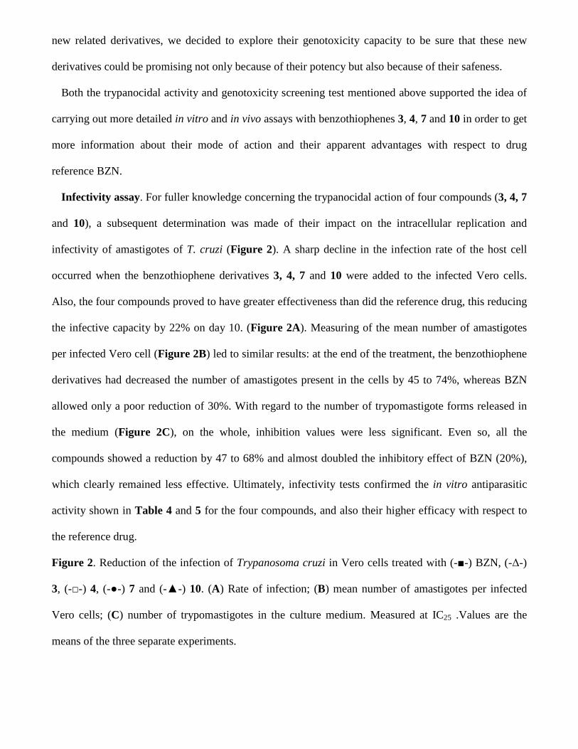
Infectivity assay. For fuller knowledge concerning the trypanocidal action of four compounds (3, 4, 7
and 10), a subsequent determination was made of their impact on the intracellular replication and
infectivity of amastigotes of T. cruzi (Figure 2). A sharp decline in the infection rate of the host cell
occurred when the benzothiophene derivatives 3, 4, 7 and 10 were added to the infected Vero cells.
Also, the four compounds proved to have greater effectiveness than did the reference drug, this reducing
the infective capacity by 22% on day 10. (Figure 2A). Measuring of the mean number of amastigotes
per infected Vero cell (Figure 2B) led to similar results: at the end of the treatment, the benzothiophene
derivatives had decreased the number of amastigotes present in the cells by 45 to 74%, whereas BZN
allowed only a poor reduction of 30%. With regard to the number of trypomastigote forms released in
the medium (Figure 2C), on the whole, inhibition values were less significant. Even so, all the
compounds showed a reduction by 47 to 68% and almost doubled the inhibitory effect of BZN (20%),
which clearly remained less effective. Ultimately, infectivity tests confirmed the in vitro antiparasitic
activity shown in Table 4 and 5 for the four compounds, and also their higher efficacy with respect to
the reference drug.
Figure 2. Reduction of the infection of Trypanosoma cruzi in Vero cells treated with (-■-) BZN, (-Δ-)
3, (-□-) 4, (-●-) 7 and (-▲-) 10. (A) Rate of infection; (B) mean number of amastigotes per infected
Vero cells; (C) number of trypomastigotes in the culture medium. Measured at IC25 .Values are the
means of the three separate experiments.
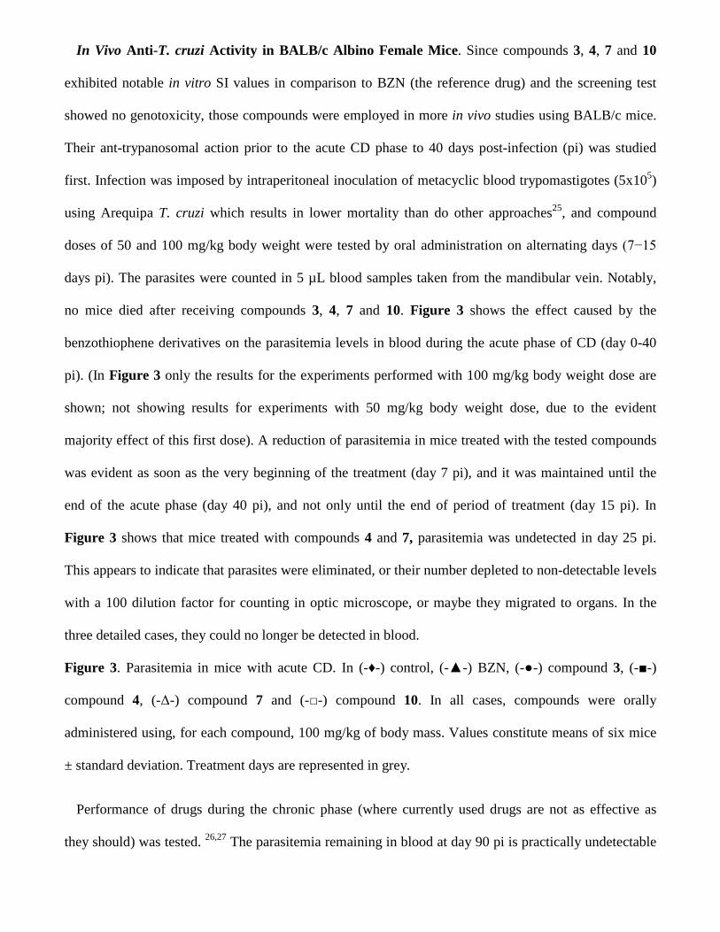
In Vivo Anti-T. cruzi Activity in BALB/c Albino Female Mice. Since compounds 3, 4, 7 and 10
exhibited notable in vitro SI values in comparison to BZN (the reference drug) and the screening test
showed no genotoxicity, those compounds were employed in more in vivo studies using BALB/c mice.
Their ant-trypanosomal action prior to the acute CD phase to 40 days post-infection (pi) was studied
first. Infection was imposed by intraperitoneal inoculation of metacyclic blood trypomastigotes (5x10⁵)
using Arequipa T. cruzi which results in lower mortality than do other approaches25, and compound
doses of 50 and 100 mg/kg body weight were tested by oral administration on alternating days (7−15
days pi). The parasites were counted in 5 µL blood samples taken from the mandibular vein. Notably,
no mice died after receiving compounds 3, 4, 7 and 10. Figure 3 shows the effect caused by the
benzothiophene derivatives on the parasitemia levels in blood during the acute phase of CD (day 0-40
pi). (In Figure 3 only the results for the experiments performed with 100 mg/kg body weight dose are
shown; not showing results for experiments with 50 mg/kg body weight dose, due to the evident
majority effect of this first dose). A reduction of parasitemia in mice treated with the tested compounds
was evident as soon as the very beginning of the treatment (day 7 pi), and it was maintained until the
end of the acute phase (day 40 pi), and not only until the end of period of treatment (day 15 pi). In
Figure 3 shows that mice treated with compounds 4 and 7, parasitemia was undetected in day 25 pi.
This appears to indicate that parasites were eliminated, or their number depleted to non-detectable levels
with a 100 dilution factor for counting in optic microscope, or maybe they migrated to organs. In the
three detailed cases, they could no longer be detected in blood.
Figure 3. Parasitemia in mice with acute CD. In (-♦-) control, (-▲-) BZN, (-●-) compound 3, (-■-)
compound 4, (-Δ-) compound 7 and (-□-) compound 10. In all cases, compounds were orally
administered using, for each compound, 100 mg/kg of body mass. Values constitute means of six mice
± standard deviation. Treatment days are represented in grey.
Performance of drugs during the chronic phase (where currently used drugs are not as effective as
they should) was tested. 26,27 The parasitemia remaining in blood at day 90 pi is practically undetectable
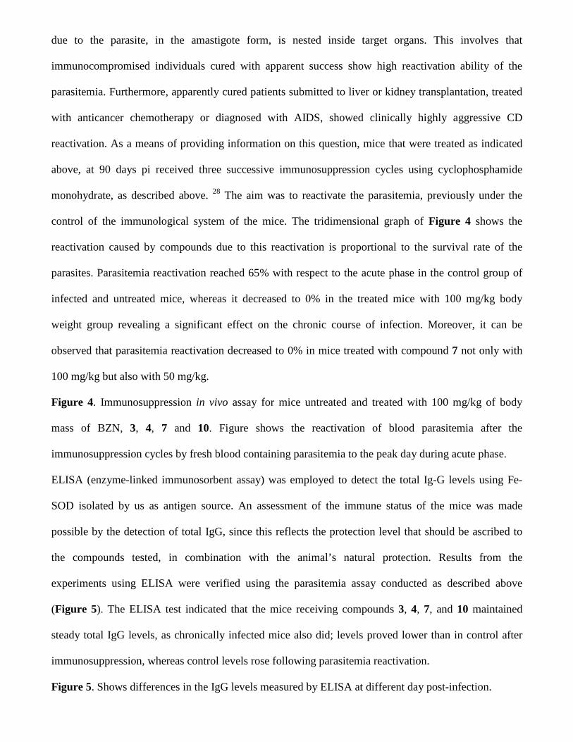
due to the parasite, in the amastigote form, is nested inside target organs. This involves that
immunocompromised individuals cured with apparent success show high reactivation ability of the
parasitemia. Furthermore, apparently cured patients submitted to liver or kidney transplantation, treated
with anticancer chemotherapy or diagnosed with AIDS, showed clinically highly aggressive CD
reactivation. As a means of providing information on this question, mice that were treated as indicated
above, at 90 days pi received three successive immunosuppression cycles using cyclophosphamide
monohydrate, as described above. 28 The aim was to reactivate the parasitemia, previously under the
control of the immunological system of the mice. The tridimensional graph of Figure 4 shows the
reactivation caused by compounds due to this reactivation is proportional to the survival rate of the
parasites. Parasitemia reactivation reached 65% with respect to the acute phase in the control group of
infected and untreated mice, whereas it decreased to 0% in the treated mice with 100 mg/kg body
weight group revealing a significant effect on the chronic course of infection. Moreover, it can be
observed that parasitemia reactivation decreased to 0% in mice treated with compound 7 not only with
100 mg/kg but also with 50 mg/kg.
Figure 4. Immunosuppression in vivo assay for mice untreated and treated with 100 mg/kg of body
mass of BZN, 3, 4, 7 and 10. Figure shows the reactivation of blood parasitemia after the
immunosuppression cycles by fresh blood containing parasitemia to the peak day during acute phase.
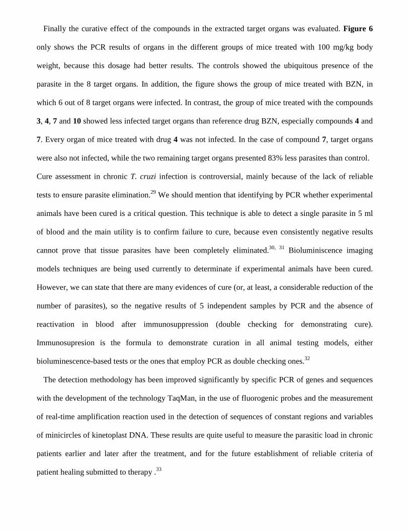
ELISA (enzyme-linked immunosorbent assay) was employed to detect the total Ig-G levels using Fe-
SOD isolated by us as antigen source. An assessment of the immune status of the mice was made
possible by the detection of total IgG, since this reflects the protection level that should be ascribed to
the compounds tested, in combination with the animal’s natural protection. Results from the
experiments using ELISA were verified using the parasitemia assay conducted as described above
(Figure 5). The ELISA test indicated that the mice receiving compounds 3, 4, 7, and 10 maintained
steady total IgG levels, as chronically infected mice also did; levels proved lower than in control after
immunosuppression, whereas control levels rose following parasitemia reactivation.
Figure 5. Shows differences in the IgG levels measured by ELISA at different day post-infection.
Finally the curative effect of the compounds in the extracted target organs was evaluated. Figure 6
only shows the PCR results of organs in the different groups of mice treated with 100 mg/kg body
weight, because this dosage had better results. The controls showed the ubiquitous presence of the
parasite in the 8 target organs. In addition, the figure shows the group of mice treated with BZN, in
which 6 out of 8 target organs were infected. In contrast, the group of mice treated with the compounds
3, 4, 7 and 10 showed less infected target organs than reference drug BZN, especially compounds 4 and
7. Every organ of mice treated with drug 4 was not infected. In the case of compound 7, target organs
were also not infected, while the two remaining target organs presented 83% less parasites than control.
Cure assessment in chronic T. cruzi infection is controversial, mainly because of the lack of reliable
tests to ensure parasite elimination.29 We should mention that identifying by PCR whether experimental
animals have been cured is a critical question. This technique is able to detect a single parasite in 5 ml
of blood and the main utility is to confirm failure to cure, because even consistently negative results
cannot prove that tissue parasites have been completely eliminated.30, 31 Bioluminiscence imaging
models techniques are being used currently to determinate if experimental animals have been cured.
However, we can state that there are many evidences of cure (or, at least, a considerable reduction of the
number of parasites), so the negative results of 5 independent samples by PCR and the absence of
reactivation in blood after immunosuppression (double checking for demonstrating cure).
Immunosupresion is the formula to demonstrate curation in all animal testing models, either
bioluminescence-based tests or the ones that employ PCR as double checking ones.32
The detection methodology has been improved significantly by specific PCR of genes and sequences
with the development of the technology TaqMan, in the use of fluorogenic probes and the measurement
of real-time amplification reaction used in the detection of sequences of constant regions and variables
of minicircles of kinetoplast DNA. These results are quite useful to measure the parasitic load in chronic
patients earlier and later after the treatment, and for the future establishment of reliable criteria of
patient healing submitted to therapy .33
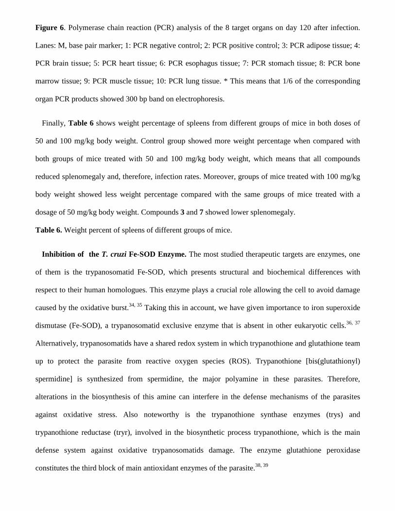
Figure 6. Polymerase chain reaction (PCR) analysis of the 8 target organs on day 120 after infection.
Lanes: M, base pair marker; 1: PCR negative control; 2: PCR positive control; 3: PCR adipose tissue; 4:
PCR brain tissue; 5: PCR heart tissue; 6: PCR esophagus tissue; 7: PCR stomach tissue; 8: PCR bone
marrow tissue; 9: PCR muscle tissue; 10: PCR lung tissue. * This means that 1/6 of the corresponding
organ PCR products showed 300 bp band on electrophoresis.
Finally, Table 6 shows weight percentage of spleens from different groups of mice in both doses of
50 and 100 mg/kg body weight. Control group showed more weight percentage when compared with
both groups of mice treated with 50 and 100 mg/kg body weight, which means that all compounds
reduced splenomegaly and, therefore, infection rates. Moreover, groups of mice treated with 100 mg/kg
body weight showed less weight percentage compared with the same groups of mice treated with a
dosage of 50 mg/kg body weight. Compounds 3 and 7 showed lower splenomegaly.
Table 6. Weight percent of spleens of different groups of mice.
Inhibition of the T. cruzi Fe-SOD Enzyme. The most studied therapeutic targets are enzymes, one
of them is the trypanosomatid Fe-SOD, which presents structural and biochemical differences with
respect to their human homologues. This enzyme plays a crucial role allowing the cell to avoid damage
caused by the oxidative burst.34, 35 Taking this in account, we have given importance to iron superoxide
dismutase (Fe-SOD), a trypanosomatid exclusive enzyme that is absent in other eukaryotic cells.36, 37
Alternatively, trypanosomatids have a shared redox system in which trypanothione and glutathione team
up to protect the parasite from reactive oxygen species (ROS). Trypanothione [bis(glutathionyl)
spermidine] is synthesized from spermidine, the major polyamine in these parasites. Therefore,
alterations in the biosynthesis of this amine can interfere in the defense mechanisms of the parasites
against oxidative stress. Also noteworthy is the trypanothione synthase enzymes (trys) and
trypanothione reductase (tryr), involved in the biosynthetic process trypanothione, which is the main
defense system against oxidative trypanosomatids damage. The enzyme glutathione peroxidase
constitutes the third block of main antioxidant enzymes of the parasite.38, 39
As mentioned above, the antiparasitic activity of other benzothiophene derivatives partly accounted
for by their inhibition of parasitic SOD. This could view as a possible target for designing new
molecules capable of acting on metal ion carried within their structure.40-42
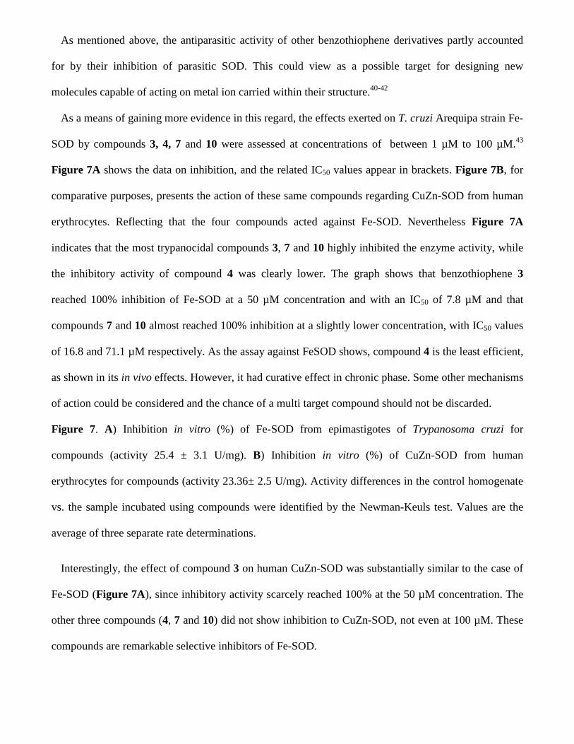
As a means of gaining more evidence in this regard, the effects exerted on T. cruzi Arequipa strain Fe-
SOD by compounds 3, 4, 7 and 10 were assessed at concentrations of between 1 µM to 100 µM.43
Figure 7A shows the data on inhibition, and the related IC50 values appear in brackets. Figure 7B, for
comparative purposes, presents the action of these same compounds regarding CuZn-SOD from human
erythrocytes. Reflecting that the four compounds acted against Fe-SOD. Nevertheless Figure 7A
indicates that the most trypanocidal compounds 3, 7 and 10 highly inhibited the enzyme activity, while
the inhibitory activity of compound 4 was clearly lower. The graph shows that benzothiophene 3
reached 100% inhibition of Fe-SOD at a 50 µM concentration and with an IC50 of 7.8 µM and that
compounds 7 and 10 almost reached 100% inhibition at a slightly lower concentration, with IC50 values
of 16.8 and 71.1 µM respectively. As the assay against FeSOD shows, compound 4 is the least efficient,
as shown in its in vivo effects. However, it had curative effect in chronic phase. Some other mechanisms
of action could be considered and the chance of a multi target compound should not be discarded.
Figure 7. A) Inhibition in vitro (%) of Fe-SOD from epimastigotes of Trypanosoma cruzi for
compounds (activity 25.4 ± 3.1 U/mg). B) Inhibition in vitro (%) of CuZn-SOD from human
erythrocytes for compounds (activity 23.36± 2.5 U/mg). Activity differences in the control homogenate
vs. the sample incubated using compounds were identified by the Newman-Keuls test. Values are the
average of three separate rate determinations.
Interestingly, the effect of compound 3 on human CuZn-SOD was substantially similar to the case of
Fe-SOD (Figure 7A), since inhibitory activity scarcely reached 100% at the 50 µM concentration. The
other three compounds (4, 7 and 10) did not show inhibition to CuZn-SOD, not even at 100 µM. These
compounds are remarkable selective inhibitors of Fe-SOD.

Metabolite excretion. Trypanosomatids proved incapable of completely degrade glucose to CO2, thus
excreting into the medium part of their hexose skeleton as partly oxidized fragments. The percentage
and nature of this excretion depended on the pathway that was used in glucose metabolism.44 In T.
cruzi, the catabolism product are mainly acetate, succinate, L-alanine and D-lactate.45
In order to obtain some information about the effects of 3, 4, 7 and 10 on the glucose metabolism of
the parasite, we registered the 1H NMR spectrum of T. cruzi epimastigotes treated with the test
compounds (Supl information); the final excretion products were qualitatively and quantitatively
identified. Figure 8 shows the results obtained and the comparison with control epimastigotes.
Excretion of some metabolites, were disturbed in the treatment with the compounds, with the succinate
being the most affected, and showing an increase of 199% for compound 10, 142% for compound 3, 22
% for 4 and an increase of 109% for compound 7; lesser alterations were observed in the other
metabolites.
All of these data could be interpreted on the basis of a change in the succinate pathways occurring in
the presence of the compounds under investigation. Otherwise, it is interesting to note that the increase
in succinate with these compounds indicates catabolic changes that could be related to mitochondria
malfunction, due to the redox stress produced by inhibition of the mitochondrion-resident Fe-SOD
enzyme.46
Figure 8. Percentages of variation among peaks of catabolites excreted by epimastigotes of
Trypanosoma cruzi exposed to compounds 3, 4, 7 and 10 at their IC25 in comparison to a control
incubated 96 h.
Mutagenicity studies. Ames test
Taking into account the great potential of compounds 4 and 7, we decided to include them in the
Ames test to explore their mutagenicity capacity before considering them for further preclinical
development. The results obtained from the Ames test for compound 4 and 7 are shown in Tables 7 and
8, respectively. The evaluated doses of the compound were determined in a previous solubility study on
the test system and 2.25 µg/plate and 0.75 µg/plate were established as the highest concentrations to be
tested in the experimental conditions for compounds 4 and 7, respectively. No signs of cytotoxicity or
precipitation were observed during the experiment. Both compounds were not mutagenic in any of the
two tested conditions, as no evidence of dose-related or more than two-fold increase in the mean
number or revertants was observed with or without metabolic activation. Therefore, compounds 4 and 7
could be pointed as interesting candidates for the development of an anti-Chagas agent
Table 7. Results of the Ames test of compound 4.
Table 8. Results of the Ames test of compound 7.
Docking study of benzothiophenes 3, 4, 7 and 10 in the T. cruzi Fe-SOD and the human CuZn-
SOD enzymes.
In order to gain insight into the binding mode of the new benzothiophene derivatives in the Fe-SOD
enzyme, docking studies were carried out using the crystal structure of both mitochondrial (PDB id
4DVH) and cytosolic (PDB id 2GPC) Fe-SOD trypanothione cruzi proteins. The results showed that the
compounds bind in a deep funnel at the top of the dimer interface, which gives access to the active site
(metal site) (Figure 9a). This cavity has already been suggested as the inhibitor binding site.47 Even
when the binding mode of the compounds in both enzymes was similar, the affinity was higher for the
mitochondrial protein. However, we believe that this is merely an artifact produced by the different
orientation of Lys36 (Lys35 in the mitochondrial enzyme), which lays inside the active site in the PDB
2GPC and outside of it in PDB 4DVH. Based on our studies, we suggest the mitochondrial protein with
PDB code 4DVH as best suited for docking studies on Fe-SOD trypanothione cruzi enzyme. The
compounds bind in an aromatic pocket formed by aminoacids Try32, His28, Trp161 and Phe119. This
hydrophobic pocket is flanked by polar residues at both sides: Glu162, Arg173, Val116 and Asn117 at
one side, and Lys35 and Gln68 at the other. Just like the fluoro substituents of compounds 4 and 7, the
nitro group of compounds 3 and 10 perform electrostatic interactions with Lys35, Gln68 and Asn117.
As expected, the ligand does not perform interactions with the metal, which binds well inside the active
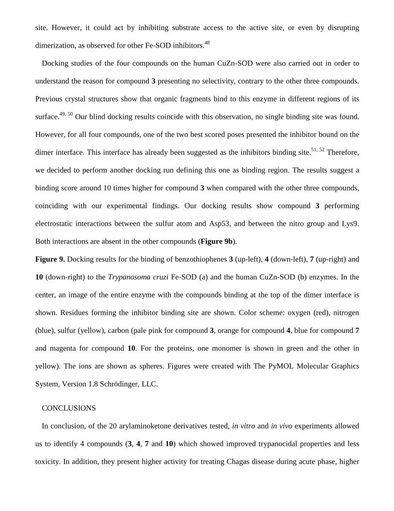
site. However, it could act by inhibiting substrate access to the active site, or even by disrupting
dimerization, as observed for other Fe-SOD inhibitors.48
Docking studies of the four compounds on the human CuZn-SOD were also carried out in order to
understand the reason for compound 3 presenting no selectivity, contrary to the other three compounds.
Previous crystal structures show that organic fragments bind to this enzyme in different regions of its
surface.49, 50 Our blind docking results coincide with this observation, no single binding site was found.
However, for all four compounds, one of the two best scored poses presented the inhibitor bound on the
dimer interface. This interface has already been suggested as the inhibitors binding site.51, 52 Therefore,
we decided to perform another docking run defining this one as binding region. The results suggest a
binding score around 10 times higher for compound 3 when compared with the other three compounds,
coinciding with our experimental findings. Our docking results show compound 3 performing
electrostatic interactions between the sulfur atom and Asp53, and between the nitro group and Lys9.
Both interactions are absent in the other compounds (Figure 9b).
Figure 9. Docking results for the binding of benzothiophenes 3 (up-left), 4 (down-left), 7 (up-right) and
10 (down-right) to the Trypanosoma cruzi Fe-SOD (a) and the human CuZn-SOD (b) enzymes. In the
center, an image of the entire enzyme with the compounds binding at the top of the dimer interface is
shown. Residues forming the inhibitor binding site are shown. Color scheme: oxygen (red), nitrogen
(blue), sulfur (yellow), carbon (pale pink for compound 3, orange for compound 4, blue for compound 7
and magenta for compound 10. For the proteins, one monomer is shown in green and the other in
yellow). The ions are shown as spheres. Figures were created with The PyMOL Molecular Graphics
System, Version 1.8 Schrödinger, LLC.
CONCLUSIONS
In conclusion, of the 20 arylaminoketone derivatives tested, in vitro and in vivo experiments allowed
us to identify 4 compounds (3, 4, 7 and 10) which showed improved trypanocidal properties and less
toxicity. In addition, they present higher activity for treating Chagas disease during acute phase, higher

curative rates and larger spectrum of action than the reference drug benznidazole. Moreover, none of the
compounds included in the toxicological studies were genotoxic or mutagenic. Therefore, they are
promising molecules for the development of an easy to synthesize anti-Chagas agent, and this allows us
to think that these compounds could be immediately implemented into a step further within the
preclinical phase. Finally, it merits mention that due to their different mechanisms of action, combined
therapies should be considered for obtaining improved efficiency.
EXPERIMENTAL SECTION
CHEMISTRY
General methods. All solvents and chemicals were used as purchased without further purification.
Thin layer chromatography (TLC) using silica Alugram SIL G/UV254 (layer: 0.2 mm) (Macherey-
Nagel GmbH&Co. KG., Düren, Germany) and detection with UV light was used to monitor reactions.
Conventional flash column chromatography was carried out with silica gel 60 (0.040_0.063 mm,
Merck). Flash column chromatography was developed on a CombiFlash®Rf (TELEDYNE ISCO,
Lincoln, USA) instrument with Silica RediSep®Rf columns. All of the synthesized compounds were
chemically characterized by infrared (IR), proton nuclear magnetic resonance (1H NMR), carbon
nuclear magnetic resonance (13C NMR) and elemental microanalyses (CHN). IR spectra were recorded
on a Nicolet Nexus FTIR (Thermo, Madison, WI, U.S.) in KBr pellets. Data are reported as vibrational
frequency (cm-1) and intensity is defined as strong (s), medium (m) and weak (w). In some cases, 2D
NMR experiments (COSY, HMBC, HMQC) were carried out for the appropriate peaks assignment.
NMR spectra were recorded in DMSO-d6 or CDCl3 solutions on a Bruker 400 Ultrashield instrument.
The data are reported as follows: chemical shift in ppm from internal tetramethylsilane (TMS) standard
on the δ scale, multiplicity (bs = broad singlet, s = singlet, d = doublet, dd = double doublet, ddd =
double double doublet, t = triplet, m = multiplet) and coupling constants (J) values given in hertz (Hz).
(See supporting information) All compounds were purified to ≥96% purity as determined by elemental
microanalysis results obtained on a CHN-900 elemental analyzer (Leco, Tres Cantos, Spain) from
vacuum-dried samples. The analytical results for C, H, and N were within ±0.4 of the theoretical values.
Melting points (mp) were determined with a Mettler FP82 + FP80 apparatus (Greifensee, Switzerland)
and are not corrected.
Synthesis. Compounds 2, 3, 6, 10, 12 and 13 were previously reported.17-19
General methodology for the preparation of arylamine Mannich base derivatives (1, 4, 5, 7-9,
11, 14-20). A solution of the substituted methylketone (1.0 eq), the corresponding arylamine (1.0 eq),
1,3-dioxolane (5 mL) and concentrated HCl (1 mL) was heated at reflux. The reaction was followed by
TLC until conversion (30 min - 2 h). NaOH 2 M was added to basic pH and the product was extracted
with CH2Cl2. The organic extracts were combined, dried over anhydrous Na2SO4, filtered and
concentrated under reduced pressure. The residue was purified by column chromatography using
CH2Cl2/methanol 95:5 (v/v) as mobile phase or Automated Flash Chromatography eluting in gradient
with CH2Cl2/methanol 99:1 (v/v). The desired compounds were precipitated with diethyl ether.
1-(benzo[b]thiophen-3-yl)-3-(4-(4-trifluoromethylphenyl)piperazin-1-yl)propan-1-one
hydrochloride (1). Yield: 14%. mp: 209-210 ºC. IR (KBr) ν cm-1: 2552 (s, νHCl); 2458 (s, νHCl); 1660 (s,
νC=O); 1335 (s, νC-F); 1166 (s, νC-F); 1113 (s, νC-F); 1073 (s, νC-F). 1H NMR (DMSO-d6, 400 MHz) δ ppm:
11.45-11.26 (bs, 1H, HCl); 9.15(s, 1H, H2); 8.61 (d, 1H, H4, J4-5=7.9 Hz); 8.12 (d, 1H, H7, J7-6=7.8 Hz);
7.57 (d, 2H, H3’+H5’, J3’-2’=8.7 Hz); 7.54-7.45 (m, 2H, H5+H6); 7.18 (d, 2H, H2’+H6’, J2’-3’=8.7 Hz);
4.10-4.00 (m, 2H, 2Hp); 3.77 (t, 2H, CO-CH2, J=6.9 Hz); 3.72-3.62 (m, 2H, 2Hp); 3.55 (t, 2H, CH2-N,
J=6.8 Hz); 3.34-3.19 (m, 4H, 2H2p+2H6p). 13C NMR (DMSO-d6, 100 MHz) δ ppm: 191.91; 151.97;
140.51; 139.36; 135.99; 133.42; 125.89; 125.47; 124.51: 123.00; 114.91 (2C); 126.30 (2C, q, J3=4.0
Hz); 124.78 (q, J1=268.0 Hz); 119.04 (q, J2=31.5 Hz); 50.52 (3C); 44.30 (2C); 34.01. Anal. calcd for
C22H21F3N2OS.HCl: C 58.08%, H 4.87%, N 6.16%. Found: C 57.81%, H 4.80%, N 6.32%.
1-(5-fluorobenzo[b]thiophen-3-yl)-3-(4-(4-trifluoromethylphenyl)piperazin-1-yl)propan-1-one
(4). Yield: 26%. mp: 143-144 ºC. IR (KBr) ν cm-1: 1664 (s, νC=O); 1331 (s, νC-F); 1243 (s, νC-F); 1107 (s,
νC-F). 1H NMR (CDCl3, 400 MHz) δ ppm: 8.48 (dd, 1H, H4, J4-F=10.3 Hz, J4-6=2.3 Hz); 8.40 (s, 1H,
H2); 7.80 (dd, 1H, H7, J7-6=8.8 Hz, J7-F=4.8 Hz); 7.48 (d, 2H, H3’+H5’, J3’-2’=8.6 Hz); 7.20 (ddd, 1H, H6,
J6-F=8.7 Hz, J6-7=8.7 Hz, J6-4=2.4 Hz); 6.92 (d, 2H, H2’+H6’, J2’-3’=8.6 Hz); 3.30-3.26 (m, 4H,
2H3p+2H5p); 3.23 (t, 2H, CO-CH2, J=7.2 Hz); 2.94 (t, 2H, CH2-N, J=7.2 Hz); 2.72-2.68 (m, 4H,
2H2p+2H6p). 13C NMR (CDCl3, 100 MHz) δ ppm: 193.73; 161.82 (d, J1=243.1 Hz); 153.20; 138.56;
137.91 (d, J3=10.2 Hz); 134.87; 134.84; 126.37 (2C, q, J3=4.0 Hz); 124.72 (q, J1=270.1 Hz); 123.27 (d,
J3=9.4 Hz); 120.55 (q, J2=32.6 Hz); 114.56 (d, J2=25.7 Hz); 114.52 (2C); 111.62 (d, J2=25.2 Hz); 53.13;
52.98 (2C); 47.98 (2C); 37.81. Anal. calcd for C22H20F4N2OS: C 60.54%, H 4.62%, N 6.42%. Found: C
60.37%, H 4.30%, N 6.39%.
1-(5-fluorobenzo[b]thiophen-3-yl)-3-(4-(4-chlorophenyl)piperazin-1-yl)propan-1-one (5). Yield:
33%. mp: 156-157 ºC. IR (KBr) ν cm-1: 1660 (s, νC=O); 1246 (s, νC-F).1H NMR (DMSO-d6, 400 MHz) δ
ppm: 9.17 (s, 1H, H2); 8.32 (dd, 1H, H4, J4-F=10.6 Hz, J4-6=2.6 Hz); 8.15 (dd, 1H, H7, J7-6=8.8 Hz, J7-
F=5.2 Hz); 7.88 (ddd, 1H, H6, J6-F=9.0 Hz, J6-7=8.9 Hz, J6-4=2.8 Hz); 7.21 (d, 2H, H3’+H5’, J3’-2’=9.2
Hz); 6.93 (d, 2H, H2’+H6’, J2’-3’=9.2 Hz); 3.27 (t, 2H, CO-CH2, J=7.0 Hz); 3.14-3.07 (m, 4H,
2H3p+2H5p); 2.80 (t, 2H, CH2-N, J=7.2 Hz); 2.62-2.54 (m, 4H, 2H2p+2H6p). 13C NMR (DMSO-d6, 100
MHz) δ ppm: 212.89; 160.96 (d, J1=240.8 Hz); 149.56; 142.13; 137.43 (d, J3=10.5 Hz); 135.06 (d,
J4=1.3 Hz); 133.64 (d, J4=4.5 Hz); 128.46 (2C); 124.57 (d, J3=9.7 Hz); 122.24; 116.70 (2C); 113.90 (d,
J2=25.2 Hz); 109.96 (d, J2=24.9 Hz); 52.64; 52.30 (2C); 47.72 (2C); 36.91. Anal. Calcd. for
C21H20ClFN2OS: C 62.60%, H 5.00%, N 6.95%. Found: C 62.23%, H 4.63%, N 6.87%.
1-(5-fluorobenzo[b]thiophen-3-yl)-3-(4-(4-fluorophenyl)piperazin-1-yl)propan-1-one (7). Yield:
18%. mp: 156-157 ºC. IR (KBr) ν cm-1: 1661 (s, νC=O); 1175 (s, νC-F); 1131 (s, νC-F). 1H NMR (CDCl3,
400 MHz) δ ppm: 8.48 (dd, 1H, H4, J4-F=10.4 Hz, J4-6= 2.6 Hz); 8.39 (s, 1H, H2); 7.79 (dd, 1H, H7, J7-
6=8.8 Hz, J7-F=4.8 Hz); 7.19 (ddd, 1H, H6, J6-F=8.6 Hz, J6-7=8.6 Hz, J6-4=2.5 Hz); 6.99-6.93 (m, 2H,
H3’+H5’); 6.89-6.84 (m, 2H, H2’+H6’); 3.22 (t, 2H, CO-CH2, J=7.3 Hz); 3.14-3.11 (m, 4H, 2H3p+2H5p);
2.94 (t, 2H, CH2-N, J=7.3 Hz); 2.71-2.68 (m, 4H, 2H2p+2H6p). 13C NMR (CDCl3, 100 MHz) δ ppm:
193.82; 161.78 (d, J1=243.8 Hz); 157.15 (d, J1=239.9 Hz); 147.86 (d, J4=2.2 Hz); 138.56; 137.89 (d,
J3=10.7 Hz); 135.11 (d, J4=1.6 Hz); 134.84 (d, J4=4.5 Hz); 123.23 (d, J3=9.5 Hz); 117.80 (2C, d, J3=7.6
Hz); 115.48 (2C, d, J2=22.0 Hz); 114.51 (d, J2=25.6 Hz); 111.60 (d, J2=25.2 Hz); 53.26 (2C); 53.15;

50.14 (2C); 37.81. Anal. Calcd. for C21H20F2N2OS: C 65.27%, H 5.22%, N 7.25%. Found: C 64.94%, H
5.18%, N 6.88%.
1-(benzo[b]thiophen-3-yl)-3-(4-benzylpiperazin-1-yl)propan-1-one hydrochloride (8). Yield:
12%. mp: 202-203 ºC. IR (KBr) ν cm-1: 2515 (m, νHCl); 2406 (m, νHCl); 1664 (s, νC=O). 1H NMR
(DMSO-d6, 400 MHz) δ ppm: 11.93 (s, 2H, HCl); 9.04 (s, 1H, H2); 8.60 (d, 1H, H4, J4-5=7.9 Hz); 7.11
(d, 1H, H7, J7-6=7.6 Hz); 7.71-7.57 (m, 2H, H5+H6); 7.56-7.36 (m, 5H, H2’-H6’); 4.36 (s, 2H, CH2-benzyl);
3.84-3.66 (m, 6H, CO-CH2+2H3p+2H5p); 3.66-3.19 (m, 6H, CH2-N+2H2p+2H6p). 13C NMR (DMSO-d6,
100 MHz) δ ppm:. 181.92; 161.21; 150.86; 140.51; 139.36; 135.99; 128.75; 125.87 (2C); 125.47 (2C);
124.98; 124.52 (2C); 123.00; 73.22; 57.70 (3C); 47.33 (2C); 36.99. Anal. Calcd. for C22H24N2OS.2HCl:
C 60.41%, H 5.99%, N 6.40%. Found: C 60.02%, H 6.08%, N 6.27%.
1-(benzo[b]thiophen-3-yl)-3-(4-(3-fluorobenzyl)piperazin-1-yl)propan-1-one hydrochloride (9).
Yield: 12%. mp: 214-215 ºC. IR (KBr) ν cm-1: 2525 (m, νHCl); 2449 (m, νHCl); 1668 (s, νC=O). 1H NMR
(DMSO-d6, 400 MHz) δ ppm: 11.45-11.26 (bs, 2H, HCl); 9.05(s, 1H, H2); 8.61 (d, 1H, H4, J4-5=7.7
Hz); 8.12 (d, 1H, H7, J7-6=7.8 Hz); 7.66-62 (m, 2H, H5+H6); 7.57-7.52 (m, 4H, H2’+H4’+H5’+H6’); 4.41
(s, 2H, CH2-benzyl); 3.85-3.65 (m, 12H, 8Hp+COCH2+CH2N). Anal. Calcd. for C22H23FN2OS.2HCl: C
58.02%, H 5.53%, N 6.15%. Found: C 58.40%, H 5.71%, N 6.00%.
1-(thiophen-3-yl)-3-(4-(3-trifluoromethylphenyl)piperazin-1-yl)propan-1-one hydrochloride
(11). Yield: 18%. mp: 177-178 ºC. IR (KBr) ν cm-1: 2459 (m, νHCl); 1682 (s, νC=O); 1166 (s, νC-F); 1120
(s, νC-F); 1076 (s, νC-F). 1H NMR (DMSO-d6, 400 MHz) δ ppm: 11.45-11.28 (bs, 1H, HCl); 8.61 (s, 1H,
H2); 7.68 (dd, 1H, H5, J5-4=4.6 Hz, J5-2=2.5 Hz); 7.55 (d, 1H, H4, J4-5=5.0 Hz); 7.47 (t, 1H, H5’, J5-4’=7.9
Hz); 7.37-7.24 (m, 2H, H2’+H6’); 7.16 (d, 1H, H4’, J4’-5’=7.3 Hz); 4.04-3.93 (m, 2H, CO-CH2); 3.70-
3.57 (m, 4H, 2H3p+2H5p); 3.52-3.43 (m, 2H, CH2-N); 3.28-3.14 (m, 4H, 2H2p+2H6p). 13C NMR
(DMSO-d6, 100 MHz) δ ppm: 190.90; 149.84; 141.07; 134.29; 130.10; 129.92 (q, J2=30.7 Hz); 127.76;
126.32; 124.28 (q, J1=272.0 Hz); 119.22; 115.65 (q, J3=3.6 Hz); 111.03 (q, J3=3.5 Hz); 50.57 (2C);
50.35; 44.86 (2C); 33.73. Anal. Calcd. for C18H19F3N2OS.HCl: C 53.40%, H 4.98%, N 6.92%. Found: C
53.02%, H 5.02%, N 6.85%.

1-(naphthalen-2-yl)-3-(4-(4-trifluoromethylphenyl)piperazin-1-yl)-propan-1-one (14). Yield:
66%. mp: 150-151 ºC. IR (KBr) ν cm-1: 1677 (s, νC=O); 1148 (s, νC-F); 1098 (s, νC-F); 1065 (s, νC-F). 1H
NMR (DMSO-d6, 400 MHz) δ ppm: 8.72 (s, 1H, H1); 8.14 (d, 1H, H8, J8-7=7.7 Hz); 8.03-7.97 (m, 3H,
H3-H5); 7.69-7.59 (m, 2H, H6+H7); 7.48 (d, 2H, H3’+H5’, J3’-2’=8.8 Hz); 7.04 (d, 2H, H2’+H6’, J2’-3’=8.8
Hz); 3.37 (t, 2H, CO-CH2, J=7.2 Hz); 3.27-3.21 (m, 4H, 2H3p+2H5p); 2.79 (t, 2H, CH2-N, J=7.2 Hz);
2.61-2.55 (m, 4H, 2H2p+2H6p). 13C NMR (DMSO-d6, 100 MHz) δ ppm: 199.19; 153.24; 135.03;
134.00; 132.23; 129.95; 129.60; 128.61; 128.26; 127.64; 126.91; 126.13 (2C, q, J3=3.8 Hz); 124.99 (q,
J1=270.1 Hz); 123.55; 117.78 (q, J2=32.0 Hz); 114.12 (2C); 52.92; 52.41 (2C); 46.96 (2C); 35.81. Anal.
Calcd. for C24H23F3N2O: C 69.89%, H 5.62%, N 6.79%. Found: C 69.82%, H 5.72%, N 6.76%.
1-(naphthalen-2-yl)-3-(4-(4-nitrophenyl)piperazin-1-yl)-propan-1-one (15). Yield: 86%. mp: 190-
191 ºC. IR (KBr) ν cm-1: 2806 (s, νC-H); 1674 (s, νC=O). 1H NMR (DMSO-d6, 400 MHz) δ ppm: 8.74 (s,
1H, H1); 8.14 (d, 1H, H8, J8-7=7.8 Hz); 8.04 (m, d, 2H, H3’+H5’, J3’-2’=9.4 Hz); 8.02-7.98 (m, 3H, H3-
H5); 7.70-7.60 (m, 2H, H6+H7); 7.03 (d, 2H, H2’+H6’, J2’-3’=9.4 Hz); 3.48-3.42 (m, 4H, 2H3p+2H5p);
3.40 (t, 2H, COCH2, J=7.2 Hz); 2.80 (t, 2H, CH2-N, J=7.2 Hz); 2.62-2.56 (m, 4H, 2H2p+2H6p). 13C
NMR (DMSO-d6, 100 MHz) δ ppm: 199.07; 154.68; 136.75; 134.98; 133.92; 132.19; 129.94; 129.56;
128.57; 128.20; 127.60; 126.86; 125.66 (2C); 123.50; 112.55 (2C); 52.75; 52.24 (2C); 46.23 (2C);
35.71. Anal. Calcd. for C23H23N3O3: C 70.93%, H 5.95%, N 10.79%. Found: C 70.99%, H 5.85%, N
10.58%.
3-(4-(4-fluorophenyl)piperazin-1-yl)-1-(naphthalen-2-yl) propan-1-one (16). Yield: 15%. mp: 98-
99 ºC. IR (KBr) ν cm-1: 2800 (m, νC-H); 1676 (s, νC=O); 1177 (s, νarC-F); 1134 (s, νarC-F). 1H NMR (CDCl3,
400 MHz) δ ppm: 8.50 (s, 1H, H1); 8.04 (dd, 1H, H8, J8-7=8.6 Hz, J8-6=1.7 Hz); 7.97 (d, 1H, H3, J3-4=8.0
Hz); 7.91 (d, 1H, H5, J5-6=8.8 Hz); 7.88 (d, 1H, H4, J4-3=8.7 Hz); 7.64-7.54 (m, 2H, H6+H7); 6.97 (dd,
2H, H3’+H5’, J3’-2’=9.0 Hz, J3’-F=8.4 Hz); 6.88 (dd, 2H, H2’+H6’, J2’-3’=9.2 Hz, J2’-F=4.6 Hz); 3.37 (t, 2H,
CO-CH2, J=7.4 Hz); 3.17-3.12 (m, 4H, 2H3p+2H5p); 2.97 (t, 2H, CH2-N, J=7.4 Hz); 2.75-2.70 (m, 4H,
2H2p+2H6p). 13C NMR (CDCl3, 100 MHz) δ ppm: 198.93; 159.17 (d, J1=239.1 Hz); 147.89 (d, J4= 2.2
Hz); 135.60; 134.22; 132.51; 129.71; 129.54; 128.51 (2C); 127.77; 126.81; 123.78; 117.82 (2C, d,
J3=7.9 Hz); 115.48 (2C, d, J2=22.1 Hz); 53.28 (2C); 53.25; 50.14 (2C); 36.33. Anal. Calcd. for
C23H23FN2O: C 76.22%, H 6.40%, N 7.73%. Found: C 76.46%, H 6.57%, N 7.60%.
1-(benzo[d][1,3]dioxol-5-yl)-3-(4-(4-trifluoromethylphenyl)piperazin-1-yl)propan-1-one (17).
Yield: 21%. mp: 107-108 ºC. IR (KBr) ν cm-1: 1669 (s, νC=O); 1158 (s, νC-F); 1111 (s, νC-F); 1072 (s, νC-
F). 1H NMR (DMSO-d6, 400 MHz) δ ppm: 7.65 (1H, d, H6, J6-7=8.2 Hz); 7.48 (d, 2H, H3’+H5’, J3’-2’=8.4
Hz); 7.47 (s, 1H, H4); 7.04 (dd, 3H, H2’+H6’+H7, J2’-3’=J7-6=8.7 Hz); 6.13 (s, 2H, 2H2); 3.28-3.2 (m, 4H,
2H3p+2H5p); 3.16 (t, 2H, CO-CH2, J=7.1 Hz); 2.70 (t, 2H, CH2-N, J=7.1 Hz); 2.59-2.52 (m, 4H,
2H2p+2H6p). 13C NMR (DMSO-d6, 100 MHz) δ ppm: 197.06; 153.16; 151.28; 147.71; 131.35; 126.01
(2C, q, J3=3.7 Hz); 124.91 (q, J1=270.5 Hz); 124.28; 117.66 (q, J2=32.0 Hz); 114.01 (2C); 107.92;
107.33; 101.89; 52.89; 52.29 (2C); 46.85 (2C); 35.45. Anal. Calcd. for C21H21F3N2O3: C 62.06%, H
5.21%, N 6.89%. Found: C 62.06%, H 5.60%, N 7.03%.
1-(benzo[d][1,3]dioxol-5-yl)-3-(4-(4-chlorophenyl)piperazin-1-yl)propan-1-one (18). Yield: 20%.
mp: 105-106 ºC. IR (KBr) ν cm-1: 1675 (s, νC=O); 1241 (s, νC-O). 1H NMR (DMSO-d6, 400 MHz) δ ppm:
7.64 (dd, 1H, H6, J6-7=8.2 Hz, J6-4=1.3 Hz); 7.46 (s, 1H, H4); 7.20 (d, 2H, H3’+H5’, J3’-2’=8.9 Hz); 7.02
(d, 1H, H7, J7-6=8.2 Hz); 6.92 (d, 2H, H2’+H6’, J2’-3’=8.9 Hz); 6.12 (s, 2H, 2H2); 3.14 (t, 2H, CO-CH2,
J=7.1 Hz); 3.11-3.05 (m, 4H, 2H3p+2H5p); 2.69 (t, 2H, CH2-N, J=7.1 Hz); 2.57-2.51 (m, 4H,
2H2p+2H6p). 13C NMR (DMSO-d6, 100 MHz) δ ppm: 197.20; 151.35; 149.78; 147.78; 131.40; 128.55
(2C); 124.38; 122.22; 116.74 (2C); 108.00; 107.38; 101.97; 52.98; 52.49 (2C); 47.95 (2C); 35.52. Anal.
Calcd. for C20H21ClN2O3: C 64.43%, H 5.68%, N 7.51%. Found: C 64.61%, H 5.86%, N 7.40%.
1-(benzo[d][1,3]dioxol-5-yl)-3-(4-(4-nitrophenyl)piperazin-1-yl)propan-1-one (19). Yield: 50%.
mp: 164-165 ºC. IR (KBr) ν cm-1: 1665 (s, νC=O); 1241 (s, νC-O). 1H NMR (DMSO-d6, 400 MHz) δ ppm:
8.04 (d, 2H, H3’+H5’, J3’-2’=9.4 Hz); 7.65 (dd, 1H, H6, J6-7=8.2 Hz, J6-4=1.4 Hz); 7.47 (s, 1H, H4); 7.06-
7.00 (m, 3H, H7+H2’+H6’); 6.13 (s, 2H, 2H2); 3.46-3.40 (m, 4H, 2H3p+2H5p); 3.16 (t, 2H, CO-CH2,
J=7.0 Hz); 2.70 (t, 2H, CH2-N, J=7.1 Hz); 2.57-2.51 (m, 4H, 2H2p+2H6p). 13C NMR (DMSO-d6, 100
MHz) δ ppm: 197.08; 154.66; 151.33; 147.75; 136.75; 131.35; 125.64 (2C); 124.35; 112.54 (2C);
107.98; 107.38; 101.94; 52.78; 52.17 (2C); 46.21 (2C); 35.42. Anal. Calcd. for C20H21N3O5: C 62.65%,
H 5.52%, N 10.96%. Found: C 62.29%, H 5.43%, N 10.61%.
1-(benzo[d][1,3]dioxol-5-yl)-3-(4-(4-fluorophenyl)piperazin-1-yl)propan-1-one (20). Yield: 19%.
mp: 96-97 ºC. IR (KBr) ν cm-1: 1676 (s, νC=O); 1242 (s, νC-O). 1132 (s, νC-F); 1109 (s, νC-F). 1H NMR
(DMSO-d6, 400 MHz) δ ppm: 7.65 (d, 1H, H6, J6-7=8.0 Hz); 7.47 (s, 1H, H4); 7.07-6.99 (m, 3H,
H3’+H5’+H7); 6.93 (dd, 2H, H2’+H6’, J2’-3’=8.9 Hz, J2’-F=4.6 Hz); 6.13 (s, 2H, 2H2); 3.14 (t, 2H, CO-
CH2, J=7.0 Hz); 3.08-2.99 (m, 4H, 2H3p+2H5p); 2.69 (t, 2H, CH2-N, J=7.1 Hz); 2.59-2.52 (m, 4H,
2H2p+2H6p). 13C NMR (DMSO-d6, 100 MHz) δ ppm: 197.10; 155.84 (d, J1=235.4 Hz); 151.26; 147.82
(d, J4=1.9 Hz); 147.71; 131.35; 124.28; 116.92 (2C, d, J3=7.5 Hz); 115.10 (2C, d, J2=21.8 Hz); 107.92;
107.32; 101.89; 52.94; 52.59 (2C); 48.85 (2C); 35.49. Anal. Calcd. for C20H21FN2O3: C 67.40%, H
5.94%, N 7.86%. Found: C 67.30%, H 6.04%, N 7.75%.
Calculation of molecular properties. The drug-likeness of the compounds was estimated with
Osiris, using the stand-alone DataWarrior program.22 The information about the methods used to
evaluate each property is available on the program web site http://www.openmolecules.org.
BIOLOGY
In vitro Trypanocidal Evaluation
Parasite strain culture. Epimastigote and trypomastigote forms: Epimastigote assays. The
epimastigote forms of T. cruzi SN3 strain (IRHOD/CO/2008/SN3, DTU I) that were isolated from the
household triatomine Rhodnius prolixus (from Guajira, Colombia).53 Epimastigotes of T. cruzi Arequipa
strain (MHOM/Pe/2011/ Arequipa, DTU V) isolated from a human and Tulahuen strain
(TINF/CH/1956/Tulahuen, DTU VI) were cultivated in vitro using trypanosome liquid medium (MTL)
containing 10% inactivated fetal bovine serum and maintained at 28°C in an air atmosphere, in Roux
flasks (Corning, USA) of 75 cm2 surface area, as described in previous studies.54 The epimastigote
forms transformed into metacyclic forms by metacyclogenesis triggered by culturing a 5-day-old
epimastigote forms that had been harvested by centrifuging (7000 g) at 10 ºC for 10 min. Next the
parasites were incubated at 28 ºC for 2 h (density of 5 x 10⁸ cells/mL) in TAU medium (190 mM NaCl,
17 mM KCl, 2 mM MgCl2, 2 mM CaCl2, 8 mM phosphate buffer, pH 6.0). The parasites were then
incubated at a dilution of 1:100 (final concentration of epimastigotes: 5 × 10⁶ cells/mL) at 28 ºC f or 96
h in TAU3AAG medium (TAU supplemented with 10 mM L-proline, 50 mM L-sodium glutamate, 2
mM L-sodium aspartate and 10 mM D-glucose) in 25 mL flasks with a layer culture medium no more
than 1 cm in deep.55
Trypomastigote Forms assays. The activity (parasite-reduction %) was compared to control with the
method described above54, with certain modifications in our lab. The test of drug action against T. cruzi
used Balb/c albino mouse blood collected during peak parasitemia (day 7). The infected blood was
diluted using uninfected mouse blood at a concentration of 4 x 10⁶ trypomastigotes/mL. Then, in RPMI
1640 medium-GIBCO (2 x 10⁶ trypomastigotes/mL), a dilution was made to 1:2. Stock solutions of the
compounds were prepared in DMSO. The IC50 values were calculated using, samples of infected blood,
adding each drug to wells in a 96-microwell plate for a final 200 µL volume for drug concentrations of
1, 10, 25, 50, and 100 µM., and the plates were then incubated for 24 h at 4- to 8 ºC. Each experiment
was replicated three times (OLYMPUS CX41) and the parasites were counted in the Neubauer chamber.
Cytotoxicity and Cell Culture Tests. Vero cells (EACC number 84113001) originally extracted from
monkey kidney, were cultured for two days in RPMI (Gibco), supplemented with 10% inactivated fetal
bovine serum in 95% humidified air, 5% CO2 at 37 ºC. The cytotoxicity test was performed for Vero
cells following a methodology described previously.54 Flow cytometry was used to determine cell
viability after 72 h of treatment. Propidium iodide solution (100 mg/mL) was added at 100 μL/well and
incubated at 28 °C for 10 min in darkness. Then, fluorescein diacetate (100 ng/mL) was added at 100
μL/well and incubated using the same conditions. The cells were collected by centrifuging (400 g for 10
min), and finally the precipitate was washed using PBS (phosphate buffered saline). Flow cytometry
was performed with a FACSVantage flow cytometer (Becton Dickinson), and the percentage of viability
was computed in comparison to control. The IC50 was calculated with linear-regression analysis
employing the Kc values corresponding to the concentrations used (0.1 to 100 μM).
In Vitro Activity. Epimastigote Forms Assays (Extracellular Forms). The compounds obtained and
BZN (the reference drug), dissolved in DMSO (Panreac, Barcelona, Spain) at a 0.01% (v/v)
concentration and assayed, proved nontoxic to the parasites and not inhibitory to their growth, according
to a procedure reported on a previous work.56 The compounds, at doses of 100, 50, 25, 10, l, 0.5, 0.25,
and 0.1 μM were added to the culture medium. For each compound, the effects against T. cruzi
epimastigote forms were assayed at 72 h with a hemocytometric chamber (Neubauer). The anti-
trypanosomatid effect is expressed as the IC50. That is, the concentration needed for 50% growth
inhibition, determined by linear regression with the Kc values of the concentrations used.
Amastigote Assays (Intracellular Forms). Vero cells grown as indicated above in RPMI medium
(Gibco), were inoculated at 1 × 10⁴ cells/well in 24-well microplates (Nunc) that had rounded coverslips
and cultured 2 days. Then the Vero cells were infected for 24 h in vitro using metacyclic forms at a 10:1
ratio. After the removal of non-phagocytic parasites by washing, the drugs (100, 50, 25, 10, 1, 0.5 0.25
and 0.1 μM) were added. Vero cells were incubated with the drugs at 37°C for 72 h in 5% CO2. The
activity of the drugs was evaluated based on the number of amastigotes in treated as well as untreated
cultures, in preparations that were methanol-fixed and Giemsa-stained. The number of amastigotes was
estimated analyzing 200 host cells in randomly taken microscopic fields. The anti trypanosomal effect
was expressed as the IC50.
In vitro genotoxicity screening
Bacteria. S. typhimurium TA1535/pSK1002 was obtained from the German Collection for
Microorganisms and Cell cultures (DSMZ). Chemicals. DMSO, dextrose, NaCl, ampicillin, o-
nitrophenol-β-D-galactopyranoside (ONPG), Na2CO3, Na2HPO4.2H2O, NaH2PO4.H2O, MgSO4.7H2O,
KCl, β-mercaptoethanol and sodium dodecyl sulphate were obtained from Sigma-Aldrich;
Bactotryptone was obtained from BD; and rat S9 mix (Mutazyme) was obtained from Moltox.
Reagents preparation for SOS/umu test. TGA culture medium was prepared dissolving 10 g
bactotryptone, 2 g dextrose and 5 g NaCl in 1 L of water and sterilizing during 30 minutes at 121 ºC. B-
buffer was prepared dissolving 20.18 g Na2HPO4.2H2O, 5.5 g NaH2PO4.H2O, 0.75 g KCl, 0.25 g

MgSO4.7H2O, 1.0 g SDS, 2.7 mL β-mercaptoethanol in 1 L water adjusting pH value to 7. Stop reagent
(Na2CO3 1 M) was prepared dissolving 106 g of Na2CO3 in 1 L water. Phosphate buffer (for ONPG
dissolution) was prepared dissolving 1.086 g Na2HPO4.2H2O and 0.538 g NaH2PO4.H2O in 100 mL
water and adjusting pH value to 7. ONPG solution was prepared dissolving 45 mg ONPG in 10 mL
phosphate buffer.
SOS/umu test. The SOS/umu test was carried out according to the method of Oda et al. (1985) and
Reifferscheid et al. (1991), with some modifications.57, 58 The test strain S. typhimurium
TA1535/pSK1002 was used. It was pre-incubated overnight in TGA medium with ampicillin (50 μg/mL)
by shaking in an incubator at 37 °C in order to obtain exponentially growing bacteria. The test was
performed in the absence and the presence of external metabolic activation system (rat S9 mix) in order
to determine the possible genotoxic effects of any metabolite. In each test performed, negative and
positive controls were included. DMSO was used as solvent control, and 4-nitroquinoline-N-oxide (4-
NQO) and 2-aminofluorene (2-AF) were used as positive controls with and without of S9 mix.
Test procedure was as follows: firstly, each compound tested was dissolved in DMSO at 40 mg/mL
and 11 serial ½ dilutions were prepared in a 96-well plate (plate A; final volume in each well was 10
μL). Next, 70 μL of water were added to each well. In another two 96-well plates (plates B; one for the
test with S9 and the other without S9), 10 μL S9 mix or 10 μL PBS, were added , respectively, followed
by the addition of 25 μL of each previously prepared compound. Finally, 90 μL of exponentially
growing bacteria was added and incubated during 4 hours by shaking at 37 ºC. After the incubation
period, absorbance at 600 nm was measured in order to evaluate toxicity on S. typhimurium
TA1535/pSK1002. Toxicity was calculated as follows:
Survival percentage = (A600 for each concentration tested/media A600 for negative control)*100
Afterwards, for the determination of β-galactosidase activity, in two new 96-well plates (plates C),
150 μL of ONPG solution were added to each well and 30 μL of the contents of each well of plates B
were transferred to these plates C. Both plates were incubated for 30 minutes by shaking at 28 ºC,
avoiding direct light exposure. After the incubation period, 120 μL of stop reagent were added and the

reaction was stopped. Absorbance (420 nm) was then measured immediately, and β-galactosidase
activity (relative units; RU) was calculated as follows:
β-galactosidase enzymatic units = A420 for each concentration tested/A600 for each concentration
tested
Finally, the induction factor (IF) was calculated as follows:
IF = β-galactosidase RU for each concentration tested/media β-galactosidase RU for negative control,
where: media β-galactosidase RU for negative control = media A420 for negative control/media A600
for negative control
In the same way, β-galactosidase relative units were calculated for both positive controls, and the test
was only considered valid if the positive controls reached an induction factor ≥2 under the given test
conditions.
Thus, a compound was consider genotoxic, as it induces the induction of the umu operon, when in any
of the conditions studied (with or without metabolic activation), the induction factor was ≥2 at non
cytotoxic concentrations (bacteria survival percentage ≥80%), presenting a dose-response relationship.
Wells in which compound precipitation was observed, were discarded from analysis.
Infectivity Assay
Vero cells were cultured under the same conditions for two days. The, cells were then infected in vitro
with T. cruzi metacyclic forms, at a 10:1 ratio. Immediately after infection, the drugs (IC25
concentrations) were added, followed by incubation at 37°C for 12 h in 5% CO2. After removal of the
drugs and nonphagocytosed parasites by washing, the infected cultures were grown in fresh medium for
10 day.41 Every 48 h, fresh culture medium was added. The activity of the drugs was determined from
the % of infected cells, the number of amastigotes per infected cell, and the number of trypomastigotes
in the medium, both in treated as well as untreated cultures, and in methanol-fixed as well as Giemsa-
stained preparations. A determination was made every 48 h of the mean number of amastigotes per
infected cell and the % of infected cells by examining 200 host cells in randomly taken microscopic
fields. The trypomastigotes in the medium were counted every 48 h in a hemocytometric chamber
(Neubauer).
In Vivo Trypanocidal Activity Assay
For the following purposes, in the in vivo stage, experimental tests were performed: i) to assess the
influence that the compounds exerted on the parasitemia levels post- treatment by a Neubauer chamber
count for 40 days as an acute-phase marker; ii) to provide a measure of the parasitemia reactivation
following immunosuppression as a chronic-phase marker; iii) to compare the levels of the chronic and
acute phases of Ig-G using ELISA as an immune-response indicator; and iv) for detection of the
parasite’s presence using PCR in target organs.
Mice Infection and Treatment. The mice were handled following the international guide for
biomedical research using experimental animals and the experiment was approved by the ethics board of
the University of Granada (Spain). Groups of six female BALB/c albino mice 8 to 10 weeks old and
weighing 25 to 30 g, were kept under standard conditions and given free access to standard chow and
water. The mice were infected intraperitoneally using 5 x 10⁵ blood trypomastigotes (T. cruzi Arequipa
strains) from the blood of mice previously infected. These animals were split in the three groups: I,
untreated infected mice constituted the positive control group; II, infected mice treated using the
reference drug made up the BZN group; and III, infected mice treated using the compounds under
investigation comprised the study group. The tested compounds were administered beginning on the day
7 after confirmation of the infection, and dosages of 50 and 100 mg/kg body mass per day were given
orally on 5 alternating days (day 7 to 15 pi). In each mouse, peripheral blood was taken from the
mandibular vein (5 μL samples) and this was diluted in 495 μL PBS solution (1:100). The circulating
parasites were counted in a Neubauer chamber every 3 days for 40 d (acute phase). The number of
bloodstream forms detected was expressed in parasites/mL.
Immunosuppression Induced by Cyclophosphamide- and Cure Assessment. At day 90 pi, the groups in
which the parasitemia levels had significantly fallen, (irrespective of the treatment the mice received)
and could not be detected by microscopic examination of fresh blood, were subjected to 3 cycles of

immunosuppression using CP (cyclophosphamide monohydrate ISOPAC®) as follows: 4 days
consecutively on which the mice were given 1 intraperitoneal injection/day (dosage 200 mg/kg·w/day),
and 3 days off. The effectiveness of this immunosuppression technique for evaluating cryptic infection
was corroborated by the mortality rate of nearly 100% in chronically untreated mice and/or by the high
parasitemia rate. The parasitemia was assessed within 1 week after the last injection of CP, following
the procedure explained above for the acute phase in order to quantify the trypomastigote forms in blood
as the reactivation rate. The mice were finally bled by heart puncture under gaseous anesthesia (CO2).
Blood was incubated for 2 hours at 37 ºC and then over night at 4º C in order to allow clotting.
Subsequently, the serum was obtained after centrifuging the supernatant twice at 1000 and 2700 g,
consecutively. The serum was used for ELISA and biochemical analysis as explained further on in this
paper. Thanks to our previous in vivo studies, we observed that the tropism of T. cruzi strain (Arequipa)
occurs in the following organs: adipose, brain, heart, esophagus, stomach, bone marrow, muscle and
lung. Therefore these 8 target organs were harvested and immediately flushed free of blood by gentle
infusion of pre-warmed PBS for the purpose of avoid contamination of collected tissue with blood
parasites. Next, they were used for DNA extraction, as explained below. In addition, spleens were
weighed to evaluate inflammation of these organs in different groups of mice.
ELISA Tests. Fe-SOD excreted from the parasite, cultured and processed as described by Lopez-
Céspedes et al.59 was used as antigen. Circulating antibodies in serum against T. cruzi, from day 40 to
day 120 post-infection (including sera post-immunosuppression), were qualitatively and quantitatively
assessed using ELISA. The serum was centrifuged from whole blood and diluted in PBS to 1:80. The
absorbance was read at 492 nm using a microplate reader (Sunrise, TECAN). All samples, in triplicate,
were analyzed in polystyrene microtiter plates. For calculating the cutoff value, the mean and standard
deviations of the optical density of the negative control sera were used.
PCR and DNA Extraction. The organs to be studied were thawed and then ground using a Potter-
Elvehjem for purification using Wizard® Genomic DNA Purification Kit (Promega). The PCR was
conducted with two primers from our laboratory (unpublished data), based on the published SOD
(superoxide dismutase) sequence of T. cruzi CL Brenner (GenBank accession No. XM_808937); this
amplifies a fragment pertaining to T. cruzi SOD gene b of c. 300 pb. The amplification was performed
in Thermal Cycler TM MyCycler thermocycler (BioRad).
Studies of the action mechanism of
Superoxide Dismutase (SOD) Inhibition Studies. T. cruzi epimastigotes cultured as indicated above
were centrifuged and collected. After the suspension of the pellet in 3 mL of STE buffer (0.25 M
sucrose, 25 mM Tris–HCl, 1 M EDTA, pH 7.8), the cells were broken up by ultrasonication (three
cycles) at 60 W every 30 s. Following 1500-g centrifugation of the homogenate at 4ºC for 5 min and,
the pellet was washed in ice-cold STE buffer three times and centrifuged (2500 g at 4ºC for 10 min)
The supernatant was collected to determine its protein concentration using the Bradford method.60The
activities of Fe-SOD and CuZn-SOD (iron and copper-zinc superoxide dismutases) (were determined by
the method of Beyer and Fridovich,61 to measure the extent to which nitrobluetetrazolium (NBT) was
reduced by superoxide ions. To each cuvette, the following was added: 1.5 mL of Triton X-100 1%
(v/v)], 2 mL of NBT (1.41 mg, 10 mL−1), and 845 μL of stock solution [3 mL of L-methionine (300 mg,
10 mL−l), as well as 30 μL of the homogenate fraction of the parasite, 10 μL riboflavin (0.44 mg, 10
mL−l), and an equivalent volume of the compound solution. For each compound, 5 concentrations were
used: 1 to 100 μM. For the control experiment, the volume was raised to 1000 μL using 50 mM
potassium phosphate buffer (pH 7.8), while 30 μL of the homogenate fraction of the parasite were added
to the mixtures that contained the compounds. A spectrophotometer was used to measure the absorbance
(A0), at 560 nm. Each cuvette was placed under lamp light for 10 min with constant stirring, and a
measurement was made of the absorbance (A1). The substrates, coenzymes, and human CuZn-SOD used
in these tests were from Sigma Chemical Co. The data were submitted to the Newman–Keuls test.62
Metabolite excretion. Cultures of epimastigotes of T. cruzi Arequipa strain at a starting concentration
of 5×10⁵ cells/mL) received the IC25 dosage of each compound (excepting control cultures). After 72 h
of incubation for at 28°C the cells underwent centrifugation (400 g) for 10 min. After supernatant
collection, the excreted metabolites were determined by 1H NMR; using sodium 2,2-dimethyl-2-

silapentane-5-sulphonate as the reference signal, and chemical shifts were expressed in ppm. The
chemical shifts used for the identification of the respective metabolites were in line with those that some
of the co-authors have reported previously.62
Mutagenicity studies
Bacteria. S. typhimurium histidine auxotrophic strains TA 98, TA 100, TA 102, TA 1535 and TA
1537 were obtained from Moltox (Trinova, Biochem GmbH). Chemicals. S9 mix (Mutazyme) was
obtained from Moltox. All the reagents were obtained from Sigma-Aldrich.
Bacterial reverse mutation test (Ames test).
The bacterial reverse mutation test, so-called the Ames test, was performed following the
recommendations of the OECD guideline 471 and using the pre-incubation version. 63 Bacteria were
exposed to 5 soluble and non-toxic concentrations of the test compound in absence or presence of an
external enzymatic metabolizing system (S9 Mix). Briefly, 100 µL of bacterial suspension were mixed
with 50 µL of each test compound solution and 500 µL of PBS or S9 mix. After 1 h of incubation at 37
ºC, 2 mL of molten top agar supplemented with histidine and biotin traces (i.e. 0.05 mM each) were
added to the mix, vortexed, and poured onto glucose minimal agar plates. Plates where then incubated
for 48 h at 37 ºC before histidine revertants colonies were counted. For-nitro-o-phenylendiamine (NPD)
and sodium azide (NaAZ) were used as positive controls in the absence of S9 mix, while 2-
aminoantracene (2-AA) and 2-aminofluorene (2-AF) were used in the presence of S9 mix. DMSO was
used as negative control. Experiments were performed using technical triplicates.
Docking study
The compounds were designed with the program Ghemical,64 using the protonation state at pH 7.4
determined by the chemicalize web server (http://www.chemicalize.org/). For the enzymes, the PDB
structures with codes 4DVH and 2GPC were used for the mitochondrial and cytosolic T. cruzi Fe-SOD
proteins, respectively; and the PDB structure with code 2C9V was used for the human CuZn-SOD.
Their protonation states at pH 7.4 were obtained with the program PDB2PQR.65 Afterwards, Gasteiger
charges for both proteins and ligand were added with Autodock.66 The docking study was performed

with the Autodock4.0 program, using the Lamarckian genetic algorithm (LGA).67 In a first step, a grid
was defined containing the whole protein. After the binding site was determined, docking runs
generating 100 poses were carried out for each protein/ligand, using a smaller grid center on the binding
site.
ASSOCIATED CONTENT
Supporting Information. 1H and 13 C NMR spectra of newly synthesized compounds. 1H NMR
spectra of the excretion products obtained after centrifugation of T. cruzi culture supernatants of
epimastigotes. “This material is available free of charge via the Internet at http://pubs.acs.org.”
AUTHOR INFORMATION
Corresponding Author
* Silvia Pérez-Silanes. Phone: (+34)948425600; e-mail: [email protected].
* Manuel Sánchez Moreno. Phone (+34)958242369; e-mail [email protected].
Author Contributions
‡ E.M. and C.J. contributed equally.
Funding Sources
This work has been carried out with the financial support of Fundación Caja Navarra (Project n. 70314),
the Institute of Tropical Health from the University of Navarra (Project API-2011/01), and financial
support from the former Spanish Ministry of Science and Innovation (MICINN) and now from the
Ministry of Economy and Competitiveness (MINECO) (project Consolider Ingenio CSD2010-00065).
Notes
The authors declare no competing financial interest.
ACKNOWLEDGMENT

The authors are grateful to Ms. María Garrido, Ms. Celia Goñi, Ms. Carmen Elizalde and Mr. Iulen
Lizarraga for their kind help. MJ is indebted to the University of Navarra for a grant. AA thanks the
Ministerio de Economía y Competitividad (‘Ramón y Cajal’ programme, 2013) of the Spanish Govern-
ment for personal support. R.M-E. is grateful for a FPU Grant (FPU14/01537) from the Ministry of
Education of Spain.
ABBREVIATIONS
2-AF, 2-aminofluorene; 4-NQO, 4-nitroquinoline-N-oxide; BZN, benznidazol; CD, Chagas Disease;
CP, Cyclophosphamide monohydrate; DMSO, Dimethylsulfoxide; ELISA, enzyme linked
immunosorbent assay; IR, Infrared; mp, melting point; NFX, nifurtimox; NTD, Neglected tropical
diseases; NMR, Nuclear magnetic resonance; PCR, Polymerase Chain Reaction; pi, post infection;
SOD, Superoxide Dismutase; Fe–SOD, Iron Superoxide Dismutase; CuZn-SOD, Cupper Zinc
Superoxide Dismutase; TMS, Tetramethylsilane; TLC, Thin layer chromatography; TPP, Target product
profile; T. cruzi, Trypanosoma cruzi; Nitroblue tetrazolium (NBT).
REFERENCES
(1) Romanha, A. J.; Castro, S. L.; Soeiro Mde, N.; Lannes-Vieira, J.; Ribeiro, I.; Talvani, A.;
Bourdin, B.; Blum, B.; Olivieri, B.; Zani, C.; Spadafora, C.; Chiari, E.; Chatelain, E.; Chaves, G.;
Calzada, J. E.; Bustamante, J. M.; Freitas-Junior, L. H.; Romero, L. I.; Bahia, M. T.; Lotrowska, M.;
Soares, M.; Andrade, S. G.; Armstrong, T.; Degrave, W.; Andrade Zde, A. In vitro and in vivo
experimental models for drug screening and development for Chagas disease. Mem. Inst. Oswaldo Cruz
2010, 105, 233-238.
(2) Rassi, A., Jr.; Rassi, A.; Marin-Neto, J. A. Chagas disease. Lancet 2010, 375, 1388-1402.
(3) Montgomery, S. P.; Starr, M. C.; Cantey, P. T.; Edwards, M. S.; Meymandi, S. K. Neglected
parasitic infections in the United States: Chagas Disease. Am. J. Trop. Med. Hyg. 2014, 90, 814-818.
(4) Jackson, Y.; Varcher Herrera, M.; Gascon, J. Economic crisis and increased immigrant mobility:
new challenges in managing Chagas disease in Europe. Bull. W. H. O. 2014, 92, 771-772.
(5) Requena-Méndez , A.; Aldasoro, E.; de Lazzari, E.; Sicuri, E.; Brown, M.; Moore, D. A. J.;
Gascon, J.; Muñoz, J. Prevalence of Chagas Disease in latin-american migrants living in Europe: A
systematic review and meta-analysis. PLoS Negl. Trop. Dis. 2015, 9(2), e0003540.
(6) WHO. Chagas disease (American trypanosomiasis).
http://www.who.int/mediacentre/factsheets/fs340/en/
(7) De Andrade, A. L.; Zicker, F.; de Oliveira, R. M.; Almeida Silva, S.; Luquetti, A.; Travassos, L.
R.; Almeida, I. C.; de Andrade, S. S.; de Andrade, J. G.; Martelli, C. M. Randomised trial of efficacy of
benznidazole in treatment of early Trypanosoma cruzi infection. Lancet 1996, 348, 1407-1413.
(8) Prata, A. Clinical and epidemiological aspects of Chagas disease. Lancet Infect. Dis. 2001, 1, 92-
100.
(9) Rojo, G.; Castillo, C.; Duaso, J.; Liempi, A.; Droguett, D.; Galanti, N.; Maya, J. D.; López-
Muñoz, R.; Kemmerling, U. Toxic and therapeutic effects of nifurtimox and benznidazol on
Trypanosoma cruzi ex vivo infection of human placental chorionic villi explants. Acta Trop. 2014, 132,
112-118.
(10) Bermudez, J.; Davies, C.; Simonazzi, A.; Real, J. P.; Palma, S. Current drug therapy and
pharmaceutical challenges for chagas disease. Acta Trop. 2016, 156, 1-16.
(11) DNDi. Target product profile. http://www.dndi.org/diseases-projects/chagas/chagas-target-
product-profile/ February 8, 2016.
(12) Ribeiro, I.; Sevcsik, A. M.; Alves, F.; Diap, G.; Don, R.; Harhay, M. O.; Chang, S.; Pecoul, B.
New, improved treatments for Chagas disease: from the R&D pipeline to the patients. PLoS Negl. Trop.
Dis. 2009, 3, e484.
(13) Hotez, P. J.; Bottazzi, M. E.; Franco-Paredes, C.; Ault, S. K.; Periago, M. R. The neglected
tropical diseases of latin america and the caribbean: a review of disease burden and distribution and a
roadmap for control and elimination. PLoS Negl. Trop. Dis. 2008, 2, e300.
(14) Swinney, D. C.; Anthony, J. How were new medicines discovered? Nat. Rev. Drug Discov.
2011, 10, 507-519.
(15) Jin, G.; Wong, S. T. C. Toward better drug repositioning: prioritizing and integrating existing
methods into efficient pipelines. Drug Discovery Today. 2014, 19, 637-644.
(16) Oficialdegui, A. M.; Martinez, J.; Perez, S.; Heras, B.; Irurzun, M.; Palop, J. A.; Tordera, R.;
Lasheras, B.; del Rio, J.; Monge, A. Design, synthesis and biological evaluation of new 3-[(4-
aryl)piperazin-1-yl]-1-arylpropane derivatives as potential antidepressants with a dual mode of action:
serotonin reuptake inhibition and 5-HT1A receptor antagonism. Il Farmaco 2000, 55, 345-353.
(17) Martinez-Esparza, J.; Oficialdegui, A. M.; Perez-Silanes, S.; Heras, B.; Orus, L.; Palop, J. A.;
Lasheras, B.; Roca, J.; Mourelle, M.; Bosch, A.; Del Castillo, J. C.; Tordera, R.; Del Rio, J.; Monge, A.
New 1-aryl-3-(4-arylpiperazin-1-yl)propane derivatives, with dual action at 5-HT1A serotonin receptors
and serotonin transporter, as a new class of antidepressants. J. Med. Chem. 2001, 44, 418-428.
(18) Orus, L.; Perez-Silanes, S.; Oficialdegui, A. M.; Martinez-Esparza, J.; Del Castillo, J. C.;
Mourelle, M.; Langer, T.; Guccione, S.; Donzella, G.; Krovat, E. M.; Poptodorov, K.; Lasheras, B.;
Ballaz, S.; Hervias, I.; Tordera, R.; Del Rio, J.; Monge, A. Synthesis and molecular modeling of new 1-
aryl-3-[4-arylpiperazin-1-yl]-1-propane derivatives with high affinity at the serotonin transporter and at
5-HT(1A) receptors. J. Med. Chem. 2002, 45, 4128-4139.
(19) Perez-Silanes, S.; Berrade, L.; Garcia-Sanchez, R. N.; Mendoza, A.; Galiano, S.; Perez-
Solorzano, B. M.; Nogal-Ruiz, J. J.; Martinez-Fernandez, A. R.; Aldana, I.; Monge, A. New 1-aryl-3-
substituted propanol derivatives as antimalarial agents. Molecules. 2009, 14, 4120-4135.
(20) Berrade, L.; Aisa, B.; Ramirez, M. J.; Galiano, S.; Guccione, S.; Moltzau, L. R.; Levy, F. O.;
Nicoletti, F.; Battaglia, G.; Molinaro, G.; Aldana, I.; Monge, A.; Perez-Silanes, S. Novel
benzo[b]thiophene derivatives as new potential antidepressants with rapid onset of action. J. Med.
Chem. 2011, 54, 3086-3090.
(21) Mendoza, A.; Perez-Silanes, S.; Quiliano, M.; Pabon, A.; Galiano, S.; Gonzalez, G.; Garavito,
G.; Zimic, M.; Vaisberg, A.; Aldana, I.; Monge, A.; Deharo, E. Aryl piperazine and pyrrolidine as
antimalarial agents. Synthesis and investigation of structure-activity relationships. Exp. Parasitol. 2011,
128, 97-103.
(22) Sander, T.; Freyss, J.; von Korff, M.; Rufener, C. DataWarrior: An open-source program for
chemistry aware data visualization and analysis. J. Chem. Inf. Model. 2015, 55, 460-473.
(23) Gonzalez, P. M., C.; Rodriguez-Gonzalez, I.; Hitos, A. B.; Rosales, M. J.; Reina, M.; Diaz, J. G.;
Gonzalez-Coloma, A.; Sanchez-Moreno, M. In vitro activity of C20-diterpenoid alkaloid derivatives in
promastigotes and intracellular amastigotes of Leishmania infantum. Int. J. Antimicrob. Agents. 2005,
25, 136-141.
(24) Nwaka, S.; Besson, D.; Ramirez, B.; Maes, L.; Matheeussen, A.; Bickle, Q.; Mansour, N. R.;
Yousif, F.; Townson, S.; Gokool, S.; Cho-Ngwa, F.; Samje, M.; Misra-Bhattacharya, S.; Murthy, P. K.;
Fakorede, F.; Paris, J. M.; Yeates, C.; Ridley, R.; Van Voorhis, W. C.; Geary, T. Integrated dataset of
screening hits against multiple neglected disease pathogens. PLoS Negl. Trop. Dis. 2011, 5, e1412.
(25) da Silva, C. F.; Batista, M. M.; Batista Dda, G.; de Souza, E. M.; da Silva, P. B.; de Oliveira, G.
M.; Meuser, A. S.; Shareef, A. R.; Boykin, D. W.; Soeiro Mde, N. In vitro and in vivo studies of the
trypanocidal activity of a diarylthiophene diamidine against Trypanosoma cruzi. Antimicrob. Agents
Chemother. 2008, 52, 3307-3314.
(26) Rodriques Coura, J.; de Castro, S. L. A critical review on Chagas disease chemotherapy. Mem.
Inst. Oswaldo Cruz. 2002, 97, 3-24.
(27) Rivera, J.; Hillis, L. D.; Levine, B. D. Reactivation of cardiac Chagas' disease in acquired
immune deficiency syndrome. Am. J. Cardiol. 2004, 94, 1102-1103.
(28) Caldas, S.; Santos, F. M.; de Lana, M.; Diniz, L. F.; Machado-Coelho, G. L.; Veloso, V. M.;
Bahia, M. T. Trypanosoma cruzi: acute and long-term infection in the vertebrate host can modify the
response to benznidazole. Exp. Parasitol. 2008, 118, 315-323.
(29) Murcia, L.; Carrilero, B.; Ferrer, F.; Roig, M.; Franco, F.; Segovia, M. Success of benznidazole
chemotherapy in chronic Trypanosoma cruzi-infected patients with a sustained negative PCR result.
Eur. J. Clin. Microbiol. Infect. Dis. 2016, 35, 1819-1827.
(30) Basquiera, A. L., Sembaj, A., Aguerri, A. M., Omelianiuk, M., Guzman, S., Barral, J. M.,
Salomone, O. A. Risk progression to chronic Chagas cardiomyopathy: influence of male sex and of
parasitaemia detected by polymerase chain reaction. Heart. 2003, 89 1186-1190.
(31) Williams, J. T., Mubiru, J. N., Schlabritz-Loutsevitch, N. E., Rubicz, R. C., VandeBerg, J. L.,
Dick, E. J., Hubbard, G. B. Polymerase chain reaction detection of Trypanosoma cruzi in Macaca
fascicularis using archived tissues. Am. J. Trop. Med. Hyg. 2009, 81, 228-234.
(32) Francisco A., J. S., Lewis M. D., White K. L. , Shackleford D. M. ,Chen G., Saunders J., Osuna-
Cabello M., Read K. D., Charman S. A., Chatelain E., Kelly J. M. Nitroheterocyclic drugs cure
experimental Trypanosoma cruzi infections more effectively in the chronic stage than in the acute stage.
Sci. Rep. 2016, 6, 35351.
(33) Britto, C., Cardoso, M. A., Marques, P., Fernandes, O., Morel, C. M. Polymerase chain reaction
detection: new insights into the diagnosis of chronic Chagas disease. Mem. Ins. Oswaldo Cruz. 1999,
94, 305-306.
(34) Maes, L.; Vanden Berghe, D.; Germonprez, N.; Quirijnen, L.; Cos, P.; De Kimpe, N.; Van
Puyvelde, L. In vitro and in vivo activities of a triterpenoid saponin extract (PX-6518) from the plant
Maesa balansae against visceral leishmania species. Antimicrob. Agents Chemother. 2004, 48, 130-136.
(35) Germonprez, N.; Maes, L.; Van Puyvelde, L.; Van Tri, M.; Tuan, D. A.; De Kimpe, N. In vitro
and in vivo anti-leishmanial activity of triterpenoid saponins isolated from Maesa balansae and some
chemical derivatives. J. Med. Chem. 2005, 48, 32-37.
(36) Hunter, W. N.; Alphey, M. S.; Bond, C. S.; Schuttelkopf, A. W. Targeting metabolic pathways
in microbial pathogens: oxidative stress and anti-folate drug resistance in trypanosomatids. Biochem.
Soc. Trans. 2003, 31, 607-610.
(37) Piacenza, L.; Zago, M. P.; Peluffo, G.; Alvarez, M. N.; Basombrio, M. A.; Radi, R. Enzymes of
the antioxidant network as novel determiners of Trypanosoma cruzi virulence. Int. J. Parasitol. 2009,
39, 1455-1464.
(38) Heby, O.; Persson, L.; Rentala, M. Targeting the polyamine biosynthetic enzymes: a promising
approach to therapy of african sleeping sickness, chagas' disease, and leishmaniasis. Amino Acids 2007,
33, 359-366.
(39) Flohe, L. The trypanothione system and its implications in the therapy of trypanosomatid
diseases. Int. J. Med. Microbiol. 2012, 302, 216-220.
(40) Sánchez-Moreno, M.; Gómez-Contreras, F.; Navarro, P.; Marín, C.; Olmo, F.; Yunta, M. J. R.;
Sanz, A. M.; Rosales, M. J.; Cano, C.; Campayo, L. Phthalazine derivatives containing imidazole rings
behave as Fe-SOD inhibitors and show remarkable anti-T. cruzi activity in immunodeficient-mouse
mode of infection. J. Med. Chem. 2012, 55, 9900-9913.
(41) Sanchez-Moreno, M.; Sanz, A. M.; Gomez-Contreras, F.; Navarro, P.; Marin, C.; Ramirez-
Macias, I.; Rosales, M. J.; Olmo, F.; Garcia-Aranda, I.; Campayo, L.; Cano, C.; Arrebola, F.; Yunta, M.
J. In vivo trypanosomicidal activity of imidazole- or pyrazole-based benzo[g]phthalazine derivatives
against acute and chronic phases of Chagas disease. J. Med. Chem. 2011, 54, 970-979.
(42) Sanz, A. M.; Gomez-Contreras, F.; Navarro, P.; Sanchez-Moreno, M.; Boutaleb-Charki, S.;
Campuzano, J.; Pardo, M.; Osuna, A.; Cano, C.; Yunta, M. J.; Campayo, L. Efficient inhibition of iron
superoxide dismutase and of Trypanosoma cruzi growth by benzo[g]phthalazine derivatives
functionalized with one or two imidazole rings. J. Med. Chem. 2008, 51, 1962-1966.
(43) Villagran, M. E.; Marin, C.; Rodriguez-Gonzalez, I.; De Diego, J. A.; Sanchez-Moreno, M. Use
of an iron superoxide dismutase excreted by Trypanosoma cruzi in the diagnosis of Chagas disease:
seroprevalence in rural zones of the state of Queretaro, Mexico. Am. J. Trop. Med. Hyg. 2005, 73, 510-
516.
(44) Ginger, M. L. Trypanosomatid biology and euglenozoan evolution: new insights and shifting
paradigms revealed through genome sequencing. Protist 2005, 156, 377-392.
(45) Cazzulo, J. J. Aerobic fermentation of glucose by trypanosomatids. FASEB J. 1992, 6, 3153-
3161.
(46) Kirkinezos, I. G. M., C. T. Reactive oxygen species and mitochondrial diseases. Semin. Cell
Develop. Biol. 2001, 12, 449-457.
(47) Muñoz, I. G. M., J. F.; Becana, M.; Montoya, G. The crystal structure of an eukaryotic iron
superoxide dismutase suggests intersubunit cooperation during catalysis. Protein Sci. 2005, 14, 387-394.
(48) Tellez-Valencia, A. O.-I., V.; Hernandez-Santoyo, A.; Perez-Montfort, R.; Costas, M.;
Rodriguez-Romero, A.; Lopez-Calahorra, F.; Tuena De Gomez-Puyou, M.; Gomez-Puyou, A. .
Inactivation of triosephosphate isomerase from Trypanosoma cruzi by an agent that perturbs its dimer
interface. J. Mol. Biol. 2004, 341, 1355-1365.
(49) Kershaw, N. M. W., G. S.; Sharma, R.; Antonyuk, S. V.; Strange, R. W.; Berry, N. G.; O'Neill,
P. M.; Hasnain, S. S. X-ray crystallography and computational docking for the detection and
development of protein-ligand interactions. Curr. Med. Chem. 2013, 20, 569-575.
(50) Antonyuk, S. S., R. W.; Hasnain, S. S. Structural discovery of small molecule binding sites in
Cu-Zn human superoxide dismutase familial amyotrophic lateral sclerosis mutants provides insights for
lead optimization. J. Med. Chem. 2010, 53, 1402-1406.
(51) Ray, S. S. N., R. J.; Brown, R. H., Jr.; Lansbury, P. T., Jr. Small-molecule-mediated stabilization
of familial amyotrophic lateral sclerosis-linked superoxide dismutase mutants against unfolding and
aggregation. Proc. Nat.l Acad. Sci. U.S.A. 2005, 102, 3639-3644.
(52) Bhatia, N. K. S., A.; Katyal, N.; Jain, N.; Khan, M. A.; Kundu, B.; Deep, S. Curcumin binds to
the pre-fibrillar aggregates of Cu/Zn superoxide dismutase (SOD1) and alters its amyloidogenic
pathway resulting in reduced cytotoxicity. Biochim. Biophys. Acta. 2015, 1854, 426-436.
(53) Tellez-Meneses, J.; Mejia-Jaramillo, A. M.; Triana-Chavez, O. Biological characterization of
Trypanosoma cruzi stocks from domestic and sylvatic vectors in Sierra Nevada of Santa Marta,
Colombia. Acta. Trop. 2008, 108, 26-34.
(54) Olmo, F.; Marin, C.; Clares, M. P.; Blasco, S.; Albelda, M. T.; Soriano, C.; Gutierrez-Sanchez,
R.; Arrebola-Vargas, F.; Garcia-Espana, E.; Sanchez-Moreno, M. Scorpiand-like azamacrocycles
prevent the chronic establishment of Trypanosoma cruzi in a murine model. Eur. J. Med. Chem. 2013,
70, 189-198.
(55) Cardoso, J.; Soares, M. J. In vitro effects of citral on trypanosoma cruzi metacyclogenesis. Mem.
Inst. Oswaldo Cruz. 2010, 105, 1026-1032.
(56) Júnior, C. O. A., R. O.; Rezende, C. A.; Costa, C. F. D.; H.Silva; Hyaric, M. L.; Pontes, A. P.;
Alves, R. J.; Romanha, A. J.; Almeida., M. V. Tripanocidal activity of lipophilic diamines and amino
alcohols. Biomed. Pharmacother. 2010, 109, 624-626.
(57) Oda, Y. N., S.; Oki, I.; Kato, T.; Shinagawa, H. Evaluation of the new system (umu-test) for the
detection of environmental mutagens and carcinogens. Mutat. Res. 1985, 147, 219-229.
(58) Reifferscheid, G. H., J.; Oda, Y.; Zahn, R. K. A microplate version of the SOS/umu-test for
rapid detection of genotoxins and genotoxic potentials of environmental samples. Mutat. Res. 1991, 253,
215-222.
(59) López-Céspedes, Á. V., E.; Álvarez, K. B.; Diego, J. A. d.; Hernández-Montiel, H. L.; Saldaña,
C.; Sānchez-Moreno, M.; Marín, C. Trypanosoma cruzi: seroprevalence detection in suburban
population of Santiago de Querétaro (Mexico). Sci. World J. 2012, 2012, Article ID 914129, 7 pages.
(60) Bradford, M. M. A rapid and sensitive method for the quantitation of microgram quantities of
protein utilizing the principle of protein-dye binding. Anal. Biochem. 1976, 72, 248-254.
(61) Beyer, W. F., Jr.; Fridovich, I. Assaying for superoxide dismutase activity: some large
consequences of minor changes in conditions. Anal. Biochem. 1987, 161, 559-566.
(62) Fernández-Becerra, C.; Sánchez-Moreno, M.; Osuna, A.; Opperdoes, F. R. Comparative aspects
of energy metabolism in plant trypanosomatids. J. Euk. Micro. 1997, 44, 523-529.
(63) OECD Guidelines for testing of chemicals Nos. 471 Bacterial reverse mutation test.; 1997.
(64) Hassinen, T. P., M. New energy terms for reduced protein models implemented in an off-lattice
force field. J. Comput. Chem. 2001, 22, 1229–1242.
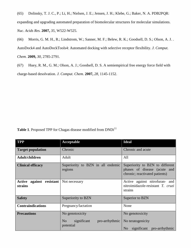
(65) Dolinsky, T. J. C., P.; Li, H.; Nielsen, J. E.; Jensen, J. H.; Klebe, G.; Baker, N. A. PDB2PQR:
expanding and upgrading automated preparation of biomolecular structures for molecular simulations.
Nuc. Acids Res. 2007, 35, W522-W525.
(66) Morris, G. M. H., R.; Lindstrom, W.; Sanner, M. F.; Belew, R. K.; Goodsell, D. S.; Olson, A. J. .
AutoDock4 and AutoDockTools4: Automated docking with selective receptor flexibility. J. Comput.
Chem. 2009, 30, 2785-2791.
(67) Huey, R. M., G. M.; Olson, A. J.; Goodsell, D. S. A semiempirical free energy force field with
charge-based desolvation. J. Comput. Chem. 2007, 28, 1145-1152.
Table 1. Proposed TPP for Chagas disease modified from DNDi11
TPP Acceptable Ideal
Target population Chronic Chronic and acute
Adult/children Adult All
Clinical efficacy Superiority to BZN in all endemic regions
Superiority to BZN to different phases of disease (acute and chronic; reactivated patients)
Active against resistant strains
Not necessary Active against nitrofuran- and nitroimidazole-resistant T. cruzi strains
Safety Superiority to BZN Superior to BZN
Contraindications Pregnancy/lactation None
Precautions No genotoxicity
No significant pro-arrhythmic potential
No genotoxicity
No teratogenicity
No significant pro-arrhythmic

potential
Presentation Oral Oral
Dosing regime Comparable to systemic antifungal treatments
Once daily for 30 days
Stability 3 years, climate zone IV 5 years, climate zone IV
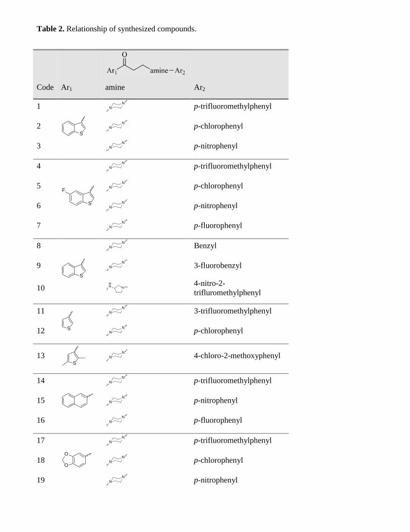
Table 2. Relationship of synthesized compounds.
Code Ar1 amine Ar2
1
p-trifluoromethylphenyl
2 p-chlorophenyl
3 p-nitrophenyl
4
p-trifluoromethylphenyl
5 p-chlorophenyl
6 p-nitrophenyl
7 p-fluorophenyl
8
Benzyl
9 3-fluorobenzyl
10 4-nitro-2-trifluromethylphenyl
11
3-trifluoromethylphenyl
12 p-chlorophenyl
13 4-chloro-2-methoxyphenyl
14
p-trifluoromethylphenyl
15 p-nitrophenyl
16 p-fluorophenyl
17
p-trifluoromethylphenyl
18 p-chlorophenyl
19 p-nitrophenyl
S
N
N
N
N
N
N
S
F
N
N
N
N
N
N
N
N
S
N
N
N
N
NHN
S
N
N
N
N
S
N
N
N
N
N
N
N
N
O
O
N
N
N
N
N
N

20 p-fluorophenyl N
N
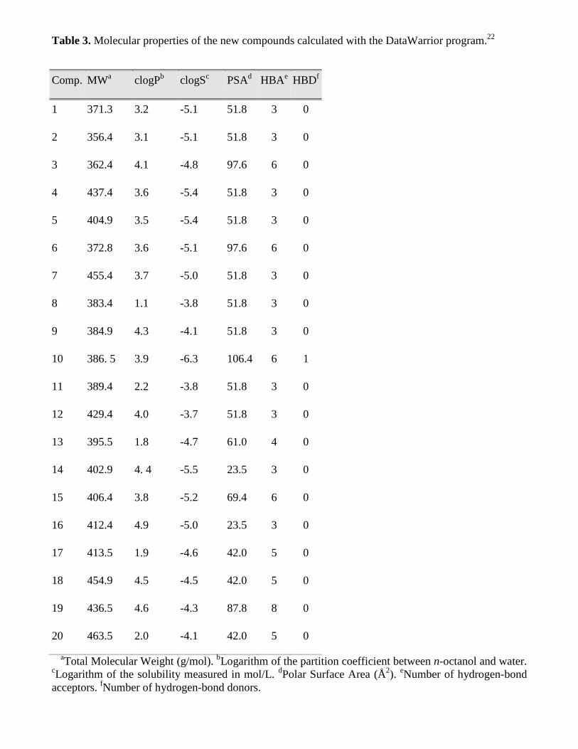
Table 3. Molecular properties of the new compounds calculated with the DataWarrior program.22
Comp. MWa clogPb clogSc PSAd HBAe HBDf
1 371.3 3.2 -5.1 51.8 3 0
2 356.4 3.1 -5.1 51.8 3 0
3 362.4 4.1 -4.8 97.6 6 0
4 437.4 3.6 -5.4 51.8 3 0
5 404.9 3.5 -5.4 51.8 3 0
6 372.8 3.6 -5.1 97.6 6 0
7 455.4 3.7 -5.0 51.8 3 0
8 383.4 1.1 -3.8 51.8 3 0
9 384.9 4.3 -4.1 51.8 3 0
10 386. 5 3.9 -6.3 106.4 6 1
11 389.4 2.2 -3.8 51.8 3 0
12 429.4 4.0 -3.7 51.8 3 0
13 395.5 1.8 -4.7 61.0 4 0
14 402.9 4. 4 -5.5 23.5 3 0
15 406.4 3.8 -5.2 69.4 6 0
16 412.4 4.9 -5.0 23.5 3 0
17 413.5 1.9 -4.6 42.0 5 0
18 454.9 4.5 -4.5 42.0 5 0
19 436.5 4.6 -4.3 87.8 8 0
20 463.5 2.0 -4.1 42.0 5 0
aTotal Molecular Weight (g/mol). bLogarithm of the partition coefficient between n-octanol and water. cLogarithm of the solubility measured in mol/L. dPolar Surface Area (Å2). eNumber of hydrogen-bond acceptors. fNumber of hydrogen-bond donors.
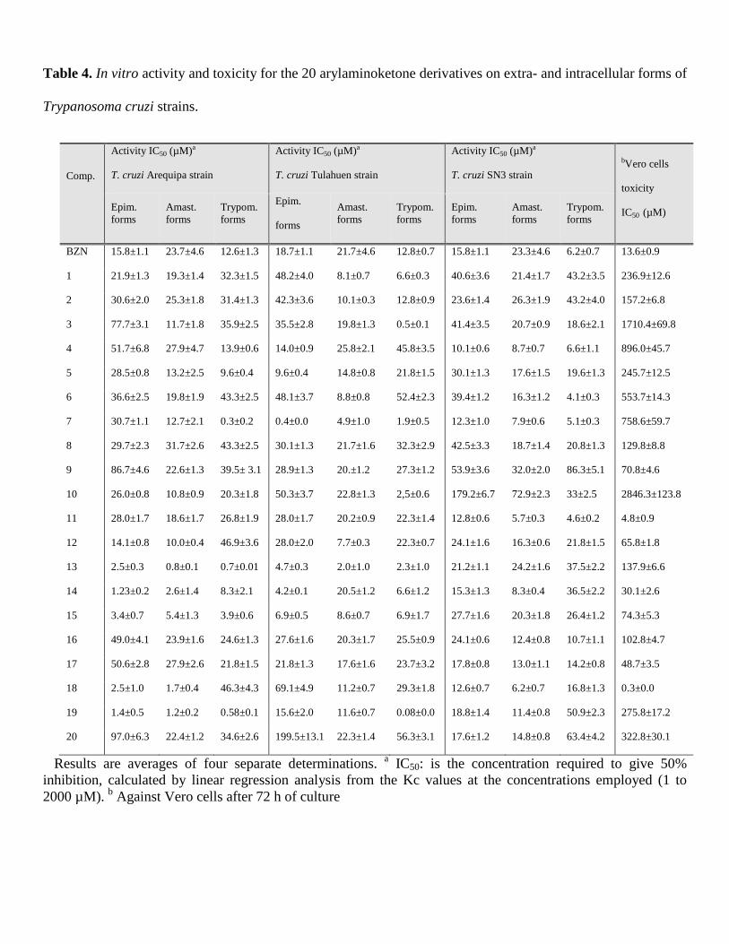
Table 4. In vitro activity and toxicity for the 20 arylaminoketone derivatives on extra- and intracellular forms of
Trypanosoma cruzi strains.
Comp.
Activity IC50 (µM)a
T. cruzi Arequipa strain
Activity IC50 (µM)a
T. cruzi Tulahuen strain
Activity IC50 (µM)a
T. cruzi SN3 strain bVero cells
toxicity
IC50 (µM) Epim. forms
Amast. forms
Trypom. forms
Epim.
forms
Amast. forms
Trypom. forms
Epim. forms
Amast. forms
Trypom. forms
BZN 15.8±1.1 23.7±4.6 12.6±1.3 18.7±1.1 21.7±4.6 12.8±0.7 15.8±1.1 23.3±4.6 6.2±0.7 13.6±0.9
1 21.9±1.3 19.3±1.4 32.3±1.5 48.2±4.0 8.1±0.7 6.6±0.3 40.6±3.6 21.4±1.7 43.2±3.5 236.9±12.6
2 30.6±2.0 25.3±1.8 31.4±1.3 42.3±3.6 10.1±0.3 12.8±0.9 23.6±1.4 26.3±1.9 43.2±4.0 157.2±6.8
3 77.7±3.1 11.7±1.8 35.9±2.5 35.5±2.8 19.8±1.3 0.5±0.1 41.4±3.5 20.7±0.9 18.6±2.1 1710.4±69.8
4 51.7±6.8 27.9±4.7 13.9±0.6 14.0±0.9 25.8±2.1 45.8±3.5 10.1±0.6 8.7±0.7 6.6±1.1 896.0±45.7
5 28.5±0.8 13.2±2.5 9.6±0.4 9.6±0.4 14.8±0.8 21.8±1.5 30.1±1.3 17.6±1.5 19.6±1.3 245.7±12.5
6 36.6±2.5 19.8±1.9 43.3±2.5 48.1±3.7 8.8±0.8 52.4±2.3 39.4±1.2 16.3±1.2 4.1±0.3 553.7±14.3
7 30.7±1.1 12.7±2.1 0.3±0.2 0.4±0.0 4.9±1.0 1.9±0.5 12.3±1.0 7.9±0.6 5.1±0.3 758.6±59.7
8 29.7±2.3 31.7±2.6 43.3±2.5 30.1±1.3 21.7±1.6 32.3±2.9 42.5±3.3 18.7±1.4 20.8±1.3 129.8±8.8
9 86.7±4.6 22.6±1.3 39.5± 3.1 28.9±1.3 20.±1.2 27.3±1.2 53.9±3.6 32.0±2.0 86.3±5.1 70.8±4.6
10 26.0±0.8 10.8±0.9 20.3±1.8 50.3±3.7 22.8±1.3 2,5±0.6 179.2±6.7 72.9±2.3 33±2.5 2846.3±123.8
11 28.0±1.7 18.6±1.7 26.8±1.9 28.0±1.7 20.2±0.9 22.3±1.4 12.8±0.6 5.7±0.3 4.6±0.2 4.8±0.9
12 14.1±0.8 10.0±0.4 46.9±3.6 28.0±2.0 7.7±0.3 22.3±0.7 24.1±1.6 16.3±0.6 21.8±1.5 65.8±1.8
13 2.5±0.3 0.8±0.1 0.7±0.01 4.7±0.3 2.0±1.0 2.3±1.0 21.2±1.1 24.2±1.6 37.5±2.2 137.9±6.6
14 1.23±0.2 2.6±1.4 8.3±2.1 4.2±0.1 20.5±1.2 6.6±1.2 15.3±1.3 8.3±0.4 36.5±2.2 30.1±2.6
15 3.4±0.7 5.4±1.3 3.9±0.6 6.9±0.5 8.6±0.7 6.9±1.7 27.7±1.6 20.3±1.8 26.4±1.2 74.3±5.3
16 49.0±4.1 23.9±1.6 24.6±1.3 27.6±1.6 20.3±1.7 25.5±0.9 24.1±0.6 12.4±0.8 10.7±1.1 102.8±4.7
17 50.6±2.8 27.9±2.6 21.8±1.5 21.8±1.3 17.6±1.6 23.7±3.2 17.8±0.8 13.0±1.1 14.2±0.8 48.7±3.5
18 2.5±1.0 1.7±0.4 46.3±4.3 69.1±4.9 11.2±0.7 29.3±1.8 12.6±0.7 6.2±0.7 16.8±1.3 0.3±0.0
19 1.4±0.5 1.2±0.2 0.58±0.1 15.6±2.0 11.6±0.7 0.08±0.0 18.8±1.4 11.4±0.8 50.9±2.3 275.8±17.2
20 97.0±6.3 22.4±1.2 34.6±2.6 199.5±13.1 22.3±1.4 56.3±3.1 17.6±1.2 14.8±0.8 63.4±4.2 322.8±30.1
Results are averages of four separate determinations. a IC50: is the concentration required to give 50% inhibition, calculated by linear regression analysis from the Kc values at the concentrations employed (1 to 2000 µM). b Against Vero cells after 72 h of culture

Table 5. Selectivity index and in vitro toxicity for the 20 derivatives of arylaminoketone intra and extracellular
of T .cruzi strains.
Comp.
Selective Indexc
T. cruzi Arequipa strain
Selective Indexc
T. cruzi Tulahuen strain
Selective Indexc
T. cruzi SN3 strain Vero cells
toxicity
IC50 (µM) Epim. forms
Amast. forms
Trypom. forms
Epim.
forms
Amast. forms
Trypom. forms
Epim. forms
Amast. forms
Trypom. forms
BZN 0.9 0.6 0.9 0.8 0.6 1.1 1.2 0.6 0.5 13.6±0.9
1 11 (12) 12 (20) 7 (8) 5 (6) 29 (49) 36 (33) 6 (5) 11 (18) 6 (11) 236.9±12.6
2 5 (6) 6 (10) 5 (6) 4 (5) 16 (26) 12 (10) 7 (6) 6 (10) 4 (7) 157.2±6.8
3 22 (25) 146 (244) 48 (53) 48 (60) 86 (144) 3420 (3109) 41 (34) 83 (138) 92 (184) 1710.4±69.8
4 17 (19) 32 (53) 864 (72) 64 (80) 35 (58) 20 (19) 89 (74) 103 (172) 136 (271) 896.0±45.7
5 9 (10) 19 (31) 26 (28) 26 (32) 17 (28) 11 (10) 8 (7) 14 (23) 12 (25) 245.7±12.5
6 15 (17) 28 (35) 13 (14) 11 (19) 63 (105) 11 (10) 14 (12) 34 (57) 135 (270) 553.7±14.3
7 25 (27) 60 (99) 2528 (2810) 1896 (3160) 155 (258) 399 (333) 62 (51) 96 (160) 149 (299) 758.6±59.7
8 4 (5) 4 (7) 3 (3) 4 (5) 6 (10) 4 (4) 3 (2) 7 (12) 6 (12) 129.8±8.8
9 1 (1) 3 (5) 2 (3) 2 (3) 3 (6) 3 (2) 1 (1) 2 (4) 1 (1.6) 70.8±4.6
10 109 (122) 263 (439) 140 (156) 57 (78) 125 (208) 1138 (1035) 16 (14) 39 (65) 87 (172) 2846.3±123.8
11 0 (0) 0 (0) 0 (0) 0 (0) 0 (0) 0 (0) 0 (0) 1 (1) 1 (2) 4.8±0.9
12 5 (5) 7 (11) 1 (2) 2 (3) 8 (14) 3 (3) 3 (2) 4 (7) 3 (6) 65.8±1.8
13 55 (61) 172 (287) 197 (219) 30 (37) 69 (115) 60 (55) 6 (5) 6 (9) 4 (7) 137.9±6.6
14 24 (27) 12 (19) 4 (4) 7 (8) 1 (2) 5 (4) 2 (2) 4 (6) 1 (2) 30.1±2.6
15 22 (24) 14 (23) 19 (21) 11 (13) 9 (14) 11 (10) 3 (2) 4 (6) 3 (6) 74.3±5.3
16 2 (2) 4 (7) 4 (5) 4 (5) 5 (8) 4 (4) 4 (4) 8 (14) 10 (19) 102.8±4.7
17 1 (1) 2 (3) 2 (2) 2 (3) 3 (5) 2 (2) 3 (2) 4 (6) 3 (7) 48.7±3.5
18 0 (0) 0 (0) 0 (0) 0 (0) 0 (0) 0 (0) 0 (0) 0 (0) 0 (0) 0.3±0.0
19 197 (219) 230 (383) 552 (528) 18 (22) 16 (26) 3447 (3134) 15 (12) 24 (40) 5 (11) 275.8±17.2
20 3 (4) 14 (24) 9 (10) 2 (2) 14 (24) 6 (5) 18 (15) 22 (36) 5 (10) 322.8±30.1
c Selectivity index =IC50 Vero cells toxicity/IC50 activity on extracellular or intracellular forms of the parasite. In brackets: number of times the compound SI exceeded the reference drug SI.

47
Table 6. Weight percent of spleens of different groups of mice.
Treatment (50 mg) Weight percent Treatment (100 mg) Weight percent
Control (untreated) 1.603 Control (untreated) 1.603
3 1.174 BZN 0.837
4 1.621 3 0.729
7 1.061 4 1.041
10 1.171 7 0.825
10 0.892

48
Table 7. Results of the Ames test of compound 4.
Treatment
Preincubation assay
Mean number of revertant colonies per plate
TA98 TA100 TA102 TA1535 TA1537
-S9 +S9 -S9 +S9 -S9 +S9 -S9 +S9 -S9 +S9
Negative control 12±4 12±0 114±24 119±6 413±80 420±130 10±1 8±3 2±2 4±1
Test dose (µg/plate)
0.03 15±1 12±1 90±20 128±8 532±69 550±65 13±2 11±2 2±1 2±1
0.08 16±3 15±2 100±3 131±18 484±61 504±25 13±3 11±5 3±2 4±3
0.25 14±2 10±5 107±13 120±16 459±28 553±85 11±4 16±2 4±3 4±2
0.75 16±1 12±2 109±13 136±4 473±42 546±63 13±4 8±1 2±1 2±1
2.25 14±2 11±3 110±12 139±4 487±29 540±76 9±1 12±3 4±3 2±2
Positive control 1608±352 1250±85 704±9 1900±164 739±36 1142±148 151±8 239±69 791±32 243±18
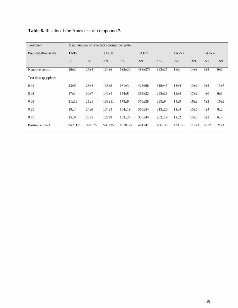
49
Table 8. Results of the Ames test of compound 7.
Treatment
Preincubation assay
Mean number of revertant colonies per plate
TA98 TA100 TA102 TA1535 TA1537
-S9 +S9 -S9 +S9 -S9 +S9 -S9 +S9 -S9 +S9
Negative control 16±3 27±4 134±6 135±20 483±279 363±27 16±1 14±3 6±3 9±1
Test dose (µg/plate)
0.01 23±5 23±4 138±3 161±3 423±58 319±45 18±4 13±2 9±2 13±5
0.03 17±1 30±7 140±4 154±8 365±22 298±23 12±4 17±2 4±0 6±1
0.08 21±13 25±1 138±11 175±9 378±28 265±6 14±5 16±2 7±2 10±2
0.25 19±4 24±6 134±4 164±19 363±54 313±20 11±4 15±5 4±4 8±2
0.75 15±6 28±5 128±9 153±27 356±44 263±19 12±2 15±8 6±2 6±4
Positive control 962±131 890±76 595±35 1078±79 491±41 486±33 423±53 112±3 79±2 21±4
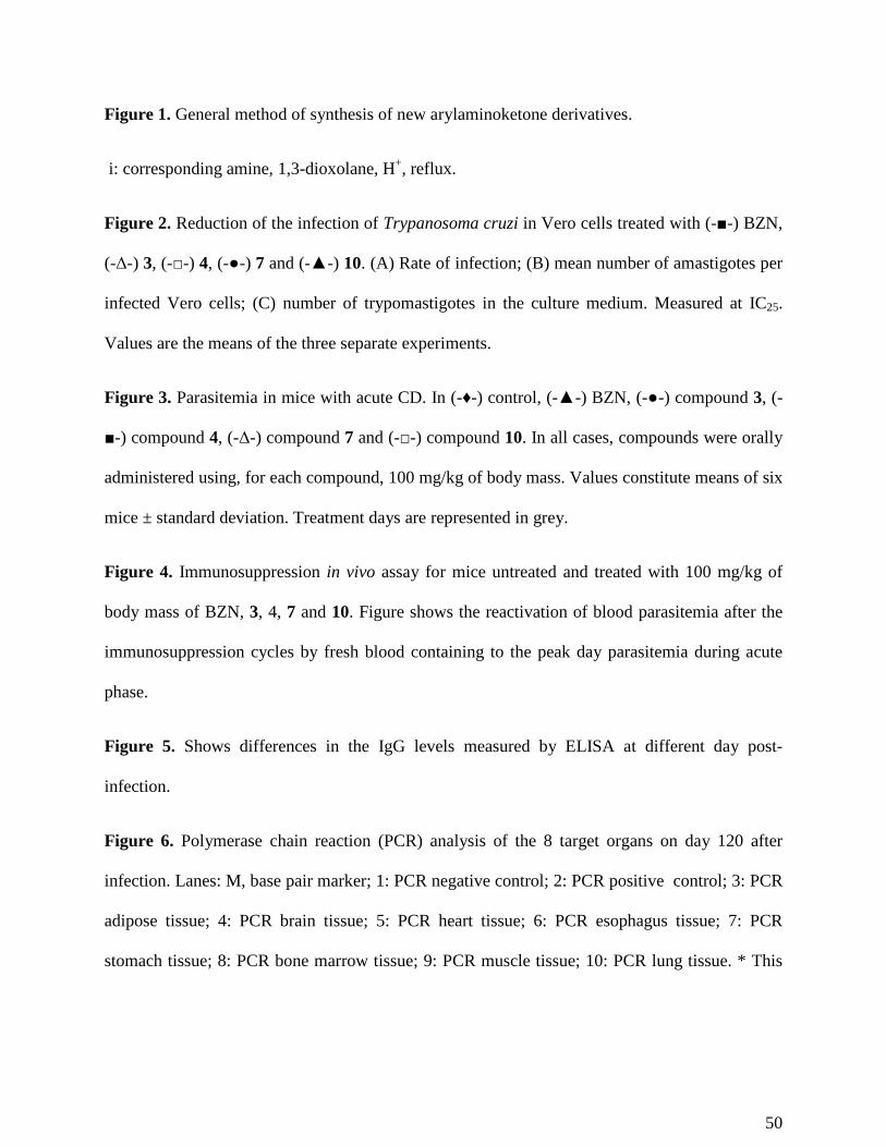
50
Figure 1. General method of synthesis of new arylaminoketone derivatives.
i: corresponding amine, 1,3-dioxolane, H+, reflux.
Figure 2. Reduction of the infection of Trypanosoma cruzi in Vero cells treated with (-■-) BZN,
(-Δ-) 3, (-□-) 4, (-●-) 7 and (-▲-) 10. (A) Rate of infection; (B) mean number of amastigotes per
infected Vero cells; (C) number of trypomastigotes in the culture medium. Measured at IC25.
Values are the means of the three separate experiments.
Figure 3. Parasitemia in mice with acute CD. In (-♦-) control, (-▲-) BZN, (-●-) compound 3, (-
■-) compound 4, (-Δ-) compound 7 and (-□-) compound 10. In all cases, compounds were orally
administered using, for each compound, 100 mg/kg of body mass. Values constitute means of six
mice ± standard deviation. Treatment days are represented in grey.
Figure 4. Immunosuppression in vivo assay for mice untreated and treated with 100 mg/kg of
body mass of BZN, 3, 4, 7 and 10. Figure shows the reactivation of blood parasitemia after the
immunosuppression cycles by fresh blood containing to the peak day parasitemia during acute
phase.
Figure 5. Shows differences in the IgG levels measured by ELISA at different day post-
infection.
Figure 6. Polymerase chain reaction (PCR) analysis of the 8 target organs on day 120 after
infection. Lanes: M, base pair marker; 1: PCR negative control; 2: PCR positive control; 3: PCR
adipose tissue; 4: PCR brain tissue; 5: PCR heart tissue; 6: PCR esophagus tissue; 7: PCR
stomach tissue; 8: PCR bone marrow tissue; 9: PCR muscle tissue; 10: PCR lung tissue. * This

51
means that 1/6 of the corresponding organ PCR products showed 300 bp band on
electrophoresis.
Figure 7. A) Inhibition in vitro (%) of Fe-SOD from epimastigotes of Trypanosoma cruzi for
compounds (activity 25.4 ± 3.1 U/mg). B) Inhibition in vitro (%) of CuZn-SOD from human
erythrocytes for compounds (activity 23.36± 2.5 U/mg). Activity differences in the control
homogenate vs. the sample incubated using compounds were identified by the Newman-Keuls
test. Values are the average of three separate rate determinations.
Figure 8. Percentages of variation among peaks of catabolites excreted by epimastigotes of
Trypanosoma cruzi exposed to compounds 3, 4, 7 and 10 at their IC25 in comparison to a control
incubated 96 h.
Figure 9. Docking results for the binding of benzothiophenes derivatives 3 (up-left), 4 (down-
left), 7 (up-right) and 10 (down-right) to the Trypanosoma cruzi Fe-SOD (a) and the human
CuZn-SOD (b) enzymes. In the center, an image of the entire enzyme with the compounds
binding at the top of the dimer interface is shown. Residues forming the inhibitor binding site are
shown. Color scheme: oxygen (red), nitrogen (blue), sulfur (yellow), carbon (pale pink for
compound 3, orange for compound 4, blue for compound 7 and magenta for compound 10. For
the proteins, one monomer is shown in green and the other in yellow). The ions are shown as
spheres. Figures were created with The PyMOL Molecular Graphics System, Version 1.8
Schrödinger, LLC.

52
Figure 1. General method of synthesis of new arylaminoketone derivatives.
i: corresponding amine, 1,3-dioxolane, H+, reflux.
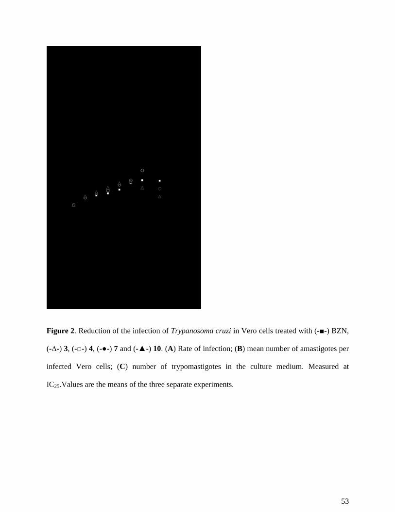
53
Figure 2. Reduction of the infection of Trypanosoma cruzi in Vero cells treated with (-■-) BZN,
(-Δ-) 3, (-□-) 4, (-●-) 7 and (-▲-) 10. (A) Rate of infection; (B) mean number of amastigotes per
infected Vero cells; (C) number of trypomastigotes in the culture medium. Measured at
IC25.Values are the means of the three separate experiments.

54
Figure 3. Parasitemia in mice with acute CD. In (-♦-) control, (-▲-) BZN, (-●-) compound 3, (-
■-) compound 4, (-Δ-) compound 7 and (-□-) compound 10. In all cases, compounds were orally
administered using, for each compound, 100 mg/kg of body mass. Values constitute means of six
mice ± standard deviation. Treatment days are represented in grey.
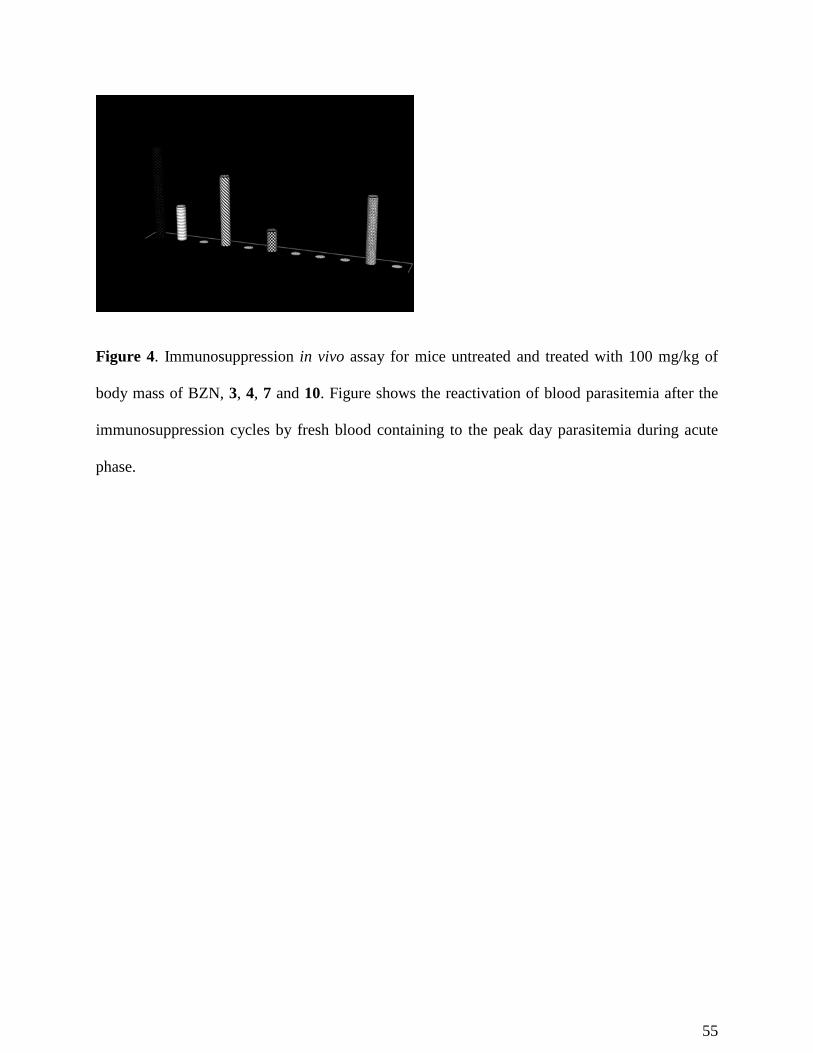
55
Figure 4. Immunosuppression in vivo assay for mice untreated and treated with 100 mg/kg of
body mass of BZN, 3, 4, 7 and 10. Figure shows the reactivation of blood parasitemia after the
immunosuppression cycles by fresh blood containing to the peak day parasitemia during acute
phase.
56
Figure 5. Shows differences in the IgG levels measured by ELISA at different day post-
infection.
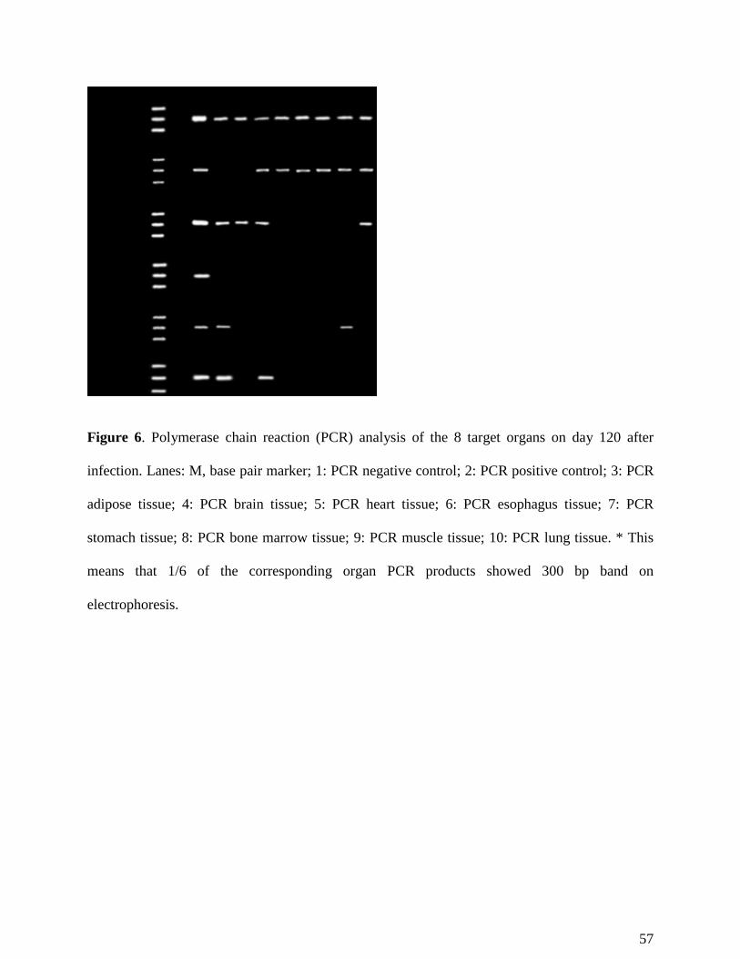
57
Figure 6. Polymerase chain reaction (PCR) analysis of the 8 target organs on day 120 after
infection. Lanes: M, base pair marker; 1: PCR negative control; 2: PCR positive control; 3: PCR
adipose tissue; 4: PCR brain tissue; 5: PCR heart tissue; 6: PCR esophagus tissue; 7: PCR
stomach tissue; 8: PCR bone marrow tissue; 9: PCR muscle tissue; 10: PCR lung tissue. * This
means that 1/6 of the corresponding organ PCR products showed 300 bp band on
electrophoresis.
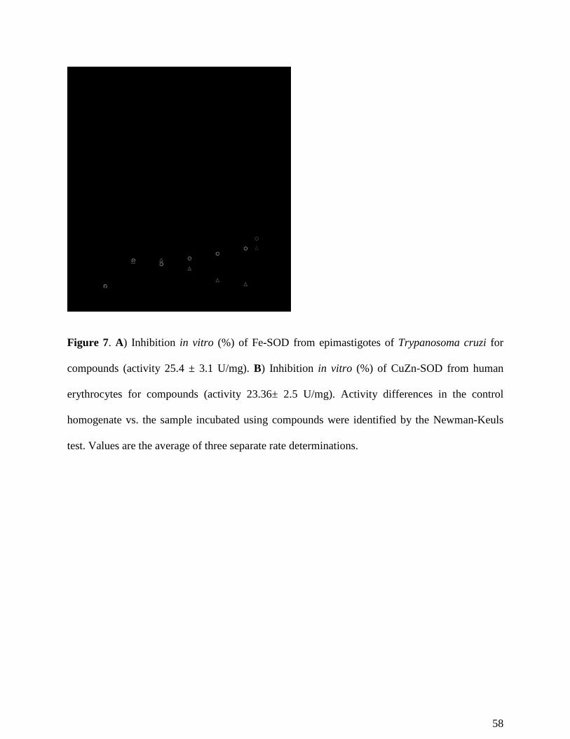
58
Figure 7. A) Inhibition in vitro (%) of Fe-SOD from epimastigotes of Trypanosoma cruzi for
compounds (activity 25.4 ± 3.1 U/mg). B) Inhibition in vitro (%) of CuZn-SOD from human
erythrocytes for compounds (activity 23.36± 2.5 U/mg). Activity differences in the control
homogenate vs. the sample incubated using compounds were identified by the Newman-Keuls
test. Values are the average of three separate rate determinations.
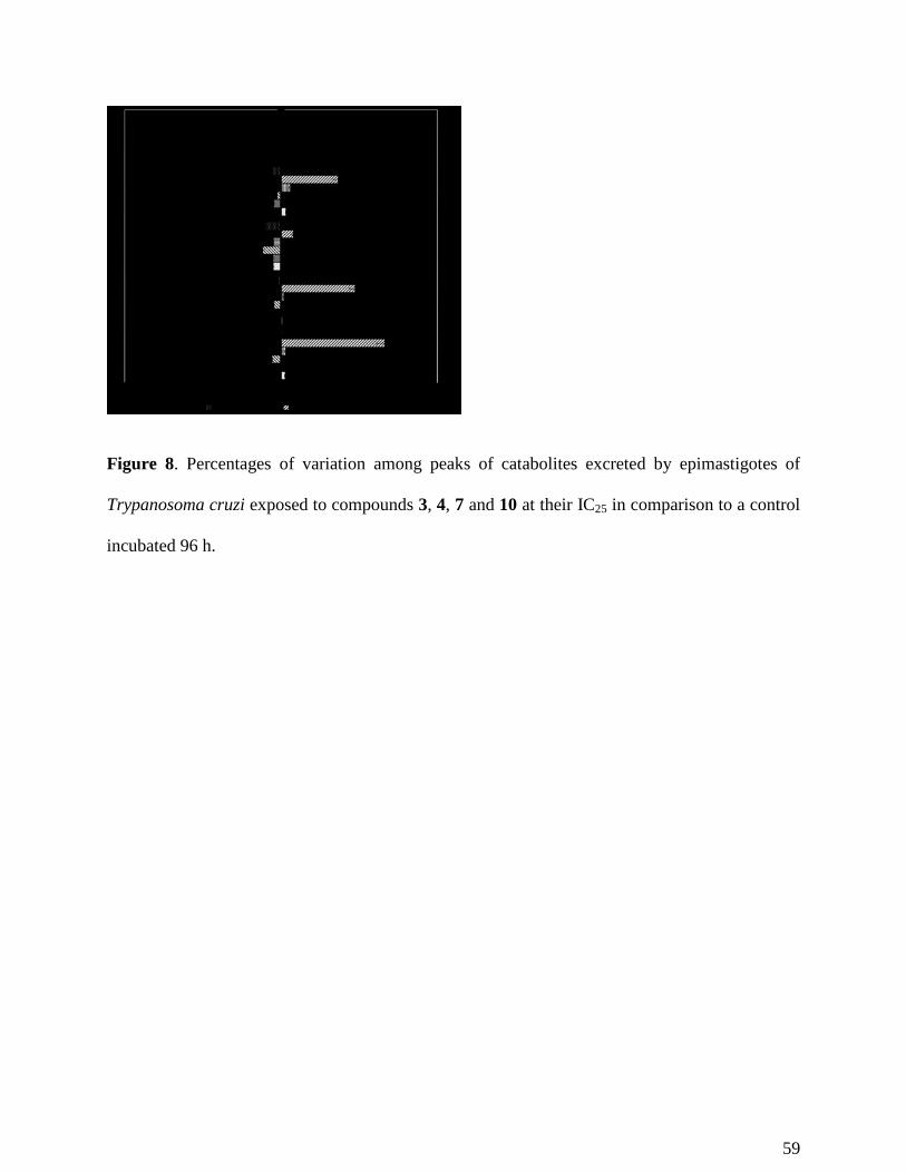
59
Figure 8. Percentages of variation among peaks of catabolites excreted by epimastigotes of
Trypanosoma cruzi exposed to compounds 3, 4, 7 and 10 at their IC25 in comparison to a control
incubated 96 h.
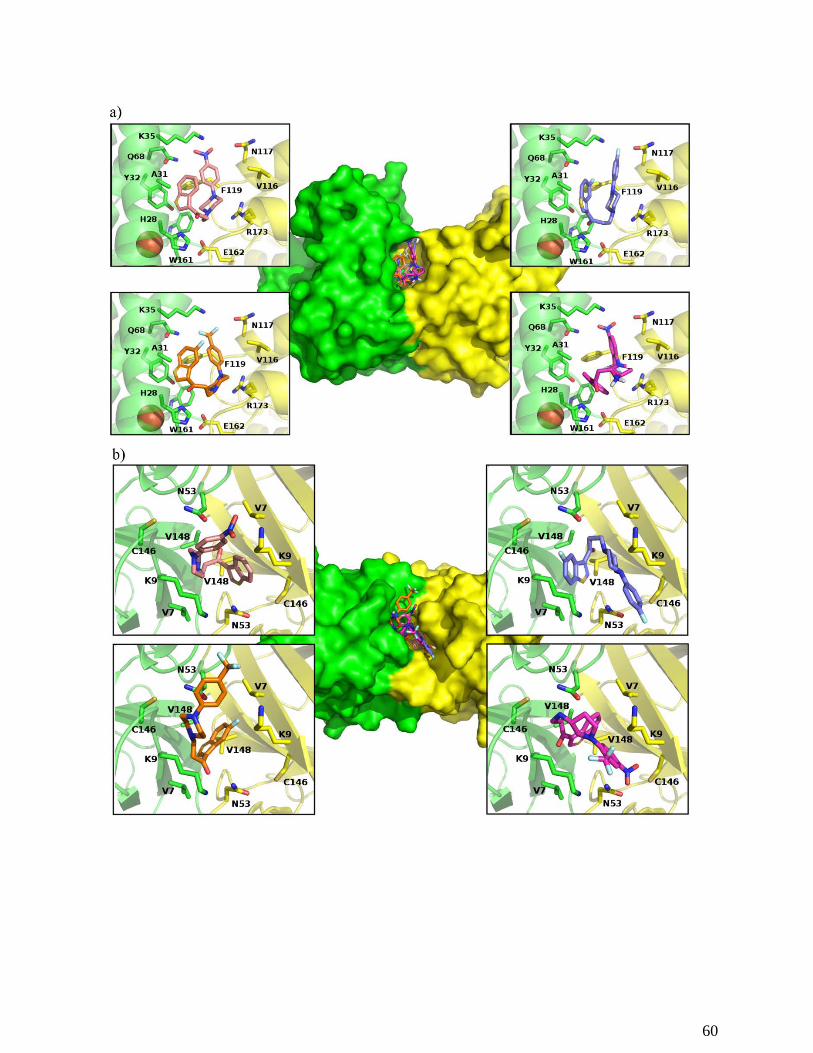
60
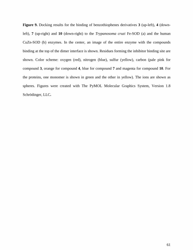
61
Figure 9. Docking results for the binding of benzothiophenes derivatives 3 (up-left), 4 (down-
left), 7 (up-right) and 10 (down-right) to the Trypanosoma cruzi Fe-SOD (a) and the human
CuZn-SOD (b) enzymes. In the center, an image of the entire enzyme with the compounds
binding at the top of the dimer interface is shown. Residues forming the inhibitor binding site are
shown. Color scheme: oxygen (red), nitrogen (blue), sulfur (yellow), carbon (pale pink for
compound 3, orange for compound 4, blue for compound 7 and magenta for compound 10. For
the proteins, one monomer is shown in green and the other in yellow). The ions are shown as
spheres. Figures were created with The PyMOL Molecular Graphics System, Version 1.8
Schrödinger, LLC.
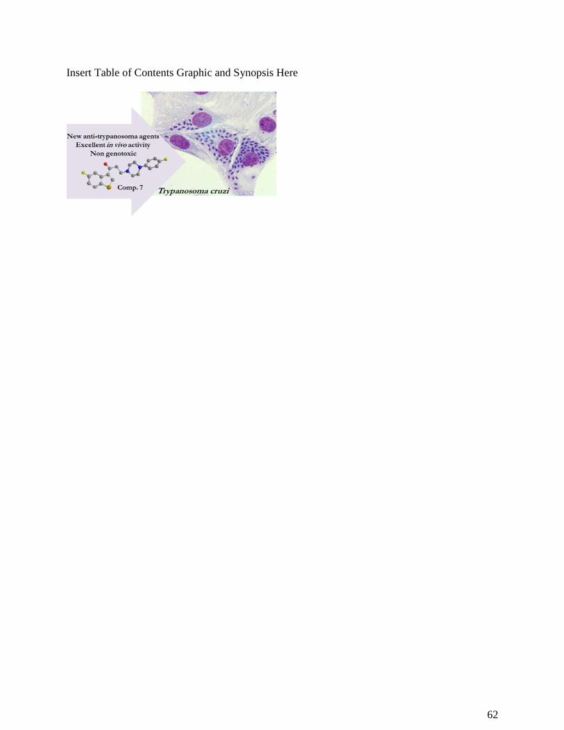
62
Insert Table of Contents Graphic and Synopsis Here
Copyright © 2022 FDOKUMEN