Camel Brucellosis: Evaluation of field sera by conventional serological tests and ELISA.

BioMed CentralParasites & Vectors
ss
Open AcceResearchDifferent serological cross-reactivity of Trypanosoma rangeli forms in Trypanosoma cruzi-infected patients seraMilene H de Moraes1, Alessandra A Guarneri1,2, Fabiana P Girardi1, Juliana B Rodrigues1, Iriane Eger1,3, Kevin M Tyler4, Mário Steindel1 and Edmundo C Grisard*1,4Address: 1Departamento de Microbiologia e Parasitologia, Universidade Federal de Santa Catarina, 88040-900, Florianópolis, Santa Catarina, Brazil, 2Instituto René Rachou, Fiocruz, Belo Horizonte, Minas Gerais, Brazil, 3Universidade do Vale do Itajaí, Itajaí, Santa Catarina, Brazil and 4Biomedical Research Centre, School of Medicine, Health Policy and Practice, University of East Anglia, Norwich, Norfolk, UK
Email: Milene H de Moraes - [email protected]; Alessandra A Guarneri - [email protected]; Fabiana P Girardi - [email protected]; Juliana B Rodrigues - [email protected]; Iriane Eger - [email protected]; Kevin M Tyler - [email protected]; Mário Steindel - [email protected]; Edmundo C Grisard* - [email protected]
* Corresponding author
AbstractBackground: American Trypanosomiasis or Chagas disease is caused by Trypanosoma cruzi whichcurrently infects approximately 16 million people in the Americas causing high morbidity andmortality. Diagnosis of American trypanosomiasis relies on serology, primarily using indirectimmunofluorescence assay (IFA) with T. cruzi epimastigote forms. The closely related butnonpathogenic Trypanosoma rangeli has a sympatric distribution with T. cruzi and is carried by thesame vectors. As a result false positives are frequently generated. This confounding factor leads toincreased diagnostic test costs and where false positives are not caught, endangers human healthdue to the toxicity of the drugs used to treat Chagas disease.
Results: In the present study, serologic cross-reactivity between the two species was comparedfor the currently used epimastigote form and the more pathologically relevant trypomastigoteform, using IFA and immunoblotting (IB) assays. Our results reveal an important decrease in crossreactivity when T. rangeli culture-derived trypomastigotes are used in IFA based diagnosis of Chagasdisease. Western blot results using sera from both acute and chronic chagasic patients presentingwith cardiac, indeterminate or digestive disease revealed similar, but not identical, antigenicprofiles.
Conclusion: This is the first study addressing the serological cross-reactivity between distinctforms and strains of T. rangeli and T. cruzi using sera from distinct phases of the Chagasic infection.Several T. rangeli-specific proteins were detected, which may have potential as diagnostic tools.
BackgroundTrypanosoma rangeli and Trypanosoma cruzi are closelyrelated and sympatric protozoan parasites that infect tri-
atomine bugs, humans and a variety of sylvatic anddomestic mammals in overlapping regions of both Southand Central America [1-3]. T. rangeli is nonpathogenic to
Published: 8 July 2008
Parasites & Vectors 2008, 1:20 doi:10.1186/1756-3305-1-20
Received: 11 June 2008Accepted: 8 July 2008
This article is available from: http://www.parasitesandvectors.com/content/1/1/20
© 2008 de Moraes et al; licensee BioMed Central Ltd. This is an Open Access article distributed under the terms of the Creative Commons Attribution License (http://creativecommons.org/licenses/by/2.0), which permits unrestricted use, distribution, and reproduction in any medium, provided the original work is properly cited.
Page 1 of 10(page number not for citation purposes)

Parasites & Vectors 2008, 1:20 http://www.parasitesandvectors.com/content/1/1/20
humans while T. cruzi is the etiological agent of Chagasdisease and infects over 16 million people in the newworld [4]. Unsurprisingly, given the sympatric distribu-tion and shared vector and host range, mixed infectionsare observed [1-3].
T. rangeli is considered a parasite of biological and epide-miological interest due to the serological cross-reactivitywith T. cruzi, which has been the subject of great contro-versy [5-8]. Several reports pointed out the sharing of anti-genic epitopes by T. cruzi and T. rangeli [9-12]. However,these studies were performed with sera from animalsimmunized with T. rangeli epimastigotes rather thaninfected or immunized with the trypomastigote formspresent in infected hosts [9-12]. It is widely accepted tohave an outstanding but underestimated impact on thediagnosis of Chagas disease [2,9-14].
A recent study on T. cruzi serodiagnosis reported theabsence of cross-reaction of T. rangeli-infected patient'ssera in some commercially available ELISA tests [15].Despite the fact that PCR-based diagnosis can provideaccurate discrimination of T. cruzi from T. rangeli [2,3],neither method is widespread in the diagnostic laborato-ries of endemic areas where IFA of cultured T. cruzi epi-mastigote forms with patient sera remains the screeningmethod of choice [16-18]. The use of at least two serolog-ical tests based on different methods, mandatory in Brazilfor almost two decades now [17], have enhanced the sen-sitivity detection. Comparative studies of the antigeniccomposition of culture-derived T. cruzi and T. rangeli epi-mastigote forms indicate that these parasites shareapproximately 60% of their soluble antigenic composi-tion [5,7,11] often resulting in a significant level of incon-clusive results [17].
Epimastigote forms are not seen in the blood of infectedindividuals and are therefore not the form against whichantibody develops, rather the forms seen on blood smearsare normally the trypomastigote forms. We consideredtherefore, whether serum screening using trypomastigotesfrom culture, rather than epimastigotes, would help toreduce misdiagnosis. We extended these studies to exam-ine the expression of cross-reactive proteins between lifecycle stages in order to reveal candidate proteins formolecular diagnostics, pathogenicity factors and possiblevaccines.
ResultsIndependent of the clinical form, all chagasic sera testedreacted with both T. cruzi and T. rangeli epimastigotes inIFA (Figs. 1 and 2). Considering reactions with titers over1:40 as positive, cross-reactions were observed for 88.46%of the serum from patients with the cardiac form of theChagas disease (Fig. 2). Interestingly, the use of T. rangeli
trypomastigote forms as antigen reduced the cross-reactiv-ity of serum from patients with the cardiac form of theChagas disease to 30.76% (8/26). The titres as well as thefluorescence intensities were variable depending on theparasite species and/or strains used but, irrespective of theform used as antigen, higher titres were obtained for T.cruzi strains (Fig. 2).
Except for the distinct and significant reactivity (p < 0.05)of serum from patients with the cardiac form of the Cha-gas disease with T. cruzi trypomastigotes from the CL or Ystrains, no other significant intra-specific variation of thereaction titres (p > 0.05) was observed among the strainsstudied and the fluorescence pattern revealed was homo-geneous (Fig. 2). However, inter-specific comparisonsrevealed a variable fluorescence pattern and significantlydifferent (p < 0.001) reaction titres between T. cruzi and T.rangeli strains for the tested sera, especially when usingtrypomastigotes as antigens (Fig. 2).
A representation of the fluorescence patterns observed inIFA is shown in Figure 1. Both T. cruzi forms showed a flu-orescence pattern varying from homogeneous cellular,including nucleus and kinetoplast when tested againstsera from patients with indeterminate form of the disease(data not shown), to strong plasma membrane labelingalone when tested with serum from patients with cardiacinvolvement (Fig. 1). Sera from all chagasic patientsshowed strong membrane fluorescence when testedagainst T. rangeli epimastigotes (Fig. 1) although, serafrom the indeterminate form patients that reacted with T.cruzi epimastigote forms at 1:40 or 1:80 dilutions resultedin negative results when tested against T. rangeli epimas-tigotes at these concentrations (Data not shown).
As expected, chagasic sera gave the strongest reaction withT. cruzi trypomastigotes regardless of patient clinical his-tory (Fig. 2). The use of trypomastigote forms from bothspecies in IFA showed a significant decrease (p < 0.001) incross-reactivity when compared to the results obtained forepimastigote forms (Fig. 2). Also, a significant intra-spe-cific variation (p < 0.05) of the reaction titers wasobserved among epimastigotes of the distinct T. cruzistrains according to the testing sera (cardiac or indetermi-nate), which was not observed among the trypomastigoteforms (Fig. 2).
Sera from chronic chagasic patients (cardiac or indetermi-nate forms) showing borderline titers of 1:40 or 1:80against T. cruzi epimastigotes, which did not react with T.rangeli epimastigotes, revealed negative results in IFA withboth T. rangeli and T. cruzi trypomastigotes.
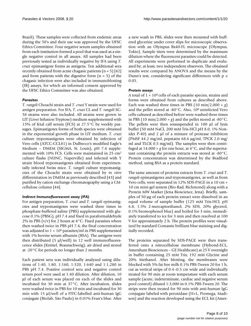
Comparative SDS-PAGE analysis revealed a protein pat-tern varying from 24 to 110 kDa for both Trypanosoma spe-
Page 2 of 10(page number not for citation purposes)

Parasites & Vectors 2008, 1:20 http://www.parasitesandvectors.com/content/1/1/20
cies (Fig. 3A). Epimastigote forms gave similar patterns,sharing a large number of prominent and faint bands. Fortrypomastigotes though, it was possible to visualize differ-ences in the electrophoretic profiles (Fig. 3A, lanes 2 and4). No major differences were observed among the pro-files of both T. rangeli forms, however, the profile of T.cruzi epimastigotes revealed two major proteins around36 and 52 kDa, as also observed on both T. rangeli forms,which were present in much lower intensity on the T. cruzitrypomastigotes profile (Fig. 3A).
Immunoblot analysis using all acute and chronic chagasicpatient sera recognized a wide range of proteins in bothparasite species and strains (data not shown), revealing aquite distinct inter-specific pattern. A representation ofthe obtained results is shown on Fig. 3B. Sera frompatients in the acute phase of Chagas disease recognizedfewer proteins on T. rangeli extracts than sera from chronicchagasic patients. Two proteins of approximately 32 and40 kDa were strongly recognized by chagasic sera (acuteand chronic) on T. cruzi extracts (Fig. 3B, dark arrows), but
were not detected in T. rangeli lysates by acute sera. A 32kDa protein was, though, weakly recognized in chronicpatient sera. Also, some intra-specific differences on anti-genic profile between T. cruzi Y strain epimastigote andtrypomastigote forms were observed (Fig. 3B).
Signal intensity for several T. rangeli antigens was variablebetween chagasic sera. Interestingly, though, a very strongband of approximately 35 kDa was recognized in both T.rangeli forms by sera from chronic patients (Fig. 3B, whitearrow).
We further considered whether chagasic sera from differ-ent pathologies showed similar antigenic profiles by uti-lizing sera from patients presenting the cardiac,indeterminate or digestive forms of the disease (Fig. 3C).Our results revealed very similar profiles for T. cruzi epi-mastigotes and trypomastigotes but a quite distinct pro-tein patterns for T. rangeli epimastigotes andtrypomastigotes, independently of the tested sera. Impor-tantly, a 35 kDa protein was recognized strongly by all
The use of trypomastigotes give better specificity than epimastigotes by IFAFigure 1The use of trypomastigotes give better specificity than epimastigotes by IFA. Indirect immunofluorescence assay using Trypanosoma cruzi and Trypanosoma rangeli trypomastigote or epimastigote forms as antigens and serum (1:80) from a chronic chagasic patient with the cardiac form of the disease. Negative controls consist of epimastigotes and non-infected patient serum (1:40).
Try
pan
oso
ma
cru
ziT
ryp
ano
som
a ra
ng
eli
EpimastigoteTrypomastigote Negative control
Page 3 of 10(page number not for citation purposes)

Parasites & Vectors 2008, 1:20 http://www.parasitesandvectors.com/content/1/1/20
chagasic sera (Fig. 3C, white arrows). Two proteins ofaround 11 and 15 kDa were recognized by the cardiac anddigestive serum in both T. rangeli forms, but not by theindeterminate sera (Fig. 3C, black arrows).
DiscussionDespite the proven cross-reactions and the existence ofmore sensitive and reliable techniques for diagnosis suchas ELISA associated to the use of recombinant antigens,IFA is still the most widely used method for Chagas dis-ease diagnosis in Brazil, as in several Latin AmericanCountries [16-18]. Thus, in the present study the serolog-ical cross-reactivity between T. cruzi and T. rangeli, epimas-tigote and trypomastigote forms, was evaluated by IFAand IB assays using sera of both acute and chronic cha-gasic patients with different clinical manifestations.
Former studies have shown the serological cross-reactivitybetween T. cruzi and T. rangeli epimastigotes by IFA, ELISAand/or IB assays, pointing that such cross-reactivity is duethe great number of soluble antigens shared by both par-asites [5,7,9-12,19]. Furthermore, sera from T. cruzi-infected patients can cross-react with other trypanosoma-tid species such as T. evansi [20] and Leishmania spp. [15]as demonstrated by western-blot, ELISA and TESA-Blot,including some commercially available kits.
In the present study we have confirmed cross-reactivity ina range of distinct serum samples and also observed thatT. cruzi and T. rangeli epimastigotes reacted with all testedsera regardless of the clinical presentation of the patient.However, the fluorescence intensity and pattern producedby the sera in IFA on parasites cells appeared to be variable
Trypomastigotes discriminate Trypanosoma cruzi from Trypanosoma rangeli over an increased titre rangeFigure 2Trypomastigotes discriminate Trypanosoma cruzi from Trypanosoma rangeli over an increased titre range. Com-parison of antibody titers obtained for chronic chagasic patients serum with cardiac or indeterminate forms tested against epi-mastigote or trypomastigote forms of T. cruzi Y (open bar) and CL (dotted bar) strains and T. rangeli Choachi (vertical lined bar) and SC-58 (horizontal lined bar) strains in indirect immunofluorescence assays. Experiments were performed in duplicates and vertical bars above data indicate mean standard error. Horizontal dotted lines indicate the 1:40 cut-off.
Cardiac serum Indeterminate serumE
pim
asti
go
teT
ryp
om
asti
go
te
Y CL SC CH0
250
500
750
1000
1250
T. cruzi
T. rangeli
Strains
Ant
ibod
y ti
ters
Y CL SC CH0
250
500
750
1000
1250
T. cruzi
T. rangeli
Strains
Ant
ibo
dy t
iter
s
Y CL SC CH0
250
500
750
1000
1250
T. cruzi
T. rangeli
*
*
Strains
An
tib
ody
tit
ers
Y CL SC CH0
250
500
750
1000
1250
T. cruzi
T. rangeli
Strains
An
tib
od
y ti
ters
Page 4 of 10(page number not for citation purposes)

Parasites & Vectors 2008, 1:20 http://www.parasitesandvectors.com/content/1/1/20
depending on the parasite species and life-cycle stage andupon the type of serum used. Such differences may beattributed both to the distinct antibody profiles of thesera, which in turn can be related to the disease form and/or to the patient immune response, as well as to the differ-ences in gene expression and antigenic composition of theparasites themselves.
It is well established that distinct profiles of anti-T. cruziantibodies may be elicited by patients with different clin-ical presentations of the disease [21-23]. This variableresponse pattern can be due to a variety of immuneresponse related mechanisms, such as the regulation ofimmunoglobulin expression. Such differences can pre-sumably influence the pathogenesis of disease and hence
Immunoblot profiles of Trypanosoma cruzi (Y strain) and Trypanosoma rangeli (Choachi strain) with chagasic seraFigure 3Immunoblot profiles of Trypanosoma cruzi (Y strain) and Trypanosoma rangeli (Choachi strain) with chagasic sera. Total protein extracts resolved in a 12% SDS-PAGE and stained by Comassie brilliant blue (A) and the immunoblot anal-ysis of the same lysates using sera from acute and chronic chagasic patients (B) or using sera from chronic chagasic patients with the cardiac, indeterminate or digestive forms of the disease (C). On panel B, arrows indicates proteins recognized by cha-gasic sera (acute and/or chronic) on T. cruzi (dark arrows) or on T. rangeli (white arrow) extracts. Arrows in panel C indicates a T. rangeli 11 and 15 kDa proteins exclusively recognized by the cardiac and digestive serum in both T. rangeli forms (dark arrows) and a 35 kDa protein (white arrows) recognized by all sera from T. cruzi-infected patients. Lanes 1 and 2 = T. cruzi Y strain epimastigote and trypomastigote forms and lanes 3 and 4 = T. rangeli Choachi strain epimastigote and trypomastigote forms, respectively. Lane 5 = Vero cells extract; MW = molecular weight marker (kDa). Asterisks indicates significant differ-ences (p < 0.05).
37.6
28.5
18.4
116.0
66.2
MW 1 2 3 4
A
1 2 3 4 5 1 2 3 4 5 1 2 3 4 5 1 2 3 4 5
22010070
50
20
30
NegativeDigestiveIndeterminateCardiac
MW
C
1 2 3 4 5 1 2 3 4 5 1 2 3 4 5MW
116.0
66.2
37.6
28.5
18.4
Acute Chronic NegativeB
Page 5 of 10(page number not for citation purposes)

Parasites & Vectors 2008, 1:20 http://www.parasitesandvectors.com/content/1/1/20
it clinical presentation but also have implications for theeffectiveness of diagnostic serology [21,23,24].
Despite the antigenic similarity of T. cruzi and T. rangeli,several studies have described species-specific polypep-tides allowing specific differentiation of these parasites[25-27]. Our observations account for both the inter- andintra-specific differences observed in IFA, identifying avariety of antigens which are specific not only to speciesbut also to strains and life cycle stages.
Even considering the antigenic differences to blood trypo-mastigotes, the use of culture-derived trypomastigotes inIFAs in this study significantly reduced the serologicalcross-reactivity between T. cruzi and T. rangeli (Figs. 1 and2). It is clear also, though, that even in trypomastigoteforms significant numbers of positive reactive antigensremain (Figs. 3B and 3C).
Regardless of the clinical presentation from which it isobtained, sera from chronic chagasic patients revealed asignificantly more intense reaction (p < 0.01) against T.cruzi trypomastigotes in IFA when compared with epimas-tigote forms (Figs. 1 and 2). This difference can beexplained by the antibody specificity, since anti-trypo-mastigote antibodies present on the evaluated sera wereproduced against blood trypomastigotes during T. cruziinfection.
Sera from patients with the indeterminate form of Chagasdisease that showed lower titers (1:40 – 1:80) against T.cruzi epimastigotes did not recognize T. cruzi and T. rangelitrypomastigote forms (Fig. 2). Since 76% of the sera fromchagasic patients revealed titres equal or in excess of1:640, but only 1.4% showed titers below 1:160, andsince all sera with high titres reacted with both epimastig-ote and trypomastigote forms of T. cruzi and T. rangeli weconclude that sera with titers below 1:40 are most likely tobe false-positive results.
Thus, considering that Chagas disease diagnosis is prima-rily based in IFA and/or ELISA assays and that variableresult may be obtained depending on the antigens andmethods used [7,15,20] the use of culture-derived trypo-mastigote forms as antigens could reduce misdiagnosis ofthe disease and the related disability-adjusted life years orDALY's [28,29].
Nevertheless, the use of such infective forms may repre-sent an increased risk to those culturing the organisms aswell as an increase in the costs of the diagnostic exams.These drawbacks reinforce the need to generate recom-binant antigens or synthetic peptides for specific diagno-sis [15].
Previous studies have pointed some species-specific anti-gens allowing specific detection of T. cruzi [30-32] or dif-ferentiation of T. cruzi and T. rangeli [7,25,26] while otherantigens seem to be shared by distinct Trypanosoma spe-cies. Among these studies, Saldaña et al. [26,27] havecompared total epimastigote extracts from T. cruzi and T.rangeli by SDS-PAGE and pointed out T. rangeli exclusiveproteins of approximately 93, 77-73, 63, 54-53 and 48kDa.
In our present work we have used sera from both acutepatients and chronic chagasic patients with distinct clini-cal presentations (Figs. 3B and 3C), as well as differentparasite life-cycle stages (epimastigotes and trypomastig-otes), being the first comparative analysis of trypomastig-ote antigens of both species.
In order to identify possible species-specific antigenspresent in both T. cruzi and T. rangeli life-cycle stages, IBassays were initially performed using sera from patientsacutely and chronically infected with T. cruzi. These exper-iments addressed the possibility of discriminating stage-specific or clinical presentation-specific antigens. Theresults indicated at least two proteins present in both T.cruzi life cycle stages but were absent in T. rangeli (Fig. 3B)and that were strongly recognized by acute and chronicsera. These proteins of approximately 32 and 40 kDa,could represent good targets for the development of spe-cific recombinant antigens, improving the diagnosis of T.cruzi in relation to T. rangeli. In addition, a 35 kDa proteinwas exclusively observed on both T. rangeli forms extractsand may also represent a new diagnostic marker candi-date. However, the amino acid sequence of the exclusiveproteins described in the present study as well as theirconservation among distinct strains remains to beaddressed.
In the present study, the antigenic detection using serafrom chronic patients showed the richest protein profilewhen compared to sera from acute patients reinforcingthe results of previous studies [24,30]. However, someproteins bands between 22 and 50 kDa (Fig. 3C) wereobserved in both parasite species and forms regardless ofthe sera used. Such proteins may constitute antigens thatelicit antibodies production in all phases and/or forms ofthe disease, which could be good targets to improve differ-ential diagnostic methods or even for vaccine develop-ment.
Only around 20% of the T. cruzi chronically infectedpatients develop symptomatic presentations such as car-diac or digestive tract involvement and, despite the effortsfor many years, no effective prognostic markers are yetavailable. Manifestations of Chagas disease range fromasymptomatic to subtle abnormalities seen on electrocar-
Page 6 of 10(page number not for citation purposes)

Parasites & Vectors 2008, 1:20 http://www.parasitesandvectors.com/content/1/1/20
diograms which may evolve to advanced congestive heartfailure and/or disabling megaesophageal or colonic syn-dromes. However, the majority (~60%) of infected indi-viduals present no clinical evidence of infection otherthan positive serology. The reasons leading to such widevariation of clinical presentations are not yet establishedbut are likely related to both host and parasite geneticsand to variation in host immunity [33,34]. Using ELISAand western blot assays, Morgan et al. [23] reported a dif-ference in the levels of some anti-T. cruzi epimastigoteantibody isotypes among patients with different clinicalpresentations, suggesting that the type of antibodies pro-duced by an individual could be related to their clinicalpresentation.
Serological cross-reactivity between different infectiousetiological agents is well reported in the literature. Con-sidering T. cruzi infection in humans, cross-reactions mayoccur in distinct levels with toxoplasmosis, hanseniasis,tuberculosis, auto-immune diseases, leishmaniasis as wellas with other trypanosomatid species as formerly reported[15,20,29,30,35].
Recent studies have comparatively tested the use of T. cruziamastigote, epimastigote and trypomastigote forms fromtwo strains as antigens in diagnostic ELISA assays, havingobserved that trypomastigote forms are as effective as epi-mastigotes concerning specificity, sensitivity and antigenstability [35,36]. In these studies, the authors discuss theproblems involving the use of infective trypomastigoteforms versus the possible clinical implications of detect-ing anti-T. cruzi antibodies directed to the bloodstreamform, reinforcing the needs for the use of recombinantantigens.
Considering the onus of Chagas disease in Latin and Cen-tral America, regarding not only the costs related to treat-ment and patient care but also the social implications,and further considering the sympatric occurrence with T.rangeli in wide geographical areas; the development of aspecies-specific detection system able to differentiate bothparasite species is of utmost relevance [15]. As a result ofthis study we believe that the use of trypomastigote formsof both species as antigens for IFA will reduce false-posi-tive diagnosis of Chagas disease in areas were T. rangelialso occurs. The consequent increment on the costs of try-pomastigote-based diagnosis will trade off against areduction of the costs associated with false-positive resultsand a direct benefit in terms of human health.
The existing serological cross-reaction between T. cruziand T. rangeli due their antigenic similarity is indeed aproblem for diagnosis but it may be an interesting pointto address protective immunological effects on Chagasdisease [8,37,38]. Recently, Basso et al. [38] tested the effi-
cacy of T. rangeli vaccination in dogs using an emulsion ofepimastigote forms from a single strain of the parasite.The challenge with T. cruzi blood trypomastigotes on vac-cinated animals revealed a reduction of the period andlevels of parasitemia, concluding that the protective effectis based on the high antigenic similarity between speciesand that T. rangeli may be a good agent to induce protec-tive immune response against T. cruzi in dogs in endemicareas [38]. Despite promising results, this study does nothave considered the antigenic variability among distinctstrains and forms as well as the possible bias on diagnosisof vaccinated individuals.
Indeed, T. rangeli is a good model to address the develop-ment of both therapeutic or prophylactic vaccines, how-ever, the antigenic variability among distinct species,strains and forms must be observed while discussing theuse antigen-based vaccines [38-40] or even genetic vac-cines [41].
Studies on the use of T. rangeli as a vaccine candidate forT. cruzi infection have reported reduction in parasitemia,clinical symptoms and mortality rates in experimentalmodels [38,39]. However, the use of distinct animal mod-els with specific strains and life-cycle stages of parasiteinoculated seem to have influence on the results of bothdiagnostic and vaccine development assays [15,38].
ConclusionIn conclusion, we recommend that positive sera for Cha-gas disease be retested with T. rangeli antigens in areaswhere overlapped distribution of these species occurs.Even considering the drawbacks of associated costs andsafety issues on culturing infective forms, our results indi-cates that the use of both T. cruzi and T. rangeli trypomas-tigotes as antigens in IFA or IB assays will help to avoidmisdiagnosis of Chagas disease.
Further studies using 2D gel electrophoresis are inprogress to obtain the differentially expressed proteins foridentification by mass spectrometry. Because of the natureby which they were defined such protein markers willlikely prove of interest both diagnostically and biologi-cally.
MethodsSerum samplesApproximately 40 serum samples were used for indirectimmunofluorescence assays (IFA). Sera from chronic cha-gasic patients with the cardiac (n = 26) or indeterminate(n = 13) forms were obtained from two distinct cryobanks(Laboratório de Protozoologia, Universidade Federal deSanta Catarina, Florianópolis, SC – Brazil and Centro deReferência para Doenças Infecciosas e Parasitárias, Uni-versidade Federal de Minas Gerais, Belo Horizonte, MG –
Page 7 of 10(page number not for citation purposes)
Parasites & Vectors 2008, 1:20 http://www.parasitesandvectors.com/content/1/1/20
Brazil). These samples were collected from endemic areasduring the 50's and their use was approved by the UFSCEthics Committee. Four negative serum samples obtainedfrom each institution formed a pool that was used as a sin-gle negative control in all assays. All samples had beenpreviously tested as individually negative by IFA using T.cruzi epimastigote forms as antigens. Ten additional serarecently obtained from acute chagasic patients (n = 5) [42]and from patients with the digestive form (n = 5) of thechagasic infection were also included in immunoblotting(IB) assays, for which an informed consent approved bythe UFSC Ethics Committee was also obtained.
ParasitesT. rangeli Choachi strain and T. cruzi Y strain were used forantigen preparation. For IFA, T. cruzi CL and T. rangeli SC-58 strains were also included. All strains were grown inLIT (Liver Infusion Tryptose) medium supplemented with15% of fetal calf serum (FCS) at 27.5°C by weekly pas-sages. Epimastigotes forms of both species were obtainedin the exponential growth phase in LIT medium. T. cruziculture trypomastigotes were obtained by infection ofVero cells (ATCC-CCL81) in Dulbecco's modified Eagle'sMedium – DMEM (SIGMA, St. Louis), pH 7.4 supple-mented with 10% FCS. Cells were maintained in 25 cm2
culture flasks (NUNC, Naperville) and infected with Ystrain blood trypomastigotes obtained from experimen-tally infected Swiss mice. T. rangeli culture trypomastig-otes of the Choachi strain were obtained by in vitrodifferentiation in DMEM as previously described [43] andpurified by cation exchange chromatography using a CM-cellulose column [44].
Indirect Immunofluorescent assay (IFA)For antigen preparation, T. cruzi and T. rangeli epimastig-otes and trypomastigotes were washed three times inphosphate-buffered saline (PBS) supplemented with glu-cose 0.1% (PBSG), pH 7.4 and fixed in paraformaldehyde2% in PBS (v/v) for 2 hours at 4°C. Fixed parasites wherethen washed twice in PBS pH 7.4, the final concentrationwas adjusted to 1 × 10⁶ parasites/ml in PBS supplementedwith 1% bovine serum albumin (BSA). The antigens werethen distributed (5 μl/well) in 12 well immunofluores-cence slides (Knittel, Braunschweig), air dried and storedat -20°C for periods no longer than 2 months.
Each patient sera was individually analyzed using dilu-tions of 1:40, 1:80, 1:160, 1:320, 1:640 and 1:1,280 inPBS pH 7.4. Positive control sera and negative controlserum pool were used at 1:40 dilution. After dilution, 10μl of each serum was placed on each of the slides andincubated for 30 min at 37°C. After incubation, slideswere washed twice in PBS for 10 min and incubated for 30min with 15 μl/well of a FITC-labelled anti-human IgGconjugate (Biolab, São Paulo) in 0.01% Evan's blue. After
a new wash in PBS, slides were then mounted with buff-ered glycerine under cover slips for microscopic observa-tion with an Olympus Bx40-FL microscope (Olympus,Tokio). Sample titers were determined by the maximumdilution where the fluorescent parasites could be detected.All experiments were performed in duplicate and evalu-ated by, at least, two independent observers. The obtainedresults were compared by ANOVA and the means by theDunn's test, considering significant differences with p <0.05.
Protein assaysA total of 1 × 10⁸ cells of each parasite species, strains andforms were obtained from cultures as described above.Each was washed three times in PBS (10 min/2,000 × g)and the pellet stored at -80°C. Also, 108 uninfected Verocells cultured as described before were washed three timesin PBS (10 min/2,000 × g) and the pellet stored at -80°C.The pellets were then ressuspended in 100 μl of lysisbuffer (50 mM NaCl, 200 mM Tris-HCl pH 8.0, 1% Non-idet P-40) and 2 μl of a mixture of protease inhibitors(PMSF 44.2 mg/ml, pepstatin 68.6 μg/ml, TPCK 2.0 mg/ml and TLCK 0.5 mg/ml). The samples were then centri-fuged at 14.000 × g for one hour, at 4°C, and the superna-tant containing the protein extracts was stored at -80°C.Protein concentration was determined by the Bradfordmethod, using BSA as a protein standard.
The same amount of proteins extracts from T. cruzi and T.rangeli epimastigotes and trypomastigotes, as well as fromVero cells, were separated in 12% SDS-PAGE in a 10 cm ×10 cm mini-gel system (Bio-Rad, Richmond) along with aProtein MW Marker (Jena Bioscience, Jena). Briefly, sam-ples of 50 μg of each protein extract were dissolved in anequal volume of sample buffer (125 mM Tris-HCl, pH6.8, 1.5% 2-mercaptoethanol, 2% SDS, 20% glycerol,0.1% bromophenol blue) and boiled for 3 min, immedi-ately transferred to ice for 3 min and then resolved at 100V for approximately 2 h. The protein profiles were visual-ized by standard Comassie brilliant blue staining and dig-itally recorded.
The proteins separated by SDS-PAGE were then trans-ferred onto a nitrocellulose membrane (Hybond-ECL,Amersham Biosciences – GE Healthcare) at 25 V overnightin buffer containing 25 mM Tris; 192 mM Glycine and20% Methanol. After blotting, the membranes wereblocked with 5% fat free milk 0.1% PBS-Tween 20 for 1 h,cut as vertical strips of 0.4–0.5 cm wide and individuallytreated for 90 min at room temperature with each serumsample (acute, indeterminate, cardiac and negative serumpool control) diluted 1:3,000 in 0.1% PBS-Tween 20. Thestrips were then treated for 90 min with anti-human IgGconjugate labeled with peroxidase (H+L, Promega, Madi-son) and the reaction developed using the ECL kit (Amer-
Page 8 of 10(page number not for citation purposes)

Parasites & Vectors 2008, 1:20 http://www.parasitesandvectors.com/content/1/1/20
sham Biosciences – GE Healthcare) according to themanufacturer's indications.
Competing interestsThe authors declare that they have no competing interests.
Authors' contributionsAll authors have equally contributed on this research andhave read and approved the final manuscript.
AcknowledgementsTo Centro de Referência para Doenças Infecciosas e Parasitárias (UFMG) for the chronic chagasic patients sera, to Dr. Carlos Roberto Zanetti and Patrícia H. Stoco for their contributions to this work. MHM was recipient of a CNPq undergraduate scholarship. AAG was a CNPq Post-Doctoral Fellow at MIP/CCB/UFSC, Brazil. ECG is currently a CNPq Post-Doctoral Fellow at BMRC/UEA, UK.
References1. D' Alessandro A, Saravia NG: Trypanosoma rangeli. In Protozoal
Disease Edited by: Gilles HM. Oxford: University Press; 1999:398-412. 2. Grisard EC, Steindel M, Guarneri AA, Eger-Mangrich I, Campbell DA,
Romanha AJ: Characterization of Trypanosoma rangeli strainsisolated in central and south America: on overview. Mem InstOswaldo Cruz 1999, 94:203-209.
3. Guhl F, Vallejo GA: Trypanosoma (Herpetosoma) rangeli Tejera,1920: an updated review. Mem Inst Oswaldo Cruz 2003,98:435-442.
4. Moncayo A, Ortiz Yanine MI: An update on Chagas disease(human American trypanosomiasis). Ann Trop Med Parasitol2006, 100:663-677.
5. Afchain D, Le Ray D, Fruit J, Capron A: Antigenic make – up ofTrypanosoma cruzi culture forms: Identification of a specificcomponent. J Parasitol 1979, 65:314-507.
6. Guhl F, Marinkelle CJ: Antibodies against Trypanosoma cruzi inmice infected with T. rangeli. Ann Trop Med Parasitol 1982,76:361.
7. O'Daly JA, Carrasco H, Fernandez V, Rodriguez MB: Comparison ofchagasic and non-chagasic myocardiopathies by ELISA andimmunoblotting with antigens of Trypanosoma cruzi andTrypanosoma rangeli. Acta Trop 1994, 56:265-287.
8. Zuñiga C, Palau T, Penin P, Gamallo C, de Diego JA: Protectiveeffect of Trypanosoma rangeli against infections with a highlyvirulent strain of Trypanosoma cruzi. Trop Med Int Health 1997,2:482-427.
9. Guhl F, Hudson L, Marinkelle CJ, Morgan SJ, Jaramillo C: Antibodyresponse to experimental Trypanosoma rangeli infection andits implications for immunodiagnosis of South Americantrypanosomiasis. Acta Trop 1985, 42:312-318.
10. Saldaña A, Sousa OE, Örn A: Immunoparasitological studies ofTrypanosoma cruzi low virulence clones from Panama:humoral immune responses and antigenic cross-reactionswith Trypanosoma rangeli in experimentally infected mice.Scand J Immunol 1995, 42:644-650.
11. Saldaña A, Sousa OE: Trypanosoma rangeli and Trypanosomacruzi: cross-reaction among their immunogenic compo-nents. Mem Inst Oswaldo Cruz 1996, 91:81-82.
12. Saldaña A, Sousa OE: Trypanosoma rangeli: epimastigote immu-nogenicity and cross-reaction with Trypanosoma cruzi. J Para-sitol 1996, 82:363-366.
13. Vasquez JE, Krusnell J, Orn A, Sousa OE, Harris RA: Serologicaldiagnosis of Trypanosoma rangeli infected patients. A com-parison of different methods and its implications for thediagnosis of Chagas' disease. Scand J Immunol 1997, 45:322-330.
14. Guhl F, Hudson L, Marinkelle CJ, Jaramillo CA, Bridge D: ClinicalTrypanosoma rangeli infection as a complication of Chagas'disease. Parasitology 1987, 94:475-484.
15. Caballero ZC, Sousa OE, Marques WP, Saez-Alquezar A, UmezawaES: Evaluation of serological tests to identify human Trypano-soma cruzi infection and cross-reactivity with Trypanosoma
rangeli and Leishmania spp. cases. Clin Vaccine Immunol 2007,14:1045-1049.
16. Luquetti AO, Ponce C, Ponce E, Esfandiari J, Schijman A, Revollo S,Añez N, Zingales B, Ramgel-Aldao R, Gonzalez A, Levin MJ, UmezawaES, Franco da Silveira J: Chagas' disease diagnosis: a multicentricevaluation of Chagas Stat-Pak, a rapid immunochromato-graphic assay with recombinant proteins of Trypanosomacruzi. Diagn Microbiol Infect Dis 2003, 46:265-271.
17. Silveira-Lacerda EP, Silva AG, Junior SF, Souza MA, Kesper N,Botelho-Filho A, Umezawa ES: Chagas' disease: application ofTESA-blot in inconclusive sera from a Brazilian blood bank.Vox Sang 2004, 87:204-207.
18. SVS/MS – Secretaria de Vigilância em Saúde do Ministério da Saúde:Consenso Brasileiro sobre a Doença de Chagas. Rev Soc BrasMed Trop 2005, 38(Suppl III):1-29.
19. Acosta L, Romanha AJ, Cosenza H, Krettli AU: Trypanosomatidisolates from Honduras: Differentiation between Trypano-soma cruzi and Trypanosoma rangeli. Am J Trop Med Hyg 1991,44:676-683.
20. Desquesnes M, Bosseno MF, Breniere SF: Detection of Chagasinfections using Trypanosoma evansi crude antigen demon-strates high cross-reactions with Trypanosoma cruzi. InfectGenet Evol 2007, 7:457-462.
21. Mineo JR, Rocha A, Costa-Cruz JM, da Silva AM, Silva DA, Goncalves-Pires MD, Lopes ER, Chapadeiro E: Total and specific anti-Trypanosoma cruzi immunoglobulin E in pericardial fluidsamples from patients with chronic Chagas' disease. Trans RSoc Trop Med Hyg 1996, 90:578-581.
22. Morgan J, Dias JC, Gontijo ED, Bahia-Oliveira L, Correa-Oliveira R,Colley DG, Powell MR: Anti-Trypanosoma cruzi antibody iso-type profiles in patients with different clinical manifestationsof Chagas' disease. Am J Trop Med Hyg 1996, 55:355-359.
23. Morgan J, Colley DG, Pinto Dias JC, Gontijo ED, Bahia-Oliveira L,Correa-Oliveira R, Powell MR: Analysis of anti-Trypanosomacruzi antibody isotype specificities by western blot in serafrom patients with different forms of Chagas' disease. J Para-sitol 1998, 84:641-643.
24. Medrano-Mercado N, Luz MR, Torrico F, Tapia G, Van Leuven F,Araujo-Jorge TC: Acute-phase proteins and serologic profilesof chagasic children from an endemic area in Bolivia. Am JTrop Med Hyg 1996, 54:154-161.
25. Saldaña A, Orn A, Henriksson J, Sousa OE: Evaluation of 4 immu-nobiochemical/molecular methods for the identification ofTrypanosoma cruzi and Trypanosoma rangeli strains. Rev MedPanama 1993, 18:41-52.
26. Saldaña A, Harris RA, Orn A, Sousa OE: Trypanosoma rangeli:identification and purification of a 48-kDa-specific antigen. JParasitol 1998, 84:67-73.
27. Saldaña A: Immunobiochemical significance of Trypanosomarangeli in the study of Trypanosoma cruzi. In From the Microbiol-ogy and Tumorbiology Center (MTC) Karolinska Institute, Stockoholm,Sweden and the Center for Research and Diagnosis of Parasitic Dis-eases (CIDEP), Faculty of Medicine, University of Panama; 1997:7-51.
28. Moncayo A: Chagas disease: current epidemiological trendsafter the interruption of vectorial and transfusional trans-mission in the Southern Cone countries. Mem Inst Oswaldo Cruz2003, 98:577-591.
29. Berrizbietia M, Ndao M, Gottschalk M, Ache A, Vasquez F, LacoutureS, Medina M, Ward BJ: Development and comparison ofenzyme immunoassays for diagnosis of Chagas' disease usingfixed forms of Trypanosoma cruzi (Epimastigotes, Amastig-otes, and Trypomastigotes) and assessment of antigen sta-bility for the three assays. J Clin Microbiol 2004, 42:1766-1769.
30. Umezawa ES, Shikanai-Yasuda MA, Stolf AM: Changes in isotypecomposition and antigen recognition of anti-Trypanosomacruzi antibodies from acute to chronic Chagas disease. J ClinLab Anal 1996, 10:407-413.
31. Umezawa ES, Bastos SF, Coura JR, Levin MJ, Gonzalez A, Rangel-Aldao R, Zingales B, Luquetti AO, da Silveira JF: An improved sero-diagnostic test for Chagas' disease employing a mixture ofTrypanosoma cruzi recombinant antigens. Transfusion 2003,43:91-97.
32. Gomes YM, Pereira VR, Nakazawa M, Rosa DS, Barros MD, FerreiraAG, Silva ED, Ogatta SF, Krieger MA, Goldenberg S: Serodiagnosisof chronic Chagas infection by using EIE-Recombinant-Cha-gas-Biomanguinhos kit. Mem Inst Oswaldo Cruz 2001, 96:497-501.
Page 9 of 10(page number not for citation purposes)

Parasites & Vectors 2008, 1:20 http://www.parasitesandvectors.com/content/1/1/20
Publish with BioMed Central and every scientist can read your work free of charge
"BioMed Central will be the most significant development for disseminating the results of biomedical research in our lifetime."
Sir Paul Nurse, Cancer Research UK
Your research papers will be:
available free of charge to the entire biomedical community
peer reviewed and published immediately upon acceptance
cited in PubMed and archived on PubMed Central
yours — you keep the copyright
Submit your manuscript here:http://www.biomedcentral.com/info/publishing_adv.asp
BioMedcentral
33. Dutra WO, Rocha MO, Teixeira MM: The clinical immunology ofhuman Chagas disease. Trends Parasitol 2005, 21:581-587.
34. Covarrubias C, Cortez M, Ferreira D, Yoshida N: Interaction withhost factors exacerbates Trypanosoma cruzi cell invasioncapacity upon oral infection. Int J Parasitol 2007, 37:1609-1616.
35. Camargo ME: Serological diagnosis – An Appraisal of Chagasdisease serodiagnosis. In Chagas disease (American Trypanosomia-sis), its impact on transfusion and clinical medicine Edited by: Wendel S,Brener Z, Camargo ME, Rassi A. São Paulo: Sociedade Brasileira deHematologia e Hemoterapia; 1992:165-178.
36. Berrizbeitia M, Ndao M, Bubis J, Gottschalk M, Ache A, Lacouture S,Medina M, Ward BJ: Field evaluation of four novel enzymeimmunoassays for Chagas' disease in Venezuela blood banks:comparison of assays using fixed-epimastigotes, fixed-trypo-mastigotes or trypomastigote excreted-secreted antigensfrom two Trypanosoma cruzi strains. Transfus Med 2006,16:419-431.
37. Basso B, Cervetta L, Moretti E, Carlier Y, Truyens C: AcuteTrypanosoma cruzi infection: IL-12, IL-18, TNF, sTNFR andNO in T. rangeli-vaccinated mice. Vaccine 2004, 22:1868-1872.
38. Basso B, Castro I, Introini V, Gil P, Truyens C, Moretti E: Vaccina-tion with Trypanosoma rangeli reduces the infectiousness ofdogs experimentally infected with Trypanosoma cruzi. Vaccine2007, 25:3855-3858.
39. Introini MV, Basso B, Moretti ER: Experimental Chagas disease.I. Study of different immunization conditions in the infectioncourse. Bol Chil Parasitol 1998, 53:45-51.
40. Basso B, Moretti ERA, Vottero-Cima E: Immune response andTrypanosoma cruzi infection in Trypanosoma rangeli – immu-nized mice. Am J Trop Med Hyg 1991, 44:413-419.
41. Garg N, Tarleton RL: Genetic immunization elicits antigen-spe-cific protective immune responses and decreases diseaseseverity in Trypanosoma cruzi infection. Infect Immun 2002,70:5547-5555.
42. Steindel M, Pacheco LK, Scholl D, Soares M, Moraes MH, Eger I, Kos-mann C, Sincero TCM, Stoco PH, Murta SMF, Carvalho-Pinto CJ,Grisard EC: Characterization of Trypanosoma cruzi isolatedfrom humans, vectors and animal reservoirs following anoutbreak of acute human Chagas disease in Brazil. Diag Micro-biol Infect Dis 2008, 60:25-32.
43. Koerich L, Emmanuelle-Machado P, Santos K, Grisard EC, Steindel M:Differentiation of Trypanosoma rangeli: high production ofinfective trypomastigote forms in vitro. Parasitology 2002,88:21-25.
44. Kanbara H, Nakabayashi T: Improved method for separation onCM-cellulose of the trypomastigote form of Trypanosomacruzi from forms grown in fibroblast cell cultures. Biken J 1983,26:63-65.
Page 10 of 10(page number not for citation purposes)
Copyright © 2022 FDOKUMEN