Thiosemicarbazones derived from 1-indanones as new anti- Trypanosoma cruzi agents
Upload
independentCategory
view
1download
0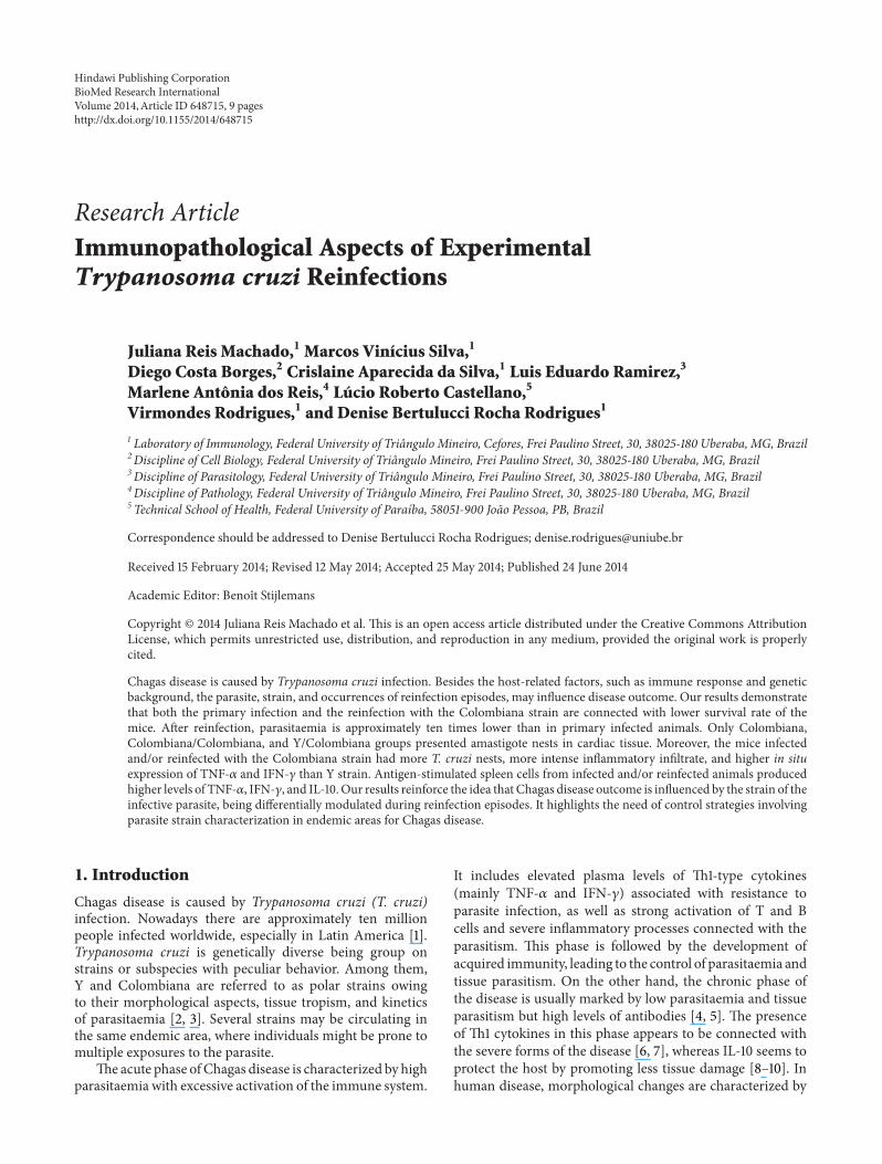
Research ArticleImmunopathological Aspects of ExperimentalTrypanosoma cruzi Reinfections
Juliana Reis Machado,1 Marcos Vinícius Silva,1
Diego Costa Borges,2 Crislaine Aparecida da Silva,1 Luis Eduardo Ramirez,3
Marlene Antônia dos Reis,4 Lúcio Roberto Castellano,5
Virmondes Rodrigues,1 and Denise Bertulucci Rocha Rodrigues1
1 Laboratory of Immunology, Federal University of Triangulo Mineiro, Cefores, Frei Paulino Street, 30, 38025-180 Uberaba, MG, Brazil2 Discipline of Cell Biology, Federal University of Triangulo Mineiro, Frei Paulino Street, 30, 38025-180 Uberaba, MG, Brazil3 Discipline of Parasitology, Federal University of Triangulo Mineiro, Frei Paulino Street, 30, 38025-180 Uberaba, MG, Brazil4Discipline of Pathology, Federal University of Triangulo Mineiro, Frei Paulino Street, 30, 38025-180 Uberaba, MG, Brazil5 Technical School of Health, Federal University of Paraıba, 58051-900 Joao Pessoa, PB, Brazil
Correspondence should be addressed to Denise Bertulucci Rocha Rodrigues; [email protected]
Received 15 February 2014; Revised 12 May 2014; Accepted 25 May 2014; Published 24 June 2014
Academic Editor: Benoıt Stijlemans
Copyright © 2014 Juliana Reis Machado et al. This is an open access article distributed under the Creative Commons AttributionLicense, which permits unrestricted use, distribution, and reproduction in any medium, provided the original work is properlycited.
Chagas disease is caused by Trypanosoma cruzi infection. Besides the host-related factors, such as immune response and geneticbackground, the parasite, strain, and occurrences of reinfection episodes, may influence disease outcome. Our results demonstratethat both the primary infection and the reinfection with the Colombiana strain are connected with lower survival rate of themice. After reinfection, parasitaemia is approximately ten times lower than in primary infected animals. Only Colombiana,Colombiana/Colombiana, and Y/Colombiana groups presented amastigote nests in cardiac tissue. Moreover, the mice infectedand/or reinfected with the Colombiana strain had more T. cruzi nests, more intense inflammatory infiltrate, and higher in situexpression of TNF-𝛼 and IFN-𝛾 than Y strain. Antigen-stimulated spleen cells from infected and/or reinfected animals producedhigher levels of TNF-𝛼, IFN-𝛾, and IL-10.Our results reinforce the idea that Chagas disease outcome is influenced by the strain of theinfective parasite, being differentially modulated during reinfection episodes. It highlights the need of control strategies involvingparasite strain characterization in endemic areas for Chagas disease.
1. Introduction
Chagas disease is caused by Trypanosoma cruzi (T. cruzi)infection. Nowadays there are approximately ten millionpeople infected worldwide, especially in Latin America [1].Trypanosoma cruzi is genetically diverse being group onstrains or subspecies with peculiar behavior. Among them,Y and Colombiana are referred to as polar strains owingto their morphological aspects, tissue tropism, and kineticsof parasitaemia [2, 3]. Several strains may be circulating inthe same endemic area, where individuals might be prone tomultiple exposures to the parasite.
The acute phase of Chagas disease is characterized by highparasitaemia with excessive activation of the immune system.
It includes elevated plasma levels of Th1-type cytokines(mainly TNF-𝛼 and IFN-𝛾) associated with resistance toparasite infection, as well as strong activation of T and Bcells and severe inflammatory processes connected with theparasitism. This phase is followed by the development ofacquired immunity, leading to the control of parasitaemia andtissue parasitism. On the other hand, the chronic phase ofthe disease is usually marked by low parasitaemia and tissueparasitism but high levels of antibodies [4, 5]. The presenceof Th1 cytokines in this phase appears to be connected withthe severe forms of the disease [6, 7], whereas IL-10 seems toprotect the host by promoting less tissue damage [8–10]. Inhuman disease, morphological changes are characterized by
Hindawi Publishing CorporationBioMed Research InternationalVolume 2014, Article ID 648715, 9 pageshttp://dx.doi.org/10.1155/2014/648715
2 BioMed Research International
mononuclear inflammatory infiltrate and fibrotic areas in thisphase [11].
Earliest studies about the role of reinfections in Chagasdisease progression pointed to the development of resistanceafter the first infection, culminating to a mild acute phase[12–14]. Nevertheless, more recent studies indicate that rein-fections may lead to the development of severe forms of thedisease [15–18], whereas other authors could not observe anyrelation in experimentally infected dogs [19]. Controversiesabout the role of reinfections in the course of Chagas diseasecan be better clarified through histopathological evaluationand measurement of cytokine production against different T.cruzi strains.
2. Materials and Methods2.1. Animals. Male C57BL/6 mice (8–10 weeks old) wereobtained and housed in the animal facility of UFTM, Uber-aba, Brazil. Mice were givenwater and food ad libitum duringthe experimental period and all procedures were approvedby the local ethical committee for animal research (CEUA—protocol number 176).
2.2. Infection and Parasitaemia. For the present study weused 75 mice, of which 40 were subcutaneously infected with3,000 forms of the Colombiana strain and 30 were infectedwith the Y strain. Five uninfected animals were kept ascontrol group. The 70 infected animals were observed for 90days until they reached the chronic phase of Chagas disease.During this time, two animals infected with the Y strain died,and 14 animals in the group infected with the Colombianastrain died.
Ninety days after primary infection of the animals withthe Colombiana strain, twelve animals were reinfected with3,000 forms of trypomastigotes of the Colombiana strain(Col/Col), eight were reinfected with the Y (Col/Y) strain,and six were not reinfected (Col). Amongst the animalsinfected with the Y strain, ten were reinfected with theY strain (Y/Y), ten were reinfected with the Colombiana(Y/Col) strain, and eight were not reinfected (Y).
We performed a direct parasitological examination tolook for trypomastigotes in the infected animals on Days7, 14, and 21 after primary infection. After the reinfection,the animals were reinfected and parasitaemia was performedagain on Days 7, 14, and 21 in accordance with protocol [20].Euthanasia was performed on day 111∘ in mice infected withY and Col strains, whereas reinfected animals from groupsY/Y, Y/Col, Col/Col, Col/Y were euthanasied on day 21 afterreinfection. The procedure was performed in CO
2chamber.
Bloodwas collected and autopsywas subsequently performedin order to collect the spleen for in vitro cell culture and theheart for in situ immunohistochemistry. The other organs ofinterest were collected and stored for further analysis.
2.3. Histological Analysis
2.3.1. Inflammatory Infiltrate. For inflammatory infiltrateanalysis we used hematoxylin and eosin (HE) stained slidesof cardiac tissue (ventricle). Qualitative analysis of infiltrate
was performed so as to classify the type of infiltrate as pre-dominantly mononuclear (macrophages and lymphocytes)or polymorphonuclear (neutrophils and eosinophils). Thecellular type observed in more than 50% of the infiltrate wasregarded as prevalent. Semiquantitative analysis of infiltratewas also performed, and inflammatory infiltrate was clas-sified as follows: mild (involvement > 25% of the tissue),moderate (25%–50% of the tissue), or severe (involvement >50% of the tissue).
2.3.2. Immunohistochemistry for Detection of T. cruzi Nests.Ventricular tissue sections were fixed in formaldehydefor immunohistochemistry, and endogenous peroxidaseblocking was performed using 3% H
2O2
in methanol.Then, rabbit anti-T. cruzi antibody (1 : 250) (in house)was added at room temperature for 2 hours. Then, theslides were incubated with peroxidase-conjugated proteinA (1 : 500) for 2 hours. To reveal the reaction we usedH2O2(0.05%) and 1mg/mL DAB (1,4-dideoxy-1,4-imino-D-
arabinitol-diaminobenzidine) (sigmaChemical Co., St Louis,MO, USA) in tris-HCl buffer (pH 7.4). The sections werecounterstained with hematoxylin and analyzed using a com-mon light microscope. Heart parasitism was quantitativelyevaluated according to the presence or absence of amastigotenests.
2.3.3. Quantification of Fibrosis. Wecarried out amorphome-tric evaluation of fibrous conjunctive tissue in heart sectionsstained with Sirius Red. The slides were analyzed usingdigital morphometry in polarized light microscope at a finalmagnification of ×400. Morphometry was performed usingKS300 Imaging System (Carl Zeiss). Fibrosis was quantifiedalong the length of the histological section and expressed inpercentage of affected tissue.
2.4. Immunological Analysis2.4.1. Spleen Cell Culture. Spleens of mice were collected andmaintained in RPMI 1640medium (GEHealth care, Uppsala,Sweden) and macerated for individualization of cells. Thesesuspended cells were washed three times by centrifugationat 400×g for 15min at 8∘C in RPMI 1640. Then, theywere counted in a Neubauer chamber and resuspended to2 × 10
6 cells/mL in RPMI 1640 medium with addition of50mM Hepes (Gibco, Grand Island, NY, USA), 5% of inac-tivated fetal bovine serum (GIBCO-US), 2mM L-glutamine(GIBCO-US), 0.05mM 2𝛽-mercaptoethanol (GIBCO-US),and 40 𝜇g/mL gentamicin (NEOQUIMICA, Anapolis, GO,BR). Then, 2 × 10⁶ cells were incubated without stimulus andwith 5 𝜇g/mL of T. cruzi antigen in 24-well culture plates (BDPharmingen, San Diego, CA, USA).The cultures were kept ina moist incubator with 5% CO
2at 37∘C for 24 and 72 hours.
The supernatants were collected and maintained at −70∘Cuntil analysis.
2.4.2. Preparation of Cardiac Tissue Homogenate. Heart tis-sue sections were immersed in PBS solution containingcomplete protease inhibitor (Sigma, St. Louis, MO, USA)and Nonidet-P40. After that, they were submitted to tissue
BioMed Research International 3
0 7 14 210
20
40
60
80
100
Days after reinfection
Surv
ival
(%)
Col/ColCol/Y
Colombiana Y
Y/ColY/Y
(a)
Parasitaemia
7 14 210
100
200
300
1500
3000 ∗
#∗
Days
Tryp
omas
tigot
es (m
L)
#
Col/ColCol/Y
Colombiana Y
Y/ColY/Y
(b)
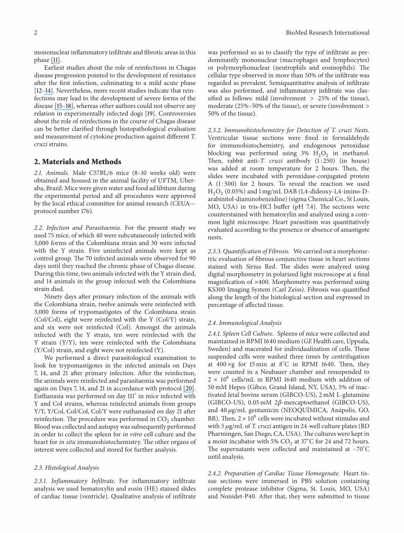
Figure 1: (a) Survival curve of reinfectedmice,𝑃 = 0.0002, Log-rank test. (b) Parasitaemia ofmice that were primarily infected and reinfectedwith theColombiana andY strains ofT. cruzi. Parasitaemia levels were evaluated by counting the number of parasites in 5 𝜇L of blood collectedfrom the tail vein. The symbols of each group represent the mean. ∗Statistical differences in animals primarily infected with the Colombianastrain or Y strain were analyzed on Days 7, 14, and 21; Mann-Whitney test. #Statistical differences among the reinfected groups (Y/Y, Y/Col,Col/Col, Col/Y) were analyzed on Days 7, 14, and 21; Kruskal-Wallis test.
homogenizer. The homogenate obtained was centrifuged at14000×g for 10 minutes and the supernatant was maintainedfor quantification of cytokines and total proteins.
2.4.3. Quantification of Cytokines in Supernatants of SpleenCell Culture and in Cardiac Tissue Homogenate Using CBA.Cytokines IL-2, IL-4, IL-5, IL-10, IL-12p70, IL-17, TNF-𝛼,and IFN-𝛾 were quantified using Cytometric Bead Array—CBA (BD Pharmingen, San Diego, CA, USA) in accordancewith the manufacturer’s specifications. The samples andthe recombinant cytokines were incubated with beads withdifferent fluorescence intensities conjugated with specificcapture antibody for each cytokine of interest. After incuba-tion, the beadswerewashedwith saline solution and analyzedin BD FACS CALIBUR flow cytometer, using CellQuestsoftware. Upon data acquisition of samples and of recombi-nant cytokines, they were analyzed using FCAP Array v2.0software (Soft Flow, USA) and the concentrations of thecytokines were measured by comparison to the standardcurve. The concentrations of cytokines in cardiac tissuehomogenate were normalized based upon the concentrationof total proteins in each homogenate and were quantifiedusing the Micro-Lowry method in accordance with themanufacturer’s instructions (Pierce, Rockford, IL, USA).
2.5. Statistical Analyses. GraphPad Prism 5.0 software(GraphPad Software, USA) was used. Mann-Whitney test(𝑈) was used for analysis between two groups, and foranalysis among more than two groups ANOVA test (𝐹) wasused for data with normal distribution and Kruskal Wallistest (𝐻) was used for data with nonnormal distribution.Qualitative variables were expressed as percentage andthe associations between them were analyzed using the
chi-square (𝜒2) test. Survival rate analyses were performedusing Log-Rank test. Results were considered statisticallysignificant when 𝑃 < 0.05.
3. Results3.1. Survival Rate. We analyzed the survival rate in animalsprimarily infected with the Y and Colombiana strains andin reinfected animals. During chronification of the disease,a survival rate of 93.3% (28/30 animals) was observed inthe animals primarily infected with the Y strain. Amongthe animals primarily infected with the Colombiana strainwe observed a survival rate of only 65% (26/40 animals)(𝑃 = 0.0055, Log-rank test). After reinfection, the groupinfected with the Colombiana strain and reinfected with thesame strain had a mortality rate of 50% (6/12) (𝑃 = 0.0002,Log-rank test). In the remaining groups we did not observemortality within the period of 21 days after reinfection(Figure 1(a)).
3.2. Parasitaemia. The animals primarily infected with theY strain but not with Colombiana strain had detectableparasitaemia since Day 7 of infection (𝑃 = 0.012;𝑈 = 0.000).On Day 14 we did not observe any difference in parasitaemiaamong the primary infected groups (𝑃 = 0.139; 𝑈 = 12.50).Nonetheless, on Day 21 increased parasitaemia was observedin the animals infected with the Colombiana strain (𝑃 =0.0006; 𝑈 = 0.000), whereas parasitemia in animals infectedwith the Y strain was not detected (Figure 1(b)).
After reinfection, the levels of parasitaemia were approxi-mately ten times lower than in primary infected animals. Par-asitaemia in animals infected with the Y strain and reinfectedwith the Y or Colombiana strains was significantly lower than
4 BioMed Research International
(a) (b)
(c) (d)
(e) (f)
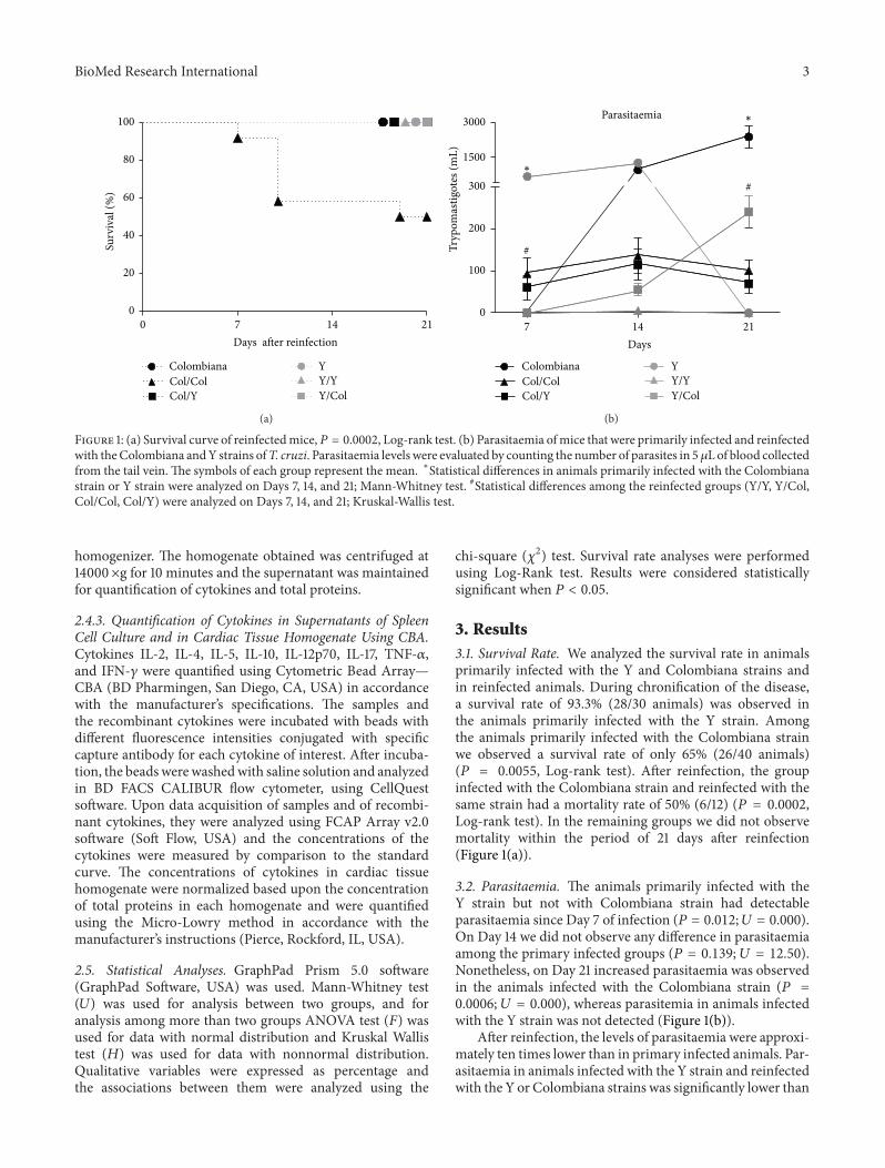
Figure 2: Histological heart sections of C57BL/6 mice. (a) Immunohistochemistry showing a T. cruzi nests in the animals infected andreinfected with the Colombiana strain (10x). (b) At a higher magnification, we noticed the details of T. cruzi nest in this same group (40x).(c) Inflammatory infiltrate in an animal infected with the Colombiana strain (HE, 10x). (d) Inflammatory infiltrate in the group infectedand/or reinfected with the Colombiana strain (HE, 20x). (e) Inflammatory infiltrate in the group infected with the Y strain (HE, 10x). (f)Inflammatory infiltrate in the group infected and reinfected with the Y strain (HE, 10x).
parasitaemia of animals infected with the Colombiana strainand reinfected with the Y or Colombiana strains on Day 7of reinfection (𝑃 = 0.035; 𝐻 = 8.567). On Day 14 only theparasitaemia in Y/Y group was significantly lower than thelevels of parasitaemia in the other reinfected groups (𝑃 =0.008; 𝐻 = 11.594). On Day 21 the levels of parasitaemia inCol/Col and Col/Y Groups were still similar to the previousdays; however, Y/Col group had significantly higher levelsof parasitaemia than the other groups, and Y/Y group hadsignificantly lower levels than the other groups (𝑃 < 0.0001;𝐻 = 23.331) (Figure 1(b)).
3.3.Histopathological Analysis. OnlyCol, Col/Col, andY/Colgroups showed amastigote nests in cardiac tissue. Moreover,
Col/Col group had a significantly higher percentage ofanimals with T. cruzi nests (𝑃 < 0.0001, 𝜒2 = 97.56) (Table 1,Figures 2(a)-2(b)).
The inflammatory infiltrate was predominantly mononu-clear (Figures 2(e)-2(f)). The groups primarily infected withthe Y strain showed a mild inflammatory infiltrate, exceptfor Y/Col group, which showed a moderate inflammatoryinfiltrate. The groups infected and/or reinfected with theColombiana strain showed a moderate-to-severe inflamma-tory infiltrate. Col/Col group showed a particularly severeinflammatory infiltrate inmore than 60% of the animals (𝑃 <0.0001,𝜒2 = 273.8) (Table 1, Figures 2(c)-2(d)), and there wasa worsening in comparison with the primary infection withthe Colombiana strain.
BioMed Research International 5
Table 1: Amastigotes nests, inflammatory infiltrate, and fibrosis in cardiac tissue.
Groups Amastigotes nests (%) Inflammatory infiltrate % Fibrosis (% area ± SEM)Mild Moderate Severe
Col 16.6 0 100 0 0.70 ± 0.13Col/Col 33.3∗ 0 33.3 66.6∗ 0.28 ± 0.04Col/Y 0 50 50 0 0.84 ± 0.79Y 0 100 0 0 0.31 ± 0.09Y/Y 0 100 0 0 0.56 ± 0.14Y/Col 10 0 100 0 0.37 ± 0.10Control — — — — 0.39 ± 0.17The qualitative variables were expressed in percentage, and the associations between themwere analyzed usingChi-square test (𝜒2), ∗𝑃 < 0.005.The percentageof fibrosis in the cardiac tissue was analyzed using ANOVA test followed by Tukey’s multiple comparison test. ∗Significant differences among Col group versusCol/Col versus Col/Y versus control group.
TNF-𝛼
Col Col/Col Col/Y Control Y Y/Y Y/Col0
50
100
150
200
250
(pg/
mg
tissu
e pro
tein
)
P = 0.0286H = 9.05
(a)
IFN-𝛾
Col Col/Col Col/Y Control Y Y/Y Y/Col0
5
10
15 P = 0.0029H = 14.00
P = 0.0023H = 14.53
(pg/
mg
tissu
e pro
tein
)
(b)
IL-10
Col Col/Col Col/Y Control Y Y/Y Y/Col0
5
10
15
20
(pg/
mg
tissu
e pro
tein
)
(c)
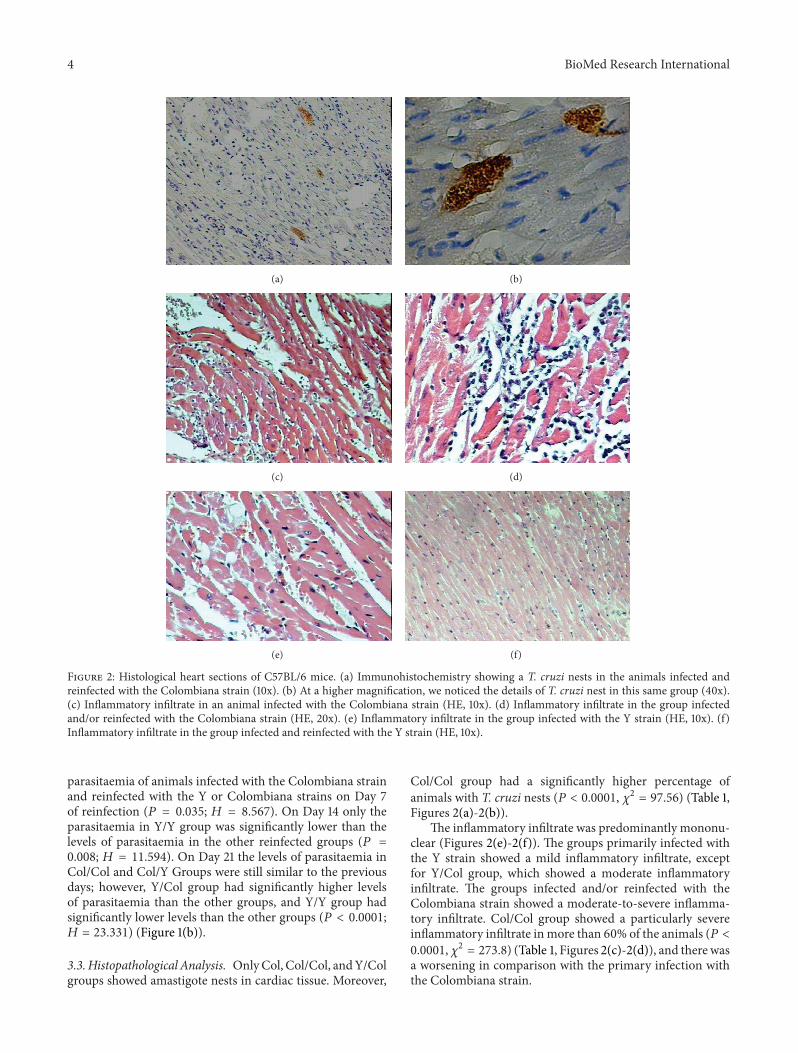
Figure 3: Production of TNF-𝛼, IFN-𝛾, and IL-10 (pg/mL) in cardiac tissue of control mice infected and reinfected with T. cruzi. Kruskal-Wallis test followed by Dunn’s multiple comparison test. Horizontal lines represent the median, bars represent 25–75 percentiles, and verticallines represent 10–90 percentiles.
We did not observe significant intensity of fibrosis amongthe groups primarily infected with the Colombiana strainand their subsequent reinfections and between these groupsand the uninfected control group (𝑃 = 0.161; 𝐹 = 1.94).Furthermore, we did not find significant differences amongthe groups primarily infected with the Y strain and theirsubsequent reinfections and between these groups and theuninfected control group (𝑃 = 0.066; 𝐹 = 2.82) (Table 1).
3.4. Immunological Analysis
3.4.1. Production of TNF-𝛼, IFN-𝛾, and IL-10 inCardiac Tissue.The expression of TNF-𝛼 was significantly higher in Col/Col
group than in the control group. Although not significant,Col/Col group proved to producemore TNF-𝛼 than the othergroups, especially the groups primarily infected with the Ystrain.The groups primarily infectedwith the Y strain did nothave a significant difference in TNF-𝛼 production in cardiactissue (Figure 3(a)).
The expression of IFN-𝛾 in cardiac tissuewas significantlyhigher in Col/Col group and in Col/Y group than in thecontrol group and higher in Y/Col group than in the controlgroup and in Y/Y group. Although not significant, the groupsinfected or reinfected with the Colombiana strain proved toproduce more IFN-𝛾 than the groups infected with the Ystrain and/or reinfected with the same strain, whose IFN-𝛾production was lower than the other groups (Figure 3(b)).
6 BioMed Research International
0
200
400
600
800
1000
Nonstimulated
P = 0.0001H = 21.1
P = 0.0005H = 17.91
P = 0.0049H = 12.47
TNF-𝛼
(pg/
mL)
T. cruzi antigens
(a)
05
1015
400
800
1200
1600
2000
Nonstimulated
P = 0.0029H = 14.04
P = 0.0020H = 14.20
P = 0.0005H = 17.82
P = 0.0027H = 14.12
IFN
-𝛾 (p
g/m
L)
T. cruzi antigens
(b)
0
100
200
300
400
500
600
2000
ColombianaCol/ColCol/YControl
YY/YY/Col
Nonstimulated T. cruzi antigens
P = 0.0005H = 17.59
P = 0.0009H = 16.42
P = 0.0088H = 11.62
P = 0.0025H = 14.36
IL-1
0 (p
g/m
L)
(c)
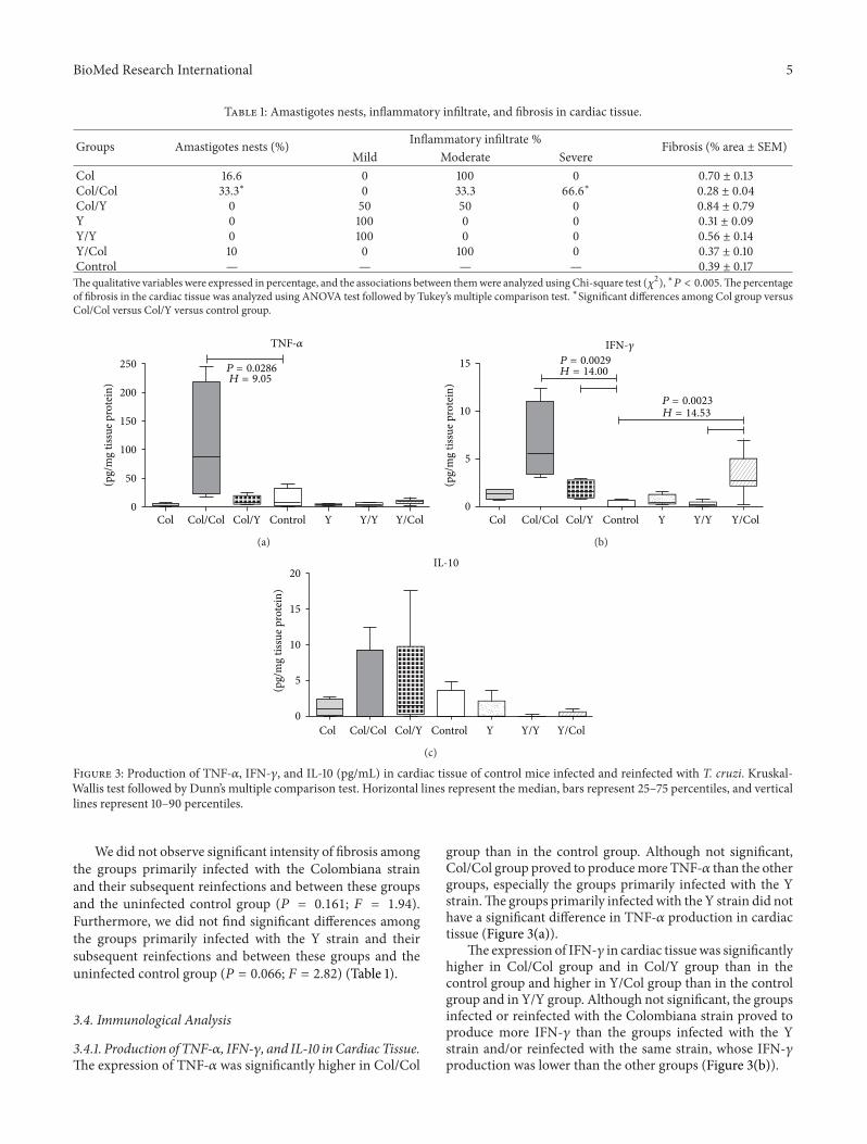
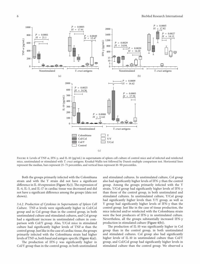
Figure 4: Levels of TNF-𝛼, IFN-𝛾, and IL-10 (pg/mL) in supernatants of spleen cell culture of control mice and of infected and reinfectedmice, unstimulated or stimulated with T. cruzi antigens. Kruskal-Wallis test followed by Dunn’s multiple comparison test. Horizontal linesrepresent the median, bars represent 25–75 percentiles, and vertical lines represent 10–90 percentiles.
Both the groups primarily infected with the Colombianastrain and with the Y strain did not have a significantdifference in IL-10 expression (Figure 3(c)).The expression ofIL-4, IL-5, and IL-17 in cardiac tissue was decreased and didnot have a significant difference among the groups (data notshown).
3.4.2. Production of Cytokines in Supernatants of Spleen CellCulture. TNF-𝛼 levels were significantly higher in Col/Colgroup and in Col group than in the control group, in bothunstimulated culture and stimulated cultures, and Col grouphad a significant increase in unstimulated culture in com-parison with Col/Y group. Also, Y/Col mice in stimulatedculture had significantly higher levels of TNF-𝛼 than thecontrol group. Just like in the case of cardiac tissue, the groupsprimarily infected with the Colombiana strain had higherlevels of TNF-𝛼, both basal and antigen-specific (Figure 4(a)).
The production of IFN-𝛾 was significantly higher inCol/Y group than in the control group, in both unstimulated
and stimulated cultures. In unstimulated culture, Col groupalso had significantly higher levels of IFN-𝛾 than the controlgroup. Among the groups primarily infected with the Ystrain, Y/Col group had significantly higher levels of IFN-𝛾than those of the control group, in both unstimulated andstimulated cultures. In unstimulated culture, Y/Col grouphad significantly higher levels than Y/Y group, as well asY group had significantly higher levels of IFN-𝛾 than thecontrol group. Just like in the case of tissue production, themice infected and/or reinfected with the Colombiana strainwere the best producers of IFN-𝛾 in unstimulated culture.Nevertheless, all the groups substantially increased IFN-𝛾production in stimulated culture (Figure 4(b)).
The production of IL-10 was significantly higher in Colgroup than in the control group, in both unstimulatedand stimulated cultures. Col group also had significantlyhigher levels of IL-10 in unstimulated culture than Col/Ygroup, and Col/Col group had significantly higher levels instimulated culture than the control group. We observed a
BioMed Research International 7
significant increase in the production of IL-10 in Y/Col groupin comparison with Y/Y group, in both unstimulated andstimulated cultures. Y/Col group also had significantly higherlevels of IL-10 in the culture stimulated with T. cruzi thanthe control group. In general, the groups infected and/orreinfected with the Colombiana strain seem to produce moreIL-10, especially in stimulated culture.
4. DiscussionAt the onset T. cruzi infection it is possible to noticesome acute phase changes such as parasitaemia and heartparasitism, both of which depend on the infecting strain [21–24]. In the present study, the animals primarily infected withthe Y strain reached peak parasitaemia on Day 14 and theselevels decreased abruptly on Day 21 of infection, whereasprimary infection with the Colombiana strain showed lowparasitaemia on the first days, with a substantial increase inthe levels up to 21 days. These results are in accordance withthe literature data [25].
Reinfected animals had much lower parasitaemia thanprimary infected animals, thus suggesting a possible pro-tection conferred by the first infection, which was welldemonstrated in Y/Y group, with undetectable parasitaemia21 days after infection. It is in accordance with previousdemonstrations that reinfected animals obtain immunolog-ical protection, thus leading to the reduction in parasitaemiaand mortality [14, 26, 27].
Animals infected and/or reinfected with the Colombianastrain showed marked parasitism, whereas those reinfectedwith the Y strain did not have T. cruzi nests in any of the stud-ied groups. Some authors argue that differences in the geneticcomposition of individual strains ofT. cruziwould determinetissue tropism [28]. Classic studies had demonstrated thatthe Y strain is connected with reticulotropism and increasedvirulence in the acute phase of the infection, whereas theColombiana strain is connected with cardiomyotropism andpathogenicity in the chronic phase [21–24].
Parasitism and inflammatory infiltrate were more severein the heart of Colombiana infected and/or reinfected ani-mals. A positive correlation between parasitism and theseverity of myocarditis has been observed [29, 30], whileaggravation of acutemyocarditis seems to depend on the con-centration and the quality of the exudate [31]. In this study,the inflammatory infiltrate was predominantly mononuclearin all groups. The severe inflammatory infiltrate observedin 66% of the animals of Col/Col group may be relatedto their higher mortality rate. Nonetheless, we did notobserve increased fibrosis in these animals, which should beassociated with the progression of cardiac insufficiency, as itis believed to lead Chagas disease patients to sudden death[32].
In Chagas disease, local immune response—representedby the inflammatory infiltrate—and systemic immuneresponse are both responsible for the symptomatology andrepercussions of the disease. In this study we analyzedthe expression of TNF-𝛼, IFN-𝛾, and IL-10, which are keycytokines in the anti-T. cruzi immune response.We observedthat the levels of TNF-𝛼 in the cardiac tissue were elevated in
Col and Col/Col groups and associated with a more severeinflammatory infiltrate and with the presence of T. cruzinests. We believe that tissue parasitism in this group inducedan increase in the expression of TNF-𝛼 in situ. TNF-𝛼activates macrophages and, thus, the production of nitricoxide, leading to the destruction of intracellular parasites[6, 33, 34].
Similarly, when TNF-𝛼 production by spleen cells wasanalyzed, the Col and Col/Col groups had a significantlyhigher production than the Y or Y/Y groups, which couldbe explained by the fact that in the latter groups there wereundetectable parasitism and mild inflammatory infiltrate,resulting in lower expression of TNF-𝛼 in the cardiac tissueand lower production by spleen cells in vitro. These resultscan be explained by a greater resistance of C57BL/6 miceto infection with the Y strain than to Colombiana strainof T. cruzi [35, 36]. Other experimental studies show thathigh levels of TNF-𝛼 in the acute phase seem to leadto cachexia and death, becoming an essential element oftissue inflammatory reaction [7, 37]. In the chronic phase ofChagas disease, TNF-𝛼 seems to be closely related to cardiacdysfunction owing to its negative inotropic effect, in bothexperimental models [38] and humans [39]. Furthermore,PBMCs of patients with chronic Chagas cardiopathy producehigh levels of TNF-𝛼, associated with higher expression ofFas and FasL, as well as with lymphocyte and myocardiocyteapoptosis [40, 41].
In the present study, the animals infected or reinfectedwith the Colombiana strain had a significantly higher expres-sion of IFN-𝛾, both in the cardiac tissue and in spleen cells.Just like in the case of TNF-𝛼, the groups with increasedparasitism and cardiac inflammatory infiltrate expressedmore IFN-𝛾 in the cardiac tissue, in association with themostevident inflammatory infiltrate, particularly Col and Col/Colgroups. Production of IFN-𝛾 by spleen cells in Col/Col grouphad a mild decrease in relation to the group that was infectedonly with the Colombiana strain.This decrease may probablybe due to cell recruitment to the cardiac tissue. Classic studieshad already shown that IFN-𝛾 and TNF-𝛼 are synergisticcytokines in the activation of macrophages and, hence, in thedestruction of intracellular parasites [42].
We did not observe a difference in the expression of IL-10 in cardiac tissue among the studied groups. However, instimulated culture, there seems to be a higher productionin the groups infected and/or reinfected with the Colom-biana strain. Interleukin-10 is a cytokine that modulatesmacrophage activity, being indirectly responsible for reducedIFN-𝛾 production and for controlling the potential tissuedamaging effects of activated macrophages [43]. In thepresent study, the groups that had the highest levels of TNF-𝛼and IFN-𝛾 also had the highest levels of IL-10. This increasemay represent a compensatory mechanism aiming to controltissue damage caused by the local strong production of IFN-𝛾 and TNF-𝛼. Some studies on other intracellular pathogensdemonstrated that the simultaneous raise of IFN-𝛾 and IL-10has a beneficial role in parasite control and in the preventionof tissue damage [7, 44, 45]. In vitro studies, particularly onChagas disease, have demonstrated that high IL-10 levels arecapable of inhibiting the intracellular destruction of T. cruzi
8 BioMed Research International
[46, 47]. Other studies using il10 knockout mice showed thatthese animals have a more efficient control over the infectionby T. cruzi, reducing parasitism levels with a significantincrease in the secretion of IFN-𝛾, TNF-𝛼, IL-12, and NO[48, 49]. However, animals with IL-10 deficiency succumbfaster to T. cruzi infection mainly due to the uncontrolledactivity of proinflammatory cytokines [49].
5. Conclusion
Our results suggest that mortality rates, tissue parasitism,inflammatory infiltrate, and expression of proinflammatorycytokines such as TNF-𝛼 and IFN-𝛾, in situ or in vitro, aredifferentially modulated by reinfections with Trypanosomacruzi Y and Colombiana strains. This reinforces the need ofcontrol strategies involving parasite strain characterization inendemic areas for Chagas disease.
Conflict of Interests
The authors declare that they have no conflict of interests.
Acknowledgments
The authors appreciate the financial support of ConselhoNacional de Desenvolvimento Cientıfico e Tecnologico(CNPq), Coordenacao de Aperfeicoamento de Pessoal deNıvel Superior (CAPES), Fundacao de Amparo a Pesquisado Estado de Minas Gerais (FAPEMIG), and Fundacao deEnsino e Pesquisa de Uberaba (FUNEPU).
References
[1] WHO, “Chagas disease (American trypanosomiasis) fact sheet(revised in June 2010),”TheWeekly Epidemiological Record, vol.85, no. 34, pp. 334–336, 2010.
[2] Z. Brener, “Comparative studies of different strains of Try-panosoma cruzi,” Annals of Tropical Medicine and Parasitology,vol. 59, pp. 19–26, 1965.
[3] Z. Brener and E. Chiari, “Morphological variations observed indifferent strains of Trypanosoma cruzi,” Revista do Instituto deMedicina Tropical de Sao Paulo, vol. 5, pp. 220–224, 1963.
[4] A. A. Fragata Filho, M. A. da Silva, and E. Boainain, “Ethiologictreatment of acute and chronic Chagas’ Disease [corrected],”Sao Paulo Medical Journal, vol. 113, no. 2, pp. 867–872, 1995.
[5] A. Rassi Jr., A. Rassi, and J. A. Marin-Neto, “Chagas disease,”The Lancet, vol. 375, no. 9723, pp. 1388–1402, 2010.
[6] J. S. Silva, G. N. R. Vespa, M. A. G. Cardoso, J. C. S. Aliberti,and F. Q. Cunha, “Tumor necrosis factor alpha mediatesresistance to Trypanosoma cruzi infection in mice by inducingnitric oxide production in infected gamma interferon-activatedmacrophages,” Infection and Immunity, vol. 63, no. 12, pp. 4862–4867, 1995.
[7] E. Roggero, A. Perez, M. Tamae-Kakazu et al., “Differentialsusceptibility to acute Trypanosoma cruzi infection in BALB/cand C57BL/6 mice is not associated with a distinct parasiteload but cytokine abnormalities,” Clinical and ExperimentalImmunology, vol. 128, no. 3, pp. 421–428, 2002.
[8] A. M. B. Bilate, V. M. Salemi, F. J. Ramires et al., “TNF blockadeaggravates experimental chronic Chagas disease cardiomyopa-thy,”Microbes and Infection, vol. 9, no. 9, pp. 1104–1113, 2007.
[9] A. R. Perez, G. H. Fontanella, A. L. Nocito, S. Revelli, and O.A. Bottasso, “Short treatment with the tumour necrosis factor-𝛼 blocker infliximab diminishes chronic chagasic myocarditisin rats without evidence of Trypanosoma cruzi reactivation,”Clinical and Experimental Immunology, vol. 157, no. 2, pp. 291–299, 2009.
[10] V. M. B. Lorena, I. M. B. Lorena, S. C. M. Braz et al.,“Cytokine levels in serious cardiopathy of chagas disease after invitro stimulationwith recombinant antigens fromTrypanosomacruzi,” Scandinavian Journal of Immunology, vol. 72, no. 6, pp.529–539, 2010.
[11] E. R. Lopes, E. Chapadeiro, Z. A. Andrade, H. O. Almeida, andA. Rocha, “Pathological anatomy of hearts from asymptomaticChagas disease patients dying in a violent manner,” Memoriasdo Instituto Oswaldo Cruz, vol. 76, no. 2, pp. 189–197, 1981.
[12] E. Brumpt, “Immunite partielle dans les infections a Try-panosoma cruzi, transmission de ce trypanosome par Cimexrotundus. Role regulateur des hotes intermediaires. Passage atravers la peau,” Bulletin de La Societe de Pathologie Exotique,vol. 6, pp. 172–176, 1913.
[13] J. T. Culbertson, M. H. Kolodny, and H. Maxwell, “Acquiredimmunity in rats against Trypanosoma cruzi,” Journal of Para-sitology, vol. 24, pp. 83–90, 1938.
[14] Z. Brener, “Some aspects of acquired immunity in mice experi-mentally infected with Trypanosoma cruzi,” Revista do Institutode Medicina Tropical de Sao Paulo, vol. 9, no. 4, pp. 233–238,1967.
[15] F. M. Hatcher, R. E. Kuhn, M. C. Cerrone, and R. C. Burton,“Increased natural killer cell activity in experimental AmericanTrypanosomiasis,”The Journal of Immunology, vol. 127, no. 3, pp.1126–1130, 1981.
[16] L. Ortiz-Ortiz, T. Ortega, R. Capın, and T.Martınez, “Enhancedmononuclear phagocytic activity during Trypanosoma cruziinfection in mice,” International Archives of Allergy and AppliedImmunology, vol. 50, no. 2, pp. 232–242, 1976.
[17] J. M. Bustamante, M. Novarese, H. W. Rivarola et al., “Reinfec-tions and Trypanosoma cruzi strains can determine the prog-nosis of the chronic chagasic cardiopathy in mice,” ParasitologyResearch, vol. 100, no. 6, pp. 1407–1410, 2007.
[18] S. G. Andrade, R. F. Campos, K. S. Castro Sobral, J. B. Mag-alhaes, R. S. Pereira Guedes, andM. L. Guerreiro, “Reinfectionswith strains of Trypanosoma cruzi, of different biodemes asa factor of aggravation of myocarditis and myositis in mice,”Revista da Sociedade Brasileira de Medicina Tropical, vol. 39, no.1, pp. 1–8, 2006.
[19] E.M.M.Machado, A. J. Fernandes, S.M. F.Murta et al., “A studyof experimental reinfection by Trypanosoma cruzi in dogs,”TheAmerican Journal of Tropical Medicine and Hygiene, vol. 65, no.6, pp. 958–965, 2001.
[20] Z. BRENER, “Therapeutic activity and criterion of cure onmiceexperimentally infected with Trypanosoma cruzi,” Revista doInstituto de Medicina Tropical de Sao Paulo, vol. 4, pp. 389–396,1962.
[21] Z. A. Andrade and C. M. P. Ramalho, “Miocardite chagasica.Estudo morfologico de 38 casos comprovados pelo encontro deparasitos nas seccoes histologicas,”GazetaMedica da Bahia, vol.66, pp. 55–67, 1966.
[22] R. Kumar, I. K. Kline, and W. H. Abelmann, “ExperimentalTrypanosoma cruzi myocarditis: relative effects upon the right

BioMed Research International 9
and left ventricles,” The American Journal of Pathology, vol. 57,no. 1, pp. 31–48, 1969.
[23] R. C. Melo and Z. Brener, “Tissue tropism of different Try-panosoma cruzi strains,” Journal of Parasitology, vol. 64, no. 3,pp. 475–482, 1978.
[24] S. G. Andrade, “Influence of Trypanosoma cruzi strain on thepathogenesis of chronic myocardiopathy inmice,”Memorias doInstituto Oswaldo Cruz, vol. 85, no. 1, pp. 17–27, 1990.
[25] S. G. Andrade, A. R. Pimentel, M. M. De Souza, and Z.A. Andrade, “Interstitial dendritic cells of the heart harborTrypanosoma cruzi antigens in experimentally infected dogs:Importance for the pathogenesis of chagasic myocarditis,” TheAmerican Journal of Tropical Medicine and Hygiene, vol. 63, no.1-2, pp. 64–70, 2000.
[26] M. T. Scott and M. Goss-Sampson, “Restricted IgG isotypeprofiles in T. cruzi infected mice and Chagas’ disease patients,”Clinical and Experimental Immunology, vol. 58, no. 2, pp. 372–379, 1984.
[27] S. G. Andrade, M. L. Carvalho, R. M. Figueira, and Z. A.Andrade, “Recovery and characterization of trypanosomasinoculated into immune animals (re-inoculation with differentstrains of T. cruzi),” Revista do Instituto de Medicina Tropical deSao Paulo, vol. 12, no. 6, pp. 395–402, 1970.
[28] L. O. Andrade, C. R. S. Machado, E. Chiari, S. D. J. Pena,and A. M. Macedo, “Differential tissue distribution of diverseclones of Trypanosoma cruzi in infected mice,” Molecular andBiochemical Parasitology, vol. 100, no. 2, pp. 163–172, 1999.
[29] M. D. L. Higuchi, M. M. Reis, V. D. Aiello et al., “Association ofan increase in CD8+ T cells with the presence of Trypanosomacruzi antigens in chronic, human, chagasic myocarditis,” TheAmerican Journal of Tropical Medicine and Hygiene, vol. 56, no.5, pp. 485–489, 1997.
[30] F. R. S. Gutierrez, P. M. M. Guedes, R. T. Gazzinelli, and J. S.Silva, “The role of parasite persistence in pathogenesis of Chagasheart disease,” Parasite Immunology, vol. 31, no. 11, pp. 673–685,2009.
[31] W. L. Tafuri, “Patogenia da doenca de chagas,” Revista doInstituto de Medicina Tropical de Sao Paulo, vol. 29, no. 4, pp.194–199, 1987.
[32] E. R. Lopes, E. Chapadeiro, W. L. Tafuri, A. O. Almeida, and D.Abraao, “Peso do coracao e tipo demorte no chagasico cronico,”Revista do Instituto de Medicina Tropical de Sao Paulo, vol. 12,no. 5, pp. 293–297, 1970.
[33] S. J. Green, S. Mellouk, S. L. Hoffman, M. S. Meltzer, andC. A. Nacy, “Cellular mechanisms of nonspecific immunityto intracellular infection: cytokine-induced synthesis of toxicnitrogen oxides from l-arginine by macrophages and hepato-cytes,” Immunology Letters, vol. 25, no. 1-3, pp. 15–19, 1990.
[34] J. A. M. Langermans, M. E. B. Van der Hulst, P. H. Nibber-ing, and R. Van Furth, “Endogenous tumor necrosis factoralpha is required for enhanced antimicrobial activity againstToxoplasma gondii and Listeria monocytogenes in recombinantgamma interferon-treated mice,” Infection and Immunity, vol.60, no. 12, pp. 5107–5112, 1992.
[35] E. N. de Gaspari, E. S. Umezawa, B. Zingales, A. M. Stolf,W. Colli, and I. A. Abrahamsohn, “Trypanosoma cruzi: serumantibody reactivity to the parasite antigens in susceptible andresistantmice,”Memorias do Instituto Oswaldo Cruz, vol. 85, no.3, pp. 261–270, 1990.
[36] D. E. Bice and R. Zeledon, “Comparison of infectivity of strainsof Trypanosoma cruzi (Chagas, 1909),” Journal of Parasitology,vol. 56, no. 4, pp. 663–670, 1970.
[37] A. R. Perez, M. Tamae-Kakazu, M. F. Pascutti et al., “Defi-cient control of Trypanosoma cruzi infection in C57BL/6 miceis related to a delayed specific IgG response and increasedmacrophage production of pro-inflammatory cytokines,” LifeSciences, vol. 77, no. 16, pp. 1945–1959, 2005.
[38] S. Hegewisch, H.-J. Weh, and D. K. Hossfeld, “TNF-inducedcardiomyopathy,” The Lancet, vol. 335, no. 8684, pp. 294–295,1990.
[39] G. Torre-Amione, S. Kapadia, J. Lee et al., “Tumor necrosisfactor-𝛼 and tumor necrosis factor receptors in the failinghuman heart,” Circulation, vol. 93, no. 4, pp. 704–711, 1996.
[40] V. Rodrigues Jr., G. S. Agrelli, S. C. Leon, D. N. Silva Teixeira, S.Tostes Jr., and D. B. Rocha-Rodrigues, “Fas/Fas-L expression,apoptosis and low proliferative response are associated withheart failure in patients with chronic Chagas’ disease,”Microbesand Infection, vol. 10, no. 1, pp. 29–37, 2008.
[41] S. Tostes Jr., D. B. Rocha-Rodrigues, G. De Araujo Pereira, andV. Rodrigues Jr., “Myocardiocyte apoptosis in heart failure inchronic Chagas’ disease,” International Journal of Cardiology,vol. 99, no. 2, pp. 233–237, 2005.
[42] M. A. Munoz-Fernandez, M. A. Fernandez, and M. Fresno,“Activation of human macrophages for the killing of intracel-lular Trypanosoma cruzi by TNF-𝛼 and IFN-𝛾 through a nitricoxide-dependent mechanism,” Immunology Letters, vol. 33, no.1, pp. 35–40, 1992.
[43] D. F. Fiorentino, A. Zlotnik, T. R. Mosmann, M. Howard, andA. O’ Garra, “IL-10 inhibits cytokine production by activatedmacrophages,” The Journal of Immunology, vol. 147, no. 11, pp.3815–3822, 1991.
[44] E. Bohn, J. Heesemann, S. Ehlers, and I. B. Autenrieth, “Earlygamma interferon mRNA expression is associated with resis-tance of mice against Yersinia enterocolitica,” Infection andImmunity, vol. 62, no. 7, pp. 3027–3032, 1994.
[45] S. Pie, P. Matsiota-Bernard, P. Truffa-Bachi, and C. Nauciel,“Gamma interferon and interleukin-10 gene expression ininnately susceptible and resistant mice during the early phaseof Salmonella typhimurium infection,” Infection and Immunity,vol. 64, no. 3, pp. 849–854, 1996.
[46] R. T. Gazzinelli, I. P. Oswald, S. Hieny, S. L. James, andA. Sher, “The microbicidal activity of interferon-𝛾-treatedmacrophages against Trypanosoma cruzi involves an L-arginine-dependent, nitrogen oxide-mediated mechanisminhibitable by interleukin-10 and transforming growth factor-𝛽,” European Journal of Immunology, vol. 22, no. 10, pp.2501–2506, 1992.
[47] J. S. Silva, P. J. Morrissey, K. H. Grabstein, K. M. Mohler, D.Anderson, and S. G. Reed, “Interleukin 10 and interferon 𝛾regulation of experimental Trypanosoma cruzi infection,” TheJournal of Experimental Medicine, vol. 175, no. 1, pp. 169–174,1992.
[48] I. A. Abrahamsohn and R. L. Coffman, “Trypanosoma cruzi:IL-10, TNF, IFN-𝛾 and IL-12 regulate innate and acquiredimmunity to infection,” Experimental Parasitology, vol. 84, no.2, pp. 231–244, 1996.
[49] C. A. Hunter, L. A. Ellis-Neyes, T. Slifer et al., “IL-10 isrequired to prevent immunehyperactivity during infectionwithTrypanosoma cruzi,”The Journal of Immunology, vol. 158, no. 7,pp. 3311–3316, 1997.
Copyright © 2022 FDOKUMEN