Glutamine treatment attenuates endoplasmic reticulum stress and apoptosis in TNBS-induced colitis

Molecular Biology of the CellVol. 14, 3529–3540, September 2003
The Interplay between Folding-facilitatingMechanisms in Trypanosoma cruziEndoplasmic ReticulumIanina Conte,* Carlos Labriola,* Juan J. Cazzulo,* Roberto Docampo,† andArmando J. Parodi*‡
*Institute for Biotechnological Research, University of San Martin, CC30, (1650) San Martin,Argentina; and †Department of Pathobiology, University of Illinois at Urbana-Champaign, Urbana,Illinois 61802-6178
Submitted April 14, 2003; Revised May 15, 2003; Accepted May 22, 2003Monitoring Editor: Reid Gilmore
Lectin (calreticulin [CRT])-N-glycan–mediated quality control of glycoprotein folding is operativein trypanosomatid protozoa but protein-linked monoglucosylated N-glycans are exclusivelyformed in these microorganisms by UDP-Glc:glycoprotein glucosyltransferase (GT)-dependentglucosylation. The gene coding for this enzyme in the human pathogen Trypanosoma cruzi wasidentified and sequenced. Even though several of this parasite glycoproteins have been identifiedas essential components of differentiation and mammalian cell invasion processes, disruption ofboth GT-encoding alleles did not affect cell growth rate of epimastigote form parasites and onlypartially affected differentiation and mammalian cell invasion. The cellular content of one of thealready identified T. cruzi glycoprotein virulence factors (cruzipain, a lysosomal proteinase) onlyshowed a partial (5–20%) decrease in GT null mutants in spite of the fact that �90% of allcruzipain molecules interacted with CRT during their folding process in wild-type cells. Althoughextremely mild cell lysis and immunoprecipitation procedures were used, no CRT-cruzipaininteraction was detected in GT null mutants but secretion of the proteinase was neverthelessdelayed because of a lengthened interaction with Grp78/BiP probably caused by the detectedinduction of this chaperone in GT null mutants. This result provides a rationale for the absence ofa more drastic consequence of GT absence. It was concluded that T. cruzi endoplasmic reticulumfolding machinery presents an exquisite plasticity that allows the parasite to surmount the absenceof the glycoprotein-specific folding facilitation mechanism.
INTRODUCTION
Most proteins following the secretory pathway in eukaryoticcells are N-glycosylated in the endoplasmic reticulum (ER). Aglycan (Glc3Man9GlcNAc2 in most cells; see below for excep-tions) is transferred to Asn residues in growing polypeptides.Glucoses are then trimmed by the action of glucosidase I (GI),which removes the external Glc unit, followed by glucosidase
II (GII), which excises both residues remaining in the glycan.Monoglucosylated N-glycans may be formed by partial deglu-cosylation of the transferred oligosaccharide or by reglucosy-lation of Glc-free glycans by the UDP-Glc:glycoprotein glu-cosyltransferase (GT; for reviews see Parodi, 2000 andTrombetta and Parodi, 2002). This enzyme is a sensor of gly-coprotein conformations because it exclusively glucosylatesN-glycans in not properly folded conformers. One or two ER-resident �-mannosidases may degrade Man9GlcNAc2 toMan8GlcNAc2 and Man7GlcNAc2, which may also be reglu-cosylated by GT. Folding glycoproteins oscillate then betweenmonoglucosylated and unglucosylated forms catalyzed by theopposing activities of GT and GII. The monoglucosylatedforms are recognized by two ER-resident lectins, calnexin(CNX), and/or calreticulin (CRT). On reaching the proper ter-tiary structures, glycoproteins become substrates of GII but notof GT. Properly folded molecules, thus liberated from the lec-tins, are then free to continue their transit to the Golgi. Proteinsthat fail to properly fold are retained in the ER and eventually
Article published online ahead of print. Mol. Biol. Cell 10.1091/mbc.E03–04–0228. Article and publication date are available atwww.molbiolcell.org/cgi/doi/10.1091/mbc.E03–04–0228.
‡ Corresponding author. E-mail address: [email protected] used: CNX, calnexin; CRT, calreticulin; CZP,cruzipain; DNJ, deoxynojirimycin; ER, endoplasmic reticulum;GI, glucosidase I; GII, glucosidase II; Grp78/BiP, glucose regu-lated protein 78/ immunoglobulin binding protein; GT, UDP-Glc:glycoprotein glucosyltransferase; SDS-PAGE, sodium dode-cylsulfate-polyacrylamide gel electrophoresis.
© 2003 by The American Society for Cell Biology 3529

transported to the cytosol, where they are degraded in theproteasomes. Interaction of monoglucosylated glycans with ERlectins not only retains misfolded glycoproteins in the ER, butalso facilitates glycoprotein folding by preventing aggregation,incorrect disulfide bond formation, and premature oligomer-ization. GT is, therefore, the key constituent of the ER qualitycontrol of glycoprotein folding, because it is the only element insuch process that discriminates between glycoprotein conformers.
Trypanosomatid protozoa are microorganisms that accord-ing to most commonly used criteria belong to an early branchof evolution. From an evolutionary point of view they aremuch more distance from mammals than fungi. Several fea-tures of the pathway leading to the formation on N-glycopro-teins in trypanosomatids reveal significant differences withthose occurring in mammalian cells (for a review see Parodi,1993). For instance trypanosomatid protozoa are the only wild-type eukaryotic cells known so far to be unable to synthesizedolichol-P-Glc; as a consequence, unglucosylated (Man6GlcNAc2,Man7GlcNAc2, or Man9GlcNAc2, depending on the species)instead of Glc3Man9GlcNAc2, is transferred in vivo in trypano-somatid cells. Nevertheless, in vivo GT-mediated transient for-mation of monoglucosylated oligosaccharides was detected inall trypanosomatid species studied so far. In vitro assaysshowed that, the same as the higher eukaryotic enzyme,trypanosomatid GTs exclusively glucosylate not properlyfolded glycoproteins (Trombetta et al., 1989). It is worth stress-ing the fact that in trypanosomatid protozoa monoglucosylatedcompounds are exclusively formed through GT-dependentglucosylation.
Other components of the lectin-mediated quality controlof glycoprotein folding as GII and CRT have also beendescribed in trypanosomatids. These parasitic protozoa ap-parently lack CNX (Bosch et al., 1988; Labriola et al., 1999). Invitro assays showed that the lectin properties of trypanoso-matid CRT did not differ from those of the same proteinfrom other species. Further, in vivo monoglucosylated N-glycan–dependent CRT-glycoprotein interaction has beendescribed (Labriola et al., 1999).
The so-called digenetic trypanosomatids, that is, thosethat have both insect and mammalian hosts have a complexlife cycle. For instance, Trypanosoma cruzi, the causativeagent of American trypanosomiasis or Chagas’ disease, oc-curs in the hematophagous insect vector digestive tract andin its rectum in the proliferative and noninfective epimas-tigote and in the nonproliferative, infective metacyclic trypo-mastigote forms, respectively, whereas in the mammalianhost the parasite may adopt the infective, nonproliferativebloodstream trypomastigote and the intracellular prolifera-tive amastigote forms. T. cruzi plasma membrane glycopro-teins are essential components of the mammalian cell-para-site interaction preceding interiorization of the protozoon(Schenkman et al., 1991; Ruiz et al., 1998; Magdesian et al.,2001). Moreover, a lysosomal glycoprotein (cruzipain [CZP],a proteinase) has been identified as one of T. cruzi virulencefactors, because it is probably involved in proteolytic pro-cesses related to differentiation.
Results herein reported show that T. cruzi ER foldingmachinery shows a remarkable plasticity that allows theparasite to surmount a deficiency in the glycoprotein-spe-cific folding facilitation mechanism.
MATERIALS AND METHODS
Cells and Culture MediaEpimastigotes of the T. cruzi CL Brener clone were grown in BHTmedium as described before (Cazzulo et al., 1985). Escherichia coliDH5� were used in cloning experiments. Bacteria were grown inLuria-Bertani medium, 0.5% NaCl, 1% tryptone (Difco, Detroit, MI),0.5% yeast extract (Difco), and 100 �g/ml ampicillin or 50 �g/mlkanamycin if necessary.
Cloning and Sequencing of T. cruzi GT-encodingGene (tcgt1)An 800-base pair fragment was amplified using T. cruzi genomicDNA as template and primers 5�-CTCCTCAGTTTAAGACGC-3�and 5�-TCGCACCAGAGCCACTCC-3� designed from the ESTTENS2248 of the T. cruzi genome project. This EST codes for aprotein fragment highly similar to a portion of the C-terminaldomains of other species GTs. The fragment was used as probe forscreening an ordered T. cruzi genomic cosmid library. Three posi-tive cosmids were detected. One of them yielded a 4000-base pairfragment on digestion with EcoRI that contained the 800-base pairfragment. The larger fragment was cloned in pBluescript II KS�(Stratagene, La Jolla, CA) and completely sequenced. Because the 5�end of this fragment lacked the initial ATG sequence, the positivecosmid was digested with SacI. The 5000-base pair fragment thusobtained was also cloned in the same plasmid and sequenced. Thegene thus completely sequenced was designated tcgt1. It receivedthe EMBL accession number AJ555866.
Constructions Used for tcgt1 DisruptionCassettes encoding two antibiotic resistance markers (neomycinphosphotransferase and hygromycin phosphotransferase) wereconstructed in pBluescript. The first gene was cloned in the plasmidHincII and EcoRI sites. A fragment of 470 base pairs of the 5� UTR ofthe glyceraldehydephosphate dehydrogenase–encoding gene(GAP), containing the splice acceptor site was cloned in position 5�of the resistance gene. Two T. cruzi GT fragments (bases 3178–3698for the first one and bases 4277–4959 for the second) were amplifiedusing the pBluescript containing the 4000-base pair fragment astemplate and primers 5�-TACGGTACCGTGTTGAGGCGC-GATGC-3� and 5�-CCAGCTCGAGCTTGCACTGCCGGTGAGG-3�(first fragment) and 5�-CTCCTCAGTTTAAGACGC-3� and 5�-ACGGGATCCCTCCAATTCGGTGTCGG-3� (second fragment).The first fragment was cloned in sites KpnI/XhoI sites upstream ofthe 470-base pair fragment mentioned above. The second fragmentwas cloned in SmaI/BamHI sites downstream of the marker gene.The second cassette conferring resistance to hygromycin was ob-tained by liberating the neomycin resistance marker gene withEcoRI and replacing it with the second marker gene in blunt. EcoRItreatment abolished the BamHI site present at the 3� end of the GAPgene 5� UTR fragment.
Parasite Transfection and SelectionEpimastigotes of the CL Brener clone (108 cells) in exponentialgrowth phase were harvested, washed with phosphate buffered-saline (PBS), and resuspended in the same solution supplementedwith 0.5 mM MgCl2 and 0.1 mM CaCl2. Parasites were mixed with100 �g of the plasmid containing the Neomycin resistance markerlinearized with KpnI/NotI and incubated for 1 h in ice. The mixturein a cuvette (0.4-cm electrode GenePulser, Bio-Rad) was subjected toelectroporation (one pulse at 0.4 kV and 500 �F) in an ElectroCellManipulator (BTX Electroporation System). Parasites were then in-cubated at room temperature for 10 min, diluted with 8 ml of BHTmedium with 10% complete serum, and incubated for 48 h. G418(Sigma, St. Louis, MO) was then added (500 �g/ml). Neomycin-resistant parasites (tcgt1�/tcgt1�) were cloned in agar-BHT plates.
I. Conte et al.
Molecular Biology of the Cell3530

A positive clone that displayed the construction correctly integratedas shown by restriction enzyme followed by Southern blottinganalysis was amplified and used for transfection of the cassetteharboring the Hygromycin resistance gene marker. Parasites resis-tant to 500 mg/ml Hygromycin B (Sigma) and G418 (tcgt1�/tcgt1�)were used for further characterization.
Infectivity AssaysRat myoblast cell (L6E9) monolayers were grown at 37°C in DMEMmedium supplemented with 2% fetal calf serum, streptomycin (100�g/ml), and penicillin (100 U/ml) in a humidified 5% CO2 atmo-sphere. Cells (5 � 104) were infected with pretreated stationaryphase wild-type (tcgt1�/tcgt1�), tcgt1 heterozygous (tcgt1�/tcgt1�,Neomycin resistant) and GT null (tcgt1�/tcgt1�, Neomycin andHygromycin resistant) epimastigotes at 5:1 and 10:1 parasite/cellratios. Cells were then washed every 48 h with Hanks’ medium toremove nonadherent parasites and DMEM medium plus 2% fetalserum was added after each wash. Cells were finally washed withPBS 16 d after infection and stained with Giemsa. Infected cells werequantified by microscopic observation. Four independent experi-ments for each parasite/cell ratio were conducted. Each experimentconsisted of three plates, each one containing 50,000 cells. The sameprocedure was used for Vero cells except that untreated epimastig-otes were used and two rounds of infection were performed. Pre-treatment of stationary phase epimastigotes was performed as fol-lows: parasites were washed twice with PBS and incubated for 30min at 37°C with a 1:20 dilution of Guinea pig serum (Sigma),Parasites were then washed twice again with PBS and resupendedin the same solution.
T. cruzi Cell Pulse-chase LabelingT. cruzi epimastigotes (2 g wet weight, exponential growth phase)were twice washed with DME/F-12 Base medium (Met, Gln, Leu,and Lys free, Sigma) supplemented with 365 mg/l Gln, 59.05 mg/lLeu, 91.25 mg/l Lys, 61.2 mg/l MgCl2, 154.5 mg/l CaCl2, and 1.2g/l NaHCO3. Parasites were resuspended in 8 ml of the samemedium and divided into two equal aliquots. Deoxynojirimycin(DNJ, Sigma) was added to one of them at a 6 mM final concentra-tion. Both aliquots were then incubated at 28°C, for 20 min. [35S]Metand [35S]Cys (1 mCi, �1000 Ci/mmol; EasyTag protein labelingmix; New England Nuclear) were added, and both aliquots wereincubated for 2 or 15 min at 28°C for immunoprecipitation orER-lysosome CZP transit experiments, respectively. Suspensionswere then subjected to low-speed centrifugation, and the pelletswere resuspended in 4 ml of T. cruzi growth medium supplementedwith 3 mM Met and 3 mM Cys. DNJ (6 mM) was added to themedium containing cells previously treated with the drug. Aliquots(0.4 ml) were withdrawn after indicated times at 28°C. For immu-noprecipitation experiments suspensions were centrifuged and cellswere lysed on addition of 350 �l of 50 mM HEPES buffer, pH 7.5containing 0.2 M NaCl, the indicated Nonidet P-40 concentrations,0.3 M iodoacetamide, 1 mM phenylmethylsufonylfluoride (PMSF,Sigma), and 100 �M trans-epoxysuccinyl-1-leucylamido(4-guanidi-no)butane (E64, Sigma; this compound irreversibly inhibits CZPactivity). The supernatants obtained upon centrifugation were sub-jected to immunoprecipitation. For ER-lysosome transit experi-ments, pellets obtained by low-speed centrifugation of 0.4-ml ali-quots were frozen at �20°C to allow plasma and lysosomal but notER membrane breakage. Immunoprecipitations with CRT anti-serum and purification of lysosomal CZP were performed as al-ready described (Labriola et al., 1995, 1999).
Grp78/BiP-CZP InteractionFor studying Grp78/BiP-CZP interaction epimastigotes (2 g, wetweight, exponential growth phase in 8 ml of growth medium) weretreated with 1 mM final concentration of cycloheximide. Aliquots
(0.8 ml) were withdrawn after indicated times, and cells in pelletsobtained upon low-speed centrifugations were lysed on addition of0.3 ml of 50 mM HEPES buffer, pH 7.5, 0.15 M NaCl, 0.1 M iodoac-etamide, and 0.5% Nonidet P-40. After 30 min at 0°C, suspensionswere centrifuged at 14,000 rpm for 10 min and the supernatantswere subjected to overnight immunoprecipitation with CZP anti-serum (1:50) at 4°C. The immunocomplexes were isolated withprotein A-Sepharose, run on 10% sodium dodecylsulfate-polyacryl-amide gel electrophoresis (SDS-PAGE) and transferred to nitrocel-lulose. Western blots were developed with Trypanosoma bruceiGrp78/BiP antiserum.
Additional Materials and MethodsT. cruzi genomic DNA was prepared as already described for T.brucei (Borst et al., 1980). Southern blots were performed as de-scribed in Sambrook et al. (1989). T. cruzi cell microsomes wereprepared as described by Bontempi et al. (1989). Rat liver GT waspurified to homogeneity according to Trombetta and Parodi (1992).Oligosaccharyltransferase, pyruvate kinase, CZP, �-mannosidase,and GII activities were assayed according to Bosch et al. (1988),Cazzulo et al. (1989), Cazzulo et al. (1990), Li and Li, (1972), andTrombetta et al. (1996), respectively. Cells were lysed, and immu-noprecipitations and Western blotting analysis were performed asdescribed before except where indicated (Labriola et al., 1999). CZPantiserum was that used previously (Labriola et al., 1999), whereasT. brucei Grp78/BiP antiserum was a generous gift of Dr. J. D. Bangs(University of Wisconsin-Madison Medical School). It cross-reactswith the T. cruzi chaperone. In vitro GT assays were performed asbefore (Trombetta et al., 1989), whereas in in vivo assays T. cruzi cellswere incubated with 0.5 mM [14C]Glc (300 Ci/mol, New EnglandNuclear) for 30 min and further chased with 0.1 M Glc for 30 min,and endo-�-N-acetylglucosaminidase H (Sigma)-sensitive N-gly-cans from whole cell glycoproteins were isolated (for further detailson cell labeling and N-glycan isolation, see Engel and Parodi, 1985).DNJ (5 mM final concentration) was added 30 min before the label.Samples were run on Whatman 1 paper chromatography withsolvents A, 1-propanol/nitromethane/water (5:2:4) and B, 1-buta-nol/pyridine/water (10:3:3).
RESULTS
Sequence Analysis of T. cruzi GTT. cruzi GT-encoding gene (tcgt1) was cloned and sequencedas described in MATERIALS AND METHODS. As almostall trypanosomatid genes sequenced so far, tcgt1 lackedintrons. The conceptual translation of the gene yielded a1657-amino acid, 188-kDa protein that displayed all aminoacid residues known so far to be required for activity (Cs1518, 1607, 1611, and 1625; Ds 1488, 1490, 1584, and 1586;Q1585; N1589, and L1587). The parasite enzyme appeared tobe at least 100 amino acids larger than similar enzymes fromyeast, fly, worm, or mammalian cells. GTs are formed by atleast two domains: the N-terminal that comprises 80% of themolecule has no homology to other known proteins, is re-quired for proper folding of the C-terminal portion, and hasbeen proposed to be involved in nonnative conformer rec-ognition; and the C-terminal or catalytic domain that binds[�-32P]5N3UDP-Glc and displays a similar size and signifi-cant similarity to members of glycosyltransferase family 8(Tessier et al., 2000; Guerin and Parodi, 2003). All GT C-terminal domains from different species share a significantsimilarity (60–70%), but much lower values occur betweenN-terminal domains. Similarities of C- and N-terminal do-mains of several species GTs are depicted in Table 1. It isworth remarking that the high eukaryote GT N-terminal
T. cruzi Glycoprotein Folding
Vol. 14, September 2003 3531

domains showed a higher similarity to the correspondingportion of the T. cruzi enzyme than to the Schizosaccharomy-ces pombe one. This result was rather surprising, because thefission yeast is much closer in evolution to the multicellularorganisms than the protozoan parasite.
Unlike all known GTs from different species, T. cruzi GTlacks an ER retrieval sequence characteristic of lumenal sol-uble ER proteins at its C termini. Because the only possiblebona fide transmembrane region corresponds to that of aninternal signal sequence (amino acids 35–52), the possibilitythat T. cruzi GT could be a type II membrane protein wasfurther studied. The enzyme was released from microsomesprepared in an isotonic medium to a 100,000 � g superna-tant on addition of Triton X-100 concentrations that solubi-lized another lumenal soluble protein (GII) but not an ERmembrane integral protein as the oligosaccharyltransferase(Figure 1), thus confirming the soluble status of the proto-zoan enzyme. It is worth mentioning that at least two otherT. cruzi lumenal soluble ER proteins (Grp78/BiP and CRT)are known to display ER retrieval sequences at their C-termini (MDDL and KEDL, respectively; Tibbetts et al., 1994;Labriola et al., 1999). Soluble ER proteins devoid of ERretrieval sequences at their C-termini are known to occur notonly in yeast and mammalian cells but also in anothertrypanosomatid protozoon as is the case of protein disulfideisomerase from Leishmania donovani (Trombetta et al., 1996;D’Alessio et al., 1999; Padilla et al., 2003). How is T. cruzi GTretained in the ER lumen remains an open question.
Characterization of T. cruzi GT Null MutantsAs a step previous to gene disruption, it was necessary todetermine the copy number of tcgt1 in the entire genome asT. cruzi is characterized for displaying several (in some caseshundreds) copies of the same gene, a fact that precludes ormakes extremely difficult the production of null mutants forthose genes. Genomic T. cruzi DNA was cleaved with threerestriction nucleases, one of them cutting 5� upstream of thegene (XhoI) and two cleaving within the gene (MluI andBamHI; Figure 2A). Fragments generated were subjected toSouthern blotting analysis using probes derived from theN-terminus (bases 34–580) and C termini (bases 4277–4805).Results obtained indicated that tcgt1 was present in a singlecopy (Figure 2B) as was suggested by detection of only threepositive cosmids harboring tcgt1 in a library in which theentire genome was represented 25 times. In addition, a timecourse digestion of T. cruzi DNA with BamHI followed bySouthern blotting analysis with the C termini-derived probedid not show the regular distribution of increasing sizebands characteristic of tandemly repeated genes in this par-asite genome (our unpublished results).
Both GT-encoding alleles were disrupted by insertion ofgenes conferring resistance to Neomycin and Hygromycinas described in MATERIALS AND METHODS (Figure 2A).Cleavage of mutant cell DNA with above mentioned restric-tion enzymes followed by Southern blotting analysis of theresulting fragments using the same probes indicated thatboth tcgt1 alleles had been effectively disrupted (Figure 2C).The absence of GT activity in mutant cells was confirmed by
Table 1. Percent similarities of GT C- and N-terminal domainsa,b
Species T. cruzi S. pombe D. melanogaster R. norvegicus H. sapiens
T. cruzi — 55.5–26.5 53.8–32.1 56.6–32.5 56.4–25.3S. pombe 62.0–18.3 65.2–21.8 64.0–19.0D. melanogaster 74.1–35.1 75.5–31.6R. norvegicus 82.5–47.6
a Values with underscore correspond to similarity values of C-terminal domains.b Similarity indexes were obtained by pairing alignment using the Lippman-Pearson method (Align, DNAStar program).
Figure 1. T. cruzi GT is a soluble protein.Microsomes isolated from wild-type T. cruzicells were treated with indicated Triton X-100concentrations and indicated enzymatic activ-ities assayed in whole extracts or in pellets,and supernatants obtained upon 100,000 � gfor 60 min centrifugations. Activities in theformer were taken as 100%. GII, GT, and OTstand for glucosidase II, UDP-Glc:glycopro-tein glucosyltransferase, and oligosaccharyl-transferase, respectively.
I. Conte et al.
Molecular Biology of the Cell3532
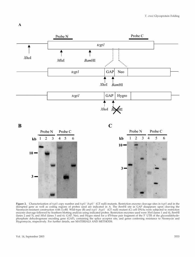
Figure 2. Characterization of tcgt1 copy number and tcgt1�/tcgt1� (GT null) mutants. Restriction enzyme cleavage sites in tcgt1 and in thedisrupted gene as well as coding regions of probes used are indicated in A. The BamHI site in GAP disappears upon cleaving theNeomycin-resistant construction with EcoRI. Wild-type (B) and tcgt1�/tcgt1� (GT null) mutant (C) cell DNAs were subjected to restrictionenzyme cleavage followed by Southern blotting analysis using indicated probes. Restriction enzymes used were XhoI (lanes 1 and 4), BamHI(lanes 2 and 5), and MluI (lanes 3 and 6). GAP, Neo, and Hygro stand for a 470-base pair fragment of the 5� UTR of the glyceraldehyde-phosphate dehydrogenase encoding gene (GAP), containing the splice acceptor site, and genes conferring resistance to Neomycin andHygromycin, respectively. For further details, see MATERIALS AND METHODS.
T. cruzi Glycoprotein Folding
Vol. 14, September 2003 3533

both in vitro and in vivo assays. Microsomes prepared fromwild-type but not from mutant cells displayed GT activity inthe presence of denatured thyroglobulin (Table 2). Incuba-tion of live cells with [14C]Glc in the presence of the GIIinhibitor DNJ led to formation of both glucosylated(Glc1Man7–9GlcNAc2) and unglucosylated (Man7–9GlcNAc2)N-glycans in the case of wild-type cells, but of only the latterspecies in the case of the GT null mutants (Figure 3, A andB). Strong acid hydrolysis confirmed the exclusive presence
of Glc units in glycans derived from wild-type cells (Figure3, C and D). The absolute absence of GT activity as revealedby above tests confirmed that although tcgt1�/tcgt1� cellshad not been cloned, they were not contaminated with het-erozygotes (tcgt1�/tcgt1�).
T. cruzi GT Null Mutants Are Not Affected in CellGrowth Rate and Viability and Only PartiallyAffected in Differentiation and InfectivityNo differences were observed in the cell growth rate ofwild-type (tcgt1�/tcgt1�), tcgt1 heterozygous (tcgt1�/tcgt1�), and GT null (tcgt1�/tcgt1�) epimastigotes in BHTmedium (our unpublished results). The GT-encoding genehas been disrupted in only other organism (S. pombe) inwhich the absence of the enzyme also did not affect cellviability and morphology under normal growth conditions(Fernandez et al., 1996). It is worth mentioning that CNX, theonly monoglucosylated N-glycan–specific lectin present inthis yeast, appeared to be essential for viability, thus indi-cating that CNX had additional roles besides facilitatingglycoprotein folding (Jannatipour and Rokeach, 1995). Twoprocedures were followed to study differentiation and infec-tivity of T. cruzi GT null mutants. In the first one rat myo-blast cell (L6E6) monolayers were infected with wild-type,
Table 2. In vitro GT activity of wild type and GT null T. cruzimutantsa
Enzyme Thyroglobulin cpm
Rat liver � 45Rat liver � 2424T. cruzi wild type � 146T. cruzi wild type � 798T. cruzi GT null � 44T. cruzi GT null � 42
a Pure rat liver GT or T. cruzi microsomes were used as enzymesource.
Figure 3. In vivo characterization of GT null (tcgt1�/tcgt1�) mutants. Wild-type (A and C) and GT null mutants (B and D) were pulse-chasedwith [14C]Glc in the presence of 5 mM DNJ. Whole cell endo-�-N-acetylglucosaminidase H-sensitive oligosaccharides were run on paperchromatography with solvent A (A and B). N-glycans were subjected to strong acid hydrolysis and run on paper chromatography withsolvent B (C and D). Standards: 1, Glc1Man9GlcNAc; 2, Man9GlcNAc; 3, Glc1Man8GlcNAc; 4, Man8GlcNAc; 5, Glc1Man7GlcNAc; 6,Man7GlcNAc; 7, Glc and 8, Man. For further details, see MATERIALS AND METHODS.
I. Conte et al.
Molecular Biology of the Cell3534

GT heterozygote (tcgt1�/tcgt1�), and GT null (tcgt1�/tcgt1�)stationary phase epimastigotes previously treated withguinea pig serum to enrich the population in infective forms(see MATERIALS AND METHODS). Two parasite/cell ra-tios were used (5:1 and 10:1). Cells were stained with Gi-emsa, and infected cells were counted. Values in Table 3correspond to the average of four experiments. In the secondprocedure, Vero cells were infected with untreated station-ary phase epimastigotes. In a first-infection experiment onlythe wild-type and heterozygote cells yielded infected cells,as judged from direct observation of unstained cells. How-ever, when supernatants were collected and cells were sub-jected to a secondary-infection experiment, observation ofGiemsa-stained cells gave results similar to those shown inTable 3. It may be concluded that absence of GT, and thenceof CRT-mediated glycoprotein folding facilitation, only par-tially affected T. cruzi differentiation and infectivity.
The Effect of GT Absence on Glycoprotein ContentThe folding process of a known virulence factor in GT nullmutant cells was studied to better understand how the par-asite cell ER reacted to the absence of one of the knownfolding facilitating mechanisms. CZP is a lysosomal protein-ase that constitutes �5% of total soluble cellular protein. Aswill be further discussed below, there is evidence indicatingthat �90% of all CZP molecules interact with CRT duringthe folding process occurring in the wild-type cell ER. Nev-ertheless, the total lysosomal CZP level was scarcely affected(5–20% decrease) in GT null mutants (Figure 4). On the otherhand, a more pronounced decrease was observed in thelevel of another lysosomal glycoprotein (�-mannosidase).The level of a cytosolic nonglycoprotein enzyme (pyruvatekinase) was not modified (Figure 4). These results confirmthat lectin-monoglycosylated glycoprotein interaction maynot be absolutely required for proper folding and that theabsence of such interaction may have differential effects onthe folding efficiency of different glycoproteins. The sameconclusions have been reached before using GII-deficientmammalian or yeast cell mutants (Reitman et al., 1982;D’Alessio et al., 1999). The model system used here (GT nulltrypanosomatid cells) obviates any effect that may have beenproduced by inhibition of normal N-glycan processing be-cause of the continued presence of Glc residues.
The Effect of GT Absence on CRT-GlycoproteinInteractionER folding and oligomerization is the rate-limiting step inthe eukaryotic cell secretion process. We have previously
determined that addition of DNJ lengthened CRT-CZP in-teraction and thence delayed arrival of the proteinase tolysosomes Labriola et al., 1995, 1999). Contrary to whathappens in other eukaryotic cells, in which addition of GIand GII inhibitors hinder lectin-glycoprotein interaction bypromoting accumulation of tri- and diglucosylated N-gly-cans, in trypanosomatid cell the inhibitors favor the tempo-ral persistence of monoglucosylated species. The same effectof DNJ addition on the arrival of CZP to lysosomes observedpreviously in Tulahuen 2 strain wild-type cells was nowreproduced in CL Brener clone cells (Figure 5, A and B). Asexpected, no effect of DNJ was detected when GT null mu-tants were used, thus confirming that the effect observed inwild-type cells was dependent on creation of monoglucosy-lated N-glycans (Figure 5, C and D). Quite unexpectedly,however, kinetics of CZP arrival to lysosomes in the pres-ence or absence of the drug were similar not to those ob-served in wild-type cells in the absence of the drug but tothose observed in its presence. This constitutes a firm indi-cation that in GT null mutants CZP molecules were beingretained in the ER by an alternative mechanisms not involv-ing a lectin-N-glycan type of interaction. Two possible pro-tein-protein interaction mechanisms were investigated, in-volving either CRT or Grp78/BiP.
CRT Does Not Interact with Unglucosylated CZPIt has been claimed that either CNX or CRT may weaklyassociate with unglucosylated glycoproteins or even withunglycosylated proteins and that such interaction has re-mained mostly undetected as it may be disrupted by therelatively high detergent concentrations usually used for celllysis previous to coimmunoprecipitation. As already de-scribed, addition of DNJ to wild-type live cells pulse-labeledwith [35S]Met plus [35S]Cys greatly enhanced the amount ofCZP that coimmunoprecipitated with CRT on addition ofCRT antiserum to cells lysed with 1% Nonidet P-40. Noimmunoprecipitation was observed either in the presence orabsence of the drug when GT null mutant cells were used(Figure 6A). We were unable to detect CZP coimmunopre-
Table 3. The effect of GT null mutation on T. cruzi infectivitya
Parasite/cell ratio Wild type Heterozygote GT null
5:1 45 � 6 50 � 5 27 � 410:1 93 � 8 106 � 11 60 � 2
a Values correspond to the average number of infected cells found infour independent experiments, each one conducted with threeplates containing 50,000 cells each.
Figure 4. Total glycoprotein content in GT null mutant cells. Cellpellets were freeze-thawed and resuspended in 50 mM Tris-HCl,pH 7.6, 0.15 M NaCl and contrifuged for 20 min at 14,000 rpm.Indicated enzymatic activities were assayed in the supernatants(cytosolic and lysosomal contents). Values in wild-type cells weretaken as 100%. PK, CZP, and �-MAN stand for pyruvate kinase,cruzipain, and �-mannosidase, respectively. Assays were per-formed with cells withdrawn from cultures at the three indicatedcell densities
T. cruzi Glycoprotein Folding
Vol. 14, September 2003 3535

cipitating with CRT antiserum when labeled mutant cellswere lysed with Nonidet P-40 concentrations as low as 0.1%(Figure 6B). The same result was observed with similar(�1%) digitonin concentrations (our unpublished results).
GT Absence Lengthens Grp78/BiP-CZP InteractionTo study the influence of the GT-created monoglucosylatedN-glycans on Grp78/BiP-CZP interaction cycloheximidewas added to wild-type and GT null T. cruzi cells to stopprotein synthesis. Cells from aliquots withdrawn after sev-eral time periods were lysed and immunoprecipitated withCZP antiserum. Immunoprecipitates were then analyzed byWestern blotting analysis using T. brucei Grp78/BiP anti-serum, which cross-reacts with the same T. cruzi protein.Results shown in Figure 7, A–C, indicate that Grp78/BiP-interaction persisted for significantly longer time periods inGT null mutants. This result agrees with the higher Grp78/BiP levels detected in these last cells when compared withthe wild-type ones (Figure 7D). It may be concluded thatabsence of CRT-glycoprotein interaction triggered inductionof alternative folding facilitation mechanisms.
DISCUSSION
Results presented show that in spite of glycoproteins beingessential components of T. cruzi complex life cycle, absenceof the ER glycoprotein-specific folding facilitation mecha-nism only had quantitative but not qualitative effects ondifferentiation and infection processes. Description of thefolding process of one of known T. cruzi glycoprotein viru-lence factors (CZP) showed that the parasite reacted to theabsence of lectin-N-glycan interaction by upregulating alter-native mechanisms that enhance ER folding efficiency.
CZP has been identified as one of T. cruzi virulence factors(Franke de Cazzulo et al., 1994). In fact, specific CZP inhib-itors are currently being tested as candidates for Chagas’disease chemotherapy (Engel et al., 1998). CZP is composedof two domains, the enzyme proper or catalytic domain andthe C-terminal extension (McGrath et al., 1995). The formerdomain has two N-glycosylation consensus sequences, atleast one of which is occupied, whereas the latter has onlyone of such sequences, which has been shown to bear anN-glycan (Cazzulo et al., 1992; Metzner et al., 1996). We havepreviously determined that �65% of glycans in either theC-terminal extension or in the catalytic domain are glucosy-lated by GT (Labriola et al., 1995). Because it has been de-termined that GT exclusively glucosylates N-glycans in theclose vicinity of structural perturbations and both CZP do-mains are known to fold independently from each other(McGrath et al., 1995; Ritter and Helenius, 2000), it wasconcluded that �90% of CZP molecules were glucosylatedin at least one of its glycans and thus interacted with CRTduring the folding process. If the second N-glycosylationconsensus sequence in the catalytic domain were occupiedand glucosylated, it may be statistically calculated that prac-tically all CZP molecules interacted with CRT. Because CZPhas seven disulfide bonds, it should be expected its properfolding to be heavily dependent on interaction with CRT if aprotein similar to ERp57 also occurs in the T. cruzi cell ER.ERp57 is a member of the protein disulfide isomerase familythat in mammalian cells is tightly associated with CNX/CRTand promotes native disulfide bond formation in monoglu-
Figure 5. Kinetics of CZP arrival to lysosomes. Wild-type (A andB) and GT null mutant (C and D) cells were pulse-chased with[35S]Met and [35S]Cys, and CZP in lysosomes were isolated, run on10% SDS-PAGE, and subjected to autoradiography. Intensity after a300-min chase was taken as 100%. Cells in B and D were pulse-chased in the presence of 6 mM DNJ. For further details, see MA-TERIALS AND METHODS.
I. Conte et al.
Molecular Biology of the Cell3536

cosylated glycoprotein folding intermediates (Oliver et al.,1999). Rather surprisingly, however, our results show thatthe absence of CRT-CZP interaction only slightly affected thetotal proteinase content. The most plausible explanation forthis result is that the absence of GT led to the ER accumu-lation of misfolded glycoproteins and thence to increasedsynthesis of other ER chaperones and folding facilitatingproteins that replaced CRT in its task of enhancing CZPfolding efficiency (unfolded protein response; Parodi, 2000;Trombetta and Parodi, 2002). It has been already observedthat inhibition of monoglucosylated N-glycan formation inmammalian and S. pombe cells either by the addition of GIIinhibitors or by the disruption of GII- or GT-encoding genesresulted in the induction of BiP mRNA (Balow et al., 1995;Pahl and Baeuerle, 1995; D’Alessio et al., 1999). In accordancewith these findings, we observed higher Grp78/BiP levels inGT null than in wild-type cells.
We have detected interaction of Grp78/BiP with CZP inboth GT-positive and -negative cells. A longer interactionwas observed, nevertheless, with the latter than with theformer cells, thus suggesting that Grp78/BiP might be re-sponsible for the similar kinetics of CZP arrival to lysosomesobserved in wild-type cells in the presence of the GII inhib-itor DNJ and in GT null mutants in the presence or absenceof the drug. Although the delay observed in CZP arrival tolysosomes in wild-type cells incubated with DNJ was due toa lengthened CRT-CZP interaction, the delay observed inmutant cells was probably due to a prolonged interactionwith Grp78/BiP and other molecular chaperones. It has beenreported that Grp78/BiP preferentially recognizes hep-tapeptides having bulky hydrophobic residues in alternat-ing positions exposed in extended protein conformations,whereas GT showed affinity for hydrophobic amino acidpatches in collapsed, molten globule-like structures (Blond-Elguindi et al., 1993; Caramelo et al., 2003). In other words, achange in the affinity of glycoproteins for Grp78/BiP toCNX/CRT would occur once sufficient primary sequenceinformation is present in the ER lumen to allow proteins tocollapse. In fact, there are several examples in which asequential glycoprotein interaction, first with Grp78/BiPand then with CNX/CRT has been observed in intact mam-malian cells (Hammond and Helenius, 1994; Kim and Ar-van, 1995; Tomita et al., 1999; Molinari and Helenius, 2000).
It may be speculated that a similar sequential interactionoccurs in normal T. cruzi cells but that in the case of GT nullmutants, higher Grp78/BiP levels triggered by the unfoldedprotein response would prolong the extended conformationto prevent glycoprotein aggregation.
Availability of GT null mutants allowed studying if pro-tein-protein, in addition to protein-glycan, recognition–me-diated T. cruzi CRT-glycoprotein interaction. There havebeen several reports in the literature suggesting that CRTand a soluble form of CNX in vitro prevented aggregationnot only of denatured monoglucosylated oligosaccharide-bearing glycoproteins as Glc1Man9GlcNAc2-soybean agglu-tinin but also of denatured proteins lacking N-oligosaccha-rides as deglycosylated soybean agglutinin, malatedehydrogenase, and citrate synthase. They also suppressedthe thermal denaturation of above-mentioned nonglycosy-lated proteins and enhanced their refolding by maintainingthe unfolded species in a folding-competent state. Both lec-tins formed stable complexes with unfolded conformers butnot with the native molecules. It was concluded that CRTand truncated CNX behaved in vitro as bona fide classicalchaperones (Ihara et al., 1999; Saito et al., 1999; Stronge et al.,2001).
Whether the conclusions reached are applicable to in vivosituations is questionable because CRT was found to displaya cooperative denaturation transition with a midpoint atabout the same temperature (44°C) at which the above-mentioned experiments were performed (Bouvier and Staf-ford 2000; Li et al., 2001). Concerning in vivo trials, it wasreported that certain glycoproteins synthesized in mamma-lian cells devoid of either GI or GII activities or in thepresence of an inhibitor of both enzymes (castanospermine;i.e., conditions that prevent formation of monoglucosylatedglycans) were immunoprecipitated with CNX antibodies ifspecial precautions were taken when cell lysis and immu-noprecipitating techniques were used (Danilczyk and Wil-liams, 2001). Results presented here show that even underthose special experimental conditions, no CRT-CZP interac-tion was detected in GT null mutant cells, thus confirmingthe exclusive lectin nature of T. cruzi CRT.
CNX/CRT-glycoprotein interaction was shown not to beessential for mammalian or fungal cell viability under nor-mal growth conditions (Reitman et al., 1982; Ray et al., 1991;
Figure 6. CRT-CZP interaction in wild-type and GT null mutant cells. Cells were pulsed for 2 min with [35S]Met plus [35S]Cys, lysed with1% Nonidet P-40 (A) or with indicated concentrations of the same detergent (NP-40) (B), and lysates were subjected to immunoprecipitationwith CRT antiserum. Immunoprecipitates were run on 10% SDS-PAGE and subjected to autoradiography. Where indicated 6 mM DNJ wasadded 30 min before the pulse. CZP and CRT stand for cruzipain and calreticulin, respectively. For further details, see MATERIALS ANDMETHODS.
T. cruzi Glycoprotein Folding
Vol. 14, September 2003 3537

Figure 7. Grp78/BiP-CZP interaction. Protein synthesis in wild-type (A) or GT null mutant (B) cells was stopped on addition of 1 mMcycloheximide, and samples were withdrawn after indicated times. Cells were lysed and subjected to immunoprecipitation with CZPantiserum. Immunoprecipitates were run on 10% SDS-PAGE and subjected to Western blotting analysis with T. brucei Grp78/BiP antiserum.In C, intensity of the first aliquot was taken as 100%. In D, 107 wild-type or GT null epimastigotes in the exponential growth phase wereharvested and heated with cracking buffer followed by SDS-PAGE and Western blotting analysis as above. For further details, seeMATERIALS AND METHODS.
I. Conte et al.
Molecular Biology of the Cell3538

Fernandez et al., 1996; D’Alessio et al., 1999). Because foldingis an error-prone but essential process, cells have alternativeways for helping protein acquisition of proper tertiary struc-tures in the ER (Parodi, 2000; Trombetta and Parodi, 2002).As a general rule, when one folding facilitation system isabsent, an alternative one fills the job. Nevertheless, thereare exceptions to the last statement and CNX/CRT-glycop-rotein interaction was reported to be necessarily required forproduction of infective HIV-1 and hepatitis B virus (Gruterset al., 1987; Fischer et al., 1996; Mehta et al., 1997) and forviability of the fission yeast S. pombe under severe ER stressconditions (Fanchiotti et al., 1998). In the first case, virusparticles produced in the absence of CNX/CRT-glycopro-tein interaction were noninfectious due to a misfolded Vi-V2loop in HIV-1 gp120. Similarly, folding of the M glycopro-tein of hepatitis virus B was severely compromised whenlectin-glycoprotein interaction was prevented and virus as-sembly was hindered. In the yeast case, a glycoprotein es-sential for cell wall assembly necessarily required CNX-glycoprotein interaction for proper folding under conditionsof severe ER stress.
Results presented herein show, therefore, that T. cruzi ERfolding machinery presents an exquisite plasticity that al-lows the parasite to surmount the absence of the glycopro-tein-specific folding facilitation mechanism.
ACKNOWLEDGMENTS
Work reported herein has received financial support from the U. S.Public Health Service (National Institutes of Health), from theHoward Hughes Medical Institute, from the United Nations Devel-opment Program/World Bank/World Health Organization SpecialProgram for Research and Training in Tropical Diseases, and fromthe National Agency for the Promotion of Science and Technology(Argentina).
REFERENCES
Balow, J.P., Weissman, J.D., and Kearse, K.P. (1995). Unique expres-sion of major histocompatibility complex class I proteins in theabsence of glucose trimming and calnexin association. J. Biol. Chem.270, 29025–29029.
Blond-Elguindi, S., Cwirla, S.E., Dower, W.J., Lipshutz, R.J., Sprang,S.R., Sambrook, J.F., and Gething, M.J. (1993). Affinity panning of alibrary of peptides displayed on bacteriophages reveals the bindingspecificity of BiP. Cell 75, 717–728.
Bontempi, E., Martinez, J., and Cazzulo, J.J. (1989). Subcellular lo-calization of cysteine proteinase from Trypanosoma cruzi. Mol. Bio-chem. Parasitol. 33, 33–48.
Borst, P., Fase-Fowler, F., Frasch, A.C.C., Hoeijmakers, J.H.J., andWeijers, P.J. (1980). Characterization of DNA from Trypanosomabrucei and related trypanosomes by restriction endonuclease diges-tion. Mol. Biochem. Parasitol. 1, 221–246.
Bosch, M., Trombetta, S., Engstrom, U., and Parodi, A.J. (1988).Characterization of dolichol diphosphate oligosaccharide:proteinoligosaccharyltransferase and of glycoprotein processing glucosi-dases occurring in trypanosomatids. J. Biol. Chem. 263, 17360–17365.
Bouvier, M., and Stafford, W.F. (2000). Probing the three-dimen-sional structure of human calreticulin. Biochemistry, 39, 14950–14959.
Caramelo, J.J., Castro, O.A., Alonso, L.G., de Prat-Gay, G., andParodi, A.J. (2003). UDP-Glc:glycoprotein glucosyltransferase recog-
nizes structured and solvent accessible hydrophobic patches inmolten globule-like folding intermediates. Proc. Natl. Acad. Sci.USA 100, 86–91.
Cazzulo, J.J., Cazzulo Franke, M.C., and Franke de Cazzulo, B.M.(1989). On the regulatory properties of the pyruvate kinase fromTrypanosoma cruzi. FEMS Microbiol. Lett. 59, 259–264.
Cazzulo, J.J., Cazzulo Franke, M.C., Martinez, J., and Franke deCazzulo, B.M. (1990). Some kinetics properties of a cysteine protein-ase (Cruzipain) from Trypanosoma cruzi. Biochim. Biophys. Acta1037, 186–191.
Cazzulo, J.J., Franke de Cazzulo, B.M., Engel, J.C., and Cannata, J.J.(1985). End products and enzyme levels of aerobic fermentation intrypanosomatids. Mol. Biochem. Parasitol. 16, 329–343.
Cazzulo, J.J., Martinez, J., Parodi, A.J., Wernstedt, C., and Hellman,U. (1992). On the post-translational modifications at the C-terminal.domain of the major cysteine proteinase (Cruzipain) from Trypano-soma cruzi. FEMS Microbiol. Lett. 100, 411–416.
D’Alessio, C., Fernandez, F., Trombetta, E.S., and Parodi, A.J. (1999).Genetic evidence for the heterodimeric structure of glucosidase II.The effect of disrupting the subunit-encoding genes on glycoproteinfolding. J. Biol. Chem. 274, 25899–25905.
Danilczyk, U.G., and Williams, D.B. (2001). The lectin chaperonecalnexin utilizes polypeptide-based interactions to associate withmany of its substrates in vivo. J. Biol. Chem. 276, 25592–25540.
Engel, J.C., Doyle, P.S., Hsieh, I., and McKerrow, J.H. (1998). Cys-teine protease inhibitors cure an experimental Trypanosoma cruziinfection. J. Exp. Med. 188, 725–734.
Engel, J. C. and Parodi, A. J. (1985). Trypanosoma cruzi cells un-dergo an alteration in protein N-glycosylation upon differentiation.J. Biol. Chem. 260, 10105–10110.
Fanchiotti, S., Fernandez, F., D’Alessio, C., and Parodi, A.J. (1998).The UDP-Glc:glycoprotein glucosyltransferase is essential forSchizosaccharomyces pombe viability under conditions of extreme en-doplasmic reticulum stress. J. Cell Biol. 143, 625–635.
Fernandez, F., Jannatipour, M., Hellman, U., Rokeach, L.A. and.Parodi, A.J. (1996). A new stress protein: synthesis of Schizosaccha-romyces pombe UDP-Glc:glycoprotein glucosyltransferase mRNA isinduced by stress conditions but the enzyme is not essential for cellviability. EMBO J. 15, 705–713.
Fischer, P.B., Karlsson, G.B., Butters, T.D., Dwek, R.A., and Platt,F.M. (1996). N-butyldeoxynojirimycin-mediated inhibition of hu-man immunodeficiency virus entry correlates with changes in anti-body recognition of the V1–V2 region of gp120. J. Virol. 70, 7143–7152.
Franke de Cazzulo, B.M., Martinez, J., North, M.J., Coombs, G.H.,and Cazzulo, J.J. (1994). Effects of proteinase inhibitors on thegrowth and differentiation of Trypanosoma cruzi. FEMS Microbiol.Lett. 124, 81–86.
Gruters, R.A., Neefjes, J.J., Tersmette, M., Tulp, A., Huisman, H.G.,Miedema, F., and Ploegh, H.L. (1987). Interference with HIV-in-duced syncytium formation and viral infectivity by inhibitors oftrimming glucosidases. Nature 330, 74–77.
Guerin, M. and Parodi, A.J. (2003). The UDP-Glc:glycoprotein glu-cosyltransferase is organized in at least two tightly bound domainsfrom yeasts to mammals. J. Biol. Chem. 278, 20540–20546.
Hammond, C., and Helenius, A. (1994). Folding of VSV G protein:sequential interaction with BiP and calnexin. Science 266, 456–458.
Ihara, Y., Cohen-Doyle, M.F., Saito, Y., and Williams, D.B. (1999).Calnexin discriminates between protein conformational states andfunctions as a molecular chaperone in vitro. Mol. Cell 4, 331–341.
T. cruzi Glycoprotein Folding
Vol. 14, September 2003 3539
Jannatipour, M. and Rokeach, L.A. (1995). The Schizosaccharomycespombe homologue of the chaperone calnexin is essential for viability.J. Biol. Chem. 270, 4845–4853.
Kim, P. S. and Arvan, P. (1995). Calnexin and BiP act as sequentialmolecular chaperones during thyroglobulin folding in the endoplas-mic reticulum. J. Cell Biol. 128, 29–38.
Labriola, C., Cazzulo, J.J., and Parodi, A.J. (1995). Retention ofglucose residues added by the UDP-Glc:glycoprotein glucosyltrans-ferase delays exit of glycoproteins from the endoplasmic reticulum.J. Cell Biol. 130, 771–779.
Labriola, C., Cazzulo, J.J., and Parodi, A.J. (1999). Trypanosoma cruzicalreticulin is a lectin that binds monoglucosylated oligosaccharidesbut not protein moieties of glycoproteins. Mol. Biol. Cell 10, 1381–1394.
Li, A., Stafford, W.F., and Bouvier, M. (2001). The metal ion bindingproperties of calreticulin modulates its conformational flexibilityand thermal stability. Biochemistry 40, 11193–11201.
Li, Y.-T. and Li, S.-C. (1972). �-mannosidase, �-N-acetylglu-cosaminidase and �-galactosidase from Jack bean meal. MethodsEnzymol. 28, 702–713.
Magdesian, M.H., Giordano, R., Ulrich, H., Juliano, M.A., Juliano,L., Schumacher, R.I., Colli, W., and Alves, M.J. (2001). Infection byTrypanosoma cruzi. Identification of a parasite ligand and its host cellreceptor. J. Biol. Chem. 276, 19382–19389.
McGrath, M.E., Eakin, A.E., Engel, J.C., McKerrow, J.H., Craik, C.S.,and Fletterick, R.J. (1995). The crystal structure of cruzain: a thera-peutic target for Chagas’ disease. J. Mol. Biol. 247, 251–259.
Mehta, A., Lu, X., Block, T.M., Blumberg, B.S., and Dwek, R.A.(1997). Hepatitis B virus (HBV) envelope glycoproteins vary dras-tically in their sensitivity to glycan processing: evidence that alter-ation of a single N-linked glycosylation site can regulate HBV se-cretion. Proc. Natl. Acad. Sci. USA 94, 1822–1827.
Metzner, S.I., Sousa, M.C., Hellman, U. Cazzulo, J.J. and Parodi, A.J.(1996). The use of UDP-Glc:glycoprotein glucosyltransferase forradiolabeling protein-linked high mannose-type oligosaccharides.Cell. Mol. Biol. 42, 631–635.
Molinari, M. and Helenius, A. (2000). Chaperone selection duringglycoprotein translocation into the endoplasmic reticulum. Science288, 331–333.
Oliver, J.D., Llewelyn Roderick, H., Llewelyn, D.H., and High, S.(1999). ERp57 functions as a subunit of specific complexes formedwith the ER lectins calreticulin and calnexin. Mol. Biol. Cell 10,2573–2582.
Padilla, A., Noiva, R., Lee, N., Krishna Mohan, K.V., Nakhasi, H.L.,and Debrabant, A. (2003). An atypical protein disulfide isomerasefrom the protozoan parasite Leishmania containing a single thiore-doxin-like domain. J. Biol. Chem. 278, 1872–1878.
Pahl, H.L., and Baeuerle, P.A. (1995). A novel signal transductionpathway from the endoplasmic reticulum to the nucleus is mediatedby transcription factor NF-kappa B. EMBO J. 14, 2580–2588.
Parodi, A.J. (1993). N-glycosylation in trypanosomatid protozoa.Glycobiology 3, 193–199.
Parodi, A.J. (2000). Protein glucosylation and its role in proteinfolding. Annu. Rev. Biochem. 69, 69–93.
Ray, M.K., Young, J., Sundaram, S., and Stanley, P. (1991). A novelglycosylation phenotype expressed by Lec23, a Chinese hamsterovary mutant deficient in �-glucosidase I. J. Biol. Chem. 266, 22818–22825.
Reitman, M.L., Trowbridge, L.S., and Kornfeld, S. (1982). A lectin-resistant mouse lymphoma cell line is deficient in glucosidase II, aglycoprotein processing enzyme. J. Biol. Chem. 257, 10357–10363.
Ritter, C., and Helenius, A. (2000). Recognition of local glycoproteinmisfolding by the ER folding sensor UDP-glucose:glycoprotein glu-cosyltransferase. Nat. Struct. Biol. 7, 278–280.
Ruiz, R.C., Favoreto, S., Jr., Dorta, M.L., Oshiro, M.E.M., Ferreira,A.T., Manque, P.M., and Yoshida, N. (1998). Infectivity of Trypano-soma cruzi strains is associated with differential expression of surfaceglycoproteins with differential Ca2� signaling activity. Biochem. J.330, 505–511.
Saito, Y., Ihara, Y., Leach, M.R., Cohen-Doyle, M.F., and Williams,D.B. (1999). Calreticulin functions in vitro as a molecular chaperonefor both glycosylated and non-glycosylated proteins. EMBO J. 18,6718–6729.
Sambrook, J., Fritsch, E.F., and Maniatis, T. (1989). Molecular Clon-ing: A Laboratory Manual. Cold Spring Harbor, NY: Cold SpringHarbor Laboratory Press.
Schenkman, S., Jiang, M.S., Hart, G.W., and Nussenzweig, V. (1991).A novel cell surface trans-sialidase of Trypanosoma cruzi generates astage-specific epitope required for invasion of mammalian cells. Cell65, 1117–1125.
Stronge, V.S., Saito, Y., Ihara, Y., and Williams, D.B. (2001). Rela-tionship between calnexin and BiP in suppressing aggregation andpromoting refolding of protein and glycoprotein substrates. J. Biol.Chem. 276, 39779–39787.
Tessier, D.C., Dignard, D., Zapun, A., Radominska-Pandya, A.,Parodi, A.J., Begeron, J.J.M., and Thomas, D.Y. (2000). Cloning andcharacterization of mammalian UDP-glucose:glycoprotein glucosyl-transferase and the development of a specific substrate for thisenzyme. Glycobiology 10, 403–412.
Tibbetts, R.S., Kim, I.Y., Olson, C.L., Barthel, L.M., Sullivan, M.A.,Winquist, A.G., Miller, S.D., and Engman, D.M. (1994). Molecularcloning and characterization of the 78-kilodalton glucose-regulatedprotein of Trypanosoma cruzi. Infect. Immun. 62, 2499–2507.
Tomita, Y., Yamashita, T., Sato, H., and Taira, H. (1999). Kinetics ofinteractions of Sendai virus envelop glycoproteins, F and HN, withendoplasmic reticulum-resident molecular chaperones, BiP, caln-exin, and calreticulin. J. Biochem. (Tokyo) 126, 1090–1100.
Trombetta, E.S., and Parodi, A.J. (2002). N-glycan processing andglycoprotein folding. Adv. Prot. Chem. 59, 303–344.
Trombetta, S., and Parodi, A.J. (1992). Purification to apparent ho-mogeneity and partial characterization of rat liver UDP-Glc:glyco-protein glucosyltransferase. J. Biol. Chem. 267, 9236–9240.
Trombetta, E.S., Simons, J.F., and Helenius, A. (1996). Endoplasmicreticulum glucosidase II is composed of a catalytic subunit, con-served from yeast to mammals, and a tightly bound noncatalyticHDEL-containing subunit. J. Biol. Chem. 271, 27509–27516.
Trombetta, S., Bosch, M., and Parodi, A.J. (1989). Glucosylation ofglycoproteins by mammalian, plant, fungal and trypanosomatidprotozoa microsomal proteins. Biochemistry 28, 8108–8116.
I. Conte et al.
Molecular Biology of the Cell3540
Copyright © 2022 FDOKUMEN